Copyright is owned by the Author of the thesis. Permission is given for a copy to be downloaded by an individual for the purpose of research and private study only. The thesis may not be reproduced elsewhere without the permission of the Author.
Transcript
Copyright is owned by the Author of the thesis. Permission is given for a copy to be downloaded by an individual for the purpose of research and private study only. The thesis may not be reproduced elsewhere without the permission of the Author.
ANIMAL SOURCES OF HUMAN
CAMPYLOBACTERIOSIS
A thesis presented in partial fulfIlment of the requirements for the
degree of
Doctor of Philosophy
in
Veterinary Public Health
At Massey University, Palmerston North,
New Zealand.
FASIUDDIN AHMED
1999
This thesis is dedicated to the memory Of
My Beloved Father
ABSTRACT
New Zealand has one of the highest reported rates of Campylobacter infections in
humans in the developed world. It is the single largest notifiable disease in all regions of
the country. Consumption of poultry meat has been widely implicated both overseas
and in New Zealand as the main cause of human infections. The potential contribution
of other animals especially cattle and sheep is less well known. The present study was
undertaken to fill this gap in knowledge.
Faecal samples from 300 cattle and 158 sheep were collected from local abattoirs and
farms plus 50 samples from the sheep slaughterhouse environment and examined for the
presence of thermophilic Campylobacter spp. Campylobacter spp. were isolated from
45% of the cattle, 44% of the sheep and 56% of the environmental samples. C. jejuni
and C. hyointestinalis were the predominating species isolated from cattle followed by
C. coli and C. lari. In sheep and in environmental samples from the sheep abattoir C.
jejuni was the only species isolated. The isolation rate and the species of
Campylobacter varied between beef and dairy cattle, bull and heifer calves, age of the
heifer calves, and time of the year. The high isolation rate of Campylobacter from the
cattle, sheep and their environment strongly suggests the possibility of these micro
organisms finding their way into milk and meat, as faecal contaminants at the farm and
slaughter level. There is also the potential to contaminate the environment and water
following disposal of abattoir effiuents and run off from farms.
The species of the isolates from human diarrhoeal cases were found to be predominantly
C. jejuni (95%) and C. coli (5%). Molecular typing of C. jejuni using Sma I generated
pulsed-field gel electrophoresis (pFGE) profiles yielded 13 to 16 different patterns in
the cattle, sheep and human isolates showing a large inter-species variation in the
isolates even from the same sources. However, indistinguishable as well as closely
related profiles (pulsotypes) were found across the isolates from cattle, sheep and
humans. The results obtained from the PFGE typing strongly indicate that cattle and
sheep may be important reservoirs of human campylobacter infections. It was also
observed that a few closely related types mostly dominate the C. jejuni populations in
the host animal species. The possibility of faecal contamination from these animals at
slaughter and thus C. jejuni entering the meat was studied.
Retail packs of beef (25), lamb (25) and chicken (50) mince purchased from local
supermarkets were examined. A combined selective enrichment and PCR based
method was evaluated to offer a rapid, sensitive and specific detection method for the
identification of C. jejuni from meats. C. jejuni was detected by culture and PCR in
44% of the chicken, 16% of the lamb and 12% of the beef mince samples. These results
lend credibility to our contention that faecal contamination of sheep and beef carcasses
at slaughter has significant implications for food safety. The much higher· rate of
detection in chicken mince may be related to a higher prevalence of infection in
chickens or to the method of processing which may facilitate spread between birds and /
or between product.
The C. jejuni isolates from the animal and human sources were also examined for
antibiotic resistance by the disc diffusion method to antibiotics commonly used for the
treatment of campylobacter infections in humans. No resistance was detected in the
cattle and sheep isolates. Two human isolates exhibited resistance to tetracycline with
MrCs of>128 J..I.g/ml. All other human isolates were found susceptible to the antibiotics
tested. The nil to negligible resistance detected in the animal and human isolates of C.
jejuni suggest that it is not a major problem in New Zealand at the present time
however, further work is required to examine the situation in more intensively farmed
species and monitor any changes in human isolates over time.
11
Acknowledgements
I consider it a privilege to record my deepest sense of gratitude to Professor Colin
Wilks for his generous support, constant encouragement and constructive counsel all
along the course of investigation and preparation of this manuscript. I would also like
to thank him for his genuine concern for my welfare.
I am indeed thankful to my co-supervisor Stan Fenwick for his help and guidance
throughout my studies as well as to my other supervisors Per Madie and Alan Mu"ay
who have been so generous with their time.
I have been associated with several people during this long period and every one has
contributed something towards this investigation. I would like to thank Jane Hunter,
Eammon Gormley, Kevin Stafford, Jacek Gwozdz and Hassan Hussein for their help.
Thanks are also due to the team of microbiologists at the Palmerston North Medical
laboratory who have provided the human isolates of Campylobacter and to Ayad
Alkaissi at the Meat works in collection of samples from animals.
Assistance in the laboratory provided by Magda Gwozdz, Kylie Walker, Jan Schrama
and Laurie Sandall is highly appreciated Special mention should be made of Peter
Wildbore who had been most helpful in procuring the most important to the trivial with
equal zeal.
I would like to specially thank Allain Scott for her readiness to help and advise in
matters of academic complexities and also to the other secretarial staff at the institute
for their help.
A big thanks to all my friends in room 2.01 and in the university for their enjoyable
company and support.
A special thanks to my wife Kavita for her constant encouragement and support in times
of great stress and in giving two lovely children Shireen and Rehan during this period
who have been the source of infinite joy.
III
The completion of this thesis is also due to the support, encouragement, and sound
advice I received from my family especially my beloved parents to all of them I am
deeply indebted
And last but not the least, I remember the Almighty who gave me strength, courage and
2.8 Antibiotic resistance among Campylobacter species .... . .. . ......... ................... 29
CHAPTER THREE: ISOLATION AND IDENTIFICATION OF
CAMPYLOBACTER SPECIES FROM FAECES OF CATTLE, SHEEP AND HUMANS •....•••••••.•••••••••.•••••.••••••••.••••••••...••.•....••••..••..•••...•...•.•.•.•• •.••••••.••••••••.•••. 34
J..Lg) were applied on the agar between the test sample and control using a disc
dispenser (Oxoid). The plate was allowed to dry for 30 minutes and then incubated in
an anaerobic jar with a Campy-Pak (BBL) gas envelope at 42°C. The plates were read
after 48 h and the diameter of the zones of inhibition around the antibiotic discs
recorded.
5.2.3. MINIMAL INHIBITORY CONCENTRA TION (MIC)
The MICs of the resistant isolates was determined by an agar dilution method (Mendez
et aI. , 1 980). Isolates were grown in blood agar plates containing 1 J..Lg/ml of
tetracycline. Growth was suspended in 0. 1 M phosphate buffer saline (PBS) to 0.5
McFarland standard. The suspension was diluted 1 : 1 0 in PBS and inoculated into
Muller-Hinton agar plates with doubling concentration of tetracycline ranging from 2
to 1 28 J..Lg/ml in duplicates. The plates were incubated at 42°C and observed after 24 h
and 48 h for growth. The highest concentration in which growth was absent was taken
as the MIC of that strain.
72
------- -
Chapter 5 Antimicrobial Susceptibilities
5.3. RESU LTS
5.3.1. RESULTS OF ANTIMICROBIAL SUSCEPTIBILITIES OF HUMAN,
CA TTLE AND SHEEP ISOLA TES
All the 100 human isolates were found sensitive to erythromycin, enrofloxacin,
gentamicin, chloramphenicol and neomycin. Two isolates were found resistant to
tetracycline.
All 35 C. jejuni isolates from cattle as well as the 1 00 sheep C. jejuni isolates were
found sensitive to all the six antibiotics tested.
5.3.2. RESULTS OF DETERMINATION OF THE MINIMAL INHIBITORY
CONCENTRA TIONS (MICs) OF THE RESISTANT ISOLA TES
The minimal inhibitory concentrations of the tetracycline resistant isolates of C. jejuni
were determined by the agar dilution method. MICs of the two resistant isolates were
found to be � 1 28 �glml. Tetracycline susceptible isolates demonstrated MICs of � 2
�glml .
The two resistant isolates were in the process of testing sub-cultured 3 times after the
first determination of resistance and still found resistant.
73
Chapter 5 Antimicrobial Susceptibilities
5.4. DISCUSSION
In modern breeding of farm animals, practised in most countries, large amounts of
antibiotics are used for a variety of reasons, including growth promotion, disease
prophylaxis, and therapy and there is an increasing concern that such widespread use of
antibiotics is contributing to the emergence of resistant bacteria that are contaminating
the food supply (WHO Report, 1 997). However, this is not at all the New Zealand
situation except for intensive industries such as pigs and poultry. Many of the 40
million sheep in New Zealand would never see antibiotics.
Infections with quinolone-resistant strains of Campylobacter organisms are now found
with increasing frequency in clinical settings, following a trend of food borne
pathogens that have become resistant to antibiotics and have caused an increase in
morbidity and mortality among humans (Tee et al. , 1995; Alder-Mosca et aI. , 1 99 1 ;
Endtz et aI. , 1 99 1 a)
In the United States, Smith et al. ( 1 999) found that resistance to nalidixic acid
increased approximately eight-fold from l .3% in 1 992 to 1 0.2% in 1 998. All isolates
that were resistant to nalidixic acid were also resistant to ciprofloxacin also 2% were
resistant to erythromycin and 6 1 % resistant to tetracycline.
Data from other countries provide evidence that the use of fluoroquinolones in poultry
production has a primary role in increasing resistance to quinolones among C. jejuni
isolates from humans. In the Netherlands, an increase in ciprofloxacin resistant human
isolates from 0% through 1 985 to 1 1 % in 1 989 paralleled the increase in ciprofloxacin
resistant isolates from retail poultry products (Endtz et al. , 1 99 1 a) .
74
Chapter 5 Antimicrobial Susceptibilities
Likewise in Spain, an increase in the prevalence of ciprofloxacin-resistant human
isolates from between 0% - 3% in 1 989 to 30% - 50% in 1 99 1 coincided with the
introduction of enrofloxacin for veterinary use in 1 990 (Reina et ai. , 1992; Sanchez et
ai., 1 994).
In the present study the antimicrobial susceptibility of C. jejuni isolates to six
antibiotics was examined by the disc diffusion method. It was simple to perfonn and
results were easily interpreted. Standard methods for susceptibility testing and
interpretation of resistance in Campylobacter spp. are lacking and different workers
have employed various methods. However, the disc diffusion method has been widely
applied with good results for campylobacter testing (Gaudreau and Gilbert, 1 997,
Huysmans and Tumidge, 1 997; Koenraad et al. , 1 995b; Vanhoof et al. , 1 984). If there
is a need to obtain quantitative data regarding the resistant isolates, then minimal
inhibitory concentrations could be determined by an agar dilution method.
All the 35 cattle and 1 00 sheep isolates were found to be susceptible to all the six
antibiotics used including enrofloxacin. This is contrary to findings in many countries
where resistance to antibiotics is being increasingly detected in animal isolates
particularly against the quinolones.
Smith et al. ( 1999) found 20 % of Campylobacter isolates from poultry to be resistant
to ciprofloxacin and Jacobs-Reitsma ( 1 997) found 29 % to be resistant to quinolones.
This may be due to the extensive systems in which these animals are raised in New
Zealand with minimal to no use of antibiotics.
The likely times when antibiotics are used in these animals would be to treat sick
animals. Antibiotic resistant bacteria are more frequently isolated from pigs and
pOUltry than from extensively grazed cattle or sheep indicating the greater use of
antimicrobial agents in intensively housed species for growth promotion (Threlfall,
1 993). A study of Campylobacter isolates from pigs and poultry in New Zealand may
give very different results and should be conducted in a suitable range of isolates.
75
Chapter 5 Antimicrobial Susceptibilities
In our study only two human isolates (2%) of C. jejuni were found resistant to
tetracycline. No resistance to the other five antibiotics was detected in human, cattle
or sheep isolates. The findings agree with a recent study done in Auckland which
reported two of 202 human isolates to be resistant to tetracycline (Dowling et al. ,
1 998).
Overseas research shows that tetracycline resistance is more common in human isolates
than among isolates from animals. In studies by Aarestrup et al. ( 1 997) 1 1 % of the
human isolates as compared to 2 % of the animal isolates were resistant to tetracycline.
Huysmans and Turnidge ( 1 997) in Australia studied 1 00 clinical isolates of
thermophilic Campylobacter species from humans and did not detect resistance to
erythromycin, gentamicin or chloramphenicol but nine strains were found resistant to
tetracycline.
The absence of resistance to important antibiotics like erythromycin and enrofloxacin
seen in the present study speaks well for the country although low level resistance was
seen in human isolates for erythromycin (1 . 5%) and ciprofloxacin (2.5%) in the
Auckland study (Dowling et aI. , 1 998). This is still very much at a lower level when
compared to the extent of resistance observed overseas.
Levels as high as 84% in Thailand (Hoge et aI., 1 998) to 34% in Spain (Velazquez et
aI. , 1 995) and 1 8 .6% in Sweden (Sjogren et aI. , 1997) to fluoroquinolones have been
reported. Similarly rates as high as 79% resistance to erythromycin have been reported
in Nigeria (Coker and Adefeso, 1 994) to 0.6 % in Japan (Tadano et al. , 1 996). It is
however advisable to monitor this situation so that appropriate action could be
instituted should resistant strains be detected in the future.
Further studies on the antibiotic susceptibility patterns of C. jejuni from intensively
farmed species in New Zealand such as poultry and pigs are needed to better
76
Chapter 5 Antimicrobial Susceptibilities
understand the situation in this country with regard the prevalence of resistance, to
come to definite conclusions about the extent of the problem if any.
77
Chapter 6 Development of PCR
CHAPTER 6
6. DEVELOPMENT OF A POLYM ERASE CHAI N R EACTION
(PCR) FOR IDENTI FICATION OF CAMPYLOBACTER JEJUNI
FROM M EAT SAM PLES.
1 . I NTRODUCTION
. The demanding growth requirements of Campylobacter spp. make their microbiological
detection and identification a time consuming and onerous task. Discrimination among
closely related thermophilic campylobacters is by biochemical and serological tests and
is labour intensive. The detection and identification of these organisms by PCR is now
being recognised as a reliable alternative to the traditional tests (Harmon et al., 1 997).
The use of PCR to screen samples of retail poultry and red meats as a rapid method of
analysis of samples from the supermarkets in addition to the culture method was
evaluated in the present study. Therefore the initial task of developing and optimising
the PCR was undertaken.
6.2. MATERIALS AND METHODS:
6.2. 1 . OLIGONUCLEOTIDE PRI M E R SELECTIO N
The objective of the development of the PCR was to identify C. jejuni, the principal
Campylobacter spp. responsible for the overwhelming majority of infections in humans.
After perusal of available literature it was decided to select and use primers directed
against the hippuricase gene previously described by Linton et al., ( 1 997), which is
unique and highly conserved in C. jejuni (Slater and Owen, 1 997; Hani and Chan,
1 995).
80
Chapter 6 Development of PCR
The primer pair was as follows:
PRIMER AH 1 : 5'- GAA GAG GGT TTG GGT GGT G -3'
PRIMER AH 2: 5 '- AGC TAG crr CGC ATA ATA ACT TG -3'
The primers were synthesised by Life technologies, Auckland, New Zealand and were
reconstituted in sterile distilled water to give 200 J.lM solution of each primer. Primers
were stored in aliquots of 50 III at -20°C.
6.2.2. DNA EXTRACTIO N FROM BACTERIA
Preparation of genomic DNA from C. jejuni was carried out by three different methods
for use as templates for the PCR. The bacteria were grown from the frozen cultures on
blood agar for 48 h at 42°C and broth cultures made by growing in Preston broth for 1 8
h at 42°C.
6.2.2. 1. DNA extraction based on proteinase-K digestion and phenol
chloroform purification
One millilitre of the broth culture was taken in a microfuge tube and centrifuged for 10
min at 2000 rpm and the supematant discarded and the pellet washed two times with TB
buffer. To the pellet was added 500 III of TE buffer and 1 00 III ( 10 mg/ml) of Lysosyme
mixed well and the tube was incubated at 37°C in a waterbath for 30 min and 1 00 III of
1 0% sodium dodecyl sulphate (SDS) added and incubated for a further 30 min.
Thereafter 5 III ( 1 0 mg/ml) Proteinase K (Boehringer Mannheim, Germany) was added
to the tube and incubated again at 56°C for 1 h.
After the incubation an equal volume of 25:24: 1 phenol:chlorofom:isoamyl alcohol
solution was added to the tube, mixed well by vortexing and centrifuged at 1 2000 rpm
for 1 0 min. The upper phase of this solution, which contained the DNA, was transferred
to a fresh tube and the extraction process repeated. Then to the solution was added an
8 1
Chapter 6 Development of PCR
equal volume of chlorofonn, mixed well and centrifuged at 1 2000 rpm for 1 0 min and
the upper phase transferred to a new tube.
The DNA in the solution was precipitated by adding an equal volume of absolute
ethanol and 1 1 1 0 volume of 3 M sodium acetate and left for 1 h at - 80°C and then
centrifuged at 1 2000 rpm for 20 min. The pellet was washed with 70 % ethanol and
vacuum dried and resuspended in 200 /-LI of TB and the DNA concentration measure
directly by the GeneQuant (Pharrnacia Biotech, Cambridge, England) DNA calculator
and 1 -5 /-LI used in the PCR or stored at -20°C for subsequent use.
6.2.2.2. DNA extraction by boiling method
The DNA was extracted by whole cell lysis brought about by boiling the bacteria. 1 ml
of the broth culture was taken in a microfuge tube and centrifuged at 8000 rpm for 5
min. Supematant was discarded and the pellet washed twice in 1 ml TB buffer. A 200
/-LI of the suspension was taken in a tube and boiled for 10 min and immediately cooled
on ice and 5 /-LI of this was used as a template in the PCR reaction.
6.2.2.3. DNA extraction using QIAGEN DNA kit
The DNA was extracted using QIAamp (QIAGEN, Gennany) rapid DNA extraction kits
using the manufacturers protocol. The procedure in brief was as follows: About I ml of
the enrichment culture was pipetted out into a 1 .5 ml microfuge tube and centrifuged for
5 min at 7500 rpm. The supematant was discarded and to the pellet was added 1 80 /-LI of
buffer ATL supplied in the kit and 20 !-ll of proteinase K stock solution (20 mglml) and
mixed by vortexing and incubated at 55°C for 1 h. After the incubation 200 /-LI of buffer
AL supplied with the kit was added and mixed thoroughly and incubated at 70°C for 10
min in a water bath. Then 2 1 0 /-LI of ethanol (96- 100%) was added to the sample and
vortexed. This mixture was applied carefully to QIAamp spin column with a collection
tube and centrifuged at 8000 rpm for 1 min. The filtrate was collected in the collection
tube and was discarded along with the collection tube. The spin column was washed
twice with 500 /-LI of buffer A W supplied with the kit at 8000 rpm for 1 min and each
time the filtrate was collected in a clean collection tube and discarded. After the wash
82
Chapter 6 Development of PCR
steps the DNA was eluted twice by applying 200 III of buffer AE preheated to 70°C to
the spin column and centrifuging at 8000 rpm for 1 min. The DNA was eluted into a
microfuge tube and the concentration determined directly by the GeneQuant DNA
calculator and used as template in the PCR or stored in the same buffer at -20°C till
further use.
6.2.3. PCR R EACTION COMPON ENTS
6.6.3. 1. Taq DNA polymerase system
During optimisation of the PCR the reaction mixture consisted of 1 0 mM Tris-HCl (pH
8 .3), 50 mM KC1, 0.2 mM dNTPs, and 2.5 U of Taq DNA polymerase (Boehringer
Mannheim, Germany) in a total volume of 25 Ill .
6.2.3.2. HotStarTaqTM DNA polymerase system.
The HotStarTaq DNA polymerase system (QIAGEN, Germany) is a modified form of
Taq DNA polymerase, which is supplied in the inactive form and is activated by heating
to 95°C for 1 5 min just before the thermal cycling begins. It was used with a 1 0x PCR
buffer (Supplied with the polymerase) and 0.2 JlM dNTPs (Boehringer Mannheim,
Germany).
6.2.4. OPTI M ISATION PROCEDU RES
6.2.4. 1. Magnesium concentration
Magnesium concentrations in the reaction were increased by the addition of sterile
MgC}z (25 mM, Boehringer Mannheim, Germany). The following concentrations were
tested: 1 .5 , 2.0, 2.5 and 3 .0, mM
6.2.4.2. Denaturation, annealing and extension times and temperature
A standard temperature of 94°C was used for denaturation. Initially, a temperature of
55°C was chosen for annealing, however, this was raised to 60°C and finally to 65°C to
eliminate non-specific priming during PCR. Primer extension was performed at a
83
Chapter 6 Development of PCR
standard 72°C. The times of denaturation, annealing and extension were kept at 1 min
each.
6.2.4.3. Number of amplification cycles
The number of cycles can affect the yield of amplification product so 25, 30 and 35
cycles were performed and evaluated.
6.2.5. STANDARD PCR REACTION COM PO NENTS AND
CONDITIONS
The components of the PCR reaction were assembled in 25 J.lI volumes containing the
following -
Addition order Quantity (J.Ll) Final Concentration
Distilled water 1 3 .8- 1 7.8
10x reaction buffer 2.5 I x
MgCl2 1 .0 3 .0 mM
dNTP's 0.5 200 Jl.M each
Primer AH 1 1 .0 1 .0 f..ll
Primer AH 2 1 .0 1 .0 f..ll
Hotstar DNA polymerase 0.2 I V
Template 1 -5 1 nglJ.lI
Master mixes of all reagents were made and aliquots placed in 0.2 ml PCR reaction
tubes to avoid reagent losses. Tubes were kept on ice until template was added. The
reactions were performed using sterile procedures and following contamination-free
guidelines. Separate rooms, equipment, reagents and disposables were used for making
up reagent mixtures, adding template, running the PCR and analysis of PCR products.
The addition of template was conducted in a class IT bio-safety cabinet (Gelman
Science, Australia) .
84
Chapter 6 Development of PCR
Amplifications were performed in 0.2 m1 PCR tubes in a programmable DNA thermal
cycler (Perkin-Elmer Cetus Instruments, Connecticut, USA). An initial temperature of
95°C for 1 5 min was used to activate the Hotstar Taq polymerase followed by 35 cycles
of denaturation, annealing and extension as follows -
peR steps
Denaturation
Annealing
Extension
Temperature
94°C
65°C
72°C
Time
1 min
1 min
1 min
After completion of the amplification cycles the tubes were cooled to 4°C to stop the
reaction.
6.2.6. SPECIFICITY AND SENSITIVITY OF TH E PCR
The in vitro detection limit or sensitivity of the PCR (defined as the lowest quantity of
template detectable in the assay) was assessed using DNA extracted from C. jejuni
reference strain NCTC 1 1 35 1 . Starting from a known quantity of DNA, estimated by
GeneQuant DNA Calculator (Pharmacia Biotech, UK), ten-fold dilutions were made
from approximately 1 ng to 1 fg and amplified under standard PCR cycling conditions.
The specificity of the PCR (defined as the ability of the PCR to amplify only DNA from
C. jejuni) was assessed using bacterial genomic DNA as a template in a standard PCR.
Species of bacteria used in the present study and their source are presented in Table 6. 1
85
Chapter 6 Development of PCR
Table 6.1 Bacterial species and sources of isolates used in the assessment of the
PCR specificity and sensitivity
BACTERIAL STRAINS
1. Salmonella brandenburg
2. Escherichia coli 0157:H7
3. Escherichia coli NCTC 10418
4. Yersinia enterocolitica
5. Yersinia pseudotuberculosis
6. Streptococcus agalactiae
7. Staphylococcus aureus
8. Brucella ovis
9. Campylobacter jejuni NCTC 1 1 35 1
10. Campylobacter coli NCTC 1 1 366
1 1 . Campylobacter lari
12. Campylobacter hyointestinalis ]
SOURCE
Reference and clinical isolates obtained
from the Veterinary Microbiology
Laboratory, Massey University,
Palmerston North.
Isolated in the present study
86
Chapter 6 Development of PCR
6.2.7. GENERATION AN D LABELLING OF PROBE
The primers AH 1 and AH 2 were employed to produce a 735 bp fragment that served
as a template of a probe, which was used to confirm the identity of the PCR products.
The template of the probe was generated by PCR using the standard PCR conditions.
After amplification, 1 5 III aliquots of amplification mixture were electrophoresed in 2%
agarose gel in TBE with molecular size markers (cI>X 1 74 RF DNNHae ID Fragments).
The 735 bp band of ethidium bromide-stained DNA was visualised on a 3 12 nm UV
transilluminator, carefully excised from the gel and transferred to an Eppendorf tube.
The PCR product was purified using a CONCER'fTM gel extraction system (Life
Technologies) according to the manufacturers instructions. After elution in TE the
concentration of the purified probe was estimated directly by GeneQuant DNA
calculator (Pharmacia Biotech, Cambridge, UK).
The purified probe was labelled using a DIG High Prime DNA labelling Kit (Boehringer
Mannheim). One Ilg of purified probe was suspended in 1 6 III of sterile distilled water
in a reaction vial and heated in a boiling water bath for 1 0 min to denature the DNA.
The probe was then rapidly cooled on ice and 4 III DIG-High Prime added and mixed
and centrifuged briefly. The mixture was incubated for 20 h at 37°C and the reaction
stopped by heating to 65°C for 1 0 min.
The efficiency of the DIG labelling was quantified by comparison with DIG labelled
control DNA supplied with the kit.
6.2.8. VALI DATION AND IDENTITY OF 735-bp PCR PRODUCT
After purification of the 735-bp PCR product using CONCER'fTM rapid PCR
purification system the sequencing of the amplicons was performed by the Massey
University DNA analysis service.
87
Chapter 6 Development of PCR
6.2.9. PCR PRODUCT ANALYSIS
After amplification, the analysis consisted of electrophoresis of PCR products in 1 .5%
agarose gel and then the identity of the amplicons generated by the AH 1 and AH 2
primers confirmed by hybridisation with the DIG-labelled probe.
Electrophoresis of peR products
Aliquots ( 1 O�l) of each amplification mixture were mixed with 2 �l of loading dye and
were electrophoresed in 1 .5% Agarose gels in 1 %TBE at 1 00 volts for 1 hr. Molecular
size markers (ct>X 1 74 RF DNAlHae III fragments, GIBCO BRL, USA) were run
concurrently. The ethidium bromide stained DNA bands were visualised on a 3 1 2 nm
UV transilluminator, and photographed using Polaroid 667 film.
Dot blot hybridisation
DNA-DNA hybridisation was performed to confirm the identity of the amplicons.
Amplification mixtures in each PCR tube were heated for 4 min at 94°C, immediately
transferred on ice, and then 1 �l aliquots of each reaction mixture deposited manually
onto a Hybond-N membrane (Amersham, OK). After DNA cross-linking by ultraviolet
l ight irradiation for 3.5 min, the membrane was hybridised for 2 h at 65°C in 1 0 ml of
Rapid-hyb buffer (Amersham, UK) containing 5 ng of the DIG-labelled 735 bp probe
per 1 ml of the buffer. After hybridisation, the membrane was washed under stringent
conditions: once in 2 X SSC, 0. 1 % SDS at room temperature for 1 0 min, fol lowed by
one wash in 1 X SSC, 0. 1 % SDS at 65°C for 10 min and finally in 0.7 X SSC, 0. 1 %
SDS at 65°C for 1 5 min.
After post-hybridisation washes, the membrane was equilibrated at room temperature
for 1 min in 40 m} of washing buffer ( 1 0 mM maleic acid, 1 5 mM NaCl; pH 7.5, 0.3%
(v/v) Tween® 20) and blocked for 60 min at room temperature in 40 ml of blocking
solution ( 1 0 mM NaCI; pH 7.5 1 % (w/v) blocking reagent). The blot was then
incubated at room temperature for 30 min in freshly prepared antibody solution (40 ml
blocking solution to which 4 �l anti-digoxigenin antibody was added), washed twice ( 1 5
min per wash) in washing buffer, equilibrated for 2 min in 40 rnl of detection buffer ( 1 0
88
Chapter 6 Development of PCR
mM Tris-HCI, 1 0 mM NaCl pH 9.5). After equilibration, the membrane was incubated
at room temperature for 2 h in 40 ml of freshly prepared colour substrate solution
containing detection buffer to which 4.5 JlI of NBT solution (75 mglml nitroblue
tetrazolium salt in 70% (v/v) dimethylformide) and 3 .5 JlI of BCIP solution (50 mglml
5-bromo-4-chloro-3-indolyl phosphate toluidinium salt in 1 00% dimethylformide) was
added per 1 ml of the buffer. The colour was developed in the dark and was stopped by
washing the membranes with 1 00 ml of TE for 1 0 min.
6.3. R ESULTS
6.3.1 . DNA EXTRACTION
DNA extracted by the phenol chloroform method resulted in a good amount of pure
DNA and the process required about 3 h to complete and the use in the amplification
reaction gave a good product. (Fig 6. 1 )
The DNA extraction using the QIAamp kit gave consistently good amounts of highly
pure DNA and the whole process took less than 90 min to complete and produced a
strong signal (product) on the gel (Fig 6. 1 ).
The use of crude DNA extracted by boiling did not give good amplification of the
product (Fig 6. 1 ).
6.3.2. POLYMERASE SYSTEMS
Two polymerase systems were evaluated. The Taq polymerase system (Bohreinger
Manheim) when used for amplifying DNA extracted from the enrichment medium in the
PCR gave a large number of non specific amplifications (Fig 6.2A).
The use of Hotstar Taq polymerase (QIAGEN) system was found to specifically amplify
the target region and the non specific reactions were eliminated (Fig 6.2B).
89
Chapter 6 Development of PCR
Figure 6.1 1.5 % Agarose gel with peR products amplified by the AH 1 and AH2 primers using DNA extracted by different methods from C.
jejuni.
735 bp
M 1 2 3 4 5 6 7
M: Molecular marker 0X 1 74 RF DNA / Hae ill fragments
8
Lane 1 and 2: 735 bp product amplified from Phenol chlorofonn extracted DNA
Lane 3 Negative reagent control
Lane 4 and 5: 735 bp product amplified from QIAGEN Kit extracted DNA
Lane 6 and 7: Faint bands visible from DNA extracted by boiling method.
90
Chapter 6 Development of PCR
Figure 6.2 1.5 % Agarose gels showing peR products using the standard Taq Polymerase system and HotStarTaq polymerase system.
M 1 A
2 3
B M 1 2 3 4
4 5 6 7
5 6 7
M : Molecular marker 0X 174 RF DNA / Hae ill fragments
Fig A: Amplification of non specific products when Taq polymerase system is used for amplification of DNA extracted from mince incoculated with C. jejuni in lanes 1 ,2,3,6
and 7 and lanes 4 and 5 containing uninoculated mince samples as negative controls.
Fig B: The same with HotStar Taq polymerase shows the elimination of non specific product amplification.
9 1
Chapter 6 Development of PCR
Figure 6.4 Sensitivity of the peR using purified DNA
Figure 6.3 Optimisation of magnesium concentration
M 1
735 bp
2 3 4 5
M : Molecular weight marker-0X 1 74 RP DNA! Hae ITI fragments
Lane 1 : C. jejuni + 1 .5 mM Mg (commercial buffer)
Lane 2 : C. jejuni +2.0 mM Mg
Lane 3 : C. jejuni + 2.5 mM Mg
Lane 4 : C. jejuni + 3.0 mM Mg
Lane 5 : reagent negative control
93
Figure 6.5 Specificity of the peR
M
735 bp
1 2 3 4
Chapter 6 Development of PCR
5 6 7 8 9
M: Molecular size marker 0X 1 74 RF DNA! Hae ITI fragments Lane 1 : C. jejuni NCTC 1 1 35 1 Lane 2 : C. coli NCTC 1 1 36 1 Lane 3 : C. lari Lane 4: C. hyointestinalis Lane 5 : E. coli Lane 6: Salmonella brandenburg Lane 7: Yersinia enterocolitica Lane 8: Brucella ovis Lane 9: Reagent negative control
94
Chapter 6 Development of PCR
6.3.3. OPTIMISATION OF PCR
A range of magnesium concentrations starting from 1 .5, 2.0, 2.5 and 3.0 � were
evaluated. A good amplification was seen at a concentration of 3 .0 � (Fig 6.3) and
was considered to be optimum for the PCR reaction.
A denaturation temperature of 94°C, annealing temperature of 65°C and extension
temperature of 72°C with a time of 1 min each were found optimum and used in the
standard PCR reaction.
Amplification using 35 cycles was seen to produce the strongest signal on the gel and
used in the standard assay.
6.3.4. SENSITIVITY AND SPECIFICITY OF PCR
The sensitivity of detection or the lowest amount of DNA that could be detected in the
standard PCR was seen to be 100 fg (Fig 6.4).
DNA from no organisms (Table 6. 1 ) except C. jejuni was amplified using the primers
AH 1 and AH 2 (6.5)
6.3.5. VALIDATION AN D I DENTITY OF PCR PRODUCT
The analysis of the sequence of the 735 bp product using the BLAST GenBank database
program confirmed the identity of the PCR product generated by the AH 1 and AH 2
primers .
95
Chapter 6 Development of PCR
6.4 DISCUSSION
Detection of Campylobacter spp. in foods takes 5-6 days and involves selective cultural
enrichment followed by isolation from selective agar (Skirrow, 1 977; Humphrey 1 986)
and finally identification by a range of biochemical and serological tests.
These limitations emphasise the need for a rapid reliable and sensitive technique for the
detection of Campylobacter in foods. The PCR has been used for the detection and
typing of Campylobacter (Birkenhead et al., 1 993), but its use in the food industry has
been l imited by the inhibition of Taq polymerase by components from food (Giesendorf
et aI., 1 992).
Therefore in the development of the PCR for the detection of C. jejuni in meat, it was
decided to employ a short enrichment before extracting the DNA so as to increase the
number of target organisms as well as remove the target bacteria from the food matrix
and thereby resolve any likely problems with inhibition of peR reaction from the
substance in the meat.
The hippuricase gene sequence was selected as the primer as it was unique to C. jejuni.
Slater and Owen ( 1 997) have shown by restriction fragment length polymorphism
analysis that the hippuricase gene of C. jejuni is highly conserved.
It was observed in the study that the PCR products were consistent and of the correct
size indicating that the amplified sequence is conserved among different strains of C.
jejuni.
The sensitivity of the peR assay was seen to be in femtogram quantities of genomic
DNA. This corresponds to fewer than four bacterial cells, on the basis of a 1 ,700 kb
genome in Campylobacter spp. (Chang and Taylor, 1 990).
t'
96
Chapter 6 Development of PCR
The assay did not generate a detectable PCR product with DNA from e. fetus, e. lan,
e. coli, e. hyointestinalis, e. upsaliensis or from other Gram-negative bacteria likely to
be encountered in the same clinical environment as e. jejuni and can therefore be
considered to be highly specific for the identification of e. jejuni.
A standard PCR buffer is suitable for most PCR assays but better results were obtained
with higher MgCh concentrations (final concentration 3.0mM) than are generally found
in commercially prepared buffers ( l .SmM). The high Mg2+ concentration can affect
primer annealing, strand disassociation temperature, product specificity and enzyme
activity and fidelity. When the Mg2+ concentration increased to 3 .0 mM the products of
the size amplified were clearly visible.
The use of chromosomal templates extracted by boiling whole cells has the advantage of
speed and convenience but is dependant on the inoculum size as too much inoculum
(>SO,OOO cel ls) can inhibit the PCR reaction (Nacharnkin et al., 1 996). It was seen that
compared to the other two methods of DNA extraction it produced a very faint signal
which could suggest a decrease in sensitivity of the PCR when it is applied to crude
DNA extracts and is in agreement with other studies (Wards et al., 1 995; Fenwick,
1 997). Possible explanations include insufficient extraction of DNA from e. jejuni, the
presence of PCR inhibitors, or a combination of both of these.
The use of HotStarTaq ™ DNA polymerase system in the PCR assay had a very
beneficial effect in eliminating the non specific amplifications seen when standard Taq
polymerase was used in the assays with DNA extracted from enrichment broth with
meat mince present. HotStarTaq ™ is a modified form of Taq DNA polymerase and is
supplied in an inactive state and can be activated by IS min incubation at 9SoC just
before the thermal cycling begins. Since it is inactive during PCR setup and prior to the
initial denaturation step non-specifically annealed primers are not extended, which
reduces amplification of nonspecific products, background, and primer-dimer formation
in each PCR cycle.
97
Chapter 6 Development of PCR
The objective of the present study was to design a peR assay for the detection of C.
jejuni in meat samples. This was successful and the application of the PCR assay to
retail meat samples is presented in Chapter 7.
98
Chapter 7 Survey of Retail Meats
CHAPTER 7
7. S U RVEY OF RETAIL M EATS FOR TH E PRESENCE OF
CAMPYLOBACTER JEJUNI BY CULTURE AND POLY M E RASE
CHAI N REACTION (PCR) M ETHODS.
7.1 . INTRODUCTION
The microbiological detection and identification of Campylobacter spp. by conventional
methods is a time consuming and delicate process due to the demanding growth
requirements of campylobacters. Discrimination among closely related thennophil ic
campylobacters by biochemical and serological tests is cumbersome. Also the
possibilities for reliable species differentiation is very limited due to the fastidious and
asaccharolytic nature of the organism. Differentiation between species is sometimes
based on a single test for example C. jejuni is differentiated from C. coli by a positive
hippurate hydrolysis reaction.
Genotype-based identification systems like the polymerase chain reaction (PCR) offer a
reliable means of identification of organisms and can cut down on the amount of time
involved. This methodology allows the amplification of a specific gene fragment by in
vitro enzymatic amplification of the target DNA. In the past few years this approach has
been evaluated by various researchers as a means of identification of Campylobacter in
faeces (Linton et al., 1 997; Oyofo et al., 1 997; Rasmussen et al., 1 996), water (Jackson
et al. , 1 996; Hemandez et aI., 1 995), sewage (Koenraad et aI., 1 995a), dairy products
(Docherty et aI., 1 996; Wegmul ler et aI., 1 993) and poultry (Ng et al., 1 997; Docherty et
al. , 1 996; Manzano et al., 1995).
Constraints commonly encountered in the PCR assays were those due to genomic
anomalies which affect the size of the PCR product, result in a false negative test or fail
99
Chapter 7 Survey of Retail Meats
to differentiate species with the primers used. Sometimes the efficacy of the assays is
affected by agents present in faecal and tissue samples with purification of bacterial
DNA required for optimum performance. Specialised materials and equipment may be
required when radiolabelled probes are used for detection of PCR products. However,
the advantages of specific and rapid detection outweigh these problems.
This study was undertaken to estimate the prevalence of C. jejuni in retail samples of
red meat and poultry mince in local supermarkets by culture and enrichment PCR. It is
possible that red meats and pOUltry are a major source of campylobacter infections
especially so since the high isolation rates of Campylobacter spp. from cattle and sheep
faeces in the Manawatu region in the earlier studies. The use of PCR and culture
simultaneously permitted a comparison between the two methods and enabled an
evaluation of the PCR with an initial enrichment step as a rapid means for identification
of C. jejuni in meat samples.
7.2. MATERIALS AN D M ETHODS
7.2. 1. COLLECTION OF SAMPLES
Sampling was carried out over a six-week period and retail packs of beef mince (n=25),
lamb mince (n=25) and pOUltry mince (n-50), were bought from five supermarkets in
Palmerston North. The mince packs were transported on ice in insulated boxes, and
processed immediately upon arrival at the laboratory.
7.2.2. PROCESSING OF SAMPLES
About 1 0 9 of each sample were homogenised in 40 ml of peptone water in a Colwarth
stomacher 400 (A.J. Seward, London, UK) for 5 minutes. One ml of the contents of the
stomacher bag was inoculated into 9 ml Preston broth (Oxoid) and incubated
microaerobically at 42°C. After about 1 8 h, a I ml portion of the broth was pipetted out
1 00
Chapter 7 Survey of Retail Meats
to be used for DNA extraction and the remainder of the broth was reincubated for
another 30 h at 42°C. After the 48 h incubation a loop-full of the Preston broth was
streaked onto a Cefoperazone charcoal deoxycholate agar (CCDA) plate. The plates
were incubated for 48 h at 42°C in a microaerophilic atmosphere. Colonies showing
typical morphology were picked up and purified by inoculating onto blood agar plates
for a further 48 h and identified biochemically.
7.2.3. PHENOTYPIC IDENTIFICA TION OF ISOLA TES
. Colonies grown on CCDA plates were presumptively identified as Campylobacter spp.
as described in Chapter 3 (3.2.3) according to their colony form, Gram stain, cell form,
motility and oxidase reaction. The species identification was carried out as described in
Chapter 3 (3 .2.4) by the following tests: hippurate hydrolysis, H2S production (triple
sugar iron agar) and sensitivity to 30 Jlg of nalidixic acid and cephalothin discs.
7.2. 4. EXTRA CTION OF DNA FOR peR
The DNA was extracted using QIAamp (QIAGEN, Germany) rapid DNA extraction kits
according to the manufacturers protocol. The procedure as given in chapter 6 (6.2.2.3)
was followed.
7.2.5. MEASUREMENT OF DNA CONCENTRA TION
The DNA concentration was measured directly by the GeneQuant DNA Calculator
(Pharmacia Biotech, Cambridge, England).
1 0 1
Chapter 7 Survey of Retail Meats
7.2. 6. POL YMERASE CHAIN REACTION
The Polymerase chain reaction was conducted on the samples using a Perkin Elmer
therrnocycler. Hot start Taq polymerase and buffer and magnesium chloride buffer was
obtained from QIAgen; dNTPs were procured from Boehringer Mannheim, primers
were synthesised by Life Technologies, Auckland, NZ and were reconstituted in sterile
distilled water to give a 200 J.lM solution of each primer. Primers were stored in
al iquots of 50 III at -20°C.
Primer 1 : 5'- GAA GAG GGT TTG GGT GGT G -3'
(Length 19 bases, GC content 57%)
Primer 2: 5'- AGC TAG CTT CGC ATA ATA ACT TG -3'
(Length 23 bases, GC content 39%)
PCR reactions were carried out in 25 III volumes as described in chapter 6 with 35
cycles of amplification.
7.2. 7. GEL ELECTROPHORESIS OF PCR PRODUCTS
Aliquots ( l01l1) of each amplification mixture were mixed with 2 III of loading dye and
were electrophoresed in 1 .5% Agarose gels in 1 %TBE at 1 00 volts for 1 hr. Molecular
size markers (<l>X I 74 RF DNAfHae III fragments, GIBCO BRL, USA) were run
concurrently. The ethidium bromide stained DNA bands were visualised on a 3 1 2· nm
UV transi l luminator, and photographed using Polaroid 667 film.
7.2.8. DOT BLOT HYBRIDISA TION
DNA-DNA hybridisation was performed to confirm the identity of the amplicons.
Amplification mixtures in each PCR tube were heated for 4 min at 94°C, immediately
transferred on ice, and then 1 III aliquots of each reaction mixture deposited manually
onto a Hybond-N membrane (Amersham, UK). The same procedure given in chapter 6
(6.2.9) was followed.
1 02
Chapter 7 Survey of Retail Meats
7.3. RESULTS
7.3. 1. RESUL TS OF THE SCREENING OF POUL TRY SAMPLES
Selective enrichment and isolation for Campylobacter spp. revealed 22 (44%) of 50
samples processed positive for Campylobacter spp.
phenotypically as 20 C. jejuni and 2 C. coli (Table 7. 1 ).
They were identified
7.3.2. RESUL TS OF THE SCREENING OF BEEF SAMPLES
The 25 beef mince samples examined revealed 4 ( 1 6%) positive for Campylobacter spp.
All the 4 isolates were typed as C. jejuni.
7.3.3. RESUL TS OF THE SCREENING OF LAMB SAMPLES
Three Iamb mince samples ( 1 2%) out of the 25 sampled were found positive for
Campylobacter spp. All of them were typed as C. jejuni.
7.3.4. RESUL TS OF THE SCREENING BY PCR
The screening of the chicken, lamb and beef mince samples after selective enrichment
and PCR with primers directed against the hippuricase gene present in C. jejuni
amplified an approximately 735 bp product in 22 chicken mince, 4 beef mince and 3
lamb mince samples as seen on the agarose gel after electrophoresis (Figs 7. 1 . 7.2 and
7.3). The products were confirmed by dot blot hybridisation. The isolates that yielded
positive results in the PCR and dot blots were those that were phenotypically identified
as C. jejuni plus the two positive samples from chicken mince that were phenotypically
identified as C. coli.
103
Table 7.1 Results of isolation of Campylobacter jejuni from different meat samples by culture and peR
M: Molecular size marker 0X 1 74 RF DNN Hae ill fragments Lanes 1, 2, 3, 4, 7, 11, 12 & 13: Poultry Mince samples showing a 735 bp product Lanes 5, 6, 8, 9 & 10: poultry mince sample DNA showing negative Lane 14: positive control C. jejuni NCTC 1 1 35 1 DNA Lane 15: reagent negative control
M 1 2 3 4 5 6 7 8 9 10 1 1 12 M
B
735 bp
M: molecular size marker 0X 174 RF DNNHae ill fragments Lanes 2, 3, 7 & 8: DNA Poultry mince samples showing a 735 bp product Lanes 1, 4, 5, 6, 9 & 10: Poultry mince samples DNA showing no product Lane 1 1 : positive control C. jejuni NCTC 1 1 35 1 DNA Lane 12: reagent negative control
1 05
Chapter 7 Survey of Retail Meats
Figure 7.2 peR of Lamb mince samples
bp
1 353 1078 872
M 1 2 3 4 5 6 7 8 9 10 1 1 1 2
M : molecular size marker 0X 1 74 RP DNA Hae ID fragments Lanes 1, 3 & 8: Lamb mince samples DNA showing a 735 bp product. Lane 1 1 : positive control C. jejuni NCTC 1 1 35 1 DNA Lane 12: reagent negative control.
106
bp
Chapter 7 Survey of Retail Meats
Figure 7.3 peR of beef mince samples
M 1 2 3 4 5 6 7 8 9 1 0 1 1 1 2 1 3 1 4 1 5 bp
1 353 1 078 872
M: molecular size marker 0X 1 74 RP DNA! Hae ill fragments Lanes 7, 8, 11 & 13: Beef mince samples DNA showing a 735 bp product Lanes 2, 3, 4, 5, 6, 9, 10, 12 & 14: Beef mince samples showing negative Lane 1: positive control C. jejuni NCTC 1 1 35 1 DNA Lane 15: reagent negative control.
1 07
Chapter 7 Survey of Retail Meats
7.4. DISCUSSION
It has been postulated that C. jejuni, a commensal in the intestinal tract of poultry and
warm blooded animals may find its way into meat as a faecal contaminant during
processing (Doyle, 1 990). Food is recognised as the most significant vehicle of human
infections (Stem and Kazmi, 1989). The examination of mince meat samples from
chicken, beef and lamb in the present study revealed the presence of C. jejuni in 44% of
the chicken, 1 6% of the beef and 12% of the lamb samples making all three types of
meat potential sources of transmission of campylobacter to consumers.
Studies of campylobacteriosis both in New Zealand and overseas (Eberhart-Phil lips et
aI., 1 997) have implicated chicken (Stem and Line, 1 992) beef (Larnmerding et aI.,
1988) and lamb (Vamam and Evans 1 99 1 ) with chicken being considered the most
common source.
Investigations of retai l meats in other countries have found the occurrence of C. jejuni in
retail meats more or less comparable to the levels of contamination seen in our study.
Fukushima et al. ( 1 987) in Japan found 50% of chicken and 1 .7% of beef samples
positive for C. jejuni and more recently Ono and Yamamoto ( 1 999), found 45.8% of
retail pOUltry positive but failed to detect the organisms in beef. In Canada Larnmerding
et aI., ( 1 988) found 5% of raw ground beef and 40% of veal specimens to be positive for
C. jejuni. However Madden et al. ( 1 998) in Northern Ireland could not detect C. jejuni
in red meats but 38% of retail packs of chicken were found positive. In Australia,
sampling of sheep carcasses and frozen bulk packs revealed 1 .3% of 885 samples
positive for Campylobacter spp. (Vanderlinde et aI., 1 999) and in the U.K. Little et al .
e 1 999) found 28% of raw meat products positive for Campylobacter spp.
The levels of contamination in the beef and lamb mince samples were found to be
higher than those found in other studies probably because our study used mince meat for
isolation, which is usually prepared from trimmings from a variety of sources and is
handled extensively. It has also been observed that one heavily contaminated piece of
108
Chapter 7 Survey of Retail Meats
meat is sufficient to contaminate the entire batch as it passes through the grinder (Jay,
1 992).
In our study the mince samples were procured from supermarkets which had their own
butchery section where the mince was made and packed. Therefore the possibility of
cross contamination of the beef and lamb mince from the chicken meat during the
mincing process cannot be ruled out.
There was concurrence between the two methods used, culture and PCR identification
in detection of Campylobacter spp. Linton et al . ( 1 997) and Giesendorf et al., ( 1992)
observed similar findings of the concurrence of culture isolation and PCR with regards
to identification of C. jejuni.
Phenotypic discrimination of the Campylobacter isolates revealed two of the isolates as
C. coli, on the basis of a negative hippurate hydrolysis test. However PCR identified
these isolates as C. jejuni.
The PCR primers used in the present study for the hippuricase gene, which is present
only in C. jejuni strains (Hani and Chan, 1 995) are capable of hybridising to genomic
DNA of phenotypically hippurate hydrolysis negative strains (Linton et al., 1 997).
There is the likel ihood that two isolates identified as C. coli on the basis of negative
hippurate hydrolysis reaction could have been atypical C. jejuni (Nicholson and Patton,
1 993). Thus the PCR is potentially more reliable in detecting all isolates of C. jejuni. It
is also possible that other surveys which have relied on isolation and identification may
have under-estimated the true prevalence and called some isolates C. coli when in fact
they were hippuricase-negative strains of C. jejuni.
Although the culture isolation method usmg Preston enrichment broth and
Campylobacter selective agar with CCDA selective supplement was found an effective
and efficient technique for the isolation of Campylobacter spp. from meat, the process is
labour intensive and time consuming. The isolation and identification process taking
about 6 days to complete. The detection of C. jejuni from meat samples util ising the
1 09
Chapter 7 Survey of Retail Meats
enrichment step and peR could be completed in less than 24 h without compromising
the accuracy and rel iability of the results. The enrichment-PeR identification using
HotstarTaq polymerase system as used in the present study has the potential to offer an
excellent means of rapidly identifying the organism from meat samples.
A short selective enrichment step in the present study was used to overcome l ikely
problems of low numbers for detection, growth of background flora and inhibition of the
peR reaction due to substances in the food. The use of pre-enrichment has been shown
to be effective in detection of Salmonella from chicken mince (Mahon et al., 1 994) and
Campylobacter from faeces (Giesendorf et al., 1 992) by peR. .
The infective dose for humans of C. jejuni is very small . It has been estimated that as
few as 500 cells of C. jejuni can cause human illness (Black et al., 1 988; Robinson,
1 98 1 ) . This means that even a very small number of Campylobacter cells in the meat
may be a potential health hazard, so a sensitive detection method is required.
The high percentage of C. jejuni contamination found in the retail meats may help to
explain the high incidence of enteritis in humans caused by Campylobacter spp. in New
Zealand (Ikram et al., 1 994; Lane and Baker 1 993). If contamination of the end product
is a reflection of infection in live animals, then control of Campylobacter contamination
on the farm, may reduce the contamination of carcass, pOUltry and red meat products at
the retail level (Kapperud et al., 1 992).
Several measures have been advocated for control of Campylobacter at the farm level.
These include adoption of strict hygiene measures including thorough cleaning and
disinfecting procedures especially at pOUltry farms and monitoring of the environment
of the farms for Campylobacter spp. (van de Giessen et al., 1 998). Another program
finding increased acceptance in certain countries is the specific pathogen free (SPF)
breeding programs to supply animals free of enteropathogens like Campylobacter
(Kuiper and Martens, 1 994). However, the big challenge is then to maintain freedom
from infection during the growing phase on farms.
1 1 0
Chapter 7 Survey of Retail Meats
At the abattoir level, the slaughtering process presents many opportunities for
contamination of the carcass by intestinal material . Practices such as 'rodding' and
'bunging', which involve sealing the oesophagus and anus in cattle and sheep, can
greatly reduce the risk of contamination during carcass dressing process. Hanging and
chilling of the carcass after slaughter can cause the surface to dry out and result in
reduction of the number of Campylobacter present due to the sensitivity of the organism
to desiccation. Radical approaches such as irradiation may also serve to eliminate the
organisms (Lambert and Maxey, 1 984).
The meat industry has now generally adopted quality assurance systems and is
implementing the Hazard Analysis Critical Control Point (HACCP) concept. There is a
need for rapid microbiological monitoring systems, which can be adapted to the
technology and logistics of specific production processes whereby contribution of
critical phases in the production chain towards end product contamination can be
estimated in order to take the necessary intervention or corrective steps. Traditional
microbiological methods are highly unlikely to meet these requirements. A PCR-based
identification of Campylobacters would offer a definite advantage.
At the retail level, in butcheries and supermarkets, the equipment associated with
grinding of meat should be thoroughly cleaned to prevent successive build up of
microbial numbers and also cleaned between grinding different meats to prevent cross
contamination. Cooked and raw meat should be kept separately with staff maintaining
adequate hygiene requirements.
Finally, consumers carry an equal responsibility in ensuring that the meat they eat is free
from Campylobacter by eating thoroughly cooked meats and taking care to avoid cross
contamination between cooked and raw meats.
1 1 1
Chapter 8 General Discussion
CHAPTER 8
8. G E N E RAL DISCUSSION I SUMMARY
The emergence of Campylobacter spp. as the leading cause of gastrointestinal infections
in humans worldwide has been swift and dramatic. They are recognised as the principal
agents of bacterial intestinal infections in many countries surpassing the traditional
organisms Salmonellae (Tauxe, 1 992). In New Zealand over the last two decades
Campylobacter has been the chief cause of human gastrointestinal infections and the
most commonly notified disease (McNicholas et al., 1 995). Since 1 993 notifications of
campylobacteriosis have consistently exceeded 200 cases per 1 00 000 population per
annum (Withington and Chambers, 1997).
Campylobacter infections vary from a mild attack of diarrhoea lasting 24 hours to a
severe il lness lasting more than a week characterised by persistent colicky abdominal
pain, blood-stained faeces and general malaise (Butzler and Oosterom, 1 99 1 ; Healing et
al., 1 992). Serious ongoing sequelae include reactive arthritis and Guillain-Barre'
syndrome (GBS). The former is estimated to complicate campylobacter infection in
approximately 1 -4% of cases and is associated with considerable ongoing morbidity
(Fan and Yu, 1993 ; Highton and Priest, 1 996). GBS is a rare but potentially fatal
neurological condition usually requiring extensive periods in hospital and prolonged
rehabilitation (Mishu and Biaser, 1993). Some 20-30% of cases of GBS have been
associated with campylobacter infection (Rees et al., 1 995).
Studies both overseas and in New Zealand have identified consumption of undercooked
chicken as a major risk factor for human infections (Eberhart-Phillips et al., 1 996;
Ikram et al., 1 994). However, the potential of cattle and sheep as a contributor to
human infection has attracted little attention. The major reservoirs of thermophilic
Campylobacter are believed to be the intestines of birds and warm-blooded animals
(Park et al., 1 99 1 ), including ruminants (Stern, 1992). The intestinal colonisation of
cattle and sheep with campylobacters is significant not only for the contamination of
1 1 2
Chapter 8 General Discussion
milk on the farm and the carcass at slaughter .but also with regards to environmental and
water contamination during disposal of abattoir effluents and slurries to land or run off
from farms (Terzieva and McFeters, 1 99 1 ) . This is of direct concern in the New
Zealand situation.
The present study was envisaged to explore the possible role of cattle and sheep in the
epidemiology of human campylobacter infections in the Manawatu region with the
overall objective to determine whether or not cattle and sheep constitute a source of
Campylobacter for humans in New Zealand. To achieve this goal the work was devised
into several parts, each represented by a chapter in the thesis. The primary work
involved the isolation of Campylobacter spp. from faeces of cattle and sheep. This was
undertaken chiefly from animals presented at abattoirs in the region, and also from some
dairy and beef farms.
The results of this study indicated an overall prevalence of 45% for Campylobacter spp.
in the faeces of cattle and sheep which was quite unexpected and contrasted with the
l imited information already published about the prevalence of Campylobacter in cattle
and sheep in New Zealand (Meanger and Marshall , 1 989). This previous study done to
estimate the seasonal prevalence of thermophil ic Campylobacter found a rate of
isolation of only 1 2% in winter to 3 1 % in autumn. The faecal sampling of cattle was
done from both young and adult groups of dairy cattle and from beef cattle sent for
slaughter from different farms in the Manawatu region with a view to get a
representative prevalence rate of Campylobacter spp. in different groups of cattle
populations in the region. The rate of Campylobacter isolations from the different
groups was found to vary between the two different dairy herds, between the different
age groups of calves and also between the bull and heifer calves. It is difficult to
speculate why this differences might occur probably factors such as access to clean
water, contact with other animals and wild birds might play a role in dissemination of
the organism. Control programs could probably look at these factors and the possibility
of rearing calves away from adult cattle.
A range of thermophillic Campylobacter species were identified from the cattle with C. jejuni and C. hyointestinalis predominating followed by C. coli and C. lari. A similar
1 1 3
Chapter 8 General Discussion
observation was made in an Australian study which estimated the prevalence of
Campylobacter in adult cattle (Grau, 1 988). However, other studies have reported a
lower isolation rate of C. hyointestinalis in Japan (Giacoboni et al., 1 993) and the U.K.
(Doyle and Roman, 1 982). The difference in the number and isolation rate between
different countries might be due to variations such as herd type, season, animal age,
feeding regimen, crowding and geography (Stem, 1 98 1 ) . The above features of sheep
and cattle farming seen in New Zealand contrast with those overseas and may account
for the higher prevalence found in this present study.
The occurrence of C. hyointestinalis and C. lari in the faeces of cattle in the present
study recognises the importance of these species in the epidemiology of human
infections here in New Zealand. These species which were originally isolated from pigs
and birds are being increasingly isolated from other animals especially cattle (Lindblom
et al., 1 990; Grau, 1 988) and are now recognised as infrequent but important human
enteric pathogens (Borczyk et al., 1 987; Edmonds et al., 1 987). It is difficult to say
whether these constitute an emerging disease or just that they were present in the past
but not isolated or identified due to unavailability of proper selective media or use of
some antibiotics in the media which suppressed their growth.
The occurrence of an isolation rate of 44% Campylobacter from sheep faecal samples
with al l isolates being typed as C. jejuni was significant as no other studies on large
numbers of sheep have been carried out and reported before in New Zealand. C. jejuni
was the only species isolated from the sheep samples and this suggests that there is
potentially an important role for sheep in the epidemiology of human infections. The
likely routes of infection in addition to contamination of meat and surface waters could
be through the direct contact of infected animals with people engaged in farming.
If there is a considerable build up of C. jejuni in the abattoir environment and
equipment then this could have serious implications for the safety of the operating staff
(Sarkar et al., 1 984), and make the contamination of meat a genuine possibility.
The occurrence of a range of thermophilic Campylobacter species in animals makes it
important that Campylobacter isolated in the medical laboratories be typed to the
1 14
Chapter 8 General Discussion
species level to ascertain the involvement of different species In causing human
infections. The current trend is to presumptively identify campylobacter based on
growth on selective media and confirmation of typical morphology. However, of the
1 05 isolates supplied from the medical laboratories for this study all were C. jejuni
except for five isolates, which were C. coli. Therefore, even if the correct species of
Campylobacter is determined this may not indicate the animal species of the source.
There is thus a need for a subtyping scheme of sufficient discriminatory ability to allow
epidemiological tracing of human and animal isolates.
A previous New Zealand study by Meanger and Marshall ( 1 989), to study the seasonal
. influence on the isolation of thermophilic Campylobacter spp. from cattle demonstrated
the highest prevalence in autumn followed by summer and winter. However data from
this present study showed that the isolation rates were more or less the same over
different seasons with only a slight increase in the summer months. This increase in
summer seems to coincide with the pattern of human clinical episodes observed which
increase in the summer months (Brieseman, 1 990) . This would suggest that cattle may
be an important source of human infection. However, the vehicle of infection may not
always be cattle meat but rather contaminated water, which may be encountered more
frequently during the summer months due to more outdoor activities.
The next phase of the study involved collection from the medical laboratory at the
Palmerston North public hospital of presumptively identified Campylobacter isolates
from diarroheal cases and these were then typed to species level in our laboratory. The
majority of the isolates were found to be C. jejuni (95%) and the rest C. coli. Our
findings are consistent with those observed elsewhere which implicate C. jejuni as the
principal campylobacter species isolated from clinical cases (Pearson and Healing,
1 992). Therefore for clinical management of human infections, presumptive
identification of isolates is probably sufficient but this is not so for epidemiological
studies.
Thereafter C. jejuni isolates from cattle, sheep and human sources were typed at the
molecular level by pulsed field gel electrophoresis (PFGE) employing the restriction
endonuclease Sma I to determine the relationship between each other and from the
1 1 5
Chapter 8 General Discussion
different isolation sources. The technique was found to be reproducible and allowed
good discrimination between the isolates.
The PFGE profiles of the C. jejuni isolates revealed a large variation both between and
within source species, with each source exhibiting between 1 3 to 1 6 different genotypic
profiles. However, a majority of the isolates of each species were found to have a few
profiles, which were more predominant suggesting the presence of a few dominant
clones. There were profiles which were indistinguishable and pulsotypes (clonally
related) to each other, between the human and cattle, and human and sheep groups as
well as a few which showed a close relationship between cattle and sheep isolates.
The appearance of closely related isolates as observed by PFGE typing suggests that
cattle and sheep offer alternative sources of Campylobacter infections in humans in
New Zealand. This is more so because of the mainly pastoral lifestyle and high number
of beef and sheep farms found in this country and the large number of people involved
in farming activities. If the rates of isolation of Campylobacter in cattle and sheep in
the Manawatu region are taken as a representative of the whole of New Zealand then
these animal species could potentially be very important sources of human infections.
The potential for Campylobacter contamination of meat from the faeces of cattle and
sheep during the slaughtering process from carriers among cattle and sheep is ever
present. To investigate the presence of these microorganisms at the retail level closest
to the consumer chain, samples of beef a.,d lamb mince as well as pOUltry mince were
examined for the presence of Campylobacter spp.
As the cultural isolation of Campylobacter is time consuming and difficult due to the
relatively fastidious nature of the organism, a short enrichment and peR based method
of identification was developed to examine the meat samples for the presence of C.
jejuni.
A survey of retail meat from six local supermarkets, revealed that both lamb and beef
mince in addition to chicken mince were contaminated with C. jejuni, reflective of the
occurrence of this organism in sheep and cattle faeces observed in our study. The
1 16
Chapter 8 General Discussion
enrichment-aided peR detection offered a sensitive, rapid and highly specific
identification method for detection of C. jejuni from meat samples
The presence of a large collection of C. jejuni isolates from animal and human sources
coupled with the burning issue of increasing occurrence of resistance in Campylobacter
spp. to antibiotics, which has been attributed to the indiscriminate use of antimicrobial
agents as growth promoters in animals, lead us to investigate this aspect with respect to
our isolates. Six antibiotics, which are commonly used in the treatment of human
infections, were chosen to test by a modified disc diffusion method.
The results indicated that resistance to antibiotics in the animal and human isolates was
notably absent except for two human isolates, which showed resistance to tetracycline.
This finding, though contrary to the findings in many countries where resistance to
antibiotics is being increasingly detected in animal isolates (Jacobs-Reitsma, 1 997),
may be due to the extensive systems in which these animals are raised in New Zealand
with minimal to no use of antibiotics. However, it is important to monitor this situation
in the future to take advantage of this fortunate status by encouraging responsible use of
antibiotics in animals and humans.
Significant practices designed to reduce the emergence of resistant bacteria in animals
include emphasis on the preventive strategies such as quality assurance programmes,
appropriate husbandry and hygiene, routine health monitoring and immunisation. Use
of therapeutic antimicrobials only when it is known or suspected that an infectious agent
is present which will be susceptible to therapy and taking care to use narrow spectrum
antimicrobials and only for as long as necessary and as short as possible.
On the basis of the results obtained and the inferences made from them, the following
summary of this thesis is presented.
1 ) There is a considerable presence of Campylobacter spp. in the faeces of cattle and
sheep that has the potential to contaminate milk on the farm, carcasses at slaughter
and surface waters and pass into the food supply.
1 1 7
Chapter 8 General Discussion
2) PFGE typing of the C. jejuni from the human and animal sources showed that there
are profiles which are indistinguishable between isolates from the same sources as
well as between the cattle and human and sheep and human sources which suggest a
close relationship between them. There is thus a good possibility that cattle and
sheep derived C. jejuni contribute to human infections.
3) The presence of C. jejuni in the beef, lamb and chicken mince samples lends
credibility to the hypothesis that faecal contamination of carcasses at slaughter by
Campylobacter from the cattle and sheep is a strong possibility and this has serious
public health implications.
4) The enrichment-based peR method of identification of C. jejuni from meat is a
viable, rapid, sensitive and highly specific method for detection of C. jejuni and
could offer an alternative to cultural isolation.
5) The antimicrobial susceptibility studies of the C. jejuni isolates from the human and
animal sources show that the resistance to the commonly used antibiotics for
treatment of human infections is infrequent but vigilance is required to monitor and
maintain this favoured status.
The study emphasised the need for affective monitoring of the prevalence of
Campylobacter spp. in the animal populations in New Zealand for the containment of
human infections. The methods used in this study can be effectively utilised to achieve
these goals.
Molecular typing using PFGE looks a promising tool which can be utilised along with
the newer computer software programs available now to type the Campylobacter
organisms isolated from various sources and maintain a national database which can be
utilised for developing epidemiological strategies to combat this disease in humans.
1 1 8
Chapter 8 General Discussion
peR aided identification of C. jejuni in meats offered a rapid alternative to culture and
could be further evaluated with the other Campylobacter spp. to estimate the extent of
their presence in food given their substantial presence in the animal populations.
Lastly, the need for a co-ordinated surveillance for the presence of antimicrobial
resistance cannot be over emphasised. If the present favourable status is to be
maintained constant efforts are required to monitor the situation in cattle and sheep and
study the extent of the problem in poultry and pigs and take effective action including
passing appropriate legislation's to limit the use of antimicrobials for growth promotion
in animals.
1 1 9
References
Aarestrup,F.M. , Nielsen, E.M. , Madsen,M. and Engberg,J . ( 1 997) Antimicrobial susceptibil ity patterns of thermophilic Campylobactel spp. from humans, pigs, cattle, and broilers in Denmark. Antimicrob. Agents Chemother. 41 , 2244-2250.
Adesiyun,A.A. , Kaminjolo,J .S. , Loregnard,R. and Kitson-Piggott,W. ( 1992) Campylobacter infections in calves, piglets, lambs and kids in Trinidad. Br. Vet. J. 1 48, 547-556.
Aeschbacher,M . and Piffaretti ,J.C. (1 989) Population genetics of human and animal enteric Campylobactel strains. Infect. Immun. 57, 1 432-1 437.
Akhtar,S.Q. (1 988) Antimicrobial sensitivity and plasmid-mediated tetracycl ine resistance in Campylobacter jejuni isolated in Bangladesh. Chemotherapy 34, 326-331 .
Alder-Mosca,H. , Luthy-Hottenstein,J. , Martinetti-Luchini ,G., Bumens,A. and Altwegg,M. (1 991 ) Development of resistance to quinolones in five patients with Campylobacteriosis treated with norfloxacin or ciprofloxacin . Eur. J. Clin. Microbiol. Infect. Dis. 10, 953-957.
Altekruse,S.F. and Swerdlow,D.L. (1 996) The changing epidemiology of foodborne diseases. American Journal of Medical Science 31 1 , 23-29 .
Ansary,A. and Radu,S. (1 992) Conjugal transfer of antibiotic resistances and plasmids from Campylobacter jejum clinical isolates. FEMS Microbiol. Lett. 70, 1 25-1 28.
Arbeit, R . D. Laboratory proceedings for the epidemiologic analysis of microorganisms. Murray, P . R . , Baron, E. J . , Pfaller, M . A. , Tenover, F. C . , and Yolken, R . H . 6, 1 90-208. 1 995. Washingtion DC, American SOCiety for Microbiology. Manual of Clinical Microbiology. Ref Type: Serial (Book,Monograph)
Atabay,H . 1 . and Corry,J .E. (1 998) The isolation and prevalence of campylobacters from dairy cattle using a variety of methods. J. Appl. Microbiol. 84, 733-740.
Barret,T.J . , Patton,C.M. and Morris,G.K. (1 988) Differentiation of Campylobacter species using phenotypic characterisation. Lab. Med. 19, 96- 1 02.
Barret,T.J . , Lior,H . and Green ,J .H . (1 994) Laboratory investigation of a multistate foodborne outbreak of Escherichia colJ 01 57:H7 by using pulsed-field gel electrophoresis and phagetyping. J. Clin. Microbiol. 32, 301 3-301 7.
1 20
Birkenhead,D. , HawkeY,P.M. , Heritage,J . , Gascoyne-Binzi,D .M . and Kite,P. (1 993) PCR for the detection and typing of campylobacters. Left. Appl. Microbiol. 1 7, 235-237.
Birnboim,H .C. and Doly,J. (1 979) A rapid alkaline extraction procedure for screening recombinant plasmid DNA. Nucleic Acids Res. 7, 1 51 3-1 523.
Black,R .E . , Levine, M.M. , Clements,M.L. , Hughes,T.P. and Blaser,M.J. (1 988) Experimental Campylobacter jejum infection in humans. J. Infect. Dis. 1 57, 472-479.
Blaser,M.J . , Cravens,J . , Powers,B.W., LaForce,F.M. and Wang,W.L. (1 979) Campylobacter enteritis associated with unpasteurized milk. Am. J. Med. 67, 71 5-71 8.
Blaser,M.J . , Hopkins,JA, Berka,R.M. , Vasi l ,M.L. and Wang,W.L. (1 983) Identification and characterization of Campylobacter jejuni outer membrane proteins. Infect. Immun. 42, 276-284.
Blaser,M.J. , Taylor,D .N . and Feldman, RA (1 983) Epidemiology of Campylobacter jejuni infections. Epidemiol. Rev. 5, 1 57-1 76.
Blaser,M .J . , Hoverson,D . , Ely, l .G. , Duncan,D.J. , Wang,W.L. and Brown,W.R . (1 984) Studies of Campylobacter jejuni i n patients with inflammatory bowel disease. Gastroenterology 86, 33-38.
Blaser,M.J . (1 990) Epidemiology and pathophysiology of Campylobacter pylori infections. Rev. Infect. Dis. 12 Suppl 1 , S99-1 06.
Blaser,M.J . (1 997) Epidemiologic and clinical features of Campylobacter jejuni infections. J. Infect. Dis. 1 76 Suppl 2 , 81 03-81 05.
Bohmer,P . (1 997) Outbreak of Campylobacteriosis at a school camp linked to a water supply. The New Zealand Public Health Reporl 4, 58-59.
Bolton,F.J. and Robertson,L. (1 982) A selective medium for isolating Campylobacter jejunilcoli. J. Clin. Pathol. 35, 462-467.
Boiton,F.J . , Dawkins,H.C. and Robertson,L. (1 982) Campylobacterjejunilcoli in abattoirs and butchers shops. J. Infect. 4, 243-245.
Bolton, F.J . , Dawkins,H .C . and Hutchinson,D.N. (1 985) Biotypes and serotypes of thermophilic campylobacters isolated from cattle, sheep and pig offal and other red meats. J. Hyg. (Lond) 95, 1 -6.
Bolton ,F.J . , Coates,D. , Hutchinson ,D .N. and Godfree,A. F. (1 987) A study of thermophil ic campylobacters in a river system. J. Appl. Bacteriol. 62, 1 67-1 76.
Bopp,C.A. , Birkness,KA, Wachsmuth, l .K. and Barrett,T.J. (1 985) In vitro antimicrobial susceptibility, plasmid analysis, and serotyping of epidemic-associated Campylobacter jejum. J. Clin. Microbiol. 21 , 4-7.
Borczyk,A.T.S. , Smith,D. and Lior,H . (1 987) Waterborne outbreak of Campylobacter laridis associated gastroenteritis. Lancet i , 1 64-1 65.
1 21
Bourke,B. , Sherman ,P .M. , Woodward,D. , Lior,H . and Chan,V.L. ( 1 996) Pulsed-field gel electrophoresis indicates genotypic heterogeneity among Campylobacter upsaliensis strains. FEMS Microbiol. Lett. 1 43, 57-61 .
Bradbury,W.C. , Pearson,A.D . , Marko,M.A. , Congi ,R.V. and Penner,J .L. ( 1984) Investigation of a Campylobacter jejuni outbreak by serotyping and chromosomal restriction endonuclease analysis. J. Clin. Microbiol. 1 9, 342-346.
Brieseman,M .A. (1 984) Raw milk consumption as a probable cause of two outbreaks of campylobacter infection. N. Z. Med. J. 97, 41 1 -4 13.
Brieseman,M .A. (1 985) The epidemiology of campylobacter infections in Christchurch 1 981 -83. N. Z. Med. J. 98, 391 -393.
Brieseman,M.A. (1 987) Town water supply as the cause of an outbreak of Campylobactef infection. N. Z. Med. J. 100, 21 2-21 3.
Brieseman,M.A. (1 990) A further study of the epidemiology of Campylobacter jejum infections. N. Z. Med. J. 103, 207-209.
Bryan,F.L. and Doyle,M.P. (1 995) Health risks and consequences of Salmonella and Campylobacter jejum in raw poultry. J. Food Prot. 58, 326-344.
Bucci,G. and Maini ,P. (1 988) [Isolation in the Ferrara province of Campylobacter thermophile from man, animals, food and the environment during the 2-year period 1 984- 1 985]. Boil. 1st. Sieroter. Milan 67, 49-56.
Butzler,J .P. and Oosterom,J. (1 99 1 ) Campylobactef: pathogenicity and significance in foods. Int. J. Food Microbiol. 12 , 1 -8.
BuzbY,J.C. and Roberts,T. (1 997) Economic costs and trade impacts of microbial foodborne il lness. World Health Statistics Quartely 50, 57-66.
Cabrita,J. , Rodrigues,J . , Braganca,F. , Morgado,C., Pires, l . and Goncalves,A.P. (1 992) Prevalence, biotypes, plasmid profile and antimicrobial resistance of Campylobacter isolated from wild and domestic animals from northeast Portugal. J. Appl. Bacteriol. 73, 279-285.
Carter,A.M . , Pacha,R . E. , Clark,G.W. and Williams,E.A. (1987) Seasonal occurrence of Campylobactef spp. in surface waters and their correlation with standard indicator bacteria. Appl. Environ. Microbiol. 53, 523-526.
Chang,N. and Taylor,D.E. (1 990) Use of pulsed-field agarose gel electrophoresis to size genomes of Campylobactef species and to construct a Sail map of Campylobacterjejuni UA580. J. Bacteriol. 172, 521 1 -521 7.
Chuma,T. , Makino,K. , Okamoto,K. and Yugi ,H . (1 997) Analysis of distribution of Campylobacter jejuni and Campylobacter coil in broilers by using restriction fragment length polymorphism of flagellin gene. J. Vet. Med. Sci. 59, 1 01 1 -1 01 5.
Coker,A.O., Olaiya,B. , Obi ,C.L. and Alabi,S.A. ( 1989) Characterization and antibiotic sensitivity of Campylobacter jejuni and Campylobacter coil isolated
1 22
from children at the Lagos University Teaching Hospital (LUTH) , Lagos, Nigeria. J. Trop. Med. Hyg. 92, 1 04-1 07.
Coker,A.O. and Adefeso,A.O. (1 994) The changing patterns of Campylobacter jejunilcoh in Lagos, N igeria after ten years. East Afr. Med. J. 71 , 437-440.
Collins,J .E. (1 997) I mpact of changing consumer lifestyles on the emergence remergence of foodborne pathogens. Emerging Infectious Diseases 3, 471 -479.
Crerar,S.K. , Dalton,C.B. , Longbottom,H.M. and Kraa,E. (1 996) Foodborne disease: current trends and future surveillance needs in Australia. Med. J. Aust. 1 65, 672-675.
DeKeyser,P . , Gossuin-Detrain ,M. , Butzler,J .P . and Sternon,J. (1 972) Acute enteritis due to related vibrio: first positive stool cultures. J. Infect. Dis. 1 25, 390-392.
Deming,M.S. , Tauxe,R.V. , Blake,P.A. , Dixon,S.E. , Fowler,B.S., Jones,T.S. , LockamY,EA, Patton,C.M. and Sikes,R.O. ( 1 987) Campylobacter enteritis at a university: transmission from eating chicken and from cats [published erratum appears in Am J Epidemiol 1 987 Dec; 1 26(6) : 1 220]. Am. J. Epidemiol. 1 26, 526-534.
Diker,K.S. , Erdeger,J. and Arda,M. (1 987) [The survival of Campylobacter coli in raw milk]. Mikrobiyol. Bul. 21 , 200-205.
Dilworth ,C. R . , Lior, H . and Belliveau, MA (1 988) Campylobacter enteritis acquired from cattle. Can. J. Public Health 79, 60-62.
Docherty,L. , Adams,M .R . , Patel ,P . and McFadden,J. (1 996) The magnetic immuno-polymerase chain reaction assay for the detection of Campylobacter in milk and poultry. Lett. Appl. Microbiol. 22, 288-292.
Dowling,J . , MacCulloch,D. and Morris,A.J. (1 998) Antimicrobial susceptibility of Campylobacterand Yersinia enterocolitica isolates [letter]. N. Z. Med. J. 1 1 1 , 281 .
Doyle,M .P. and Roman,D .J . (1 982) Recovery of Campylobacter jejuni and Campylobacter coli from inoculated foods by selective enrichment. Appl. Environ. Microbiol. 43, 1 343-1 353.
Doyle, M . P . CampylobacterjejunJ. Cliver, D. O. 1 , 209. 1 990. San Deigo, Academic Press. Ref Type: Serial (Book,Monograph)
Dupont,H.L . , Ericsson,C.D. , Robinson,A. and Johnson, P.C. (1 987) Current problems in antimicrobial therapy for bacterial enteric infection. Am. J. Med. 82, 324-328.
1 23
Eberhart-Phillips,J . , Walker,N . , Garrett ,N. , Bell ,D. , Sinclair,D. , Rainger,W. and Bates,M. (1 997) Campylobacteriosis in New Zealand: results of a case-control study. J. Epidemiol. Community Health 51 , 686-691 .
Edmonds,P. , Patton,C.M. , Griffin ,P.M. , Barrett,T.J. , Schmid ,G.P . , Baker,C .N . , Lambert,MA and Brenner,D.J. (1 987) Campylobacter hyointestinalis associated with human gastrointestinal disease in the United States. J. Clin. Microbiol. 25, 685-691 .
Elegbe, l .A. , Juba,A. and Adebayo,J.O. (1 987) Species and serotypes of Campylobactel from domestic animals in N igeria. J. Diarrhoeal Dis. Res. 5, 97- 101 .
Endtz ,H .P . , Ruijs,G.J . , van Klingeren,B. , Jansen,W.H. , van der,RT. and Mouton ,RP. (1 99 1 ) Quinolone resistance in campylobacter isolated from man and poultry following the introduction of fluoroquinolones in veterinary medicine. J. Antimicrob. Chemother. 27, 1 99-208.
Endtz,H . P., Ruijs,G.J . , Zwinderman,A.H . , van der,RT., Biever,M . and Mouton, R.P . (1 991 ) Comparison of six media, including a semisolid agar, for the isolation of various Campylobactel species from stool specimens [see comments] . J. Clin. Microbiol. 29, 1 007-1 01 0.
Endtz ,H .P . , Broeren,M. and Mouton,RP. (1 993) In vitro susceptibility of quinolone-resistant Campylobacter jejunito new macrolide antibiotics. Eur. J. Clin. Microbiol. Infect. Dis. 12 , 48-50.
Evans,M.R . , Roberts,R.J. , R ibeiro,C.D. , Gardner,D. and KembreY,D. ( 1 996) A milk-borne campylobacter outbreak following an educational farm visit. Epidemio/' Infect. 1 17, 457-462.
Eyers,M. , Chapelle,S., Van Camp,G. , Goossens,H. and De Wachter,R ( 1 994) Discrimination among thermophilic Campylobacter species by polymerase chain reaction amplification of 23S rRNA gene fragments. J. Clin. Microbiol. 32, 1 623.
FaheY,T. , Morgan,D. , Gunneburg,C. , Adak,G.K. , Majid,F. and Kaczmarski,E . (1 995) An outbreak of Campylobacter jejuni enteritis associated with failed milk pasteurisation. J. Infect. 31 , 1 37-1 43.
Fan, P. T. and Vu, D. T. Y. Reiter's Syndrome. Kelly, W. N . , Harris, E. D . , Ruddy, S. , and Sledge, C . B. 1 993. Philadelphia, Saunders. Textbook of Rheumatology. Ref Type: Serial (Book,Monograph)
Feizabadi ,M.M. , Robertson, l .D. , Hope,A. , Cousins,D.V. and Hampson,D.J. (1 997) Differentiation of Australian isolates of Mycobacterium tuberculosis using pulsed-field gel electrophoresis. Aust. Vet. J. 75, 887-889.
Fennel l ,C.L. , Rompalo,A.M. , Totten,PA, Bruch ,K.L. , Flores,B.M. and Stamm,W. E. (1 986) Isolation of " Campylobacter hyointestinalis" from a human. J. Clin. Microbiol. 24, 1 46-1 48.
1 24
Fenwick, S. G. Yersinia enterocolitica. infections in people and other animals. A New Zealand study. 1 42-1 75. 1 997. Massey University, Palmerston North, New Zealand. Ref Type: Thesis/Dissertation
Fitzgerald,C. , Owen, R.J. and StanleY,J . (1 996) Comprehensive ribotyping scheme for heat-stable serotypes of Campylobacter jejunJ. J. Clin. Microbiol. 34, 265-269.
Fraser,A.D. , Brooks,B.W. , Garcia ,M.M. and Uor,H. (1 992) Molecular discrimination of Campylobacter coli serogroup 20 biotype I (Uor) strains. Vet. Microbiol. 30, 267-280.
Fujimoto,S. , Vuki ,N . , Itoh,T. and Amako,K. (1 992) Specific serotype of Campylobacter jejum associated with Guil lain- Barre syndrome [letter] . J. Infect. Dis. 165, 1 83.
Fujimoto,S. , Allos,B.M . , Misawa,N . , Patton,C .M. and Blaser,M.J. (1 997) Restriction fragment length polymorphism analysis and random amplified polymorphic DNA analysis of Campylobacter jejuni strains isolated from patients with Guillain-Barre syndrome. J. Infect. Dis. 176, 1 1 05-1 1 08.
Fukushima,H . , Hoshina,K. , Nakamura,R. and Ito,V. (1 987) Raw beef, pork and chicken in Japan contaminated with Salmonella sp. , Campylobacter sp. , Yersinia enterocolitica, and Clostridium perfringens- -a comparative study. Zentralbl. Bakteriol. Mikrobiol. Hyg. [B) 1 84, 60-70.
Garcia,M.M., Uor,H . , Stewart ,R.B. , Ruckerbauer,G.M. , Trudel,J .R. and Skljarevski ,A. (1 985) Isolation, characterization , and serotyping of Campylobacter jejum and Campylobacter coli from slaughter cattle. Appl. Environ. Microbiol. 49, 667-672.
Gaudreau,C. and Gilbert,H. (1 997) Comparison of disc diffusion and agar di lution methods for antibiotic susceptibility testing of Campylobacter jejuni subsp. jejuni and Campylobacter colJ. J. Antimicrob. Chemother. 39, 707-71 2.
Gaudreau,C. and Gilbert,H . (1 998) Antimicrobial resistance of clinical strains of Campylobacter jejuni subsp. jejum isolated from 1 985 to 1 997 in Quebec, Canada. Antimicrob. Agents Chemother. 42, 21 06-21 08.
Gaunt, P .N . and Piddock,L.J. (1 996) Ciprofloxacin resistant Campylobacter spp. in humans: an epidemiological and laboratory study. J. Antimicrob. Chemother. 37, 747-757.
Gebhart,C.J . , Ward,G.E. , Chang,K. and Kurtz,H.J. (1 983) Campylobacter hyointestinalis (new species) isolated from swine with lesions of proliferative ileitis. Am. J. Vet. Res. 44, 361 -367.
Gebhart,C .J. , Edmonds,P. , Ward,G.E., Kurtz,H.J . and Brenner,D.J. (1 985) " Campylobacter hyointestinalis" sp. nov. : a new species of Campylobacter found in the intestines of pigs and other animals. J. Clin. Microbiol. 21 , 71 5-720.
1 25
Giacoboni,G . I . , Itoh,K., Hirayama,K., Takahashi ,E . and Mitsuoka,T. (1 993) Comparison of fecal Campylobactel in calves and cattle of different ages and areas in Japan. J. Vet. Med. Sci. 55, 555-559.
Gibson,J. , Lorenz,E. and Owen,R .J . (1 997) Lineages within Campylobacter jejuni defined by numerical analysis of pulsed-field gel electrophoretic DNA profiles. J. Med. Microbiol. 46, 1 57-1 63.
Gibson,J.R . , Fitzgerald ,C. and Owen,R .J . (1 995) Comparison of PFGE, ribotyping and phage-typing in the epidemiological analysis of Campylobacter jejum serotype HS2 infections. Epidemiol. Infect. 1 1 5, 21 5-225.
Giesendorf,B.A. , Quint,W.G., Henkens,M.H . , Stegeman,H. , Huf,F.A. and N iesters,H .G. (1 992) Rapid and sensitive detection of Campylobacterspp. in chicken products by using the polymerase chain reaction. Appl. Environ. Microbiol. 58, 3804-3808.
Giesendorf,B.A., Van Belkum,A., Koeken,A., Stegeman,H. , Henkens,M.H. , Van der,P.J . , Goossens,H. , Niesters,H.G. and Quint,W.G. (1 993) Development of species-specific DNA probes for Campylobacter jejuni, Campylobacter coli, and Campylobacter lari by polymerase chain reaction fingerprinting. J. Clin. Microbiol. 31 , 1 541 -1 546.
Glunder,G. , Neumann,U. and Braune,S. (1 992) Occurrence of Campylobacter spp. in young gulls, duration of Campylobacter infection and reinfection by contact. Zentralbl. Veterinarmed. [8j 39, 1 1 9- 1 22.
Gold,H.S. and Moellering,R.C. (1 996) Antimicrobial-drug resistance. New England Journal of Medicine 335, 1 445-1 453.
Gomez-Garces,J.L. , Cogollos,R. and Alos,J.L. (1 995) Susceptibilities of fluoroquinolone-resistant strains of Campylobacter jejunl to 1 1 oral antimicrobial agents. Antimicrob. Agents Chemother. 39, 542-544.
Goodman,D.J. and Wise,K.A. (1 990) Peritonitis caused by Campylobacter jejum and serologically confirmed in a patient being treated with continuous ambulatory peritoneal dialysis. J. Infect. 21 , 71 -75.
Goossens,H. , Vlaes,L. , De Boeck,M. , Pot,B., Kersters,K. , LeVY,J . , de Mol,P . , Butzler,J.P. and Vandamme,P. ( 1990) Is "Campylobacter upsaliensis" an unrecognised cause of human diarrhoea? Lancet 335, 584-586.
Goossens,H. and Butzler,J .P . (1 99 1 ) Isolation of Campylobacter spp. from stool specimens with a semisolid medium [letter; comment] . J. Clin. Microbiol. 29, 2681 -2682.
Goossens, H. and Butzler, J. Isolation and Identification of Campylobacter species. Nachamkin, I , Blaser, M . J . , and Tomkins, L. S. 1 , 93-1 09. 1 992. Washington DC, American Society for Microbiology. Campylobacter jejuni:Current Status and Future Trends. Ref Type: Serial (Book,Monograph)
1 26
Grajewski,B.A., Kusek,J .W. and Gelfand,H. M. (1 985) Development of a bacteriophage typing system for Campylobacter jejunJ and Campylobacter coli. J. Clin. Microbiol. 22, 1 3-1 8.
Grau,F.H. (1 988) Campylobacter jejum and Campylobacter hyointestinalis in the intestinal tract and on the carcasses of calves and cattle. J. Food Prot. 51 , 857-861 .
Grau, F. H . CampylobacterjejunVcoh. Buckle, K. A. 4, 1 36-1 51 . 1 99 1 . NSW, AIFST Food Microbiology Group. Foodborne Microrganisms of Pulbic Health Significance. Ref Type: Serial (Book,Monograph)
Green ,L.E. , Wyat,J .M. and Morgan,K.L. (1 990) Terminal ileitis in lambs. Vet. Rec. 127, 1 1 9.
Griffiths, P.L. and Park,R .W. (1 990) Campylobacters associated with human diarrhoeal disease. J. Appl. Bacteriol. 69, 281 -301 .
Gun-Munro,J . , Rennie,R .P. , ThornleY,J .H . , R ichardson,H.L. , Hodge,D . and Lynch,J. (1 987) Laboratory and clinical evaluation of isolation media for Campylobacter jejum. J. Clin. Microbiol. 25, 2274-22n.
Gur,D . , Hascelik,G. , Akyon,Y., Akalin ,H .E. and Diker,S. (1 989) [In vitro susceptibil ity of Campylobacter jejum and Campylobacter coli to quinolone antibiotics]. Mikrobiyol. Bul. 23, 1 85-1 89.
Hald ,B. and Madsen,M. (1 997) Healthy puppies and kittens as carriers of Campylobacter spp., with special reference to Campylobacter upsaliensis. J. Clin. Microbiol. 35 , 3351 -3352.
Hani ,E.K. and Chan,V.L. (1 995) Expression and characterization of Campylobacter jejuni benzoylglycine amidohydrolase (Hippuricase) gene in Escherichia coil. J. Bacteriol. 1n, 2396-2402.
Hanninen,M.L. , Niskanen,M. and Korhonen,L. (1 998) Water as a reservoir for Campylobacter jejum infection in cows studied by serotyping and pulsed-field gel electrophoresis (PFGE). Zentralbl. Veterinarmed. [Bj 45, 37-42.
Hanninen,M.L. , Pajarre,S. , Klossner,M .L. and Rautelin ,H. (1 998) Typing of human Campylobacter jejum isolates in Finland by pulsed- field gel electrophoresis. J. Clin. Microbiol. 36, 1 787-1 789.
Harmon,K.M. , Ransom,G.M. and Wesley , l .v . (1 997) Differentiation of Campylobacter jejum and Campylobacter coh by polymerase chain reaction. Mol. Cell Probes 1 1 , 1 95-200.
Harris,N .V. , Thompson,D. , Martin,D.C. and Nolan,C.M. (1 986) A survey of Campylobacter and other bacterial contaminants of pre- market chicken and retai l poultry and meats, King County, Washington. Am. J. Public Health 76, 401 -406.
1 27
Harsono,K.D . , Kaspar,C.W. and Luchansky,J.B. (1 993) Comparison and genomic sizing of Escherichia coli 01 57:H7 isolates by pulsed-field gel electrophoresis. Appl. Environ. Microbiol. 59, 3141 -31 44.
Healing,T.D . , Greenwood ,M.H. and Pearson,A.D. (1 992) Campylobacter and enteritis. Rev. Med. Microbiol. 3, 1 59-1 67.
Hernandez,J . , Alonso,J .L. , Fayos,A. , Amoros, l . and Owen,R .J . ( 1 995) Development of a PCR assay combined with a short enrichment culture for detection of Campylobacter jejuni in estuarine surface waters. FEMS Microbiol. Left. 127, 201 -206.
Highton,J. and Priest ,D. (1 996) Reactive Arthritis:characterstics in southern New Zealand. N. Z. Med. J. 109, 93-95.
Hoge,C.W., Gambel,J.M. , Srijan,A. , Pitarangsi,C. and Echeverria, P. ( 1 998) Trends in antibiotic resistance among diarrheal pathogens isolated in Thailand over 1 5 years. Clin. Infect. Dis. 26, 341 -345.
Hosaka,S., Uchiyama,S., Akahoshi,T. , Kondo,H. , Shimauchi,C. , Sasahara,T. and Inoue,M. (1 997) Yersinia enterocolitica serotype 0:8 septecemia in an otherwise healthy adult:analysis of chromosomal DNA pattern by pulsed-field gel electrophoresis. J. Clin. Microbiol. 35, 3346-3347.
Hum,S. , Quinn,K. , Brunner,J . and On,S.L. (1 997) Evaluation of a PCR assay for identification and differentiation of Campylobacter fetus subspecies. Aust. Vet. J. 75, 827-831 .
Humphrey,T.J. (1 986) Techniques for the optimum recovery of cold injured Campylobacter jejuni from milk or water. J. Appl. Bacterio/. 61 , 1 25-1 32.
Humphrey,T.J. and Beckett,P. (1 987) Campylobacter jejuni in dairy cows and raw milk. Epidemio/. Infect. 98, 263-269.
Humphrey,T.J . (1 995) Human campylobacter infections: epidemiology and control. Sci. Prog. 78 ( Pt 2), 135-1 46.
Hunter, J . E. B . Apramycin-resistant Escherichia coli in farm animals and man . 53-54. 1 994. University of Liverpool, United Kingdom. Ref Type: Thesis/Dissertation
Hutchinson,D. N. and Bolton,F.J. (1 984) Improved blood free selective medium for the isolation of Campylobacter jejuni from faecal specimens [letter] . J. Clin. Pathol. 37, 956-957.
Hutchinson,D.N. , Bolton, F.J . , H inchliffe, P .M. , Dawkins,H.C. , HorsleY,S.D. , Jessop,E.G. , Robertshaw,P.A. and Counter,D .E. (1 985) Evidence of udder excretion of Campylobacter jejuni as the cause of milk-borne campylobacter outbreak. J. Hyg. (Lond) 94, 205-21 5.
Huysmans,M.B. and Turnidge,J .D. ( 1 997) Disc susceptibility testing for thermomphilic campylobacters. Pathology 29, 209-21 6.
1 28
Ikram,R . , Chambers,S. , Mitchel l ,P . , Brieseman,MA and I kam,O.H. (1 994) A case control study to determine risk factors for campylobacter infection in Christchurch in the summer of 1 992-3. N. Z. Med. J. 107, 430-432.
Iriarte,P. and Owen, R.J. (1 996) PCR-RFLP analysis of the large subunit (23S) ribosomal RNA genes of Campylobacter jejum. Lett. Appl. Microbial. 23, 1 63-1 66.
Isenberg, H. D. The clinical microbiology procedure handbook. 1 992 . Washington DC, American Society for Microbiology. Ref Type: Serial (Book,Monograph)
Jackson,C.J. , Fox,A.J. , Wareing,D .R . , Hutchinson,D.N. and Jones,D.M. (1 996) The application of genotyping techniques to the epidemiological analysis of Campylobacterjejum. Epidemiol. Infect. 1 17, 233-244.
Jacobs-Reitsma,W.F. (1 997) Aspects of epidemiology of Campylobacter in poultry. Vet. Q. 19, 1 1 3-1 1 7.
Jay, J. M . Modern Food Microbiology. 4. 1 992. New York, Van Nostrand Reinhold. Ref Type: Serial (Book,Monograph)
Jimenez,A. , Velaquez,J.B. , Rodriguez,J . , Chomon,B. and Vil la,T.G. ( 1 994) Biotyping of Campylobacter jejuni and Campylobacter coli infections in Spain. J. Infect. 29, 305-31 0.
Johnson,A.P . , Burns,L. , Woodford,N. , Threlfal l ,E.J. , Naidoo,J. , Cooke,E.M. and George,R .O. (1 994) Gentamicin resistance in clinical isolates of Escherichia coli encoded by genes of Veterinary origin. J. Med. Microbiol. 40, 221 -226.
Jones, D.M. , Abbott,J .D . , Painter,M .J. and Sutcliffe,E.M. ( 1 984) A comparison of biotypes and serotypes of Campylobactel sp. isolated from patients with enteritis and from animal and environmental sources. J. Infect. 9, 51 -58.
Jones,K. , Betaieb,M. and Telford,D.R. (1 990) Correlation between environmental monitoring of thermophilic campylobacters in sewage effluent and the incidence of Campylobacter infection in the community. J. Appl. Bacteriol. 69, 235-240.
Jones,K. , Howard,S. and Wallace,J.S. (1 999) Intermittent shedding of thermophilic campylobacters by sheep at pasture. J. Appl. Microbiol. 86, 531 -536.
Kaferstein, F. and Abdussalaam,M. (1 999) Food safety in the 21 st century. Bulletin World Health Organisation n, 347-351 .
Kakkar,M. and Dogra,S.C. (1 990) Prevalence of Campylobactel infections in animals and children in Haryana, India. J. Diarrhoeal Dis. Res. 8, 34-36.
Kakoyiannis,C.K., Winter,P.J. and Marshal l ,R.B. (1 988) The relationship between intestinal Campylobactel species isolated from animals and humans as determined by BRENDA. Epidemiol. Infect. 100, 379-387.
1 29
Kapperud,G., Skjerve,E. , Bean,N .H . , Ostroff,S.M. and Lassen,J. (1 992) Risk factors for sporadic Campylobacter infections: results of a case- control study in southeastern Norway. J. Clin. Microbiol. 30, 31 1 7-31 21 .
Karmali ,M .A. and Fleming,P.C. (1 979) Application of the Fortner principle to isolation of Campylobactel from stools. J. Clin. Microbiol. 1 0, 245-247.
Karmali ,M .A. , Simor,A. E. , Roscoe,M. , Fleming, P.C. , Smith,S.S. and Lane,J . (1 986) Evaluation of a blood-free, charcoal-based, selective medium for the isolation of Campylobactel organisms from feces. J. Clin. Microbiol. 23, 456-459.
Ketley ,J . M. (1 995) The 1 6th C. L. Oakley Lecture. Virulence of Campylobacter species: a molecular genetic approach. J. Med. Microbiol. 42, 31 2-327.
Khakhria, R . and Lior,H. (1 992) Extended phage-typing scheme for Campylobacter jejuni and Campylobacter coh. Epidemiol. Infect. 1 08, 403-41 4.
Kievits,T., van Gemen,B. , Schukkink,R . , Dirks,M . , Adriaanse,H . , Malek,L . , Sooknanan,R. and Lens,P. (1 991 ) NASBA, isothermal enzymatic in vitro nucleic acid amplification optimised for the diagnosis of HIV-1 infection. J. Virological Methods 35, 273-286.
Koenraad,P .M. , Ayling,R . , Hazeleger,W.C. , Rombouts,F.M. and Newel l ,D.G. (1 995) The speciation and subtyping of campylobacter isolates from sewage plants and waste water from a connected poultry abattoir using molecular techniques. Epidemiol. Infect. 1 1 5, 485-494.
Koenraad,P .M. , Jacobs-Reitsma,W.F., Van der,L.T., Beumer,R .R . and Rombouts,F.M. (1 995) Antibiotic susceptibility of campylobacter isolates from sewage and poultry abattoir drain water. Epidemiol. Infect. 1 1 5, 475-483.
Korhonen,L.K. and Martikainen, P .J. (1 991 ) Comparison of the survival of Campylobacter jejum and Campylobacter coli in culturable form in surface water. Can. J. Microbiol. 37, 530-533.
Korlath,J.A., Osterholm,M.T., JudY,L.A. , Forfang,J .C. and Robinson,R .A. (1 985) A point-source outbreak of campylobacteriosis associated with consumption of raw milk. J. Infect. Dis. 152, 592-596.
Ko rol ik, V. , Chang, G. and Coloe, P. J. Antibiotic resistance in Australian isolates of Campylobacter from humans and animals. PO 3.28. 1 996. Sydney,Australia, Australian Society of Microbiology. Ref Type: Conference Proceeding
KUiper,C.J . and Martens,M.R . (1 994) [Specific pathogen-free health programs]. Tijdschr. Diergeneeskd. 1 1 9, 390-393.
Kuroki ,S. , Saida,T. , Nukina,M. , Haruta,T. , Yoshioka,M . , Kobayashi,Y. and Nakanishi,H . (1 993) Campylobacter jejuni strains from patients with
1 30
Guillain-Barre syndrome belong mostly to Penner serogroup 1 9 and contain beta-N- acetylglucosamine residues. Ann. Neurol. 33, 243-247.
Kursteiner, P. , Schifferli ,D . , Lanz ,E . , Burgi. l . and Erb,C. (1 985) [Isolation of Campylobacter species in slaughter animals and in a slaugtherhouse environment]. Schweiz. Arch. Tierheilkd. 1 27, 231 -238.
Lambert,J .D. and Maxey, R.B. (1 984) Effect of gamma radiation on Campylobacter jejum. J. Food Sci. 49, 665-667.
Lammerding,A.M. , Garcia,M.M. , Mann,E.D. , Robinson,Y., Dorward ,W.J. and Truscott ,R .B. (1 988) Prevalence of Salmonella and thermophilic Campylobacter in fresh pork, beef, veal and poultry in Canada. J. Food Prot. 51 , 47-52.
Lander,K.P. and GiII ,K.P. (1 980) Experimental infection of the bovine udder with Campylobacter coliljejum. J. Hyg. (Lond) 84, 421 -428.
Lane,L. and Baker,M. (1 993) "Are we experiencing an epidemic of Campylobactel infections". Communicable Diseases New Zealana 93.
Lee,C.Y. , Tai ,C.L. , Lin,S.C . and Chen,Y.T. (1 994) Occurrence of plasmids and tetracycline resistance among Campylobacter jejuni and Campylobacter coli isolated from whole market chickens and clinical samples. Int. J. Food Microbiol. 24, 1 61 -1 70.
Lekowska-Kochaniak,A. , Rozynek,E. and Popowski,J. (1 996) Antibiotic resistance of Campylobacter jejum with reference to plasmid profi les of clinical and chicken isolates. Acta Microbiol. Pol. 45, 249-259.
Lind,L. , Sjogren,E. , Welinder-Olsson,C. and Kaijser,B. (1 989) Plasmids and serogroups in Campylobacter jejum. APMIS 97, 1 097-1 1 02 .
Lindblom,G.B. , JohnY,M . , Khal i l ,K. , Mazhar,K. , Ruiz-Palacios,G.M . and Kaijser,B. (1 990) Enterotoxigenicity and frequency of Campylobacter jejuni, C. coli and C. laridis in human and animal stool isolates from different countries. FEMS Microbiol. Left. 54, 1 63-1 67.
Linton,A.H. (1 986) Flow of resistance genes in the environment from animals to man. J. Antimicrob. Chemother. 18 Suppl C, 1 89-1 97.
Linton ,D. , Lawson,A.J . , Owen,R.J. and StanleY,J. (1 997) PCR detection, identification to species level, and fingerprinting of Campylobacter jejum and Campylobacter coli direct from diarrheic samples. J. Clin. Microbiol. 35, 2568-2572.
Lior,H . , Woodward,D.L. , Edgar,J.A. , Laroche,L.J. and GiII ,P . ( 1982) Serotyping of Campylobacter jejuni by slide agglutination based on heat- labile antigenic factors. J. Clin. Microbiol. 1 5 , 761 -768.
Lior,H. (1 984) New, extended biotyping scheme for Campylobacter jejuni, Campylobacter coli, and " Campylobacter laridis' . J. Clin. Microbiol. 20, 636-640.
1 31
Lior,H. and Woodward,D.L. (1 993) A serotyping scheme for Campylobacter upsaliensis. Acta gastroenterol belg 56(Suppl), 28.
Little ,C . , Gillespie , l . , de Louvois,J. and Mitchel l ,R. (1 999) M icrobiological investigation of halal butchery products and butchers' premises [In Process Citation]. Commun. Dis. Public Health 2, 1 1 4-1 1 8.
Logan,S.M. and Trust,T.J. (1 982) Outer membrane characteristics of Campylobacter jejum. Infect. Immun. 38, 898-906.
Lorenz,E . , Lastovica,A. and Owen,R.J. (1 998) Subtyping of Campylobacter jejuni Penner serotypes 9, 38 and 63 from human infections, animals and water by pulsed field gel electrophoresis and flagellin gene analysis. Left. Appl. Microbiol. 26, 1 79-1 82.
MacFadyean, J. and Stockman, S. Report of the Departmental Committee Appointed by the Board of Agriculture and Fisheries to Enquire into Epizootic Abortions. 3. 1 91 3. London, HMSO. Ref Type: Report
Madden ,R .H . , Moran,L. and Scates, P. ( 1996) Sub-typing of animal and human Campylobacter spp. using RAPD . Left. Appl. Microbiol. 23, 1 67-1 70.
Madden, R .H ., Moran,L. and Scates,P. (1 998) Frequency of occurrence of Campylobacter spp. in red meats and poultry in Northern Ireland and their subsequent subtyping using polymerase chain reaction-restriction fragment length polymorphism and the random amplified polymorphic DNA method. J. Appl. Microbiol. 84, 703-708.
Mahendru ,M. , Prasad,K.N. , Dhole,T.N. and Ayyagari ,A. (1 997) Rapid identification of Campylobacter jejum strains by polymerase chain reaction & their restriction fragment length polymorphism analysis. Indian J. Med. Res. 1 05, 9-1 4.
Mahon,J. , Murphy,C.K. and Barrow,P.A. (1 994) Comparison of multiplex PCR and standard bacteriological methods of detecting Salmonella on chicken skin. Left. Appl. Microbiol. 19 , 1 69.
Manavathu ,E.K. , H iratsuka,K. and Taylor,D. E. (1 988) Nucleotide sequence analysis and expression of a tetracycline- resistance gene from Campylobacter jejum. Gene 62, 1 7-26.
Mandal ,B.K. , Ell is ,M.E . , Dunbar,E.M. and Whale,K. ( 1 984) Double-blind placebo-controlled trial of erythromycin in the treatment of clinical campylobacter infection. J. Antimicrob. Chemother. 1 3, 61 9-623.
Manser,P.A. and Dalziel , R.W. (1 985) A survey of Campylobacter in animals. J. Hyg. (Lond) 95, 1 5-21 .
1 32
Manzano,M . , Pipan,C. , Botta,G. and Comi,G. (1 995) Polymerase chain reaction assay for detection of Campylobacter coli and Campylobacter jejuni in poultry meat. Zentralbl. Hyg. Umweltmed. 197, 370-386.
Martin ,W.T. , Patton,C.M. , Morris,G.K., Potter,M .E. and Puhr,N .D. (1 983) Selective enrichment broth medium for isolation of Campylobacter jejunl. J. Clin. Microbiol. 17 , 853-855.
Mazurier,S . , van de,GA, Heuvelman,K. and Wernars,K. (1 992) RAPD analysis of Campylobactel isolates: DNA fingerprinting without the need to purify DNA. Lett. Appl. Microbiol. 14, 260-262.
McNicholas,AM. , Bates,M., Kiddle, E. and Wright,J. (1 995) Is New Zealand's recent increase in campylobacteriosis due to changes in laboratory procedures? A survey of 69 medical laboratories. N. Z. Med. J. 1 08, 459-461 .
Meanger,J.D. and Marshal l ,R .B. (1 989) Seasonal prevalence of thermophilic Campylobacter infections in dairy cattle and a study of infection of sheep. New Zealand Veterinary Journal 37, 1 8-20.
Mendez,B. , Tachibana,C. and Levy,S.B. (1 980) Heterogeneity of tetracycline resistance determinants. Plasmio 3, 99-1 08.
Mishu, B . , Patton, C. M. and Tauxe, R. V. Clinical and epidemiologic features of non-jejuni and non-coli Campylobactel species. Nachamkin, I , Blaser, M . J . , and Tompkins, L . S. 31 -41 . 1 992. Washington DC, American Society for Microbiology. Campylobacter jejuni: current status and future trends. Ref Type: Serial (Book,Monograph)
Mishu ,B. and Blaser,M.J. (1 993) Role of infection due to Campylobacter jejuni in the initiation of Guillain-Barre syndrome. Clin. Infect. Dis. 17, 1 04-1 08.
Morgan,G. , Chadwick,P. , Lander,K.P. and GiII ,K.P. (1 985) Campylobacter jejuni mastitis in a cow: a zoonosis-related incident [letter] . Vet. Rec. 1 16, 1 1 1 .
Morris, G. K. and Patton, C. M. Campylobactel. Lennette, E. H . , Balows, A, Hausler, W. J . , and Shadomy, H . J. 4, 302-308. 1 985. Washington DC, American Society for Microbiology. Manual of Clinical M icrobiology. Ref Type: Serial (Book,Monograph)
Morris,J.G. and Potter,M. (1 997) Emergence of new pathogens as a function of changes in host susceptibility. Emerging Infectious Diseases 3, 435-441 .
Myers,L.L. , Firehammer,B.D. , Border,M .M. and Shoop,D.S. ( 1984) Prevalence of enteric pathogens in the feces of healthy beef calves. Am. J. Vet. Res. 45, 1 544-1 548.
Nachamkin, I . Campylobactel and Acrobactel. Murray, P . R . , Baron, E. J . , Pfaller, M . A , Tenover, F . C . , and Yolken, R. H . 6 , 483-491 . 1 995. Washington DC, American· Society for Microbiology. Manual of Cl inical Microbiology. Ref Type: Serial (Book,Monograph)
133
Nachamkin, l . , Ung,H. and Patton,C.M. (1 996) Analysis of HL and 0 serotypes of Campylobacter strains by the flagellin gene typing system. J. Clin. Microbiol. 34, 277-281 .
Ng,L.K. , Kingombe,C . I . , Yan,W. , Taylor,D.E. , Hiratsuka,K., Malik,N . and Garcia ,M .M. (1 997) Specific detection and confirmation of Campylobacter jejum by DNA hybridization and PCR. Appl. Environ. Microbiol. 63, 4558-4563.
Nicholson ,MA and Patton,C.M. ( 1 993) Evaluation of commercial antisera for serotyping heat-labile antigens of Campylobacter jejuni and Campylobacter coIl. J. Clin. Microbiol. 31 , 900-903.
Nielsen,E .M. , Engberg,J. and Madsen,M. (1 997) Distribution of serotypes of Campylobacter jejuni and C. coli from Danish patients, poultry, cattle and swine. FEMS Immunol. Med. Microbiol. 19, 47-56.
Nijsten,R . , London ,N . , van den Bofaard,A. and Stobberingh, E (1 994) Antibiotic resistance among Escherichia coli isolated from pig farmers and abattoir workers. Epidemiol. Infect. 1 1 3, 45-52.
Nishimura ,M. , Nukina,M. , Yuan,J .M . , Shen,B.Q. , Ma,J.J . , Ohta,M. , Saida,T. and Uchiyama,T. (1 996) PCR-based restriction fragment length polymorphism (RFLP) analysis and serotyping of Campylobacter jejuni isolates from diarrheic patients in China and Japan. FEMS Microbiol. Lett. 142, 1 33-1 38.
Olsen,J .E . , Skov,M .N . , Threlfall ,E .J . and Brown,D.J . (1 994) Clonal lines of Salmonella enterica serotype Enteritidis documented by IS200-, ribo-, pulsed-field gel electrophoresis and RFLP 1yping. J. Med. Microbiol. 40, 1 5-22.
On,S.L . , N ielsen,EM. , Engberg,J. and Madsen,M. (1 998) Validity of Smal-defined genotypes of Campylobacter jejum examined by Sai l , Kpnl , and BamHI polymorph isms: evidence of identical clones infecting humans, poultry, and cattle. Epidemiol. Infect. 120, 231 -237.
On ,S.L. , AtabaY,H . 1 . and Corry,J,E. (1 999) Clonality of Campylobacter sputorum bv. paraureolyticus determined by macrorestriction profiling and biotyping, and evidence for long-term perSistent infection in cattle. Epidemiol. Infect. 1 22, 1 75-1 82.
Ono,K., Masaki ,H. and Tokumaru,Y. ( 1 995) Isolation of Campylobacter spp. from slaughtered cattle and swine on blood-free selective medium. J. Vet. Med. Sci. 57, 1 085-1 087.
Ono,K. and Yamamoto,K. (1 999) Contamination of meat with Campylobacter jejuni in Saitama, Japan. Int. J. Food Microbiol. 47, 21 1 -2 19 .
Oosterom,J . , De Wilde,G A, De Boer, E, De Blaauw,L.H . and Karman,H. (1 983) Survival of Campylobacter jejuni during poultry processing and pig slaughtering. J. Food Prot. 46, 702-706.
Orr,K.E. , Lightfoot ,N .F. , Sisson,P .R . , Harkis,BA, Tweddle,J .L, Boyd ,P . , Carroll ,A., Jackson,C.J . , Wareing,D .R . and Freeman,R. (1 995) Direct milk excretion of Campylobacter jejunl in a dairy cow causing cases of human
Owen,RJ. , Costas,M . and Dawson,C. (1 989) Application of different chromosomal DNA restriction digest fingerprints to specific and subspecific identification of Campylobacter isolates. J. Clin. Microbiol. 27, 2338-2343.
Owen,RJ. , Hernandez,J. and Bolton,F. (1 990) DNA restriction digest and ribosomal RNA gene patterns of Campylobacter jejum: a comparison with bio-, sero-, and bacteriophage-types of United Kingdom outbreak strains. Epidemiol. Infect. 1 05, 265-275.
Owen, RJ. and Hernandez,J. (1 990) Genotypic variation in 'Campylobacter upsa/iensis' from blood and faeces of patients in different countries. FEMS Microbiol. Left. 60, 5-1 0.
Owen,R.J . , Desai ,M. and Garcia,S. (1 993) Molecular typing of thermotolerant species of Campylobactel with ribosomal RNA gene patterns. Res. Microbiol. 144, 709-720.
Owen,RJ. , Fitzgerald,C. , Sutherland,K. and Borman,P. ( 1 994) Flagellin gene polymorphism analysis of Campylobacter jejum infecting man and other hosts and comparison with biotyping and somatic antigen serotyping. Epidemiol. Infect. 1 1 3, 221 -234.
Oyofo,BA, Abd el Salam,S.M. , Churilla,A.M. and Wasty,M.O. (1 997) Rapid and sensitive detection of Campylobactel spp. from chicken using the polymerase chain reaction. Zentralbl. Bakteriol. 285, 480-485.
Park,R .W., Griffiths,P .L. and Moreno,G.S. ( 1 99 1 ) Sources and survival of campylobacters: relevance to enteritis and the food industry. Soc. Appl. Bacteriol. Symp. Ser. 20, 97S-1 06S.
Patton,C.M. , Wachsmuth, I .K. , Evins,G.M. , Kiehlbauch,JA , Plikaytis ,B .D . , Troup,N . , Tompkins,L. and Lior,H . (1 99 1 ) Evaluation of 10 methods to distinguish epidemic-associated Campylobacter strains. J. Clin. Microbiol. 29, 680-688.
Patton, C. M . and Wachsmuth, I . K. Typing Schemes:Are current methods useful? Nachamkin, I, Blaser, M . I . , and Tomkins, L. S. 1 , 1 1 0- 1 28. 1 992. Washington DC, American Society for M icrobiology. Campylobacter jejuni: Current Status and Future Trends. Ref Type: Serial (Book,Monograph)
Pearson,A.D . and Healing,T.D. (1 992) The surveillance and control of campylobacter infection. Commun. Ois. Rep. COR Rev. 2, R 1 33-R 1 39.
PebodY,R.G. , Ryan,M.J. and Wall, P.G. (1 997) Outbreaks of campylobacter infection: rare events for a common pathogen. Commun. Ois. Rep. COR Rev. 7, R33-R37.
Penner,J .L . and HennessY,J.N . (1 980) Passive hemagglutination technique for serotyping Campylobacter fetus subsp. jejunl on the basis of soluble heat-stable antigens. J. C/in. Microbiol. 12, 732-737.
1 35
Penner,J.L. , Hennessy,J.N. and Congi,R .V. (1 983) Serotyping of Campylobacter jejum and Campylobacter coh on the basis of thermostable antigens. Eur. J. Clin. Microbio/. 2, 378-383.
Piazza,D. and Lasta,JA (1 986) Presence of zoonotic campylobacters in cattle and swine for consumption in Argentina. Vet. Res. Commun. 10, 303-308.
Porter, l .A. and Reid,T.M . (1 980) A milk-borne outbreak of Campylobacter infection. J. Hyg. (Lond) 84, 41 5-41 9.
Potter ,M.E. , Blaser,M .J . , Sikes,R .K. , Kaufmann,A.F. and Wells,J.G. ( 1 983) Human Campylobacter infection associated with certified raw milk. Am. J. Epidemiol. 1 1 7, 475-483.
Prescott ,J . F. and Bruin-Mosch,C.W. (1 981 ) Carriage of Campylobacter jejuni in healthy and diarrheic animals. Am. J. Vet. Res. 42, 1 64-1 65.
Prescott,J.F. and Munroe,D.L. (1 982) Campylobacter jejum enteritis in man and domestic animals. J. Am. Vet. Med. Assoc. 181 , 1 524-1 530.
Rasmussen,H.N . , Olsen,J . E. , Jorgensen,K. and Rasmussen,O.F. (1 996) Detection of Campylobacter jejunJ and Campylobacter coli in chicken faecal samples by PCR. Left. Appl. Microbiol. 23, 363-366.
Rautelin,H . , Renkonen,O.V. and Kosunen,T.U. (1 99 1 ) Emergence of fluoroquinolone resistance in Campylobacter jejuni and Campylobacter coh in subjects from Finland. Antimicrob. Agents Chemother. 35, 2065-2069.
Rees,J .H . , Soudain,S.E . , Gregson, NA and Hughes,RA (1 995) Campylobacter jejuni infection and Guillain-Barre syndrome [see comments] . N. Engl. J. Med. 333, 1 374-1 379.
Reina,J . , Borrell ,N . and Serra,A. ( 1 992) Emergence of resistance to erythromycin and fluoroquinolones in thermotolerant Campylobacter strains isolated from feces 1 987-1 991 . Eur. J. Clin. Microbiol. Infect. Dis. 1 1 , 1 1 63-1 1 66.
Reina,J . , Ros,M.J. and Serra,A. (1 994) Susceptibi lities to 1 0 antimicrobial agents of 1 ,220 Campylobactel strains isolated from 1 987 to 1 993 from feces of pediatric patients. Antimicrob. Agents Chemother. 38, 291 7-2920.
Reller, L. B . , Mirret, S. and Reimer, L. G. Controlled evaluation of an improved selective medium for isolation of Campylobacter jejum. Abst . C-274, 357. 1 983. Annual meeting of American Soceity of Microbiology. 1 983. Ref Type: Conference Proceeding
Robinson,DA and Jones,D.M. (1 981 ) Milk-borne campylobacter infection. Br. Med. J. (Clin. Res. Ed) 282, 1 374-1 376.
Robinson,D.A. (1 981 ) Infective dose of Campylobacter jejum in milk. Br. Med. J. (Clin. Res. Ed) 282, 1 584.
1 36
Rogol ,M . , Shpak,B. , Rothman,D. and Sechter, l . (1 985) Enrichment medium for isolation of Campylobacter jejuni-Campylobacter coIl. Appl. Environ. Microbiol. 50, 1 25-1 26.
Rogol,M . and Sechter, l . (1 987) Serogroups of thermophilic campylobacters from humans and from non- human sources, Israel 1 982-1 985. Epidemiol. Infect. 99, 275-282.
Rol lins,D.M. and Colwell , R .R . (1 986) Viable but nonculturable stage of Campylobacter jejum and its role in survival in the natural aquatic environment. Appl. Environ. Microbiol. 52, 531 -538.
Rosef,O. , Gondrosen, B., Kapperud,G. and Underdal,B. (1 983) Isolation and characterization of Campylobacter jejuni and Campylobacter coli from domestic and wild mammals in Norway. Appl. Environ. Microbiol. 46, 855-859.
Rosef,O. , Kapperud,G. , Lauwers,S. and Gondrosen,B. (1 985) Serotyping of Campylobacter jejuni, Campylobacter coli, and Campylobacter laridis from domestic and wild animals. Appl. Environ. Microbiol. 49, 1 507-1 51 0.
Salama,S.M. , Bolton,F.J. and Hutchinson,D.N. (1 990) Application of a new phagetyping scheme to campylobacters isolated during outbreaks. Epidemiol. Infect. 1 04, 405-41 1 .
Salama,S.M . , Tabor,H . , R ichter,M . and Taylor,D .E . (1 992) Pulsed-field gel electrophoresis for epidemiologic studies of Campylobacter hyointestinalis isolates. J. Clin. Microbiol. 30, 1 982-1 984.
Salama,S.M. , Garcia,M.M. and Taylor,D.E. (1 992) Differentiation of the subspecies of Campylobacter fetus by genomic sizing. Int. J. Syst. Bacteriol. 42, 446-450.
Salloway,S. , Mermel,L.A. , Seamans,M. , Aspinall ,G.O. , Nam Shin,J .E . , Kurjanczyk,L.A. and Penner,J .L. (1 996) Miller-Fisher syndrome associated with Campylobacter jejuni bearing lipopolysaccharide molecules that mimic human ganglioside GD3. Infect. Immun. 64, 2945-2949.
Sanchez ,R . , Fernandez-Baca,V. , Diaz,M.D. , Munoz,P. , Rodriguez-Creixems,M. and Bouza,E. (1 994) Evolution of susceptibi l ities of Campylobactef spp. to quinolones and macrolides. Antimicrob. Agents Chemother. 38, 1 879-1 882.
Sandstedt,K. and Wierup,M. (1 981 ) Concomitant occurrence of Campylobactef and parvoviruses in dogs with gastroenteritis. Vet. Res. Commun. 4, 271 -273.
Sarkar,R .K. , Chowdhury,S. , Nair,G.B. and Pal ,S.C. (1 984) Prevalence of Campylobacter jejum in an abattoir. Indian J. Med. Res. 80, 41 7-420.
Schwartz,D. , Goossens,H. , LeVY,J . , Butzler,J .P . and Goldhar,J. (1 993) Plasmid profiles and antimicrobial susceptibility of Campylobacter jejuni isolated from Israeli children with diarrhea. Zentralbl. Bakteriol. 279, 368-376.
1 37
Schwartz,D.C. and Cantor,C.R. (1 984) Seperation of Yeast chromosome sized DNAs by pulsed-field gel electrophoresis. Cell 37, 67-75.
Scoli ,C. , Esposito ,S. , Anzilott i ,G .P .A. and Pennucci,C. ( 1 980) Antibiotic resistance and transfer in enterobacteriaceae of avian origin. Boil. 1st. Sieroter. Milan 59, 4-1 1 .
Sebald,M. and Veron,M. (1 963) Teneur en bases de 1 ADN et classification vibrions. Ann. Inst. PasteUl 1 05, 897-91 0.
Selander,R .K. , Caugant,D.A., Ochman,H. , Musser,J .M . , Gilmour,M.N . and Whittam,T.S. (1986) Methods of multilocus enzyme electrophoresis for bacterial population genetics and systemics. Applied and Environmental Microbiology 51 , 873-884.
Shane,S.M. ( 1 992) The significance of Campylobacter jejuni infection in poultry: a review. Avian Pathology. 21 , 1 89-2 1 3.
Shi,Z.Y. , UU,P.Y. , Lau,Y.J. , Un,Y.H . , HU,B.S. and Tsai,H .N . ( 1 996) Comparison of polymerase chain reaction and pulsed-field gel electrophoresis for the epidemiological typing of Campylobacter jejuni. Diagn. Microbiol. Infect. Dis. 26, 1 03-1 08.
Sicinschi,L. ( 1 996) [The antibiotic susceptibil ity of Campylobactel strains isolated in Moldova. The possibilities for estimation and the results] . Bacteriol. Virusol. Parazitol. Epidemiol. 41 , 1 23-1 29.
Sjogren,E . , Lindblom,G.B. and Kaijser,B. (1 997) Norfloxacin resistance in Campylobacter jejum and Campylobactel coh isolates from Swedish patients. J. Antimicrob. Chemother. 40, 257-261 .
Skirrow,M.B. (1 977) Campylobactel enteritis: a "new" disease. Br. Med. J. 2, 9-1 1 .
Skirrow,M.B. and Benjamin ,J. (1 980) '1 001 ' Campylobacters: cultural characteristics of intestinal campylobacters from man and animals. J. Hyg. (Lond) 85, 427-442 .
Skirrow,M.B. (1 991 ) Epidemiology of Campylobacter enteritis. Int. J. Food Microbiol. 12 , 9-1 6.
Skirrow,M.B. (1 994) Diseases due to Campylobacter, Helicobacter and related bacteria. J. Comp Pathol. 1 1 1 , 1 1 3-1 49.
Slater,E . and Owen,R .J . (1 998) Subtyping of Campylobacter jejuni Penner heat-stable (HS) serotype 1 1 isolates from human infections. J. Med. Microbiol. 47, 353-357.
Slater, E.R. and Owen,R.J. ( 1 997) Restriction fragment length polymorphism analysis shows that the hippuricase gene of Campylobacter jejum is highly conserved. Left. Appl. Microbiol. 25, 274-278.
Smith,C.L. and Cantor,C.R. (1 987) Purification, specific fragmentation and seperation of large DNA molecules. Meth. Enzymol. 1 55, 449-467.
1 38
Smith, C. L . , Klco, S. R. and Cantor, C. R. Pulsed-field gel electrophoresis and the technology of large DNA molecules. Davies, K. 1 988. Washington DC, IRL Press. Genomic analysis: a practical approach. Ref Type: Serial (Book,Monograph)
Smith,K.E. , Besser,J .M. , Hedberg,C.W., Leano,F.T., Bender,J.B. , Wicklund,J .H. , Johnson,B.P. , Moore,K.A. and Osterholm,M.T. (1 999) Quinolone-resistant Campylobaeter jejum infections in Minnesota, 1 992- 1 998. Investigation Team [see comments). N. Engl. J. Med. 340, 1 525-1 532.
Smith,S. / . , Coker,A.O. and Olukoya,D.K. (1 997) Biotyping of Campylobaeter strains isolated in Lagos, Nigeria using the modified Preston biotype. Z. Naturforseh. [C. j 52, 259-263.
StanleY,J. , Jones,C., Burnens,A. and Owen,R .J . (1 994) Distinct genotypes of human and canine isolates of Campylobacter upsaliensis determined by 1 6S rRNA gene typing and plasmid profi ling. J. Clin. Microbiol. 32, 1 788-1 794.
StanleY,K.N . , Wallace,J.S., Currie,J . E. , Diggle ,P .J . and Jones,K. (1 998) The seasonal variation of thermophilic campylobacters in beef cattle, dairy cattle and calves. J. Appl. Microbiol. 85, 472-480.
StanleY,K.N . , Wal/ace,J.S., Currie,J .E . , Diggle,P.J . and Jones,K. (1 998) Seasonal variation of thermophi lic campylobacters in lambs at slaughter. J. Appl. Microbiol. 84, 1 1 1 1 -1 1 1 6.
Steele,M. , McNab,B. , Fruhner,L . , DeGrandis,S. , Woodward,D. and Odumeru,J.A. (1 998) Epidemiological typing of Campylobaetel isolates from meat processing plants by pulsed-field gel electrophoresis, fatty acid profi le typing, serotyping, and biotyping. Appl. Environ. Microbiol. 64, 2346-2349.
Stehr-Green,J.K., Nichol/s,C., McEwan,S. , Payne,A. and Mitchel/ ,P. (1 991 ) Waterborne outbreak of Campylobacter jejum in Christchurch: the importance of a combined epidemiologic and microbiologic investigation . N. Z Med. J. 1 04, 356-358.
Stern,N.J . (1 981 ) Recovery rates of Campylobacter fetus ssp.jejunJ on eviserated pork, lamb and beef carcasses. J. Food Sei. 46, 1 29 1 .
Stern ,N.J . , Hernandez,M.P. , Blankenship,L. , Deibel ,K.E. , Dorres,S. , Doyle,M.P. , Ng,H. , Pierson,M.D. , Sofos,J.N . , Sveum,W.H . and Westhoff,D.C. (1 985) Prevalence and distribution of Campylobacter jejum and C.co/J in meats. J. Food Prof. 48, 595-599.
Stern , N . J. and Kazmi, S. U. CampylobacterjejunJ. Doyle, M. P. 71 -1 1 0. 1 989. Ney York, Marcel Dekker. Bacterial Foodborne Pathogens. Ref Type: Serial (Book,Monograph)
Stern , N. J. Reservoirs for Campylobacter jejuni and approaches for intervention in poultry. 49-60. 1 992. Washington DC, American Society for M icrobiology. Campylobacter jejuni:current status and future trends. Ref Type: Serial (Book,Monograph)
1 39
Stern.N.J. and Line,J .E. (1 992) Comparison of three methods for recovery of Campylobactel species from broiler carcasses. J. Food Prot. 55, 663-666.
Suzuki ,Y., Ishihara,M. , Saito,M., Ishikawa,N. and Yokochi,T. (1 994) Discrimination by means of pulsed-field gel electrophoresis between strains of Campylobacter jejum Lior type 4 derived from sporadic cases and from outbreaks of infection. J. Infect. 29, 1 83-1 87.
Tadano,K., Shingaki ,M. , Saito,K. , Takahashi ,M . , Kai ,A, Yanagawa,Y. , Itoh,T., Ohta,K. and Kudoh,Y. (1 996) [Evolution of susceptibi l ities of Campylobacter jejum isolated from diarrhoeal cases to fluoroquinolones in Tokyo] . Kansenshogaku Zasshl 70, 1 227-1 233.
Tauxe,R.v., Patton,C.M. , Edmonds,P. , Barrett,T.J . , Brenner,D.J. and Blake,P.A. (1 985) I l lness associated with Campylobacter laridis, a newly recognized Campylobactel species. J. Clin. Microbiol. 21 , 222-225.
Tauxe, R. V. Epidemiology of Campylobacter jejuni infections in the United States and other industrial nations. Nachamkin, I . , Blaser, M . J . , and Tompkins, L. S. 9-1 2. 1 992. Washington DC, American Society for Microbiology. Campylobacter jejuni:current and future trends. Ref Type: Serial (Book,Monograph)
Tay,S.T. , Puthucheary,S.D. , Devi,S. and Kautner, 1 . ( 1 995) Characterisation of Campylobacters from Malaysia. Singapore Med. J. 36, 282-284.
Taylor,D.E. , Hiratsuka,K. , Ray,H. and Manavathu ,E.K. ( 1 987) Characterization and expression of a cloned tetracycline resistance determinant from Campylobacter jejuni plasmid pUA466. J. Bacteriol. 1 69, 2984-2989.
Taylor,O.E . (1 992) Genetics of Campylobactel and Helicobactel. Annu. Rev.
Microbio/. 46, 35-64.
Taylor, D. E. ( 1 992) Antimicrobial Resistance of Campylobacter jejuni and Campylobacter coli to Tetracycline, Chloramphenicol, and Erythromycin. In Campylobacter jejuni:Current Status and Future Trends ed. Nachamkin , l . , Blaser,M.J. and Tomkins,L.S. pp. 74-86. Washington DC: American SOCiety for Microbiology.
Taylor, D. N. and Blaser, M. P. Campylobactel infections. Evans, A. S. and Brachman , P. S. 1 51 - 1 72. 1 991 . New York, P lenum Publishing Corp. Bacterial Infections in Humans. Ref Type: Serial (Book,Monograph)
Tee,W. , M ijch,A., Wright,E. and Yung,A (1 995) Emergence of multidrug resistance in Campylobacter jejum isolates from three patients infected with human immunodeficiency virus. Clin. Infect. Dis. 21 , 634-638.
Tenover,F.C. , Arbeit ,R.D. , Goering,R.V. , Mickelsen,P.A, Murray,B.E . , Persing,D.H. and Swaminathan,B. (1 995) I nterpreting chromosomal DNA
1 40
restriction patterns produced by pulsed field gel electrophoresis: Criteria for bacterial strain typing. J. Clin. Microbiol. 33, 2233-2239.
Terzieva,S. 1 . and McFeters,GA (1 991 ) Survival and injury of Escherichia coli, Campylobacter jejum, and Yersinia enterocolitica in stream water. Can. J. Microbiol. 37, 785-790.
Threlfal l ,E .J . , Rowe,B. and Ward,L.R. (1 993) A comparison of multiple drug resistance in Salmonellas from humans and food animals in England and Wales, 1 987 and 1 990. Epidemiol. lnfect. 1 1 1 , 1 89-1 97.
Todd E.C.D. (1 997) Epidemiology of foodborne diseases: a worldwide review. World Health Statistics Quarterly 50, 30-50.
Tollefson,L. , Angulo,F.J. and Fedorka-Cray,P.J. (1 998) National surveillance for antibiotic resistance in zoonotic enteric pathogens. Vet. Clin. North Am. Food Anim Pr act. 14, 1 41 -1 50.
Tomancova, l . , Steinhauser, l . and Matyas,Z. (1 987) [Campylobacter jejuni and its incidence in meat packaged under warm conditions]. Vet. Med. (Praha) 32, 541 -546.
Turkson,P.K. , Lindqvist,K.J. and Kapperud,G. (1 988) Isolation of Campylobacter spp. and Yersinia enterocolitica from dom.estic animals and human patients in Kenya. APMIS 96, 141 -1 46.
Tumbul l ,R.C.B. and Rose.P (1 982) Campylobacter jejunl in raw red meats: a Public Health Laboratory Survey. J. Hyg. Camb 88, 29-37.
Ursing,J . , Sandstedt,K. and Hansson,E. (1 984) Genetic and phenotypic characteristics of a new group of Campylobactel isolated from pigs and cattle. Acta Pathol. Microbiol. Immunol. Scand. rBj 92, 71 -72.
Uyttendaele,M . , Schukkink,R . , van Gemen,B. and Debevere,J. (1 994) Identification of Campylobacter jejuni. Campylobacter coh and Campylobacter lari by the nucleic acid amplification system NASBAR. J. Appl. Bacteriol. n, 694-701 .
Uyttendaele,M . , Schukkink,R . , van Gemen,B. and Debevere,J. (1 995) Detection of Campylobacter jejuni added to foods by using a combined selective enrichment and nucleic acid sequence-based amplification (NASBA) . Appl. Environ. Microbiol. 61 , 1 341-1 347.
Van Camp,G . , De Peer,V.V. , Nicolai,S. , Neefs,J .M . , Vandamme,P. and DeWachter,R. (1 993) Stucture of 1 6S and 23S ribosomal RNA genes in Campylobacter species:phylogenetic analysis of the genus Campylobactel and presence of internal transcribed spacers. Syst. Appl. Microbiol. 16, 361 -368.
van de Giessen,A.W. , Tilburg,J.J . , R itmeester,W.S. and Van der,P.J. ( 1 998) Reduction of campylobacter infections in broiler flocks by application of hygiene measures. Epidemiol. Infect. 121 , 57-66.
1 41
Van Donkersgoed,J. , Jantzen.E. and Chirinotrejo,M. ( 1990) Prevalence of Campylobacter jejum in pronghorns and muledeer in Southern Saskatchewen. Can. Vet. J. 31 , 302-303.
Vandamme,P. and deLeY,J. (1 991 ) Proposal for a new family, Campylobacteraceae. Int. J. Syst. Bacteriol. 41 , 451 -455.
Vanderlinde,P.B. , ShaY,B. and MurraY,J. (1 999) Microbiological status of Australian sheep meat [ In Process Citation]. J. Food Prot. 62, 380-385.
Vanhoof, R . , Dierickx,R. , Coignau,H . , Hubrechts,J .M. , Kaufman,L. and Butzler,J .P . (1 984) Disk sensitivity testing for Campylobacter jejum [letter] . Eur. J. Clin. Microbiol. 3, 1 60-1 62.
Varga,J . , Fodor,L. , Fazekas,B. , Rady,M. and Ha�os, l . (1 986) Biochemical and serological characterization of Campylobacter strains isolated from aborted bovine fetuses. Acta Vet. Hung. 34, 55-59.
Varnam, A. H. and Evans, M. G. ( 1 991 ) Foodborne Pathogens: an illustrated text Wolfe Publishing Ltd .
Velazquez,J .B. , Jimenez,A., Chomon,B. and Villa,T.G. (1 995) Incidence and transmission of antibiotic resistance in Campylobacter jejum and Campylobacter coh. J. Antimicrob. Chemother. 35, 1 73-1 78.
Wachsmuth, l .K. , Kiehlbauch,J.A., Bopp,C.A., Cameron,D .N . , Strockbine,N .A. , Wells,J .G. and Blake,P.A. (1 991 ) The use of plasmid profi les and nucleic acid probes in epidemiologic investigations of foodborne, diarrheal diseases. Int. J. Food Microbiol. 12, 77-89.
Waegel,A. and Nachamkin , l . (1 996) Detection and molecular typing of Campylobacter jejuni in fecal samples by polymerase chain reaction. Mol. Cell Probes 10, 75-80.
Wards,B.J . , Collins,D.M. and deLisle,G.W. ( 1 995) Detection of Mycobacterium bovis in tissues by polymerase chain reaction. Vet. Microbiol. 43, 227-240.
Waterman,S.C., Park,R . W. and Bramley,A.J. (1 984) A search for the source of Campylobacter jejuni in milk. J. Hyg. (Lond) 93, 333-337.
Wegmuller,B. , LuthY,J . and Candrian,U. (1 993) Direct polymerase chain reaction detection of Campylobacter jejuni and Campylobacter coli in raw milk and dairy products [published erratum appears in Appl Environ Microbiol 1 993 Dec;59(1 2) :4378]. Appl. Environ. Microbiol. 59, 2 1 61 -2 1 65.
Wesley , l .V. , Sanderson,T.P. , Larson,D.J. , Harmon,K.M. , Andrews,J .J. , Miskimins, D.W. and Zeman,D .H. ( 1 997) Application of multiplex polymerase chain reaction for rapid identification of Campylobacter jejuni and C coli associated with reproductive failure. Am. J. Vet. Res. 58, 1 070-1 075.
Whelan,C.D. , Monaghan,P . , Girdwood,R.W. and Fricker,C .R . (1 988) The significance of wild birds (Larus sp.) in the epidemiology of Campylobacter infections in humans. Epidemiol. Infect. 101 , 259-267.
1 42
------ --
WHO. The medical impact of the use of anti microbials in food animals. 1 997. Ref Type: Report
Winters,D.K. , O'Leary,A. E. and Slavik,M.F. (1 997) Rapid PCR with nested primers for direct detection of Campylobacter jejuni in chicken washes. Mol. Cell Probes 1 1 , 267-271 .
Withington,S.G. and Chambers,S.T. (1 997) The cost of campylobacteriosis in New Zealand in 1 995. N. Z Med. J. 1 10, 222-224.
Wrights,J .M. , Brett,M. and Bennet,J. (1 998) Laboratory investigation and comparison of Salmonella brandenburg cases in New Zealand. Epidemiol. Infect. 121 , 49-55.
Yan,W., Chang,N. and Taylor,D.E. (1 991 ) Pulsed-field gel electrophoresis of Campylobacter jejum and Campylobacter coli genomic DNA and its epidemiologic application. J. Infect. Dis. 163, 1 068-1 072.
Yogasundram,K. , Shane,S.M. and Harrington,K.S. (1989) Prevalence of Campylobacter jejum in selected domestic and wild birds in Louisiana. Avian Dis. 33, 664-667.
Yuki ,N . , Taki ,T. , Takahashi ,M . , Saito,K., Tai,T., M iyatake,T. and Handa,S. (1 994) Penner's serotype 4 of Campylobacter jejum has a lipopolysaccharide that bears a GM1 ganglioside epitope as well as one that bears a GD1 a epitope. Infect. Immun. 62, 21 01 -21 03.
Zanetti , F. , Varol i ,O. , Stampi ,S. and De Luca,G. ( 1 996) Prevalence of thermophilic Campylobacter and Arcobacter butzlen in food of animal orig in . Int. J. Food Microbiol. 33, 31 5-321 .
1 43
Preston Enrichment Broth
Composition per litre:
Nutrient broth No.2 (Oxoid) 25 g
Distilled water 925 ml
APPENDIX
A. MEDIA
Dissolve in distilled water, sterilise and cool to 50°C.
Add 50 ml of sterile defibrinated sheep blood.
Add I vial of reconstituted Preston campylobacyter supplement (Oxoid S 1 1 7E)
Add 1 vial of reconstituted campylobacter growth supplement (Oxoid SR 084E)
Aseptically dispense in 1 0 rnl amounts into sterile bijoux bottles.
Campylobacter Blood-Free Selective Agar Base
Campylobacter blood free selective agar base (Oxoid CM 739)
Typical formula (gIL)
Nutrient broth No 2 25.0
Bacteriological charcoal 4.0
Casein hydrolysate 3.0
Sodium desoxycholate 1 .0
Ferrous sulphate 0.25
Sodium pyruvate 0.25
Agar 1 2.0
22.75 g is dissolved in 500ml of distilled water and autoclaved.
Cooled to 50°C and aseptically added 1 vial of CCDA selective supplement SR 1 55
(Oxoid)
1 44
Blood Agar No 2 (Oxoid)
Protease peptone
Liver digest
Yeast extract
Sodium chloride
Agar
g/litre
1 5 .0
2.5
5.0
5.0
1 2.0
Suspend 40 g in 1 litre distilled water and autoclave.
Cool to 50°C and add 7% defibrinated sheep blood.
Muller-Hinton Agar
gilt
Beef dehydrated infusion form 300.0
Casein hydrolysate
Starch
Agar
1 7.5
1 .5
1 7 .0
Suspend 38 g in 1 litre distilled water. Bring to boil to dissolve.
Steril ise by autoclaving.
Trypticase Soya Broth (Difco)
gII
Bacto Tryptone 1 7.0
Bacto Soytone 3.0
Bacto Dextrose 2.5
Sodium chloride 5.0
Dipotassium phosphate 2.5
Suspend 30 g in 1 litre distilled water and sterilise by autoclaving.
1 45
Muller-Hinton Broth
Composition g!l
Beef, dehydrated infusion form 300.0
Casein hydrolysate
Starch
pH 7.4
1 7.5
1 5 .0
Dissolve 2 1 g in 1 litre distilled water and sterilise by autoclaving.
15 % Glycerol Broth
Nutrient broth No 2 (Oxoid) 25 gIlt
1 5 % vlv glycerol
Triple Sugar Iron Agar (Oxoid)
g!l
Lab lemco powder 3.0
Yeast extract 3 .0
Peptone 20.0
NaCI 5.0 Lactose 1 0.0
Sucrose 1 0.0
Glucose 1 .0
Ferric citrate 0.3
Sodium thiosulphate 0.3
Phenol red q.s
Agar 1 2.0
pH 7.4
Suspend 65 g in 1 It of distilled water. Bring to dissolve and mix well and distribute and
autoclave. Allow the medium to set in sloped form with a butt 1 inch deep.


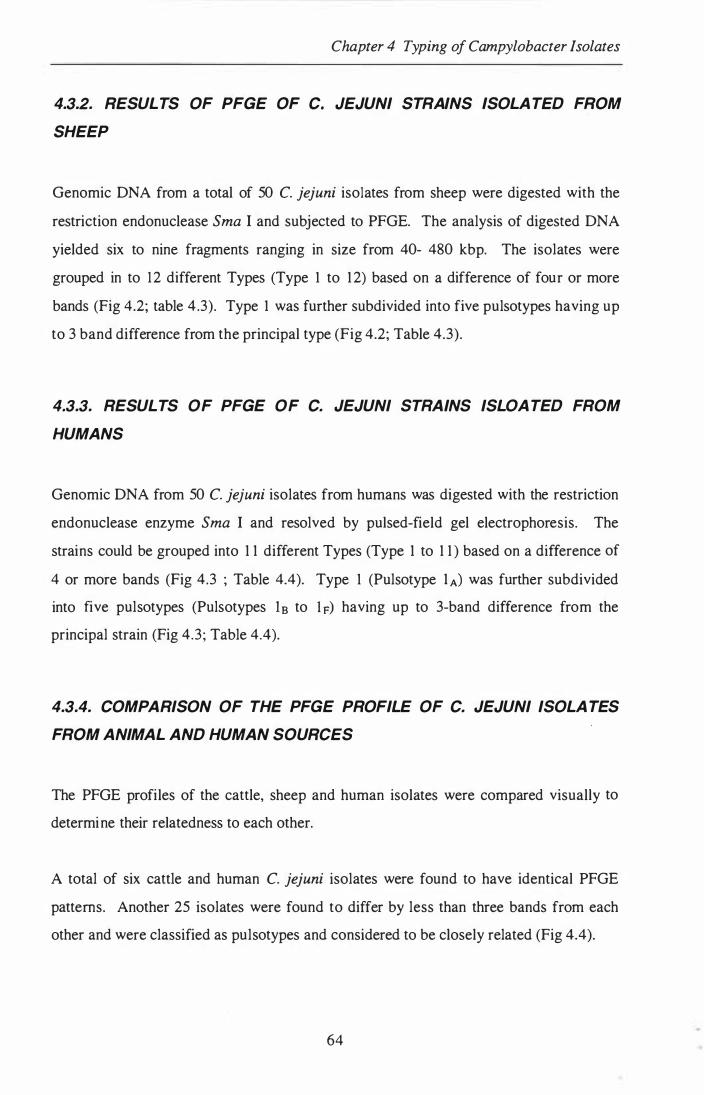


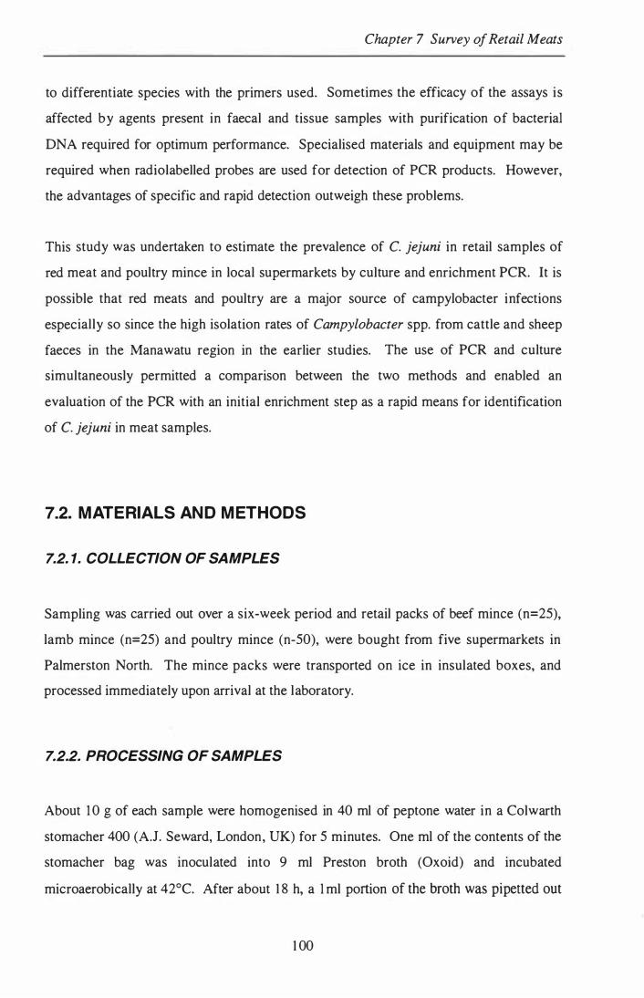


![Health ConsultationTrowbridge 1997]. Sources of dust from inside the house include: 1. The breakdown of plant and animal materials including food debris, animal hairs/dander, feathers,](https://static.documents.pub/doc/80x56/5f1f3cb5403c6f2a160f2f27/health-trowbridge-1997-sources-of-dust-from-inside-the-house-include-1-the-breakdown.jpg)


