REVIEW
Animals in the Zika Virus Life Cycle: What to
Expect from Megadiverse Latin American
Countries
Marina Galvão Bueno1*, Nadia Martinez1, Lıvia Abdalla1, Claudia Nunes Duarte dos
Santos2, Marcia Chame1*
1 Fundacão Oswaldo Cruz, Programa Institucional Biodiversidade e Saude, Rio de Janeiro, Brazil,
2 Laboratorio de Virologia Molecular, Instituto Carlos Chagas, Fundacão Oswaldo Cruz, Curitiba, Parana,
Brazil
* [email protected] (MGB); [email protected] (MC)
Abstract
Zika virus (ZIKV) was first isolated in 1947 in primates in Uganda, West Africa. The virus
remained confined to the equatorial regions of Africa and Asia, cycling between infecting
monkeys, arboreal mosquitoes, and occasionally humans. The ZIKV Asiatic strain was
probably introduced into Brazil in or around late 2013. Presently, ZIKV is in contact with the
rich biodiversity in all Brazilian biomes, bordering on other Latin American countries. Infec-
tions in Brazilian primates have been reported recently, but the overall impact of this virus
on wildlife in the Americas is still unknown. The current epidemic in the Americas requires
knowledge on the role of mammals, especially nonhuman primates (NHPs), in ZIKV trans-
mission to humans. The article discusses the available data on ZIKV in host animals and
issues of biodiversity, rapid environmental change, and impact on human health in megadi-
verse Latin American countries. The authors reviewed scientific articles and recent news
stories on ZIKV in animals, showing that 47 animal species from three orders (mammals,
reptiles, and birds) have been investigated for the potential to establish a sylvatic cycle. The
review aims to contribute to epidemiological studies and the knowledge on the natural his-
tory of ZIKV. The article concludes with questions that require urgent attention in epidemio-
logical studies involving wildlife in order to understand their role as ZIKV hosts and to
effectively control the epidemic.
Introduction
Zika virus (ZIKV) is an emerging flavivirus from the same family as the West Nile (WNV),
Japanese encephalitis (JEV), dengue (DENV), and yellow fever viruses (YFV) [1, 2]. ZIKV is
an RNA virus, mostly transmitted to humans by bites from infected Aedes spp., especially
Aedes aegypti, a highly competent and anthropophilic vector species [3] that also transmits
DENV and Chikungunya virus (CHIKV) [4]. Other Aedes species have been implicated in
ZIKV transmission, mainly in sylvatic cycles, including Ae. africanus, Ae. albopictus, Ae. api-coargenteus, and Ae. furcifer [5, 6, 7, 8].
PLOS Neglected Tropical Diseases | DOI:10.1371/journal.pntd.0005073 December 22, 2016 1 / 13
a11111
OPENACCESS
Citation: Bueno MG, Martinez N, Abdalla L, Duarte
dos Santos CN, Chame M (2016) Animals in the
Zika Virus Life Cycle: What to Expect from
Megadiverse Latin American Countries. PLoS Negl
Trop Dis 10(12): e0005073. doi:10.1371/journal.
pntd.0005073
Editor: Archie C. A. Clements, University of
Queensland, AUSTRALIA
Published: December 22, 2016
Copyright: © 2016 Galvao Bueno et al. This is an
open access article distributed under the terms of
the Creative Commons Attribution License, which
permits unrestricted use, distribution, and
reproduction in any medium, provided the original
author and source are credited.
Funding: The authors received no specific funding
for this work.
Competing Interests: The authors have declared
that no competing interests exist.
ZIKV was first identified in 1947 in primates during a YFV study in Uganda [5]. The first
reports of infected humans appeared five years later in Uganda and Tanzania [9], but the infec-
tion remained limited to equatorial regions of Africa and Asia, cycling between infective mon-
keys, arboreal mosquitoes, and occasionally humans [10, 11]. Mosquitoes captured annually
since 1965 in Senegal have shown that ZIKV amplifies cyclically every four years, which indi-
cates that it is the “dominant periodicity" of the ZIKV in Senegal [12]. ZIKV outbreaks in
humans occurred in 2007 on the island of Yap, in Micronesia, and in Gabon [13, 6], and
another outbreak occurred in 2013 in French Polynesia [14].
Recent phylogenetic and molecular studies suggest a single introduction of the ZIKV Asi-
atic strain into the Americas (Brazil) between May and December 2013 [15] and in February
2014 in Chile [16]. In early 2015, several patients in Northeast Brazil presented DENV-like
symptoms, and molecular diagnosis revealed autochthonous ZIKV infection [17].
ZIKV has invaded a geographic area that comprises the huge Brazilian biomes, bordering
on other Latin American countries. Althouse et al. [18] modeled the ZIKV transmission
dynamics, estimating the numbers of primates and mosquitos needed to maintain a wild
ZIKV cycle. Six thousand primates and 10,000 mosquitoes are enough to support a ZIKV
transmission cycle. Based on the number of Brazilian primate species, the proximity of these
and other small mammal species to urban and rural areas, and the wide distribution of Ae.aegypti,Ae. albopictus, and other mosquito genera like Culex [19, 20] andHaemagogusthroughout the country, ZIKV spillover to wild primates is a potentially real scenario [21]. A
wildlife cycle would launch new transmission dynamics with unknown impacts on other ani-
mal species, including humans.
This review aims to describe the available data on ZIKV infection in host animals and its
relationship to biodiversity, rapid environmental changes, and the impact on human health in
megadiverse Latin American countries.
Methods
Recent advances in scientific research have emerged since ZIKV became pandemic. We
searched for scientific articles and news stories on research involving ZIKV in animals using
PubMed citation and index, the Fiocruz Library database, the Scopus database, and websites
for news stories in the mainstream lay press.
Results and Discussion
Animals as ZIKV hosts
Few studies have focused on the role of animals as hosts for ZIKV. Some authors claim that
there is no solid evidence of wild mammals, such as nonhuman primates (NHPs), as reservoirs
for ZIKV. Meanwhile, studies have reported ZIKV antibodies in livestock like goats and sheep,
rodents [22], and lions and ungulates like Artiodactyla, Perissodactyla, and Proboscidea [23].
In 1971, ZIKV antibodies were detected in primates from the Cercopithecidae family in Nige-
ria [24]. Several studies suggest that DENV, CHIKV, and ZIKV adapted from an ancestral
enzootic transmission cycle involving NHPs and a broad spectrum of species from genus
Aedes (Stegomyia, aegypti) as vectors in an urban/peri-urban cycle [25].
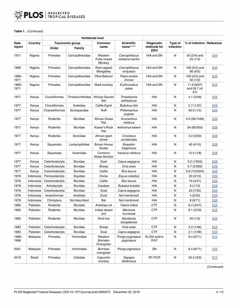
ZIKV infection has also been identified in other naturally and experimentally susceptible
animal species (Table 1 and Fig 1). Sera from 172 domestic animals and 157 wild rodents were
tested for ZIKV in Pakistan, showing that sheep, goats, some rodent species, and one human
living in the same area tested positive for ZIKV antibodies [22].
A study in Kenya in 1977 focused on the potential role of livestock (goats, sheep, and cattle)
and wild vertebrates (2,424 small mammals, 1,202 birds, 18 reptiles) in maintaining arbovirus
PLOS Neglected Tropical Diseases | DOI:10.1371/journal.pntd.0005073 December 22, 2016 2 / 13
Table 1. Chronological ZIKV natural and experimental assay infection in vertebrate hosts in the world.
Vertebrate host
Date
report
Country Taxonomic group Common
name
Scientific
name***Diagnostic
methods for
ZIKV
Type of
infection
% of infection Reference
Order Family
1947 Uganda Primates Cercopithecidae Rhesus
monkey
(sentinela)
Macaca mulatta Virus isolation N 16.7 (1/6) [5]
1952* London Rodentia Caviidae Guineapigs Cavia sp. EA Not clearly
mentioned
[9]
1952* London Lagomorpha Leporidae Rabbit Not mentioned Serology EA 100 (4/4) [9]
1952* London Rodentia Muridae Swiss albino
mice
Mus musculus EA Not clearly
mentioned
[9]
1952* London Rodentia Cricetidae Cotton-rats Sigmodon
hispidus hispidus
EA Not clearly
mentioned
[9]
1952* London Primates Cercopithecidae Rhesus
monkey
Macaca mulatta EA Not clearly
mentioned
[9]
1952** London Primates Cercopithecidae Red-tailed
monkey
Cercopithecus
ascanius schmidti
EA Not clearly
mentioned
[9]
1952** London Primates Cercopithecidae Grivet monkey Cercopithecus
aethiops centralis
EA Not clearly
mentioned
[9]
1955* EUA Chiroptera Pteropodidae Cave bat Myotus lucifugus EA 80 (16/20) [28]
1968 Kenya Artiodactyla Bovidae Gazelle not mentioned HIA N 8.3 (1/12) [23]
1968 Kenya Artiodactyla Bovidae Kongoni Alcelaphus
buselaphus
HIA N 27.3 (6/22) [23]
1968 Kenya Carnivora Felidae Lion Panthera leo HIA N 50 (1/2) [23]
1968 Kenya Artiodactyla Bovidae Wildebeest Connochaetes
taurinus
HIA N 22.2 (4/18) [23]
1968 Uganda Primates Cercopithecidae African green
monkey
Cercopithecus
aethiops
HIA N 64.6 (115/
178)
[23]
1968 Uganda Primates Cercopithecidae Red-tailed
Monkey
Cercopithecus
ascanius
HIA N 21.4 (3/14) [23]
1968 Uganda Rodentia Muridae Abyssinian
grass rat
Arvicanthis
abyssinicus
HIA N 4.6 (2/43) [23]
1968 Zambia Cetartiodactyla Bovidae African buffalo Syncerus caffer HIA N 88.9 (8/9) [23]
1968 Zambia Artiodactyla Hippopotamidae Hippo not mentioned HIA N 57.1 (20/35) [23]
1968 Zambia Proboscidea Elephantidae Elephant not mentioned HIA N 46.5 (54/116) [23]
1968 Zambia Artiodactyla Bovidae Impala Aepyceros
melampus
HIA N 33.3 (1/3) [23]
1968 Kenya Perissodactyla Equidae Zebra not mentioned HIA N 5.5 (1/18) [23]
1968 Kenya Proboscidea Elephantidae Elephant not mentioned HIA N 40.8 (31/76) [23]
1969–
1970
Uganda Primates Cercopithecidae Red-tailed
monkey
Cercopithecus
ascanius schmidti
HIA and SN N 38.1 (54/142)
and 52.1 (74/
142)
[7]
1969–
1970
Uganda Primates Cercopithecidae Colobus Colobus
abyssinicus
uellensis
HIA and SN N 45.4 (5/11)
and 54.5 (6/
11)
[7]
1969–
1970
Uganda Primates Cercopithecidae Mangabey Cercocebus
albigena johnstoni
HIA and SN N 50 (2/4) and
75 (3/4)
[7]
1969–
1971
Nigeria Primates Cercopithecidae African green
monkey
Chlorocebus
aethiops
HIA and SN N 55.5 (5/9) and
66.6 (6/9)
[24]
1969–
1971
Nigeria Primates Cercopithecidae Mona Monkey Cercopithecus
mona
HIA and SN N 36.1 (13/36)
and 41.7 (15/
36)
[24]
(Continued )
PLOS Neglected Tropical Diseases | DOI:10.1371/journal.pntd.0005073 December 22, 2016 3 / 13
Table 1. (Continued)
Vertebrate host
Date
report
Country Taxonomic group Common
name
Scientific
name***Diagnostic
methods for
ZIKV
Type of
infection
% of infection Reference
Order Family
1971 Nigeria Primates Cercopithecidae Western
Putty-nosed
Monkey
Cercopithecus
nictitans martini
HIA and SN N 50 (2/4) and
25 (1/4)
[24]
1969 Nigeria Primates Cercopithecidae Red-capped
Mangabey
Cercopithecus
torquatus
HIA and SN N 100 (5/5) and
80 (4/5)
[24]
1969–
1971
Nigeria Primates Cercopithecidae Olive Baboon Papio anubis
choras
HIA and SN N 100 (2/2) and
50 (1/2)
[24]
1969–
1971
Nigeria Primates Cercopithecidae Wadi monkey Erythrocebus
patas
HIA and SN N 11.9 (8/67)
and 59.7 (4/
67)
[24]
1977 Kenya Ciconiiformes Threskiornithidae African Sacred
Ibis
Threskiornis
aethiopicus
HIA N 4.1 (2/49) [29]
1977 Kenya Ciconiiformes Ardeidae Cattle Egret Bubulcus ibis HIA N 2.7 (1/37) [29]
1977 Kenya Charadriformes Scolopacidae Ruff Philomachus
pugnax
HIA N 50.0 (1/2) [29]
1977 Kenya Rodentia Muridae African Grass
Rat
Arvicanthus
niloticus
HIA N 4.0 (58/1446) [29]
1977 Kenya Rodentia Muridae Kaiser’s Rock
Rat
Aethomys kaiseri HIA N 34 (85/250) [29]
1977 Kenya Rodentia Soricidae African giant
shrew
Crocidura
occidentalis
HIA N 3.2 (2/63) [29]
1977 Kenya Squamata Lamprophiidae Brown House
Snake
Boaedon
fuliginosus
HIA N 40 (4/10) [29]
1977 Kenya Squamata Varanidae Common
Water Monitor
Varanus niloticus HIA N 12.5 (1/8) [29]
1977 Kenya Cetartiodactyla Bovidae Goat Capra aegagrus HIA N 0.2 (1/655) [29]
1977 Kenya Cetartiodactyla Bovidae Sheep Ovis aries HIA N 0.7 (2/283) [29]
1977 Kenya Cetartiodactyla Bovidae Cattle Bos taurus HIA N 0.6 (15/2324) [29]
1978 Indonesia Perissodactyla Equidae Horse Equus caballus HIA N 20 (3/15) [30]
1978 Indonesia Cetartiodactyla Bovidae Cattle Bos taurus HIA N 10 (4/41) [30]
1978 Indonesia Artiodactyla Bovidae Carabao Bubalus bubalis HIA N 8 (1/13) [30]
1978 Indonesia Cetartiodactyla Bovidae Goat Capra aegagrus HIA N 20 (7/35) [30]
1978 Indonesia Anseriformes Anatidae Duck Not mentioned HIA N 4 (2/52) [30]
1978 Indonesia Chiroptera Not described Bat Not mentioned HIA N 8 (6/71) [30]
1983 Pakistan Rodentia Muridae Antelope rat Tatera indica CTF N 6.4 (3/47) [22]
1983 Pakistan Rodentia Muridae Indian desert
jird
Meriones
hurrianae
CTF N 6.1 (2/33) [22]
1983 Pakistan Rodentia Muridae Sind rice Bandicota
bengalensis
CTF N 50 (1/2) [22]
1983 Pakistan Cetartiodactyla Bovidae Sheep Ovis aries CTF N 2.2 (1/46) [22]
1983 Pakistan Cetartiodactyla Bovidae Goat Capra aegagrus CTF N 2.1 (1/48) [22]
1996–
1998
Malaysia Primates Hominidae Western
Bornean
Orangutan
Pongo pygmaeus
pygmaeus
ELISA and/or
IFAT
N 8.4 (6/71) [31]
2001 Malaysia Primates Hominidae Bornean
orangutan
Pongo pigmaeus SN N 8.4 (6/71) [32]
2016 Brazil Primates Cebidae Capuchin
monkey
Sapajus
libidinosus
RT-PCR N 33.3 (3/9) [21]
(Continued )
PLOS Neglected Tropical Diseases | DOI:10.1371/journal.pntd.0005073 December 22, 2016 4 / 13
transmission. Hemagglutination inhibition assays showed that domestic animals (0.4%), wild
birds (0.4%), small wild mammals (5.9%), and reptiles (27.7%) tested positive for ZIKV [29].
Serologic studies should be interpreted carefully in view of possible cross-reactions with
other antigenic flavivirus, despite studies suggesting that plaque reduction neutralization test
Table 1. (Continued)
Vertebrate host
Date
report
Country Taxonomic group Common
name
Scientific
name***Diagnostic
methods for
ZIKV
Type of
infection
% of infection Reference
Order Family
2016 Brazil Primates Callitrichidae Marmoset Callithrix jacchus RT-PCR N 26.7 (4/15) [21]
Abbreviations: N, natural; EA, experimental assay; ELISA, enzyme-linked immunosorbent assay; RT-PCR, real-time polymerase chain reaction; HIA,
hemagglutination inhibiting antibodies; IFAT, immunofluorescence antibody test; SN, serum neutralization; CTF, complement fixation test, EUA, United
States of America.
*Intracerebral inoculation.
**Subcutaneous inoculation.
***The scientific names follow the exact description of the original reference and not the current taxonomic classification.
doi:10.1371/journal.pntd.0005073.t001
Fig 1. Historical time-line of ZIKV spread in humans and animals in the world. Colored countries have reported autochthonous vector-
borne human cases, and those labeled with specific years and animal silhouettes have reported diagnosed cases of ZIKV in naturally
infected animals. Human cases are according to references [26, 27], and the list of animal species is described in Table 1.
doi:10.1371/journal.pntd.0005073.g001
PLOS Neglected Tropical Diseases | DOI:10.1371/journal.pntd.0005073 December 22, 2016 5 / 13
(PRNT) does not cross react and is the most specific serological test for the proper serological
identification of flaviviruses [33, 34, 35].
Regarding ZIKV infection of sylvatic animals, the presence of positive animals for antibod-
ies does not necessarily means that they are viremic, and they may not be able to transmit the
virus to a mosquito, but more studies are required to properly address this issue [36]. In the
case of the sylvatic cycle of YFV (also a flavivirus) in the Americas, when monkeys become
infected, they present overt clinical signs and a viremia high enough to transmit virus to the
mosquito vectors [37].
Unlike humans, wild mammals with ZIKV infection display few clinical signs. In a sentinel
study in Uganda in 1947, primates showed only mild pyrexia. All monkeys inoculated by dif-
ferent routes developed neutralizing antibodies by day 14 after inoculation [5]. In the same
study, Swiss mice became ill and one animal died following intracerebral inoculation [9]. Such
inoculation is not a natural transmission route, and authors point out that some species of
wild and laboratory rodents are resistant to some flavivirus infections due to innate genetic
resistance [38].
Most primates identified as ZIKV-positive in the wild or in sentinel studies are from Old
World species. Phylogenetic analysis shows that humans are more closely related to Old
World primate species, especially chimpanzees and orangutans [39]. Diseases that can be
transmitted between closely related species often increase the relative risk [40, 41]. NHPs thus
deserve special attention because of their close relatedness to humans and potential disease
exchange [42].
Favoretto et. al. [21], using real-time PCR, showed that 29% (7/24) of the New World pri-
mates, Callithrix jacchus and Sapajus libidinosus, in Ceara State in Northeast Brazil were
infected with ZIKV. They also showed that the ZIKV genome sequence from monkeys was
100% similar to the ZIKV circulating in humans in South America, suggesting that primates
sharing the habitat with humans could act as ZIKV hosts, as in the YFV sylvatic cycle in Brazil.
Besides the use of primates as sentinels in ZIKV studies, some experimental work has been
performed with other mammals. Cotton-rats, guinea pigs, and rabbits showed no clinical signs
of infection after intracerebral inoculation [9]. An experiment in 1955 aimed to determine the
susceptibility of cave bats to ZIKV and showed that these bats are susceptible to ZIKV by
intraperitoneal, intradermal, intracerebral, and intrarectal exposure, but not by intranasal
exposure [28].
Barr et al. [43] infected cell cultures from different animal species with ZIKV and showed
that 17 were susceptible to the virus, developing a cytopathic effect seven days post infection.
Some of the cell cultures were from domestic animals and others from Old World wild pri-
mates, while nine were from wild animals species found in the Americas: Tabarida brasiliensis,Sylvilagus floridanus,Urocyon cinerorgeneus, Odocoileus hemionus, Procyon lotor, Didelphis vir-giniana, Dasypus novemcinctus,Marmota monax, and Neovison vison. Most of these animals
are peri-domestic and sympatric to mosquito vectors. The authors also argued that with suffi-
ciently high viremia, these animals could serve as hosts. However, they also indicated that the
virus strain used in the experiment lacks some characteristics of the ZIKV currently circulating
in the field, and that the virus in the laboratory does not mirror natural infection.
Public policy and elimination efforts in the Americas are based mainly on vector control
and personal protection measures, so the high number of wild species with the potential to
establish a sylvatic cycle would make elimination extremely difficult, if not impossible [18].
We thus need studies on ZIKV in wild and domestic animals in the Americas, both to under-
stand their potential role as hosts in the natural cycle and to target surveillance for enzootic
ZIKV transmission.
PLOS Neglected Tropical Diseases | DOI:10.1371/journal.pntd.0005073 December 22, 2016 6 / 13
Biodiversity, animal hosts, and diseases
Human health relates closely to environmental health, defined here as the relationship between
the health of domestic animals, wildlife, and the environment. Most etiological agents (60.3%)
circulate between animals and humans, and 71.8% of emerging diseases are caused by patho-
gens originating in wildlife [44]. A recent study associated 2,107 etiological agents with dis-
eases in humans and animals [45].
Recent efforts by the Convention on Biological Diversity and the World Health Organization
have addressed scientific and political discussions on the relationship between human health
and biodiversity. Such relationships include global concern over the importance of emerging
zoonotic diseases originating in wildlife. Environmental changes, including loss of biodiversity,
can favor emerging diseases originating from wildlife and act as the source of selective forces in
new genetic variations, leading to spillover and infecting humans [46]. This justifies actions to
improve knowledge on biodiversity and pathogens and to monitor them to anticipate problems.
The current ZIKV epidemic in Brazil requires understanding of the role of mammals, espe-
cially primates, in viral transmission to humans, especially when this interface occurs in frag-
mented forest areas, as described by Favoretto et al. [21]. Such areas are usually bordered or
surrounded by farmland and human settlements and by dense urban and unstructured areas
that can increase contact between humans, wildlife, and domestic animals and occasionally
promote disease spillover [47, 48]. Wild animals, especially primates, can thus be considered
sentinels for pathogens of human health concern [48, 49]. ZIKV is an example of spillover,
because this virus adapted from an ancestral transmission cycle involving NHPs to an urban/
peri-urban cycle, with humans as the main host.
Brazil is a megadiverse country with 357 million hectares of tropical forest and other highly
biodiverse biomes [50]. Not surprisingly, Brazil has more primate species than any other coun-
try. Its 53 species account for 27% of the world’s primates [51].
Some NHP species occupy urban forests due to habitat fragmentation and have close con-
tact with humans and domestic animals. Examples include primates from the Callitrichinae
(Callithrix, Leontopithecus, and Saguinus), Cebinae (Cebus), and Atelidae families (Alouattaand Brachyteles) [52]. Favoretto et al. [21] were the first to report ZIKV in NHPs in Northeast
Brazil, highlighting that these New World primates can act as potential ZIKV hosts in the
Americas. Many questions remain unanswered. Does ZIKV impact the health of NHPs? Are
NHPs living in urban fragments of forest more prone to ZIKV infection than those in pre-
served areas? Can naturally infected neotropical primates transmit ZIKV to mosquito vectors
and thus help keep the virus circulating in the Americas?
Barr et al. [43] demonstrated the feasibility of infection in cell cultures from other mamma-
lian species like carnivores, armadillos, rodents, and bats, thus raising the possibility of a trans-
mission network shaped by biological and ecological factors. These factors include vector and
host density and behavior, virulence, viral load, immunity, genetic variation, climate change,
competition between biological communities, and anthropogenic forces like urbanization,
sanitation, limited access to health services, poverty, and mistreatment of animals [38].
Considering the current epidemiological scenario with simultaneous circulation of the
arboviruses ZIKV, DENV, and CHIKV and the fact that Brazil has a large NHP population,
there is an urgent need to answer these questions to evaluate the impact of diseases like Zika
on the NHP population in Brazil and elsewhere in the Americas. YFV, another flavivirus that
circulates in a sylvatic cycle in the Americas, has a great impact on primate populations, espe-
cially those of genus Alouatta [53], which exhibit disease signs after infection and act as senti-
nel primates for viral circulation and for implementation of control measures like human
vaccination campaigns.
PLOS Neglected Tropical Diseases | DOI:10.1371/journal.pntd.0005073 December 22, 2016 7 / 13
The pandemic ZIKV strain differs significantly from the African strain mainly in two
regions of the genome. These acquired genetic markers increase its fitness for replication in
the human host [4]. Whether these mutations also alter the infectivity in NHPs remains to be
determined. The role of wild primates and other mammals in ZIKV epidemiology thus
requires urgent investigation.
The complex epidemiological panorama currently experienced in several countries of
South America, with the co-circulation of three arbovirus, ZIKV, DENV, and CHIKV, of high
impact on public health, highlights the importance of a robust epidemiological surveillance.
During 2014, two strains of CHIKV were introduced in Brazil: the Asian and the African
(East/Central/South Africa [ECSA]) strains, both transmitted by Ae. aegypti.As seen during the 2005 CHIKV outbreak in La Reunion Island, where the predominant
mosquito species was Ae. albopictus, the viruses quickly acquired an E1-A226V mutation,
increasing viral fitness to infect Ae. albopictus, which became the principal vector [54]. The
Brazilian CHIKV strains analyzed so far did not display mutations that increase CHIKV trans-
missibility and persistence in Ae. albopictus [55]. However, the elevated density and wide dis-
tribution of Ae. albopictus in Latin America warns the risk of the ECSA strain adapt to this
vector [56]. Moreover, the abundance of naïve primate (and maybe other small mammals) spe-
cies and culicids species in South American forests creates the scenario for the establishment
of an enzootic cycle, as seen in Africa and Asia, where there is evidence of a sylvatic CHIKV
transmission cycle involving NHPs and mosquitoes [57].
Another relevant issue is the development of diagnostic tests for the detection of ZIKV
infection in wild mammals, enabling unequivocal results without cross-reactivity with other
flavivirus infections.
Final Comments and Research Perspectives
Despite the growth of epidemiological knowledge in the last century, health interventions still
mainly react to emergency events involving specific diseases in the human population, with
some mitigation efforts [46]. The current ZIKV epidemic is no exception. We cannot expect to
completely block the emergence of diseases, considering vector spread due to our limited
capacity to reverse climate change, the globalization of goods and people, and our mode of
production and consumption of natural resources. This situation is particularly paradoxical in
megadiverse countries like Brazil.
The driving forces in the spread of diseases apply to the ZIKV epidemic, including anthro-
pogenic activities, climatic change, intense human movement, loss of biodiversity, habitat
destruction, land use change, introduction of invasive species, urban development, lack of
knowledge on the role of animals in maintaining the sylvatic cycle, clinical manifestations, and
wildlife trafficking [46].
We need to understand the diversity of pathogens in nature and correlate them with biolog-
ical communities, pathogenic and genetic characteristics, and anthropic impacts in areas
where disease transmission occurs. DENV is a good example of how a combination of envi-
ronmental changes, genetic characteristics, and human mobility propels the spread of viruses
in Brazil. A new lineage of DENV entered in the country through Caribbean through the
northern/northeast and spread rapidly to the rest of Brazil, especially through the aerial trans-
portation of humans and/or mosquito vectors [58]. In parallel, this example allows us to sug-
gest that the spread of ZIKV to other biomes in the Americas and outside Brazil may also be
related to these factors, and that these should be highlighted.
The ZIKV epidemic illustrates the importance of monitoring and predicting the pathogens
arising from wild animals and biodiversity. Based on the above and the results of other studies,
PLOS Neglected Tropical Diseases | DOI:10.1371/journal.pntd.0005073 December 22, 2016 8 / 13
we pose several questions and hypotheses that emerge from this discussion and that require
investigation:
1. What other wild animals besides primates could be infected by ZIKV in Americas? What is
their role in maintaining and transmitting the virus to mosquito vectors? Which species
can act as hosts?
2. Does the virus circulate at higher levels in wild animals inhabiting forest fragments adjacent
to urban areas? What role do these animals play in maintaining the virus in areas close to
humans?
3. Which wild hosts help keep the virus circulating in the Americas?
4. Do neotropical primates play a special role in the ZIKV epidemic?
5. Does ZIKV impact wild animal populations and biodiversity? Does it cause disease and
mortality in these animals?
Infectious diseases have important implications for animal and human health and biodiver-
sity. Public health and biodiversity needs are misaligned and need to be rebalanced.
Rather than merely attacking and solving epidemic situations, as in the current ZIKV global
health emergency, we need to predict and prevent future emerging diseases. Studies of wild
hosts are troublesome and costly, especially when they require long-term monitoring. Funding
also needs to be targeted for these studies. Future laboratory, field, and eco-epidemiological
research should focus on wildlife hosts to elucidate their role in ZIKV epidemiology in the
Americas and enhance the epidemic’s control.
Key Learning Points
• A remarkable diversity of wildlife species involving three orders, namely mammals,
reptiles, and birds, totaling 47 different species, were investigated for their potential to
establish a sylvatic ZIKV cycle.
• A study showed that New World primates in Northeast Brazil were infected and that
the ZIKV genome sequence in monkeys was 100% similar to the ZIKV circulating in
humans in South America. Studies are thus needed on ZIKV in wild and domestic ani-
mals in the Americas, both to understand their potential role as hosts in the natural
cycle and to target surveillance for enzootic ZIKV transmission.
• Environmental changes, including loss of biodiversity, can favor emerging diseases
originating from wildlife and act as selective forces in new genetic variations leading to
spillover and infection in humans. This justifies actions to improve knowledge on bio-
diversity and pathogens and to monitor them to anticipate problems.
• Driving forces in the spread of diseases apply to the ZIKV epidemic, including anthro-
pogenic activities, climatic change, intense human movement, loss of biodiversity,
habitat destruction, land use change, introduction of invasive species, urban develop-
ment, lack of knowledge on the role of animals in maintaining the sylvatic cycle, clini-
cal manifestations, and wildlife trafficking.
• Future laboratory, field, and epidemiological research should focus on wildlife hosts to
elucidate their role in ZIKV epidemiology in the Americas and enhance the epidemic’s
control.
PLOS Neglected Tropical Diseases | DOI:10.1371/journal.pntd.0005073 December 22, 2016 9 / 13
Acknowledgments
We would like to thank Dr. Fernando Dias de Avila Pires for constructive comments.
References1. International comittee on taxonomy of viruses. Virus taxonomy. 2014. Release 2015. http://www.
ictvonline.org/virustaxonomy.asp.
2. Pierson TC, Diamond MS. Flaviviruses. In: Knipe DM, Howley PM, editors, Fields Virol, Lippincott Wil-
liams & Wilkins, Philadelphia; 2013. pp. 747–794.
3. Albuquerque, C. Fiocruz identifica mosquitos Aedes aegypti naturalmente infectados pelo vırus Zika.
Portal Fiocruz. 25 May 2016. http://portal.fiocruz.br/pt-br/content/fiocruz-identifica-mosquitos-aedes-
aegypti-naturalmente-infectados-pelo-virus-zika. Accessed 27 May 2016.
4. Russell PK. The Zika Pandemic—A Perfect Storm? PLoS Negl Trop Dis. 2016; 10: e0004589. doi: 10.
1371/journal.pntd.0004589 PMID: 26991663
5. Dick GW, Kitchen SF, Haddow AJ. Zika virus. I. Isolations and serological specificity. Trans R Soc Trop
Med Hyg. 1952; 46: 509–520. PMID: 12995440
6. Grard G, Caron M, Mombo IM, Nkoghe D, Ondo SM, Jiolle D, et al. Zika Virus in Gabon (Central Africa)–
2007: A New Threat from Aedes albopictus? PLoS Negl Trop Dis. 2014; 8: e2681. doi: 10.1371/journal.
pntd.0002681 PMID: 24516683
7. McCrae AW, Kirya BG, Yellow fever and Zika virus epizootics and enzootics in Uganda. Trans R Soc
Trop Med Hyg. 1982; 76(4):552–62. PMID: 6304948
8. Faye O, Freire CCM, Iamarino A, Oliveira JVC, Faye O, Oliveira JVC, et al. Molecular Evolution of Zika
Virus during Its Emergence in the 20thCentury. PLoS Negl Trop Dis; 2014; 8: e2636. doi: 10.1371/
journal.pntd.0002636 PMID: 24421913
9. Dick GW. Zika virus. II. Pathogenicity and physical properties. Trans R Soc Trop Med Hyg. 1952; 46:
521–534. PMID: 12995441
10. Hayes EB. Zika virus outside Africa. Emerg. Infect. Dis. 2009; 15: 1347–1350. doi: 10.3201/eid1509.
090442 PMID: 19788800
11. Zanluca C, Santos CND. Zika virus—an overview. Microbes Infect. 2016; 18: 295–301. doi: 10.1016/j.
micinf.2016.03.003 PMID: 26993028
12. Althouse BM, Hanley KA, Diallo M, Sall AA, Ba Y, Faye O, et al. Impact of climate and mosquito vector
abundance on sylvatic arbovirus circulation dynamics in Senegal. Am J Trop Med Hyg. 2015; 92: 88–
97. doi: 10.4269/ajtmh.13-0617 PMID: 25404071
Top Five Papers
1. Favoretto S, Araujo D, Oliveira D, Duarte N, Mesquita F, Zanotto P, et al. First detec-
tion of Zika virus in neotropical primates in Brazil: a possible new reservoir. In press.
2016. BioRxiv.
2. Dick GW, Kitchen SF, Haddow AJ. Zika virus. I. Isolations and serological specificity.
Trans R Soc Trop Med Hyg. 1952; 46: 509–520.
3. Althouse BM, Vasilakis N, Sall AA, Diallo M, Weaver S, Hanley KA. Potential for
Zika virus to establish a sylvatic transmission cycle in the Americas. In press. 2016.
BioRxiv.
4. Henderson BE, Hewitt LE, Lule M. Serology of wild mammals. In: Virus Research
Institute 409 Annual Report. East African Printer, Nairobi, Kenya. 1968, pp. 48–51.
5. Karesh WB, Formenty P. Infectious diseases. In: WHO. Connecting Global Priorities:
Biodiversity and Human Health. A State of Knowledge Review, 2015, pp.28.
PLOS Neglected Tropical Diseases | DOI:10.1371/journal.pntd.0005073 December 22, 2016 10 / 13
13. Ioos S, Mallet H-P, Leparc Goffart I, Gauthier V, Cardoso T, Herida M. Current Zika virus epidemiology
and recent epidemics. Medecine Mal. Infect. 2014; 44: 302–307.
14. World Health Organization. Epidemiological Update Zika virus infection. 2015. http://www.paho.org/hq/
index.php?option=com_docman&task=doc_view&Itemid=270&gid=32021&lang=en.
15. Faria NR, Azevedo RSS, Kraemer MUG, Souza R, Cunha MS, Hill SC et al. Zika virus in the Americas:
Early epidemiological and genetic findings. Science. 2016; 351: 1371–1494.
16. Tognarelli J, Ulloa S, Villagra E, Lagos J, Aguayo C, Fasce R, et al. A report on the outbreak of Zika
virus on Easter Island, South Pacific, 2014. Arch Virol. 2016; 161: 665–668. doi: 10.1007/s00705-015-
2695-5 PMID: 26611910
17. Zanluca C, Melo VC, Mosimann AL, Santos GI, Santos CN, Luz K. First report of autochthonous trans-
mission of Zika virus in Brazil. Mem Inst Oswaldo Cruz. 2015; 110: 569–572. doi: 10.1590/0074-
02760150192 PMID: 26061233
18. Althouse BM, Vasilakis N, Sall AA, Diallo M, Weaver S, Hanley KA. Potential for Zika virus to establish a
sylvatic transmission cycle in the Americas. In press. 2016. BioRxiv. http://dx.doi.org/10.1101/047175.
Accessed 5 Apr. 2016.
19. Diallo D, Sall AA, Diagne CT, Faye O, Faye O, Ba Y, et al. Zika Virus Emergence in Mosquitoes in
Southeastern Senegal, 2011. PLoS ONE. 2014; 9: e109442. doi: 10.1371/journal.pone.0109442
PMID: 25310102
20. Vogel, G. Scientific sleuths hunt for Zika-carrying mosquitoes. Science. 1 Jun 2016. http://www.
sciencemag.org/news/2016/06/scientific-sleuths-hunt-zika-carrying-mosquitoes. Accessed 29 Aug
2016.
21. Favoretto S, Araujo D, Oliveira D, Duarte N, Mesquita F, Zanotto P, et al. First detection of Zika virus in
neotropical primates in Brazil: a possible new reservoir. In press. 2016. BioRxiv. http://dx.doi.org/10.
1101/049395. Accessed 20 April 2016.
22. Darwish MA, Hoogstraal H, Roberts TJ, Ahmed IP, Omar F. A sero-epidemiological survey for certain
arboviruses (Togaviridae) in Pakistan. Trans R Soc Trop Med Hyg. 1983; 77: 442–445. PMID:
6314612
23. Henderson BE, Hewitt LE, Lule M. Serology of wild mammals. In: Virus Research Institute 409 Annual
Report. East African Printer, Nairobi, Kenya. 1968, pp. 48–51. http://dx.doi.org/10.2471/BLT.16.
171082.
24. Monath TP, Kemp GE. Importance of non-human primate in yellow fever epidemiology in Nigeria. Trop
Geogr Med. 1973; 25:28–38. PMID: 4632785
25. Musso D, Cao-Lormeau VM, Gubler DJ. Zika virus: following the path of dengue and chikungunya? Lan-
cet. 2015; 386: 243–244. doi: 10.1016/S0140-6736(15)61273-9 PMID: 26194519
26. Kindhauser MK, Allen T, Frank V, Santhana RS, Dye C. Zika: the origin and spread of a mosquito-borne
virus [In press]. Bull World Health Organ. 2016; 171082. http://dx.doi.org/10.2471/BLT.16.171082
27. World Health Organization. 2016 Jun 9 [cited 20 June 2016]. In: World Health Organization [Internet].
Atlanta: WHO 2016. http://apps.who.int/iris/bitstream/10665/208877/1/zikasitrep_9Jun2016_eng.pdf?
ua
28. Reagan RL, Rumbaugh H, Nelson H, Brueckner AL. Effect of Zika virus and Bwanba virus in the cave
bat (Myotus lucifugus). Trans Am Microsc Soc. 1955; 74: 77–79.
29. Johnson BK, Chanas AC, Shockle P, Squires EJ, Gardner P, Wallace C, et al. Arbovirus isolations
from, and serological studies on, wild and domestic vertebrates from Kano Plain, Kenya. Trans. R. Soc.
Trop. Med. Hyg. 1977; 71: 512–517. PMID: 605465
30. Olson JG, Ksiazek TG, Gubler DJ, Lubis SI, Simanjuntak G, Lee VH, Nalim S, et al. A survey for arbo-
viral antibodies in sera of humans and animals in Lombok, Republic of Indonesia. Ann Trop Med Parasi-
tol. 1983; 77:131–137. PMID: 6309104
31. Kilbourn AM, Karesh WB, Wolfe ND, Bosi EJ, Cook RA, Andau M. Health evaluation of free-ranging
and semi-captive orangutans (Pongo pygmaeus pygmaeus) in Sabah, Malaysia. JWD. 2003; 39: 73–
83, http://dx.doi.org/10.7589/0090-3558-39.1.73
32. Wolfe N, Kilbourn AM, Karesh WB, Rahman HA, Bosi EJ, Cropp B et al. Sylvatic transmission of arbovi-
ruses among Bornean Orangutans. Am J Trop Med Hyg. 2001; 64: 310–316. PMID: 11463123
33. Gennaro AD, Lorusso A, Casaccia C, Conte A, Monaco F, Savini G. Serum Neutralization Assay Can
Efficiently Replace Plaque Reduction Neutralization Test for Detection and Quantitation of West Nile
Virus Antibodies in Human and Animal Serum Samples. Clin Vaccine Immunol. 2014; 1: 1460–1462.
34. Mansfield KL, Horton DL, Johnson N, Li L, Barrett AD, Smith DJ et al. Flavivirus-induced antibody
cross-reactivity. J Gen Virol. 2011; 92: 2821–2829. doi: 10.1099/vir.0.031641-0 PMID: 21900425
PLOS Neglected Tropical Diseases | DOI:10.1371/journal.pntd.0005073 December 22, 2016 11 / 13
35. Lanciotti RS, Kosoy OL, Laven JJ, Velez JO, Lambert AJ, Johnson AJ et al. Genetic and Serologic
Properties of Zika Virus Associated with an Epidemic, Yap State, Micronesia, 2007. Emerg Infect Dis.
2008; 14: 1232–1239. doi: 10.3201/eid1408.080287 PMID: 18680646
36. Rabe IB, Staples JE, Villanueva J et al. Interim Guidance for Interpretation of Zika Virus Antibody Test
Results. MMWR Morb Mortal Wkly Rep 2016; 65. http://dx.doi.org/10.15585/mmwr.mm6521e1.
37. Vasconcelos PF, Costa ZG, Travassos Da Rosa ES, Luna E, Rodrigues SG, Barros VL et al. Epidemic
of jungle yellow fever in Brazil, 2000: implications of climatic alterations in disease spread. J Med Virol.
2001; 65: 598–604. PMID: 11596099
38. Kuno G, Chang GJJ. Biological Transmission of Arboviruses: Reexamination of and New Insights into
Components, Mechanisms, and Unique Traits as Well as Their Evolutionary Trends. CMR. 2005; 18:
608–637.
39. Haviland WA, Prins HEL, Walrath D, McBride B. Anthropology: The Human Challenge. In: Living pri-
mates. Belmont: Wadsworth, Cengange Learning. Wadsworth/Cengage Learning; 2014. 61 pp. ISBN-
13: 9781133941323.
40. Pedersen AB, Davies TJ. Cross-Species Pathogen Transmission and Disease Emergence in Primates
2009; 6: 496–508.
41. Davies TJ, Pedersen AB. Phylogeny and geography predict pathogen community similarity in wild pri-
mates and humans Proc. R. Soc. B. 2008; 275: 1695–1701
42. Unwin S, Ancrenaz M, Bailey W. Handling, anesthesia, health evaluation and biological sampling. In:
Setchell JM, Curtis DJ, editors. Field and laboratory methods in primatology: A Practical Guide. 2nd ed.
Cambridge, UK: Cambridge University Press; 2011. pp. 147–168.
43. Barr KL, Anderson BD, Long MT. Working with Zika and Usutu Viruses in Vitro. In press. 2016. BioRxiv.
http://dx.doi.org/10.1101/040139. Accessed 18 Feb. 2016
44. Jones KE, Patel NG, Levy MA, Storeygard A, Balk D, Gittleman JL, et al. Global trends in emerging
infectious diseases. Nature. 2008; 451: 990–993. doi: 10.1038/nature06536 PMID: 18288193
45. Wardeh M, Risley C, McIntyre MK, Setzkorn C, Baylis M. Database of host-pathogen and related spe-
cies interactions, and their global distribution. Sci data. 2015.
46. Karesh WB, Formenty P. Infectious diseases. In: WHO. Connecting Global Priorities: Biodiversity and
Human Health. A State of Knowledge Review: 2015. pp
47. Osofsky SA, Kock RA, Kock MD, Kalema-Zikusoka G, Grahn R, Leyland T, et al. Building support for
protected areas using a ‘one health’ perspective. In: Friends for life: New partners in support of pro-
tected areas, McNeely JA, editor. Gland, Switzerland and Cambridge, United Kingdom: IUCN.
2005. pp. 65–79.
48. Wolfe ND, Escalante AA, Karesh WB, Kilbourn A, Spielman A, Lal AA: Wild primate populations in
emerging infectious disease research: the missing link? Emerg Infect Dis 1998; 4: 149–158. doi: 10.
3201/eid0402.980202 PMID: 9621185
49. Gillespie TR, Chapman CA, Forest fragmentation, the decline of an endangered primate, and changes
in host–parasite interactions relative to an unfragmented forest. 2008; 70: 222–230.
50. Bohrer CBA. Mata Atlantica e formacões associadas. In: Serra MV, Serra MTF, editors. Guia de His-
toria Natural do Rio de Janeiro. Rio de Janeiro: Ed. Cidade Viva; 2012. pp. 138–157.
51. Mittermeier R. Primate Diversity and the Tropical Forest. In: Wilson EO, Peter FM, editors. Biodiversity.
Harvard University; National Academy of Sciences/Smithsonian Institution; 1999. pp. 521.
52. Marsh LK. Primates in Fragments. Ecology and Conservation. New York: Kluwer Academic/ Plenum
Publishers; 2003.
53. Daszak P, Cunningham AA, Hyatt AD. Emerging infectious diseases of wildlife-threats to biodiversity
and human health. Science. 2000; 287: 443. PMID: 10642539
54. Santhosh SR, Dash PK, Parida MM, Khan M, Tiwari M, Lakshmana Rao PV. Comparative full genome
analysis revealed E1: A226V shift in 2007 Indian Chikungunya virus isolates. Virus Res. 2008; 135:
36–41. doi: 10.1016/j.virusres.2008.02.004 PMID: 18384900
55. Tsetsarkin KA, Weaver SC. Sequential adaptive mutations enhance efficient vector switching by Chi-
kungunya virus and its epidemic emergence. PLoS Pathog. 2011; 7: e1002412. doi: 10.1371/journal.
ppat.1002412 PMID: 22174678
56. Vega-Rua A, Zouache K, Girod R, Failloux AB, Lourenco-de-Oliveira R. High level of vector compe-
tence of Aedes aegypti and Aedes albopictus from ten American countries as a crucial factor in the
spread of Chikungunya virus. Journal of virology. 2014; 88: 6294–6306. doi: 10.1128/JVI.00370-14
PMID: 24672026
PLOS Neglected Tropical Diseases | DOI:10.1371/journal.pntd.0005073 December 22, 2016 12 / 13
57. Nunes MR, Faria NR, Vasconcelos JM, Golding N, Kraemer MU, Oliveira LF et al. Emergence and
potential for spread of Chikungunya virus in Brazil. BMC Med. 2015; 13: 102. doi: 10.1186/s12916-015-
0348-x PMID: 25976325
58. Nunes MRT, Palacios G, Faria NR, Sousa EC Jr, Pantoja JA, Rodrigues SG et al. Air Travel Is Associ-
ated with Intracontinental Spread of Dengue Virus Serotypes 1–3 in Brazil. PLoS Negl Trop Dis. 2014;
8: e2769. doi: 10.1371/journal.pntd.0002769 PMID: 24743730
PLOS Neglected Tropical Diseases | DOI:10.1371/journal.pntd.0005073 December 22, 2016 13 / 13