, 20120102, published 5 August 2013 371 2013 Phil. Trans. R. Soc. A Turro and M. H. Levitt M. Concistrè, S. Mamone, M. Denning, G. Pileio, X. Lei, Y. Li, M. Carravetta, N. J. determined by solid-state NMR 60 O@C 2 Anisotropic nuclear spin interactions in H References 02.full.html#ref-list-1 http://rsta.royalsocietypublishing.org/content/371/1998/201201 This article cites 22 articles, 3 of which can be accessed free Subject collections (42 articles) spectroscopy collections Articles on similar topics can be found in the following Email alerting service here in the box at the top right-hand corner of the article or click Receive free email alerts when new articles cite this article - sign up http://rsta.royalsocietypublishing.org/subscriptions go to: Phil. Trans. R. Soc. A To subscribe to on August 19, 2013 rsta.royalsocietypublishing.org Downloaded from
Transcript
, 20120102, published 5 August 2013371 2013 Phil. Trans. R. Soc. A Turro and M. H. LevittM. Concistrè, S. Mamone, M. Denning, G. Pileio, X. Lei, Y. Li, M. Carravetta, N. J. determined by solid-state NMR
This article cites 22 articles, 3 of which can be accessed free
Subject collections
(42 articles)spectroscopy � collectionsArticles on similar topics can be found in the following
Email alerting service herein the box at the top right-hand corner of the article or click Receive free email alerts when new articles cite this article - sign up
http://rsta.royalsocietypublishing.org/subscriptions go to: Phil. Trans. R. Soc. ATo subscribe to
on August 19, 2013rsta.royalsocietypublishing.orgDownloaded from
ResearchCite this article: Concistrè M, Mamone S,Denning M, Pileio G, Lei X, Li Y, Carravetta M,Turro NJ, Levitt MH. 2013 Anisotropic nuclearspin interactions in H2O@C60 determined bysolid-state NMR. Phil Trans R Soc A 371:20120102.http://dx.doi.org/10.1098/rsta.2012.0102
One contribution of 13 to a Theo MurphyMeeting Issue ‘Nanolaboratories: physics andchemistry of small-molecule endofullerenes’.
Subject Areas:spectroscopy
Keywords:solid-state NMR, endohedral water fullerene,spin interaction
Anisotropic nuclear spininteractions in H2O@C60determined by solid-state NMRM. Concistrè1, S. Mamone1, M. Denning1, G. Pileio1,
X. Lei2, Y. Li2, M. Carravetta1, N. J. Turro2,† and
M. H. Levitt1
1School of Chemistry, University of Southampton, Highfield,Southampton SO17 1BJ, UK2Department of Chemistry, Columbia University, 3000 Broadway,New York, NY 10027, USA
We report a solid-state NMR study of the anisotropicnuclear spin interactions in H2O@C60 at roomtemperature. We find evidence of significant dipole–dipole interactions between the water protons, andalso a proton chemical shift anisotropy (CSA)interaction. The principal axes of these interactiontensors are found to be perpendicular. The magnitudeof the CSA is too large to be explained by a model inwhich the water molecules are partially aligned withrespect to an external axis. The evidence indicates thatthe observed CSA is caused by a distortion of thegeometry or electronic structure of the fullerene cages,in response to the presence of the endohedral water.
1. IntroductionClosed fullerene cages encapsulating small molecules arestable substances that can be synthesized via molecularsurgery [1–3]. These extraordinary compounds, availablein macroscopic quantities, offer the almost uniquepossibility to study molecules in a well-insulated,well-defined and highly symmetric environment. Theguest molecules encapsulated in these endohedralfullerenes behave as quantum rotors, whose rotationaland translational levels are mixed together by theconfinement provided by the cage, strongly influencingtheir spectroscopic properties. Studies of this kindhave already been performed on samples of H2@C60,HD@C60 and H2O@C60, which have been investigatedby far-infrared spectroscopy, inelastic neutron scatteringand nuclear magnetic resonance (NMR) [4–13].
2013 The Author(s) Published by the Royal Society. All rights reserved.
on August 19, 2013rsta.royalsocietypublishing.orgDownloaded from
The endohedral water fullerene H2O@C60 is a particularly interesting system since waterexhibits ortho–para spin isomerism and also a strong electric dipole moment. The C60 cageprovides an environment of icosahedral symmetry, which is expected to lead to a triplydegenerate ortho-H2O ground state. Nevertheless, physical studies by a range of techniques,including infrared spectroscopy, inelastic neutron scattering and cryogenic NMR have shown thatthe degeneracy of the ortho-H2O ground state is lifted in H2O@C60 [4]. This implies a reductionin symmetry, which could be due to partial orientation of the water electric dipoles by a localelectric field, or to distortion of the geometry or electronic distribution in the fullerene cage, or toa combination of these effects.
The broken symmetry of the water environment in H2O@C60 leads to a residual dipolarcoupling between the water protons, which gives rise to dipole-induced spinning sidebands incryogenic magic-angle-spinning (MAS) NMR spectra [4]. The sideband pattern of the MAS NMRspectrum at 20 K is consistent with randomly oriented proton pairs coupled by a dipole–dipolemagnetic interaction of −5.5 kHz [4]. The residual dipole–dipole coupling interaction may beinterpreted in terms of a splitting in the ortho-H2O ground state by an energy of about 0.5 meV,which matches observations by inelastic neutron scattering [4].
The question arises as to whether this symmetry-breaking effect is specific to the cryogenicregime, or whether it arises also at high temperature. In this paper, we describe MAS NMR spectrataken at room temperature, which show clear evidence of nuclear anisotropic spin interactionsin H2O@C60. In order to investigate the source of these spinning sidebands, we estimated theresidual dipolar coupling between the water protons using a double-quantum-filtered dipolarrecoupling (DQfDR) technique [14]. We show that the residual dipole–dipole coupling coexistswith a chemical shift anisotropy (CSA) interaction. The magnitude of the observed CSA isinconsistent with partial orientation of the water molecules, and is attributed to distortion orpolarization of the enclosing cage.
2. Material and methods
(a) SampleThe sample of H2O@C60 (figure 1a) was synthesized according to the method of Kurotobi &Murata [3]. The solution NMR spectrum of the last intermediate prior to cage closure indicatedthat about 60% of the C60 cages are occupied by water molecules.
The sample was purified in two stages. In the first stage (p1), the sample was purified bycolumn chromatography, eluted with CS2/pentane = 2/1 and dissolved in CS2. A precipitate wasformed by adding pentane. The precipitate was centrifuged and heated at 180◦C under vacuumfor 3 days. The solid-state 1H MAS NMR spectrum of the dry powder clearly shows not only theendohedral water peak but also a strong peak from a protonated impurity (figure 1b).
An additional purification step (p2) was performed as follows: (i) the sample was washed withdiethyl ether to remove ether-soluble impurities; (ii) the sample was heated to a temperature of250◦C and a pressure of approximately 2 × 10⁻⁵ mbar for 4 h to remove occluded solvent; (iii) thesample was sublimed onto the walls of a clean 6 mm outer-diameter silica tube. The sublimationwas carried out by heating the sample from room temperature to 550◦C at 1◦C min−1 and thenmaintaining this temperature for 12 h.
The solid-state 1H MAS NMR spectrum of the sample after procedures p1 and p2 is shown infigure 1c. The intensity of the protonated impurity peak is greatly reduced compared with thatin figure 1b, indicating the almost complete removal of protonated impurities. The weak broadfeature is attributed to protonated parts of the MAS rotor assembly and the NMR probe.
In addition to the large improvement in purity, the half-width at half-height of the endohedralproton peak is reduced from approximately 0.15 ppm in figure 1b to approximately 0.04 ppmin figure 1c. This indicates that sublimation leads to a more homogeneous and better orderedsolid-state structure.
on August 19, 2013rsta.royalsocietypublishing.orgDownloaded from
Figure 1. (a) The molecular structure of endohedral water fullerene, H2O@C60; (b) 1H MAS NMR spectrum of H2O@C60 afterpurificationmethod p1; and (c) 1HMAS NMR spectrum of H2O@C60 after purificationmethods p1 and p2. The spinning frequencywas 8.00 kHz in both cases and the spectra have the same vertical scale. The peak of endohedral water is at−4.7 ppm. (Onlineversion in colour.)
All results presented below were obtained on a 2.4 mg sample of H2O@C60 that had undergoneboth procedures p1 and p2, packed in a 4 mm outer-diameter zirconia rotor.
(b) EquipmentThe solid-state NMR experiments reported in this paper were performed on a Varian Infinity+spectrometer equipped with a 9.4 T magnet, corresponding to a 1H resonance frequency of400 MHz. All experiments were performed with the sample at room temperature.
(c) Spectroscopic techniques(i) Static proton NMR
Proton NMR spectra were obtained without sample rotation by acquiring the NMR signalinduced by a 90◦ pulse. The proton pulse duration was 3.5 μs.
(ii) Magic-angle-spinning NMR
Proton MAS NMR spectra were obtained by rotating the sample at the ‘magic angle’ arctan√
2 �54.7◦ with respect to the static magnetic field and applying a 90◦ pulse. The rotation frequenciesused in these experiments were between 1.00 and 5.00 kHz. All chemical shift scales are referencedto the 1.8 ppm proton peak of solid adamantane.
(iii) Double-quantum-filtered dipolar recoupling
A variety of methods are available for studying 1H–1H dipolar couplings in solids [15–17].In the present work, symmetry-based dipolar recoupling was used to estimate the residualproton–proton coupling. The pulse sequence is sketched in figure 2. The sequence is performedunder MAS conditions and is rotor synchronized. The dipolar recoupling is induced by using asupercycled symmetry-based sequence of the form SR202
9 [18]. Repeated SR2029 supercycles are
applied for an excitation time interval, τexc, during which double-quantum coherence builds up.A 90◦ phase-shifted sequence of SR202
9 supercycles is applied for the reconversion time interval,τrec, followed by a 90◦ pulse, which induces an NMR signal. Signals passing through double-quantum coherence are isolated by a double-quantum filter, implemented by a 16-step phasecycle.
on August 19, 2013rsta.royalsocietypublishing.orgDownloaded from
Figure 2. Double-quantum-filtered dipolar recoupling sequence used to measure residual dipolar couplings in [email protected] supercycle SR2029 consists of four symmetry-based recoupling cycles of the form R202±9, with overall phase shiftsof {0, 0,π ,π}. Each R202±9 cycle consists of 10 pairs of two-element composite pulses, with phase shifts of ±9π /20.The radiofrequency field strength is adjusted to match the duration of each R202±9 cycle with two periods of the MASrotor. The complete pulse sequence involves a double-quantum excitation sequence of duration τexc, which convertsequilibrium magnetization to double-quantum coherence. This is followed by a double-quantum reconversion sequence,which converts double-quantum coherence back to longitudinal magnetization, which is transformed into observabletransverse magnetization by a π /2 pulse. Phase cycling is used to suppress NMR signals that did not pass through double-quantum coherence.
The SR2029 supercycle consists of the following scheme [18]:
SR2029 =
[R202
9R202−9
]0
[R202
−9R2029]π
,
which combines sign alternation of the winding number with a sequence of overall phaseshifts. As described in Webber et al. [15], this supercycle compensates second-order terms in therecoupled Hamiltonian, which is important for the measurement of small dipolar couplings, atthe expense of removing the γ -encoding feature of the pure R202
9 pulse sequence. Althoughthis reduces the maximum double-quantum efficiency, this loss is more than compensated by theincreased robustness [18].
All double-quantum recoupling experiments were performed at a spinning frequency of8.00 kHz. The double-quantum excitation interval τexc was incremented from 0 to 7.25 ms in stepsof 0.25 ms. The double-quantum reconversion interval τrec was decremented by the same amount,so that the total recoupling interval τexc + τrec was kept constant at 7.25 ms (symmetric protocol[19]). The integrated signal plotted versus τexc gives rise to the double-quantum build-up curvethat is analysed to yield the value of the dipolar coupling constant.
(d) Numerical simulationsAll numerical simulations were run using the SIMPSON simulation package [20].
on August 19, 2013rsta.royalsocietypublishing.orgDownloaded from
Figure 3. (a) Static 1H spectrumof H2O@C60 obtained from4196 accumulated transients. (b) 1HMAS spectra of the same sampleat the indicated spinning frequencies. The MAS spectra are derived from 256 accumulated transients.
(i) Proton magic-angle-spinning spectra
Proton MAS spectra were simulated for the experimental spinning frequency of 1.00 kHz. Thepowder average was simulated by using the SIMPSON crystal file rep100 together with 10 evenlydistributed γ angles. The simulations were performed for randomly oriented proton pairs withdipolar coupling and CSA interactions. Both the dipolar coupling and CSA interactions weredescribed by uniaxial tensors, with the relative orientation of the unique principal axes describedby the angle β. The angle β was varied in a series of simulations (see below).
(ii) Double-quantum-filtered dipolar recoupling simulations
The DQfDR experiment was simulated for randomly oriented pairs of dipolar-coupled protonswith no CSA. The powder average was simulated by using the SIMPSON crystal file bcr20together with 20 evenly distributed γ angles. A grid of simulations was compiled by varying thedipolar coupling constant in the range −600 Hz < b/(2π ) < −400 Hz in steps of 20 Hz. The dipolarcoupling was estimated by comparing the grid of simulations to the experimental data. It wasverified by simulation that inclusion of a 2.9 ppm CSA interaction has no effect on the estimateddipolar coupling constant.
3. Results
(a) Static 1H NMRThe static solid-state 1H NMR spectrum of H2O@C60 displays a broad peak with a linewidthof several kHz (figure 3a). The lineshape is due to long-range dipole–dipole interactions andcontains no useful local information.
on August 19, 2013rsta.royalsocietypublishing.orgDownloaded from
(b) Magic-angle-spinning 1H NMRMAS NMR averages out anisotropic interactions such as dipole–dipole couplings and chemicalshift anisotropies, providing well-resolved NMR peaks for solid samples, at the isotropic chemicalshift frequencies. The sample rotation generates periodic spinning sidebands displaced fromthe isotropic chemical shifts by multiples of the spinning frequency. The amplitudes of thespinning sidebands may be analysed to obtain information on the anisotropic local nuclear spininteractions.
A set of 1H MAS spectra of H2O@C60, obtained at three different spinning frequencies, isshown in figure 3b. The proton MAS spectra display a strong narrow peak at the chemical shift of−4.7 ppm, which is very close to the reported chemical shift for H2O@C60 in solution (−4.81 ppm[3]). Spinning sidebands are visible at low (less than 5 kHz) spinning frequencies, indicating thatresidual anisotropic interactions are present, but with a smaller magnitude than those observedat cryogenic temperatures [4].
Two kinds of spin interactions can contribute to the spinning sideband pattern observed atlow spinning frequency: through-space dipolar coupling and CSA. The contributions of thesetwo different interactions may not be disentangled without additional spectroscopic data.
(c) Double-quantum dipolar recouplingThe effect of dipole–dipole coupling interactions may be distinguished from CSA interactionsby using selective dipolar recoupling methods, for example, those based on the rotationalsymmetry of the spin interactions [19,21]. Symmetry-based double-quantum recoupling wasused previously on a highly asymmetric open-cage form of the endofullerene H2@C60 inorder to investigate the anisotropy of the H2 rotation [14]. Since the anisotropic interactionsin H2O@C60 are much smaller than those encountered in [14], a modified double-quantumprocedure was used for the current work, based on the robust error-compensated supercycleSR202
9, as described in §2.The double-quantum recoupling procedure results in a plot of peak intensity versus the
double-quantum excitation time τexc. The results for H2O@C60 are shown in figure 4, wherethe filled circles represent the experimental amplitudes of the water proton peak. The double-quantum-filtered spectral amplitude is at a maximum when the excitation and reconversionintervals are equal (τexc = τrec = 7.25 ms). The dependence of the spectral amplitude on theexcitation and reconversion times away from this maximum may be analysed to estimate thedipole–dipole coupling constant.
The value of the dipolar coupling constant is retrieved by comparing the experimental build-up curve with numerical simulations. The signal areas of the simulated spectra as a function ofτexc are reported in figure 4 as solid curves. The best match between simulation and experimentsoccurs for a dipolar coupling constant |b/(2π )| = 500 Hz. The confidence limits on this dipolarcoupling constant are approximately ±10 Hz.
(d) Estimation of chemical shift anisotropyA numerical simulation of the NMR spectrum generated by randomly oriented proton pairsat a spinning frequency of 1.00 kHz, and with a dipolar coupling b/(2π ) = −500 Hz, is shownin figure 5a (second from the bottom). The simulated spinning sidebands have a significantlysmaller intensity than in the experimental spectrum (figure 5a, grey spectrum at the bottom).The poor match between simulation and experiment indicates that the dipole–dipole coupling,as estimated by the double-quantum recoupling experiment, cannot be solely responsible for theMAS spinning sideband intensities. A CSA interaction must contribute as well.
The magnitude and orientation of the CSA were estimated by comparing 1H MAS spectrawith simulations, at a spinning frequency of 1.00 kHz (figure 5). For simplicity, the simulationsassumed randomly oriented pairs of protons with uniaxial dipolar and chemical shift tensors. The
on August 19, 2013rsta.royalsocietypublishing.orgDownloaded from
Figure 4. Double-quantum-filtered signal areas as a function of the recoupling interval τexc obtained in a symmetric double-quantum recoupling procedure, using the pulse sequence SR2029 at a spinning frequency of 1.00 kHz (figure 2). Circles representthe experimental values. Solid curves from grey to black represent numerical simulations for dipolar coupling constants|b/(2π )| = 460, 480, 500, 520 and 540 Hz. The data were normalized to 1 at the maximum recoupling efficiency.
(a) (b)
–4 –2 0 2 4offset frequency (kHz)
–4 –2 0 2 4offset frequency (kHz)
da=4 ppm
da=3 ppm
da=2 ppm
da=1 ppm
da=0 ppm
experiment experiment
b=90°
b=60°
b=30°
b=0°
Figure 5. Simulated 1H MAS spectra (black lines) of randomly oriented proton pairs at a spinning frequency of 1.00 kHz, witha dipolar coupling constant b/(2π )= −500 Hz and an additional CSA interaction. The CSA tensor is assumed to be uniaxial,with magnitude δa = δzz − δiso, and with the unique axis at an angle β with respect to the 1H–1H dipole–dipole couplingtensor. (a) Simulations forβ = 90◦ at the indicated values of δa; and (b) simulations for δa = 3.1 ppm at the indicated valuesofβ . The experimental spectra for H2O@C60 are shown at the bottom of each panel.
value of the dipolar coupling constant was set to −500 Hz, as determined by the double-quantumrecoupling experiment. The motionally averaged chemical shift tensors of the two protons wereassumed to be identical and uniaxial. The unique principal axis of the CSA tensor was assumedto subtend an angle β with the unique principal axis of the dipole–dipole coupling tensor. TheCSA δa (defined as δa = δzz − δiso, where δzz is the principal axis of the chemical shift tensor thatis most distant from the isotropic value δiso) and the angle β were varied independently in a setof simulations and compared with the experimental spectrum.
on August 19, 2013rsta.royalsocietypublishing.orgDownloaded from
Figure 6. Contour map of the χ 2 statistic equation (3.1) plotted versus the CSA δa and the angle β between the principalaxes of the chemical shift and dipole–dipole interactions. Low values of χ 2 are indicated by dark shading. The darkest arearepresents the 68% confidence region. This plot shows that the 1H CSA tensor is most likely to be perpendicular to the 1H–1Hdipolar coupling tensor.
The best match between experiment and simulations was obtained for |δa| = 3.1 ppm andβ = 90◦. The simulated spectrum is insensitive to the signs of both the CSA δa and the dipole–dipole coupling in the best-fitted region, so these signs cannot be determined unambiguously.The confidence limits on
{δa, β
}were estimated by a χ2 analysis [22] using
χ2(δa, β) =2∑
i=−2
(ISi (δa, β) − IE
i )2, (3.1)
where IEi indicates the area of the ith-order sideband in the experimental spectrum and IS
i (δa, β) isthe area of the ith-order sideband in the spectrum simulated for given values of the parametersδa and β. A plot of χ2 against {δa, β} is shown in figure 6.
Rough 68% confidence limits on the parameters were estimated from the bounds of theregion. The estimated CSA parameters, and their confidence limits, are |δa| = 3.1 ± 0.3 ppm andβ = 90◦ ± 40◦. This indicates that the unique principal axis of the CSA tensor is approximatelyperpendicular to the dipole–dipole coupling tensor.
(e) DiscussionThe double-quantum 1H dipolar recoupling experiment finds clear evidence of a 1H–1H dipolarcoupling interaction in H2O@C60 at room temperature, with magnitude |b/(2π )| = 500 ± 10 Hz.Analysis of the MAS 1H spectrum indicates the simultaneous presence of a 1H CSA interaction,of magnitude 3.1 ppm, and with a unique principal axis that is perpendicular to that of the dipole–dipole coupling tensor.
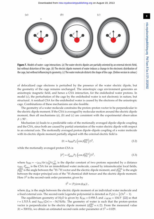
What is the physical interpretation of these results?Three competing physical models are sketched in figure 7. Model (a) represents a situation
in which the enclosing cage retains its icosahedral symmetry. A local electric field, with originoutside the cage, partially orients the endohedral water molecules. This partial orientation leadsto a residual dipole–dipole coupling, and also a residual CSA interaction, since the 1H CSAassociated with the water molecule itself is not averaged to zero. In model (b), the distribution
on August 19, 2013rsta.royalsocietypublishing.orgDownloaded from
Figure 7. Models of water–cage interactions. (a) The water electric dipoles are partially oriented by an external electric field,but without distortion of the cage. (b) The electric dipole moment of water induces a change in the electronic distribution ofthe cage, but without influencing its geometry. (c) Thewatermolecule distorts the shape of the cage. (Online version in colour.)
of delocalized cage electrons is perturbed by the presence of the water electric dipole, butthe geometry of the cage remains unchanged. The anisotropic cage environment generates ananisotropic magnetic field, and hence a CSA interaction, for the endohedral water protons. Inmodel (c), the perturbation of the cage by the endohedral water is not electronic in nature, butstructural. A residual CSA for the endohedral water is caused by the electrons of the anisotropiccage. Combinations of these mechanisms are also feasible.
The geometry of a water molecule constrains the proton–proton vector to be perpendicular tothe electric dipole moment. If the CSA is averaged by molecular motion around the electric dipolemoment, then all mechanisms (a), (b) and (c) are consistent with the experimental observationβ � 90◦.
Mechanism (a) leads to a predictable ratio of the motionally averaged dipole–dipole couplingand the CSA, since both are caused by partial orientation of the water electric dipole with respectto an external axis. The motionally averaged proton dipole–dipole coupling of a water moleculewith its electric dipole moment partially aligned with the external electric field is
〈b〉 = bHHP2
(cos βHH
PD
)S2, (3.2)
while the motionally averaged proton CSA is
⟨δa⟩ = δa
intraP2
(cos βCSA
PD
)S2, (3.3)
where bHH = −(μ0/4π)γ 2H�r−3
HH is the dipolar constant of two protons separated by a distancerHH, δa
intra is the CSA for an immobilized water molecule, caused by intramolecular local fields,βHH
PD is the angle between the 1H–1H vector and the electric dipole moment, and βCSAPD is the angle
between the major principal axis of the 1H chemical shift tensor and the electric dipole moment.Here S2 is the second-rank order parameter, given by
S2 = 〈P2(cos βDE)〉 ,
where βDE is the angle between the electric dipole moment of an individual water molecule anda fixed external axis. The second-rank Legendre polynomial is denoted as P2(x) = 1
2 (3x2 − 1).The equilibrium geometry of H2O is given by ROH = 0.958 Å and � HOH = 104.5◦ [22] so that
r = 1.515 Å and bHH/(2π ) = −34.5 kHz. The geometry of water is such that the proton–protonvector is perpendicular to the electric dipole moment (βHH
PD = π/2). From the measured value|b| = 500 Hz, we obtain an estimated second-rank order parameter of S2 = 0.029.
on August 19, 2013rsta.royalsocietypublishing.orgDownloaded from
The intramolecular contribution to the CSA may be estimated from solid-state NMRmeasurements of immobilized water molecules in calcium sulfate dihydrate (gypsum), providingδa
intra = 3.4 ppm [23]. If the CSA principal axis is assumed to be along the OH bond, then theCSA orientation angle is estimated to be βCSA
PD � 12 × 104.5◦ � 52.3◦. These pieces of information
may be combined to obtain a theoretical value of the motionally averaged CSA under model(a):
⟨δa⟩ (a) � 0.024 ppm. This is much smaller than the experimental estimate δa = 3.1 ppm. We
conclude that model (a) is not tenable.The observed proton CSA for H2O@C60 is therefore unlikely to be associated with the water
molecule itself, but must be mainly generated by the cage. This implies that the electronicdistribution of the cage is distorted (mechanism (b)) or that the geometry of the cage is distorted(mechanism (c)), or a combination of the two.
4. ConclusionThis work investigated the magnitude and relative orientation of anisotropic spin interactionsin H2O@C60 at room temperature by means of solid-state NMR spectroscopy and numericalsimulations. We obtained evidence for a significant residual 1H CSA as well as a residual proton–proton dipolar coupling. The presence of these spin interactions at room temperature couldbe due either to a partial alignment of the water molecules or to a distortion of the cage’sgeometry or electronic structure. Our data exclude the hypothesis of partial water alignmentwithin unperturbed fullerene cages, supporting models in which the water molecule leads toa geometrical distortion of the surrounding cage, or a polarization of cage electrons.
In principle, quantum calculations could determine the electronic configuration and geometryof the H2O@C60 complex. Furthermore, distortion of the cage breaks the icosahedral symmetryand removes the equivalence of the 13C sites. High-resolution 13C NMR spectra may resolve 13Cchemical shifts due to non-equivalent carbons in cages of reduced symmetry. Studies of this kindare in progress.
Acknowledgements. M.C. acknowledges a University Research Fellowship from the Royal Society (UK); we alsothank E. Carignani (Pisa, Italy) for experimental help.Funding statement. We thank EPSRC (UK) and The National Science Foundation (USA) for funds.
References1. Rubin Y. 1999 Ring opening reactions of fullerenes: designed approaches to endohedral metal
complexes. Top. Curr. Chem. 199, 67–91. (doi:10.1007/3-540-68117-5_2)2. Komatsu K, Murata M, Murata Y. 2005 Encapsulation of molecular hydrogen in fullerene C60
by organic synthesis. Science 307, 238–240. (doi:10.1126/science.1106185)3. Kurotobi K, Murata Y. 2011 A single molecule of water encapsulated in fullerene C60. Science
333, 613–616. (doi:10.1126/science.1206376)4. Beduz C et al. 2012 Quantum rotation of ortho and para-water encapsulated in a fullerene cage.
Proc. Natl Acad. Sci. USA 109, 12 894–12 898. (doi:10.1073/pnas.1210790109)5. Carravetta M, Johannessen OG, Levitt MH, Heinmaa I, Stern R, Samoson A, Horsewill AJ,
Murata Y, Komatsu K. 2006 Cryogenic NMR spectroscopy of endohedral hydrogen–fullerenecomplexes. J. Chem. Phys. 124, 104507. (doi:10.1063/1.2174012)
6. Carravetta M et al. 2007 Solid-state NMR of endohedral hydrogen–fullerene complexes. Phys.Chem. Chem. Phys. 9, 4879–4894. (doi:10.1039/b707075f)
7. Mamone S et al. 2009 Rotor in a cage: infrared spectroscopy of an endohedral hydrogen–fullerene complex. J. Chem. Phys. 130, 081103. (doi:10.1063/1.3080163)
8. Horsewill AJ et al. 2010 Inelastic neutron scattering of a quantum translator–rotatorencapsulated in a closed fullerene cage: isotope effects and translation–rotation coupling inH2@C60 and HD@C60. Phys. Rev. B. 82, 081410. (doi:10.1103/PhysRevB.82.081410)
9. Mamone S, Chen JY-C, Bhattacharyya R, Levitt MH, Lawler RG, Horsewill AJ, Rõõm T,Bacic Z, Turro NJ. 2011 Theory and spectroscopy of an incarcerated quantum rotor: theinfrared spectroscopy, inelastic neutron scattering and nuclear magnetic resonance of H2@C60at cryogenic temperature. Coord. Chem. Rev. 255, 938–948. (doi:10.1016/j.ccr.2010.12.029)
on August 19, 2013rsta.royalsocietypublishing.orgDownloaded from
10. Ge M et al. 2011 Interaction potential and infrared absorption of endohedral H2 in C60. J. Chem.Phys. 134, 054507. (doi:10.1063/1.3535598)
11. Ge M et al. 2011 Infrared spectroscopy of endohedral HD and D2 in C60. J. Chem. Phys 135,114511. (doi:10.1063/1.3637948)
12. Xu M, Sebastianelli F, Bacic Z, Lawler R, Turro NJ. 2008 H2, HD, and D2 inside C60: coupledtranslation–rotation eigenstates of the endohedral molecules from quantum five-dimensionalcalculations. J. Chem. Phys. 129, 064313. (doi:10.1063/1.2967858)
13. Xu M, Sebastianelli F, Gibbons BR, Bacic Z, Lawler R, Turro NJ. 2009 Coupled translation–rotation eigenstates of H2 in C60 and C70 on the spectroscopically optimized interactionpotential: effects of cage anisotropy on the energy level structure and assignments. J. Chem.Phys. 130, 224306. (doi:10.1063/1.3152574)
14. Levitt MH. 2002 Symmetry-based pulse sequences in magic-angle spinning solid-state NMR.In Encyclopedia of Nuclear Magnetic Resonance, vol. 9 (supplementary volume), Advances inNMR (eds DM Grant, RK Harris), pp. 165–195. Chichester, UK: John Wiley & Sons.
15. Webber AL et al. 2010 Complete 1H resonance assignment of β-maltose from 1H-1HDQ-SQ CRAMPS and 1H (DQ-DUMBO)–13C SQ refocused INEPT 2D solid-state NMRspectra and first principles GIPAW calculations. Phys. Chem. Chem. Phys. 12, 6970–6983.(doi:10.1039/c001290d)
16. Brown SP, Spiess HW. 2001 Advanced solid-state NMR methods for the elucidation ofstructure and dynamics of molecular, macromolecular, and supramolecular systems. Chem.Rev. 101, 4125–4155. (doi:10.1021/cr990132e)
17. Feike M, Demco DE, Graf R, Gottwald J, Hafner S, Spiess HW. 1996 Broadband multiple-quantum NMR spectroscopy. J. Magn. Reson. A 122, 214–221. (doi:10.1006/jmra.1996.0197)
18. Kristiansen PE, Carravetta M, Lai WC, Levitt MH. 2004 A robust pulse sequence for thedetermination of small homonuclear dipolar couplings in magic-angle spinning NMR. Chem.Phys. Lett. 390, 1–7. (doi:10.1016/j.cplett.2004.03.075)
19. Carravetta M, Eden M, Brinkmann A, Zhao X, Levitt MH. 2000 Symmetry principles for thedesign of radiofrequency pulse sequences in the nuclear magnetic resonance of rotating solids.Chem. Phys. Lett. 321, 205–215. (doi:10.1016/S0009-2614(00)00340-7)
20. Bak M, Rasmussen JT, Nielsen NC. 2000 SIMPSON: a general simulation program for solid-state NMR spectroscopy. J. Magn. Reson. 147, 296–330. (doi:10.1006/jmre.2000.2179)
21. Levitt MH. 2008 Symmetry in the design of NMR multiple-pulse sequences. J. Chem. Phys.128, 052205. (doi:10.1063/1.2831927)
22. Bunker PR, Jenesen P. 2006 Molecular symmetry and spectroscopy. Ottawa, Canada: NRCResearch Press.
23. McKnett CL, Dybowski CR, Vaughan RW. 1975 High resolution proton NMR of gypsum,CaSO4 · 2H2O. J. Chem. Phys. 63, 4578–4581. (doi:10.1063/1.431266)
on August 19, 2013rsta.royalsocietypublishing.orgDownloaded from