22
Ankle Injuries in the Athlete Michelle Wolcott, MD Assistant Professor, Department of Orthopaedics Team Physician for the University of Colorado Buffaloes And University of Denver Pioneers
| Date post: | 23-Dec-2015 |
| Category: |
Documents |
| Upload: | megan-carter |
| View: | 217 times |
| Download: | 1 times |

Ankle Injuries in the AthleteAnkle Injuries in the Athlete
Michelle Wolcott, MDAssistant Professor, Department of Orthopaedics
Team Physician for the University of Colorado Buffaloes
And University of Denver Pioneers
Michelle Wolcott, MDAssistant Professor, Department of Orthopaedics
Team Physician for the University of Colorado Buffaloes
And University of Denver Pioneers

TreatmentTreatment
• Chronic ankle sprains
–Functional rehabilitation
• Role in recovery
• May attempt for as long as 6 months
• Studies have shown that delayed
functional rehab can still be successful
• Chronic ankle sprains
–Functional rehabilitation
• Role in recovery
• May attempt for as long as 6 months
• Studies have shown that delayed
functional rehab can still be successful

Functional RehabilitationFunctional Rehabilitation
Matsusaka, AJSMMatsusaka, AJSM
TreatmentTreatment
• Chronic ankle instability
–Mechanical instability
• Objective measurement of instability
–Functional instability
• Subjective measurement of instability
• Chronic ankle instability
–Mechanical instability
• Objective measurement of instability
–Functional instability
• Subjective measurement of instability

TreatmentTreatment
Chronic Ankle Instability
–Surgical treatment (req. in 10-20%)
• Nonanatomic
– tenodeses
• Anatomic
–Repair/imbrication of tissues
Chronic Ankle Instability
–Surgical treatment (req. in 10-20%)
• Nonanatomic
– tenodeses
• Anatomic
–Repair/imbrication of tissues

TreatmentTreatment
• Nonanatomic
–Evans
procedure
• Average of ATFL
& CFL resistance
vectors
• Nonanatomic
–Evans
procedure
• Average of ATFL
& CFL resistance
vectors
DeLee & DrezDeLee & Drez

TreatmentTreatment
• Nonanatomic
–Watson-Jones
procedure
• Uses peroneus
brevis tendon to
recreate ATFL
• Nonanatomic
–Watson-Jones
procedure
• Uses peroneus
brevis tendon to
recreate ATFL
DeLee & DrezDeLee & Drez

TreatmentTreatment
• Nonanatomic
–Chrisman-Snook
• ½ peroneus
brevis tendon
used to recreate
ATFL and CFL
• Nonanatomic
–Chrisman-Snook
• ½ peroneus
brevis tendon
used to recreate
ATFL and CFLDeLee & DrezDeLee & Drez

TreatmentTreatment
• Anatomic
– modified Brostrom
(Gould)
• Anatomic repair of
ATFL, CFL with
reinforcement using
lateral extensor
retinaculum
• Anatomic
– modified Brostrom
(Gould)
• Anatomic repair of
ATFL, CFL with
reinforcement using
lateral extensor
retinaculum
DeLee & DrezDeLee & Drez

Treatment

TreatmentTreatment
• Anatomic vs Nonanatomic
– Evans procedure 1913
• Karlsson, JBJS - 50% excellent, good results
at long term follow-up
–Watson-Jones
• Barbari, F&A; Van Der Rijt, JBJS – good short-
term results, inconsistent long term results
• Anatomic vs Nonanatomic
– Evans procedure 1913
• Karlsson, JBJS - 50% excellent, good results
at long term follow-up
–Watson-Jones
• Barbari, F&A; Van Der Rijt, JBJS – good short-
term results, inconsistent long term results

Treatment
• Anatomic vs. Nonanatomic– Chrisman-Snook• Snook, JBJS; Sammarco, AJSM – 80-90% good
or excellent results at 10 yrs• Decreased ROM and sural nerve injury not
considered in results–modified Brostrom (Gould) • Karlsson, JBJS; Sjolin, F&A – 86-95% good or
excellent results at 10 yrs with equivalent results for acute vs chronic rpr

Risk FactorsRisk Factors
• Axial/foot alignment
• Plantar/dorsiflexion strength
• Inversion/Eversion strength
• Gender/sport – No significant difference
• Axial/foot alignment
• Plantar/dorsiflexion strength
• Inversion/Eversion strength
• Gender/sport – No significant difference

PreventionPrevention
• Taping
– Shown to be effective for initial stabilization
– Aids in proprioception
• Braces
– Shown to be effective in athletes with h/o
previous sprains
• Taping
– Shown to be effective for initial stabilization
– Aids in proprioception
• Braces
– Shown to be effective in athletes with h/o
previous sprains
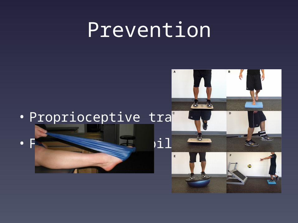
Prevention
• Proprioceptive training
• Functional rehabiliation

Syndesmotic Ligament Injury
Syndesmotic Ligament Injury
• Partial or complete rupture
– Often associated with other injuries
• Mechanism of injury
– Usually dorsiflexion/ext rotation
• Diagnosis
– Pain over syndesmosis
– Positive squeeze test
– Radiographic evaluation
• Partial or complete rupture
– Often associated with other injuries
• Mechanism of injury
– Usually dorsiflexion/ext rotation
• Diagnosis
– Pain over syndesmosis
– Positive squeeze test
– Radiographic evaluation

Syndesmotic Ligament Injury
Syndesmotic Ligament Injury
• Treatment
– Partial
• No clear consensus
• Healing rates highly variable
– Related to extent of injury
• Rate of return ranges from 2 wks to 6 mos
– Complete
• Surgical stabilization
• Treatment
– Partial
• No clear consensus
• Healing rates highly variable
– Related to extent of injury
• Rate of return ranges from 2 wks to 6 mos
– Complete
• Surgical stabilization

Syndesmotic Ligament Injury
Deltoid Ligament InjuryDeltoid Ligament Injury
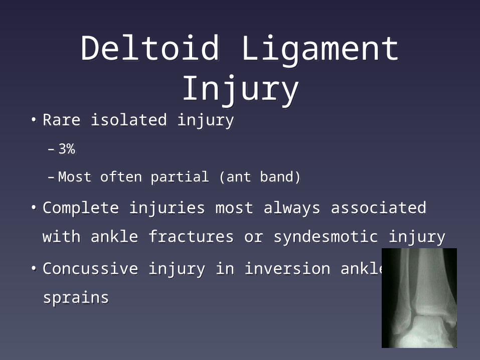
• Rare isolated injury
– 3%
–Most often partial (ant band)
• Complete injuries most always associated
with ankle fractures or syndesmotic injury
• Concussive injury in inversion ankle sprains
• Rare isolated injury
– 3%
–Most often partial (ant band)
• Complete injuries most always associated
with ankle fractures or syndesmotic injury
• Concussive injury in inversion ankle sprains

ConclusionsConclusions
• Ankle injuries very common in the athletic
population
• Majority recover with functional rehab despite
Grade of injury
• Associated injuries largely responsible for chronic
pain
• Primary vs secondary repair yields equivalent
results
• Ankle injuries very common in the athletic
population
• Majority recover with functional rehab despite
Grade of injury
• Associated injuries largely responsible for chronic
pain
• Primary vs secondary repair yields equivalent
results

Thank You!Thank You!


