Page 1
Cancers 2012, 4, 1180-1211; doi:10.3390/cancers4041180
OPEN ACCESS
cancers ISSN 2072-6694
www.mdpi.com/journal/cancers
Article
Annotating Cancer Variants and Anti-Cancer Therapeutics
in Reactome
Marija Milacic 1, Robin Haw
1,*, Karen Rothfels
1, Guanming Wu
1, David Croft
2,
Henning Hermjakob 2, Peter D’Eustachio
3 and Lincoln Stein
1
1 Informatics and Bio-computing Platform, Ontario Institute for Cancer Research, Toronto, ON,
M5G0A3, Canada; E-Mails: [email protected] (M.M.); [email protected] (K.R.);
[email protected] (G.W.); [email protected] (L.S.) 2 European Bioinformatics Institute, Wellcome Trust Genome Campus, Hinxton, Cambridge,
CB10 1SD, UK; E-Mails: [email protected] (D.C.); [email protected] (H.H.) 3 Department of Biochemistry, NYU School of Medicine, New York, NY 10016, USA;
E-Mail: Peter.D’[email protected]
* Author to whom correspondence should be addressed; E-Mail: [email protected] ;
Tel.: +1-647-260-7985; Fax: +1-416-977-1118.
Received: 8 October 2012; in revised form: 31 October 2012 / Accepted: 2 November 2012 /
Published: 8 November 2012
Abstract: Reactome describes biological pathways as chemical reactions that closely
mirror the actual physical interactions that occur in the cell. Recent extensions of our data
model accommodate the annotation of cancer and other disease processes. First, we have
extended our class of protein modifications to accommodate annotation of changes in
amino acid sequence and the formation of fusion proteins to describe the proteins involved
in disease processes. Second, we have added a disease attribute to reaction, pathway, and
physical entity classes that uses disease ontology terms. To support the graphical
representation of “cancer” pathways, we have adapted our Pathway Browser to display
disease variants and events in a way that allows comparison with the wild type pathway,
and shows connections between perturbations in cancer and other biological pathways. The
curation of pathways associated with cancer, coupled with our efforts to create other
disease-specific pathways, will interoperate with our existing pathway and network
analysis tools. Using the Epidermal Growth Factor Receptor (EGFR) signaling pathway as
an example, we show how Reactome annotates and presents the altered biological behavior
of EGFR variants due to their altered kinase and ligand-binding properties, and the mode
of action and specificity of anti-cancer therapeutics.
OPEN ACCESS
Page 2
Cancers 2012, 4 1181
Keywords: pathway database; pathway visualization; network visualization; cancer
annotation; EGFR signaling
1. Introduction
The development of a malignantly transformed cell from a normal cell is a complex multi-step
process that remains incompletely understood [1,2]. “Bottom-up” studies of relevant processes such as
control of cell division, cell migration, tissue remodeling, and cell death have allowed the
identification and characterization of many individual genes whose malfunction due to mutation or
misregulation is associated with malignant transformation [3–6]. More recently, the development of
high-throughput studies that exploit the availability of whole-genome sequencing has enabled
“top-down” studies to systematically catalogue somatically mutated genes and altered patterns of gene
expression in individual tumors [7,8]. These studies have confirmed the importance of genes identified
as key players in the “bottom-up” studies, but have also suggested roles for additional genes and gene
combinations not previously associated with processes relevant to malignancy.
Pathway databases have been effectively used to annotate our “bottom-up” understanding of
molecular details of processes relevant to cell growth, differentiation, migration, and death. Here, we
describe one such database, Reactome, focusing on extensions to this basic annotation strategy to
allow the capture of details of disease processes, and on the development of data analysis tools to
support the annotation and interpretation of gene sets identified in top-down studies.
Reactome is an open-source, open access, curated and peer-reviewed biological knowledgebase of
human reactions, pathways and processes that serves as a platform for pathway visualization and
analysis [9–12]. Reactome provides information about proteins and small molecules and how they
participate in pathways to coordinate cellular events. The Reactome database employs a reductionist
data model, which represents biology as reactions that convert input physical entities into output
physical entities. The Reactome definition of a “reaction” is broad, including binding, dissociation,
translocation and degradation, in addition to biochemical transformations of proteins and small
molecules. Reactions are linked in causal chains to form pathways which in turn are grouped to
represent larger biological processes like intermediary metabolism, innate immunity, solute transport,
GPCR signal transduction, and apoptosis [13,14].
Reactome curators, in collaboration with outside expert researchers, annotate new pathways. The
molecular details of every reaction are traceable to experimental evidence in the primary literature. If
an event has not been directly studied in human systems, the appropriate non-human reaction is
annotated and the homologous human one is inferred from it. Every pathway module is peer-reviewed
by an additional expert. New and revised modules are publicly released to the Reactome website every
quarter. Pathways, reactions, protein and small molecule entities are cross referenced with accession
numbers and identifiers to a number of well-established databases, including NCBI Gene [15],
Ensembl [16] and UniProt databases [17], UCSC Genome Browser [18], and ChEBI [19]. Physical
entities and events are further linked to “Molecular Function”, “Biological Process” and “Cellular
Component” ontology terms found in Gene Ontology (GO) [20].
Page 3
Cancers 2012, 4 1182
Currently, the pathways in Reactome cover about 25% of the gene products encoded in the human
genome, and contain the normal versions of many pathways that can be abnormally activated in
cancer, such as “Signaling by EGFR” [21], “Signaling by FGFR” [22], “Signaling by NOTCH” [23],
“PIP3 Activates AKT Signaling” [24], “RAF/MAP Kinase Cascade” [25]. We have also annotated a
number of pathways that can be inactivated in cancer, such as pathways involving TP53: “Apoptosis” [26]
and “Cell Cycle Checkpoints” [27], as well as pathways involving the RB1 protein family: “Mitotic
G1-G1/S phases” [28].
Here, we use the epidermal growth factor receptor (EGFR), fibroblast growth factor receptor
(FGFR) and PI3K/AKT signaling pathways to illustrate Reactome annotation of cancer pathways.
EGFR and FGFR are transmembrane receptor tyrosine kinases. EGFR is activated by several growth
factors, including the epidermal growth factor (EGF) [29]. FGFR family members (FGFR1, FGFR2,
FGFR3 and FGFR4) are activated by 18 of 22 existing human fibroblast growth factors (FGFs), with
each FGFR showing different affinity for individual FGFs [30]. Growth factor binding induces a
conformational change that enables dimerization and trans-autophosphorylation on C-tail tyrosine
residues of EGFR [31] and FGFRs [32–34]. Phosphorylated tyrosines in the C-tails of EGFR and
FGFR serve as docking sites for downstream effectors that, upon binding to phosphorylated receptors,
activate intracellular signaling cascades that regulate cellular proliferation, differentiation, and
survival [30,35,36]. One of the intracellular signaling cascades downstream of EGFR and FGFRs is
PI3K/AKT signaling [37,38]. PI3K class IA enzymes are heterodimers composed of a regulatory
subunit p85 (encoded by PIK3R1, PIK3R2 or PIK3R3) and a catalytic subunit p110 (encoded by
PIK3CA, PIK3CB or PIK3CD) [39]. The catalytic p110 subunit of PI3K becomes activated when
inhibitory contacts with the p85 regulatory subunit are relieved by p85 binding to phosphorylated
adaptor proteins recruited to activated EGFR or FGFRs [40,41]. Active PI3K class I enzymes
phosphorylate PIP2 (phosphatidylinositol-4,5-bisphophate), converting it into PIP3 (phosphatidylinositol-
3,4,5-trisphosphate), a reaction negatively regulated by PTEN phosphatase [42]. PIP3 serves as a
second messenger that activates AKT (AKT1, AKT2 or AKT3) [43]. AKT family members are
cytosolic and nuclear serine/threonine protein kinases involved in phosphorylation-mediated regulation
of numerous proteins involved in cell survival and growth [39].
EGFR, FGFRs, PIK3CA, PIK3R1 and AKT1 are proto-oncogenes, frequently activated in cancer
through gain-of-function mutations and/or overexpression. PTEN is an established tumor suppressor
gene, with a frequent loss of function in cancer [44]. Gain-of-function mutations in EGFR [45,46] and
FGFRs [47–51] usually act by conferring ligand-independent activation or by increasing tyrosine
kinase catalytic activity. Mutations in PIK3R1 or PIK3CA abolish inhibitory interactions between the
regulatory and catalytic subunit of PI3K [52–56], resulting in PI3K activity in the absence of growth
factor stimulation. AKT1 gain of function mutations increase AKT1 affinity for PIP2, allowing AKT1
activation in the absence of PI3K activity and PIP3 generation [57]. PTEN loss-of-function mutations
usually affect the phosphatase domain, impairing PTEN catalytic activity and removal of PIP3 [58].
Small molecule therapeutics and recombinant antibodies are being developed as potential
treatments for cancers driven by increased activity of EGFR, FGFR and/or PI3K/AKT. Gefitinib and
erlotinib, small tyrosine kinase inhibitors, are approved for the clinical treatment of cancers harboring
specific EGFR mutations. A recombinant antibody, cetuximab, is approved for the clinical treatment of
Page 4
Cancers 2012, 4 1183
cancers that overexpress wild-type EGFR [59]. Small molecules that inhibit the catalytic activity of
FGFRs [60], PI3K and AKT [61] are currently undergoing clinical trials or are in pre-clinical development.
We have extended the Reactome data model and enhanced the web tools to permit the annotation
and visualization of the altered biological behavior of protein variants. These enhancements can be
applied to any molecular abnormality due to germline or somatic mutation, as well as to abnormalities
due to expression of foreign proteins encoded by genomes of infectious agents like viruses or
intracellular parasites.
2. Results and Discussion
2.1. Annotation of Cancer-Perturbed Pathways
Pathways that stimulate cell growth, cell division and survival, and maintenance of undifferentiated
state are activated in cancer through gain-of-function mutations in participating proto-oncogenes
and/or their overexpression. On the other hand, pathways that negatively regulate cell division, growth
and survival, or promote cellular differentiation are inactivated through loss-of-function mutations in
tumor suppressor genes and/or their downregulation. To capture these two groups of cancer effectors,
we have added new classes of data to the Reactome database.
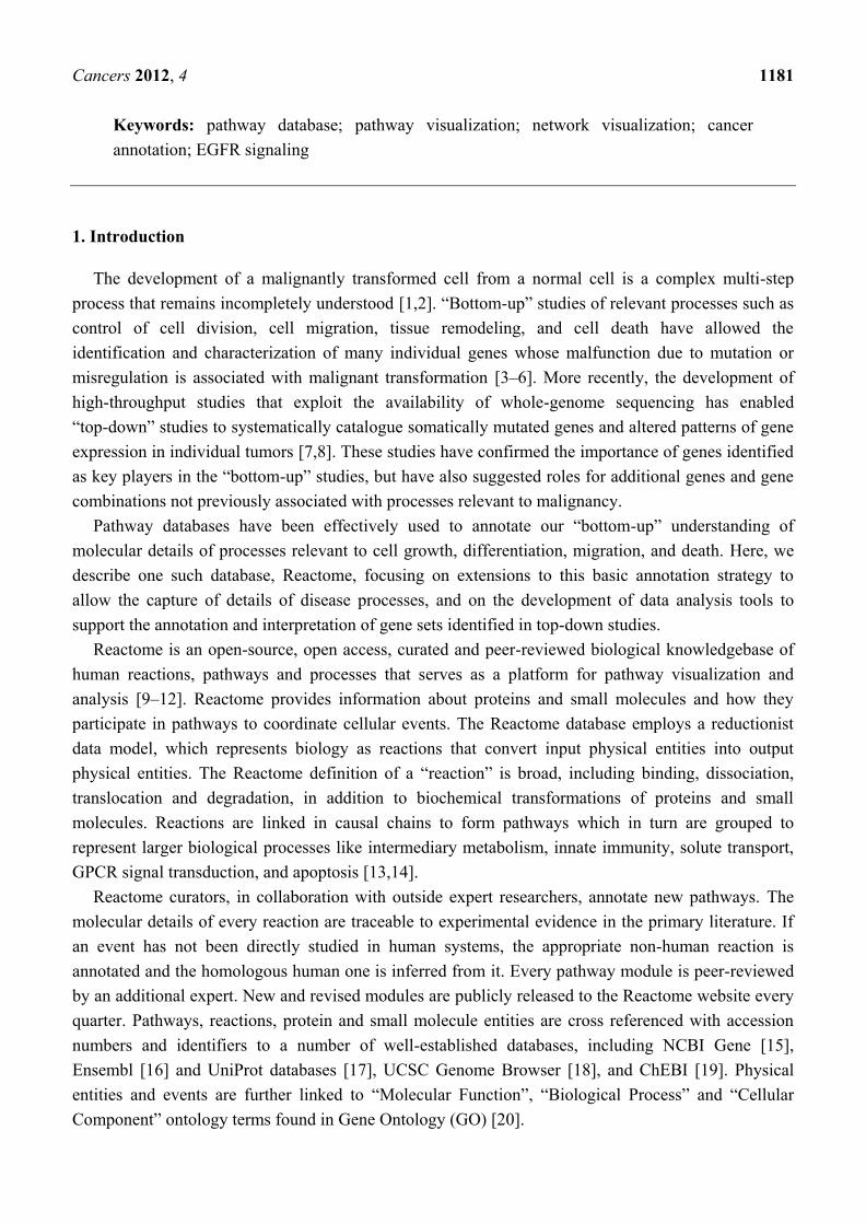
2.1.1. Extension of Protein Modifications to Accommodate Annotation of Changes in Amino Acid
Sequence
The protein modification class in the Reactome data model was constructed to support annotation of
covalent co- and post-translational modifications of proteins such as the phosphorylation of serine
residues. To allow for annotation of mutant proteins, two new subclasses of modifications were
introduced: Replaced Residue and Fragment Modification (Figure 1a). The Replaced Residue class is
used to annotate amino acid substitutions in a protein sequence. A Replaced Residue instance
associates a specific coordinate of a protein sequence with two PSI-MOD ontology [62] attributes: the
first identifies the amino acid found at that position in the normal protein and the second attribute
identifies the amino acid that replaces it in the mutant protein. For example, the most frequently found
mutation in EGFR is the substitution of a leucine residue at position 858 with an arginine residue in the
catalytic domain of EGFR. This mutation disrupts autoinhibitory interactions, facilitating adoption of
an active conformation [63]. The Reactome record for EGFR L858R (Figure 1b) indicates this amino
acid substitution.
The FragmentModification subclass includes two subclasses, FragmentInsertionModification and
FragmentDeletionModification. FragmentInsertionModification is used to annotate insertions of amino
acid residues in a protein sequence. FragmentDeletionModification is used to annotate removal of
amino acid residues. PIK3R1 Y463_L466del is a variant of the PI3K regulatory subunit p85alpha
found in endometrial cancer (Figure 1c). This PIK3R1 mutant lacks four amino acid residues in the
inter-SRC homology 2 (iSH2) domain. PIK3R1 is able to bind the catalytic subunit of PI3K, PIK3CA
(p110alpha), but does not inhibit it, resulting in the constitutive activity of PI3K, in the absence of
growth factors [64]. The deletion coordinates are indicated in the Reactome record for PIK3R1
Y463_L466del mutant.
Page 5
Cancers 2012, 4 1184
Figure 1. Annotation of cancer mutations. Reactome records are not displayed in their
entirety due to space limitations. (a) Subclasses of protein modifications contained in
Reactome class Abstract Modified Residue. Currently, Reactome website displays all
subclasses of protein modifications in the single field “Post-translational modification(s)”.
Future changes to the website will allow chemical modifications to be distinguished from
effects of mutations. (b) Reactome record for EGFR L858R caused by a missense mutation
that replaces leucine residue at position 858 with arginine. (c) Reactome record for PIK3R1
Y463_L466del caused by an in-frame intragenic deletion in PIK3R1 that removes amino
acid residues from position 463 to position 466, as captured by the Fragment Deletion
Modification instance. (d) Reactome record for BCR-FGFR1 fusion protein. Truncation of
the wild-type BCR protein sequence is shown by altered end coordinate. FGFR1 fragment
fused to BCR is annotated as an insertion using FragmentInsertionModification class.
Page 6
Cancers 2012, 4 1185
The FragmentModification class can also be used to annotate fusion proteins. For example, the
translocation t(8;22)(p11;q11) in chronic myeloid leukemia produces a BCR-FGFR1 fusion that
consists of the first four exons of BCR and exons 9–18 of FGFR1 [65]. The BCR-FGFR1 fusion
protein is annotated as an Entity with Accessioned Sequence (Figure 1d) that consists of a truncated
BCR protein, starting at position 1 and ending at position 584 of the reference UniProt sequence
P11274 (human BCR). Then, a FragmentInsertionModification instance defines insertion of amino
acids 429–822 of the UniProt reference sequence P11362 (human FGFR1) at position 585 of BCR
(Figure 1d).
On the Reactome website, selecting a physical entity or an event node by clicking on a pathway
diagram brings up a record for that particular instance in the details pane, which appears by clicking on
the yellow triangle at the bottom of the Pathway Browser page. Selecting EGFRvIII in the diagram
(Figure 2a), brings up Reactome information on this mutant protein, as well as interactive cross
references that direct users to other Reactome website pages or other databases of interest (Figure 2b).
Each cancer-related disease variant record cross-references available records in the Catalogue of
Somatic Mutations in Cancer (COSMIC) database (Table 1) [66]. The EGFRvIII record displayed on
Reactome website links to COSMIC record 21351, which provides information on nucleotide sequence
changes and tumor samples in which this mutation was reported.
2.1.2. Associating Disease Attributes with Physical Entities and Events
All physical entities related to disease variants, such as proteins, sets of proteins, and protein
complexes are tagged with disease attributes (Table 1), using a term from the Disease Ontology (DO) [67].
This DO record provides, when possible, a link to the synonymous disease record in the National
Cancer Institute Thesaurus (NCIt) [68]. The disease attribute of the physical entity is assigned to all
reactions and pathways in which it participates.
Besides providing information on disease involvement of specific proteins and directing users to
more detailed disease descriptions, a disease attribute annotation enables users to search Reactome
database for proteins and events associated with a specific disease. For example, in Figure 2b, a DO
instance “adult glioblastoma multiforme” is associated with EGFRvIII. Clicking on the “adult
glioblastoma multiforme” link displayed on Reactome website (Figure 2b) provides a DO identifier for
this disease instance (3075) and also lists all other proteins in Reactome database whose mutant forms
are associated with adult glioblastoma multiforme (Figure 2c). Thus, Reactome provides cancer
researchers with a quick access to cancer type-specific disease variants and information on the
mechanism of action for each variant annotated.
2.1.3. Mode of Action and Specificity of Anti-Cancer Therapeutics
The Reactome data model allows for annotation of small molecules and antibodies used as
anti-cancer therapeutics, as well as the annotation of their specific mode of action. We have annotated
nine small tyrosine kinase inhibitors (TKIs) used to inhibit EGFR kinase activity in
cancer [59,69], as well as the recombinant antibody cetuximab [70] (Figure 3a). In addition, we
annotated five benzaquinoid ansamycins that inhibit the HSP90 chaperone protein that stabilizes
EGFR mutant proteins [71], twelve anti-FGFR TKIs [60], one anti-FGFR recombinant antibody [72],
Page 7
Cancers 2012, 4 1186
ten small molecules that inhibit the catalytic subunit of PI3K [61], and three small molecules that
inhibit AKT [61] (Table 2).
Figure 2. Disease information presented interactively on the Reactome website. (a)
Selecting an entity in the pathway diagram, EGFRvIII mutant in this case and opening the
Reactome details pane by clicking on the yellow triangle at the bottom of the Pathway
Browser page brings up a record for the selected instance. A pathway hierarchy displayed
on the left hand side shows how the selected instance is related to the rest of the pathway
content. (b) Reactome record, displayed in the details pane, provides information on the
selected entity, including cross-references to other databases such as COSMIC, UniProt,
GO. (c) Cross-reference to Disease Ontology: clicking on a disease attribute, such as “adult
glioblastoma multiforme”, provides a Disease Ontology (DO) identifier for this disease
instance (3075) and lists all proteins in Reactome database associated with adult
glioblastoma multiforme.
Page 8
Cancers 2012, 4 1187
Figure 3. Mode of action and specificity of anti-EGFR cancer therapeutics. (a) Anti-EGFR
therapeutics differ in their specificity for EGFR cancer variants, as well as in their mode of
action (non-covalent vs. covalent binding). (b) Classification of EGFR-binding small
tyrosine kinase inhibitors (TKIs) according to spectrum and reversibility of their binding.
For each anti-EGFR TKI, we specify whether it associates with the EGFR catalytic domain through
formation of a covalent (irreversible) bond or through a non-covalent interaction (reversible). We also
specify whether a TKI is EGFR-specific or whether it can inhibit other receptor tyrosine kinases
besides EGFR (EGFRplus). Each small molecule instance we annotate is associated with the Chemical
Entities of Biological Interest (ChEBI) database identifier [19]. On the Reactome website, a link to a
corresponding ChEBI record is displayed after the name of each small molecule. Clicking on the
ChEBI link associated with gefitinib (Figure 3b) directs the user to the gefitinib information in ChEBI,
displaying its molecular structure and additional information not directly captured by Reactome.
Page 9
Cancers 2012, 4 1188
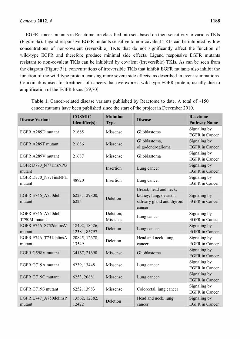
EGFR cancer mutants in Reactome are classified into sets based on their sensitivity to various TKIs
(Figure 3a). Ligand responsive EGFR mutants sensitive to non-covalent TKIs can be inhibited by low
concentrations of non-covalent (reversible) TKIs that do not significantly affect the function of
wild-type EGFR and therefore produce minimal side effects. Ligand responsive EGFR mutants
resistant to non-covalent TKIs can be inhibited by covalent (irreversible) TKIs. As can be seen from
the diagram (Figure 3a), concentrations of irreversible TKIs that inhibit EGFR mutants also inhibit the
function of the wild-type protein, causing more severe side effects, as described in event summations.
Cetuximab is used for treatment of cancers that overexpress wild-type EGFR protein, usually due to
amplification of the EGFR locus [59,70].
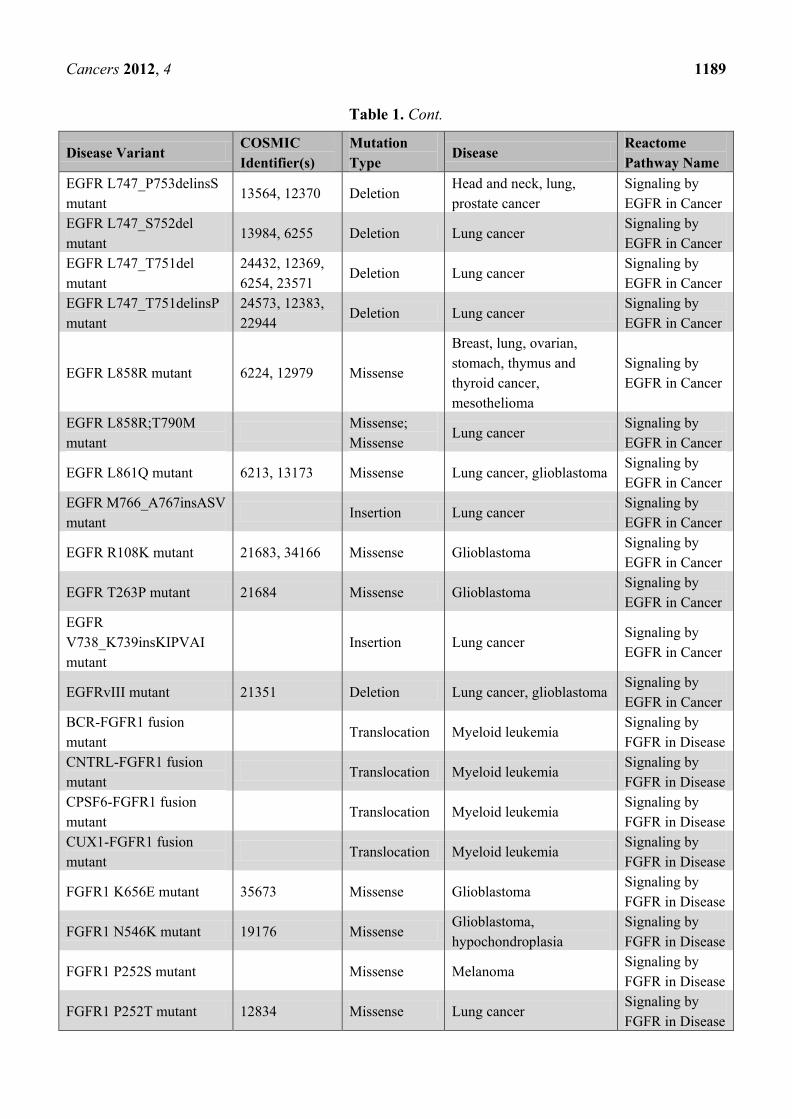
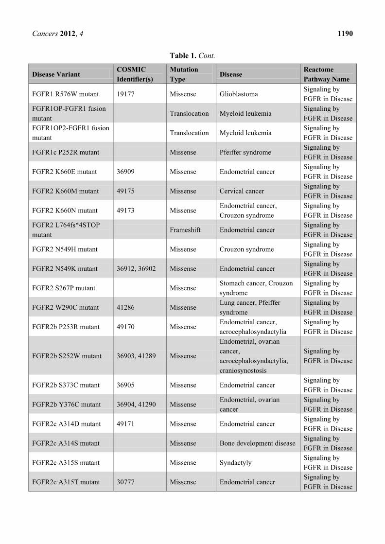
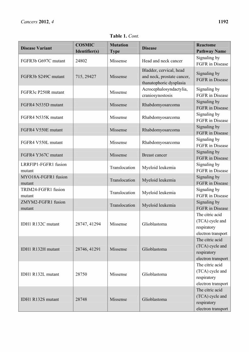
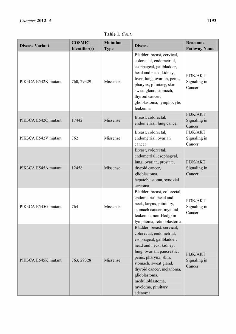
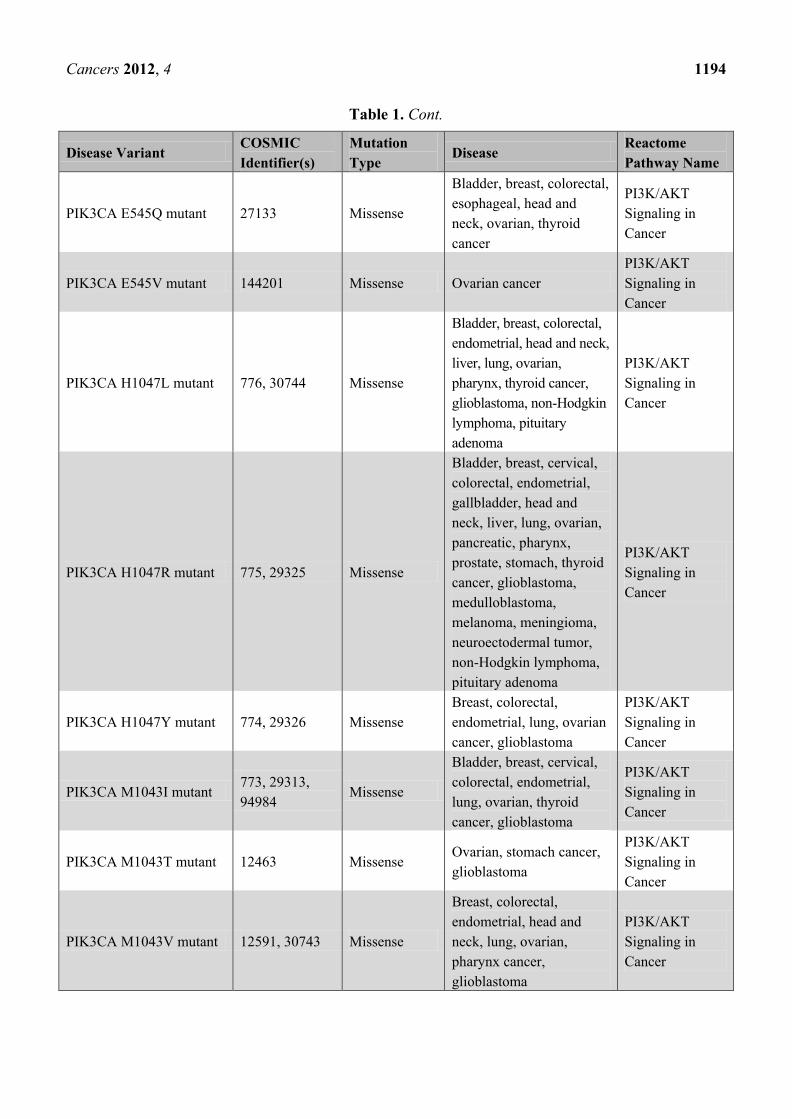
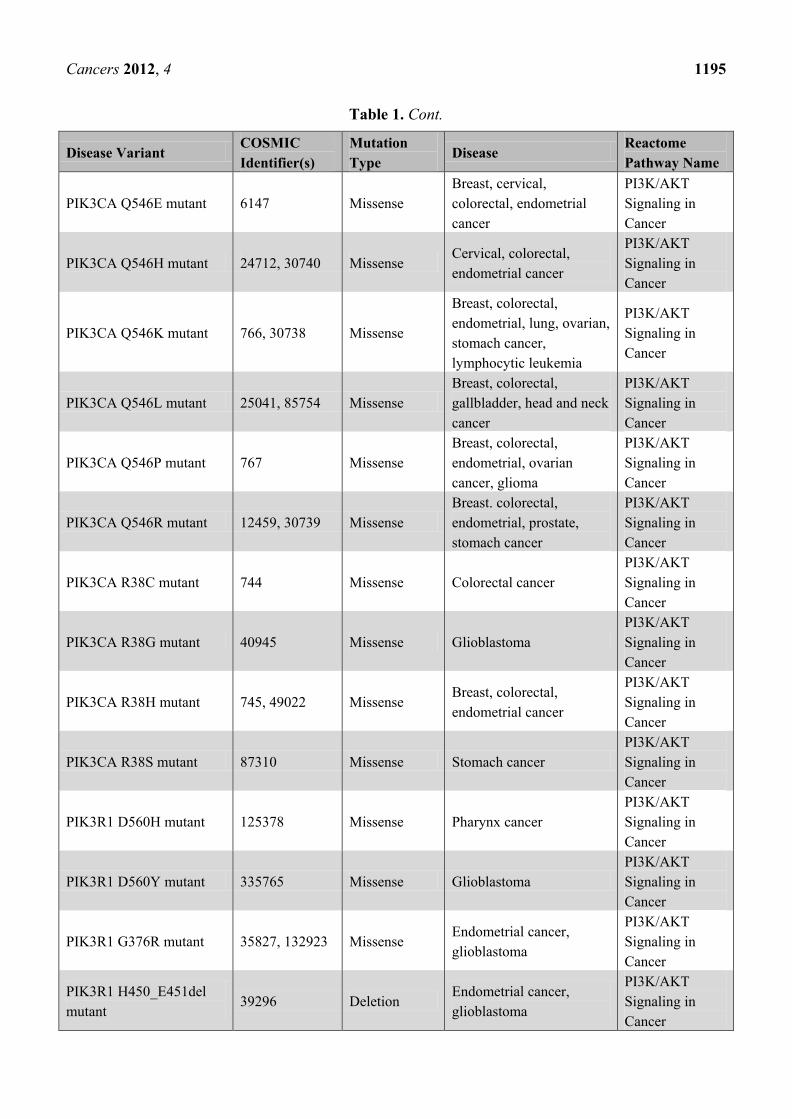
Table 1. Cancer-related disease variants published by Reactome to date. A total of ~150
cancer mutants have been published since the start of the project in December 2010.
Disease Variant COSMIC
Identifier(s)
Mutation
Type Disease
Reactome
Pathway Name
EGFR A289D mutant 21685 Missense Glioblastoma Signaling by
EGFR in Cancer
EGFR A289T mutant 21686 Missense Glioblastoma,
oligodendroglioma
Signaling by
EGFR in Cancer
EGFR A289V mutant 21687 Missense Glioblastoma Signaling by
EGFR in Cancer
EGFR D770_N771insNPG
mutant Insertion Lung cancer
Signaling by
EGFR in Cancer
EGFR D770_N771insNPH
mutant 48920 Insertion Lung cancer
Signaling by
EGFR in Cancer
EGFR E746_A750del
mutant
6223, 129800,
6225 Deletion
Breast, head and neck,
kidney, lung, ovarian,
salivary gland and thyroid
cancer
Signaling by
EGFR in Cancer
EGFR E746_A750del;
T790M mutant
Deletion;
Missense Lung cancer
Signaling by
EGFR in Cancer
EGFR E746_S752delinsV
mutant
18492, 18426,
12384, 85797 Deletion Lung cancer
Signaling by
EGFR in Cancer
EGFR E746_T751delinsA
mutant
20845, 12678,
13549 Deletion
Head and neck, lung
cancer
Signaling by
EGFR in Cancer
EGFR G598V mutant 34167, 21690 Missense Glioblastoma Signaling by
EGFR in Cancer
EGFR G719A mutant 6239, 13448 Missense Lung cancer Signaling by
EGFR in Cancer
EGFR G719C mutant 6253, 20881 Missense Lung cancer Signaling by
EGFR in Cancer
EGFR G719S mutant 6252, 13983 Missense Colorectal, lung cancer Signaling by
EGFR in Cancer
EGFR L747_A750delinsP
mutant
13562, 12382,
12422 Deletion
Head and neck, lung
cancer
Signaling by
EGFR in Cancer
Page 10
Cancers 2012, 4 1189
Table 1. Cont.
Disease Variant COSMIC
Identifier(s)
Mutation
Type Disease
Reactome
Pathway Name
EGFR L747_P753delinsS
mutant 13564, 12370 Deletion
Head and neck, lung,
prostate cancer
Signaling by
EGFR in Cancer
EGFR L747_S752del
mutant 13984, 6255 Deletion Lung cancer
Signaling by
EGFR in Cancer
EGFR L747_T751del
mutant
24432, 12369,
6254, 23571 Deletion Lung cancer
Signaling by
EGFR in Cancer
EGFR L747_T751delinsP
mutant
24573, 12383,
22944 Deletion Lung cancer
Signaling by
EGFR in Cancer
EGFR L858R mutant 6224, 12979 Missense
Breast, lung, ovarian,
stomach, thymus and
thyroid cancer,
mesothelioma
Signaling by
EGFR in Cancer
EGFR L858R;T790M
mutant
Missense;
Missense Lung cancer
Signaling by
EGFR in Cancer
EGFR L861Q mutant 6213, 13173 Missense Lung cancer, glioblastoma Signaling by
EGFR in Cancer
EGFR M766_A767insASV
mutant Insertion Lung cancer
Signaling by
EGFR in Cancer
EGFR R108K mutant 21683, 34166 Missense Glioblastoma Signaling by
EGFR in Cancer
EGFR T263P mutant 21684 Missense Glioblastoma Signaling by
EGFR in Cancer
EGFR
V738_K739insKIPVAI
mutant
Insertion Lung cancer Signaling by
EGFR in Cancer
EGFRvIII mutant 21351 Deletion Lung cancer, glioblastoma Signaling by
EGFR in Cancer
BCR-FGFR1 fusion
mutant Translocation Myeloid leukemia
Signaling by
FGFR in Disease
CNTRL-FGFR1 fusion
mutant Translocation Myeloid leukemia
Signaling by
FGFR in Disease
CPSF6-FGFR1 fusion
mutant Translocation Myeloid leukemia
Signaling by
FGFR in Disease
CUX1-FGFR1 fusion
mutant Translocation Myeloid leukemia
Signaling by
FGFR in Disease
FGFR1 K656E mutant 35673 Missense Glioblastoma Signaling by
FGFR in Disease
FGFR1 N546K mutant 19176 Missense Glioblastoma,
hypochondroplasia
Signaling by
FGFR in Disease
FGFR1 P252S mutant Missense Melanoma Signaling by
FGFR in Disease
FGFR1 P252T mutant 12834 Missense Lung cancer Signaling by
FGFR in Disease
Page 11
Cancers 2012, 4 1190
Table 1. Cont.
Disease Variant COSMIC
Identifier(s)
Mutation
Type Disease
Reactome
Pathway Name
FGFR1 R576W mutant 19177 Missense Glioblastoma Signaling by
FGFR in Disease
FGFR1OP-FGFR1 fusion
mutant Translocation Myeloid leukemia
Signaling by
FGFR in Disease
FGFR1OP2-FGFR1 fusion
mutant Translocation Myeloid leukemia
Signaling by
FGFR in Disease
FGFR1c P252R mutant Missense Pfeiffer syndrome Signaling by
FGFR in Disease
FGFR2 K660E mutant 36909 Missense Endometrial cancer Signaling by
FGFR in Disease
FGFR2 K660M mutant 49175 Missense Cervical cancer Signaling by
FGFR in Disease
FGFR2 K660N mutant 49173 Missense Endometrial cancer,
Crouzon syndrome
Signaling by
FGFR in Disease
FGFR2 L764fs*4STOP
mutant Frameshift Endometrial cancer
Signaling by
FGFR in Disease
FGFR2 N549H mutant Missense Crouzon syndrome Signaling by
FGFR in Disease
FGFR2 N549K mutant 36912, 36902 Missense Endometrial cancer Signaling by
FGFR in Disease
FGFR2 S267P mutant Missense Stomach cancer, Crouzon
syndrome
Signaling by
FGFR in Disease
FGFR2 W290C mutant 41286 Missense Lung cancer, Pfeiffer
syndrome
Signaling by
FGFR in Disease
FGFR2b P253R mutant 49170 Missense Endometrial cancer,
acrocephalosyndactylia
Signaling by
FGFR in Disease
FGFR2b S252W mutant 36903, 41289 Missense
Endometrial, ovarian
cancer,
acrocephalosyndactylia,
craniosynostosis
Signaling by
FGFR in Disease
FGFR2b S373C mutant 36905 Missense Endometrial cancer Signaling by
FGFR in Disease
FGFR2b Y376C mutant 36904, 41290 Missense Endometrial, ovarian
cancer
Signaling by
FGFR in Disease
FGFR2c A314D mutant 49171 Missense Endometrial cancer Signaling by
FGFR in Disease
FGFR2c A314S mutant Missense Bone development disease Signaling by
FGFR in Disease
FGFR2c A315S mutant Missense Syndactyly Signaling by
FGFR in Disease
FGFR2c A315T mutant 30777 Missense Endometrial cancer Signaling by
FGFR in Disease
Page 12
Cancers 2012, 4 1191
Table 1. Cont.
Disease Variant COSMIC
Identifier(s)
Mutation
Type Disease
Reactome
Pathway Name
FGFR2c P253R mutant 49170 Missense Endometrial cancer,
acrocephalosyndactylia
Signaling by
FGFR in Disease
FGFR2c S252W mutant 41289, 36903 Missense
Endometrial, ovarian
cancer,
acrocephalosyndactylia,
craniosynostosis
Signaling by
FGFR in Disease
FGFR2c S372C mutant Missense Beare-Stevenson cutis
gyrata syndrome
Signaling by
FGFR in Disease
FGFR2c W290G mutant Missense Crouzon syndrome,
Pfeiffer syndrome
Signaling by
FGFR in Disease
FGFR2c Y375C mutant Missense Beare-Stevenson cutis
gyrata syndrome
Signaling by
FGFR in Disease
FGFR3 795fs*139STOP
mutant Frameshift
Multiple myeloma,
thanatophoric dysplasia
Signaling by
FGFR in Disease
FGFR3 A391E mutant 721 Missense Bladder cancer, Crouzon
syndrome
Signaling by
FGFR in Disease
FGFR3 G370C mutant 716, 35897 Missense Bladder cancer,
thanatophoric dysplasia
Signaling by
FGFR in Disease
FGFR3 G380R mutant 24842, 24812 Missense Bladder cancer, multiple
myeloma, achondroplasia
Signaling by
FGFR in Disease
FGFR3 G382D mutant 727 Missense Multiple myeloma Signaling by
FGFR in Disease
FGFR3 K650E mutant 719, 35899 Missense
Bladder, testicular cancer,
multiple myeloma,
thanatophoric dysplasia
Signaling by
FGFR in Disease
FGFR3 K650M mutant 720, 85791 Missense
Bladder, testicular cancer,
multiple myeloma,
thanatophoric dysplasia
Signaling by
FGFR in Disease
FGFR3 K650N mutant Missense Bladder, testicular cancer,
hypochondroplasia
Signaling by
FGFR in Disease
FGFR3 K650Q mutant 726 Missense Bladder cancer,
hypochondroplasia
Signaling by
FGFR in Disease
FGFR3 K650T mutant 731 Missense Bladder, testicular cancer,
hypochondroplasia
Signaling by
FGFR in Disease
FGFR3 R248C mutant 714, 35896 Missense
Bladder cancer, multiple
myeloma, thanatophoric
dysplasia
Signaling by
FGFR in Disease
FGFR3 S371C mutant 17461, 35898 Missense Bladder cancer,
thanatophoric dysplasia
Signaling by
FGFR in Disease
FGFR3 Y373C mutant 718, 29428 Missense
Bladder cancer, multiple
myeloma, thanatophoric
dysplasia
Signaling by
FGFR in Disease
Page 13
Cancers 2012, 4 1192
Table 1. Cont.
Disease Variant COSMIC
Identifier(s)
Mutation
Type Disease
Reactome
Pathway Name
FGFR3b G697C mutant 24802 Missense Head and neck cancer Signaling by
FGFR in Disease
FGFR3b S249C mutant 715, 29427 Missense
Bladder, cervical, head
and neck, prostate cancer,
thanatophoric dysplasia
Signaling by
FGFR in Disease
FGFR3c P250R mutant Missense Acrocephalosyndactylia,
craniosynostosis
Signaling by
FGFR in Disease
FGFR4 N535D mutant Missense Rhabdomyosarcoma Signaling by
FGFR in Disease
FGFR4 N535K mutant Missense Rhabdomyosarcoma Signaling by
FGFR in Disease
FGFR4 V550E mutant Missense Rhabdomyosarcoma Signaling by
FGFR in Disease
FGFR4 V550L mutant Missense Rhabdomyosarcoma Signaling by
FGFR in Disease
FGFR4 Y367C mutant Missense Breast cancer Signaling by
FGFR in Disease
LRRFIP1-FGFR1 fusion
mutant Translocation Myeloid leukemia
Signaling by
FGFR in Disease
MYO18A-FGFR1 fusion
mutant Translocation Myeloid leukemia
Signaling by
FGFR in Disease
TRIM24-FGFR1 fusion
mutant Translocation Myeloid leukemia
Signaling by
FGFR in Disease
ZMYM2-FGFR1 fusion
mutant Translocation Myeloid leukemia
Signaling by
FGFR in Disease
IDH1 R132C mutant 28747, 41294 Missense Glioblastoma
The citric acid
(TCA) cycle and
respiratory
electron transport
IDH1 R132H mutant 28746, 41291 Missense Glioblastoma
The citric acid
(TCA) cycle and
respiratory
electron transport
IDH1 R132L mutant 28750 Missense Glioblastoma
The citric acid
(TCA) cycle and
respiratory
electron transport
IDH1 R132S mutant 28748 Missense Glioblastoma
The citric acid
(TCA) cycle and
respiratory
electron transport
Page 14
Cancers 2012, 4 1193
Table 1. Cont.
Disease Variant COSMIC
Identifier(s)
Mutation
Type Disease
Reactome
Pathway Name
PIK3CA E542K mutant 760, 29329 Missense
Bladder, breast, cervical,
colorectal, endometrial,
esophageal, gallbladder,
head and neck, kidney,
liver, lung, ovarian, penis,
pharynx, pituitary, skin
sweat gland, stomach,
thyroid cancer,
glioblastoma, lymphocytic
leukemia
PI3K/AKT
Signaling in
Cancer
PIK3CA E542Q mutant 17442 Missense Breast, colorectal,
endometrial, lung cancer
PI3K/AKT
Signaling in
Cancer
PIK3CA E542V mutant 762 Missense
Breast, colorectal,
endometrial, ovarian
cancer
PI3K/AKT
Signaling in
Cancer
PIK3CA E545A mutant 12458 Missense
Breast, colorectal,
endometrial, esophageal,
lung, ovarian, prostate,
thyroid cancer,
glioblastoma,
hepatoblastoma, synovial
sarcoma
PI3K/AKT
Signaling in
Cancer
PIK3CA E545G mutant 764 Missense
Bladder, breast, colorectal,
endometrial, head and
neck, larynx, pituitary,
stomach cancer, myeloid
leukemia, non-Hodgkin
lymphoma, retinoblastoma
PI3K/AKT
Signaling in
Cancer
PIK3CA E545K mutant 763, 29328 Missense
Bladder, breast. cervical,
colorectal, endometrial,
esophageal, gallbladder,
head and neck, kidney,
lung, ovarian, pancreatic,
penis, pharynx, skin,
stomach, sweat gland,
thyroid cancer, melanoma,
glioblastoma,
medulloblastoma,
myeloma, pituitary
adenoma
PI3K/AKT
Signaling in
Cancer
Page 15
Cancers 2012, 4 1194
Table 1. Cont.
Disease Variant COSMIC
Identifier(s)
Mutation
Type Disease
Reactome
Pathway Name
PIK3CA E545Q mutant 27133 Missense
Bladder, breast, colorectal,
esophageal, head and
neck, ovarian, thyroid
cancer
PI3K/AKT
Signaling in
Cancer
PIK3CA E545V mutant 144201 Missense Ovarian cancer
PI3K/AKT
Signaling in
Cancer
PIK3CA H1047L mutant 776, 30744 Missense
Bladder, breast, colorectal,
endometrial, head and neck,
liver, lung, ovarian,
pharynx, thyroid cancer,
glioblastoma, non-Hodgkin
lymphoma, pituitary
adenoma
PI3K/AKT
Signaling in
Cancer
PIK3CA H1047R mutant 775, 29325 Missense
Bladder, breast, cervical,
colorectal, endometrial,
gallbladder, head and
neck, liver, lung, ovarian,
pancreatic, pharynx,
prostate, stomach, thyroid
cancer, glioblastoma,
medulloblastoma,
melanoma, meningioma,
neuroectodermal tumor,
non-Hodgkin lymphoma,
pituitary adenoma
PI3K/AKT
Signaling in
Cancer
PIK3CA H1047Y mutant 774, 29326 Missense
Breast, colorectal,
endometrial, lung, ovarian
cancer, glioblastoma
PI3K/AKT
Signaling in
Cancer
PIK3CA M1043I mutant 773, 29313,
94984 Missense
Bladder, breast, cervical,
colorectal, endometrial,
lung, ovarian, thyroid
cancer, glioblastoma
PI3K/AKT
Signaling in
Cancer
PIK3CA M1043T mutant 12463 Missense Ovarian, stomach cancer,
glioblastoma
PI3K/AKT
Signaling in
Cancer
PIK3CA M1043V mutant 12591, 30743 Missense
Breast, colorectal,
endometrial, head and
neck, lung, ovarian,
pharynx cancer,
glioblastoma
PI3K/AKT
Signaling in
Cancer
Page 16
Cancers 2012, 4 1195
Table 1. Cont.
Disease Variant COSMIC
Identifier(s)
Mutation
Type Disease
Reactome
Pathway Name
PIK3CA Q546E mutant 6147 Missense
Breast, cervical,
colorectal, endometrial
cancer
PI3K/AKT
Signaling in
Cancer
PIK3CA Q546H mutant 24712, 30740 Missense Cervical, colorectal,
endometrial cancer
PI3K/AKT
Signaling in
Cancer
PIK3CA Q546K mutant 766, 30738 Missense
Breast, colorectal,
endometrial, lung, ovarian,
stomach cancer,
lymphocytic leukemia
PI3K/AKT
Signaling in
Cancer
PIK3CA Q546L mutant 25041, 85754 Missense
Breast, colorectal,
gallbladder, head and neck
cancer
PI3K/AKT
Signaling in
Cancer
PIK3CA Q546P mutant 767 Missense
Breast, colorectal,
endometrial, ovarian
cancer, glioma
PI3K/AKT
Signaling in
Cancer
PIK3CA Q546R mutant 12459, 30739 Missense
Breast. colorectal,
endometrial, prostate,
stomach cancer
PI3K/AKT
Signaling in
Cancer
PIK3CA R38C mutant 744 Missense Colorectal cancer
PI3K/AKT
Signaling in
Cancer
PIK3CA R38G mutant 40945 Missense Glioblastoma
PI3K/AKT
Signaling in
Cancer
PIK3CA R38H mutant 745, 49022 Missense Breast, colorectal,
endometrial cancer
PI3K/AKT
Signaling in
Cancer
PIK3CA R38S mutant 87310 Missense Stomach cancer
PI3K/AKT
Signaling in
Cancer
PIK3R1 D560H mutant 125378 Missense Pharynx cancer
PI3K/AKT
Signaling in
Cancer
PIK3R1 D560Y mutant 335765 Missense Glioblastoma
PI3K/AKT
Signaling in
Cancer
PIK3R1 G376R mutant 35827, 132923 Missense Endometrial cancer,
glioblastoma
PI3K/AKT
Signaling in
Cancer
PIK3R1 H450_E451del
mutant 39296 Deletion
Endometrial cancer,
glioblastoma
PI3K/AKT
Signaling in
Cancer
Page 17
Cancers 2012, 4 1196
Table 1. Cont.
Disease Variant COSMIC
Identifier(s)
Mutation
Type Disease
Reactome
Pathway Name
PIK3R1 K459del mutant 87216 Deletion Endometrial cancer
PI3K/AKT
Signaling in
Cancer
PIK3R1 N564D mutant 42912 Missense Colorectal, endometrial
cancer, glioblastoma
PI3K/AKT
Signaling in
Cancer
PIK3R1 N564K mutant 35808 Missense Glioblastoma
PI3K/AKT
Signaling in
Cancer
PIK3R1 R574_T576del
mutant 87219 Deletion Endometrial cancer
PI3K/AKT
Signaling in
Cancer
PIK3R1 R574I mutant 85927 Missense Colorectal cancer
PI3K/AKT
Signaling in
Cancer
PIK3R1 R574T mutant 87544 Missense Bladder, breast cancer
PI3K/AKT
Signaling in
Cancer
PIK3R1 Y463_L466del
mutant 87228 Deletion Endometrial cancer
PI3K/AKT
Signaling in
Cancer
AKT1 E17K mutant 33765, 34142 Missense Breast, colorectal, ovarian
cancer
PI3K/AKT
Signaling in
Cancer
PTEN R130G mutant 5219 Missense Endometrial, lung, ovarian
cancer, glioblastoma
PI3K/AKT
Signaling in
Cancer
PTEN R130Q mutant 5033 Missense
Breast, colorectal,
endometrial, ovarian,
thyroid cancer, glioma,
histiocytoma
PI3K/AKT
Signaling in
Cancer
PTEN R130L mutant 5216 Missense Breast, endometrial
cancer, Cowden syndrome
PI3K/AKT
Signaling in
Cancer
PTEN C124S mutant 5224, 5271 Missense Endometrial, thyroid
cancer, glioblastoma
PI3K/AKT
Signaling in
Cancer
PTEN C124R mutant Missense Thyroid adenoma,
Cowden syndrome
PI3K/AKT
Signaling in
Cancer
PTEN R173H mutant 5039 Missense Endometrial, ovarian
cancer, glioma
PI3K/AKT
Signaling in
Cancer
Page 18
Cancers 2012, 4 1197
Table 1. Cont.
Disease Variant COSMIC
Identifier(s)
Mutation
Type Disease
Reactome
Pathway Name
PTEN R173C mutant 5089, 24682 Missense
Endometrial cancer,
glioblastoma, lymphocytic
leukemia, melanoma
PI3K/AKT
Signaling in
Cancer
PTEN R173P mutant 12735 Missense Testicular cancer
PI3K/AKT
Signaling in
Cancer
PTEN S170N mutant 5045 Missense Endometrial cancer,
glioblastoma
PI3K/AKT
Signaling in
Cancer
PTEN S170R mutant Missense Bannayan-Riley-
Ruvalcaba syndrome
PI3K/AKT
Signaling in
Cancer
PTEN H123Y mutant 5078 Missense Endometrial cancer
PI3K/AKT
Signaling in
Cancer
PTEN G129E mutant 28917 Missense Endometrial cancer
PI3K/AKT
Signaling in
Cancer
PTEN G129R mutant 5092 Missense Thyroid cancer,
glioblastoma
PI3K/AKT
Signaling in
Cancer
PTEN H93Y mutant 5043 Missense Endometrial cancer,
glioma, medulloblastoma
PI3K/AKT
Signaling in
Cancer
PTEN H93A mutant Missense Cancer
PI3K/AKT
Signaling in
Cancer
PTEN H93R mutant 5060 Missense Glioblastoma, autism
spectrum disorders
PI3K/AKT
Signaling in
Cancer
PTEN H93D mutant 5283 Missense Endometrial cancer
PI3K/AKT
Signaling in
Cancer
PTEN H93Q mutant 5186 Missense Glioblastoma
PI3K/AKT
Signaling in
Cancer
PTEN R130P mutant 5277 Missense Breast, endometrial,
glioblastoma
PI3K/AKT
Signaling in
Cancer
PTEN C124F mutant 13578 Missense Lung cancer
PI3K/AKT
Signaling in
Cancer
Page 19
Cancers 2012, 4 1198
Table 1. Cont.
Disease Variant COSMIC
Identifier(s)
Mutation
Type Disease
Reactome
Pathway Name
PTEN C124Y mutant 5140 Missense Stomach cancer
PI3K/AKT
Signaling in
Cancer
PTEN S170I mutant 5218 Missense Glioblastoma
PI3K/AKT
Signaling in
Cancer
PTEN S170G mutant 5063 Missense Glioblastoma
PI3K/AKT
Signaling in
Cancer
PTEN G129V mutant 5276 Missense Endometrial cancer
PI3K/AKT
Signaling in
Cancer
PTEN R130* mutant 21342, 5152 Nonsense
Cervical, colorectal,
endometrial, lung, ovarian,
prostate, thyroid cancer,
glioblastoma,
medulloblastoma,
leimyosarcoma
PI3K/AKT
Signaling in
Cancer
PTEN R233* mutant 5154, 21343 Nonsense
Cervical, colorectal,
endometrial, lung, ovarian
cancer, glioblastoma,
histiocytoma, lymphocytic
leukemia,
PI3K/AKT
Signaling in
Cancer
PTEN R335* mutant 5775, 5151 Nonsense
Head and neck, stomach
cancer, glioblastoma,
melanoma, Burkitt
lymphoma, lymphocytic
leukemia
PI3K/AKT
Signaling in
Cancer
Table 2. Anti-cancer therapeutics published by Reactome to date. A total of 39 small
molecule inhibitors and 2 recombinant antibodies have been published since the start of the
project in December 2010.
Anti-Cancer Therapeutic Reference Molecule Identifier Specificity Reactome Pathway Name
17-AAG ChEBIa:64153 HSP90 Signaling by EGFR in Cancer
17-DMAG ChEBI:65324 HSP90 Signaling by EGFR in Cancer
Afatinib ChEBI:61390 EGFR, ERBB2 Signaling by EGFR in Cancer
Canertinib ChEBI:61399 Pan-ERBB Signaling by EGFR in Cancer
Cetuximab Recombinant antibody EGFR Signaling by EGFR in Cancer
Erlotinib ChEBI:114785 EGFR Signaling by EGFR in Cancer
Gefitinib ChEBI:49668 EGFR Signaling by EGFR in Cancer
Geldanamycin ChEBI:5292 HSP90 Signaling by EGFR in Cancer
HKI-272 ChEBI:61390 EGFR, ERBB2 Signaling by EGFR in Cancer
Page 20
Cancers 2012, 4 1199
Table 2. Cont.
Anti-Cancer Therapeutic Reference Molecule Identifier Specificity Reactome Pathway Name
Herbimycin A ChEBI:5674 HSP90 Signaling by EGFR in Cancer
Lapatinib ChEBI:49603 EGFR, ERBB2 Signaling by EGFR in Cancer
Pelitinib ChEBI:38927 EGFR Signaling by EGFR in Cancer
Vandetanib ChEBI:49960 EGFR, VEGFR Signaling by EGFR in Cancer
WZ4002 ChEBI:61400 EGFR Signaling by EGFR in Cancer
IPI-504 Pending HSP90 Signaling by EGFR in Cancer
AZ 2171 ChEBI:556867 FGFR, PDGFR,
VEGFR. KIT
Signaling by FGFR in
Disease
Brivanib ChEBI:443041 FGFR, VEGFR Signaling by FGFR in
Disease
Brivanib alaninate ChEBI:270995 FGFR, VEGFR Signaling by FGFR in
Disease
Dovitinib ChEBI:594834
FGFR, FLT3,
VEGFR,
PDGFR, KIT,
CSFR
Signaling by FGFR in
Disease
E3810 Pending FGFR, VEGFR Signaling by FGFR in
Disease
E7080 ChEBI:816009 FGFR VEGFR,
PDGFR
Signaling by FGFR in
Disease
Masitinib ChEBI:63450 FGFR3,
PDGFR, KIT
Signaling by FGFR in
Disease
GP369 Recombinant antibody FGFR2b Signaling by FGFR in
Disease
Midostaurin ChEBI:63452
FGFR, FLT3,
PDGFR,
VEGFR, KIT,
PKCA
Signaling by FGFR in
Disease
PD173074 ChEBI:63448 Pan-FGFR Signaling by FGFR in
Disease
AZD4547 ChEBI:63453 Pan-FGFR Signaling by FGFR in
Disease
BGJ398 ChEBI:63451 Pan-FGFR Signaling by FGFR in
Disease
SU5402 ChEBI:63449 FGFR, VEGFR Signaling by FGFR in
Disease
GSK1059615 Pending Pan-PI3K PI3K/AKT Signaling in
Cancer
BEZ235 Pending PI3K Class I,
mTOR
PI3K/AKT Signaling in
Cancer
BGT226 Pending PI3K Class I,
mTOR
PI3K/AKT Signaling in
Cancer
BKM120 Pending PI3K Class I PI3K/AKT Signaling in
Cancer
Page 21
Cancers 2012, 4 1200
Table 2. Cont.
Anti-Cancer Therapeutic Reference Molecule Identifier Specificity Reactome Pathway Name
XL765 Pending PI3K Class I,
mTOR
PI3K/AKT Signaling in
Cancer
XL147 Pending PI3K Class I PI3K/AKT Signaling in
Cancer
GDC0941 ChEBI:65326 PI3K Class I PI3K/AKT Signaling in
Cancer
PX-866 ChEBI:65345
PIK3CA,
PIK3CD,
PIK3CG
PI3K/AKT Signaling in
Cancer
LY294002 ChEBI:65329 Pan-PI3K PI3K/AKT Signaling in
Cancer
wortmannin ChEBI:52289 Pan-PI3K PI3K/AKT Signaling in
Cancer
Perifosine ChEBI:428891 AKT PI3K/AKT Signaling in
Cancer
MK2206 ChEBI:716367 AKT PI3K/AKT Signaling in
Cancer
Triciribine ChEBI:65310 AKT PI3K/AKT Signaling in
Cancer
2.2. Other Disease Pathways in Reactome
In addition to cancer, Reactome also collects and provides information on communicable diseases.
Currently featured infection-related Reactome pathways are “HIV Infection”, “Influenza Infection”,
“Botulinum Neurotoxicity”, and “Latent Infection with Mycobacterium tuberculosis”. The pathway
“Signaling by FGFR in Disease” contains, besides information on FGFR in cancer, the information on
FGFR mutations and their functional implication in various developmental disorders, such as Pfeiffer
syndrome and Crouzon syndrome. Reactome has recently published “Abnormal Metabolism in
Phenylketonuria” and “Mucopolysaccharidoses” pathways, thereby introducing metabolic genetic diseases.
2.3. Enhancing the Reactome Pathway Browser for Display of Disease Variants
The Reactome Pathway Browser, based upon the Systems Biology Graphical Notation (SBGN) [73],
permits the navigation and analysis of Reactome data, in a similar manner to Google Maps. SBGN is a
standard graphical representation of biological pathway and network models. The Pathway Browser
was adapted to enable display of disease variants and disease-related events involving proteins. A
pathway diagram is shared between a wild-type pathway, for example “Signaling by EGFR”, and the
corresponding disease pathway, “Signaling by EGFR in Cancer”. A disease attribute, attached to
events involving cancer, instructs the browser to hide disease events when a user selects a wild-type
pathway view (Figure 4a). When a user selects a disease pathway view, disease events appear in the
diagram while all normal events are shaded gray. All disease events and physical entities with disease
tags are outlined in red for easier visualization (Figure 4b).
Page 22
Cancers 2012, 4 1201
Figure 4. Display of wild-type and disease pathway diagrams. (a) A cancer disease
attribute, assigned to events involved in cancer, instructs the browser to hide disease events
when a user selects to view a wild-type pathway. (b) When a user selects to view a disease
pathway, disease events appear in the wild type diagram, while all normal events are
shaded. All disease events and physical entities with disease tags are outlined in red for
easier visualization.
Physical entity and reaction nodes within the pathway diagrams are interactive. Clicking on either
feature displays specific information and additional links out to external databases in the “Details”
Panel, which opens by clicking on the yellow triangle at the bottom of the Pathway Browser page
(Figure 2). Context sensitive menus, accessible through the right click on a selected entity, provide
additional information about the physical entity in the pathway: a catalogue of other pathways in
Reactome in which the selected entity participates; a list of the entities that contribute to the
macromolecular complex; a catalogue of interactors of the selected entity; and the option to export a
list of interactors of the selected entity. The latter two features of the context sensitive menu increase
protein coverage and associated variant annotations. The Molecular Interaction Overlay (MI Overlay),
accessible through “Analyze, Annotate & Upload” button of the Pathway Browser, displays proteins
interacting with the manually annotated protein components of a Reactome pathway. This network
overlay tool employs PSICQUIC (Proteomics Standard Initiative Common QUery InterfaCe) to apply
an interactive display of interaction data from an external database such as IntAct [74] into Reactome
pathway diagrams. Other sources of interaction data include protein-protein and protein-drug/small
molecule interactions; a user-supplied list can also be displayed. By displaying interaction data from
Page 23
Cancers 2012, 4 1202
ChEMBL, a database of bioactive drug-like molecules (Figure 5) [75], the MI Overlay feature
provides an opportunity to identify protein variant-drug interactions, identify novel cancer targets or
off-target effects, or pharmaceuticals that can moderate perturbed reactions or pathways experimentally.
Figure 5. AKT1 E17K mutant-small molecule interactions. When ChEMBL is selected as
the interaction database, the MI Overlay displays small molecules from ChEMBL as
interactors of AKT1 E17K variant protein of the PI3K/AKT Signaling in Cancer pathway.
The nodes of the mini network are interactive; clicking the node to the left of the green
arrow will link out to the Staurosporine protein kinase inhibitor record at ChEMBL.
2.4. Reactome Cancer-Perturbed Pathways Support Pathway Visualization and Analysis
The Pathway Browser provides an intuitive and interactive pathway visualization system,
promoting a variety of web-based data analyses of user-supplied experimental data. The Pathway
Analysis tool provides two alternate functions to analyze lists of genes. First, in the identifier (ID)
mapping mode, a user-supplied set of gene or protein identifiers can be mapped to Reactome events.
Second, in the overrepresentation analysis mode, users can determine which pathways are statistically
overrepresented in a gene/protein list. The Expression Analysis tool will aid with the biological
interpretation of large-scale cancer genome sequencing, genomics and proteomics experiments. For
example, this tool allows users to visualize expression data (or any other numeric value, e.g.,
differential expression) superimposed on the Reactome pathway diagram. Reactome applies an
orthology-based computational algorithm to curated human data to infer pathways in 22 diverse model
organisms. The Species Comparison tool allows users to visually compare and contrast human
pathways with these predicted model organism pathways. As additional cancer-perturbed pathways are
added to Reactome, this method of “inferred” curation will provide a platform from which to study
molecular disease mechanisms across the evolutionary spectrum. Reactome data is available for
downloading and manipulation by third party visualization and analysis tools, including Cytoscape,
Vanted and CellDesigner [76–78].
Page 24
Cancers 2012, 4 1203
3. Experimental Section
Using the previously curated human EGFR pathway, which included a number of annotations for
EGFR and downstream signaling by SHC1, GRB2, PLCG1 and CBL, as a template from which to
extend the EGFR pathway, we imported this dataset into the Reactome Curator Tool [11]. Briefly, the
curator tool provides Reactome curators with all the necessary tools to access the Reactome database
and annotate data in agreement with the Reactome data model. Curators identified research articles and
reviews in PubMed that were relevant to the annotation of the cancer-perturbed EGFR, FGFR and
PI3K/AKT pathways. Once publications had been reviewed, a list of cancer-related proteins, small
molecules and macromolecular complexes was prepared. Additional queries were performed in
UniProt and ChEBI to identify the reference entity proteins and small molecules, respectively that
would be used to construct the reactions of the cancer-perturbed EGFR pathway. Additional attributes
of a reaction were captured. For example, details of the input and output entity(s), the catalytic or
regulatory protein(s), the cellular location(s) of the reactants, a textual summation describing the
reaction and the supporting literature reference(s). The Disease Ontology terms that match literature
references and COSMIC records for annotated cancer variants were assigned as disease attributes to
physical entities and events involving these mutant proteins. Oncogenic overexpression of proteins as a
consequence of gene amplification is usually not explicitly shown in pathway diagrams, but is captured
in text summations that accompany cancer pathways.
4. Conclusions
Reactome is a highly reliable, curated database of biological pathways. Through our website, we
provide access to pathway and network data analysis tools for visualizing pathway data and
interpreting experimental data sets. All Reactome data and software is openly available with no
licensing required.
In view of the potential applicability of pathway and network analyses to identify and characterize
novel cancer targets, Reactome has integrated and expanded the pathway gene product-function
annotation and pathway curation to promote comprehensive and effective characterization of cancer
targets, their related relationships and pathways. Our curation efforts thus far have focused on the
EGFR pathway (including the EGFR, ERBB2, ERBB3, ERBB4 receptors), FGFR and PI3K-AKT
signaling and their downstream effector genes. Reactome curators will enhance our curation of other
cancer-perturbed pathways, such as apoptosis, cell cycle checkpoints, and other signaling pathways,
including BMP, PDGF, NOTCH, VEGF, WNT, Rho-GTPase, and TGF-beta. Furthermore, as the
Ontario Institute for Cancer Research and its partners in the International Cancer Genome Consortium
(ICGC) [79,80] sequence various tumor genomes, new cancer-related candidate pathways will be
identified and curated into Reactome. Existing Reactome pathways are updated on a regular basis, and
additional cancer variants and anti-cancer drugs implicated in EGFR, FGFR and PI3K/AKT pathways
will be included as information on their function becomes available.
Reactome is not the only pathway database to curate pathway data relevant to cancer and disease.
Cancer-perturbed signaling pathways can be found in KEGG, Panther, MetaCyc, and NCI-PID [81–84].
The Reactome data model, however, provides a more detailed framework for the curation of the
Page 25
Cancers 2012, 4 1204
knowledge relevant to cancer-related pathways, a visualization environment to display pathway data,
and a suite of analysis tools for the interpretation of experimental cancer data sets.
A number of other bioinformatics databases such as Mouse Genome Informatics (MGI) [85] and
Comparative Toxicogenomics Database (CTD) [86] have established disease curation pipelines,
employing OMIM. OMIM is a detail-orientated database of disease annotation, widely used by the
clinical community, but it lacks the structure and features of an ontology that would otherwise make it
a perfect data source to systematically reference disease. Curation of human disease requires an
establishment of a widely accessible and structured vocabulary (or ontology) that consists of
knowledge that is familiar to Reactome’s end user, flexible to future Reactome annotation updates, and
open to semantic reasoning. One such ontology is the Disease Ontology. Reactome will continue to
work with the research community to support the development and continuous improvement of human
disease ontologies and will link out to the relevant cancer and disease-related databases, to advance our
own annotation consistency. In future versions of Reactome, we may also cross-reference NCIt [68]
directly for cancer-related physical entities and events. The Disease Ontology does provide NCIt
identifiers when possible, but disease terms captured by the Disease Ontology and NCIt do not
completely overlap. Cross-referencing different ontologies will make our disease annotations more
comprehensive and stable. Since some amount of overlap exists between disease terms in any disease
ontology, the overlap is reflected in our current annotation of disease attributes. This is not ideal and
we are developing guidelines to standardize the use of disease terms in Reactome. As far as anti-cancer
therapeutics are concerned, we do not capture their approval for clinical use other than in text
summations, as this is outside the scope of Reactome project. However, cross-referencing a drug
database, such as PharmaGKB [87] would provide Reactome users with easy access to clinically
relevant drug information, and is currently under our consideration.
We are working on further improvements to the Reactome pathway browser to produce more
compact images and to be able to share one diagram between the wild-type pathway and several
disease pathways with different etiologies. Furthermore, we are making additions to the Molecular
Interaction Overlay to promote visual linkages between pathway entities and disease annotations, such
as OMIM. Network-based methods have been used extensively in genomic and proteomic studies to
analyze challenging and complex datasets. Reactome provides the Functional Interaction (FI) network
plug-in for Cytoscape, which can identify network patterns related to diseases, including cancer [88].
Future expansion of the FI network with interactions based upon Reactome cancer-related pathways
should significantly improve coverage, enhance the functionality of the analysis, and enrich the
functional annotations supported by the FI network plug-in. Reactome will continue to develop novel
and useful technologies for the querying, visualization and analysis of experimental datasets, in the
context of not only normal but also disease pathways.
Acknowledgments
The data, data model, and data analysis tools described in this article are the product of the
collaborative work of curators and software developers at the Ontario Institute for Cancer Research
(Lincoln Stein, Michael Caudy, Marc Gillespie, Robin Haw, Marija Milacic, Bruce May, Karen
Rothfels, Heeyeon Song, Joel Weiser, Guanming Wu), the European Bioinformatics Institute (Henning
Page 26
Cancers 2012, 4 1205
Hermjakob, David Croft, Antonio Fabregat-Mundo, Phani Garapati, Bijay Jassal, Steven Jupe) and the
NYU School of Medicine (Peter D’Eustachio, Lisa Matthews, Veronica Shamovsky). We are grateful
to the many scientists who collaborated with us as authors and reviewers to build the content on the
knowledgebase, and to our colleagues at GO, ChEBI, and UniProt. We are especially grateful to Lisa
Matthews and Marc Gillespie for helpful discussions of disease annotation strategies and to two
anonymous reviewers for their comments on an earlier version of this manuscript. This work was
supported by grants from the National Human Genome Research Institute at the National Institutes of
Health [grant number U41 HG003751], the European Union FP7 project “LipidomicNet” [grant
agreement number 202272, and Ontario Research (GL2) Fund. Funding for this open access charge:
National Human Genome Research Institute at the National Institutes of Health [grant number U41
HG003751].
References
1. Vogelstein, B.; Fearon, E.R.; Hamilton, S.R.; Kern, S.E.; Preisinger, A.C.; Leppert, M.;
Nakamura, Y.; White, R.; Smits, A.M.; et al. Genetic alterations during colorectal-tumor
development. N. Engl. J. Med. 1988, 319, 525–532.
2. Shah, S.P.; Roth, A.; Goya, R.; Oloumi, A.; Ha, G.; Zhao, Y.; Turashvili, G.; Ding, J.; Tse, K.;
Haffari, G.; et al. The clonal and mutational evolution spectrum of primary triple-negative breast
cancers. Nature 2012, 486, 395–399.
3. Hahn, S.A.; Schutte, M.; Hoque, A.T.; Moskaluk, C.A.; da Costa, L.T.; Rozenblum, E.;
Weinstein, C.L.; Fischer, A.; Yeo, C.J.; Hruban, R.H.; et al. DPC4, a candidate tumor suppressor
gene at human chromosome 18q21.1. Science 1996, 271, 350–353.
4. Hockenbery, D.; Nunez, G.; Milliman, C.; Schreiber, R.D.; Korsmeyer, S.J. Bcl-2 is an inner
mitochondrial membrane protein that blocks programmed cell death. Nature 1990, 348, 334–336.
5. Lew, D.J.; Dulic, V.; Reed, S.I. Isolation of three novel human cyclins by rescue of G1 cyclin
(Cln) function in yeast. Cell 1991, 66, 1197–1206.
6. Motokura, T.; Bloom, T.; Kim, H.G.; Juppner, H.; Ruderman, J.V.; Kronenberg, H.M.; Arnold, A.
A novel cyclin encoded by a bcl1-linked candidate oncogene. Nature 1991, 350, 512–515.
7. Govindan, R.; Ding, L.; Griffith, M.; Subramanian, J.; Dees, N.D.; Kanchi, K.L.; Maher, C.A.;
Fulton, R.; Fulton, L.; Wallis, J.; et al. Genomic landscape of non-small cell lung cancer in
smokers and never-smokers. Cell 2012, 150, 1121–1134.
8. Greenman, C.; Stephens, P.; Smith, R.; Dalgliesh, G.L.; Hunter, C.; Bignell, G.; Davies, H.;
Teague, J.; Butler, A.; Stevens, C.; et al. Patterns of somatic mutation in human cancer genomes.
Nature 2007, 446, 153–158.
9. Croft, D.; O’Kelly, G.; Wu, G.; Haw, R.; Gillespie, M.; Matthews, L.; Caudy, M.; Garapati, P.;
Gopinath, G.; Jassal, B.; et al. Reactome: A database of reactions, pathways and biological
processes. Nucleic Acids Res. 2011, 39, D691–D697.
10. Matthews, L.; Gopinath, G.; Gillespie, M.; Caudy, M.; Croft, D.; de Bono, B.; Garapati, P.;
Hemish, J.; Hermjakob, H.; Jassal, B.; et al. Reactome knowledgebase of human biological
pathways and processes. Nucleic Acids Res. 2009, 37, D619–D622.
Page 27
Cancers 2012, 4 1206
11. Vastrik, I.; D’Eustachio, P.; Schmidt, E.; Gopinath, G.; Croft, D.; de Bono, B.; Gillespie, M.;
Jassal, B.; Lewis, S.; Matthews, L.; et al. Reactome: A knowledge base of biologic pathways and
processes. Genome Biol. 2007, 8, R39.
12. Joshi-Tope, G.; Gillespie, M.; Vastrik, I.; D’Eustachio, P.; Schmidt, E.; de Bono, B.; Jassal, B.;
Gopinath, G.R.; Wu, G.R.; Matthews, L.; et al. Reactome: A knowledgebase of biological
pathways. Nucleic Acids Res. 2005, 33, D428–D432.
13. Gillespie, M.; Shamovsky, V.; D’Eustachio, P. Human and chicken TLR pathways: Manual
curation and computer-based orthology analysis. Mamm. Genome 2010, 22, 130–138.
14. Jassal, B.; Jupe, S.; Caudy, M.; Birney, E.; Stein, L.; Hermjakob, H.; D’Eustachio, P. The
systematic annotation of the three main GPCR families in Reactome. Database (Oxford) 2010,
2010, baq018.
15. Maglott, D.; Ostell, J.; Pruitt, K.D.; Tatusova, T. Entrez Gene: Gene-centered information at
NCBI. Nucleic Acids Res. 2011, 39, D52–D57.
16. Flicek, P.; Aken, B.L.; Ballester, B.; Beal, K.; Bragin, E.; Brent, S.; Chen, Y.; Clapham, P.;
Coates, G.; Fairley, S.; et al. Ensembl’s 10th year. Nucleic Acids Res. 2010, 38, D557–D562.
17. Consortium, T.U. Reorganizing the protein space at the Universal Protein Resource (UniProt).
Nucleic Acids Res. 2012, 40, D71–D75.
18. Fujita, P.A.; Rhead, B.; Zweig, A.S.; Hinrichs, A.S.; Karolchik, D.; Cline, M.S.; Goldman, M.;
Barber, G.P.; Clawson, H.; Coelho, A.; et al. The UCSC Genome Browser database: Update
2011. Nucleic Acids Res. 2011, 39, D876–D882.
19. De Matos, P.; Alcantara, R.; Dekker, A.; Ennis, M.; Hastings, J.; Haug, K.; Spiteri, I.; Turner, S.;
Steinbeck, C. Chemical Entities of Biological Interest: An update. Nucleic Acids Res. 2010, 38,
D249–D254.
20. Consortium, G.O. The Gene Ontology in 2010: Extensions and refinements. Nucleic Acids Res.
2010, 38, D331–D335.
21. Reactome-Signaling by EGFR. Available online: http://www.reactome.org/cgi-bin/eventbrowser_
st_id?ST_ID=REACT_9417/ (accessed on 20 September 2012).
22. Reactome-Signaling by FGFR. Available online: http://www.reactome.org/cgi-bin/eventbrowser_
st_id?ST_ID=REACT_9470/ (accessed on 20 September 2012).
23. Reactome-Signaling by NOTCH. Available online: http://www.reactome.org/cgi-bin/
eventbrowser_st_id?ST_ID=REACT_299/ (accessed on 20 September 2012).
24. Reactome-PIP3 Activates AKT Signaling. Available online: http://www.reactome.org/cgi-bin/
eventbrowser_st_id?ST_ID=REACT_75829/ (accessed on 20 September 2012).
25. Reactome-RAF/MAP Kinase Cascade. Available online: http://www.reactome.org/cgi-bin/
eventbrowser_st_id?ST_ID=REACT_634/ (accessed on 20 September 2012).
26. Reactome-Apoptosis. Available online: http://www.reactome.org/cgi-bin/eventbrowser_
st_id?ST_ID=REACT_578/ (accessed on 20 September 2012).
27. Reactome-Cell Cycle Checkpoints. Available online: http://www.reactome.org/cgi-bin/
eventbrowser_st_id?ST_ID=REACT_1538/ (accessed on 20 September 2012).
28. Reactome-Mitotic G1-G1/S phases. Available online: http://www.reactome.org/cgi-bin/
eventbrowser_st_id?ST_ID=REACT_21267/ (accessed on 20 September 2012).
Page 28
Cancers 2012, 4 1207
29. Sherrill, J.M.; Kyte, J. Activation of epidermal growth factor receptor by epidermal growth factor.
Biochemistry 1996, 35, 5705–5718.
30. Eswarakumar, V.P.; Lax, I.; Schlessinger, J. Cellular signaling by fibroblast growth factor
receptors. Cytokine Growth Factor Rev. 2005, 16, 139–149.
31. Ferguson, K.M. Structure-based view of epidermal growth factor receptor regulation. Annu. Rev.
Biophys. 2008, 37, 353–373.
32. Furdui, C.M.; Lew, E.D.; Schlessinger, J.; Anderson, K.S. Autophosphorylation of FGFR1 kinase
is mediated by a sequential and precisely ordered reaction. Mol. Cell 2006, 21, 711–717.
33. Hart, K.C.; Robertson, S.C.; Donoghue, D.J. Identification of tyrosine residues in constitutively
activated fibroblast growth factor receptor 3 involved in mitogenesis, Stat activation, and
phosphatidylinositol 3-kinase activation. Mol. Biol. Cell 2001, 12, 931–942.
34. Mohammadi, M.; Dikic, I.; Sorokin, A.; Burgess, W.H.; Jaye, M.; Schlessinger, J. Identification
of six novel autophosphorylation sites on fibroblast growth factor receptor 1 and elucidation of
their importance in receptor activation and signal transduction. Mol. Cell. Biol. 1996, 16, 977–989.
35. Avraham, R.; Yarden, Y. Feedback regulation of EGFR signalling: Decision making by early and
delayed loops. Nat. Rev. Mol. Cell Biol. 2011, 12, 104–117.
36. Schlessinger, J. Common and distinct elements in cellular signaling via EGF and FGF receptors.
Science 2004, 306, 1506–1507.
37. Ong, S.H.; Hadari, Y.R.; Gotoh, N.; Guy, G.R.; Schlessinger, J.; Lax, I. Stimulation of
phosphatidylinositol 3-kinase by fibroblast growth factor receptors is mediated by coordinated
recruitment of multiple docking proteins. Proc. Natl. Acad. Sci. USA 2001, 98, 6074–6079.
38. Rodrigues, G.A.; Falasca, M.; Zhang, Z.; Ong, S.H.; Schlessinger, J. A novel positive feedback
loop mediated by the docking protein Gab1 and phosphatidylinositol 3-kinase in epidermal
growth factor receptor signaling. Mol. Cell. Biol. 2000, 20, 1448–1459.
39. Manning, B.D.; Cantley, L.C. AKT/PKB signaling: Navigating downstream. Cell 2007, 129,
1261–1274.
40. Burke, J.E.; Vadas, O.; Berndt, A.; Finegan, T.; Perisic, O.; Williams, R.L. Dynamics of the
phosphoinositide 3-kinase p110delta interaction with p85alpha and membranes reveals aspects of
regulation distinct from p110alpha. Structure 2011, 19, 1127–1137.
41. Mandelker, D.; Gabelli, S.B.; Schmidt-Kittler, O.; Zhu, J.; Cheong, I.; Huang, C.H.; Kinzler, K.W.;
Vogelstein, B.; Amzel, L.M. A frequent kinase domain mutation that changes the interaction
between PI3Kalpha and the membrane. Proc. Natl. Acad. Sci. USA 2009, 106, 16996–17001.
42. Maehama, T.; Dixon, J.E. The tumor suppressor, PTEN/MMAC1, dephosphorylates the lipid
second messenger, phosphatidylinositol 3,4,5-trisphosphate. J. Biol. Chem. 1998, 273, 13375–13378.
43. Scheid, M.P.; Marignani, P.A.; Woodgett, J.R. Multiple phosphoinositide 3-kinase-dependent
steps in activation of protein kinase B. Mol. Cell. Biol. 2002, 22, 6247–6260.
44. Hollander, M.C.; Blumenthal, G.M.; Dennis, P.A. PTEN loss in the continuum of common
cancers, rare syndromes and mouse models. Nat. Rev. Cancer 2011, 11, 289–301.
45. Greulich, H.; Chen, T.H.; Feng, W.; Janne, P.A.; Alvarez, J.V.; Zappaterra, M.; Bulmer, S.E.;
Frank, D.A.; Hahn, W.C.; Sellers, W.R.; et al. Oncogenic transformation by inhibitor-sensitive
and -resistant EGFR mutants. PLoS Med. 2005, 2, e313.
Page 29
Cancers 2012, 4 1208
46. Fernandes, H.; Cohen, S.; Bishayee, S. Glycosylation-induced conformational modification
positively regulates receptor-receptor association: A study with an aberrant epidermal growth
factor receptor (EGFRvIII/DeltaEGFR) expressed in cancer cells. J. Biol. Chem. 2001, 276,
5375–5383.
47. Wesche, J.; Haglund, K.; Haugsten, E.M. Fibroblast growth factors and their receptors in cancer.
Biochem. J. 2011, 437, 199–213.
48. Weiss, J.; Sos, M.L.; Seidel, D.; Peifer, M.; Zander, T.; Heuckmann, J.M.; Ullrich, R.T.; Menon, R.;
Maier, S.; Soltermann, A.; et al. Frequent and focal FGFR1 amplification associates with
therapeutically tractable FGFR1 dependency in squamous cell lung cancer. Sci. Transl. Med.
2010, 2, 62ra93.
49. Turner, N.; Grose, R. Fibroblast growth factor signalling: From development to cancer. Nat. Rev.
Cancer 2010, 10, 116–129.
50. Cappellen, D.; de Oliveira, C.; Ricol, D.; de Medina, S.; Bourdin, J.; Sastre-Garau, X.; Chopin, D.;
Thiery, J.P.; Radvanyi, F. Frequent activating mutations of FGFR3 in human bladder and cervix
carcinomas. Nat. Genet. 1999, 23, 18–20.
51. Neilson, K.M.; Friesel, R. Ligand-independent activation of fibroblast growth factor receptors by
point mutations in the extracellular, transmembrane, and kinase domains. J. Biol. Chem. 1996,
271, 25049–25057.
52. Sun, M.; Hillmann, P.; Hofmann, B.T.; Hart, J.R.; Vogt, P.K. Cancer-derived mutations in the
regulatory subunit p85alpha of phosphoinositide 3-kinase function through the catalytic subunit
p110alpha. Proc. Natl. Acad. Sci. USA 2010, 107, 15547–15552.
53. Jaiswal, B.S.; Janakiraman, V.; Kljavin, N.M.; Chaudhuri, S.; Stern, H.M.; Wang, W.; Kan, Z.;
Dbouk, H.A.; Peters, B.A.; Waring, P.; et al. Somatic mutations in p85alpha promote
tumorigenesis through class IA PI3K activation. Cancer Cell 2009, 16, 463–474.
54. Huang, C.H.; Mandelker, D.; Schmidt-Kittler, O.; Samuels, Y.; Velculescu, V.E.; Kinzler, K.W.;
Vogelstein, B.; Gabelli, S.B.; Amzel, L.M. The structure of a human p110alpha/p85alpha
complex elucidates the effects of oncogenic PI3Kalpha mutations. Science 2007, 318, 1744–1748.
55. Miled, N.; Yan, Y.; Hon, W.C.; Perisic, O.; Zvelebil, M.; Inbar, Y.; Schneidman-Duhovny, D.;
Wolfson, H.J.; Backer, J.M.; Williams, R.L. Mechanism of two classes of cancer mutations in the
phosphoinositide 3-kinase catalytic subunit. Science 2007, 317, 239–242.
56. Zhao, J.J.; Liu, Z.; Wang, L.; Shin, E.; Loda, M.F.; Roberts, T.M. The oncogenic properties of
mutant p110alpha and p110beta phosphatidylinositol 3-kinases in human mammary epithelial
cells. Proc. Natl. Acad. Sci. USA 2005, 102, 18443–18448.
57. Carpten, J.D.; Faber, A.L.; Horn, C.; Donoho, G.P.; Briggs, S.L.; Robbins, C.M.; Hostetter, G.;
Boguslawski, S.; Moses, T.Y.; Savage, S.; et al. A transforming mutation in the pleckstrin
homology domain of AKT1 in cancer. Nature 2007, 448, 439–444.
58. Han, S.Y.; Kato, H.; Kato, S.; Suzuki, T.; Shibata, H.; Ishii, S.; Shiiba, K.; Matsuno, S.;
Kanamaru, R.; Ishioka, C. Functional evaluation of PTEN missense mutations using in vitro
phosphoinositide phosphatase assay. Cancer Res. 2000, 60, 3147–3151.
59. Pao, W.; Chmielecki, J. Rational, biologically based treatment of EGFR-mutant non-small-cell
lung cancer. Nat. Rev. Cancer 2010, 10, 760–774.
Page 30
Cancers 2012, 4 1209
60. Greulich, H.; Pollock, P.M. Targeting mutant fibroblast growth factor receptors in cancer. Trends
Mol. Med. 2011, 17, 283–292.
61. Liu, P.; Cheng, H.; Roberts, T.M.; Zhao, J.J. Targeting the phosphoinositide 3-kinase pathway in
cancer. Nat. Rev. Drug Discov. 2009, 8, 627–644.
62. Montecchi-Palazzi, L.; Beavis, R.; Binz, P.A.; Chalkley, R.J.; Cottrell, J.; Creasy, D.; Shofstahl,
J.; Seymour, S.L.; Garavelli, J.S. The PSI-MOD community standard for representation of protein
modification data. Nat. Biotechnol. 2008, 26, 864–866.
63. Yun, C.H.; Boggon, T.J.; Li, Y.; Woo, M.S.; Greulich, H.; Meyerson, M.; Eck, M.J. Structures of
lung cancer-derived EGFR mutants and inhibitor complexes: Mechanism of activation and
insights into differential inhibitor sensitivity. Cancer Cell 2007, 11, 217–227.
64. Urick, M.E.; Rudd, M.L.; Godwin, A.K.; Sgroi, D.; Merino, M.; Bell, D.W. PIK3R1 (p85alpha) is
somatically mutated at high frequency in primary endometrial cancer. Cancer Res. 2011, 71,
4061–4067.
65. Demiroglu, A.; Steer, E.J.; Heath, C.; Taylor, K.; Bentley, M.; Allen, S.L.; Koduru, P.; Brody, J.P.;
Hawson, G.; Rodwell, R.; et al. The t(8;22) in chronic myeloid leukemia fuses BCR to FGFR1:
Transforming activity and specific inhibition of FGFR1 fusion proteins. Blood 2001, 98,
3778–3783.
66. Forbes, S.A.; Bindal, N.; Bamford, S.; Cole, C.; Kok, C.Y.; Beare, D.; Jia, M.; Shepherd, R.;
Leung, K.; Menzies, A.; et al. COSMIC: Mining complete cancer genomes in the Catalogue of
Somatic Mutations in Cancer. Nucleic Acids Res. 2011, 39, D945–D950.
67. Schriml, L.M.; Arze, C.; Nadendla, S.; Chang, Y.W.; Mazaitis, M.; Felix, V.; Feng, G.; Kibbe, W.A.
Disease Ontology: A backbone for disease semantic integration. Nucleic Acids Res. 2012, 40,
D940–D946.
68. De Coronado, S.; Wright, L.W.; Fragoso, G.; Haber, M.W.; Hahn-Dantona, E.A.; Hartel, F.W.;
Quan, S.L.; Safran, T.; Thomas, N.; Whiteman, L. The NCI Thesaurus quality assurance life
cycle. J. Biomed. Inform. 2009, 42, 530–539.
69. Lee, J.C.; Vivanco, I.; Beroukhim, R.; Huang, J.H.; Feng, W.L.; DeBiasi, R.M.; Yoshimoto, K.;
King, J.C.; Nghiemphu, P.; Yuza, Y.; et al. Epidermal growth factor receptor activation in
glioblastoma through novel missense mutations in the extracellular domain. PLoS Med. 2006,
3, e485.
70. Li, S.; Schmitz, K.R.; Jeffrey, P.D.; Wiltzius, J.J.; Kussie, P.; Ferguson, K.M. Structural basis for
inhibition of the epidermal growth factor receptor by cetuximab. Cancer Cell 2005, 7, 301–311.
71. Stebbins, C.E.; Russo, A.A.; Schneider, C.; Rosen, N.; Hartl, F.U.; Pavletich, N.P. Crystal
structure of an Hsp90-geldanamycin complex: Targeting of a protein chaperone by an antitumor
agent. Cell 1997, 89, 239–250.
72. Bai, A.; Meetze, K.; Vo, N.Y.; Kollipara, S.; Mazsa, E.K.; Winston, W.M.; Weiler, S.; Poling, L.L.;
Chen, T.; Ismail, N.S.; et al. GP369, an FGFR2-IIIb-specific antibody, exhibits potent antitumor
activity against human cancers driven by activated FGFR2 signaling. Cancer Res. 2010, 70,
7630–7639.
73. Le Novere, N.; Hucka, M.; Mi, H.; Moodie, S.; Schreiber, F.; Sorokin, A.; Demir, E.; Wegner, K.;
Aladjem, M.I.; Wimalaratne, S.M.; et al. The Systems Biology Graphical Notation. Nat. Biotechnol.
2009, 27, 735–741.
Page 31
Cancers 2012, 4 1210
74. Aranda, B.; Achuthan, P.; Alam-Faruque, Y.; Armean, I.; Bridge, A.; Derow, C.; Feuermann, M.;
Ghanbarian, A.T.; Kerrien, S.; Khadake, J.; Kerssemakers, J.; et al. The IntAct molecular
interaction database in 2010. Nucleic Acids Res. 2010, 38, D525–D531.
75. Overington, J. ChEMBL. An interview with John Overington, team leader, chemogenomics at the
European Bioinformatics Institute Outstation of the European Molecular Biology Laboratory
(EMBL-EBI). Interview by Wendy A. Warr. J. Comput. Aided Mol. Des. 2009, 23, 195–198.
76. Funahashi, A.; Tanimura, N.; Morohashi, M.; Kitano, H. CellDesigner: A process diagram editor
for gene-regulatory and biochemical networks. BioSilico 2003, 1, 159–162.
77. Killcoyne, S.; Carter, G.W.; Smith, J.; Boyle, J. Cytoscape: A community-based framework for
network modeling. Methods Mol. Biol. 2009, 563, 219–239.
78. Junker, B.H.; Klukas, C.; Schreiber, F. VANTED: A system for advanced data analysis and
visualization in the context of biological networks. BMC Bioinformatics 2006, 7, 109.
79. Hudson, T.J.; Anderson, W.; Artez, A.; Barker, A.D.; Bell, C.; Bernabe, R.R.; Bhan, M.K.; Calvo, F.;
Eerola, I.; Gerhard, D.S.; et al. International network of cancer genome projects. Nature 2010,
464, 993–998.
80. International Cancer Genome Consortium. Available online: http://www.icgc.org (accessed on
20 September 2012).
81. Caspi, R.; Altman, T.; Dale, J.M.; Dreher, K.; Fulcher, C.A.; Gilham, F.; Kaipa, P.; Karthikeyan, A.S.;
Kothari, A.; Krummenacker, M.; et al. The MetaCyc database of metabolic pathways and
enzymes and the BioCyc collection of pathway/genome databases. Nucleic Acids Res. 2010, 38,
D473–D479.
82. Kanehisa, M.; Goto, S.; Furumichi, M.; Tanabe, M.; Hirakawa, M. KEGG for representation and
analysis of molecular networks involving diseases and drugs. Nucleic Acids Res. 2010, 38,
D355–D360.
83. Mi, H.; Dong, Q.; Muruganujan, A.; Gaudet, P.; Lewis, S.; Thomas, P.D. PANTHER version 7:
Improved phylogenetic trees, orthologs and collaboration with the Gene Ontology Consortium.
Nucleic Acids Res. 2010, 38, D204–D210.
84. Schaefer, C.F.; Anthony, K.; Krupa, S.; Buchoff, J.; Day, M.; Hannay, T.; Buetow, K.H. PID: The
Pathway Interaction Database. Nucleic Acids Res. 2009, 37, D674–D679.
85. Bello, S.M.; Richardson, J.E.; Davis, A.P.; Wiegers, T.C.; Mattingly, C.J.; Dolan, M.E.; Smith, C.L.;
Blake, J.A.; Eppig, J.T. Disease model curation improvements at Mouse Genome Informatics.
Database (Oxford) 2012, 2012, bar063.
86. Davis, A.P.; King, B.L.; Mockus, S.; Murphy, C.G.; Saraceni-Richards, C.; Rosenstein, M.;
Wiegers, T.; Mattingly, C.J. The Comparative Toxicogenomics Database: Update 2011. Nucleic
Acids Res. 2011, 39, D1067–D1072.
87. Whirl-Carrillo, M.; McDonagh, E.M.; Hebert, J.M.; Gong, L.; Sangkuhl, K.; Thorn, C.F.;
Altman, R.B.; Klein, T.E. Pharmacogenomics knowledge for personalized medicine. Clin.
Pharmacol. Ther. 2012, 92, 414–417.
Page 32
Cancers 2012, 4 1211
88. Wu, G.; Feng, X.; Stein, L. A human functional protein interaction network and its application to
cancer data analysis. Genome Biol. 2010, 11, R53.
© 2012 by the authors; licensee MDPI, Basel, Switzerland. This article is an open access article
distributed under the terms and conditions of the Creative Commons Attribution license
(http://creativecommons.org/licenses/by/3.0/).