Msp1/ATAD1 in ProteinQuality Control andRegulation of SynapticActivitiesLan Wang1 and Peter Walter1,21Department of Biochemistry and Biophysics, University of California at San Francisco, SanFrancisco, California 94143, USA; email: [email protected], [email protected] Hughes Medical Institute, University of California at San Francisco, San Francisco,California 94122, USA
Annu. Rev. Cell Dev. Biol. 2020. 36:141–64
First published as a Review in Advance onSeptember 4, 2020
The Annual Review of Cell and Developmental Biologyis online at cellbio.annualreviews.org
Mitochondrial function depends on the efficient import of proteins synthe-sized in the cytosol. When cells experience stress, the efficiency and faith-fulness of the mitochondrial protein import machinery are compromised,leading to homeostatic imbalances and damage to the organelle. Yeast Msp1(mitochondrial sorting of proteins 1) and mammalian ATAD1 (ATPase fam-ily AAA domain–containing 1) are orthologous AAA proteins that, fueledby ATP hydrolysis, recognize and extract mislocalized membrane proteinsfrom the outer mitochondrial membrane. Msp1 also extracts proteins thathave become stuck in the import channel. The extracted proteins are tar-geted for proteasome-dependent degradation or, in the case of mistargetedtail-anchored proteins, are given another chance to be routed correctly. Inaddition, ATAD1 is implicated in the regulation of synaptic plasticity, medi-ating the release of neurotransmitter receptors from postsynaptic scaffoldsto allow their trafficking.Here we discuss how structural and functional spe-cialization imparts the unique properties that allow Msp1/ATAD1 ATPasesto fulfill these diverse functions and also highlight outstanding questions inthe field.
1. AN OVERVIEW OF Msp1-MEDIATED MITOCHONDRIALPROTEIN QUALITY CONTROL
Mitochondria are complex organelles that serve numerous functions, including ATP synthesis,iron–sulfur cluster assembly, metabolism, proteostasis, and apoptosis (Pfanner et al. 2019). Theycontain over a thousand different proteins, localized in four different compartments: the outer mi-tochondrial membrane (OMM), the inner mitochondrial membrane (IMM), the intermembranespace (IMS), and the matrix space. The vast majority of these proteins are encoded in the nucleusand imported into the organelle from the cytosol (Wiedemann & Pfanner 2017). To ensure theirproper function, mitochondria employ multiple mechanisms for proteins to be recognized, im-ported, and then sorted into the correct compartment. However, all protein targeting reactionsare prone to mistakes. To prevent the mistargeted proteins from accumulating in the organelle,mitochondria must recognize and reject noncognate proteins. Recent studies have revealed thatthe yeast AAA (ATPase associated with diverse cellular activities) protein Msp1 (mitochondrialsorting of proteins 1) functions in mitochondria to ensure proper protein import and to correctsuch targeting mistakes, establishing it as a key factor in mitochondrial protein quality control.In addition,Msp1’s mammalian homolog ATAD1 (ATPase family AAA domain–containing 1) hasbeen implicated in having a crucial role in the regulation of synaptic activities in neurons. In thisreview, we synthesize the current knowledge of the Msp1-mediated mitochondrial protein qualitycontrol pathways and highlight the outstanding questions in the field.
1.1. The Extraction of Mislocalized Tail-Anchored Proteins from the OuterMitochondrial Membrane
Membrane proteins constitute about one-third of the cell’s proteome (Wallin & Heijne 2008).Upon synthesis, cells must precisely route each membrane protein to the correct intracellular lo-cale to ensure its proper function.The successful delivery of a membrane protein to its appropriateorganelle requires matching its signal sequence with the targeting machinery that recognizes it.
142 Wang • Walter
Ann
u. R
ev. C
ell D
ev. B
iol.
2020
.36:
141-
164.
Dow
nloa
ded
from
ww
w.a
nnua
lrev
iew
s.or
g A
cces
s pr
ovid
ed b
y 26
01:6
43:8
97f:
5619
:18d
4:7c
a2:5
0a6:
48ea
on
10/0
8/20
. For
per
sona
l use
onl
y.
For example, endoplasmic reticulum (ER)-targeted proteins possess N-terminal signal sequencesthat are recognized by the signal-recognition particle (SRP) upon emerging from the ribosome.They are then delivered to the ER membrane, where they are cotranslationally inserted into thebilayer by the ER-resident Sec61 translocon (Akopian et al. 2013). The signal sequence is usuallyproteolytically removed. The cotranslational targeting mechanism ensures the specificity of thetargeting reaction. By contrast, tail-anchored (TA) proteins cannot use this mechanism, becausethe targeting information in their C-terminal membrane anchors only becomes exposed outsidethe ribosome after the termination of protein synthesis.
TA proteins constitute about 3–5% of the eukaryotic membrane proteome (Beilharz et al.2003, Kalbfleisch et al. 2007, Kriechbaumer et al. 2009). They are integral membrane proteinsthat are embedded in the membrane exclusively via a hydrophobic stretch in the C-terminus. TAproteins function in important physiological processes such as apoptosis (Bcl-2 family proteins),protein import [translocase of the outer membrane 20 (Tom20)], vesicle targeting and fusion [sol-uble N-ethylmaleimide-sensitive factor (NSF) attachment protein receptor (SNARE) proteins],and organelle biogenesis [peroxisomal membrane protein 15 (Pex15)]. Their organelle-specificinformation is encoded at the extreme C-terminus; hence, these proteins are posttranslationallytargeted to organellar membranes (Borgese & Fasana 2011). The hydrophobic sequences serve asboth targeting signals and membrane anchors.
Dedicated cellular machinery delivers TA proteins to their cognate organellar membranes. ForTA proteins that travel to or through the ER, the Get proteins [in yeast; transmembrane domainrecognition complex (TRC) proteins in humans] deliver them to the ER membrane. In partic-ular, Get3 loads the newly synthesized TA proteins in an ATP-dependent manner (Schuldineret al. 2008) and hands them over to the ER-resident receptors Get1 and Get2 that are thoughtto form a membrane insertase complex (Denic 2012). After insertion, they stay in the ER, travelto peroxisomes, or populate the membranes of organelles in the secretory pathway such as theGolgi apparatus and the plasma membrane. By contrast, the insertion of TA proteins into the mi-tochondria is Get independent. For a long time, no mitochondrial insertase was discovered forTA proteins and the process was thought to occur spontaneously (Chio et al. 2017); however, thisnotion was challenged by a recent study (Doan et al. 2020) which reported that the mitochondrialimport complex could assist the import of tail-anchored proteins.
Partitioning between the ER- and the mitochondria-targeted TA proteins is determined by acombination of several properties encoded at the extreme C-terminus, including the transmem-brane domain (TMD) and the C-terminal element (CTE) that follow it. On average, the ERTA proteins have longer transmembrane regions that are more hydrophobic and have a higherpropensity to form an alpha helix, whereas the OMM TA proteins have TMDs that are shorterand less hydrophobic with a lower helical propensity (Beilharz et al. 2003, Lee et al. 2014, Raoet al. 2016). In addition, some OMM TA proteins are enriched in positively charged amino acidsin the CTE (Chio et al. 2017).
Besides these subtle differences, the biophysical properties of the ER- and the mitochondria-targeted TA proteins are rather similar, which causes their mistargeting. Mistargeting is exagger-ated in cells with malfunctioning Get machinery (Okreglak & Walter 2014) but also occurs at abaseline level in normal cells. This necessitates a proofreading mechanism especially on the mito-chondria, where TA protein insertion could occur spontaneously. This function is carried out byMsp1.
The first hint of the function of Msp1 in TA protein quality control came from its stronggenetic interaction with the Get proteins. Two independent studies (Chen et al. 2014, Okreglak& Walter 2014) showed that while deleting Msp1 or a gene from the Get pathway alone doesnot result in a significant growth defect, deleting both causes a synthetic growth defect in yeast,
www.annualreviews.org • Msp1/ATAD1 in Protein Quality Control 143
Ann
u. R
ev. C
ell D
ev. B
iol.
2020
.36:
141-
164.
Dow
nloa
ded
from
ww
w.a
nnua
lrev
iew
s.or
g A
cces
s pr
ovid
ed b
y 26
01:6
43:8
97f:
5619
:18d
4:7c
a2:5
0a6:
48ea
on
10/0
8/20
. For
per
sona
l use
onl
y.
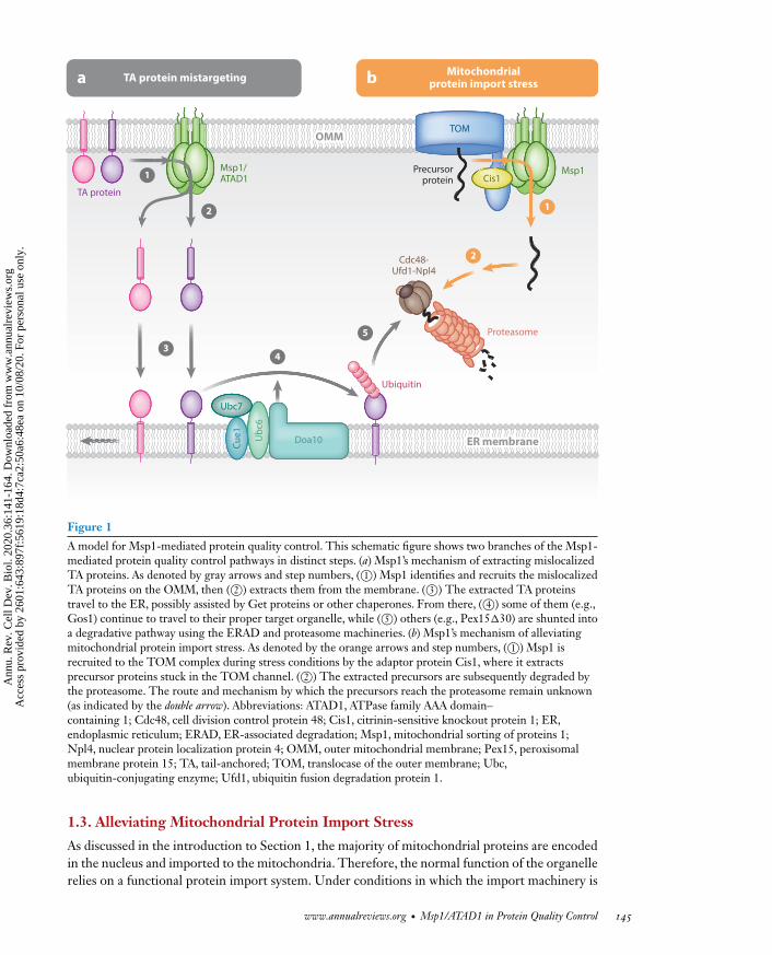
indicating that the Get proteins and Msp1 function in parallel, genetically linked pathways. Imag-ing of the peroxisomal TA protein Pex15 showed that in get3� cells a small fraction of Pex15 ismislocalized to mitochondria (Chen et al. 2014, Okreglak &Walter 2014). The overexpression ofMsp1 in these cells leads to the depletion of mislocalized Pex15 (Chen et al. 2014). By contrast,the deletion of Msp1 greatly exacerbates the mislocalization effect (Okreglak & Walter 2014),indicating that Msp1 counters the effect of TA protein mislocalization caused by get3 deletion.In vitro reconstitution experiments showed that Msp1 achieves this effect by extracting the TAproteins from the OMM in an ATP-dependent manner (Wohlever et al. 2017). The same functionhas been shown in the human ATAD1 in mammalian cells (Chen et al. 2014), suggesting that therole of Msp1 in extracting mislocalized TA proteins is phylogenetically conserved. Upon extrac-tion from the OMM, some of the TA proteins are rescued; they then travel to the ER, from wherethey continue to travel to their proper destination (Matsumoto et al. 2019).Others, when reachingthe ER, are degraded by the ER-associated degradation (ERAD) machinery (Dederer et al. 2019,Matsumoto et al. 2019). Whether the TA proteins travel to the ER via a direct handover fromMsp1 to the Get protein insertion machinery or via a different route is unknown. In summary,Msp1 surveys the OMM for mislocalized TA proteins and removes them from the membrane.This initiates either the redistribution of the TA proteins to their cognate destination organellesor degradation, thus achieving protein quality control to maintain cell health (Figure 1).
Msp1 was initially discovered nearly three decades ago in a screen for factors that sort proteinsbetween the OMM and the IMM (hence the protein’s name) (Nakai et al. 1993). However, effortsto validate the role Msp1 plays in this mechanism were not successful, suggesting that its over-expression in the screening regime may have artificially perturbed protein sorting. Only recentlyhave the structure and function of Msp1 in protein quality control gradually come to light.
1.2. Msp1 as a Member of the AAA Family
Even though its function remained a mystery until recently, phylogenetic structural analyses(Frickey & Lupas 2004) indicated that Msp1 is a member of the large AAA protein family. Thereare about 20 different AAA proteins in yeast or human cells and about 60 in plant cells (Ogura &Wilkinson 2001). As their name suggests, they perform a wide variety of functions, such as degrad-ing proteins (proteasome), disassembling SNARE complexes (NSF), and moving proteins acrossthe membrane [cell division control protein 48 (Cdc48)]. Within this family, Msp1 clusters withkatanin, spastin, Vps4, and fidgetin in one of six major subfamilies called the meiotic clade (MC).Recently, a number of high-resolution structures of AAA proteins (de la Peña et al. 2018, Donget al. 2018, Gates et al. 2017, Han et al. 2017, Puchades et al. 2017, Twomey et al. 2019) have re-vealed a commonmechanism of substrate unfolding: typically, the AAA protein forms a hexamericassembly and binds to its substrate in a central channel. The protein subunits undergo sequentialATP hydrolysis around the hexamer to generate mechanical forces that pull the substrate in two–amino acid steps through the channel, thereby unfolding it.Msp1 follows this general mechanismbut in evolution has acquired special features that allow it to recognize and extract mislocalizedmembrane proteins.
Since the first reports on its activity in extracting mislocalized TA proteins, the field has madesignificant progress elucidating Msp1’s mechanism of action. In Section 2 of this review, we sum-marize the current knowledge of the three major steps of the Msp1-mediated TA protein qualitycontrol pathway: (a) substrate recognition and recruitment, (b) substrate extraction, and (c) sub-strate degradation or resorting. We also discuss the structure and function of Msp1 within thecontext of the larger AAA protein family, and how its unique structural features are adapted to itsfunction (Wang et al. 2020).
1.3. Alleviating Mitochondrial Protein Import Stress
As discussed in the introduction to Section 1, the majority of mitochondrial proteins are encodedin the nucleus and imported to the mitochondria. Therefore, the normal function of the organellerelies on a functional protein import system. Under conditions in which the import machinery is
www.annualreviews.org • Msp1/ATAD1 in Protein Quality Control 145
Ann
u. R
ev. C
ell D
ev. B
iol.
2020
.36:
141-
164.
Dow
nloa
ded
from
ww
w.a
nnua
lrev
iew
s.or
g A
cces
s pr
ovid
ed b
y 26
01:6
43:8
97f:
5619
:18d
4:7c
a2:5
0a6:
48ea
on
10/0
8/20
. For
per
sona
l use
onl
y.
overwhelmed, several cellular pathways sense the stress and restore the organelle’s protein importcapacity. Among them is the mitochondrial compromised protein import response (mitoCPR)(Weidberg & Amon 2018). As a crucial part of mitoCPR,Msp1 is recruited to the TOM complexto clear precursor proteins that clog the translocase channel (Tom40), thus restoring its abilityto import proteins (Figure 1). This discovery expanded the function of Msp1 from extractingmislocalized TA proteins to general quality control on the OMM. In Section 3, we review thecurrent knowledge ofMsp1’s role inmitoCPR as well as its emerging role in general mitochondrialprotein quality control.
2. MECHANISM FOR EXTRACTING MISLOCALIZEDTAIL-ANCHORED PROTEINS
As discussed in Section 1.1,Msp1/ATAD1 recognizes and extracts themislocalizedTA proteins fordegradation or resorting. In this section, we synthesize the current knowledge about this processin three steps: (a) substrate recognition and recruitment, (b) substrate extraction, and (c) substratedegradation and resorting.
2.1. Substrate Recognition and Recruitment
The proper function of Msp1 depends on its ability to precisely distinguish endogenous mito-chondrial TA proteins frommislocalized TA proteins, as extracting endogenousmitochondrial TAproteins (such as the small TOM complex proteins) could wreak havoc on the organelle. A majorquestion remains in the field about how such discrimination is achieved. In addition,Msp1/ATAD1localizes to both the peroxisomal membrane and the OMM (Chen et al. 2014, Okreglak &Walter 2014).Why doesMsp1 only extract the Pex15molecules that aremislocalized to theOMMand ignore those that normally exist on the peroxisome?What features of mitochondria-localizedPex15 mark it as foreign? In this section, we discuss the current models for Msp1 substrate recog-nition and the biological evidence for each of them.We approach this question from a structuralpoint of view and discuss how the domain organization and structure of Msp1 shed light on itssubstrate-recognition mechanism.
2.1.1. Current models for substrate recognition. Several models have been proposed to ex-plain how Msp1 selects its substrate. One model posits that a specific structural feature servesas a signal that renders a protein sensitive to Msp1 (which we termed the extracton signal), andthat it is recognized by Msp1 directly in the substrate. Evidence for the extracton model comesfrom experiments using Pex15�30, a version of Pex15 in which 30 amino acids in its CTE havebeen removed, causing its mislocalization tomitochondria and degradation in anMsp1-dependentmanner (Okreglak & Walter 2014). Based on using Pex15�30 as a model substrate, Msp1 wasproposed to recognize a 12–amino acid hydrophobic sequence in the juxtamembrane region ofthe substrate (Li et al. 2019). A minimal construct containing only the hydrophobic sequence,the transmembrane helix, and the shortened CTE is degraded in an Msp1-dependent manner,whereas deletion of this patch from full-length Pex15�30 renders it insensitive to Msp1. The in-sertion of this segment into the juxtamembrane regions of the mitochondrial TA proteins (Fis1 orGem1) rendered them sensitive toMsp1-dependent degradation, suggesting that the hydrophobicpatch contains both essential and sufficient information for recognition by Msp1 (Li et al. 2019).On Msp1, in vitro cross-linking studies showed that hydrophobic amino acids contained in theN-terminal domain (N-domain) directly contact the substrate (Li et al. 2019). These amino acidsare distributed throughout the N-domain: some are found in the intramembrane space (IMS),some in the transmembrane anchor, and others in the linker that follows (see Figure 2e for the
146 Wang • Walter
Ann
u. R
ev. C
ell D
ev. B
iol.
2020
.36:
141-
164.
Dow
nloa
ded
from
ww
w.a
nnua
lrev
iew
s.or
g A
cces
s pr
ovid
ed b
y 26
01:6
43:8
97f:
5619
:18d
4:7c
a2:5
0a6:
48ea
on
10/0
8/20
. For
per
sona
l use
onl
y.
IMS
Cytosol
c
90° 90°
baM2
(ATP)
M4(ATP)
M3 (ATP)
M5 (ADP)
M6 (Apo)
M1(ATP)
M2(ATP)
M4(ATP)
M3 (ATP)
M5 (ADP)
M6 (Apo)
M1
Δ30-Msp1 (closed) Δ30-Msp1 (open)
Seam
Seam
ooM6 (ApM6 (Ap
e
TMD LD1 31 117 411Pore-loop 1
Pore-loop 2
AAA-large AAA-small
12 287
N-domain Core-ATPase domain
Pore-loop 3
Cryo-EM structures
Fishhook
Substrate
α1α0L1 L2
OMMOMM
TMD
180°
ATP
Representativesubunit (M4)
d
Figure 2
The structure of Msp1. Cryo-EM structures of (a) Msp1 (open conformation) and (b) Msp1 (closed conformation) complexes areshown in top and side views (PDB ID: 6PDW and 6PDY). Both structures lack the first 30 amino acids, which include thetransmembrane domain of Msp1. Each subunit (from M1 to M6) is assigned a distinct color, and the substrate is shown in black. Thenucleotide bound to each subunit is indicated in parentheses; Apo indicates that there is no bound nucleotide. The spiral seam of theopen conformations is denoted with dashed lines. In panel b, the map for the mobile subunit M1 is depicted at two thresholds: solidpink for σ = 5.3 (the same for the rest of the five subunits) and translucent pink for σ = 2.5, indicating that its weak density is due to theflexibility of this subunit and not to its complete dissociation from the complex. (c) Cutaway view of the open conformation revealingthe substrate density (black; highlighted in white dashed lines) in the central pore. (d ) A representative Msp1 subunit (M4) with domainsand structural elements colored according to panel e. ATP is shown in purple. (e) A schematic illustration of the individual domains andstructural elements of Msp1. The amino acid numbering is based on Msp1 from Chaetomium thermophilum. Abbreviations: cryo-EM,cryo-electron microscopy; IMS, intermembrane space; LD, linker domain; Msp1, mitochondrial sorting of proteins 1; N-domain,N-terminal domain; PDB, Protein Data Bank; TMD, transmembrane domain. Figure adapted from Wang et al. (2020).
domain organization).Mutations of these amino acids inactivateMsp1, causing a synthetic growthdefect with �get3 (Li et al. 2019). Collectively, these results strongly indicate that the N-domainof Msp1 recognizes a special feature, an extracton signal, that is presented by a hydrophobic seg-ment on the substrate.The extracton signal needs to be displayed in a particular geometry, becauseappending it to the N-terminus of Fis1 did not render Fis1 sensitive to Msp1 (Li et al. 2019).
www.annualreviews.org • Msp1/ATAD1 in Protein Quality Control 147
Ann
u. R
ev. C
ell D
ev. B
iol.
2020
.36:
141-
164.
Dow
nloa
ded
from
ww
w.a
nnua
lrev
iew
s.or
g A
cces
s pr
ovid
ed b
y 26
01:6
43:8
97f:
5619
:18d
4:7c
a2:5
0a6:
48ea
on
10/0
8/20
. For
per
sona
l use
onl
y.
In addition to the hydrophobic amino acids on Msp1, a single negatively charged amino acid(aspartate 12) residing in the IMS was found to interact with positively charged amino acids in theCTE of Pex15�30 (Li et al. 2019). This interaction adds to the strength of the substrate-Msp1association yet does not provide the basis for discrimination between endogenous TA proteinson the OMM and mislocalized TA proteins, as many mitochondrial TA proteins have enrichedpositive charges in their CTEs.
The extracton model suggests that an interaction partner for Pex15 exists on the peroxisomethat shields thisMsp1-recognizable feature. Indeed, a separate study (Weir et al. 2017) showed thatPex3 interacts with Pex15 on the peroxisomal membrane and removing Pex3 results in the Msp1-dependent degradation of Pex15 on the peroxisome. Pex3 is a docking factor on the peroxisomalmembrane important for peroxisomal protein import (Mast et al. 2020). The structure of Pex3(Sato et al. 2010, Schmidt et al. 2010) shows that a cluster of hydrophobic amino acids is presentin the membrane-proximal region that might shield the hydrophobic segment on Pex15. Thus, itis reasonable to infer from these two independent studies that Pex3 protects Pex15 from Msp1-mediated extraction through shielding its hydrophobic extracton signal.
A weakness of the extracton model is that it is currently based on studies of a single modelsubstrate, Pex15�30, so its generality has not been established. Among the Msp1 TA proteinsubstrates discovered so far [Pex15 (Chen et al. 2014, Okreglak & Walter 2014), Gos1 (Chenet al. 2014), Fmp32 (Dederer et al. 2019), Frt1 (Li et al. 2019), and Ysy6 (Li et al. 2019)], Pex15is the only one with a recognizable hydrophobic segment at the juxtamembrane region. TheseTA protein substrates also do not possess recognizable common sequences or structural motifs.Therefore, other mechanisms for recognition are likely to exist.
The alternative but not mutually exclusive model hypothesizes that Msp1 selects its substratesbased on their monomeric existence; i.e., Msp1 selects proteins that are not part of a larger pro-tein complex.We termed this model the orphanmodel. By fusing oligomerizationmodules such asthe FKBP or FRB domains to Pex15�30, researchers induced the dimerization of two Pex15�30molecules and showed that,whereas themonomeric Pex15�30molecules are efficiently degraded,artificially induced dimers are resistant to Msp1-mediated degradation (Dederer et al. 2019). Insupport of this model, the overexpression of Gem1 [a mitochondrial TA protein that is a compo-nent of the ER-mitochondria tethering complex ERMES (ER-mitochondria encounter structure)(Kornmann et al. 2009)] results in its Msp1-dependent degradation, as it outnumbers cognatecomplex partners (Dederer et al. 2019).
One explanation for these results is that theMsp1-recognizable feature in the substrate (such asthe hydrophobic segment of Pex15�30) is occluded in a dimer or a higher-order protein complex.In this case, the extracton and orphan models converge on the same mechanistic explanation.An alternative explanation is that the extra transmembrane helices in a protein complex provideenough anchoring power to the membrane that it exceeds the pulling force of Msp1. In otherwords, Msp1 does not select its substrate based on a sequence motif or a structural feature but,rather, specificity is achieved based solely on the energy it takes to extract a protein from thelipid bilayer. Teasing out between the two models will require measuring Msp1’s activity towardcarefully designed substrates.
Based on the orphan model, all of the mitochondrial TA proteins would have to exist aspart of bigger protein complexes (or oligomers) to evade extraction by Msp1. In yeast, there aresix known mitochondrial TA proteins: Tom5, Tom6, Tom7, Tom22, Fis1, and Fmp32 (Burri &Lithgow 2004). Among those, the TOM proteins are firmly embedded in the TOM complex.Fis1 mediates mitochondrial fission and has known interaction partners, including Drp1 (Losónet al. 2013). Fmp32 is less well studied and is thought to be an assembly factor for cytochromec oxidase (Paupe et al. 2015). This protein is rapidly degraded by Msp1 under physiological
148 Wang • Walter
Ann
u. R
ev. C
ell D
ev. B
iol.
2020
.36:
141-
164.
Dow
nloa
ded
from
ww
w.a
nnua
lrev
iew
s.or
g A
cces
s pr
ovid
ed b
y 26
01:6
43:8
97f:
5619
:18d
4:7c
a2:5
0a6:
48ea
on
10/0
8/20
. For
per
sona
l use
onl
y.
conditions, perhaps because it exists mainly in a monomeric state. If so, it would be interesting toask whether inducing its dimerization would make it resistant to Msp1 extraction.
In addition to the functional evidence, the structure and domain organization of Msp1 providefurther insight into its substrate recognition mechanism, which we review in detail next. In itsrole in relieving protein import stress, a completely different model for substrate recognition isproposed for Msp1: it is recruited by an adaptor protein to the TOM complex to extract theprecursor proteins that clog the Tom40 channel. We discuss this process in Section 3.
2.1.2. The structural basis for Msp1’s substrate recognition. A large number of mechanisticstudies on AAA proteins (Augustin et al. 2009,Hanson &Whiteheart 2005) have provided us witha framework to understand the mechanism of Msp1. AAA proteins typically contain an N-domainthat is responsible for substrate recruitment and selection followed by one or two classic AAAdomains that form oligomers and harness the chemical energy of ATP hydrolysis to generate me-chanical force. Unlike the typically highly conserved AAA domains, the N-domains show a largedegree of variance among members of the AAA protein family, thus specializing each member forits particular function. In this section, we discuss the recently published cryo-electron microscopy(cryo-EM) structures of the Msp1-substrate complex with a focus on the N-domain, which shedslight on Msp1’s substrate selection and recruitment mechanism.We also discuss the structure anddomain organization of Msp1 in the context of the AAA protein family to delineate how its uniqueN-domain structure adapts the AAA domain engine to the special function of Msp1.
2.1.2.1. The overall structure of the Msp1-substrate complex. Recently, we solved a first set ofstructures of Msp1 in complex with a peptide substrate by cryo-EM (Wang et al. 2020). The struc-tures contain the cytosolic domain of the Chaetomium thermophilumMsp1 lacking its N-terminalmembrane anchor (�30-Msp1). Two different conformations were identified: an open conforma-tion and a closed conformation (Figure 2a,b). In the open conformation, �30-Msp1 formed ahomohexamer in which the six subunits (M1–M6) rotate and translate progressively to assembleinto a right-handed open spiral with an open seam between the top (M1) and the bottom (M6)subunits, an assembly seen in many other AAA protein structures including katanin and spastin(Figure 2a). In the closed conformation, the Msp1 subunits assemble into the same spiral confor-mation, except for M1, which closes the seam of the spiral (Figure 2b). The cryo-EM density forM1 in the closed state is weak, indicating that the structure is an ensemble that represents a con-tinuum of multiple states in the Msp1 reaction cycle. A 10-mer peptide was found in the centralcavity formed by the hexamer that likely presents an averaged composite of many Escherichia colipeptides that copurified with Msp1 (Figure 2c). The well-resolved peptide backbone allowed usto closely examine the mechanism of substrate interaction in the central pore, which we discussin detail in Section 2.2.
2.1.2.2. The structure of Msp1’s linker domain. Each Msp1 subunit consists of an N-domainfollowed by a core-ATPase domain. In Msp1, the N-domain is composed of a transmembraneanchor (not included in our structures) followed by a linker domain (LD). The LD contains twohelices (α0 and α1) and two loops (L1 and L2) (Figure 2e). Helix α0 and loop L1 fold into afishhook-shaped motif that is also observed in the structure of katanin (Zehr et al. 2017). In theopen conformation, the α0 helices in subunits M1–M5 are radially organized with their N-terminipointing toward the spiral’s center, whereas the helix α0 in M6 is disordered (Figure 3a). TheN-terminal regions of the α0 helices fromM1 toM5 converge in a central hub,where they contacteach other, with M1 being closest to andM5 farthest from the membrane.Helix α0M6 is unfolded,presumably because M6 occupies the lowest position in the spiral and therefore is positioned too
www.annualreviews.org • Msp1/ATAD1 in Protein Quality Control 149
Ann
u. R
ev. C
ell D
ev. B
iol.
2020
.36:
141-
164.
Dow
nloa
ded
from
ww
w.a
nnua
lrev
iew
s.or
g A
cces
s pr
ovid
ed b
y 26
01:6
43:8
97f:
5619
:18d
4:7c
a2:5
0a6:
48ea
on
10/0
8/20
. For
per
sona
l use
onl
y.
a
c
M2 M5 M6
...
M6
M5
M4 M3
M2
M1
SeamSeS
M6Seam
M6
Possibility 2:The monomer recruits the substrateand then assembles into the hexamer.
Possibility 1:The hexamer preassembles and then recruits the substrate at M6.
b
OMMOMM
Central hubα0L1Amino acids that are validated to interact with the substrateOther hydrophobic amino acids that may interact with the substrate
Figure 3
The structural basis for the extracton model of substrate recruitment. (a) A cryo-electron microscopy map of �30-Msp1 (open)showing the arrangement of the fishhook motifs in the spiral. Subunits M1–M5 show high density for the entire fishhook motif (helixα0 and loop L1), whereas M6 shows density for L1 but not for α0. The structure shows the fishhook motifs of different subunits to beradially organized, with their N-termini pointing to the center of the spiral. (b) Surface representations of individual subunitshighlighting amino acids in the linker domains (LDs) that are likely to engage with the hydrophobic substrate. These amino acids areburied by α0 in M2–M5 but are exposed in M6 where α0 is melted. (c) Two possible mechanistic explanations of the extracton model,shown in the outer mitochondrial membrane (OMM). (Left) In possibility 1, in the preassembled hexamer, the amino acids that interactwith the substrate (identified in Li et al. 2019) by cross-linking or immunoprecipitation form a patch at the seam of the spiral, wherethey recruit the substrate using hydrophobic interactions. (Right) In possibility 2, monomeric Msp1 recruits the substrate andsubsequently assembles into a hexamer. Figure adapted from Wang et al. (2020).
far away to participate in this stabilizing interaction at the central hub. The melting and refoldingof α0 allow the subunits to remain stably inserted in the membrane during Msp1’s translocationcycle (a process discussed in detail in Section 2.2).
The conformational change of helix α0 suggests a plausible mechanism for substrate recruit-ment by the extracton model, which posits that a series of hydrophobic amino acids in the N-domain of Msp1 directly engage the substrate Pex15 (Li et al. 2019). Mapping these amino acidsto the structure shows that many of them lie in the LD. Intriguingly, they are only exposed at thesurface in M6 [due to the melting of helix α0 (Figure 3b)] but are mostly buried by the foldedhelices α0 in M1–M5, suggesting that M6 is the only Msp1 subunit that exposes the possiblesubstrate-binding site.
150 Wang • Walter
Ann
u. R
ev. C
ell D
ev. B
iol.
2020
.36:
141-
164.
Dow
nloa
ded
from
ww
w.a
nnua
lrev
iew
s.or
g A
cces
s pr
ovid
ed b
y 26
01:6
43:8
97f:
5619
:18d
4:7c
a2:5
0a6:
48ea
on
10/0
8/20
. For
per
sona
l use
onl
y.
Binding could happen either before or after monomeric Msp1 assembles into a hexamer; thatis, either the hexamer preassembles in regions where the local Msp1 concentration is high enoughand then recruits the substrate at the binding site in M6 (Figure 3c), or, alternatively, Msp1binds the substrate as a monomer. This latter notion is supported by the crystal structure of themonomeric Saccharomyces cerevisiae Msp1 (Wohlever et al. 2017), in which half of the LD is dis-ordered, perhaps due to the lack of the stabilization interaction in the central hub present only inthe oligomer. This suggests that helix α0 may sample both the folded and the melted states in themonomeric form and thus could bind the substrate in its melted state and subsequently assembleinto the M6 position of a hexamer (Figure 3c).
Interestingly, many of the hydrophobic amino acids in M6 line the open seam of the hexamer(Figure 3c). This arrangement could allow Msp1 to align the bound substrate with the seam,where it can be conveniently threaded into the central pore. Thus the observed conformationalchanges of helix α0 provide an intuitive mechanism for substrate recruitment toMsp1’s entry gate.Also, the membrane-proximal position of the LD of M6 provides an intuitive explanation for thegeometric requirement that the extracton signal must be located next to the membrane.
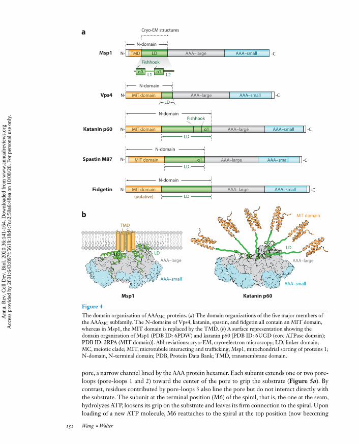
2.1.2.3. A comparison of the N-domain of Msp1 to those of other AAA proteins. Accordingto the orphan model, Msp1 may lack intrinsic substrate specificity and could select its substratespurely on an energetic basis. A closer look at the N-domain of Msp1 provides support for this no-tion, as comparison ofMsp1’s N-domain to those of other AAA proteins has revealed some uniquefeatures.Within the AAAMC clade (Figure 4a), the N-domains of katanin, spastin, Vps4, and fid-getin all contain amicrotubule-interacting and trafficking (MIT) domain that specifically interactswith their substrates [microtubules for katanin, spastin, and possibly fidgetin; ESCRT (endoso-mal sorting complexes required for transport) III peptides for Vps4]. Outside of the AAAMC clade,the N-domain of NSF mediates the interaction with its cofactor SNAP (soluble NSF attachmentprotein) proteins, which recruit substrate SNAREs; the N-domain of Cdc48 binds to cofactorsubiquitin fusion degradation protein 1 (Ufd1)/nuclear protein localization protein 4 (Npl4) thatspecifically interact with ubiquitin. By contrast, Msp1’s N-domain lacks a module that targets itto a specific protein (Figure 4a).
Despite its simple makeup, the N-domain notably contains a transmembrane segment thatlimits Msp1’s substrates to those embedded in or associated with the membrane to which Msp1is attached. This is similar to a few other membrane-anchored AAA proteases such as FtsH andYme1, both of which also appear to lack N-domains that would provide substrate specificity andwhich degrade a wide range of proteins present on the same membrane.
In summary, both the domain organization and the functional data suggest that the N-domainof Msp1 may not overly restrict its substrate recognition but rather may allow it to extract manydifferent substrates localized to the OMM, some of which likely remain to be discovered. Themain determinant of its substrate recognition may not be overly constrained by a sequence motifor structural feature but rather, as posited by the orphan model, may be primarily limited by theenergy expense required to extract the protein from the membrane. Further insights will be aidedby determining the biochemical and biophysical requirements for Msp1’s substrate selection aswell as by comprehensive proteomic analyses of the full spectrum of Msp1 substrates.
2.2. Substrate Extraction
2.2.1. The overall extraction mechanism. Recent advances in single-particle cryo-EM haveenabled the determination of many substrate-bound AAA protein structures at high resolu-tion (Puchades et al. 2020). Many of these structures converge on a common hand-over-handmechanism of protein unfolding: Upon recruitment, the substrate is threaded into the central
www.annualreviews.org • Msp1/ATAD1 in Protein Quality Control 151
Ann
u. R
ev. C
ell D
ev. B
iol.
2020
.36:
141-
164.
Dow
nloa
ded
from
ww
w.a
nnua
lrev
iew
s.or
g A
cces
s pr
ovid
ed b
y 26
01:6
43:8
97f:
5619
:18d
4:7c
a2:5
0a6:
48ea
on
10/0
8/20
. For
per
sona
l use
onl
y.
Vps4
Katanin p60
Spastin M87
Fidgetin
Msp1
α1α0L1 L2
TMD
MIT domain
MIT domain
MIT domain AAA–small AAA–large
MIT domain AAA–large
AAA–large
AAA–large
AAA–large
AAA–small
AAA–small
AAA–small
AAA–small
(putative)
LD
N-domain
N-domain
LD
LD
LD
LD
N-domain
N-domain
Cryo-EM structures
Msp1 Katanin p60
AAA–small
a
bTMD
LD
AAA–large
Fishhook
Fishhook
N-
N-
N-
N-
N-
-C
-C
-C
-C
-C
N-domain
AAA–small
MIT domain
LD
AAA–large
α1
α1
Figure 4
The domain organization of AAAMC proteins. (a) The domain organizations of the five major members ofthe AAAMC subfamily. The N-domains of Vps4, katanin, spastin, and fidgetin all contain an MIT domain,whereas in Msp1, the MIT domain is replaced by the TMD. (b) A surface representation showing thedomain organization of Msp1 (PDB ID: 6PDW) and katanin p60 [PDB ID: 6UGD (core ATPase domain);PDB ID: 2RPA (MIT domain)]. Abbreviations: cryo-EM, cryo-electron microscopy; LD, linker domain;MC, meiotic clade; MIT, microtubule interacting and trafficking; Msp1, mitochondrial sorting of proteins 1;N-domain, N-terminal domain; PDB, Protein Data Bank; TMD, transmembrane domain.
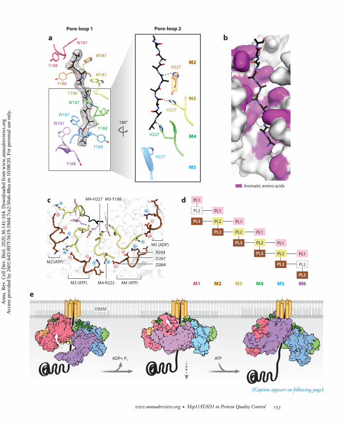
pore, a narrow channel lined by the AAA protein hexamer. Each subunit extends one or two pore-loops (pore-loops 1 and 2) toward the center of the pore to grip the substrate (Figure 5a). Bycontrast, residues contributed by pore-loops 3 also line the pore but do not interact directly withthe substrate. The subunit at the terminal position (M6) of the spiral, that is, the one at the seam,hydrolyzes ATP, loosens its grip on the substrate and leaves its firm connection to the spiral. Uponloading of a new ATP molecule, M6 reattaches to the spiral at the top position (now becoming
152 Wang • Walter
Ann
u. R
ev. C
ell D
ev. B
iol.
2020
.36:
141-
164.
Dow
nloa
ded
from
ww
w.a
nnua
lrev
iew
s.or
g A
cces
s pr
ovid
ed b
y 26
01:6
43:8
97f:
5619
:18d
4:7c
a2:5
0a6:
48ea
on
10/0
8/20
. For
per
sona
l use
onl
y.
W187
W187
W187
W187
W187
Y188
Y188
Y188
Y188
Y188
W187
Y188
180°
H227
H227
H227
H227
Pore-loop 1 Pore-loop 2
M2M2
M3
M4
M5
PL1
PL2
PL3
PL1
PL2
PL3
PL1
PL2
PL3
PL1
PL2
PL3
PL1
PL2
PL3
PL1
PL2
PL3
M1 M2M2 M3 M4 M5 M6
dM4-H227 M5-Y188
M4-R222 M4 (ATP)M3 (ATP)
+
_
M5 (ADP)
M2 (ATP)
R264D267D269
c
e
ADP+ Pi ATP
_
_
_
__
_
_
+
+
+ +
++
+
a b
OMMOMM
Aromatic amino acids
(Caption appears on following page)
www.annualreviews.org • Msp1/ATAD1 in Protein Quality Control 153
Ann
u. R
ev. C
ell D
ev. B
iol.
2020
.36:
141-
164.
Dow
nloa
ded
from
ww
w.a
nnua
lrev
iew
s.or
g A
cces
s pr
ovid
ed b
y 26
01:6
43:8
97f:
5619
:18d
4:7c
a2:5
0a6:
48ea
on
10/0
8/20
. For
per
sona
l use
onl
y.
Figure 5 (Figure appears on preceding page)
Substrate extraction mechanism. (a) Pore-loops 1 (PL1s) form a spiral staircase around the substrate (with peptide density shown as ablack mesh). PL2s form a second staircase below PL1s. H227s form hydrogen bonds with the peptide backbone carbonyls (dashed lines).(b) A surface representation of the central pore showing that the peptide (in stick representation) is surrounded by interdigitatingaromatic amino acids (purple) in the central pore. (c) An illustration of the PLs showing that PL2s interact with PL1s through π–πstacking and with PL3s via electrostatic and polar interactions. The π–π stacking interaction is shown with dashed lines, and thepositive charges are shown in blue while negative charges are in red. PL1s are shown in pink, PL2s in light yellow, and PL3s in brown.(d ) A schematic diagram showing the interactions (depicted by lines) between PLs. The disordered PL2s in M1 and M6 are shown indashed boxes. (e) A model for substrate extraction showing the upward translocation of the bottom subunit (M6) and the downwardtranslocation of the substrate through the central pore. A tail-anchored (TA) protein substrate is shown in black. The Msp1(mitochondrial sorting of proteins 1) subunits are shown in surface representation and are colored as in Figure 2. The folded α0 helix isshown in cartoon representation (green) and the positions of the N-terminal transmembrane regions are schematically indicated. Figureadapted from Wang et al. (2020).
M1), where its pore-loops reengage the substrate (Figure 5e). Each upward translocation eventlike this allows the protein to move along the peptide substrate in two amino acid steps, and, inMsp1’s case, to extract the substrate stepwise out of the membrane. During the movement of theterminal subunit, pore-loops from the other subunits remain engaged with the substrate, pre-venting it from backsliding. The pore-loops hold the substrate in an almost linearly extendedconformation imposed by the shape of the central pore. In this way, Msp1 (and AAA proteins ingeneral) effectively unfolds its substrate as it traverses the pore.
2.2.2. The structural features that adapt Msp1 to its function. Msp1 follows the conservedgeneral mechanism of protein unfolding established for AAA proteins, yet several structural fea-tures specialize it for its unique role of extracting transmembrane proteins from the lipid bilayer.First, the KX1X2G motif in pore-loop 1 is highly conserved across the entire AAA protein family,with X1 being an aromatic amino acid and X2 usually a nonaromatic hydrophobic amino acid. Thearomatic amino acid intercalates between the substrate’s side chains and form a staircase aroundthe substrate. By contrast, in Msp1/ATAD1, both X1 and X2 are aromatic amino acids (Msp1:W187 and Y188), and both are in direct contact with the substrate’s side chains (Figure 5a).Second, pore-loop 2 is highly variable across the AAA protein family and is usually disordered orretracted from the substrate. By contrast, the pore-loops 2 of Msp1 are in close contact with thesubstrate, forming a second staircase around the substrate just below pore-loops 1. In addition,a conserved histidine (Msp1: H227) forms a hydrogen bond with the backbone carbonyl of thesubstrate (Figure 5a). In total, not just one but three aromatic amino acids from each subunittightly engage the substrate (Figure 5b), making the central pore of Msp1 more hydrophobic andsterically constrained than those in other AAA proteins.
A recent study (Rodriguez-Aliaga et al. 2016) on the ClpXP motor showed that the bulkinessof the pore-loops positively correlates with their grip on the substrate but inversely correlatesto the substrate pulling velocity. As Msp1 needs to extract the hydrophobic membrane anchorsfrom the lipid bilayer, it would seem beneficial for it to exert a stronger grip on its substrateusing bulky pore-loops to prevent backsliding. Similar to Msp1, other AAA proteins that extractmembrane proteins, such as Cdc48 (Cooney et al. 2019, Twomey et al. 2019), Yme1 (Puchadeset al. 2017), and AFG3L2 (Puchades et al. 2019), all have second aromatic amino acids in additionto the conserved ones in pore-loops 1 that directly interact with the substrate, suggesting thatdistantly related AAA proteins may have converged on similar solutions to increase their grip onmembrane protein substrates.
In addition to providing extra grip, the pore-loop 2 ofMsp1 is also uniquely positioned to sensethe position of the subunit in the spiral. It is involved in an interconnected network of electrostaticinteractions involving pore-loops 1, 2, and 3 (Figure 5c,d ). Its stabilization requires interactions
154 Wang • Walter
Ann
u. R
ev. C
ell D
ev. B
iol.
2020
.36:
141-
164.
Dow
nloa
ded
from
ww
w.a
nnua
lrev
iew
s.or
g A
cces
s pr
ovid
ed b
y 26
01:6
43:8
97f:
5619
:18d
4:7c
a2:5
0a6:
48ea
on
10/0
8/20
. For
per
sona
l use
onl
y.
from subunits on both sides. As a result, pore-loops 2 in the central four subunits (M2–M5) areordered, and those in subunits capping the spiral (M1 andM6) are disordered due to the lack of aninteraction partner on opposite sides of the seam (Figure 5d ). This allows M6 to sense its bottomposition in the spiral and initiate its dissociation from the substrate. The importance of the aminoacids in all three pore-loops has been validated experimentally (Wang et al. 2020).
In addition to the sophisticated network of pore-loop–pore-loop interactions, Msp1 uses aunique mechanism for communication between subunits. A few AAA proteins (such as Yme1 andthose found in the cap structure of the proteasome) use an intersubunit-signaling (ISS) motif thatcontains a signature DGF tripeptide to sense the nucleotide state and transmit this information tothe pore-loops. In Msp1, as in other AAAMC proteins, the crucial phenylalanine in the ISS motif isreplaced by an aliphatic amino acid, which is predicted to hamper transmission of the informationon nucleotide state by this mechanism. Instead, Msp1 possesses a short loop insertion C-terminalto the traditionally defined ISS motif [the nucleotide communication loop (NCL)] that sensesthe nucleotide hydrolysis and communicates it to the adjacent subunit. Together with the meltingof pore-loop 2 when it reaches the M6 position, the NCL prompts the terminal subunit for itsupward translocation. Although this loop seems to be unique to the C. thermophilumMsp1 and isnot conserved in the Msp1 family, it is also found in similar proteins such as katanin. Whether italso plays a role there in the transmission of nucleotide states remains to be determined.
In summary, while following the general hand-over-hand mechanism,Msp1 has acquired a fewstructural modifications to the conserved AAA fold. These modifications are uniquely suited to itsrole as a membrane protein extractase.
Despite the rich mechanistic insights provided by these structures, several questions remain.First, a recent study (Han et al. 2019) showed that the central pore of Vps4 is able to engage acircular peptide, an indication that it could unfold internal sequences. Can Msp1 also hold twopolypeptide chains in its pore at once and hence unfold internal sequences that are part of a foldedstructure? The structure of Msp1 shows that the bulky pore-loops encase the substrate tightly andleave almost no space to fit another peptide. If Msp1’s central pore can be shown to similarly ac-commodate an extra peptide chain, what conformational changes are required for this to happen?Second, given that the NCL is not an evolutionarily conserved feature across the Msp1/ATAD1subfamily, what mechanism is employed by the Msp1 orthologs to communicate the nucleotidestate between subunits? Lastly, doesMsp1’s transmembrane helix play a role in substrate extractionbeyond aligning the extractase on the surface of the membrane?
2.3. Substrate Degradation or Resorting
While a significant amount of work has expanded our understanding of how Msp1 selects andextracts its substrates, the fate of the extracted TA proteins has remained a mystery until very re-cently. Previous studies (Chen et al. 2014, Okreglak &Walter 2014) showed that mislocalized TAproteins are proteolytically degraded upon Msp1 extraction. Given that Msp1 itself does not havea protease function, the degradation must require the channeling of the extracted protein to thedegradative machineries. A recent study (Matsumoto et al. 2019) showed that the degradation ofthe Msp1 substrates is proteasome mediated. To identify factors involved in this process, the au-thors conducted a limited screen of the ubiquitin-proteasome-related genes that revealed that theER-resident E3 ubiquitin ligase Doa10, its associated E2 ubiquitin-conjugating enzymes Ubc6and Ubc7, and their tethering factor Cue1 are all required for the degradation of Pex15�30. Inaddition, Cdc48 and its cofactors Ufd1, Npl4, and the tethering factor Ubx2 are also required forits degradation. Interestingly, these proteins constitute the cytosolic branch of the ERAD pathway(ERAD-C), which degrades the ER-membrane proteins with misfolded cytosolic domains. The
www.annualreviews.org • Msp1/ATAD1 in Protein Quality Control 155
Ann
u. R
ev. C
ell D
ev. B
iol.
2020
.36:
141-
164.
Dow
nloa
ded
from
ww
w.a
nnua
lrev
iew
s.or
g A
cces
s pr
ovid
ed b
y 26
01:6
43:8
97f:
5619
:18d
4:7c
a2:5
0a6:
48ea
on
10/0
8/20
. For
per
sona
l use
onl
y.
surprising finding that the ERAD machinery is required for the degradation of Msp1 substratesindicates that Msp1 drives the substrates from the OMM to the ER membrane (Figure 1). Inline with this notion, the authors (Matsumoto et al. 2019) showed that the deletion of Msp1 fromcells lacking a functional ERAD-C machinery (�ubc7) leads to the accumulation of Pex15�30 onthe OMM, whereas the overexpression of Msp1 in these cells leads to its enrichment on the ER.In contrast to Pex15�30, another Msp1 substrate, mitochondrially mislocalized Gos1, escapesdegradation by ERAD and ends up on the Golgi apparatus, its cognate organelle of residence.These observations are explained by the notion that Pex15�30 is an artificial protein that is seenby the ERAD system as foreign, whereas Gos1 is an endogenous protein whose targeting to theER is on pathway during its normal biogenesis, therefore allowing it to escape the degradation. Analternative, albeit less interesting, explanation is that during extraction, Msp1 unfolds part of itssubstrates, and the observed differences are due to different refolding efficiencies of different sub-strates: Pex15�30 may remain partially unfolded by the time it reaches the ER, which causes themolecule to be marked by ERAD-C as a defective protein substrate ready to be degraded, whereasGos1 may refold by the time it reaches the ER (perhaps due to its strong helical propensity) andthus escape degradation.
The involvement of ERAD-C was independently shown by a separate study (Dederer et al.2019) in which researchers used the fluorescent timer strategy to identify genes whose absenceleads to a longer half-life of the substrate. From a genome-wide screen and follow-up experiments,the authors also determined that the Doa10 complex is responsible for degrading Pex15�30. Inaddition, the researchers showed that a small fraction of TA protein substrates are degraded bythe vacuole.
These two independent studies unambiguously showed that the ERAD-C pathway plays animportant role in the degradation of Msp1 substrates. The requirement for the collaboration oftwo independent quality control systems on two separate membranes to degrade the mislocal-ized TA proteins remains surprising. The possibility to correct mistakes by sending mislocalizedproteins such as Gos1 back to the ER may offer an explanation. How proteins are shuttled fromMsp1 to the ER still remains a mystery, as proteins containing transmembrane sequences are un-likely to diffuse through the cytoplasmic space unchaperoned. Msp1 extraction may therefore beobligatorily coupled to a transport system, perhaps hitchhiking on the Get machinery, to reachthe ER. Alternatively, Msp1 may release its substrates at ER-mitochondria contact sites to allowdirect delivery. Nevertheless, ERAD-C and the proteasome complete the Msp1-assisted proteinquality control pathway.
3. THE ROLE OF Msp1 IN ALLEVIATING MITOCHONDRIALPROTEIN IMPORT STRESS
All but a few mitochondrial proteins are encoded in the nuclear genome and thus need to beimported across the mitochondrial membranes. Together the multicomponent translocase of theouter membrane (TOM) and that of the inner membrane (TIM) constitute one of the main mi-tochondrial protein import machineries. Typically, a protein is targeted to the TOM complex bya short peptide at the N-terminus [the mitochondrial targeting sequence (MTS)] (Neupert &Herrmann 2007). Powered by the electrochemical potential across the IMM and ATP hydrolysis,the protein traverses the TOMand then the TIM channels and finally emerges in thematrix space.In the cases in which the protein possesses an internal hydrophobic sequence, it stalls the transferin the TIM23 channel, from which it is laterally released into the IMM.Maintaining unimpededflow in the import channels is essential, as the clogging of these channels not only leads to a failurein obtaining the necessary proteins for the organelle’s function but also causes the accumulation of
156 Wang • Walter
Ann
u. R
ev. C
ell D
ev. B
iol.
2020
.36:
141-
164.
Dow
nloa
ded
from
ww
w.a
nnua
lrev
iew
s.or
g A
cces
s pr
ovid
ed b
y 26
01:6
43:8
97f:
5619
:18d
4:7c
a2:5
0a6:
48ea
on
10/0
8/20
. For
per
sona
l use
onl
y.
unimported precursor proteins in the cytosol. Such accumulation sequesters essential chaperonesand leads to a failure of protein homeostasis. Despite the severe outcome, until recently little wasknown about how the organelle protects itself in the face of such import stress.
The mitoCPR protects the mitochondria when the import machinery is overwhelmed. Theoverexpression of proteins containing both the N-terminal MTS and an internal hydrophobicsequence (a bipartite signal) leads to the TIM23 channel becoming clogged due to the slow lateralrelease of the hydrophobic sequence into the IMM. This defect consequently causes a clog in theTOM channel, preventing more proteins from being imported and processed. In response to thisimport stress, cells activate the mitoCPR, which is mediated by the transcription factor Pdr3.
As a crucial part of the mitoCPR,Msp1 is recruited to the TOM complex by Cis1.CIS1 is oneof the most strongly induced genes in response to PDR3 activation. It accumulates at the surfaceof the mitochondria under import stress. Upon recruitment,Msp1 extracts the precursor proteinsfrom the import channel, and they are degraded in a proteasome-dependent manner (Figure 1).Consequently, Cis1 and Msp1 together restore the ability of the mitochondria to import proteinsduring the period of import stress. Mechanistically, it is still a mystery how Msp1 gains access tothe precursor proteins stuck in the Tom40 channel. Whether Cis1 or other factors are requiredto help shuttle these precursors to Msp1 remains to be understood.
The function of Msp1 in mediating protein import is apparently independent from its func-tion in extracting mislocalized TA proteins. Overexpressing Cis1 in the �get3 background causesa growth phenotype resembling the �get3/�msp1 double knockout, indicating that Msp1 is redis-tributed to the TOM complex by binding to Cis1, which precludes it from extracting TA proteins.
Msp1’s function in assisting with mitochondrial protein import stress is supported by severallines of evidence. First, MSP1 has displayed a strong genetic interaction with several genes en-coding subunits of the TOM complex under respiratory conditions: TOM5,TOM20, and TOM40(Basch et al. 2020). Both Tom5 and Tom20 are known to bind incoming precursor proteins(Bausewein et al. 2017, Söllner et al. 1989), and their absence leads to the accumulation of im-properly channeled precursor proteins at the main import channel (Tom40). Second, in vitro im-port assays show that Msp1 is required for the removal of mitochondria-targeted DHFR, a modelsubstrate whose core can be stabilized by methotrexate and then is particularly hard to unfold,thus clogging the import channel (Basch et al. 2020). Finally, the overexpression of Msp1 leads toreduced levels of several IMS proteins whose import is less efficient, suggestingMsp1may activelyremove proteins that do not easily pass the channel (Basch et al. 2020). In summary, by extractingproteins that are stuck in the import channel, Msp1 clears the path for more precursor proteinsand alleviates the mitochondrial import stress.
Upon extraction by Msp1, the precursor proteins are degraded in a proteasome-dependentmanner (Basch et al. 2020); however, the route by which the substrate proteins reach the protea-some remains ill defined. Immunoprecipitation followed by mass spectrometry (IP-MS) showedthat the proteasomal subunit Rpn10 interacts with Msp1 under nonstress conditions (Basch et al.2020), suggesting that extracted precursor proteins are directly handed over to the proteasome.This suggests that precursor protein ubiquitylation is coordinated with Msp1 extraction at theOMM,which is different from that of TA proteins that become ubiquitylated at the ERmembraneafter Msp1 extraction (Dederer et al. 2019). However, a similar IP-MS experiment described pre-viously (Chen et al. 2014) failed to uncover any proteasomal subunits, perhaps due to differentMsp1 expression and enrichment methods. A direct proteasomal association thus requires furthervalidation, and the ubiquitylation machinery required for the degradation remains to be defined.
Finally, how cells sense the protein import stress and relay that information from themitochon-dria to the nucleus remains a mystery.Recently, the activation of Pdr3 was shown to be the last stepof a reaction cascade: it begins with the activation of Hsf1 (Boos et al. 2019), the key transcription
www.annualreviews.org • Msp1/ATAD1 in Protein Quality Control 157
Ann
u. R
ev. C
ell D
ev. B
iol.
2020
.36:
141-
164.
Dow
nloa
ded
from
ww
w.a
nnua
lrev
iew
s.or
g A
cces
s pr
ovid
ed b
y 26
01:6
43:8
97f:
5619
:18d
4:7c
a2:5
0a6:
48ea
on
10/0
8/20
. For
per
sona
l use
onl
y.
factor driving the heat shock response. Hsf1 is normally repressed by binding to molecular chap-erones and is activated when the chaperones are sequestered by unimported precursor proteins.Upon activation, Hsf1 induces the expression of a proteasomal subunit Rpn4, a transcription fac-tor that in turn induces the expression of Pdr3. Thus, mitoCPR is part of a multilayered cellularresponse to mitochondrial protein import stress.Whether Pdr3 can be activated by a mechanismindependent of Rpn4 or Hsf1, that is, whether it can sense the import stress directly, and whatrole Msp1 plays in this process remain open questions.
Likewise, whether Msp1/ATAD1 is involved in protein import quality control in highereukaryotes remains an open question. Although Cis1 does not have an apparent ortholog inmammalian cells, a different adaptor protein could recruit Msp1 to the TOM complex. This issupported by the fact that Cis1 deletion only has a mild effect on mitoCPR in yeast (Weidberg& Amon 2018). Alternatively, Msp1 could have an intrinsic affinity to the TOM complex and theadaptor(s) may enhance binding during import stress. Interestingly, a loss-of-function mutationin the Caenorhabditis elegansMsp1 (Mspn-1) causes the upregulation of the ATFS-1 transcriptionfactor (Basch et al. 2020), which is a hallmark of the mitochondrial unfolded protein response(UPRmt).
Whereas Msp1’s essential role in facilitating mitochondrial import under stress is well estab-lished, its role under nonstress conditions is less well defined. A recent study (Mårtensson et al.2019) described a protein quality control pathway termed mitoTAD (mitochondria translocation-associated degradation). Under normal conditions, the soluble AAA protein Cdc48 is recruited tothe OMM by its adaptor Ubx2 to remove proteins that stall the TOM complex and extract themfor degradation. The study showed that the parallel deletion of UBX2 andMSP1 causes the syn-thetic growth defect in yeast, suggesting that Msp1 has a role in a pathway parallel to mitoTAD.Although under the described experimental conditions Msp1 is not pulled down with Tom40, itsinvolvement in protein import control has not been ruled out.
Finally, a separate study (Chen et al. 2014) identified a number of ribosomal subunits that areenriched in pull-downs ofMsp1E193Q in wild-type cells but not in�get3 cells, suggesting thatMsp1might associate with ribosomes at the mitochondrial surface under normal conditions, potentiallyengaged in some form of cotranslational protein quality control, and is repurposed to extract TAproteins when their mislocalization is induced. A comprehensive identification of the full set ofMsp1 substrates and interacting partners under nonstress and stress conditions promises to shedfurther light on the functional scope of Msp1 activity.
4. ATAD1 AND THE REGULATION OF SYNAPTIC ACTIVITIES
Msp1 is highly conserved across eukaryotic species. The yeast Msp1 shares a high degree of simi-larity with itsmammalian homologATAD1 (also namedThorase afterThor, the hammer-wieldingNorse god of thunder, in reference to its proposed function of being a molecular hammer thatbreaks down receptor protein complexes), with the key amino acids in the pore-loops and thenucleotide-binding pocket strictly conserved (Wang et al. 2020). Functionally, in addition to ex-tracting mislocalized TA proteins from the OMM, recent studies (Dai et al. 2010; Zhang et al.2011a,b) have established ATAD1 as a neuroprotective gene, although compared to its role in mi-tochondrial protein quality control, the mechanistic details of its neuroprotective activities are lesswell understood.
ATAD1 emerged as a hit from a two-step functional screen for genes whose expressionwere (a) induced in neurons during sublethal oxygen-glucose deprivation (OGD) conditions and(b) cytoprotective when cells were subjected to a subsequent, more severe oxidative stress (Daiet al. 2010). Multiple lines of evidence confirmed ATAD1’s neuroprotective function: in neurons,
158 Wang • Walter
Ann
u. R
ev. C
ell D
ev. B
iol.
2020
.36:
141-
164.
Dow
nloa
ded
from
ww
w.a
nnua
lrev
iew
s.or
g A
cces
s pr
ovid
ed b
y 26
01:6
43:8
97f:
5619
:18d
4:7c
a2:5
0a6:
48ea
on
10/0
8/20
. For
per
sona
l use
onl
y.
the knockdown of ATAD1 resulted in a decreased survival rate during OGD, whereas its overex-pression increased the survival rate during OGD or excitotoxic exposure to the neurotransmitterN-methyl-D-aspartate (NMDA) (Dai et al. 2010). In animals, mice overexpressing ATAD1 hadfewer neurological deficits after a stroke than their wild-type counterparts, whereas ATAD1−/−
mice showed enhanced neurological deficits and eventually died from seizure-like syndromes. Inhumans, exome sequencing of patients with familial neurological disorders such as schizophreniaand encephalopathy revealed a strong correlation between ATAD1 mutations and these diseases(Piard et al. 2018, Umanah et al. 2017).
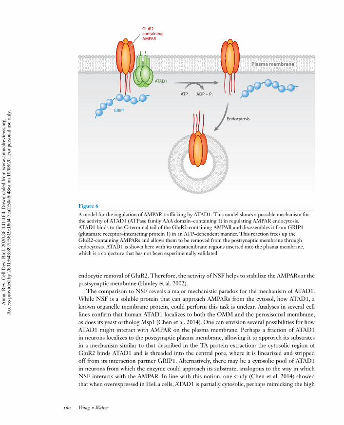
The prevailing theory to explain ATAD1’s neuroprotective function is that it works throughregulating receptor trafficking at the synapse, an activity that is seemingly independent of its func-tion inmitochondrial protein quality control (Zhang et al. 2011b). In particular, ATAD1 is thoughtto regulate the trafficking of the AMPA receptors (AMPARs), the most ubiquitously present neu-rotransmitter (glutamate)-gated ion channel in the nervous system. The number of AMPARsdisplayed at the cell surface determines, in part, the strength of the synapse and is dynamicallyregulated by the receptors’ insertion into and removal from the postsynaptic membrane. Thedysregulation of these processes underlies many neurological disorders, including schizophrenia(Rubio et al. 2012) and epilepsy (Rogawski 2013). Multiple lines of evidence suggest that ATAD1frees the AMPAR from its interaction partner GRIP1 (glutamate receptor–interacting protein 1),a scaffolding protein that stabilizes AMPAR at the postsynaptic membrane, thus licensing the en-docytic removal of the AMPARs (Figure 6) (Zhang et al. 2011b). These lines of evidence are asfollows:
1. Immunoprecipitation experiments in both HEK293 cells and mouse brain lysates showedthatGluR2,one of the four subunits that constitute the tetrameric AMPAR, forms a complexwith ATAD1 and GRIP1;
2. In vitro reconstitution assays with purified components showed that ATAD1 could disas-semble the GluR2-GRIP1 complex in an ATP-dependent manner; and
3. In neurons, the overexpression of ATAD1 (but not ATAD1 bearing mutations that inacti-vated its ATPase activity) caused a decrease in the surface AMPAR level, whereas its deletionled to an increase in surface-displayed AMPARs.
ATAD1 not only regulates the steady state level of surface AMPARs under normal conditionsbut also is required for the active endocytosis of these receptors in response to a stimulus. Whentreated with bicuculline, a molecule that inhibits the GABAA receptor (an inhibitory neurotrans-mitter receptor), wild-type neurons internalized a significant portion of AMPARs (excitatory re-ceptors) from the cell surface. This process, which is called synaptic downscaling, allows neuronsto adjust the levels of neuroreceptors to retarget their baseline activity properties in the face of acontinued perturbation. In ATAD1−/− neurons, however, bicuculline treatment did not affect thesurface AMPAR level, indicating that ATAD1 is required for synaptic downscaling (Zhang et al.2011b).
In addition to the biochemical and cell biological evidence mentioned earlier in this section,the function of ATAD1 in regulating AMPAR trafficking is also supported by pharmacologicalevidence. The treatment of ATAD1−/− mice as well as patients with ATAD1 mutations with anAMPAR antagonist was found to mitigate neurological defects (Ahrens-Nicklas et al. 2017).
The proposed mechanism of action for ATAD1 to unravel the binding interaction betweenGluR2 and GRIP1 closely resembles that of the AAA protein NSF, which has been extensivelycharacterized for its role in disassembling SNARE complexes to free up SNAREproteins for reuse.Similar to the function of ATAD1,NSF disrupts the interaction betweenGluR2 and its interactionpartner PICK1 in an ATP-dependent manner. However, unlike GRIP1, PICK1 facilitates the
www.annualreviews.org • Msp1/ATAD1 in Protein Quality Control 159
Ann
u. R
ev. C
ell D
ev. B
iol.
2020
.36:
141-
164.
Dow
nloa
ded
from
ww
w.a
nnua
lrev
iew
s.or
g A
cces
s pr
ovid
ed b
y 26
01:6
43:8
97f:
5619
:18d
4:7c
a2:5
0a6:
48ea
on
10/0
8/20
. For
per
sona
l use
onl
y.
GluR2-containing AMPAR
Plasma membranePlasma membrane
Endocytosis
ATAD1
GRIP1
ATP ADP + Pi
Figure 6
A model for the regulation of AMPAR trafficking by ATAD1. This model shows a possible mechanism forthe activity of ATAD1 (ATPase family AAA domain–containing 1) in regulating AMPAR endocytosis.ATAD1 binds to the C-terminal tail of the GluR2-containing AMPAR and disassembles it from GRIP1(glutamate receptor–interacting protein 1) in an ATP-dependent manner. This reaction frees up theGluR2-containing AMPARs and allows them to be removed from the postsynaptic membrane throughendocytosis. ATAD1 is shown here with its transmembrane regions inserted into the plasma membrane,which is a conjecture that has not been experimentally validated.
endocytic removal of GluR2. Therefore, the activity of NSF helps to stabilize the AMPARs at thepostsynaptic membrane (Hanley et al. 2002).
The comparison to NSF reveals a major mechanistic paradox for the mechanism of ATAD1.While NSF is a soluble protein that can approach AMPARs from the cytosol, how ATAD1, aknown organelle membrane protein, could perform this task is unclear. Analyses in several celllines confirm that human ATAD1 localizes to both the OMM and the peroxisomal membrane,as does its yeast ortholog Msp1 (Chen et al. 2014). One can envision several possibilities for howATAD1 might interact with AMPAR on the plasma membrane. Perhaps a fraction of ATAD1in neurons localizes to the postsynaptic plasma membrane, allowing it to approach its substratesin a mechanism similar to that described in the TA protein extraction: the cytosolic region ofGluR2 binds ATAD1 and is threaded into the central pore, where it is linearized and strippedoff from its interaction partner GRIP1. Alternatively, there may be a cytosolic pool of ATAD1in neurons from which the enzyme could approach its substrate, analogous to the way in whichNSF interacts with the AMPAR. In line with this notion, one study (Chen et al. 2014) showedthat when overexpressed in HeLa cells, ATAD1 is partially cytosolic, perhaps mimicking the high
160 Wang • Walter
Ann
u. R
ev. C
ell D
ev. B
iol.
2020
.36:
141-
164.
Dow
nloa
ded
from
ww
w.a
nnua
lrev
iew
s.or
g A
cces
s pr
ovid
ed b
y 26
01:6
43:8
97f:
5619
:18d
4:7c
a2:5
0a6:
48ea
on
10/0
8/20
. For
per
sona
l use
onl
y.
ATAD1 expression level in the brain.We note that ATAD1’s N-terminal transmembrane domainis only mildly hydrophobic, adding plausibility to this scenario as it may weaken ATAD1’s mem-brane association and allow it to exist in equilibrium between soluble and membrane-anchoredstates.
In addition to its proposed role of directly catalyzing the dissociation of GluR2 from GRIP1,ATAD1’s neuroprotective activity may result from its role in mitochondrial quality control. Thedeletion of ATAD1 in mice causes mitochondrial fragmentation and mitochondrial protein loss inthe brain (Chen et al. 2014), suggesting that it is critical to maintaining mitochondrial function.As mitochondrial dysfunction can cause neurological diseases such as Parkinson’s disease (Sekine& Youle 2018), the major neurological defects observed in animals and patients with defectiveATAD1 may be attributed, at least partially, to ATAD1-related mitochondrial damage.
5. CONCLUDING REMARKS
Cells spend a significant amount of energy to maintain protein homeostasis. Over the past fewyears, a combination of genetic, cell biological, biochemical, and structural studies have estab-lishedMsp1 as a key regulator of mitochondrial protein homeostasis. Since the initial discovery ofits function (Chen et al. 2014, Okreglak &Walter 2014), the field has made considerable advancesin understanding the mechanism of Msp1-mediated membrane protein extraction. The first ad-vance involved identifying how Msp1 distinguishes the substrate from the nonsubstrate, that isto say, how Msp1 acts precisely on the mislocalized proteins and ignores ones that are correctlytargeted to the OMM (Dederer et al. 2019, Fresenius &Wohlever 2019, Li et al. 2019,Weir et al.2017). The second advance was examining how Msp1 overcomes the energetic barrier of extract-ing hydrophobic sequences from the greasy lipid bilayer (Wang et al. 2020,Wohlever et al. 2017).The third advance was tracking the fate of the extracted TA proteins and identifying the otherplayers involved in this pathway (Dederer et al. 2019, Matsumoto et al. 2019). Going beyond TAprotein extraction, the discovery of the mitoCPR pathway in yeast expanded the function of Msp1to include the regulation ofmitochondrial protein import (Weidberg&Amon 2018); the discoveryof ATAD1 as a neuroprotective molecule expanded ATAD1’s function to the regulation of synap-tic activities (Dai et al. 2010, Zhang et al. 2011b). As the mechanistic details of these processesare lacking, however, how their mechanisms and the mechanism of TA protein extraction can beunified is yet unknown. Finally, it also remains to be understood what role Msp1/ATAD1 playsunder different stress conditions and in different cell types, and how it is integrated into the com-plex cellular stress response network. To this end, defining the full proteome of Msp1/ATAD1’ssubstrates under different cellular contexts will provide valuable insights.
DISCLOSURE STATEMENT
The authors are not aware of any affiliations, memberships, funding, or financial holdings thatmight be perceived as affecting the objectivity of this review.
ACKNOWLEDGMENTS
Wewant to thank Jared Rutter,MatthewWohlever, andHannah Toutkoushian for critical readingof this manuscript. This work was supported by National Institutes of Health grant GM032384to P.W. P.W. is an investigator of the Howard Hughes Medical Institute. L.W. is a fellow of theDamon Runyon Cancer Research Foundation (grant DRG-2312-17).
www.annualreviews.org • Msp1/ATAD1 in Protein Quality Control 161
Ann
u. R
ev. C
ell D
ev. B
iol.
2020
.36:
141-
164.
Dow
nloa
ded
from
ww
w.a
nnua
lrev
iew
s.or
g A
cces
s pr
ovid
ed b
y 26
01:6
43:8
97f:
5619
:18d
4:7c
a2:5
0a6:
48ea
on
10/0
8/20
. For
per
sona
l use
onl
y.
LITERATURE CITED
Ahrens-Nicklas RC,UmanahGKE, SondheimerN,Deardorff MA,Wilkens AB, et al. 2017. Precision therapyfor a new disorder of AMPA receptor recycling due to mutations in ATAD1.Neurol. Genet. 3(1):e130
Akopian D, Shen K, Zhang X, Shan S. 2013. Signal recognition particle: an essential protein-targeting ma-chine. Annu. Rev. Biochem. 82:693–721
Augustin S, Gerdes F, Lee S, Tsai FTF, Langer T, Tatsuta T. 2009. An intersubunit signaling network coor-dinates ATP hydrolysis by m-AAA proteases.Mol. Cell 35(5):574–85
Basch M,Wagner M, Rolland S, Carbonell A, Zeng R, et al. 2020. Msp1 cooperates with the proteasome forextraction of arrested mitochondrial import intermediates.Mol. Biol. Cell 31(8):753–67
Bausewein T, Mills DJ, Langer JD, Nitschke B, Nussberger S, Kühlbrandt W. 2017. Cryo-EM structure ofthe TOM core complex from Neurospora crassa. Cell 170(4):693–700.e7
Beilharz T, Egan B, Silver PA, Hofmann K, Lithgow T. 2003. Bipartite signals mediate subcellular targetingof tail-anchored membrane proteins in Saccharomyces cerevisiae. J. Biol. Chem. 278(10):8219–23
Borgese N, Fasana E. 2011. Targeting pathways of C-tail-anchored proteins. Biochim. Biophys. Acta Biomembr.1808(3):937–46
Burri L, Lithgow T. 2004. A complete set of SNAREs in yeast. Traffic 5(1):45–52Chen Y, Umanah GKE, Dephoure N, Andrabi SA, Gygi SP, et al. 2014. Msp1/ATAD1 maintains mi-
tochondrial function by facilitating the degradation of mislocalized tail-anchored proteins. EMBO J.33(14):1548–64
Chio US, Cho H, Shan S-O. 2017. Mechanisms of tail-anchored membrane protein targeting and insertion.Annu. Rev. Cell Dev. Biol. 33:417–38
Cooney I, Han H, Stewart MG, Carson RH,Hansen DT, et al. 2019. Structure of the Cdc48 segregase in theact of unfolding an authentic substrate. Science 365(6452):502–5
Dai C, Liang D,Li H, Sasaki M,Dawson TM,Dawson VL. 2010. Functional identification of neuroprotectivemolecules. PLOS ONE 5(11):e15008
de la Peña AH, Goodall EA, Gates SN, Lander GC, Martin A. 2018. Substrate-engaged 26S proteasomestructures reveal mechanisms for ATP-hydrolysis–driven translocation. Science 362(6418):eaav0725
Dederer V, Khmelinskii A, Huhn AG, Okreglak V, Knop M, Lemberg MK. 2019. Cooperation of mitochon-drial and ER factors in quality control of tail-anchored proteins. eLife 8:e45506
Denic V. 2012. A portrait of the GET pathway as a surprisingly complicated young man. Trends Biochem. Sci.37(10):411–17
DoanKN,Grevel A,MårtenssonCU,Ellenrieder L,ThorntonN, et al. 2020.Themitochondrial import com-plex MIM functions as main translocase for α-helical outer membrane proteins. Cell Rep. 31(4):107567
Dong Y,Zhang S,WuZ,Li X,WangWL, et al. 2018.Cryo-EM structures and dynamics of substrate-engagedhuman 26S proteasome.Nature 565(7737):49–55
Fresenius HL,Wohlever ML. 2019. Sorting out how Msp1 maintains mitochondrial membrane proteostasis.Mitochondrion 49:128–34
Frickey T, Lupas AN. 2004. Phylogenetic analysis of AAA proteins. J. Struct. Biol. 146(1–2):2–10Gates SN, Yokom AL, Lin J, Jackrel ME, Rizo AN, et al. 2017. Ratchet-like polypeptide translocation mech-
anism of the AAA+ disaggregase Hsp104. Science 357(6348):273–79HanH, Fulcher JM,Dandey VP, Iwasa JH, SundquistWI, et al. 2019. Structure of Vps4 with circular peptides
and implications for translocation of two polypeptide chains by AAA+ ATPases. eLife 8:e44071Han H, Monroe N, Sundquist WI, Shen PS, Hill CP. 2017. The AAA ATPase Vps4 binds ESCRT-III sub-
strates through a repeating array of dipeptide-binding pockets. eLife 6:e31324Hanley JG, Khatri L, Hanson PI, Ziff EB. 2002. NSF ATPase and α-/β-SNAPs disassemble the AMPA
receptor-PICK1 complex.Neuron 34(1):53–67Hanson PI,Whiteheart SW. 2005. AAA+ proteins: have engine, will work.Nat. Rev.Mol. Cell Biol. 6(7):519–29Kalbfleisch T, Cambon A, Wattenberg BW. 2007. A bioinformatics approach to identifying tail-anchored
proteins in the human genome. Traffic 8(12):1687–94
162 Wang • Walter
Ann
u. R
ev. C
ell D
ev. B
iol.
2020
.36:
141-
164.
Dow
nloa
ded
from
ww
w.a
nnua
lrev
iew
s.or
g A
cces
s pr
ovid
ed b
y 26
01:6
43:8
97f:
5619
:18d
4:7c
a2:5
0a6:
48ea
on
10/0
8/20
. For
per
sona
l use
onl
y.
Kornmann B, Currie E, Collins SR, Schuldiner M, Nunnari J, et al. 2009. An ER-mitochondria tetheringcomplex revealed by a synthetic biology screen. Science 325(5939):477–81
Lee J, Kim DH,Hwang I. 2014. Specific targeting of proteins to outer envelope membranes of endosymbioticorganelles, chloroplasts, and mitochondria. Front. Plant Sci. 5:173
Li L, Zheng J, Wu X, Jiang H. 2019. Mitochondrial AAA-ATPase Msp1 detects mislocalized tail-anchoredproteins through a dual-recognition mechanism. EMBO Rep. 20(4):e46989
Mast FD, Rachubinski RA, Aitchison JD. 2020. Peroxisome prognostications: exploring the birth, life, anddeath of an organelle. J. Cell Biol. 219(3):e201912100
Mårtensson CU, Priesnitz C, Song J, Ellenrieder L, Doan KN, et al. 2019. Mitochondrial proteintranslocation-associated degradation.Nature 569(7758):679–83
Matsumoto S,Nakatsukasa K,Kakuta C,Tamura Y, Esaki M, Endo T. 2019.Msp1 clears mistargeted proteinsby facilitating their transfer from mitochondria to the ER.Mol. Cell 76(1):191–205.e10
Nakai M, Endo T, Hase T, Matsubara H. 1993. Intramitochondrial protein sorting. Isolation and charac-terization of the yeast MSP1 gene which belongs to a novel family of putative ATPases. J. Biol. Chem.268(32):24262–69
NeupertW,Herrmann JM. 2007.Translocation of proteins into mitochondria.Annu. Rev. Biochem. 76:723–49Ogura T, Wilkinson AJ. 2001. AAA+ superfamily ATPases: common structure-diverse function. Genes Cells
6(7):575–97Okreglak V,Walter P. 2014. The conserved AAA-ATPase Msp1 confers organelle specificity to tail-anchored
proteins. PNAS 111(22):8019–24Paupe V, Prudent J, Dassa EP, Rendon OZ, Shoubridge EA. 2015. CCDC90A (MCUR1) is a cytochrome c
oxidase assembly factor and not a regulator of themitochondrial calciumuniporter.CellMetab. 21(1):109–16
Pfanner N, Warscheid B, Wiedemann N. 2019. Mitochondrial proteins: from biogenesis to functional net-works.Nat. Rev. Mol. Cell Biol. 20(5):267–84
Piard J,UmanahGKE,Harms FL,Abalde-Atristain L,AmramD, et al. 2018.A homozygous ATAD1mutationimpairs postsynaptic AMPA receptor trafficking and causes a lethal encephalopathy.Brain 141(3):651–61
Puchades C, Ding B, Song A, Wiseman RL, Lander GC, Glynn SE. 2019. Unique structural features of themitochondrial AAA+ protease AFG3L2 reveal the molecular basis for activity in health and disease.Mol.Cell 75(5):1073–85.e6
Puchades C,Rampello AJ, ShinM,GiulianoCJ,WisemanRL, et al. 2017. Structure of themitochondrial innermembrane AAA+ protease YME1 gives insight into substrate processing. Science 358(6363):eaao0464
Puchades C, Sandate CR, Lander GC. 2020. The molecular principles governing the activity and functionaldiversity of AAA+ proteins.Nat. Rev. Mol. Cell Biol. 21(1):43–58
Rao M, Okreglak V, Chio US, Cho H, Walter P, et al. 2016. Multiple selection filters ensure accurate tail-anchored membrane protein targeting. eLife 5:e21301
Rodriguez-Aliaga P, Ramirez L, Kim F, Bustamante C, Martin A. 2016. Substrate-translocating loops regu-late mechanochemical coupling and power production in AAA+ protease ClpXP. Nat. Struct. Mol. Biol.23(11):974–81
Rogawski MA. 2013. AMPA receptors as a molecular target in epilepsy therapy. Acta Neurol. Scand. Suppl.127(197):9–18
Rubio MD,Drummond JB,Meador-Woodruff JH. 2012. Glutamate receptor abnormalities in schizophrenia:implications for innovative treatments. Biomol. Ther. 20(1):1–18
Sato Y, Shibata H, Nakatsu T, Nakano H, Kashiwayama Y, et al. 2010. Structural basis for docking of perox-isomal membrane protein carrier Pex19p onto its receptor Pex3p. EMBO J. 29(24):4083–93
Schmidt F, Treiber N, Zocher G, Bjelic S, Steinmetz MO, et al. 2010. Insights into peroxisome function fromthe structure of PEX3 in complex with a soluble fragment of PEX19. J. Biol. Chem. 285(33):25410–17
Schuldiner M, Metz J, Schmid V, Denic V, Rakwalska M, et al. 2008. The GET complex mediates insertionof tail-anchored proteins into the ER membrane. Cell 134(4):634–45
www.annualreviews.org • Msp1/ATAD1 in Protein Quality Control 163
Ann
u. R
ev. C
ell D
ev. B
iol.
2020
.36:
141-
164.
Dow
nloa
ded
from
ww
w.a
nnua
lrev
iew
s.or
g A
cces
s pr
ovid
ed b
y 26
01:6
43:8
97f:
5619
:18d
4:7c
a2:5
0a6:
48ea
on
10/0
8/20
. For
per
sona
l use
onl
y.
Sekine S,Youle RJ. 2018.PINK1 import regulation; a fine system to conveymitochondrial stress to the cytosol.BMC Biol. 16:2
Söllner T,GriffithsG,Pfaller R,PfannerN,NeupertW.1989.MOM19, an import receptor formitochondrialprecursor proteins. Cell 59(6):1061–70
Twomey EC, Ji Z,Wales TE, Bodnar NO, Ficarro SB, et al. 2019. Substrate processing by the Cdc48 ATPasecomplex is initiated by ubiquitin unfolding. Science 365(6452):eaax1033
Umanah GKE, Pignatelli M, Yin X, Chen R, Crawford J, et al. 2017. Thorase variants are associatedwith defects in glutamatergic neurotransmission that can be rescued by Perampanel. Sci. Transl. Med.9(420):eaah4985
Wallin E,Heijne GV. 2008. Genome-wide analysis of integral membrane proteins from eubacterial, archaean,and eukaryotic organisms. Protein Sci. 7(4):1029–38
Wang L, Myasnikov A, Pan X, Walter P. 2020. Structure of the AAA protein Msp1 reveals mechanism ofmislocalized membrane protein extraction. eLife 9:e54031
Weidberg H, Amon A. 2018. MitoCPR—a surveillance pathway that protects mitochondria in response toprotein import stress. Science 360(6385):eaan4146
Weir NR, Kamber RA, Martenson JS, Denic V. 2017. The AAA protein Msp1 mediates clearance of excesstail-anchored proteins from the peroxisomal membrane. eLife 6:e28507
Wiedemann N, Pfanner N. 2017. Mitochondrial machineries for protein import and assembly. Annu. Rev.Biochem. 86:685–714
Wohlever ML, Mateja A, McGilvray PT, Day KJ, Keenan RJ. 2017. Msp1 is a membrane protein dislocasefor tail-anchored proteins.Mol. Cell 67(2):194–202.e6
Zehr E, Szyk A, Piszczek G, Szczesna E, Zuo X, Roll-Mecak A. 2017. Katanin spiral and ring structures shedlight on power stroke for microtubule severing.Nat. Struct. Mol. Biol. 24(9):717–25
Zhang J, Yang J, Wang H, Sherbini O, Keuss MJ, et al. 2011a. The AAA+ ATPase thorase is neuroprotectiveagainst ischemic injury. J. Cereb. Blood Flow Metab. 39(9):1836–48
Zhang J, Wang Y, Chi Z, Keuss MJ, Pai Y-ME, et al. 2011b. The AAA+ ATPase thorase regulates AMPAreceptor-dependent synaptic plasticity and behavior. Cell 145(2):284–99
164 Wang • Walter
Ann
u. R
ev. C
ell D
ev. B
iol.
2020
.36:
141-
164.
Dow
nloa
ded
from
ww
w.a
nnua
lrev
iew
s.or
g A
cces
s pr
ovid
ed b
y 26
01:6
43:8
97f:
5619
:18d
4:7c
a2:5
0a6:
48ea
on
10/0
8/20
. For
per
sona
l use
onl
y.
CB36_FrontMatter ARjats.cls September 17, 2020 8:35