Journal of Neurology, Neurosurgery, and Psychiatry, 1974, 37, 455-462 Anomalous stress reactions in patients suffering from depression and anxiety HARRY BRIERLEY AND ROBIN JAMIESON From the Professorial Unit of Psychological Medicine, General Hospital, Newcastle upon Tyne SYNOPSIS An experiment is reported in which the forearm muscle blood flow of a group of patients with mixed depressions is compared with that of a group of patients with anxiety states. The blood flow was measured under relaxed conditions and during the presentation of a noise 'stress'. The measurements obtained under stressed conditions show that, while the blood flow of those with anxiety states fell with repetition of the stress noise, that of the depressive patients increased. A similar pattern was shown by the relaxed measurements but here the difference was not statistically significant. There was also suggestion that stress produced a fall in blood flow in depressive subjects and an increase in patients with anxiety states. These results tend to support the hypothesis that depressive patients show something akin to a freeze response to stress, while patients with anxiety states show an arousal response. Many workers have considered the significance of physiological activity levels. Kelly and Walter (1968), for example, have made out a case for regarding forearm muscle blood flow as a measure of anxiety. Duffy (1962), concerned with the whole range of physiological indices, uses the term 'activation' as having similar meaning to 'arousal', 'energy mobilization', and 'excitation'. Lader and Matthews (1968) were more cautious and use the term 'arousal' in a ' non-theoretical way' so that anxiety is an aspect of over-arousal as compared with the simple alertness of moderate arousal. Precisely what aspects of behaviour are represented by changes in physiological indices are not well established and Lader's conservative use of the term 'arousal' appears to reflect best the significance of psychophysiological measure- ments in the present state of knowledge. Increases in these levels apparently brought about by a variety of 'stresses', such as noise, mental arithmetic, and emotion, have commonly been observed. There is a temptation to assume that psychophysiological values represent anxiety in some form, so that where subjects with agitated depression are shown to have higher levels of blood flow than those with retarded depression this has been interpreted as reflecting the higher anxiety component exhibited by the 455 agitated depressives. However, as Kelly and Walter (1968) have shown, 'basal' forearm blood flow values are not very highly related to anxiety as measured by either clinical judgement or conventional test methods, correlations with these standards being of the order of less than 025. It seems, therefore, that despite significant differences in blood flow between various clinical groups as shown by Kelly these differences cannot be attributed to anxiety alone in any conventional sense of the term. Moreover the term 'stresser' in this context does not need to imply any discomfort, since similar increases can be brought about by relatively pleasant stimuli-for example, rhythmical music or erotic fantasies, which appear to affect the level of alertness of the subject. Brierley (1969) reported an investigation of the forearm muscle blood flow of patients suffering from phobic anxiety. Lader and Wing (1964) had suggested that if it were found that phobic patients showed increasing arousal as a result of continued stress a positive feedback cycle might be produced. That is to say, the more aroused the subject became the greater would be the arousal effect of a given stimulus. Such a situation could well be the mechanism of phobic panics. The findings in Brierley's experiment were anomalous. Phobic patients showed a Protected by copyright. on December 18, 2021 by guest. http://jnnp.bmj.com/ J Neurol Neurosurg Psychiatry: first published as 10.1136/jnnp.37.4.455 on 1 April 1974. Downloaded from
Transcript
Journal of Neurology, Neurosurgery, and Psychiatry, 1974, 37, 455-462
Anomalous stress reactions in patients sufferingfrom depression and anxiety
HARRY BRIERLEY AND ROBIN JAMIESON
From the Professorial Unit ofPsychological Medicine, General Hospital, Newcastle upon Tyne
SYNOPSIS An experiment is reported in which the forearm muscle blood flow ofa group of patientswith mixed depressions is compared with that of a group of patients with anxiety states. The bloodflow was measured under relaxed conditions and during the presentation of a noise 'stress'. Themeasurements obtained under stressed conditions show that, while the blood flow of those withanxiety states fell with repetition of the stress noise, that of the depressive patients increased. Asimilar pattern was shown by the relaxed measurements but here the difference was not statisticallysignificant. There was also suggestion that stress produced a fall in blood flow in depressive subjectsand an increase in patients with anxiety states. These results tend to support the hypothesis thatdepressive patients show something akin to a freeze response to stress, while patients with anxietystates show an arousal response.
Many workers have considered the significanceof physiological activity levels. Kelly and Walter(1968), for example, have made out a case forregarding forearm muscle blood flow as a
measure of anxiety. Duffy (1962), concernedwith the whole range of physiological indices,uses the term 'activation' as having similarmeaning to 'arousal', 'energy mobilization', and'excitation'. Lader and Matthews (1968) were
more cautious and use the term 'arousal' in a' non-theoretical way' so that anxiety is an
aspect of over-arousal as compared with thesimple alertness of moderate arousal. Preciselywhat aspects of behaviour are represented bychanges in physiological indices are not wellestablished and Lader's conservative use of theterm 'arousal' appears to reflect best thesignificance of psychophysiological measure-ments in the present state ofknowledge. Increasesin these levels apparently brought about by a
variety of 'stresses', such as noise, mentalarithmetic, and emotion, have commonly beenobserved. There is a temptation to assume thatpsychophysiological values represent anxiety insome form, so that where subjects with agitateddepression are shown to have higher levels ofblood flow than those with retarded depressionthis has been interpreted as reflecting thehigher anxiety component exhibited by the
455
agitated depressives. However, as Kelly andWalter (1968) have shown, 'basal' forearmblood flow values are not very highly related toanxiety as measured by either clinical judgementor conventional test methods, correlations withthese standards being of the order of less than025. It seems, therefore, that despite significantdifferences in blood flow between variousclinical groups as shown by Kelly thesedifferences cannot be attributed to anxiety alonein any conventional sense of the term. Moreoverthe term 'stresser' in this context does not needto imply any discomfort, since similar increasescan be brought about by relatively pleasantstimuli-for example, rhythmical music orerotic fantasies, which appear to affect the levelof alertness of the subject.
Brierley (1969) reported an investigation of theforearm muscle blood flow of patients sufferingfrom phobic anxiety. Lader and Wing (1964) hadsuggested that if it were found that phobicpatients showed increasing arousal as a result ofcontinued stress a positive feedback cycle mightbe produced. That is to say, the more arousedthe subject became the greater would be thearousal effect of a given stimulus. Such asituation could well be the mechanism of phobicpanics. The findings in Brierley's experimentwere anomalous. Phobic patients showed a
Protected by copyright.
on Decem
ber 18, 2021 by guest.http://jnnp.bm
j.com/
J Neurol N
eurosurg Psychiatry: first published as 10.1136/jnnp.37.4.455 on 1 A
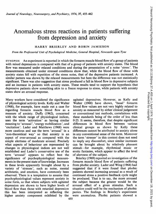
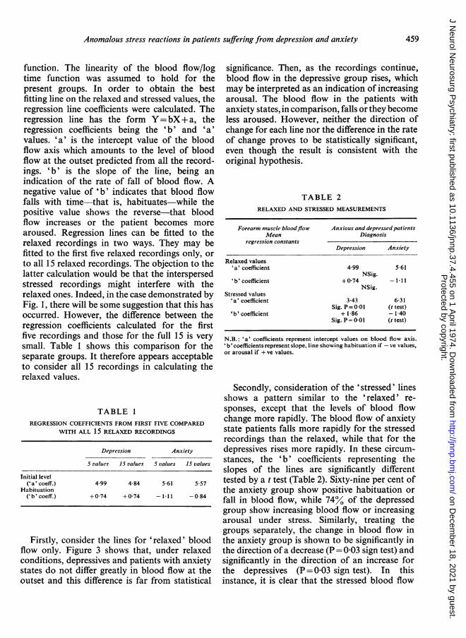
FIG. 1. Mrs. E. Stressedand unstressed bloodflowlevels.
smaller increase in forearm muscle blood flowas a result of exposure to a noise stimulus as astresser than did normal subjects. Also repeatedblood flow measurements showed a decreasewith time and repetition of the stresser-that is,they showed habituation. Thus, the hypothesis ofpositive feedback was not supported by thatexperiment.
It might have been anticipated that a noisestresser would produce an increase in blood flowlike that resulting from stressed mental arithmeticwhich Kelly (1966) has reported. This would beparallel with the positive arousal increasesshown by Lader and Wing (1964) for thepsychogalvanic reflex. However, a continuationof Brierley's investigation found a number ofpatients who exhibited an apparently anomalousfall in blood flow under experimental stress,compared with their relaxed levels. Three otherfactors seemed associated with this phenomenon.Firstly. these patients appeared to exhibit moredepressed affect. Secondly, unlike the patientsreported in Brierley (1969), the blood flow inthese patients actually increased with time duringthe measurement session that is, they becamemore aroused and did not habituate. Thirdly, a
number of these patients became upset andcontinuation of the measurement appearedlikely to give rise to panic.
A record demonstrating forearm blood flow,measured during the presentation of a noisestresser, consistently below the relaxed level ofblood flow is shown in Fig. 1. Here the stressedreadings have an average of 7-30 0/min ascompared with the relaxed level of 10.30/ min.(This difference is statistically significant astested by the Mann-Whitney U Test).
This patient was a 44 year old woman whosediagnosis was 'a depressive illness in a woman ofan obsessional personality who has a tendency tobe rigid and anxiety prone'. She had been underintermittent psychiatric care for over 12 yearsand initially she was believed to suffer from arecurrent endogenous depression. More recently,hypochondriacal preoccupations, complaints ofdefective eyesight, low back pain, and genito-urinary problems, etc, had resulted in examina-tions by numerous departments. She was arather aggressive lady who steadfastly refused toaccept herself as suffering from any psychiatricdisability. At times she threatened suicide anddemanded 'an operation for the pain'. Sheexpressed dissatisfaction with all doctors whoexamined and treated her and refused to remainin hospital for treatment. Her husband, abuilder's labourer, reported that her moodchanged dramatically at times. Normally, sheenjoyed going with him to a club and she liked
Anomalous stress reactions in patients suffering from depression and anxiety
playing the piano. When she was ill she foughtwith him, accused him of being alcoholic and agambler. She would also believe she was clair-voyant and at times her suspicious behaviourcaused family difficulties.
Figure 1 shows, in addition, the way that Mrs.E's forearm blood flow increased rapidly overthe first five recordings in contrast with the wayin which a normal record would demonstrate afall in blood flow.
These observations were reported by Jamieson(1970). He postulated that the increased arousalresponse to a stress signal was akin to the familiarmobilization-for-flight reaction in fear, while theresponse of reduced arousal level was of the'freeze' type. Such freeze behaviour in fear isfamiliar in the animal kingdom and, of course,even humans describe themselves as 'petrified'or 'frozen to the spot' in fear. However, there islittle, if any, existing evidence of the existence ofsuch freeze responses in humans but the evidenceJamieson presented seemed to illustrate a patternof physiological stress response of this type.The upshot of these observations was that it
was decided to test two hypotheses:1. That patients suffering from depression do
not show the habituation of forearm muscleblood flow exhibited by patients with anxietystates but show an increase in flow during themeasurement period.
2. That patients with anxiety states respond toa stress with an increase in forearm blood flowwhile depressives respond with a reduction inflow level.
METHOD
Forearm muscle blood flow is measured by occludingthe venous return of blood from the forearm by asphygmomanometer cuff fitted to the upper arm andmeasuring the increase in volume in the forearm. Itis necessary to fit a cuff at the wrist to occlude the
arterial flow to the hand, since the flow to the hand isprimarily through the skin rather than muscle. Thevolume increase of the forearm is measured in avariety of ways. Most workers-for example, Kelly-have employed the waterbath plethysmographwhich encases the forearm in a metal tank and hencelargely immobilizes the patient. The techniqueemployed by Whitney (1953) uses a mercury-in-rubber strain gauge. This is a 0*5 mm bore siliconerubber tube filled with mercury and placed round theforearm. An increase in forearm circumferenceresults in a change in electrical resistance in themercury and this change can be calibrated to givethe change in the arm circumference. Making theassumption that the arm is roughly cylindrical, thevolume change can be calculated from the circum-ference change. Whitney has provided evidence thatthis method yields results which are very similar tothe water-bath technique.
This strain gauge method was used for theexperiment. It should not be forgotten that mostpatients view such procedures as to some extentstressful and, if valid estimates of relaxed blood floware sought, the less elaborate the apparatus and thepreparation the better. The setting up of the twocuffs and the strain gauge on the patient is a matter ofnot more than one or two minutes and is quite with-out discomfort, restricting the patient only to a verysmall degree.
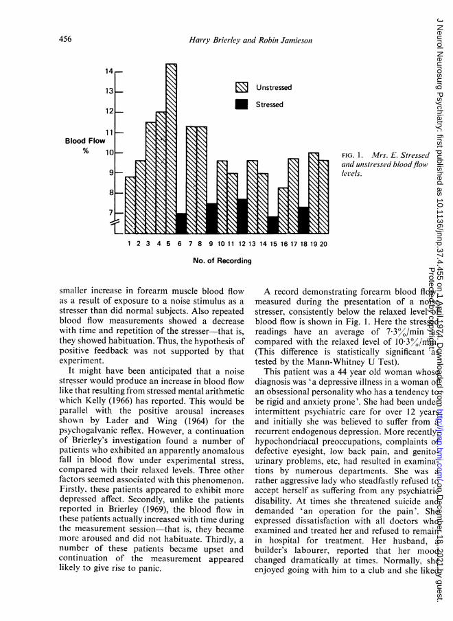
In the present experiment the patient lay on adivan bed in a separate dimly illuminated and sound-proofed room with all the recording apparatusisolated in an adjoining laboratory. The patient wasobserved through a one-way screen, a two-wayintercom providing communication when necessary.Measurements were made on the left arm which wassupported by pillows.The sequence of relaxed and stressed measure-
ments is shown in Fig. 2. This shows that eachrecording sequence begins with a 30 second restperiod. The distal cuff then inflates to a pressure of180 mm mercury. Fifteen seconds later the proximalcuff inflates to 80 mm mercury and the recordingbegins simultaneously. At each third recording fromthe sixth onwards the stress signal also commences
with the inflation of the upper cuff. The pressuresemployed were found by experiment to be the mostgenerally satisfactory.Thus 15 relaxing recordings and five stress
recordings were obtained, as in Brierley (1969), butthe total time was shortened from 30 to 20 minutesby limiting the actual recording time. The stresserused in the previous experiment was the amplifiedsound of a ticking clock but experiments showedthat a pure tone of 1,000 Hz at approximately 80decibels could be substituted. As such a signal wasmore readily reproduced it was adopted.
SUBJECTS The subjects were 19 depressed patientsand 29 suffering from anxiety states. The mean age ofthe anxiety state patients was 31 6 years, SD= 12-8and the mean age of the depressed group was 38-7years, SD= 9 7. Again, as in the previous investiga-tions, no evidence was found of a relationshipbetween age and forearm blood flow within thesegroups; none of the blood flow parameters usedcorrelated higher than 0-19 with age. The depressedgroup was composed of 11 males and eight females,and the anxiety group 10 males and 19 females.The patients were referred by a number of depart-
mental consultant psychiatrists and the clinicaldiagnosis was accepted as defining these two groups.No attempt was made to examine intra-groupdifferences-for example, between generalizedanxieties and phobias or between endogenous and
0
00
0
o-0
3
2
1
reactive depressions. A wide range of types anddegrees of anxiety and depression was included butall were regarded as materially disabled patients. Itwas not, of course, desirable to discontinue treatmentfor the present research so almost all subjects werecurrently taking psychotropic drugs of some kind.Many also would have night sedation prescribed.The number and variety of such drugs was such as toprevent any meaningful analysis. However, patientsunder treatment with electroconvulsive therapy wereavoided, although a number subsequently receivedthis treatment. The groups were, therefore, fairlyrepresentative of patients under treatment fordepression or anxiety in an acute psychiatric unit.The patients were informed of the research nature
of the experiment and instructed to relax if possible.They were forewarned that they would hear a noiselike the whistle of a TV set and this was explained asbeing used to ensure that they were awake during therecording. Patients who were severely disturbed orclearly fearful of the experimental conditions wereexcluded.
RESULTS
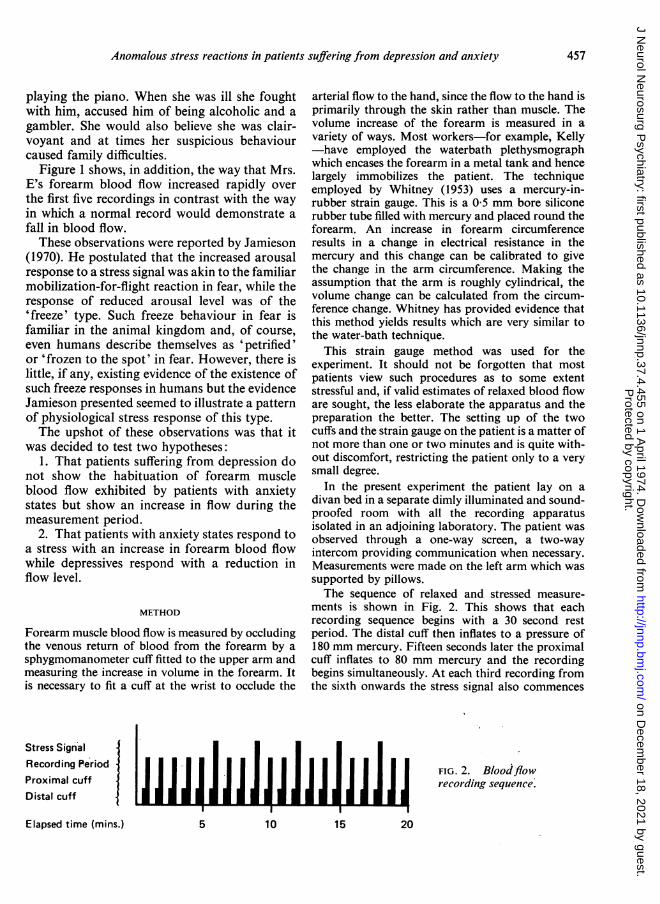
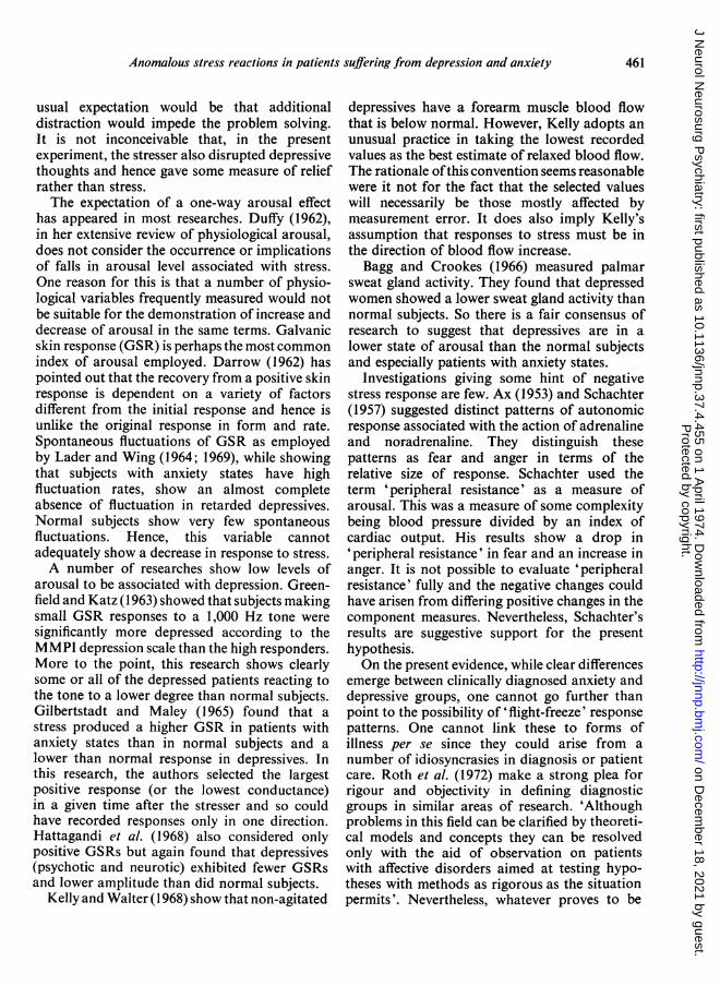
As shown by Brierley (1969), the forearm bloodflow level for the anxiety group showed atendency to fall throughout the recording session.If plotted as blood flow against log time scale,this fall in blood flow appeared as a straight line
FIG. 3. Habituation curves offorearm bloodflow in patients withanxiety states and depression.
I II I I I III1 2 3 4 5 10 20
log tine units
458
Protected by copyright.
on Decem
ber 18, 2021 by guest.http://jnnp.bm
j.com/
J Neurol N
eurosurg Psychiatry: first published as 10.1136/jnnp.37.4.455 on 1 A
Anomalous stress reactions in patients suffering from depression and anxiety
function. The linearity of the blood flow/logtime function was assumed to hold for thepresent groups. In order to obtain the bestfitting line on the relaxed and stressed values, theregression line coefficients were calculated. Theregression line has the form Y=bX+ a, theregression coefficients being the 'b' and 'a'values. 'a' is the intercept value of the bloodflow axis which amounts to the level of bloodflow at the outset predicted from all the record-ings. 'b' is the slope of the line, being anindication of the rate of fall of blood flow. Anegative value of 'b' indicates that blood flowfalls with time-that is, habituates-while thepositive value shows the reverse-that bloodflow increases or the patient becomes morearoused. Regression lines can be fitted to therelaxed recordings in two ways. They may befitted to the first five relaxed recordings only, orto all 15 relaxed recordings. The objection to thelatter calculation would be that the interspersedstressed recordings might interfere with therelaxed ones. Indeed, in the case demonstrated byFig. 1, there will be some suggestion that this hasoccurred. However, the difference between theregression coefficients calculated for the firstfive recordings and those for the full 15 is verysmall. Table 1 shows this comparison for theseparate groups. It therefore appears acceptableto consider all 15 recordings in calculating therelaxed values.
TABLE 1
REGRESSION COEFFICIENTS FROM FIRST FIVE COMPAREDWITH ALL 15 RELAXED RECORDINGS
Depression Anxiety
S values 15 values 5 values 15 values
Initial level('a' coeff.) 4 99 4-84 5-61 5 57
Habituation('b'coeff.) + 0 74 + 0 74 -1 11 -0-84
Firstly, consider the lines for 'relaxed' bloodflow only. Figure 3 shows that, under relaxedconditions, depressives and patients with anxietystates do not differ greatly in blood flow at theoutset and this difference is far from statistical
significance. Then, as the recordings continue,blood flow in the depressive group rises, whichmay be interpreted as an indication of increasingarousal. The blood flow in the patients withanxiety states, in comparison, falls or they becomeless aroused. However, neither the direction ofchange for each line nor the difference in the rateof change proves to be statistically significant,even though the result is consistent with theoriginal hypothesis.
TABLE 2RELAXED AND STRESSED MEASUREMENTS
Forearm muscle bloodflow Anxious and depressed patientsMean Diagnosis
regression constantsDepression Anxiety
Relaxed values'a' coefficient 4 99 5-61
NSig.'b' coefficient + 0 74 -111
NSig.Stressed values
'a' coefficient 3-43 6-31Sig. P= 0 01 (t test)
'b' coefficient + 1-86 -1-40Sig. P= 0 01 (t test)
N.B.: 'a' coefficients represent intercept values on blood flow axis.'b'coefficients represent slope, line showing habituation if -ve values,or arousal if + ve values.
Secondly, consideration of the 'stressed' linesshows a pattern similar to the 'relaxed' re-sponses, except that the levels of blood flowchange more rapidly. The blood flow of anxietystate patients falls more rapidly for the stressedrecordings than the relaxed, while that for thedepressives rises more rapidly. In these circum-stances, the 'b' coefficients representing theslopes of the lines are significantly differenttested by a t test (Table 2). Sixty-nine per cent ofthe anxiety group show positive habituation orfall in blood flow, while 74%0 of the depressedgroup show increasing blood flow or increasingarousal under stress. Similarly, treating thegroups separately, the change in blood flow inthe anxiety group is shown to be significantly inthe direction of a decrease (P= 003 sign test) andsignificantly in the direction of an increase forthe depressives (P= 003 sign test). In thisinstance, it is clear that the stressed blood flow
459
Protected by copyright.
on Decem
ber 18, 2021 by guest.http://jnnp.bm
j.com/
J Neurol N
eurosurg Psychiatry: first published as 10.1136/jnnp.37.4.455 on 1 A
change differentiates between the depressives andthe patients with anxiety states.
Thirdly, one can examine the direction of theresponse to stress. The 'a' coefficients of theregression lines represent the initial level ofblood flow predicted from the complete patternof measurements throughout the session. Thus,the 'a' coefficients probably represent the bestindex of the initial level of blood flow understress and under relaxation conditions and willindicate the direction of change of blood flow asa result of the stress signal and as the patientresponds at the beginning of the recordings.Figure 3 suggests that the stress produces a fall inthe forearm blood flow of depressives and a risein anxiety states as was hypothesized. However,statistical tests of the direction of the change inblood flow from relaxed to stressed conditionsassessed like this just failed to reach statisticalsignificance. In the depressed group, 5300 ofsubjects showed a fall in blood flow associatedwith stress, while only 2800 of patients withanxiety states showed a fall in blood flow.
Finally, one may look at the initial blood flowlevels as measured by regression 'a' coefficients.Under relaxed conditions the initial blood flowswere not significantly different but again understressed conditions the differing responses tostress result in a divergence of the initial bloodflow levels of the two groups. Thus, theseestimates of the initial stressed response show alevel significantly higher in the anxiety groupthan in the depressive group as shown by a t test(Table 2). Of course, as the measurementscontinue and the blood flow of one group fallsand the other rises, this difference disappears asshown in Fig. 3.
DISCUSSION
The evidence presented here indicates differencesbetween the patterns of forearm blood flowunder stress exhibited by patients with anxietystates and depression. Interpretation of thesedifferences is difficult at this stage. It cannot besaid how far these features are transient sequelaeof illness or how far they are representative offundamental modes of adaptation of the subjectsinvolved. The differences found may be per-manent predispositions associated with theappearance of symptoms of depression or
anxiety under stress, subjects tending to becomeover-aroused under stress suffering from anxietystates, while those who show a lessening ofarousal level suffer depression, possibly withretardation.
Previous work on psychophysiology has notreported this effect. However, close examinationof other researches finds similar results obscuredeither by the particular physiological variablesemployed, the measurement techniques, or theunderlying expectations in the experiment.Dykman et al. (1968) have pointed to the
fact that a field of research so apparentlyobjective as psychophysiological measurement'is remarkable for the results which are in-conclusive, contradicting or paradoxical...'.One of the most curious features in suchresearches is the consistency of the assumptionthat the subject not subjected to a controlledlaboratory stress is, in fact, in a 'no stress'condition. This is manifestly untrue and thepresent results show variations of arousal levelto occur over a period of 20 minutes which arenot a direct result of the planned experimentalstress. This being so, there is certainly no reasonto make the very common assumption that allresponses to a deliberately applied stress musthave the form of an increase in arousal. In fact,the response might be either a rise or a fall in thecurrent level of arousal which might suggest amore complex repertoire of human stressresponse with 'flight' and 'freeze' characteristics.Clearly, the present result would lead one toexpect conflicting research findings if the changesassociated with adaptation to the experimentare not taken into account. No measurement ofrelaxation and stress response over a short periodof time and neglecting the pattern of change isadequate. It, therefore, is far from simple toensure that experiments in psychophysiologyemploying stress of any kind can be regarded asquite comparable. The full specification of theregression functions as here seems important atleast.
In other fields, Foulds (1952) and Hethering-ton (1956) have provided some evidence that thedisrupting of the 'flood of depressive thoughts'in the depressed patient improves his mentalfunctioning. Foulds, for instance, showed thatthe tick of a metronome improved the testperformance of depressives where perhaps the
460
Protected by copyright.
on Decem
ber 18, 2021 by guest.http://jnnp.bm
j.com/
J Neurol N
eurosurg Psychiatry: first published as 10.1136/jnnp.37.4.455 on 1 A
Anomalous stress reactions in patients suffering from depression and anxiety
usual expectation would be that additionaldistraction would impede the problem solving.It is not inconceivable that, in the presentexperiment, the stresser also disrupted depressivethoughts and hence gave some measure of reliefrather than stress.The expectation of a one-way arousal effect
has appeared in most researches. Duffy (1962),in her extensive review of physiological arousal,does not consider the occurrence or implicationsof falls in arousal level associated with stress.One reason for this is that a number of physio-logical variables frequently measured would notbe suitable for the demonstration of increase anddecrease of arousal in the same terms. Galvanicskin response (GSR) is perhaps the mostcommonindex of arousal employed. Darrow (1962) haspointed out that the recovery from a positive skinresponse is dependent on a variety of factorsdifferent from the initial response and hence isunlike the original response in form and rate.Spontaneous fluctuations of GSR as employedby Lader and Wing (1964; 1969), while showingthat subjects with anxiety states have highfluctuation rates, show an almost completeabsence of fluctuation in retarded depressives.Normal subjects show very few spontaneousfluctuations. Hence, this variable cannotadequately show a decrease in response to stress.A number of researches show low levels of
arousal to be associated with depression. Green-field and Katz (1963) showed that subjects makingsmall GSR responses to a 1,000 Hz tone weresignificantly more depressed according to theMMPI depression scale than the high responders.More to the point, this research shows clearlysome or all of the depressed patients reacting tothe tone to a lower degree than normal subjects.Gilbertstadt and Maley (1965) found that astress produced a higher GSR in patients withanxiety states than in normal subjects and alower than normal response in depressives. Inthis research, the authors selected the largestpositive response (or the lowest conductance)in a given time after the stresser and so couldhave recorded responses only in one direction.Hattagandi et al. (1968) also considered onlypositive GSRs but again found that depressives(psychotic and neurotic) exhibited fewer GSRsand lower amplitude than did normal subjects.
Kelly and Walter (1968) show that non-agitated
depressives have a forearm muscle blood flowthat is below normal. However, Kelly adopts anunusual practice in taking the lowest recordedvalues as the best estimate of relaxed blood flow.The rationale ofthis convention seems reasonablewere it not for the fact that the selected valueswill necessarily be those mostly affected bymeasurement error. It does also imply Kelly'sassumption that responses to stress must be inthe direction of blood flow increase.Bagg and Crookes (1966) measured palmar
sweat gland activity. They found that depressedwomen showed a lower sweat gland activity thannormal subjects. So there is a fair consensus ofresearch to suggest that depressives are in alower state of arousal than the normal subjectsand especially patients with anxiety states.
Investigations giving some hint of negativestress response are few. Ax (1953) and Schachter(1957) suggested distinct patterns of autonomicresponse associated with the action of adrenalineand noradrenaline. They distinguish thesepatterns as fear and anger in terms of therelative size of response. Schachter used theterm 'peripheral resistance' as a measure ofarousal. This was a measure of some complexitybeing blood pressure divided by an index ofcardiac output. His results show a drop in'peripheral resistance' in fear and an increase inanger. It is not possible to evaluate 'peripheralresistance' fully and the negative changes couldhave arisen from differing positive changes in thecomponent measures. Nevertheless, Schachter'sresults are suggestive support for the presenthypothesis.On the present evidence, while clear differences
emerge between clinically diagnosed anxiety anddepressive groups, one cannot go further thanpoint to the possibility of 'flight-freeze' responsepatterns. One cannot link these to forms ofillness per se since they could arise from anumber of idiosyncrasies in diagnosis or patientcare. Roth et al. (1972) make a strong plea forrigour and objectivity in defining diagnosticgroups in similar areas of research. 'Althoughproblems in this field can be clarified by theoreti-cal models and concepts they can be resolvedonly with the aid of observation on patientswith affective disorders aimed at testing hypo-theses with methods as rigorous as the situationpermits'. Nevertheless, whatever proves to be
461
Protected by copyright.
on Decem
ber 18, 2021 by guest.http://jnnp.bm
j.com/
J Neurol N
eurosurg Psychiatry: first published as 10.1136/jnnp.37.4.455 on 1 A
the significance of a particular pattern ofpsychophysiological arousal characterizing a
clinical group, it seems quite possible that it maybe among the most objective features of thatgroup.
CONCLUSIONS
The present paper shows that under the influenceof a 1,000 Hz stress forearm muscle blood flowdifferentiates groups of mixed depressives andanxiety states. The depressives at the outsetshow lower blood flow values but, as the measure-ment session continues, these rise, while those ofthe patients with anxiety states fall. There is a
suggestion that depressives show a fall in bloodflow from their non-stressed level, in response tothe stress, compared with the increase shown bythose with anxiety states. These differing patternsillustrate the possibility of flight-freeze patternsof stress response in clinical subjects.The whole investigation emphasizes the need
for precise delineation of diagnostic groups aswell as the impracticability of properly defininglevels of arousal by psychophysiological methodsunless the variation over a material period oftime is considered.
REFERENCES
Ax, A. F. (1953). The physiological differentiation betweenfear and anger in humans. Psychosomatic Medicine, 15,433-442.
Bagg, C. E., and Crookes, T. G. (1966). Palmar digitalsweating in women suffering from depression. BritishJournal of Psychiatry, 112, 1251-1255.
Brierley, H. (1969). The habituation of forearm muscle bloodflow in phobic subjects. Journal ofNeurology, Neurosurgery,and Psychiatry, 32, 15-20.
Darrow, C. W. (1962). The rationale for treating the change in
GSR as a change in conductance. Paper read to Symposiumon Problems in Electrodermal Measurement. AmericanPsychological Association, St. Louis.
Dykman, R. A., Reese, W. G., Galbrecht, C. R., Ackerman,P. T., and Sundermann, R. S. (1968). Autonomic responsesin psychiatric patients. Annals of the New York Academy ofScience, 147, 237-303.
Duffy, E. (1962). Activation and Behavior. Wiley: New York.Foulds, G. A. (1952). Temperamental differences in maze
performance. Part 2. British Journal of Psychology, 43,33-41.
Gilberstadt, H., and Maley, M. (1965). GSR, clinical stateand psychiatric diagnosis. Journal of Clinical Psychology,21, 235-238.
Greenfield, N. S., Katz, D., Alexander, A. A., and Roessler,R. (1963). The relationship between physiological andpsychological responsivity: depression and the galvanicskin response. Journal ofNervous and Mental Disease, 136,535-539.
Hattagandi, S., Lidsky, A., Lee, H., and Ban, T. A. (1968).Orienting-reflex behavior and clinical psychopathology.Conditional Reflex, 3, 29-33.
Hetherington, R. (1956). The effects of E.C.T. on the efficiencyand retentivity of depressed patients. British Journal ofMedical Psychology, 29, 258-269.
Jamieson, R. (1970). Orienting reflexes and the stress responsein the measurement of anxiety. Paper read to LondonConference of British Psychological Society.
Kelly, D. H. W. (1966). Measurement of anxiety by forearmblood flow. British Journal of Psychiatrv, 112, 789-798.
Kelly, D. H. W., and Walter, C. J. S. (1968). The relationshipbetween clinical diagnosis and anxiety, assessed by forearmblood flow and other measurements. British Journal ofPsychiatry, 114, 611-626.
Lader, M. H., and Wing, L. (1964). Habituation of thepsycho-galvanic reflex in patients with anxiety states and innormal subjects. Journal of Neuirology, Neurosurgery, andPsychiatry, 27, 210-218.
Lader, M. H., and Wing, L. (1969). Physiological measures inagitated and retarded depressed patients. Journal ofPsychiatric Research, 7, 89-100.
Lader, M. H., and Mathews, A. M. (1968). A physiologicalmodel of phobic anxiety and desensitization. BehaviourResearch and Therapy, 6, 411-421.
Roth, M., Gurney, C., Garside, R. F., and Kerr, T. A. (1972).Studies in the classification of affective disorders. BritishJournal of Psychiatry, 121, 147-161.
Schachter, J. (1957). Pain, fear, and anger in hypertensivesand normotensives. Psychosomatic Medicine, 19, 17-29.
Whitney, R. J. (1953). The measurement ofvolume changes inhuman limbs. Journal of Physiology, 121, 1-27.
462
Protected by copyright.
on Decem
ber 18, 2021 by guest.http://jnnp.bm
j.com/
J Neurol N
eurosurg Psychiatry: first published as 10.1136/jnnp.37.4.455 on 1 A