Studies of patients with semantic impairment suggest that the most anterior portions of the temporal cortices critically support human conceptual knowledge. The purest documented semantic syndrome, semantic dementia (SD; Hodges, Patterson, Oxbury, & Funnell, 1992; Snowden, Goulding, & Neary, 1989), arises from progressive dete- rioration of the anterior temporal cortex, frequently more pronounced in the left hemisphere, but always involving both (Davies, Graham, Xuereb, Williams, & Hodges, 2004; Mummery et al., 2000). Patients with SD are impaired on any task requiring knowledge about the meanings of words and objects, including picture naming, word–picture match- ing, category and property verification (Rogers et al., 2004; Rogers, Watling, Hodges, & Patterson, 2005; Snowden et al., 1989; Warrington, 1975), matching pictures or words on the basis of thematic associations (Hodges, Graham, & Patterson, 1995), sorting words or pictures (Hodges et al., 1992; Rogers et al., 2004), drawing-to-name and delayed copying of drawings of familiar objects (Bozeat et al., 2003; Rogers et al., 2004), sound–picture matching (Bozeat, Lambon Ralph, Patterson, Garrard, & Hodges, 2000), demonstrating the correct use of objects (Hodges, Bozeat, Lambon Ralph, Patterson, & Spatt, 2000), object reality decision (Rogers, Lambon Ralph, Hodges, & Patterson, 2003), and so on (see Patterson & Hodges, 2000). These deficits are typically observed for all semantic categories (Garrard, Lambon Ralph, & Hodges, 2002) and are appar- ent in all modalities of testing but are specific to semantic knowledge; other cognitive faculties are reasonably well preserved in the disorder (Hodges, Garrard, & Patterson, 1998). The striking consistency of both the cognitive and the neural abnormalities in SD strongly suggests that the 201 Copyright 2006 Psychonomic Society, Inc. Correspondence concerning this article should be addressed to T. T. Rogers, Department of Psychology, University of Wisconsin, W. J. Brogden Hall, 1202 W. Johnson St., Madison, WI 53702 (e-mail: [email protected]). Anterior temporal cortex and semantic memory: Reconciling findings from neuropsychology and functional imaging TIMOTHY T. ROGERS MRC Cognition and Brain Sciences Unit, Cambridge, England and University of Wisconsin, Madison, Wisconsin JULIA HOCKING and UTA NOPPENEY Functional Imaging Laboratory, London, England ANDREA MECHELLI Functional Imaging Laboratory, London, England and King’s College London, London, England MARIA LUISA GORNO-TEMPINI University of California, San Francisco, California KARALYN PATTERSON MRC Cognition and Brain Sciences Unit, Cambridge, England and CATHY J. PRICE Functional Imaging Laboratory, London, England Studies of semantic impairment arising from brain disease suggest that the anterior temporal lobes are critical for semantic abilities in humans; yet activation of these regions is rarely reported in functional imaging studies of healthy controls performing semantic tasks. Here, we combined neuropsychological and PET functional imaging data to show that when healthy subjects identify concepts at a specific level, the regions activated correspond to the site of maximal atrophy in patients with relatively pure semantic impairment. The stimuli were color photographs of common animals or vehicles, and the task was cat- egory verification at specific (e.g., robin), intermediate (e.g., bird), or general (e.g., animal ) levels. Spe- cific, relative to general, categorization activated the antero-lateral temporal cortices bilaterally, despite matching of these experimental conditions for difficulty. Critically, in patients with atrophy in precisely these areas, the most pronounced deficit was in the retrieval of specific semantic information. Cognitive, Affective, & Behavioral Neuroscience 2006, 6 (3), 201-213
Transcript
Studies of patients with semantic impairment suggest that the most anterior portions of the temporal cortices critically support human conceptual knowledge The purest documented semantic syndrome semantic dementia (SD Hodges Patterson Oxbury amp Funnell 1992 Snowden Goulding amp Neary 1989) arises from progressive dete-rioration of the anterior temporal cortex frequently more pronounced in the left hemisphere but always involving both (Davies Graham Xuereb Williams amp Hodges 2004 Mummery et al 2000) Patients with SD are impaired on any task requiring knowledge about the meanings of words and objects including picture naming wordndashpicture match-ing category and property verification (Rogers et al 2004
Rogers Watling Hodges amp Patterson 2005 Snowden et al 1989 Warrington 1975) matching pictures or words on the basis of thematic associations (Hodges Graham amp Patterson 1995) sorting words or pictures (Hodges et al 1992 Rogers et al 2004) drawing-to-name and delayed copying of drawings of familiar objects (Bozeat et al 2003 Rogers et al 2004) soundndashpicture matching (Bozeat Lambon Ralph Patterson Garrard amp Hodges 2000) demonstrating the correct use of objects (Hodges Bozeat Lambon Ralph Patterson amp Spatt 2000) object reality decision (Rogers Lambon Ralph Hodges amp Patterson 2003) and so on (see Patterson amp Hodges 2000) These deficits are typically observed for all semantic categories (Garrard Lambon Ralph amp Hodges 2002) and are appar-ent in all modalities of testing but are specific to semantic knowledge other cognitive faculties are reasonably well preserved in the disorder (Hodges Garrard amp Patterson 1998) The striking consistency of both the cognitive and the neural abnormalities in SD strongly suggests that the
201 Copyright 2006 Psychonomic Society Inc
Correspondence concerning this article should be addressed to T T Rogers Department of Psychology University of Wisconsin W J Brogden Hall 1202 W Johnson St Madison WI 53702 (e-mail ttrogerswiscedu)
Anterior temporal cortex and semantic memory Reconciling findings from neuropsychology
and functional imaging
TimoThy T RogeRsMRC Cognition and Brain Sciences Unit Cambridge England
and University of Wisconsin Madison Wisconsin
JuliA hocking and uTA noppeneyFunctional Imaging Laboratory London England
AndReA mechelliFunctional Imaging Laboratory London England
and Kingrsquos College London London England
mARiA luisA goRno-TempiniUniversity of California San Francisco California
kARAlyn pATTeRsonMRC Cognition and Brain Sciences Unit Cambridge England
and
cAThy J pRiceFunctional Imaging Laboratory London England
studies of semantic impairment arising from brain disease suggest that the anterior temporal lobes are critical for semantic abilities in humans yet activation of these regions is rarely reported in functional imaging studies of healthy controls performing semantic tasks here we combined neuropsychological and peT functional imaging data to show that when healthy subjects identify concepts at a specific level the regions activated correspond to the site of maximal atrophy in patients with relatively pure semantic impairment The stimuli were color photographs of common animals or vehicles and the task was cat-egory verification at specific (eg robin) intermediate (eg bird) or general (eg animal) levels spe-cific relative to general categorization activated the antero-lateral temporal cortices bilaterally despite matching of these experimental conditions for difficulty critically in patients with atrophy in precisely these areas the most pronounced deficit was in the retrieval of specific semantic information
bilateral anterior temporal cortices are critical for amodal and domain-general aspects of semantic processing (Rogers et al 2004) that is they contribute to semantic processing for all kinds of concepts and for all modalities of reception and expression In line with this view other brain diseases that can affect the anterior temporal lobes (aTLs) such as alzheimerrsquos disease and herpes simplex viral encephalitis also often disrupt semantic memory although never as se-lectively as in SD (Hodges amp Patterson 1995)
Functional neuroimaging has offered a rather start-lingly different picture of the neural representation of se-mantic knowledge in three respects First the majority of research has yielded left-sided rather than bilateral cortical activations for semantic tasks (Devlin et al 2002 Joseph 2001 Martin amp Chao 2001 Thompson-Schill 2003) Second functional imaging results have indicated that semantic knowledge is encoded in a widely distributed cortical network with different regions specialized to repre-sent particular kinds of information (Martin amp Chao 2001 Tranel Damasio amp Damasio 1997) particular categories of object (Caramazza amp Mahon 2003 Perani et al 1995) or both (Humphreys amp Forde 2001 Thompson-Schill aguirre Drsquoesposito amp Farah 1999)mdashleading some re-searchers to suggest that no single region supports semantic abilities for all modalities and categories (eg Thompson-Schill 2003) Third and perhaps most puzzling although anterior temporal activation has been associated with sen-tence comprehension (Crinion Lambon Ralph Warburton Howard amp Wise 2003 Davis amp Johnsrude 2003 Mazoyer et al 1993 Vandenberghe Nobre amp Price 2002) famous face recognition (Gorno-Tempini amp Price 2001 Gorno-Tempini et al 1998 Grabowski et al 2001 Tranel et al 1997) and a few other semantic tasks (Devlin et al 2000 Gauthier anderson Tarr Skudlarski amp Gore 1997 Mum-mery et al 1999 Ricci et al 1999 Tyler et al 2004 Van-denberghe Price Wise Josephs amp Frackowiak 1996) the vast majority of functional imaging studies have reported posterior temporal andor frontal activations for semantic tasks with no mention of the anterior temporal cortex (for recent literature reviews see Joseph 2001 Martin amp Chao 2001 Thompson-Schill 2003)
To summarize neuropsychological research on SD might lead one to conclude that although many other re-gions undoubtedly contribute the anterior temporal cortex in both hemispheres is critical for semantic representation and processing across all stimulus modalities and for all types of conceptual knowledge From functional imaging research on normal adults in contrast one might conclude that no single region contributes to semantic memory for all modalities and categories and further that the widely distributed network responsible for different aspects of semantic processing is left-lateralized and includes the posterior temporal and frontal cortex but not the aTL How is this conundrum to be resolved
A Convergence Theory of Semantic ProcessingWe have previously proposed that the aTL regions af-
fected in SD serve in the healthy brain to mediate commu-nication among the modality-specific regions distributed
throughout the cortex that encode explicit representations of object attributes (McClelland amp Rogers 2003 Rogers et al 2004)mdasha hypothesis that is supported both by neu-roanatomical (Gloor 1997) and computational (Rogers amp McClelland 2004) considerations and by the neuropsy-chological phenomena described previously When this cross-modal ldquohubrdquo degrades as a consequence of brain disease the ability to map between surface formsmdashfor instance to generate an itemrsquos name from its visual image or vice versamdashis compromised On this view the aTLs critically support semantic task performance for all mo-dalities of reception and expression and all categories of objects as suggested by the impairments observed in SD Perhaps less intuitively this hypothesis also offers a clue regarding the discrepancy in the data from neuropsychol-ogy and functional imaging
The key observation is that in SD knowledge about prop-erties that individuate a specific concept from its semantic neighbors (eg the stripes of a zebra) is always more vul-nerable than knowledge about properties shared by related concepts (eg the fact that a zebra has four legs) This pat-tern has been documented in tasks as varied as naming wordndashpicture matching drawing object recognition col-oring lexical decision and object use (Papagno amp Capitani 2001 Patterson et al 2006 Rogers et al 2004 Snowden Neary amp Mann 1996 Warrington 1975) Moreover this pervasive tendency does not simply reflect the overall dif-ficulty of retrieving more specific relative to more general information For instance healthy controls are typically faster and more accurate in classifying objects at the basic level (eg bird ) than at more general levels (Rosch Mer-vis Gray Johnson amp Boyes-Braem 1976) but the reverse is true for patients with SD (Hodges et al 1995 Rogers Watling et al 2005) Thus tasks that require objects to be classified with greater precision appear to exert greater de-mands on the neural system affected in SD even when they are not more difficult overall One basis for the discrepancy between neuropsychology and functional imaging then may be that functional imaging studies have not tended to use semantic tasks that require very specific classification of the stimulus
The few functional imaging studies that do report aTL effects appear to be generally consistent with this idea al-though the evidence to date is equivocal and other inter-pretations have been offered For instance most of these studies concern recognition or naming of famous or fa-miliar people andor buildings (H Damasio Grabowski Tranel amp Hichwa 1996 Gorno-Tempini amp Price 2001 Gorno-Tempini et al 1998 Grabowski et al 2001 Na-kamura et al 2000 Nakamura et al 2001 Sugiura et al 2001)mdashsuggesting to some researchers that the aTLs are dedicated to the representation and processing of lexical or semantic information about unique entities but are not oth-erwise involved in semantic memory (Tranel et al 1997) as we and others have previously noted (Gauthier et al 1997 Gorno-Tempini amp Price 2001 Gorno-Tempini et al 1998) however this pattern may not reflect functional spe-cialization for unique items per se but may instead arise from a more general sensitivity within the aTLs to specific
aNTeRIOR TeMPORaL CORTeX IN SeMaNTICS 203
classification That is recognition of unique entities may constitute a form of very specific recognition carried out by the very same processes that support classification of nonunique items into fairly specific semantic categories
To our knowledge only two previous functional imag-ing studies have directly compared different levels of clas-sification for nonunique items In the first Gauthier et al (1997) used fMRI to investigate temporal lobe activations in subjects classifying line drawings of common objects at the subordinate (eg flamingo) or basic (eg bird ) level The authors found no selective anterior temporal activation in the direct contrast of specific to basic-level categoriza-tion however such an effect was observed bilaterally after activations from a second purely verbal semantic task were subtracted out although suggestive the results are some-what difficult to interpret given that the effect was apparent only in a double subtraction with another semantic task Furthermore the subjects were slower and less accurate to respond in the specific relative to the general condition hence the impact of the specificity manipulation was po-tentially confounded with task difficulty
a second study compared basic and general classifi-cation (Tyler et al 2004) Subjects silently named pho-tographs of objects at the basic level (eg dog) or at a very general level (eg living thing) Using event-related fMRI the authors observed activation on the medial sur-face of the left aTL within the perirhinal cortex This contrasts with the lateral anterior temporal activation ob-served for subordinate relative to basic categorization by Gauthier et al (1997) Furthermore although patients with SD have dramatically reduced volume in both me-dial and lateral anterior temporal regions (Davies et al 2004 Mummery et al 2000) patients with antero-medial damage sparing the lateral cortex do not show the same all-encompassing semantic impairments observed in SD (Levy Bayley amp Squire 2004 Moss Rodd Stamatakis Bright amp Tyler 2005)
In sum there appears to be little evidence from func-tional imaging to refute the view of semantic processing suggested by behavioral impairments in SDmdashthat the aTLs contribute to semantic memory for all kinds of ob-jects and are most strongly taxed by tasks that require spe-cific classification of the stimulus Nor however is there strong evidence from imaging to support this view and a consideration of the behavioral and imaging literatures together raises several open empirical questions about the role of the anterior temporal cortex in semantic memory (1) are the aTLs activated by specific relative to more general semantic classification tasks when overall dif-ficulty is controlled (2) If so are the areas of activation in healthy controls situated medially laterally or both (3) are patterns of anterior temporal activation in seman-tic processing observed bilaterally as the behavioral pro-file of SD would suggest or are they left-lateralized as most functional imaging studies show (4) are patterns of aTL activation observed across all domains of concep-tual knowledge as suggested by SD or are they selective to some categories as they appear to be in other parts of the brain from some functional imaging studies (Chao
Haxby amp Martin 1999) (5) are the aTL regions acti-vated by unique-item identification different from regions activated by specific classification (Tranel et al 1997) or the same (Gauthier et al 1997)
In the present work we addressed these questions with positron emission tomography (PeT) since it is notori-ously difficult to get a good signal from the aTLs in fMRI especially at higher field strengths (Devlin et al 2000) We used a category verification paradigm in which healthy subjects categorized pictures of animals and artifacts at specific intermediate and general levels (see Figure 1) On each trial the subjects decided whether a color photo-graph matched a general name (eg animal or vehicle) an intermediate name (eg bird or car) or a specific name (eg robin or Volkswagen) a baseline task was also in-cluded to identify activation common to all the levels of categorization Prior to the PeT study stimulus items were normed in a behavioral pilot study to ensure that the sub-jects were equally fast and accurate at verifying category membership (1) at the most general and most specific lev-els and (2) for animals and vehicles at both intermediate and specific levels (see the Method section for details)
To permit rigorous tests of the questions posed above we first identified regions of interest (ROIs) in the aTLs on the basis of gray matter loss in a group of 6 patients with SD We then investigated functional activation evoked in these regions (in normal individuals) by specific relative to more general semantic categorization for both animals and vehicles Finally to assess whether specific classifi-cation engaged the same regions as unique-item identi-fication we will report functional imaging data from a previous study of naming unique faces versus nonunique items (Gorno-Tempini Cipolotti amp Price 2000) that were collected on the same scanner and analyzed with the same procedures as in the present task
MeThod
PatientsThe site of maximal atrophy in SD was determined on the basis
of structural T1-weighted MRI anatomical brain images from the 6 patients previously reported by Mummery et al (2000) and 60 neu-rologically normal control subjects all the images were acquired using a 2T Siemens Vision system with identical protocols for each subject Using standard procedures in SPM2 (Wellcome Department of Imaging Neuroscience London wwwfilionuclacukspm) all 66 images were spatially normalized segmented into gray and white matter and smoothed with a Gaussian kernel of 12 mm3 To identify the gray matter loss in each individual patient his or her image was compared with those of 10 age- and sex-matched neurologically normal controls The six differences between the patients and their individual control groups were then averaged to yield a T map of the mean amount of gray matter reduction across patients (Gitelman ashburner Friston Tyler amp Price 2001)
healthy SubjectsTwelve male subjects (age 19ndash39 years mean age 25) partici-
pated in the functional imaging study all were right-handed native english speakers were free from any history of neurological disease or mental illness and were not on any medication The study was approved by the local hospital ethics committee and the adminis-tration of Radioactive Substances advisory Committee (aRSaC
204 ROGeRS eT aL
UK) all the subjects in the imaging experiment gave written in-formed consent prior to receiving a PeT scanning session consisting of 12 measurements The behavioral task was also piloted outside the scanner on a separate set of 12 male subjects also ranging in age from 19 to 39 years selected from the volunteer subject pool at the MRC Cognition and Brain Sciences Unit in Cambridge
StimuliThe stimuli consisted of 48 color photographs of real animals
and vehicles including the following robins kingfishers and other birds labradors Pekinese and other dogs BMWs Morris Travellers (a well-known automobile in the UK) and other cars and ferries yachts and other boats Target items for the specific categories were successfully named at the specific level by university undergradu-ates with greater than 72 agreement
Task and designIn each trial the subjects viewed a category label followed by a
color photograph and were asked to indicate by buttonpress whether the photograph matched the word Category labels could be specific names (eg labrador BMW ) intermediate names (eg dog car) or general names (eg animal vehicle) Over subjects exactly the same stimuli were observed in all three experimental conditions For spe-cific trials distractors (ie trials that should yield a no response) were from the same semantic category as the probe word (eg for labrador the distractor was a different breed of dog) For intermediate trials the distractors were from a different category in the same superordinate domain (eg for dog the distractor was a different kind of animal) and for general trials the distractors were from the contrasting seman-tic domain (eg for animal the distractor was a vehicle)
Trials were blocked in a design manipulating semantic domain (animal or vehicle) and level of specificity (general intermediate or specific) each 16-trial block (one PeT scan) included two dif-ferent category labels at the same level of specificity ordered at random For instance in one specific block the subjects viewed the probes labrador and Pekingese each occurring eight times in ran-dom order and followed by a matching or nonmatching dog picture In a separate intermediate block the probes dog and bird appeared eight times apiece in random order and in a third general block the probes animal and vehicle occurred eight times each
The 48 items were arranged into 4 different blocks in each spec-ificity condition yielding 12 experimental blocks total with the same 48 items appearing in all the task conditions Three baseline blocks were also constructed in which the subjects viewed the word left or right followed by a scrambled photograph and pressed the left or right response button accordingly The 12 experimental and 3 baseline blocks yielded a total of 15 scans however due to the limi-tations of PeT each subject could complete at most 12 scans Scans were therefore distributed across subjects as follows each subject completed all 4 specific blocks 2 of the 4 intermediate blocks and 3 of the 4 general blocks with the assignment of intermediate- and general-level blocks counterbalanced across subjects This design yielded 9 experimental scans per subject with the 48 photographs viewed an equivalent number of times in each condition across sub-jects all the subjects also completed all 3 baseline scans so that all the subjects received 12 scans and across subjects the same set of photographs was viewed in all three task conditions The order of the 12 scans was determined randomly for each subject
Finally to assess the relative speed and accuracy with which the subjects could classify stimuli at specific intermediate and general levels the task was first piloted outside of the scanner although all the subjects in the PeT study were encouraged to respond quickly and accurately requirements of PeT can elicit long response times (RTs) Specifically it is necessary to control the duration and timing of exposure to stimuli during the imaging sessions so that observed differences in functional activation cannot be attributed to these fac-tors Consequently the stimuli appeared on-screen for a constant duration regardless of a subjectrsquos response and there was a fixed relatively long intertrial interval (25-sec duration from the start of one trial to the start of the next) These conditions can lead subjects to respond relatively slowly and to fall into a ldquorhythmrdquo of responding locked to the timing of the trials so that behavioral data collected from the scanning session may not accurately reflect the relative difficulty of the various conditions The behavioral pilot therefore differed from the scanning task in two respects First whereas in the imaging session the picture remained on-screen for a fixed duration in the behavioral pilot it disappeared as soon as a response was de-tected at which point the trial ended Second the intertrial interval was much shorter (500 msec from the response to the beginning of the next trial) We anticipated that these minor changes to the
t
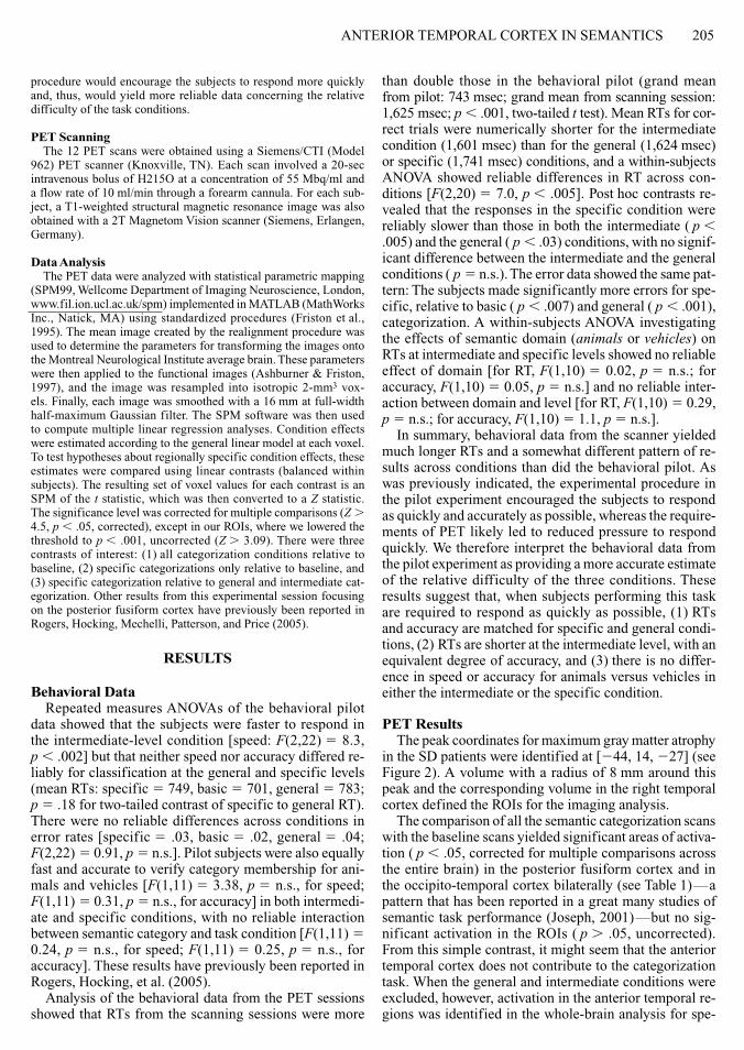
kingfisherrobin
Specific Intermediate General
birddog
animalvehicle
Figure 1 Some examples of the stimuli used and a schematic of the block design on each trial a word was viewed followed by a picture and the subjects were asked to indicate whether the picture matched the word
aNTeRIOR TeMPORaL CORTeX IN SeMaNTICS 205
procedure would encourage the subjects to respond more quickly and thus would yield more reliable data concerning the relative difficulty of the task conditions
PeT ScanningThe 12 PeT scans were obtained using a SiemensCTI (Model
962) PeT scanner (Knoxville TN) each scan involved a 20-sec intravenous bolus of H215O at a concentration of 55 Mbqml and a flow rate of 10 mlmin through a forearm cannula For each sub-ject a T1-weighted structural magnetic resonance image was also obtained with a 2T Magnetom Vision scanner (Siemens erlangen Germany)
data AnalysisThe PeT data were analyzed with statistical parametric mapping
(SPM99 Wellcome Department of Imaging Neuroscience London wwwfilionuclacukspm) implemented in MaTLaB (MathWorks Inc Natick Ma) using standardized procedures (Friston et al 1995) The mean image created by the realignment procedure was used to determine the parameters for transforming the images onto the Montreal Neurological Institute average brain These parameters were then applied to the functional images (ashburner amp Friston 1997) and the image was resampled into isotropic 2-mm3 vox-els Finally each image was smoothed with a 16 mm at full-width half-maximum Gaussian filter The SPM software was then used to compute multiple linear regression analyses Condition effects were estimated according to the general linear model at each voxel To test hypotheses about regionally specific condition effects these estimates were compared using linear contrasts (balanced within subjects) The resulting set of voxel values for each contrast is an SPM of the t statistic which was then converted to a Z statistic The significance level was corrected for multiple comparisons (Z 45 p 05 corrected) except in our ROIs where we lowered the threshold to p 001 uncorrected (Z 309) There were three contrasts of interest (1) all categorization conditions relative to baseline (2) specific categorizations only relative to baseline and (3) specific categorization relative to general and intermediate cat-egorization Other results from this experimental session focusing on the posterior fusiform cortex have previously been reported in Rogers Hocking Mechelli Patterson and Price (2005)
ReSulTS
Behavioral dataRepeated measures aNOVas of the behavioral pilot
data showed that the subjects were faster to respond in the intermediate-level condition [speed F(222) 5 83 p 002] but that neither speed nor accuracy differed re-liably for classification at the general and specific levels (mean RTs specific 5 749 basic 5 701 general 5 783 p 5 18 for two-tailed contrast of specific to general RT) There were no reliable differences across conditions in error rates [specific 5 03 basic 5 02 general 5 04 F(222) 5 091 p 5 ns] Pilot subjects were also equally fast and accurate to verify category membership for ani-mals and vehicles [F(111) 5 338 p 5 ns for speed F(111) 5 031 p 5 ns for accuracy] in both intermedi-ate and specific conditions with no reliable interaction between semantic category and task condition [F(111) 5 024 p 5 ns for speed F(111) 5 025 p 5 ns for accuracy] These results have previously been reported in Rogers Hocking et al (2005)
analysis of the behavioral data from the PeT sessions showed that RTs from the scanning sessions were more
than double those in the behavioral pilot (grand mean from pilot 743 msec grand mean from scanning session 1625 msec p 001 two-tailed t test) Mean RTs for cor-rect trials were numerically shorter for the intermediate condition (1601 msec) than for the general (1624 msec) or specific (1741 msec) conditions and a within-subjects aNOVa showed reliable differences in RT across con-ditions [F(220) 5 70 p 005] Post hoc contrasts re-vealed that the responses in the specific condition were reliably slower than those in both the intermediate ( p 005) and the general ( p 03) conditions with no signif-icant difference between the intermediate and the general conditions ( p 5 ns) The error data showed the same pat-tern The subjects made significantly more errors for spe-cific relative to basic ( p 007) and general ( p 001) categorization a within-subjects aNOVa investigating the effects of semantic domain (animals or vehicles) on RTs at intermediate and specific levels showed no reliable effect of domain [for RT F(110) 5 002 p 5 ns for accuracy F(110) 5 005 p 5 ns] and no reliable inter-action between domain and level [for RT F(110) 5 029 p 5 ns for accuracy F(110) 5 11 p 5 ns]
In summary behavioral data from the scanner yielded much longer RTs and a somewhat different pattern of re-sults across conditions than did the behavioral pilot as was previously indicated the experimental procedure in the pilot experiment encouraged the subjects to respond as quickly and accurately as possible whereas the require-ments of PeT likely led to reduced pressure to respond quickly We therefore interpret the behavioral data from the pilot experiment as providing a more accurate estimate of the relative difficulty of the three conditions These results suggest that when subjects performing this task are required to respond as quickly as possible (1) RTs and accuracy are matched for specific and general condi-tions (2) RTs are shorter at the intermediate level with an equivalent degree of accuracy and (3) there is no differ-ence in speed or accuracy for animals versus vehicles in either the intermediate or the specific condition
PeT ResultsThe peak coordinates for maximum gray matter atrophy
in the SD patients were identified at [244 14 227] (see Figure 2) a volume with a radius of 8 mm around this peak and the corresponding volume in the right temporal cortex defined the ROIs for the imaging analysis
The comparison of all the semantic categorization scans with the baseline scans yielded significant areas of activa-tion ( p 05 corrected for multiple comparisons across the entire brain) in the posterior fusiform cortex and in the occipito-temporal cortex bilaterally (see Table 1)mdasha pattern that has been reported in a great many studies of semantic task performance (Joseph 2001)mdashbut no sig-nificant activation in the ROIs ( p 05 uncorrected) From this simple contrast it might seem that the anterior temporal cortex does not contribute to the categorization task When the general and intermediate conditions were excluded however activation in the anterior temporal re-gions was identified in the whole-brain analysis for spe-
206 ROGeRS eT aL
cific categorization relative to baseline in both the left [254 6 226] (Z 5 36) and the right [48 22 228] (Z 5 31) hemispheres
The selectivity of this response was confirmed by direct comparison of the specific with general and intermediate categorization with the left peak at [250 8 222] (Z 5 39) and the right peak at [50 8 222] (Z 5 30) These ac-tivations fall within the area of atrophy in the SD patients with peak activation in the left corresponding almost ex-actly to the maximal atrophy in the patients (see Figure 2 panel a) Within the ROI (8-mm radius) around [244 14 227] and the corresponding region in the right hemi-sphere [144 14 227] activation for the specific the
general and intermediate conditions was significant after correction for multiple comparisons in both hemispheres (left p 001 right p 03) This effect of specificity was observed for both animal and vehicle categories there was no main effect of semantic category and no interac-tion between category and domain (see Figure 2 panel B) No effect of specificity was observed in the medial aTL regions previously investigated in a naming paradigm by Tyler et al (2004) even using small-volume correction ( p 05)
To verify that the effect of specificity did not solely re-flect differences in activation between the specific and the intermediate conditions we also contrasted specific and
A
B
Mean gray matterreduction in SD
Specific (intermediate andgeneral) p 001uncorrected forwhole brain
12
10
8
6
4
2
04
3
2
1
0
Left [ndash50 8 ndash22]
25
00
ndash25
Eff
ect S
ize
20
00
ndash20
Eff
ect S
ize
Right [50 8 ndash22]
baseline baseline
specific
Animals Vehicles Intermixed
general specific general
inter-mediate
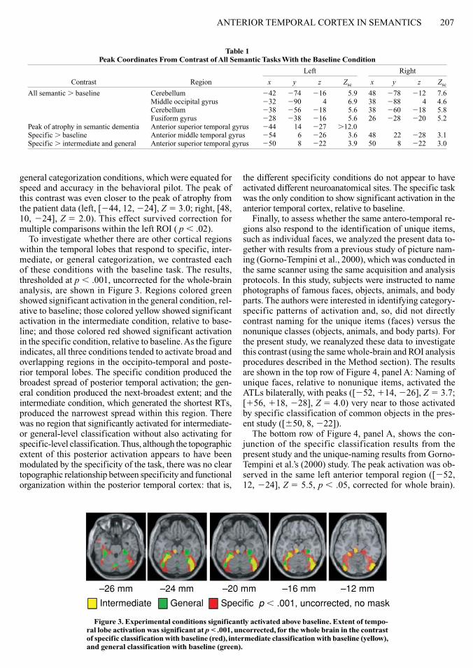
inter-mediate
Figure 2 localization of atrophy and activation (A) Top row Mean gray matter reduction for 6 patients with semantic dementia (Sd) The peak was identified at [244 14 227] a volume of 8 mm around this peak and the corresponding region in the right hemisphere were defined as regions of interest for the PeT analysis The crosshairs indicate the peak of activation from the functional im-aging study contrasting specific with intermediate and general categorization Bottom row Whole-brain activation for healthy controls significant at p lt 001 uncorrected contrasting the specific with the intermediate and general categorization conditions A corresponding peak in the right hemisphere did not surmount the whole-brain threshold but fell within the region of interest and was statistically reliable with small-volume correction The crosshairs indicate the peak of maximal atrophy from the volumetric analysis of Sd data (B) Mean-centered effect sizes (percentage of regional blood flow change) at the left and right peaks for the six different stimulus conditions in our experiment specific categorization for animals and for vehicles intermediate categorization for animals and vehicles general categorization and the baseline task The effect was observed for both animal and vehicle scans
aNTeRIOR TeMPORaL CORTeX IN SeMaNTICS 207
general categorization conditions which were equated for speed and accuracy in the behavioral pilot The peak of this contrast was even closer to the peak of atrophy from the patient data (left [244 12 224] Z 5 30 right [48 10 224] Z 5 20) This effect survived correction for multiple comparisons within the left ROI ( p 02)
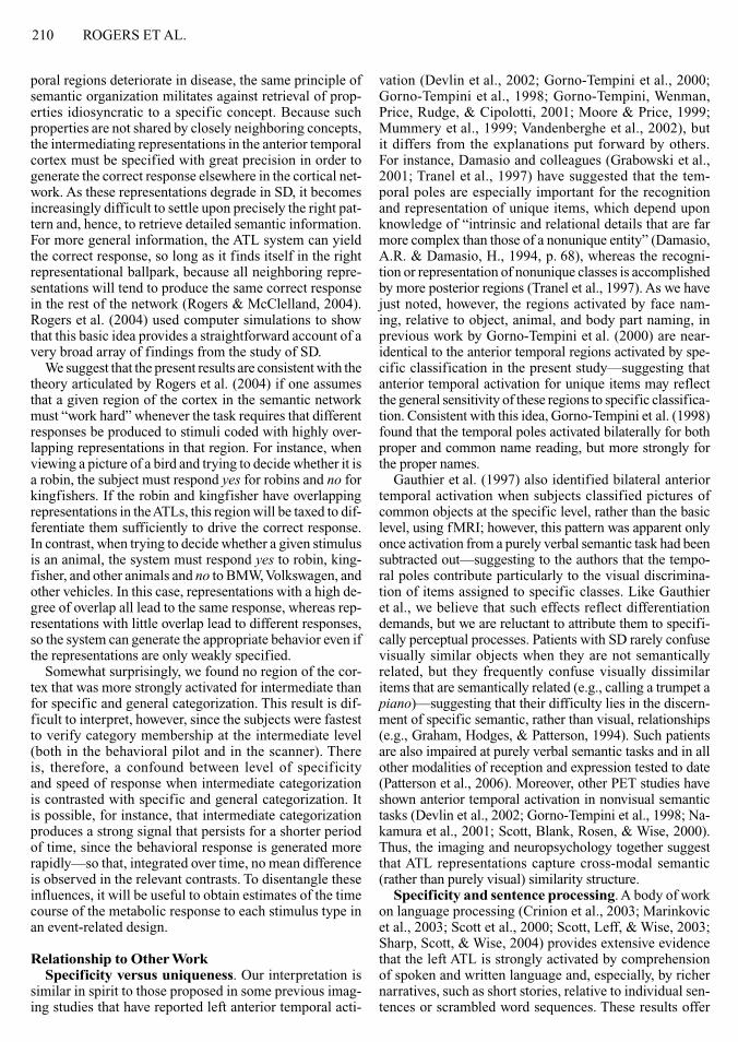
To investigate whether there are other cortical regions within the temporal lobes that respond to specific inter-mediate or general categorization we contrasted each of these conditions with the baseline task The results thresholded at p 001 uncorrected for the whole-brain analysis are shown in Figure 3 Regions colored green showed significant activation in the general condition rel-ative to baseline those colored yellow showed significant activation in the intermediate condition relative to base-line and those colored red showed significant activation in the specific condition relative to baseline as the figure indicates all three conditions tended to activate broad and overlapping regions in the occipito-temporal and poste-rior temporal lobes The specific condition produced the broadest spread of posterior temporal activation the gen-eral condition produced the next-broadest extent and the intermediate condition which generated the shortest RTs produced the narrowest spread within this region There was no region that significantly activated for intermediate- or general-level classification without also activating for specific-level classification Thus although the topographic extent of this posterior activation appears to have been modulated by the specificity of the task there was no clear topographic relationship between specificity and functional organization within the posterior temporal cortex that is
the different specificity conditions do not appear to have activated different neuroanatomical sites The specific task was the only condition to show significant activation in the anterior temporal cortex relative to baseline
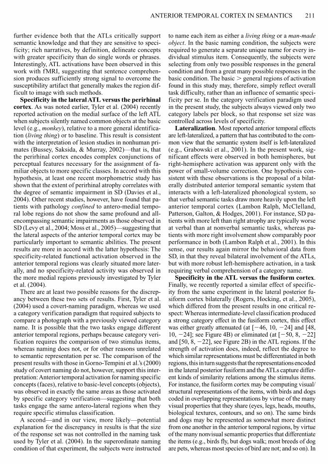
Finally to assess whether the same antero-temporal re-gions also respond to the identification of unique items such as individual faces we analyzed the present data to-gether with results from a previous study of picture nam-ing (Gorno-Tempini et al 2000) which was conducted in the same scanner using the same acquisition and analysis protocols In this study subjects were instructed to name photographs of famous faces objects animals and body parts The authors were interested in identifying category-specific patterns of activation and so did not directly contrast naming for the unique items (faces) versus the nonunique classes (objects animals and body parts) For the present study we reanalyzed these data to investigate this contrast (using the same whole-brain and ROI analysis procedures described in the Method section) The results are shown in the top row of Figure 4 panel a Naming of unique faces relative to nonunique items activated the aTLs bilaterally with peaks ([252 114 226] Z 5 37 [156 118 228] Z 5 40) very near to those activated by specific classification of common objects in the pres-ent study ([650 8 222])
The bottom row of Figure 4 panel a shows the con-junction of the specific classification results from the present study and the unique-naming results from Gorno-Tempini et alrsquos (2000) study The peak activation was ob-served in the same left anterior temporal region ([252 12 224] Z 5 55 p 05 corrected for whole brain)
Table 1 Peak Coordinates From Contrast of All Semantic Tasks With the Baseline Condition
Peak of atrophy in semantic dementia anterior superior temporal gyrus 244 14 227 120Specific baseline anterior middle temporal gyrus 254 6 226 36 48 22 228 31Specific intermediate and general anterior superior temporal gyrus 250 8 222 39 50 8 222 30
ndash26 mm ndash24 mm ndash20 mm ndash16 mm ndash12 mm
Intermediate General Specific p 001 uncorrected no mask
Figure 3 experimental conditions significantly activated above baseline extent of tempo-ral lobe activation was significant at p lt 001 uncorrected for the whole brain in the contrast of specific classification with baseline (red) intermediate classification with baseline (yellow) and general classification with baseline (green)
208 ROGeRS eT aL
Two corresponding peaks in the right did not reach a cor-rected level of significance in the whole-brain analysis (Peak 1 [58 12 224] Z 5 35 p 5 ns Peak 2 [50 14 234] Z 5 35 p 5 ns) but aligned closely to the right- hemisphere ROI Within these regions activations in both hemispheres were reliable (left [250 12 226] Z 5 54 p 001 corrected right [50 8 224] Z 5 34 p 01 corrected) Thus the same aTL regions most affected in SD appear to be activated in normal individuals both by specific classification of nonunique animals and vehicles and by silent naming of individual people
Figure 4 panel B shows the mean-centered effect sizes for the different conditions in the present experiment and the silent-naming experiment in Gorno-Tempini et al (2000) at the peak voxel of the contrast of the conjunction of specific general classification for the present study
and silent naming of faces relative to other categories in Gorno-Tempini et al (2000) (left [246 10 224] right [48 10 224]) These are very close to the coordinates of peak atrophy in the patients [246 14 227] The effect size for silent naming of individual people is approximately equal to that observed for specific categorization of animals and vehicles in the present experiment and is substantially larger than silent naming of any other category of object
diSCuSSion
The results show that the aTL regions most affected in SD were significantly activated in healthy individuals classifying pictures of objects and animals at a specific level relative to more general levels The activation was bilateral and was equally robust for categorization of
A
B
Naming faces animals objects andbody parts
Conjunction of(faces animals objectsand body parts) with(specific intermediateand general categorization)
Figure 4 naming faces versus objects animals and body parts (A) Top Silent naming of unique faces versus nonunique items (animals objects and body parts) from Gorno-Tempini Cipolotti and Price (2000) Bottom Conjunction of (specific gt general categorization) from the present study and (naming faces gt naming animals objects and body parts) from Gorno-Tempini et al (2000) note that in the case of the conjunction the color scale for the t values relates to the minimal t (B) Mean centered effect sizes (percentages of regional blood flow change) at the peak voxel
aNTeRIOR TeMPORaL CORTeX IN SeMaNTICS 209
animals and vehicles and it appears in the same regions engaged by silent naming of unique faces relative to non-unique objects animals and body parts Thus we have shown functional activation in healthy subjects as a result of semantic processing that directly parallels the type of semantic knowledge that is most degraded in SD
On the basis of the behavioral data from the pilot exper-iment the specificity-related activation does not appear to be attributable to the overall difficulty of specific clas-sification when the subjects were encouraged to respond as quickly and accurately as possible the most general and most specific conditions were matched for speed and accuracy in responding and hence were matched for dif-ficulty in this respect In the behavioral results from the scanning session itself however specific classification elicited significantly longer RTs than did intermediate or general classification so that we cannot rule out the possi-bility that difficulty contributed to the observed pattern of activation Under this interpretation it is still notable that the activation peak appears in the aTLs indicating that these regions are indeed involved in at least the specific condition of the task
The results suggest that the previously noted puzzling discrepancy between functional neuroimaging and neu-ropsychology arises from a confluence of methodologi-cal factors that together conspire against observing aTL activation in functional imaging First most commonly used semantic tasks do not require very precise identifica-tion of the stimulus as is illustrated in Figure 3 anterior temporal activation is not much apparent in association with intermediate- or general-level classification Second it is difficult to acquire a good signal in this region with fMRI the most commonly used technique for these inves-tigations (Devlin et al 2002 Devlin et al 2000) In the present work we used PeT because it does not suffer from this susceptibility artifact Third to gain statistical power in testing their hypotheses many investigators limit their analyses to cortical regions of a priori interest which tend to be regions that are easily activated across different tasks and imaging methods areas that are difficult to observe in whole-brain studies are less likely to be used as ROIs in subsequent studies hence the statistical power of small-volume correction is only rarely brought to bear on these areas (see Joseph 2001) We have addressed this chal-lenge in the present study by using structural imaging data from a patient population rather than previous functional imaging results to identify regions of a priori interest
By addressing these three methodological factors we show that functional imaging and neuropsychology do in fact accord fairly well Both suggest that left and right an-terior temporal regions contribute to semantic processing for living and nonliving things especially in tasks requir-ing specific classification
interpretation of the effectThe specificity effect reported in this article indicates
that the antero-lateral temporal cortices are more strongly activated by fairly specific classification of items relative to more general classification Perhaps the most clear-cut
interpretation of the effect arises from the proposal of Martin and Chao (2001) that there exists a topographic gradient in the infero-temporal cortex so that more spe-cific concepts are represented more rostrally and more general concepts are represented more caudally This no-tion provides an intuitive account of observations from SD in that pathology in the disease typically spreads backward from the temporal pole so that the fine-to-coarse deterioration of conceptual knowledge coincides with an anterior-to-posterior dissolution of the temporal cortex The present data are partially consistent with this view in that the specific classification task engages more anterior regions relative to baseline than do the interme-diate and general tasks The one inconsistency is shown in Figure 3 We found no region between the temporal pole and the posterior temporal cortex that responded signifi-cantly above baseline for intermediate and general but not specific classification as the specificity gradient hypoth-esis would predict
The present results are also consistent with the theory of normal and degraded semantic representation and pro-cessing proposed by Rogers et al (2004) This theory builds on the fairly widespread view that knowledge about the particular properties of objects is encoded in percep-tual and motor representations distributed throughout the cortexmdashwith for instance knowledge about the character-istic shapes of objects coded in visual regions responsible for shape perception knowledge about characteristic pat-terns of movement stored in regions responsible for motion perception knowledge about colors represented in regions dedicated to color perception knowledge about actions in motor regions and so on (Martin amp Chao 2001) accord-ing to Rogers et al (2004) these widely distributed percep-tual motor and language representations do not interact with one another directly Instead they interact by means of a representational ldquohubrdquo in the aTLsmdashso that for instance to make inferences about the visual appearance of an object when given its name perception of the phonological form of the word first provokes activation of an amodal repre-sentation in the aTLs which then passes activation on to the shape color and motion regions that code information about the visual properties of the item denoted by the word On this view the aTLs serve to mediate communication among the various perceptual motor and language rep-resentations that constitute the cortical semantic network They compute these mappings for all kinds of items and for all modalities of reception and expression
Rogers et al (2004) argued that the systematic erosion of detailed semantic information observed in SD arises because the intermediating representations encoded in the aTLs capture the degree of semantic relatedness among known concepts On this view closely related items (eg a robin and a kingfisher) are represented with quite simi-lar patterns of activity in the aTL whereas semantically unrelated items (eg a robin and a yacht) are represented with dissimilar patterns (Hinton amp Shallice 1991 Plaut amp Shallice 1993 Rogers amp McClelland 2004) In the healthy system this similarity structure promotes seman-tic generalization and induction but when anterior tem-
210 ROGeRS eT aL
poral regions deteriorate in disease the same principle of semantic organization militates against retrieval of prop-erties idiosyncratic to a specific concept Because such properties are not shared by closely neighboring concepts the intermediating representations in the anterior temporal cortex must be specified with great precision in order to generate the correct response elsewhere in the cortical net-work as these representations degrade in SD it becomes increasingly difficult to settle upon precisely the right pat-tern and hence to retrieve detailed semantic information For more general information the aTL system can yield the correct response so long as it finds itself in the right representational ballpark because all neighboring repre-sentations will tend to produce the same correct response in the rest of the network (Rogers amp McClelland 2004) Rogers et al (2004) used computer simulations to show that this basic idea provides a straightforward account of a very broad array of findings from the study of SD
We suggest that the present results are consistent with the theory articulated by Rogers et al (2004) if one assumes that a given region of the cortex in the semantic network must ldquowork hardrdquo whenever the task requires that different responses be produced to stimuli coded with highly over-lapping representations in that region For instance when viewing a picture of a bird and trying to decide whether it is a robin the subject must respond yes for robins and no for kingfishers If the robin and kingfisher have overlapping representations in the aTLs this region will be taxed to dif-ferentiate them sufficiently to drive the correct response In contrast when trying to decide whether a given stimulus is an animal the system must respond yes to robin king-fisher and other animals and no to BMW Volkswagen and other vehicles In this case representations with a high de-gree of overlap all lead to the same response whereas rep-resentations with little overlap lead to different responses so the system can generate the appropriate behavior even if the representations are only weakly specified
Somewhat surprisingly we found no region of the cor-tex that was more strongly activated for intermediate than for specific and general categorization This result is dif-ficult to interpret however since the subjects were fastest to verify category membership at the intermediate level (both in the behavioral pilot and in the scanner) There is therefore a confound between level of specificity and speed of response when intermediate categorization is contrasted with specific and general categorization It is possible for instance that intermediate categorization produces a strong signal that persists for a shorter period of time since the behavioral response is generated more rapidlymdashso that integrated over time no mean difference is observed in the relevant contrasts To disentangle these influences it will be useful to obtain estimates of the time course of the metabolic response to each stimulus type in an event-related design
Relationship to other WorkSpecificity versus uniqueness Our interpretation is
similar in spirit to those proposed in some previous imag-ing studies that have reported left anterior temporal acti-
vation (Devlin et al 2002 Gorno-Tempini et al 2000 Gorno-Tempini et al 1998 Gorno-Tempini Wenman Price Rudge amp Cipolotti 2001 Moore amp Price 1999 Mummery et al 1999 Vandenberghe et al 2002) but it differs from the explanations put forward by others For instance Damasio and colleagues (Grabowski et al 2001 Tranel et al 1997) have suggested that the tem-poral poles are especially important for the recognition and representation of unique items which depend upon knowledge of ldquointrinsic and relational details that are far more complex than those of a nonunique entityrdquo (Damasio aR amp Damasio H 1994 p 68) whereas the recogni-tion or representation of nonunique classes is accomplished by more posterior regions (Tranel et al 1997) as we have just noted however the regions activated by face nam-ing relative to object animal and body part naming in previous work by Gorno-Tempini et al (2000) are near- identical to the anterior temporal regions activated by spe-cific classification in the present studymdashsuggesting that anterior temporal activation for unique items may reflect the general sensitivity of these regions to specific classifica-tion Consistent with this idea Gorno-Tempini et al (1998) found that the temporal poles activated bilaterally for both proper and common name reading but more strongly for the proper names
Gauthier et al (1997) also identified bilateral anterior temporal activation when subjects classified pictures of common objects at the specific level rather than the basic level using fMRI however this pattern was apparent only once activation from a purely verbal semantic task had been subtracted outmdashsuggesting to the authors that the tempo-ral poles contribute particularly to the visual discrimina-tion of items assigned to specific classes Like Gauthier et al we believe that such effects reflect differentiation demands but we are reluctant to attribute them to specifi-cally perceptual processes Patients with SD rarely confuse visually similar objects when they are not semantically related but they frequently confuse visually dissimilar items that are semantically related (eg calling a trumpet a piano)mdashsuggesting that their difficulty lies in the discern-ment of specific semantic rather than visual relationships (eg Graham Hodges amp Patterson 1994) Such patients are also impaired at purely verbal semantic tasks and in all other modalities of reception and expression tested to date (Patterson et al 2006) Moreover other PeT studies have shown anterior temporal activation in nonvisual semantic tasks (Devlin et al 2002 Gorno-Tempini et al 1998 Na-kamura et al 2001 Scott Blank Rosen amp Wise 2000) Thus the imaging and neuropsychology together suggest that aTL representations capture cross-modal semantic (rather than purely visual) similarity structure
Specificity and sentence processing a body of work on language processing (Crinion et al 2003 Marinkovic et al 2003 Scott et al 2000 Scott Leff amp Wise 2003 Sharp Scott amp Wise 2004) provides extensive evidence that the left aTL is strongly activated by comprehension of spoken and written language and especially by richer narratives such as short stories relative to individual sen-tences or scrambled word sequences These results offer
aNTeRIOR TeMPORaL CORTeX IN SeMaNTICS 211
further evidence both that the aTLs critically support semantic knowledge and that they are sensitive to speci-ficity rich narratives by definition delineate concepts with greater specificity than do single words or phrases Interestingly aTL activations have been observed in this work with fMRI suggesting that sentence comprehen-sion produces sufficiently strong signal to overcome the susceptibility artifact that generally makes the region dif-ficult to image with such methods
Specificity in the lateral ATl versus the perirhinal cortex as was noted earlier Tyler et al (2004) recently reported activation on the medial surface of the left aTL when subjects silently named common objects at the basic level (eg monkey) relative to a more general identifica-tion (living thing) or to baseline This result is consistent with the interpretation of lesion studies in nonhuman pri-mates (Bussey Saksida amp Murray 2002)mdashthat is that the perirhinal cortex encodes complex conjunctions of perceptual features necessary for the assignment of fa-miliar objects to more specific classes In accord with this hypothesis at least one recent morphometric study has shown that the extent of perirhinal atrophy correlates with the degree of semantic impairment in SD (Davies et al 2004) Other recent studies however have found that pa-tients with pathology confined to antero-medial tempo-ral lobe regions do not show the same profound and all- encompassing semantic impairments as those observed in SD (Levy et al 2004 Moss et al 2005)mdashsuggesting that the lateral aspects of the anterior temporal cortex may be particularly important to semantic abilities The present results are more in accord with the latter hypothesis The specificity-related functional activation observed in the anterior temporal regions was clearly situated more later-ally and no specificity-related activity was observed in the more medial regions previously investigated by Tyler et al (2004)
There are at least two possible reasons for the discrep-ancy between these two sets of results First Tyler et al (2004) used a covert-naming paradigm whereas we used a category verification paradigm that required subjects to compare a photograph with a previously viewed category name It is possible that the two tasks engage different anterior temporal regions perhaps because category veri-fication requires the comparison of two stimulus items whereas naming does not or for other reasons unrelated to semantic representation per se The comparison of the present results with those in Gorno-Tempini et alrsquos (2000) study of covert naming do not however support this inter-pretation anterior temporal activation for naming specific concepts (faces) relative to basic-level concepts (objects) was observed in exactly the same areas as those activated by specific category verificationmdashsuggesting that both tasks engage the same antero-lateral regions when they require specific stimulus classification
a secondmdashand in our view more likelymdashpotential explanation for the discrepancy in results is that the size of the response set was not controlled in the naming task used by Tyler et al (2004) In the superordinate naming condition of that experiment the subjects were instructed
to name each item as either a living thing or a man-made object In the basic naming condition the subjects were required to generate a separate unique name for every in-dividual stimulus item Consequently the subjects were selecting from only two possible responses in the general condition and from a great many possible responses in the basic condition The basic general regions of activation found in this study may therefore simply reflect overall task difficulty rather than an influence of semantic speci-ficity per se In the category verification paradigm used in the present study the subjects always viewed only two category labels per block so that response set size was controlled across levels of specificity
lateralization Most reported anterior temporal effects are left-lateralized a pattern that has contributed to the com-mon view that the semantic system itself is left-lateralized (eg Grabowski et al 2001) In the present work sig-nificant effects were observed in both hemispheres but right-hemisphere activation was apparent only with the power of small-volume correction One hypothesis con-sistent with these observations is the proposal of a bilat-erally distributed anterior temporal semantic system that interacts with a left-lateralized phonological system so that verbal semantic tasks draw more heavily upon the left anterior temporal cortex (Lambon Ralph McClelland Patterson Galton amp Hodges 2001) For instance SD pa-tients with more left than right atrophy are typically worse at verbal than at nonverbal semantic tasks whereas pa-tients with more right involvement show comparably poor performance in both (Lambon Ralph et al 2001) In this sense our results again mirror the behavioral data from SD in that they reveal bilateral involvement of the aTLs but with more robust left-hemisphere activation in a task requiring verbal comprehension of a category name
Specificity in the ATl versus the fusiform cortex Finally we recently reported a similar effect of specific-ity from the same experiment in the lateral posterior fu-siform cortex bilaterally (Rogers Hocking et al 2005) which differed from the present results in one critical re-spect Whereas intermediate-level classification produced a strong category effect in the fusiform cortex this effect was either greatly attenuated (at [246 10 224] and [48 10 224] see Figure 4B) or eliminated (at [250 8 222] and [50 8 222] see Figure 2B) in the aTL regions If the strength of activation does indeed reflect the degree to which similar representations must be differentiated in both regions this in turn suggests that the representations encoded in the lateral posterior fusiform and the aTLs capture differ-ent kinds of similarity relations among the stimulus items For instance the fusiform cortex may be computing visual structural representations of the items with birds and dogs coded in overlapping representations by virtue of the many visual properties that they share (eyes legs heads mouths biological textures contours and so on) The same birds and dogs may be represented as somewhat more distinct from one another in the anterior temporal regions by virtue of the many nonvisual semantic properties that differentiate the items (eg birds fly but dogs walk most breeds of dog are pets whereas most species of bird are not and so on) In
212 ROGeRS eT aL
this case the fusiform cortex would be more susceptible to a crowding effect when processing animals at an intermedi-ate level than would the anterior temporal regions
although somewhat speculative there is some a pos-teriori support for this hypothesis Specifically Rogers et al (2004) assessed (1) the degree of overlap in verbal descriptions of 64 items from the Snodgrass and Vander-wart (1980) corpus and (2) the degree of overlap in the vi-sual attributes depicted when subjects were asked to draw the same 64 items The analysis showed that for the 24 animal items included in the sample there was more over-lap in the visual features produced in the drawings (mean Jaccardrsquos distance of 079 for all animal pairs) than in the verbal descriptions (mean Jaccardrsquos distance of 088 for all animal pairs see Rogers et al 2004 Figure 2)mdashsuggesting that these items tend to have more visual than nonvisual properties in common
Thus the differences in patterns of activation between the fusiform and the aTLs may be understood as follows (1) Both areas respond strongly when the task requires dif-ferentiation of items with similar representations (2) the fusiform cortex codes visual similarity whereas the ante-rior temporal cortex encodes ldquosemanticrdquo similarity which is influenced by knowledge of nonvisual characteristics and (3) animals from different intermediate categories in our study tend to have more overlap in visual than in se-mantic representations so that crowding effects for the intermediate categorization of animals are stronger in the posterior fusiform than in the anterior temporal cortices In other words the different patterns of activation indicate that different kinds of similarity structure are encoded by different parts of the cortical semantic network
ConclusionWe observed bilateral anterior temporal activation for
specific relative to more general classification for both living things and artifacts in regions closely aligned to those affected in patients with SD who fail at tasks requir-ing specific concept knowledge The effect cannot reflect simple task difficulty because in contrast to previous studies specific and general conditions were matched for speed and accuracy in a behavioral study that pressured subjects to respond as quickly and accurately as possible Moreover the regions activated were nearly identical to those involved in the naming of unique faces relative to nonunique items Imaging and neuropsychology together thus suggest that anterior temporal regions in both hemi-spheres critically support the retrieval of specific semantic information for all classes of objects
ReFeRenCeS
Ashburner J amp Friston K [J] (1997) Multimodal image coregistra-tion and partitioning a unified framework NeuroImage 6 209-217
Bozeat S Lambon Ralph M A Graham K S Patterson K Wilkin H Rowland J et al (2003) a duck with four legs In-vestigating the structure of conceptual knowledge using picture draw-ing in semantic dementia Cognitive Neuropsychology 20 27-47
Bozeat S Lambon Ralph M A Patterson K Garrard P amp Hodges J R (2000) Nonverbal semantic impairment in semantic dementia Neuropsychologia 38 1207-1215
Bussey T J Saksida L M amp Murray E A (2002) Perirhinal cor-tex resolves feature ambiguity in complex visual discriminations Eu-ropean Journal of Neuroscience 15 365-374
Caramazza A amp Mahon B Z (2003) The organization of concep-tual knowledge The evidence from category-specific semantic defi-cits Trends in Cognitive Sciences 7 354-361
Chao L L Haxby J V amp Martin A (1999) attribute-based neural substrates in temporal cortex for perceiving and knowing about ob-jects Nature Neuroscience 2 913-919
Crinion J T Lambon Ralph M A Warburton E A Howard D amp Wise R J S (2003) Temporal lobe regions engaged during nor-mal speech comprehension Brain 126 1193-1201
Damasio A R amp Damasio H (1994) Cortical systems underlying knowledge retrieval In C Koch (ed) Large-scale neuronal theories of the brain (pp 61-74) Cambridge Ma MIT Press
Damasio H Grabowski T J Tranel D amp Hichwa R D (1996) a neural basis for lexical retrieval Nature 380 499-505
Davies R R Graham K S Xuereb J H Williams G B amp Hodges J R (2004) The human perirhinal cortex and semantic memory European Journal of Neuroscience 20 2441-2446
Davis M amp Johnsrude I (2003) Hierarchical processing in spoken language comprehension Journal of Neuroscience 23 3423-3431
Devlin J T Russell R P Davis M H Price C J Moss H E Fadili M J amp Tyler L K (2002) Is there an anatomical basis for category-specificity Semantic memory studies in PeT and fMRI Neuropsychologia 40 54-75
Devlin J T Russell R P Davis M H Price C J Wilson J Moss H E et al (2000) Susceptibility-induced loss of signal Com-paring PeT and fMRI on a semantic task NeuroImage 11 589-600
Friston K J Holmes A P Worsley K J Poline J B Frith C D amp Frackowiak R S J (1995) Statistical parametric maps in functional imaging a general linear approach Human Brain Map-ping 2 189-210
Garrard P Lambon Ralph M A amp Hodges J R (2002) Semantic dementia a category-specific paradox In e M Forde amp G W Hum-phreys (eds) Category specificity in brain and mind (pp 149-179) Hove UK Psychology Press
Gauthier I Anderson A W Tarr M J Skudlarski P amp Gore J C (1997) Levels of categorization in visual recognition studied with functional MRI Current Biology 7 645-651
Gitelman D R Ashburner J Friston K Tyler L amp Price C J (2001) Voxel based mophometry of Herpes simplex encephalitis NeuroImage 13 623-631
Gloor P (1997) The temporal lobe and limbic system New York Ox-ford University Press
Gorno-Tempini M L Cipolotti L amp Price C J (2000) Category differences in brain activation studies Where do they come from Pro-ceedings of the Royal Society of London Series B 267 1253-1258
Gorno-Tempini M L amp Price C J (2001) Identification of famous faces and buildings a functional neuroimaging study of semantically unique items Brain 124 2087-2097
Gorno-Tempini M L Price C J Josephs O Vandenberghe R Cappa S F Kapur N amp Frackowiak R S (1998) The neural systems sustaining face and proper-name processing Brain 121 2103-2118
Gorno-Tempini M [L] Wenman R Price C [J] Rudge P amp Cipolotti L (2001) Identification without naming a functional neuroimaging study of an anomic patient Journal of Neurology Neu-rosurgery amp Psychiatry 70 397-400
Grabowski T J Damasio H Tranel D Boles Ponto L L Hichwa R D amp Damasio A R (2001) a role for left temporal pole in the retrieval of words for unique entities Human Brain Map-ping 13 199-212
Graham K S Hodges J R amp Patterson K (1994) The relation-ship between comprehension and oral reading in progressive fluent aphasia Neuropsychologia 32 299-316
Hinton G E amp Shallice T (1991) Lesioning an attractor network Investigations of acquired dyslexia Psychological Review 98 74-95
Hodges J R Bozeat S Lambon Ralph M A Patterson K amp Spatt J (2000) The role of conceptual knowledge in object use evidence from semantic dementia Brain 123 1913-1925
Hodges J R Garrard P amp Patterson K (1998) Semantic de-
aNTeRIOR TeMPORaL CORTeX IN SeMaNTICS 213
mentia and Pick complex In a Kertesz amp D G Munoz (eds) Pickrsquos disease and Pick complex New York Wiley-Liss
Hodges J R Graham N amp Patterson K (1995) Charting the progression in semantic dementia Implications for the organisation of semantic memory Memory 3 463-495
Hodges J R amp Patterson K (1995) Is semantic memory consis-tently impaired early in the course of alzheimerrsquos disease Neuroana-tomical and diagnostic implications Neuropsychologia 33 441-459
Hodges J R Patterson K Oxbury S amp Funnell E (1992) Semantic dementia Progressive fluent aphasia with temporal lobe atrophy Brain 115 1783-1806
Humphreys G W amp Forde E M (2001) Hierarchies similarity and interactivity in object recognition On the multiplicity of ldquocategory-specificrdquo deficits in neuropsychological populations Behavioral amp Brain Sciences 24 453-509
Joseph J E (2001) Functional neuroimaging studies of category speci-ficity in object recognition a critical review and meta-analysis Cog-nitive Affective amp Behavioral Neuroscience 1 119-136
Lambon Ralph M A McClelland J L Patterson K Gal- ton C J amp Hodges J R (2001) No right to speak The relation-ship between object naming and semantic impairment Neuropsycho-logical evidence and a computational model Journal of Cognitive Neuroscience 13 341-356
Levy D A Bayley P J amp Squire L R (2004) The anatomy of semantic knowledge Medial vs lateral temporal lobe Proceedings of the National Academy of Sciences 101 6710-6715
Marinkovic K Dhond R P Dale A M Glessner M Carr V amp Halgren E (2003) Spatiotemporal dynamics of modality- specific and supramodal word processing Neuron 38 487-497
Martin A amp Chao L L (2001) Semantic memory in the brain Struc-ture and processes Current Opinion in Neurobiology 11 194-201
Mazoyer B M Tzourio N Frak V Syrota A Murayama N Levrier O et al (1993) The cortical representation of speech Journal of Cognitive Neuroscience 5 467-479
McClelland J L amp Rogers T T (2003) The parallel distributed processing approach to semantic cognition Nature Reviews Neurosci-ence 4 310-322
Moore C amp Price C (1999) a functional neuroimaging study of the variables that generate category-specific object processing differ-ences Brain 122 943-962
Moss H E Rodd J M Stamatakis E A Bright P amp Tyler L K (2005) anteromedial temporal cortex supports fine grained differen-tiation among objects Cerebral Cortex 15 626-627
Mummery C J Patterson K Price C J Ashburner J Frack-owiak R S J amp Hodges J (2000) a voxel-based morphometry study of semantic dementia Relationship between temporal lobe atro-phy and semantic memory Annals of Neurology 47 36-45
Mummery C J Patterson K Wise R J S Vandenberghe R Price C J amp Hodges J R (1999) Disrupted temporal lobe con-nections in semantic dementia Brain 122 61-73
Nakamura K Kawashima R Sato N Nakamura A Sugiura M Kato T et al (2000) Functional delineation of the human occipito-temporal areas related to faces and scene processing a PeT study Brain amp Cognition 123 1903-1912
Nakamura K Kawashima R Sugiura M Kato T Nakamura A Hatano K et al (2001) Neural substrates for recognition of fa-miliar voices a PeT study Neuropsychologia 39 1047-1054
Papagno C amp Capitani E (2001) Slowly progressive aphasia a four-year follow-up study Neuropsychologia 39 678-686
Patterson K amp Hodges J (2000) Semantic dementia One window on the structure and organisation of semantic memory In J Cermak (ed) Handbook of neuropsychology Vol 2 Memory and its disor-ders (pp 313-333) amsterdam elsevier
Patterson K Lambon Ralph M A Jeffries E A Woollams A Jones R Hodges J R amp Rogers T T (2006) ldquoPresemanticrdquo cog-nition in semantic dementia Six deficits in search of an explanation Journal of Cognitive Neuroscience 18 169-183
Perani D Cappa S F Bettinardi V Bressi S Gorno-Tempini M Matarrese M amp Fazio F (1995) Different neural systems for the recognition of animals and man-made tools NeuroReport 6 1637-1641
Plaut D C amp Shallice T (1993) Deep dyslexia a case study of con-nectionist neuropsychology Cognitive Neuropsychology 10 377-500
Ricci P T Zelkowicz B J Nebes R D Meltzer C C Min- tun M A amp Becker J T (1999) Functional neuroanatomy of se-mantic memory Recognition of semantic associations NeuroImage 9 88-96
Rogers T T Hocking J Mechelli A Patterson K amp Price C (2005) Fusiform activation to animals is driven by the process not the stimulus Journal of Cognitive Neuroscience 17 434-445
Rogers T T Lambon Ralph M A Garrard P Bozeat S Mc-Clelland J L Hodges J R amp Patterson K (2004) The struc-ture and deterioration of semantic memory a computational and neu-ropsychological investigation Psychological Review 111 205-235
Rogers T T Lambon Ralph M A Hodges J R amp Patterson K (2003) Object recognition under semantic impairment The effects of conceptual regularities on perceptual decisions Language amp Cogni-tive Processes 18 625-662
Rogers T T amp McClelland J L (2004) Semantic cognition A par-allel distributed processing approach Cambridge Ma MIT Press
Rogers T T Watling L Hodges J R amp Patterson K (2005 april) A basic-level disadvantage for speeded category verification Paper presented at the meeting of the Cognitive Neuroscience Society New York
Rosch E Mervis C B Gray W Johnson D amp Boyes-Braem P (1976) Basic objects in natural categories Cognitive Psychology 8 382-439
Scott S Blank C C Rosen S amp Wise R (2000) Identification of a pathway for intelligible speech in the left temporal lobe Brain 124 83-95
Scott S Leff A amp Wise R J (2003) Going beyond the information given a neural system supporting semantic interpretation NeuroImage 19 870-876
Sharp D J Scott S amp Wise R J (2004) Monitoring and the con-trolled processing of meaning Distinct prefrontal systems Cerebral Cortex 14 1-10
Snodgrass J G amp Vanderwart M (1980) a standardized set of 260 pictures Norms for name agreement image agreement familiarity and visual complexity Journal of Experimental Psychology Learn-ing Memory amp Cognition 6 174-215
Snowden J S Goulding P J amp Neary D (1989) Semantic de-mentia a form of circumscribed temporal atrophy Behavioural Neu-rology 2 167-182
Snowden J S Neary D amp Mann D M A (1996) Frontotemporal lobar degeneration Frontotemporal dementia progressive aphasia semantic dementia New York Churchill Livingstone
Sugiura M Kawashima R Nakamura K Sato N Nakamura A Kato T et al (2001) activation reduction in anterior temporal cortices during repeated recognition of faces of personal acquain-tances NeuroImage 13 877-890
Thompson-Schill S L (2003) Neuroimaging studies of semantic mem-ory Inferring ldquohowrdquo from ldquowhererdquo Neuropsychologia 41 280-292
Thompson-Schill S L Aguirre G K DrsquoEsposito M amp Farah M J (1999) a neural basis for category and modality specificity of semantic knowledge Neuropsychologia 37 671-676
Tranel D Damasio H amp Damasio A R (1997) a neural basis for the retrieval of conceptual knowledge Neuropsychologia 35 1319-1327
Tyler L K Stamatakis E A Bright P Acres K Abdallah S Rodd J M amp Moss H E (2004) Processing objects at different lev-els of specificity Journal of Cognitive Neuroscience 16 351-362
Vandenberghe R Nobre A C amp Price C J (2002) The response of left temporal cortex to sentences Journal of Cognitive Neurosci-ence 14 550-560
Vandenberghe R Price C Wise R Josephs O amp Fracko- wiak R S J (1996) Functional anatomy of a common semantic sys-tem for words and pictures Nature 383 254-255
Warrington E K (1975) Selective impairment of semantic memory Quarterly Journal of Experimental Psychology 27 635-657
(Manuscript received July 27 2005 revision accepted for publication February 18 2006)
202 ROGeRS eT aL
bilateral anterior temporal cortices are critical for amodal and domain-general aspects of semantic processing (Rogers et al 2004) that is they contribute to semantic processing for all kinds of concepts and for all modalities of reception and expression In line with this view other brain diseases that can affect the anterior temporal lobes (aTLs) such as alzheimerrsquos disease and herpes simplex viral encephalitis also often disrupt semantic memory although never as se-lectively as in SD (Hodges amp Patterson 1995)
Functional neuroimaging has offered a rather start-lingly different picture of the neural representation of se-mantic knowledge in three respects First the majority of research has yielded left-sided rather than bilateral cortical activations for semantic tasks (Devlin et al 2002 Joseph 2001 Martin amp Chao 2001 Thompson-Schill 2003) Second functional imaging results have indicated that semantic knowledge is encoded in a widely distributed cortical network with different regions specialized to repre-sent particular kinds of information (Martin amp Chao 2001 Tranel Damasio amp Damasio 1997) particular categories of object (Caramazza amp Mahon 2003 Perani et al 1995) or both (Humphreys amp Forde 2001 Thompson-Schill aguirre Drsquoesposito amp Farah 1999)mdashleading some re-searchers to suggest that no single region supports semantic abilities for all modalities and categories (eg Thompson-Schill 2003) Third and perhaps most puzzling although anterior temporal activation has been associated with sen-tence comprehension (Crinion Lambon Ralph Warburton Howard amp Wise 2003 Davis amp Johnsrude 2003 Mazoyer et al 1993 Vandenberghe Nobre amp Price 2002) famous face recognition (Gorno-Tempini amp Price 2001 Gorno-Tempini et al 1998 Grabowski et al 2001 Tranel et al 1997) and a few other semantic tasks (Devlin et al 2000 Gauthier anderson Tarr Skudlarski amp Gore 1997 Mum-mery et al 1999 Ricci et al 1999 Tyler et al 2004 Van-denberghe Price Wise Josephs amp Frackowiak 1996) the vast majority of functional imaging studies have reported posterior temporal andor frontal activations for semantic tasks with no mention of the anterior temporal cortex (for recent literature reviews see Joseph 2001 Martin amp Chao 2001 Thompson-Schill 2003)
To summarize neuropsychological research on SD might lead one to conclude that although many other re-gions undoubtedly contribute the anterior temporal cortex in both hemispheres is critical for semantic representation and processing across all stimulus modalities and for all types of conceptual knowledge From functional imaging research on normal adults in contrast one might conclude that no single region contributes to semantic memory for all modalities and categories and further that the widely distributed network responsible for different aspects of semantic processing is left-lateralized and includes the posterior temporal and frontal cortex but not the aTL How is this conundrum to be resolved
A Convergence Theory of Semantic ProcessingWe have previously proposed that the aTL regions af-
fected in SD serve in the healthy brain to mediate commu-nication among the modality-specific regions distributed
throughout the cortex that encode explicit representations of object attributes (McClelland amp Rogers 2003 Rogers et al 2004)mdasha hypothesis that is supported both by neu-roanatomical (Gloor 1997) and computational (Rogers amp McClelland 2004) considerations and by the neuropsy-chological phenomena described previously When this cross-modal ldquohubrdquo degrades as a consequence of brain disease the ability to map between surface formsmdashfor instance to generate an itemrsquos name from its visual image or vice versamdashis compromised On this view the aTLs critically support semantic task performance for all mo-dalities of reception and expression and all categories of objects as suggested by the impairments observed in SD Perhaps less intuitively this hypothesis also offers a clue regarding the discrepancy in the data from neuropsychol-ogy and functional imaging
The key observation is that in SD knowledge about prop-erties that individuate a specific concept from its semantic neighbors (eg the stripes of a zebra) is always more vul-nerable than knowledge about properties shared by related concepts (eg the fact that a zebra has four legs) This pat-tern has been documented in tasks as varied as naming wordndashpicture matching drawing object recognition col-oring lexical decision and object use (Papagno amp Capitani 2001 Patterson et al 2006 Rogers et al 2004 Snowden Neary amp Mann 1996 Warrington 1975) Moreover this pervasive tendency does not simply reflect the overall dif-ficulty of retrieving more specific relative to more general information For instance healthy controls are typically faster and more accurate in classifying objects at the basic level (eg bird ) than at more general levels (Rosch Mer-vis Gray Johnson amp Boyes-Braem 1976) but the reverse is true for patients with SD (Hodges et al 1995 Rogers Watling et al 2005) Thus tasks that require objects to be classified with greater precision appear to exert greater de-mands on the neural system affected in SD even when they are not more difficult overall One basis for the discrepancy between neuropsychology and functional imaging then may be that functional imaging studies have not tended to use semantic tasks that require very specific classification of the stimulus
The few functional imaging studies that do report aTL effects appear to be generally consistent with this idea al-though the evidence to date is equivocal and other inter-pretations have been offered For instance most of these studies concern recognition or naming of famous or fa-miliar people andor buildings (H Damasio Grabowski Tranel amp Hichwa 1996 Gorno-Tempini amp Price 2001 Gorno-Tempini et al 1998 Grabowski et al 2001 Na-kamura et al 2000 Nakamura et al 2001 Sugiura et al 2001)mdashsuggesting to some researchers that the aTLs are dedicated to the representation and processing of lexical or semantic information about unique entities but are not oth-erwise involved in semantic memory (Tranel et al 1997) as we and others have previously noted (Gauthier et al 1997 Gorno-Tempini amp Price 2001 Gorno-Tempini et al 1998) however this pattern may not reflect functional spe-cialization for unique items per se but may instead arise from a more general sensitivity within the aTLs to specific
aNTeRIOR TeMPORaL CORTeX IN SeMaNTICS 203
classification That is recognition of unique entities may constitute a form of very specific recognition carried out by the very same processes that support classification of nonunique items into fairly specific semantic categories
To our knowledge only two previous functional imag-ing studies have directly compared different levels of clas-sification for nonunique items In the first Gauthier et al (1997) used fMRI to investigate temporal lobe activations in subjects classifying line drawings of common objects at the subordinate (eg flamingo) or basic (eg bird ) level The authors found no selective anterior temporal activation in the direct contrast of specific to basic-level categoriza-tion however such an effect was observed bilaterally after activations from a second purely verbal semantic task were subtracted out although suggestive the results are some-what difficult to interpret given that the effect was apparent only in a double subtraction with another semantic task Furthermore the subjects were slower and less accurate to respond in the specific relative to the general condition hence the impact of the specificity manipulation was po-tentially confounded with task difficulty
a second study compared basic and general classifi-cation (Tyler et al 2004) Subjects silently named pho-tographs of objects at the basic level (eg dog) or at a very general level (eg living thing) Using event-related fMRI the authors observed activation on the medial sur-face of the left aTL within the perirhinal cortex This contrasts with the lateral anterior temporal activation ob-served for subordinate relative to basic categorization by Gauthier et al (1997) Furthermore although patients with SD have dramatically reduced volume in both me-dial and lateral anterior temporal regions (Davies et al 2004 Mummery et al 2000) patients with antero-medial damage sparing the lateral cortex do not show the same all-encompassing semantic impairments observed in SD (Levy Bayley amp Squire 2004 Moss Rodd Stamatakis Bright amp Tyler 2005)
In sum there appears to be little evidence from func-tional imaging to refute the view of semantic processing suggested by behavioral impairments in SDmdashthat the aTLs contribute to semantic memory for all kinds of ob-jects and are most strongly taxed by tasks that require spe-cific classification of the stimulus Nor however is there strong evidence from imaging to support this view and a consideration of the behavioral and imaging literatures together raises several open empirical questions about the role of the anterior temporal cortex in semantic memory (1) are the aTLs activated by specific relative to more general semantic classification tasks when overall dif-ficulty is controlled (2) If so are the areas of activation in healthy controls situated medially laterally or both (3) are patterns of anterior temporal activation in seman-tic processing observed bilaterally as the behavioral pro-file of SD would suggest or are they left-lateralized as most functional imaging studies show (4) are patterns of aTL activation observed across all domains of concep-tual knowledge as suggested by SD or are they selective to some categories as they appear to be in other parts of the brain from some functional imaging studies (Chao
Haxby amp Martin 1999) (5) are the aTL regions acti-vated by unique-item identification different from regions activated by specific classification (Tranel et al 1997) or the same (Gauthier et al 1997)
In the present work we addressed these questions with positron emission tomography (PeT) since it is notori-ously difficult to get a good signal from the aTLs in fMRI especially at higher field strengths (Devlin et al 2000) We used a category verification paradigm in which healthy subjects categorized pictures of animals and artifacts at specific intermediate and general levels (see Figure 1) On each trial the subjects decided whether a color photo-graph matched a general name (eg animal or vehicle) an intermediate name (eg bird or car) or a specific name (eg robin or Volkswagen) a baseline task was also in-cluded to identify activation common to all the levels of categorization Prior to the PeT study stimulus items were normed in a behavioral pilot study to ensure that the sub-jects were equally fast and accurate at verifying category membership (1) at the most general and most specific lev-els and (2) for animals and vehicles at both intermediate and specific levels (see the Method section for details)
To permit rigorous tests of the questions posed above we first identified regions of interest (ROIs) in the aTLs on the basis of gray matter loss in a group of 6 patients with SD We then investigated functional activation evoked in these regions (in normal individuals) by specific relative to more general semantic categorization for both animals and vehicles Finally to assess whether specific classifi-cation engaged the same regions as unique-item identi-fication we will report functional imaging data from a previous study of naming unique faces versus nonunique items (Gorno-Tempini Cipolotti amp Price 2000) that were collected on the same scanner and analyzed with the same procedures as in the present task
MeThod
PatientsThe site of maximal atrophy in SD was determined on the basis
of structural T1-weighted MRI anatomical brain images from the 6 patients previously reported by Mummery et al (2000) and 60 neu-rologically normal control subjects all the images were acquired using a 2T Siemens Vision system with identical protocols for each subject Using standard procedures in SPM2 (Wellcome Department of Imaging Neuroscience London wwwfilionuclacukspm) all 66 images were spatially normalized segmented into gray and white matter and smoothed with a Gaussian kernel of 12 mm3 To identify the gray matter loss in each individual patient his or her image was compared with those of 10 age- and sex-matched neurologically normal controls The six differences between the patients and their individual control groups were then averaged to yield a T map of the mean amount of gray matter reduction across patients (Gitelman ashburner Friston Tyler amp Price 2001)
healthy SubjectsTwelve male subjects (age 19ndash39 years mean age 25) partici-
pated in the functional imaging study all were right-handed native english speakers were free from any history of neurological disease or mental illness and were not on any medication The study was approved by the local hospital ethics committee and the adminis-tration of Radioactive Substances advisory Committee (aRSaC
204 ROGeRS eT aL
UK) all the subjects in the imaging experiment gave written in-formed consent prior to receiving a PeT scanning session consisting of 12 measurements The behavioral task was also piloted outside the scanner on a separate set of 12 male subjects also ranging in age from 19 to 39 years selected from the volunteer subject pool at the MRC Cognition and Brain Sciences Unit in Cambridge
StimuliThe stimuli consisted of 48 color photographs of real animals
and vehicles including the following robins kingfishers and other birds labradors Pekinese and other dogs BMWs Morris Travellers (a well-known automobile in the UK) and other cars and ferries yachts and other boats Target items for the specific categories were successfully named at the specific level by university undergradu-ates with greater than 72 agreement
Task and designIn each trial the subjects viewed a category label followed by a
color photograph and were asked to indicate by buttonpress whether the photograph matched the word Category labels could be specific names (eg labrador BMW ) intermediate names (eg dog car) or general names (eg animal vehicle) Over subjects exactly the same stimuli were observed in all three experimental conditions For spe-cific trials distractors (ie trials that should yield a no response) were from the same semantic category as the probe word (eg for labrador the distractor was a different breed of dog) For intermediate trials the distractors were from a different category in the same superordinate domain (eg for dog the distractor was a different kind of animal) and for general trials the distractors were from the contrasting seman-tic domain (eg for animal the distractor was a vehicle)
Trials were blocked in a design manipulating semantic domain (animal or vehicle) and level of specificity (general intermediate or specific) each 16-trial block (one PeT scan) included two dif-ferent category labels at the same level of specificity ordered at random For instance in one specific block the subjects viewed the probes labrador and Pekingese each occurring eight times in ran-dom order and followed by a matching or nonmatching dog picture In a separate intermediate block the probes dog and bird appeared eight times apiece in random order and in a third general block the probes animal and vehicle occurred eight times each
The 48 items were arranged into 4 different blocks in each spec-ificity condition yielding 12 experimental blocks total with the same 48 items appearing in all the task conditions Three baseline blocks were also constructed in which the subjects viewed the word left or right followed by a scrambled photograph and pressed the left or right response button accordingly The 12 experimental and 3 baseline blocks yielded a total of 15 scans however due to the limi-tations of PeT each subject could complete at most 12 scans Scans were therefore distributed across subjects as follows each subject completed all 4 specific blocks 2 of the 4 intermediate blocks and 3 of the 4 general blocks with the assignment of intermediate- and general-level blocks counterbalanced across subjects This design yielded 9 experimental scans per subject with the 48 photographs viewed an equivalent number of times in each condition across sub-jects all the subjects also completed all 3 baseline scans so that all the subjects received 12 scans and across subjects the same set of photographs was viewed in all three task conditions The order of the 12 scans was determined randomly for each subject
Finally to assess the relative speed and accuracy with which the subjects could classify stimuli at specific intermediate and general levels the task was first piloted outside of the scanner although all the subjects in the PeT study were encouraged to respond quickly and accurately requirements of PeT can elicit long response times (RTs) Specifically it is necessary to control the duration and timing of exposure to stimuli during the imaging sessions so that observed differences in functional activation cannot be attributed to these fac-tors Consequently the stimuli appeared on-screen for a constant duration regardless of a subjectrsquos response and there was a fixed relatively long intertrial interval (25-sec duration from the start of one trial to the start of the next) These conditions can lead subjects to respond relatively slowly and to fall into a ldquorhythmrdquo of responding locked to the timing of the trials so that behavioral data collected from the scanning session may not accurately reflect the relative difficulty of the various conditions The behavioral pilot therefore differed from the scanning task in two respects First whereas in the imaging session the picture remained on-screen for a fixed duration in the behavioral pilot it disappeared as soon as a response was de-tected at which point the trial ended Second the intertrial interval was much shorter (500 msec from the response to the beginning of the next trial) We anticipated that these minor changes to the
t
kingfisherrobin
Specific Intermediate General
birddog
animalvehicle
Figure 1 Some examples of the stimuli used and a schematic of the block design on each trial a word was viewed followed by a picture and the subjects were asked to indicate whether the picture matched the word
aNTeRIOR TeMPORaL CORTeX IN SeMaNTICS 205
procedure would encourage the subjects to respond more quickly and thus would yield more reliable data concerning the relative difficulty of the task conditions
PeT ScanningThe 12 PeT scans were obtained using a SiemensCTI (Model
962) PeT scanner (Knoxville TN) each scan involved a 20-sec intravenous bolus of H215O at a concentration of 55 Mbqml and a flow rate of 10 mlmin through a forearm cannula For each sub-ject a T1-weighted structural magnetic resonance image was also obtained with a 2T Magnetom Vision scanner (Siemens erlangen Germany)
data AnalysisThe PeT data were analyzed with statistical parametric mapping
(SPM99 Wellcome Department of Imaging Neuroscience London wwwfilionuclacukspm) implemented in MaTLaB (MathWorks Inc Natick Ma) using standardized procedures (Friston et al 1995) The mean image created by the realignment procedure was used to determine the parameters for transforming the images onto the Montreal Neurological Institute average brain These parameters were then applied to the functional images (ashburner amp Friston 1997) and the image was resampled into isotropic 2-mm3 vox-els Finally each image was smoothed with a 16 mm at full-width half-maximum Gaussian filter The SPM software was then used to compute multiple linear regression analyses Condition effects were estimated according to the general linear model at each voxel To test hypotheses about regionally specific condition effects these estimates were compared using linear contrasts (balanced within subjects) The resulting set of voxel values for each contrast is an SPM of the t statistic which was then converted to a Z statistic The significance level was corrected for multiple comparisons (Z 45 p 05 corrected) except in our ROIs where we lowered the threshold to p 001 uncorrected (Z 309) There were three contrasts of interest (1) all categorization conditions relative to baseline (2) specific categorizations only relative to baseline and (3) specific categorization relative to general and intermediate cat-egorization Other results from this experimental session focusing on the posterior fusiform cortex have previously been reported in Rogers Hocking Mechelli Patterson and Price (2005)
ReSulTS
Behavioral dataRepeated measures aNOVas of the behavioral pilot
data showed that the subjects were faster to respond in the intermediate-level condition [speed F(222) 5 83 p 002] but that neither speed nor accuracy differed re-liably for classification at the general and specific levels (mean RTs specific 5 749 basic 5 701 general 5 783 p 5 18 for two-tailed contrast of specific to general RT) There were no reliable differences across conditions in error rates [specific 5 03 basic 5 02 general 5 04 F(222) 5 091 p 5 ns] Pilot subjects were also equally fast and accurate to verify category membership for ani-mals and vehicles [F(111) 5 338 p 5 ns for speed F(111) 5 031 p 5 ns for accuracy] in both intermedi-ate and specific conditions with no reliable interaction between semantic category and task condition [F(111) 5 024 p 5 ns for speed F(111) 5 025 p 5 ns for accuracy] These results have previously been reported in Rogers Hocking et al (2005)
analysis of the behavioral data from the PeT sessions showed that RTs from the scanning sessions were more
than double those in the behavioral pilot (grand mean from pilot 743 msec grand mean from scanning session 1625 msec p 001 two-tailed t test) Mean RTs for cor-rect trials were numerically shorter for the intermediate condition (1601 msec) than for the general (1624 msec) or specific (1741 msec) conditions and a within-subjects aNOVa showed reliable differences in RT across con-ditions [F(220) 5 70 p 005] Post hoc contrasts re-vealed that the responses in the specific condition were reliably slower than those in both the intermediate ( p 005) and the general ( p 03) conditions with no signif-icant difference between the intermediate and the general conditions ( p 5 ns) The error data showed the same pat-tern The subjects made significantly more errors for spe-cific relative to basic ( p 007) and general ( p 001) categorization a within-subjects aNOVa investigating the effects of semantic domain (animals or vehicles) on RTs at intermediate and specific levels showed no reliable effect of domain [for RT F(110) 5 002 p 5 ns for accuracy F(110) 5 005 p 5 ns] and no reliable inter-action between domain and level [for RT F(110) 5 029 p 5 ns for accuracy F(110) 5 11 p 5 ns]
In summary behavioral data from the scanner yielded much longer RTs and a somewhat different pattern of re-sults across conditions than did the behavioral pilot as was previously indicated the experimental procedure in the pilot experiment encouraged the subjects to respond as quickly and accurately as possible whereas the require-ments of PeT likely led to reduced pressure to respond quickly We therefore interpret the behavioral data from the pilot experiment as providing a more accurate estimate of the relative difficulty of the three conditions These results suggest that when subjects performing this task are required to respond as quickly as possible (1) RTs and accuracy are matched for specific and general condi-tions (2) RTs are shorter at the intermediate level with an equivalent degree of accuracy and (3) there is no differ-ence in speed or accuracy for animals versus vehicles in either the intermediate or the specific condition
PeT ResultsThe peak coordinates for maximum gray matter atrophy
in the SD patients were identified at [244 14 227] (see Figure 2) a volume with a radius of 8 mm around this peak and the corresponding volume in the right temporal cortex defined the ROIs for the imaging analysis
The comparison of all the semantic categorization scans with the baseline scans yielded significant areas of activa-tion ( p 05 corrected for multiple comparisons across the entire brain) in the posterior fusiform cortex and in the occipito-temporal cortex bilaterally (see Table 1)mdasha pattern that has been reported in a great many studies of semantic task performance (Joseph 2001)mdashbut no sig-nificant activation in the ROIs ( p 05 uncorrected) From this simple contrast it might seem that the anterior temporal cortex does not contribute to the categorization task When the general and intermediate conditions were excluded however activation in the anterior temporal re-gions was identified in the whole-brain analysis for spe-
206 ROGeRS eT aL
cific categorization relative to baseline in both the left [254 6 226] (Z 5 36) and the right [48 22 228] (Z 5 31) hemispheres
The selectivity of this response was confirmed by direct comparison of the specific with general and intermediate categorization with the left peak at [250 8 222] (Z 5 39) and the right peak at [50 8 222] (Z 5 30) These ac-tivations fall within the area of atrophy in the SD patients with peak activation in the left corresponding almost ex-actly to the maximal atrophy in the patients (see Figure 2 panel a) Within the ROI (8-mm radius) around [244 14 227] and the corresponding region in the right hemi-sphere [144 14 227] activation for the specific the
general and intermediate conditions was significant after correction for multiple comparisons in both hemispheres (left p 001 right p 03) This effect of specificity was observed for both animal and vehicle categories there was no main effect of semantic category and no interac-tion between category and domain (see Figure 2 panel B) No effect of specificity was observed in the medial aTL regions previously investigated in a naming paradigm by Tyler et al (2004) even using small-volume correction ( p 05)
To verify that the effect of specificity did not solely re-flect differences in activation between the specific and the intermediate conditions we also contrasted specific and
A
B
Mean gray matterreduction in SD
Specific (intermediate andgeneral) p 001uncorrected forwhole brain
12
10
8
6
4
2
04
3
2
1
0
Left [ndash50 8 ndash22]
25
00
ndash25
Eff
ect S
ize
20
00
ndash20
Eff
ect S
ize
Right [50 8 ndash22]
baseline baseline
specific
Animals Vehicles Intermixed
general specific general
inter-mediate
inter-mediate
Figure 2 localization of atrophy and activation (A) Top row Mean gray matter reduction for 6 patients with semantic dementia (Sd) The peak was identified at [244 14 227] a volume of 8 mm around this peak and the corresponding region in the right hemisphere were defined as regions of interest for the PeT analysis The crosshairs indicate the peak of activation from the functional im-aging study contrasting specific with intermediate and general categorization Bottom row Whole-brain activation for healthy controls significant at p lt 001 uncorrected contrasting the specific with the intermediate and general categorization conditions A corresponding peak in the right hemisphere did not surmount the whole-brain threshold but fell within the region of interest and was statistically reliable with small-volume correction The crosshairs indicate the peak of maximal atrophy from the volumetric analysis of Sd data (B) Mean-centered effect sizes (percentage of regional blood flow change) at the left and right peaks for the six different stimulus conditions in our experiment specific categorization for animals and for vehicles intermediate categorization for animals and vehicles general categorization and the baseline task The effect was observed for both animal and vehicle scans
aNTeRIOR TeMPORaL CORTeX IN SeMaNTICS 207
general categorization conditions which were equated for speed and accuracy in the behavioral pilot The peak of this contrast was even closer to the peak of atrophy from the patient data (left [244 12 224] Z 5 30 right [48 10 224] Z 5 20) This effect survived correction for multiple comparisons within the left ROI ( p 02)
To investigate whether there are other cortical regions within the temporal lobes that respond to specific inter-mediate or general categorization we contrasted each of these conditions with the baseline task The results thresholded at p 001 uncorrected for the whole-brain analysis are shown in Figure 3 Regions colored green showed significant activation in the general condition rel-ative to baseline those colored yellow showed significant activation in the intermediate condition relative to base-line and those colored red showed significant activation in the specific condition relative to baseline as the figure indicates all three conditions tended to activate broad and overlapping regions in the occipito-temporal and poste-rior temporal lobes The specific condition produced the broadest spread of posterior temporal activation the gen-eral condition produced the next-broadest extent and the intermediate condition which generated the shortest RTs produced the narrowest spread within this region There was no region that significantly activated for intermediate- or general-level classification without also activating for specific-level classification Thus although the topographic extent of this posterior activation appears to have been modulated by the specificity of the task there was no clear topographic relationship between specificity and functional organization within the posterior temporal cortex that is
the different specificity conditions do not appear to have activated different neuroanatomical sites The specific task was the only condition to show significant activation in the anterior temporal cortex relative to baseline
Finally to assess whether the same antero-temporal re-gions also respond to the identification of unique items such as individual faces we analyzed the present data to-gether with results from a previous study of picture nam-ing (Gorno-Tempini et al 2000) which was conducted in the same scanner using the same acquisition and analysis protocols In this study subjects were instructed to name photographs of famous faces objects animals and body parts The authors were interested in identifying category-specific patterns of activation and so did not directly contrast naming for the unique items (faces) versus the nonunique classes (objects animals and body parts) For the present study we reanalyzed these data to investigate this contrast (using the same whole-brain and ROI analysis procedures described in the Method section) The results are shown in the top row of Figure 4 panel a Naming of unique faces relative to nonunique items activated the aTLs bilaterally with peaks ([252 114 226] Z 5 37 [156 118 228] Z 5 40) very near to those activated by specific classification of common objects in the pres-ent study ([650 8 222])
The bottom row of Figure 4 panel a shows the con-junction of the specific classification results from the present study and the unique-naming results from Gorno-Tempini et alrsquos (2000) study The peak activation was ob-served in the same left anterior temporal region ([252 12 224] Z 5 55 p 05 corrected for whole brain)
Table 1 Peak Coordinates From Contrast of All Semantic Tasks With the Baseline Condition
Peak of atrophy in semantic dementia anterior superior temporal gyrus 244 14 227 120Specific baseline anterior middle temporal gyrus 254 6 226 36 48 22 228 31Specific intermediate and general anterior superior temporal gyrus 250 8 222 39 50 8 222 30
ndash26 mm ndash24 mm ndash20 mm ndash16 mm ndash12 mm
Intermediate General Specific p 001 uncorrected no mask
Figure 3 experimental conditions significantly activated above baseline extent of tempo-ral lobe activation was significant at p lt 001 uncorrected for the whole brain in the contrast of specific classification with baseline (red) intermediate classification with baseline (yellow) and general classification with baseline (green)
208 ROGeRS eT aL
Two corresponding peaks in the right did not reach a cor-rected level of significance in the whole-brain analysis (Peak 1 [58 12 224] Z 5 35 p 5 ns Peak 2 [50 14 234] Z 5 35 p 5 ns) but aligned closely to the right- hemisphere ROI Within these regions activations in both hemispheres were reliable (left [250 12 226] Z 5 54 p 001 corrected right [50 8 224] Z 5 34 p 01 corrected) Thus the same aTL regions most affected in SD appear to be activated in normal individuals both by specific classification of nonunique animals and vehicles and by silent naming of individual people
Figure 4 panel B shows the mean-centered effect sizes for the different conditions in the present experiment and the silent-naming experiment in Gorno-Tempini et al (2000) at the peak voxel of the contrast of the conjunction of specific general classification for the present study
and silent naming of faces relative to other categories in Gorno-Tempini et al (2000) (left [246 10 224] right [48 10 224]) These are very close to the coordinates of peak atrophy in the patients [246 14 227] The effect size for silent naming of individual people is approximately equal to that observed for specific categorization of animals and vehicles in the present experiment and is substantially larger than silent naming of any other category of object
diSCuSSion
The results show that the aTL regions most affected in SD were significantly activated in healthy individuals classifying pictures of objects and animals at a specific level relative to more general levels The activation was bilateral and was equally robust for categorization of
A
B
Naming faces animals objects andbody parts
Conjunction of(faces animals objectsand body parts) with(specific intermediateand general categorization)
Figure 4 naming faces versus objects animals and body parts (A) Top Silent naming of unique faces versus nonunique items (animals objects and body parts) from Gorno-Tempini Cipolotti and Price (2000) Bottom Conjunction of (specific gt general categorization) from the present study and (naming faces gt naming animals objects and body parts) from Gorno-Tempini et al (2000) note that in the case of the conjunction the color scale for the t values relates to the minimal t (B) Mean centered effect sizes (percentages of regional blood flow change) at the peak voxel
aNTeRIOR TeMPORaL CORTeX IN SeMaNTICS 209
animals and vehicles and it appears in the same regions engaged by silent naming of unique faces relative to non-unique objects animals and body parts Thus we have shown functional activation in healthy subjects as a result of semantic processing that directly parallels the type of semantic knowledge that is most degraded in SD
On the basis of the behavioral data from the pilot exper-iment the specificity-related activation does not appear to be attributable to the overall difficulty of specific clas-sification when the subjects were encouraged to respond as quickly and accurately as possible the most general and most specific conditions were matched for speed and accuracy in responding and hence were matched for dif-ficulty in this respect In the behavioral results from the scanning session itself however specific classification elicited significantly longer RTs than did intermediate or general classification so that we cannot rule out the possi-bility that difficulty contributed to the observed pattern of activation Under this interpretation it is still notable that the activation peak appears in the aTLs indicating that these regions are indeed involved in at least the specific condition of the task
The results suggest that the previously noted puzzling discrepancy between functional neuroimaging and neu-ropsychology arises from a confluence of methodologi-cal factors that together conspire against observing aTL activation in functional imaging First most commonly used semantic tasks do not require very precise identifica-tion of the stimulus as is illustrated in Figure 3 anterior temporal activation is not much apparent in association with intermediate- or general-level classification Second it is difficult to acquire a good signal in this region with fMRI the most commonly used technique for these inves-tigations (Devlin et al 2002 Devlin et al 2000) In the present work we used PeT because it does not suffer from this susceptibility artifact Third to gain statistical power in testing their hypotheses many investigators limit their analyses to cortical regions of a priori interest which tend to be regions that are easily activated across different tasks and imaging methods areas that are difficult to observe in whole-brain studies are less likely to be used as ROIs in subsequent studies hence the statistical power of small-volume correction is only rarely brought to bear on these areas (see Joseph 2001) We have addressed this chal-lenge in the present study by using structural imaging data from a patient population rather than previous functional imaging results to identify regions of a priori interest
By addressing these three methodological factors we show that functional imaging and neuropsychology do in fact accord fairly well Both suggest that left and right an-terior temporal regions contribute to semantic processing for living and nonliving things especially in tasks requir-ing specific classification
interpretation of the effectThe specificity effect reported in this article indicates
that the antero-lateral temporal cortices are more strongly activated by fairly specific classification of items relative to more general classification Perhaps the most clear-cut
interpretation of the effect arises from the proposal of Martin and Chao (2001) that there exists a topographic gradient in the infero-temporal cortex so that more spe-cific concepts are represented more rostrally and more general concepts are represented more caudally This no-tion provides an intuitive account of observations from SD in that pathology in the disease typically spreads backward from the temporal pole so that the fine-to-coarse deterioration of conceptual knowledge coincides with an anterior-to-posterior dissolution of the temporal cortex The present data are partially consistent with this view in that the specific classification task engages more anterior regions relative to baseline than do the interme-diate and general tasks The one inconsistency is shown in Figure 3 We found no region between the temporal pole and the posterior temporal cortex that responded signifi-cantly above baseline for intermediate and general but not specific classification as the specificity gradient hypoth-esis would predict
The present results are also consistent with the theory of normal and degraded semantic representation and pro-cessing proposed by Rogers et al (2004) This theory builds on the fairly widespread view that knowledge about the particular properties of objects is encoded in percep-tual and motor representations distributed throughout the cortexmdashwith for instance knowledge about the character-istic shapes of objects coded in visual regions responsible for shape perception knowledge about characteristic pat-terns of movement stored in regions responsible for motion perception knowledge about colors represented in regions dedicated to color perception knowledge about actions in motor regions and so on (Martin amp Chao 2001) accord-ing to Rogers et al (2004) these widely distributed percep-tual motor and language representations do not interact with one another directly Instead they interact by means of a representational ldquohubrdquo in the aTLsmdashso that for instance to make inferences about the visual appearance of an object when given its name perception of the phonological form of the word first provokes activation of an amodal repre-sentation in the aTLs which then passes activation on to the shape color and motion regions that code information about the visual properties of the item denoted by the word On this view the aTLs serve to mediate communication among the various perceptual motor and language rep-resentations that constitute the cortical semantic network They compute these mappings for all kinds of items and for all modalities of reception and expression
Rogers et al (2004) argued that the systematic erosion of detailed semantic information observed in SD arises because the intermediating representations encoded in the aTLs capture the degree of semantic relatedness among known concepts On this view closely related items (eg a robin and a kingfisher) are represented with quite simi-lar patterns of activity in the aTL whereas semantically unrelated items (eg a robin and a yacht) are represented with dissimilar patterns (Hinton amp Shallice 1991 Plaut amp Shallice 1993 Rogers amp McClelland 2004) In the healthy system this similarity structure promotes seman-tic generalization and induction but when anterior tem-
210 ROGeRS eT aL
poral regions deteriorate in disease the same principle of semantic organization militates against retrieval of prop-erties idiosyncratic to a specific concept Because such properties are not shared by closely neighboring concepts the intermediating representations in the anterior temporal cortex must be specified with great precision in order to generate the correct response elsewhere in the cortical net-work as these representations degrade in SD it becomes increasingly difficult to settle upon precisely the right pat-tern and hence to retrieve detailed semantic information For more general information the aTL system can yield the correct response so long as it finds itself in the right representational ballpark because all neighboring repre-sentations will tend to produce the same correct response in the rest of the network (Rogers amp McClelland 2004) Rogers et al (2004) used computer simulations to show that this basic idea provides a straightforward account of a very broad array of findings from the study of SD
We suggest that the present results are consistent with the theory articulated by Rogers et al (2004) if one assumes that a given region of the cortex in the semantic network must ldquowork hardrdquo whenever the task requires that different responses be produced to stimuli coded with highly over-lapping representations in that region For instance when viewing a picture of a bird and trying to decide whether it is a robin the subject must respond yes for robins and no for kingfishers If the robin and kingfisher have overlapping representations in the aTLs this region will be taxed to dif-ferentiate them sufficiently to drive the correct response In contrast when trying to decide whether a given stimulus is an animal the system must respond yes to robin king-fisher and other animals and no to BMW Volkswagen and other vehicles In this case representations with a high de-gree of overlap all lead to the same response whereas rep-resentations with little overlap lead to different responses so the system can generate the appropriate behavior even if the representations are only weakly specified
Somewhat surprisingly we found no region of the cor-tex that was more strongly activated for intermediate than for specific and general categorization This result is dif-ficult to interpret however since the subjects were fastest to verify category membership at the intermediate level (both in the behavioral pilot and in the scanner) There is therefore a confound between level of specificity and speed of response when intermediate categorization is contrasted with specific and general categorization It is possible for instance that intermediate categorization produces a strong signal that persists for a shorter period of time since the behavioral response is generated more rapidlymdashso that integrated over time no mean difference is observed in the relevant contrasts To disentangle these influences it will be useful to obtain estimates of the time course of the metabolic response to each stimulus type in an event-related design
Relationship to other WorkSpecificity versus uniqueness Our interpretation is
similar in spirit to those proposed in some previous imag-ing studies that have reported left anterior temporal acti-
vation (Devlin et al 2002 Gorno-Tempini et al 2000 Gorno-Tempini et al 1998 Gorno-Tempini Wenman Price Rudge amp Cipolotti 2001 Moore amp Price 1999 Mummery et al 1999 Vandenberghe et al 2002) but it differs from the explanations put forward by others For instance Damasio and colleagues (Grabowski et al 2001 Tranel et al 1997) have suggested that the tem-poral poles are especially important for the recognition and representation of unique items which depend upon knowledge of ldquointrinsic and relational details that are far more complex than those of a nonunique entityrdquo (Damasio aR amp Damasio H 1994 p 68) whereas the recogni-tion or representation of nonunique classes is accomplished by more posterior regions (Tranel et al 1997) as we have just noted however the regions activated by face nam-ing relative to object animal and body part naming in previous work by Gorno-Tempini et al (2000) are near- identical to the anterior temporal regions activated by spe-cific classification in the present studymdashsuggesting that anterior temporal activation for unique items may reflect the general sensitivity of these regions to specific classifica-tion Consistent with this idea Gorno-Tempini et al (1998) found that the temporal poles activated bilaterally for both proper and common name reading but more strongly for the proper names
Gauthier et al (1997) also identified bilateral anterior temporal activation when subjects classified pictures of common objects at the specific level rather than the basic level using fMRI however this pattern was apparent only once activation from a purely verbal semantic task had been subtracted outmdashsuggesting to the authors that the tempo-ral poles contribute particularly to the visual discrimina-tion of items assigned to specific classes Like Gauthier et al we believe that such effects reflect differentiation demands but we are reluctant to attribute them to specifi-cally perceptual processes Patients with SD rarely confuse visually similar objects when they are not semantically related but they frequently confuse visually dissimilar items that are semantically related (eg calling a trumpet a piano)mdashsuggesting that their difficulty lies in the discern-ment of specific semantic rather than visual relationships (eg Graham Hodges amp Patterson 1994) Such patients are also impaired at purely verbal semantic tasks and in all other modalities of reception and expression tested to date (Patterson et al 2006) Moreover other PeT studies have shown anterior temporal activation in nonvisual semantic tasks (Devlin et al 2002 Gorno-Tempini et al 1998 Na-kamura et al 2001 Scott Blank Rosen amp Wise 2000) Thus the imaging and neuropsychology together suggest that aTL representations capture cross-modal semantic (rather than purely visual) similarity structure
Specificity and sentence processing a body of work on language processing (Crinion et al 2003 Marinkovic et al 2003 Scott et al 2000 Scott Leff amp Wise 2003 Sharp Scott amp Wise 2004) provides extensive evidence that the left aTL is strongly activated by comprehension of spoken and written language and especially by richer narratives such as short stories relative to individual sen-tences or scrambled word sequences These results offer
aNTeRIOR TeMPORaL CORTeX IN SeMaNTICS 211
further evidence both that the aTLs critically support semantic knowledge and that they are sensitive to speci-ficity rich narratives by definition delineate concepts with greater specificity than do single words or phrases Interestingly aTL activations have been observed in this work with fMRI suggesting that sentence comprehen-sion produces sufficiently strong signal to overcome the susceptibility artifact that generally makes the region dif-ficult to image with such methods
Specificity in the lateral ATl versus the perirhinal cortex as was noted earlier Tyler et al (2004) recently reported activation on the medial surface of the left aTL when subjects silently named common objects at the basic level (eg monkey) relative to a more general identifica-tion (living thing) or to baseline This result is consistent with the interpretation of lesion studies in nonhuman pri-mates (Bussey Saksida amp Murray 2002)mdashthat is that the perirhinal cortex encodes complex conjunctions of perceptual features necessary for the assignment of fa-miliar objects to more specific classes In accord with this hypothesis at least one recent morphometric study has shown that the extent of perirhinal atrophy correlates with the degree of semantic impairment in SD (Davies et al 2004) Other recent studies however have found that pa-tients with pathology confined to antero-medial tempo-ral lobe regions do not show the same profound and all- encompassing semantic impairments as those observed in SD (Levy et al 2004 Moss et al 2005)mdashsuggesting that the lateral aspects of the anterior temporal cortex may be particularly important to semantic abilities The present results are more in accord with the latter hypothesis The specificity-related functional activation observed in the anterior temporal regions was clearly situated more later-ally and no specificity-related activity was observed in the more medial regions previously investigated by Tyler et al (2004)
There are at least two possible reasons for the discrep-ancy between these two sets of results First Tyler et al (2004) used a covert-naming paradigm whereas we used a category verification paradigm that required subjects to compare a photograph with a previously viewed category name It is possible that the two tasks engage different anterior temporal regions perhaps because category veri-fication requires the comparison of two stimulus items whereas naming does not or for other reasons unrelated to semantic representation per se The comparison of the present results with those in Gorno-Tempini et alrsquos (2000) study of covert naming do not however support this inter-pretation anterior temporal activation for naming specific concepts (faces) relative to basic-level concepts (objects) was observed in exactly the same areas as those activated by specific category verificationmdashsuggesting that both tasks engage the same antero-lateral regions when they require specific stimulus classification
a secondmdashand in our view more likelymdashpotential explanation for the discrepancy in results is that the size of the response set was not controlled in the naming task used by Tyler et al (2004) In the superordinate naming condition of that experiment the subjects were instructed
to name each item as either a living thing or a man-made object In the basic naming condition the subjects were required to generate a separate unique name for every in-dividual stimulus item Consequently the subjects were selecting from only two possible responses in the general condition and from a great many possible responses in the basic condition The basic general regions of activation found in this study may therefore simply reflect overall task difficulty rather than an influence of semantic speci-ficity per se In the category verification paradigm used in the present study the subjects always viewed only two category labels per block so that response set size was controlled across levels of specificity
lateralization Most reported anterior temporal effects are left-lateralized a pattern that has contributed to the com-mon view that the semantic system itself is left-lateralized (eg Grabowski et al 2001) In the present work sig-nificant effects were observed in both hemispheres but right-hemisphere activation was apparent only with the power of small-volume correction One hypothesis con-sistent with these observations is the proposal of a bilat-erally distributed anterior temporal semantic system that interacts with a left-lateralized phonological system so that verbal semantic tasks draw more heavily upon the left anterior temporal cortex (Lambon Ralph McClelland Patterson Galton amp Hodges 2001) For instance SD pa-tients with more left than right atrophy are typically worse at verbal than at nonverbal semantic tasks whereas pa-tients with more right involvement show comparably poor performance in both (Lambon Ralph et al 2001) In this sense our results again mirror the behavioral data from SD in that they reveal bilateral involvement of the aTLs but with more robust left-hemisphere activation in a task requiring verbal comprehension of a category name
Specificity in the ATl versus the fusiform cortex Finally we recently reported a similar effect of specific-ity from the same experiment in the lateral posterior fu-siform cortex bilaterally (Rogers Hocking et al 2005) which differed from the present results in one critical re-spect Whereas intermediate-level classification produced a strong category effect in the fusiform cortex this effect was either greatly attenuated (at [246 10 224] and [48 10 224] see Figure 4B) or eliminated (at [250 8 222] and [50 8 222] see Figure 2B) in the aTL regions If the strength of activation does indeed reflect the degree to which similar representations must be differentiated in both regions this in turn suggests that the representations encoded in the lateral posterior fusiform and the aTLs capture differ-ent kinds of similarity relations among the stimulus items For instance the fusiform cortex may be computing visual structural representations of the items with birds and dogs coded in overlapping representations by virtue of the many visual properties that they share (eyes legs heads mouths biological textures contours and so on) The same birds and dogs may be represented as somewhat more distinct from one another in the anterior temporal regions by virtue of the many nonvisual semantic properties that differentiate the items (eg birds fly but dogs walk most breeds of dog are pets whereas most species of bird are not and so on) In
212 ROGeRS eT aL
this case the fusiform cortex would be more susceptible to a crowding effect when processing animals at an intermedi-ate level than would the anterior temporal regions
although somewhat speculative there is some a pos-teriori support for this hypothesis Specifically Rogers et al (2004) assessed (1) the degree of overlap in verbal descriptions of 64 items from the Snodgrass and Vander-wart (1980) corpus and (2) the degree of overlap in the vi-sual attributes depicted when subjects were asked to draw the same 64 items The analysis showed that for the 24 animal items included in the sample there was more over-lap in the visual features produced in the drawings (mean Jaccardrsquos distance of 079 for all animal pairs) than in the verbal descriptions (mean Jaccardrsquos distance of 088 for all animal pairs see Rogers et al 2004 Figure 2)mdashsuggesting that these items tend to have more visual than nonvisual properties in common
Thus the differences in patterns of activation between the fusiform and the aTLs may be understood as follows (1) Both areas respond strongly when the task requires dif-ferentiation of items with similar representations (2) the fusiform cortex codes visual similarity whereas the ante-rior temporal cortex encodes ldquosemanticrdquo similarity which is influenced by knowledge of nonvisual characteristics and (3) animals from different intermediate categories in our study tend to have more overlap in visual than in se-mantic representations so that crowding effects for the intermediate categorization of animals are stronger in the posterior fusiform than in the anterior temporal cortices In other words the different patterns of activation indicate that different kinds of similarity structure are encoded by different parts of the cortical semantic network
ConclusionWe observed bilateral anterior temporal activation for
specific relative to more general classification for both living things and artifacts in regions closely aligned to those affected in patients with SD who fail at tasks requir-ing specific concept knowledge The effect cannot reflect simple task difficulty because in contrast to previous studies specific and general conditions were matched for speed and accuracy in a behavioral study that pressured subjects to respond as quickly and accurately as possible Moreover the regions activated were nearly identical to those involved in the naming of unique faces relative to nonunique items Imaging and neuropsychology together thus suggest that anterior temporal regions in both hemi-spheres critically support the retrieval of specific semantic information for all classes of objects
ReFeRenCeS
Ashburner J amp Friston K [J] (1997) Multimodal image coregistra-tion and partitioning a unified framework NeuroImage 6 209-217
Bozeat S Lambon Ralph M A Graham K S Patterson K Wilkin H Rowland J et al (2003) a duck with four legs In-vestigating the structure of conceptual knowledge using picture draw-ing in semantic dementia Cognitive Neuropsychology 20 27-47
Bozeat S Lambon Ralph M A Patterson K Garrard P amp Hodges J R (2000) Nonverbal semantic impairment in semantic dementia Neuropsychologia 38 1207-1215
Bussey T J Saksida L M amp Murray E A (2002) Perirhinal cor-tex resolves feature ambiguity in complex visual discriminations Eu-ropean Journal of Neuroscience 15 365-374
Caramazza A amp Mahon B Z (2003) The organization of concep-tual knowledge The evidence from category-specific semantic defi-cits Trends in Cognitive Sciences 7 354-361
Chao L L Haxby J V amp Martin A (1999) attribute-based neural substrates in temporal cortex for perceiving and knowing about ob-jects Nature Neuroscience 2 913-919
Crinion J T Lambon Ralph M A Warburton E A Howard D amp Wise R J S (2003) Temporal lobe regions engaged during nor-mal speech comprehension Brain 126 1193-1201
Damasio A R amp Damasio H (1994) Cortical systems underlying knowledge retrieval In C Koch (ed) Large-scale neuronal theories of the brain (pp 61-74) Cambridge Ma MIT Press
Damasio H Grabowski T J Tranel D amp Hichwa R D (1996) a neural basis for lexical retrieval Nature 380 499-505
Davies R R Graham K S Xuereb J H Williams G B amp Hodges J R (2004) The human perirhinal cortex and semantic memory European Journal of Neuroscience 20 2441-2446
Davis M amp Johnsrude I (2003) Hierarchical processing in spoken language comprehension Journal of Neuroscience 23 3423-3431
Devlin J T Russell R P Davis M H Price C J Moss H E Fadili M J amp Tyler L K (2002) Is there an anatomical basis for category-specificity Semantic memory studies in PeT and fMRI Neuropsychologia 40 54-75
Devlin J T Russell R P Davis M H Price C J Wilson J Moss H E et al (2000) Susceptibility-induced loss of signal Com-paring PeT and fMRI on a semantic task NeuroImage 11 589-600
Friston K J Holmes A P Worsley K J Poline J B Frith C D amp Frackowiak R S J (1995) Statistical parametric maps in functional imaging a general linear approach Human Brain Map-ping 2 189-210
Garrard P Lambon Ralph M A amp Hodges J R (2002) Semantic dementia a category-specific paradox In e M Forde amp G W Hum-phreys (eds) Category specificity in brain and mind (pp 149-179) Hove UK Psychology Press
Gauthier I Anderson A W Tarr M J Skudlarski P amp Gore J C (1997) Levels of categorization in visual recognition studied with functional MRI Current Biology 7 645-651
Gitelman D R Ashburner J Friston K Tyler L amp Price C J (2001) Voxel based mophometry of Herpes simplex encephalitis NeuroImage 13 623-631
Gloor P (1997) The temporal lobe and limbic system New York Ox-ford University Press
Gorno-Tempini M L Cipolotti L amp Price C J (2000) Category differences in brain activation studies Where do they come from Pro-ceedings of the Royal Society of London Series B 267 1253-1258
Gorno-Tempini M L amp Price C J (2001) Identification of famous faces and buildings a functional neuroimaging study of semantically unique items Brain 124 2087-2097
Gorno-Tempini M L Price C J Josephs O Vandenberghe R Cappa S F Kapur N amp Frackowiak R S (1998) The neural systems sustaining face and proper-name processing Brain 121 2103-2118
Gorno-Tempini M [L] Wenman R Price C [J] Rudge P amp Cipolotti L (2001) Identification without naming a functional neuroimaging study of an anomic patient Journal of Neurology Neu-rosurgery amp Psychiatry 70 397-400
Grabowski T J Damasio H Tranel D Boles Ponto L L Hichwa R D amp Damasio A R (2001) a role for left temporal pole in the retrieval of words for unique entities Human Brain Map-ping 13 199-212
Graham K S Hodges J R amp Patterson K (1994) The relation-ship between comprehension and oral reading in progressive fluent aphasia Neuropsychologia 32 299-316
Hinton G E amp Shallice T (1991) Lesioning an attractor network Investigations of acquired dyslexia Psychological Review 98 74-95
Hodges J R Bozeat S Lambon Ralph M A Patterson K amp Spatt J (2000) The role of conceptual knowledge in object use evidence from semantic dementia Brain 123 1913-1925
Hodges J R Garrard P amp Patterson K (1998) Semantic de-
aNTeRIOR TeMPORaL CORTeX IN SeMaNTICS 213
mentia and Pick complex In a Kertesz amp D G Munoz (eds) Pickrsquos disease and Pick complex New York Wiley-Liss
Hodges J R Graham N amp Patterson K (1995) Charting the progression in semantic dementia Implications for the organisation of semantic memory Memory 3 463-495
Hodges J R amp Patterson K (1995) Is semantic memory consis-tently impaired early in the course of alzheimerrsquos disease Neuroana-tomical and diagnostic implications Neuropsychologia 33 441-459
Hodges J R Patterson K Oxbury S amp Funnell E (1992) Semantic dementia Progressive fluent aphasia with temporal lobe atrophy Brain 115 1783-1806
Humphreys G W amp Forde E M (2001) Hierarchies similarity and interactivity in object recognition On the multiplicity of ldquocategory-specificrdquo deficits in neuropsychological populations Behavioral amp Brain Sciences 24 453-509
Joseph J E (2001) Functional neuroimaging studies of category speci-ficity in object recognition a critical review and meta-analysis Cog-nitive Affective amp Behavioral Neuroscience 1 119-136
Lambon Ralph M A McClelland J L Patterson K Gal- ton C J amp Hodges J R (2001) No right to speak The relation-ship between object naming and semantic impairment Neuropsycho-logical evidence and a computational model Journal of Cognitive Neuroscience 13 341-356
Levy D A Bayley P J amp Squire L R (2004) The anatomy of semantic knowledge Medial vs lateral temporal lobe Proceedings of the National Academy of Sciences 101 6710-6715
Marinkovic K Dhond R P Dale A M Glessner M Carr V amp Halgren E (2003) Spatiotemporal dynamics of modality- specific and supramodal word processing Neuron 38 487-497
Martin A amp Chao L L (2001) Semantic memory in the brain Struc-ture and processes Current Opinion in Neurobiology 11 194-201
Mazoyer B M Tzourio N Frak V Syrota A Murayama N Levrier O et al (1993) The cortical representation of speech Journal of Cognitive Neuroscience 5 467-479
McClelland J L amp Rogers T T (2003) The parallel distributed processing approach to semantic cognition Nature Reviews Neurosci-ence 4 310-322
Moore C amp Price C (1999) a functional neuroimaging study of the variables that generate category-specific object processing differ-ences Brain 122 943-962
Moss H E Rodd J M Stamatakis E A Bright P amp Tyler L K (2005) anteromedial temporal cortex supports fine grained differen-tiation among objects Cerebral Cortex 15 626-627
Mummery C J Patterson K Price C J Ashburner J Frack-owiak R S J amp Hodges J (2000) a voxel-based morphometry study of semantic dementia Relationship between temporal lobe atro-phy and semantic memory Annals of Neurology 47 36-45
Mummery C J Patterson K Wise R J S Vandenberghe R Price C J amp Hodges J R (1999) Disrupted temporal lobe con-nections in semantic dementia Brain 122 61-73
Nakamura K Kawashima R Sato N Nakamura A Sugiura M Kato T et al (2000) Functional delineation of the human occipito-temporal areas related to faces and scene processing a PeT study Brain amp Cognition 123 1903-1912
Nakamura K Kawashima R Sugiura M Kato T Nakamura A Hatano K et al (2001) Neural substrates for recognition of fa-miliar voices a PeT study Neuropsychologia 39 1047-1054
Papagno C amp Capitani E (2001) Slowly progressive aphasia a four-year follow-up study Neuropsychologia 39 678-686
Patterson K amp Hodges J (2000) Semantic dementia One window on the structure and organisation of semantic memory In J Cermak (ed) Handbook of neuropsychology Vol 2 Memory and its disor-ders (pp 313-333) amsterdam elsevier
Patterson K Lambon Ralph M A Jeffries E A Woollams A Jones R Hodges J R amp Rogers T T (2006) ldquoPresemanticrdquo cog-nition in semantic dementia Six deficits in search of an explanation Journal of Cognitive Neuroscience 18 169-183
Perani D Cappa S F Bettinardi V Bressi S Gorno-Tempini M Matarrese M amp Fazio F (1995) Different neural systems for the recognition of animals and man-made tools NeuroReport 6 1637-1641
Plaut D C amp Shallice T (1993) Deep dyslexia a case study of con-nectionist neuropsychology Cognitive Neuropsychology 10 377-500
Ricci P T Zelkowicz B J Nebes R D Meltzer C C Min- tun M A amp Becker J T (1999) Functional neuroanatomy of se-mantic memory Recognition of semantic associations NeuroImage 9 88-96
Rogers T T Hocking J Mechelli A Patterson K amp Price C (2005) Fusiform activation to animals is driven by the process not the stimulus Journal of Cognitive Neuroscience 17 434-445
Rogers T T Lambon Ralph M A Garrard P Bozeat S Mc-Clelland J L Hodges J R amp Patterson K (2004) The struc-ture and deterioration of semantic memory a computational and neu-ropsychological investigation Psychological Review 111 205-235
Rogers T T Lambon Ralph M A Hodges J R amp Patterson K (2003) Object recognition under semantic impairment The effects of conceptual regularities on perceptual decisions Language amp Cogni-tive Processes 18 625-662
Rogers T T amp McClelland J L (2004) Semantic cognition A par-allel distributed processing approach Cambridge Ma MIT Press
Rogers T T Watling L Hodges J R amp Patterson K (2005 april) A basic-level disadvantage for speeded category verification Paper presented at the meeting of the Cognitive Neuroscience Society New York
Rosch E Mervis C B Gray W Johnson D amp Boyes-Braem P (1976) Basic objects in natural categories Cognitive Psychology 8 382-439
Scott S Blank C C Rosen S amp Wise R (2000) Identification of a pathway for intelligible speech in the left temporal lobe Brain 124 83-95
Scott S Leff A amp Wise R J (2003) Going beyond the information given a neural system supporting semantic interpretation NeuroImage 19 870-876
Sharp D J Scott S amp Wise R J (2004) Monitoring and the con-trolled processing of meaning Distinct prefrontal systems Cerebral Cortex 14 1-10
Snodgrass J G amp Vanderwart M (1980) a standardized set of 260 pictures Norms for name agreement image agreement familiarity and visual complexity Journal of Experimental Psychology Learn-ing Memory amp Cognition 6 174-215
Snowden J S Goulding P J amp Neary D (1989) Semantic de-mentia a form of circumscribed temporal atrophy Behavioural Neu-rology 2 167-182
Snowden J S Neary D amp Mann D M A (1996) Frontotemporal lobar degeneration Frontotemporal dementia progressive aphasia semantic dementia New York Churchill Livingstone
Sugiura M Kawashima R Nakamura K Sato N Nakamura A Kato T et al (2001) activation reduction in anterior temporal cortices during repeated recognition of faces of personal acquain-tances NeuroImage 13 877-890
Thompson-Schill S L (2003) Neuroimaging studies of semantic mem-ory Inferring ldquohowrdquo from ldquowhererdquo Neuropsychologia 41 280-292
Thompson-Schill S L Aguirre G K DrsquoEsposito M amp Farah M J (1999) a neural basis for category and modality specificity of semantic knowledge Neuropsychologia 37 671-676
Tranel D Damasio H amp Damasio A R (1997) a neural basis for the retrieval of conceptual knowledge Neuropsychologia 35 1319-1327
Tyler L K Stamatakis E A Bright P Acres K Abdallah S Rodd J M amp Moss H E (2004) Processing objects at different lev-els of specificity Journal of Cognitive Neuroscience 16 351-362
Vandenberghe R Nobre A C amp Price C J (2002) The response of left temporal cortex to sentences Journal of Cognitive Neurosci-ence 14 550-560
Vandenberghe R Price C Wise R Josephs O amp Fracko- wiak R S J (1996) Functional anatomy of a common semantic sys-tem for words and pictures Nature 383 254-255
Warrington E K (1975) Selective impairment of semantic memory Quarterly Journal of Experimental Psychology 27 635-657
(Manuscript received July 27 2005 revision accepted for publication February 18 2006)