Page 1
RESEARCH ARTICLE
Anti-angiogenic drug loaded liposomes:
Nanotherapy for early atherosclerotic lesions
in mice
Isabel Pont1, Aracely Calatayud-Pascual2, Alicia Lopez-Castellano2, Elena P. Albelda3,
Enrique Garcıa-España1, Luis Martı-Bonmatı4, Juan C. Frias4,5*, M. Teresa Albelda4*
1 Instituto de Ciencia Molecular, Universidad de Valencia, Valencia, Spain, 2 Departamento de Fisiologıa,
Farmacologıa y Toxicologıa, Universidad Cardenal Herrera-CEU, CEU Universities, Alfara del Patriarca,
Spain, 3 Departamento anestesiologıa, Hospital Lluıs Alcanyıs, Xàtiva, Valencia, Spain, 4 Grupo de
investigacion biomedica en imagen, Hospital Universitario y Politecnico La Fe, Valencia, Spain,
5 Departamento de Ciencias Biomedicas, Universidad Cardenal Herrera-CEU, CEU Universities, Alfara del
Patriarca, Spain
* [email protected] (MTA); [email protected] (JCF)
Abstract
Fumagillin-loaded liposomes were injected into ApoE-KO mice. The animals were divided
into several groups to test the efficacy of this anti-angiogenic drug for early treatment of ath-
erosclerotic lesions. Statistical analysis of the lesions revealed a decrease in the lesion size
after 5 weeks of treatment.
Introduction
Over the past decade, nanoparticles have been explored for both drug delivery and imaging
applications. The term theranostic was coined to describe this emerging technology, where an
imaging moiety is combined with a therapeutic effect within a single nanoparticle. For example,
different nanoparticles have been employed for the detection and treatment of cancer. How-
ever, the use of nanoparticles for the treatment of atherosclerotic plaques remain scarce [1,2].
In our approach, liposomes were chosen as carriers based on the flexibility that these types
of nanoparticles offer during their preparation. Their size can be easily modified allowing the
incorporation of several imaging agents and homing molecules to selected targets into the
phospholipid bilayer [3]. In addition, their hollow core enables incorporation of lipophilic or
hydrophilic therapeutic drugs to generate a multimodal theranostic agent.
Fumagillin is a mycotoxin produced by Aspergillus fumagatus (Fig 1). It is a selective inhib-
itor of endothelium cell proliferation and migration. It inhibits methionine aminopeptidase 2
(MetAP-2) that is responsible for the cleavage of the N-terminal methionine residue from
nascent proteins [4]. It is a drug with poor solubility and consequently its instability limits its
potential for clinical translation, although some promising results have shown suppression of
the inflammatory cytokine production via the local production of NO [5]. Other results sug-
gest that MetAP-2 inhibition by fumagillin perturbed angiogenesis in zebrafish embryos [6].
It is well established that cardiovascular disease (CVD) is the main responsible of global
deaths in the world. Atherosclerosis is a chronic systemic inflammatory disease characterized
PLOS ONE | https://doi.org/10.1371/journal.pone.0190540 January 16, 2018 1 / 11
a1111111111
a1111111111
a1111111111
a1111111111
a1111111111
OPENACCESS
Citation: Pont I, Calatayud-Pascual A, Lopez-
Castellano A, Albelda EP, Garcıa-España E, Martı-
Bonmatı L, et al. (2018) Anti-angiogenic drug
loaded liposomes: Nanotherapy for early
atherosclerotic lesions in mice. PLoS ONE 13(1):
e0190540. https://doi.org/10.1371/journal.
pone.0190540
Editor: Michael J Lipinski, Medstar Washington
Hospital Center, UNITED STATES
Received: November 20, 2017
Accepted: December 15, 2017
Published: January 16, 2018
Copyright: © 2018 Pont et al. This is an open
access article distributed under the terms of the
Creative Commons Attribution License, which
permits unrestricted use, distribution, and
reproduction in any medium, provided the original
author and source are credited.
Data Availability Statement: All relevant data are
within the paper and its Supporting Information
files.
Funding: Financial support by the Spanish
MINECO, ISCIII, FEDER (Projects Consolider-
Ingenio CSD2010-000652010 (EGE) and Project
CP13/00216 (MTA), and «Unidad de Excelencia
Marıa de Maeztu» MDM-2015-0538) and Fundacio
Gent per Gent (JCF).
Page 2
by the accumulation of several types of cells (macrophages, T cells, mast cells) and deposition
of cholesterol-rich apolipoprotein B-containing lipoproteins within the subendothelial space
in the arterial walls [7]. The most important consequences of atherosclerotic plaque rupture
are myocardial infarction and stroke. Different imaging techniques have provided enough
data that support the detection of atherosclerotic plaques at early stages in specific situations
[8]. For example, the ability to specifically image macrophages with immunoliposomes may
enable improved detection and characterization of atherosclerosis since the amount of
enhancement observed by MRI is related to the macrophage content in the plaque [3,9].
Therefore, it seems that early detection of these vascular lesions is achievable in order to avoid
complications derived from the atherogenic process. A key biological feature of the atheroscle-
rotic process is the expansion of microvascular networks of vasa vasorum confined to the
adventitia and outer media into the thickened intimal layer of the atheroma. Angiogenesis
occurs in association with remodeling and protease activation in the surrounding tissues [10].
Consequently, several inhibiting compounds such as fumagillin, endostatin, PLP [11], thalido-
mide, U0126, and TGFβ [12] have shown to slow down the development of plaque formation
and inflammation, as tested in animal models.
The aim of this study was to test the action of the anti-angiogenic agent fumagillin in early
atherosclerotic lesions and to observe if the addition of the therapeutic drug fumagillin attenu-
ates the growth of the atheromata. To carry out the study, three different types of contrast
agents were prepared: plain liposomes (L), liposomes with fumagillin (LF), and immunolipo-
somes with fumagillin (ILF) including antibody CD36 for the targeting of macrophages.
Material and methods
Phospholipids (1-palmitoyl-2-oleoyl-sn-glycero-3-phosphocholine (POPC), 1, 2-dipalmitoyl-
sn-glycero-3-phosphoethanolamine-N-7-nitro-2-1, 3-benzoxadiazol-4-yl (DPPE-NBD), 1,
2-dipalmitoyl-sn-glycero-3-phosphoethanolamine-N-biotinyl (DPPE-Biotin), were purchased
from Avanti Polar Lipids, Inc. (Alabaster, AL, USA). Solvents, CD36 antibody and fumagillin
were acquired from Aldrich Chemical Co (St. Louis, MO, USA). The Spectra/Por membrane
(Cellulose MWCO: 20,000 Da) was used for dialysis (Spectrum Medical Industries, Inc.,
Laguna Hills, CA, USA)
Synthesis of liposomes
Liposomes were made from the phospholipid 1-palmitoyl-2-oleoyl-sn-glycero-3-phosphocho-
line (POPC), 1, 2-dipalmitoyl-sn-glycero-3-phosphoethanolamine-N-7-nitro-2-1, 3-benzoxa-
diazol-4-yl (DPPE-NBD), 1, 2-Dipalmitoyl-sn-glycero-3-phosphoethanolamine-N-biotinyl
Fig 1. Chemical structure of fumagillin.
https://doi.org/10.1371/journal.pone.0190540.g001
Anti-angiogenic drug loaded liposomes: Nanotherapy for early atherosclerotic lesions in mice
PLOS ONE | https://doi.org/10.1371/journal.pone.0190540 January 16, 2018 2 / 11
Competing interests: The authors have declared
that no competing interests exist.
Page 3
(DPPE-Biotin), a surfactant (Tween 80), and an aliphatic Gd complex (Gd-AAZTA-C17) with
a molar ratio of 78.5/2/0.5/12/7. The lipid mixture was dissolved in a 1:1 chloroform/methanol
solution (5 mL) and evaporated under nitrogen flux yielding a thin film that was then rehy-
drated. Thereafter, the lipid film was heated and sonicated twice for 15 min at 70W at 90%
duty cycle. Since the liposome platform is very labile, a homing phospholipid that includes a
biotin moiety was added for conjugation with biotinylated antibodies via avidin bridge. In
order to increase the signals from MRI and fluorescence an antibody targeting the macrophage
scavenger receptor-B (CD36) was attached to the liposomes. Gd-AAZTA-C17 was synthesized
according to Gianolio et al.[13]
Characterization of liposomes
Dynamic light scattering was performed on a Malvern instrument (Zetasizer, Nano-S Malvern
Instruments, Westborough, MA) to determine the hydrodynamic diameter of a suspension of
liposomes and reported as the mean Z-averaged diameter, and polydispersity index (PDI)
from a cumulants analysis of three measurements. The number of gadolinium ions per lipo-
some was determined by inductively coupled plasma optical emission spectroscopy (Maxxam
Analytics, Burnaby, British Columbia, Canada) and this data was used to determine the num-
ber of gadolinium molecules per liposome.
HPLC fumagillin determination
Fumagillin loading was determined by HPLC analysis. HPLC of the liposome sample was run
by triplicate using a NH4H2PO4:CH3CN (50:50 v/v, pH 4.8) mixture in a Kromasil1 C18 col-
umn (4.0 x 250 mm) at room temperature with a UV/VIS diode-array detector measuring
absorbance set up at 351 nm (Chromatographic conditions are specified in Table 1 and the
resulting chromatogram is shown in Fig 2). The synthesized liposomes used as therapeutic
agents (LF and ILF) contained 24.8 μg of fumagillin/mL, which represents an average of 120
molecules of fumagillin per liposome [14].
Experimental animals and quantification of atherosclerosis burden
ApoE-KO engineering modified 8 week-old mice (Charles River Laboratories) were fed with
an atherogenic high fat diet for 9 weeks (4 weeks prior to randomization to treatment groups
and 5 weeks during treatment). Animal care and procedures were in accordance with institu-
tional guidelines and regulations of Centro Nacional de Investigaciones Cardiovasculares
(CNIC) and were approved by the Centro Nacional de Investigaciones Cardiovasculares Ani-
mal Care and Ethics Committee and regional authorities. Fat-fed mice were euthanized and
aortas were removed after in situ perfusion with PBS followed by 4% paraformaldehyde/PBS.
Table 1. Chromatographic conditions for the analysis of fumagillin content by HPLC-UV.
Component Chromatographic Conditions
Column Kromasil1 (250 x 4.0 mm, 5 μm)
Mobile phase [A:B (v/v)]a 50:50
pH 4.8
Flow rate 1.0 mL�min-1
Injection volume 50 μL
UV detection 351 nm
aMobile phase [A (ammonium di-hydrogen phosphate): B (acetonitrile)]
https://doi.org/10.1371/journal.pone.0190540.t001
Anti-angiogenic drug loaded liposomes: Nanotherapy for early atherosclerotic lesions in mice
PLOS ONE | https://doi.org/10.1371/journal.pone.0190540 January 16, 2018 3 / 11
Page 4
The entire aortas from the root to abdominal aorta, were dissected free, fixed overnight and
stained with Oil Red O (0.2% Oil Red O in 80% MeOH, Sigma) [15]. An operator who was
blinded to mouse genotype quantified the extent of atherosclerosis by computer-assisted mor-
phometric analysis (SigmaScan Pro5, Aspire Software International, Ashburn, Virginia) of
whole-mounted aorta.
Magnetic resonance imaging experiments
In vivo imaging was performed with a Philips 1.5T system. Animals were anesthetized and
scanned 24 hours after the injection of the different formulations (L, LF, and ILF). Four con-
tinuous axial images were obtained with T1 GEMS sequence. The imaging parameters were as
follows: TR = 4 ms, TE = 2.34 ms, TI = 200 ms, and a flip angle of 35˚. The images had a slice
thickness of 1 mm, FOV 22 x 22 mm, matrix size 256 x 256, and a resolution of 0.0895 mm/
pixel.
Statistical analysis
Data were analyzed using SPSS software version 22 [16]. The inhibitory effects on plaque
growth of the plain (without fumagillin) and theranostic liposomes (with fumagillin) were
compared with the Mann-Whitney test. P values< 0.05 were considered statistically signifi-
cant. Results are given as mean ±SD.
Results
The above commented unique features of liposomes, such as their small size, biodegradability,
biocompatibility, low toxicity, high carrying capacity, and ease of preparation and surface
modification, make them excellent drug delivery vehicles [17]. Formulation included
DPPE-NBD, a phospholipid that incorporates a fluorescent probe in its polar head, and the
lipophilic complex Gd-AAZTA-C17 for fluorescent and MR imaging visualization, respec-
tively. The Gd-AAZTA-C17 is a contrast agent based on a modified diazepine with a hydration
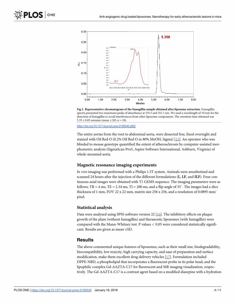
Fig 2. Representative chromatogram of the fumagillin sample obtained after liposome extraction. Fumagillin
spectra presented two maximum peaks of absorbance at 335.5 and 351.1 nm. We used a wavelength of 351nm for the
detection of fumagillin to avoid interferences from other liposome components. The retention time obtained was
5.35 ± 0.05 minutes (mean ± SD; n = 10).
https://doi.org/10.1371/journal.pone.0190540.g002
Anti-angiogenic drug loaded liposomes: Nanotherapy for early atherosclerotic lesions in mice
PLOS ONE | https://doi.org/10.1371/journal.pone.0190540 January 16, 2018 4 / 11
Page 5
number of q = 2 water molecules and renders high relaxivity when compared with typical
based macrocycle or open chain polyamine agents [13,18]. An antibody able to target the mac-
rophage scavenger receptor-B (CD36) was coupled to the surface of liposomes for specific
delivery of immunoliposomes to the lesion. The diameter of the liposomes was determined by
dynamic light scattering resulting in 98.7 nm with a PDI of 0.149.
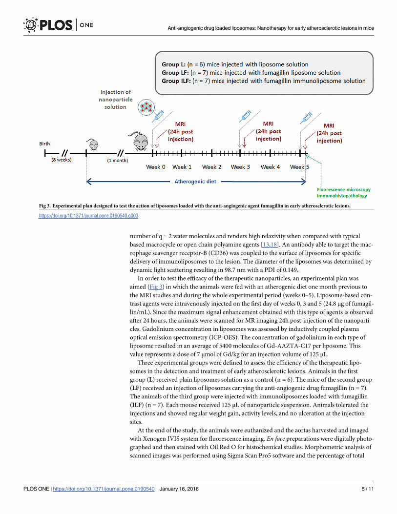
In order to test the efficacy of the therapeutic nanoparticles, an experimental plan was
aimed (Fig 3) in which the animals were fed with an atherogenic diet one month previous to
the MRI studies and during the whole experimental period (weeks 0–5). Liposome-based con-
trast agents were intravenously injected on the first day of weeks 0, 3 and 5 (24.8 μg of fumagil-
lin/mL). Since the maximum signal enhancement obtained with this type of agents is observed
after 24 hours, the animals were scanned for MR imaging 24h post-injection of the nanoparti-
cles. Gadolinium concentration in liposomes was assessed by inductively coupled plasma
optical emission spectrometry (ICP-OES). The concentration of gadolinium in each type of
liposome resulted in an average of 5400 molecules of Gd-AAZTA-C17 per liposome. This
value represents a dose of 7 μmol of Gd/kg for an injection volume of 125 μL.
Three experimental groups were defined to assess the efficiency of the therapeutic lipo-
somes in the detection and treatment of early atherosclerotic lesions. Animals in the first
group (L) received plain liposomes solution as a control (n = 6). The mice of the second group
(LF) received an injection of liposomes carrying the anti-angiogenic drug fumagillin (n = 7).
The animals of the third group were injected with immunoliposomes loaded with fumagillin
(ILF) (n = 7). Each mouse received 125 μL of nanoparticle suspension. Animals tolerated the
injections and showed regular weight gain, activity levels, and no ulceration at the injection
sites.
At the end of the study, the animals were euthanized and the aortas harvested and imaged
with Xenogen IVIS system for fluorescence imaging. En face preparations were digitally photo-
graphed and then stained with Oil Red O for histochemical studies. Morphometric analysis of
scanned images was performed using Sigma Scan Pro5 software and the percentage of total
Fig 3. Experimental plan designed to test the action of liposomes loaded with the anti-angiogenic agent fumagillin in early atherosclerotic lesions.
https://doi.org/10.1371/journal.pone.0190540.g003
Anti-angiogenic drug loaded liposomes: Nanotherapy for early atherosclerotic lesions in mice
PLOS ONE | https://doi.org/10.1371/journal.pone.0190540 January 16, 2018 5 / 11
Page 6
area of the arch and thoracic aorta covered by plaque was calculated. At the time of euthanasia,
there were no difference in body weight between animals in different experimental groups.
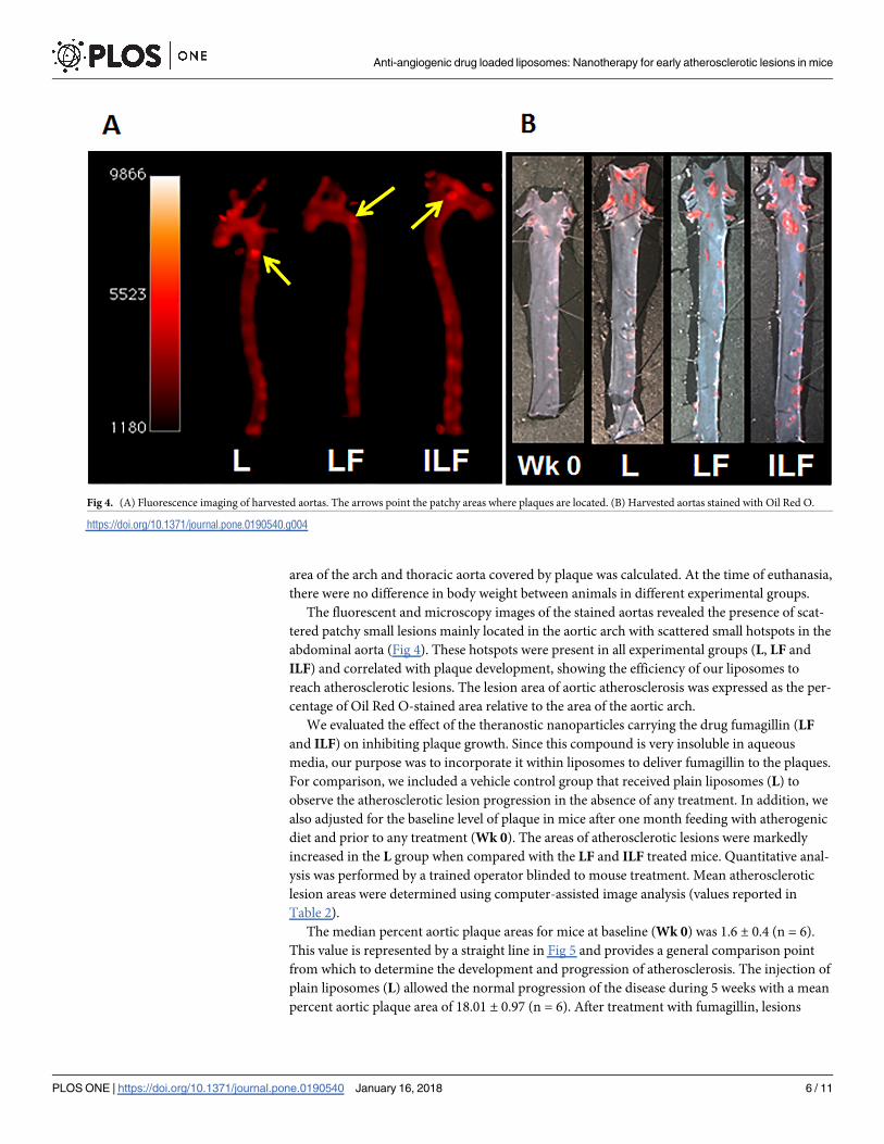
The fluorescent and microscopy images of the stained aortas revealed the presence of scat-
tered patchy small lesions mainly located in the aortic arch with scattered small hotspots in the
abdominal aorta (Fig 4). These hotspots were present in all experimental groups (L, LF and
ILF) and correlated with plaque development, showing the efficiency of our liposomes to
reach atherosclerotic lesions. The lesion area of aortic atherosclerosis was expressed as the per-
centage of Oil Red O-stained area relative to the area of the aortic arch.
We evaluated the effect of the theranostic nanoparticles carrying the drug fumagillin (LF
and ILF) on inhibiting plaque growth. Since this compound is very insoluble in aqueous
media, our purpose was to incorporate it within liposomes to deliver fumagillin to the plaques.
For comparison, we included a vehicle control group that received plain liposomes (L) to
observe the atherosclerotic lesion progression in the absence of any treatment. In addition, we
also adjusted for the baseline level of plaque in mice after one month feeding with atherogenic
diet and prior to any treatment (Wk 0). The areas of atherosclerotic lesions were markedly
increased in the L group when compared with the LF and ILF treated mice. Quantitative anal-
ysis was performed by a trained operator blinded to mouse treatment. Mean atherosclerotic
lesion areas were determined using computer-assisted image analysis (values reported in
Table 2).
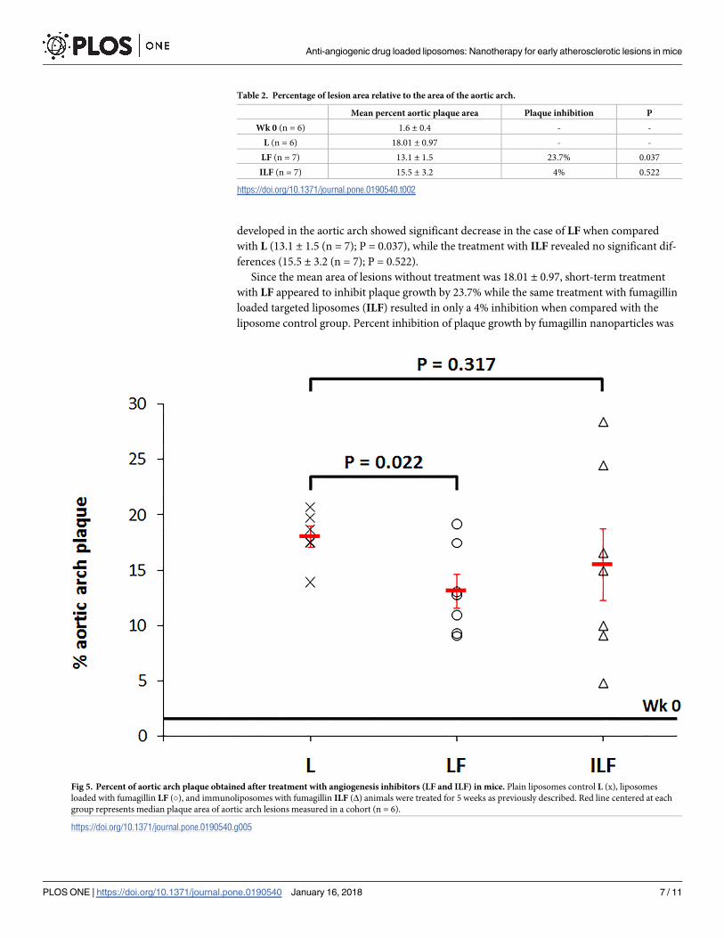
The median percent aortic plaque areas for mice at baseline (Wk 0) was 1.6 ± 0.4 (n = 6).
This value is represented by a straight line in Fig 5 and provides a general comparison point
from which to determine the development and progression of atherosclerosis. The injection of
plain liposomes (L) allowed the normal progression of the disease during 5 weeks with a mean
percent aortic plaque area of 18.01 ± 0.97 (n = 6). After treatment with fumagillin, lesions
Fig 4. (A) Fluorescence imaging of harvested aortas. The arrows point the patchy areas where plaques are located. (B) Harvested aortas stained with Oil Red O.
https://doi.org/10.1371/journal.pone.0190540.g004
Anti-angiogenic drug loaded liposomes: Nanotherapy for early atherosclerotic lesions in mice
PLOS ONE | https://doi.org/10.1371/journal.pone.0190540 January 16, 2018 6 / 11
Page 7
developed in the aortic arch showed significant decrease in the case of LF when compared
with L (13.1 ± 1.5 (n = 7); P = 0.037), while the treatment with ILF revealed no significant dif-
ferences (15.5 ± 3.2 (n = 7); P = 0.522).
Since the mean area of lesions without treatment was 18.01 ± 0.97, short-term treatment
with LF appeared to inhibit plaque growth by 23.7% while the same treatment with fumagillin
loaded targeted liposomes (ILF) resulted in only a 4% inhibition when compared with the
liposome control group. Percent inhibition of plaque growth by fumagillin nanoparticles was
Table 2. Percentage of lesion area relative to the area of the aortic arch.
Mean percent aortic plaque area Plaque inhibition P
Wk 0 (n = 6) 1.6 ± 0.4 - -
L (n = 6) 18.01 ± 0.97 - -
LF (n = 7) 13.1 ± 1.5 23.7% 0.037
ILF (n = 7) 15.5 ± 3.2 4% 0.522
https://doi.org/10.1371/journal.pone.0190540.t002
Fig 5. Percent of aortic arch plaque obtained after treatment with angiogenesis inhibitors (LF and ILF) in mice. Plain liposomes control L (x), liposomes
loaded with fumagillin LF (�), and immunoliposomes with fumagillin ILF (Δ) animals were treated for 5 weeks as previously described. Red line centered at each
group represents median plaque area of aortic arch lesions measured in a cohort (n = 6).
https://doi.org/10.1371/journal.pone.0190540.g005
Anti-angiogenic drug loaded liposomes: Nanotherapy for early atherosclerotic lesions in mice
PLOS ONE | https://doi.org/10.1371/journal.pone.0190540 January 16, 2018 7 / 11
Page 8
calculated according to Eq (1) [10]
% inhibition ¼ 100 � 1 �median percent plaque area treated � 0:25
median percent plaque area control � 0:25
� �� �
ð1Þ
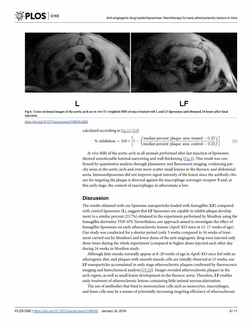
In vivo MRI of the aortic arch in all animals performed after last injection of liposomes
showed unnoticeable luminal narrowing and wall thickening (Fig 6). This result was con-
firmed by quantitative analysis through planimetry and fluorescent imaging, evidencing pat-
chy areas in the aortic arch and even more scatter small lesions in the thoracic and abdominal
aorta. Immunoliposomes did not improve signal intensity of the lesion since the antibody cho-
sen for targeting the plaque is directed against the macrophage scavenger receptor B and, at
this early stage, the content of macrophages in atheromata is low.
Discussion
The results obtained with our liposome nanoparticles loaded with fumagillin (LF) compared
with control liposomes (L), suggest that LF liposomes are capable to inhibit plaque develop-
ment to a similar percent (23.7%) obtained in the experiment performed by Moulton using the
fumagillin derivative TNP-470. Nevertheless, our approach aimed to investigate the effect of
fumagillin liposomes on early atherosclerotic lesions (ApoE-KO mice at 12–17 weeks of age).
Our study was conducted for a shorter period (only 5 weeks compared to 16 weeks of treat-
ment carried out by Moulton) and lower doses of the anti-angiogenic drug were injected only
three times during the whole experiment (compared to higher doses injected each other day
during 16 weeks in Moulton study.
Although fatty streaks normally appear at 8–20 weeks of age in ApoE-KO mice fed with an
atherogenic diet, and plaques with smooth muscle cells are initially observed at 15 weeks, our
LF nanoparticles accumulated in early stage atherosclerotic plaques confirmed by fluorescence
imaging and histochemical analysis.[19,20]. Images revealed atherosclerotic plaques in the
arch region, as well as small lesion development in the thoracic aorta. Therefore, LF enables
early treatment of atherosclerotic lesions containing little intimal neovascularization.
The use of antibodies that bind to mononuclear cells such as monocytes, macrophages,
and foam cells may be a means of potentially increasing targeting efficiency of atherosclerosis
Fig 6. Cross-sectional images of the aortic arch on in vivoT1-weighted MRI of mice treated with L and LF liposomes and obtained 24 hours after final
injection.
https://doi.org/10.1371/journal.pone.0190540.g006
Anti-angiogenic drug loaded liposomes: Nanotherapy for early atherosclerotic lesions in mice
PLOS ONE | https://doi.org/10.1371/journal.pone.0190540 January 16, 2018 8 / 11
Page 9
since these cells have been shown to play a key role in the progression of disease [21]. CD36
is a glycoprotein that is present in the membrane of macrophages, thus antiCD36 targets the
macrophage scavenger receptor-B facilitating the in vivo imaging of atherosclerotic plaques
according to the amount of macrophages accumulated in the lesion. This was demonstrated by
Lipinski et al showing signal enhancement in advanced plaque imaging studies [19]. Taking
this into consideration, we synthesized immunoliposomes including antiCD36 loaded with
fumagillin (ILF) for better targeting and therapeutic efficiency. However, ILF produced no sig-
nificant inhibition compared with control liposomes (L), which can be attributed to the very
early stage of the plaques. In our in vivo experiment, the small percentage of atherosclerotic
lesions present in the aortic arch just reflected the low macrophages content in the lesion.
Treatment with immunoliposomes resulted in poor targeting and consequently in little
decrease of plaque development. This was evidenced with a smaller inhibition percentage than
in the case of non-targeted liposomes (LF). Additionally, it must be taking into consideration
that intravenous administration of nanoparticles able to target CD36 protein can be retained
in the liver by binding to scavenger receptors expressed on the surface of Kuppfer cells [22] or
to any other cells that express CD36 such as endothelial cells, smooth muscle cells, adipocytes,
and platelets.
Regarding to the choice of the therapeutic agent, the important role of angiogenesis in ath-
erosclerosis has generated wide interest during the last years since many studies suggest that
angiogenesis constitutes a potential target for the treatment of atherosclerosis [23,24]. Novel
and specific therapies based on the administration of anti-angiogenic agents have shown to
decrease both neovascular proliferation and plaque development in animal models of athero-
sclerosis [25,26]. Effective inhibitors of angiogenesis include fumagillin and synthetic deriva-
tives. Fumagillin has proven to exhibit anti-angiogenic effects in cancer therapies [27,28], and
more recently research has shown that the administration of TNP-470, a water soluble form of
fumagillin, resulted in significant reduction in plaque growth [10]. However, there was no sig-
nificant effect at the very early stages of atherosclerotic plaque formation and furthermore, sys-
temic administration of high doses of TNP-470 during prolonged period of time is associated
with neurotoxic effects [29].
For the purpose of diminishing side effects, we have developed targeted (ILF) and non-tar-
geted (LF) fumagillin loaded liposomes as theranostic strategy against plaque progression.
Since fumagillin is highly hydrophobic, the compound will probably be entrapped within the
phospholipid bilayer of the nanoparticle. Fumagillin is probably delivered into the cells by a
mechanism referred to as “contact-facilitated drug delivery” (CFDD), a process in which the
liposome containing the therapeutic drug facilitates the interaction and hemifusion with the
target cell surface phospholipids promoting the passive transfer of the drug [12].
Conclusion
We have developed liposome-based nanoparticles able to reach atherosclerotic lesions. Anti-
angiogenesis treatment with fumagillin-loaded liposomes appears to inhibit atherosclerotic
plaque growth, suggesting that even early treatment can significantly attenuate the develop-
ment of atherosclerosis. The use of antiCD36 immunoliposomes did not significantly inhibit
the development of atherosclerosis compared with a naked liposome control group.
Supporting information
S1 Table. Percent of aortic arch plaque data.
(PDF)
Anti-angiogenic drug loaded liposomes: Nanotherapy for early atherosclerotic lesions in mice
PLOS ONE | https://doi.org/10.1371/journal.pone.0190540 January 16, 2018 9 / 11
Page 10
S1 Fig. Graphical abstract figure.
(TIF)
Acknowledgments
We would like to thank Drs LJ Jimenez-Borreguero, J Ruiz Cabello and V Andres for their
help with the animal experiments at CNIC in Madrid.
Author Contributions
Conceptualization: Isabel Pont.
Investigation: Isabel Pont, Aracely Calatayud-Pascual, Alicia Lopez-Castellano, Elena P.
Albelda, Enrique Garcıa-España, Luis Martı-Bonmatı, Juan C. Frias, M. Teresa Albelda.
Methodology: Isabel Pont, Aracely Calatayud-Pascual, Alicia Lopez-Castellano, Elena P.
Albelda, Enrique Garcıa-España, Luis Martı-Bonmatı, Juan C. Frias, M. Teresa Albelda.
Supervision: Elena P. Albelda, Enrique Garcıa-España, Luis Martı-Bonmatı, Juan C. Frias, M.
Teresa Albelda.
Validation: Elena P. Albelda, Juan C. Frias, M. Teresa Albelda.
Writing – original draft: Juan C. Frias, M. Teresa Albelda.
Writing – review & editing: Enrique Garcıa-España, Luis Martı-Bonmatı, Juan C. Frias, M.
Teresa Albelda.
References1. Tang J, Lobatto ME, Read JC, Mieszawska AJ, Fayad ZA, Mulder WJ, Nanomedical Theranostics in
Cardiovascular Disease. Curr Cardiovasc Imaging Rep. 2012; 5(1):19–25. https://doi.org/10.1007/
s12410-011-9120-6 PMID: 22308199
2. Winter PM, Caruthers SD, Zhang H, Williams TA, Wickline SA, Lanza GM Antiangiogenic synergism of
integrin-targeted fumagillin nanoparticles and atorvastatin in atherosclerosis. JACC Cardiovasc Imag-
ing. 2008; 1(5):624–34. https://doi.org/10.1016/j.jcmg.2008.06.003 PMID: 19356492
3. Lipinski MJ, Amirbekian V, Frias JC, Aguinaldo JG, Mani V, Briley-Saebo KC, et al. MRI to detect ath-
erosclerosis with gadolinium-containing immunomicelles targeting the macrophage scavenger receptor.
Magn Reson Med. 2006; 56(3):601–10. https://doi.org/10.1002/mrm.20995 PMID: 16902977
4. Sin N, Meng L, Wang MQ, Wen JJ, Bornmann WG, Crews CM. The anti-angiogenic agent fumagillin
covalently binds and inhibits the methionine aminopeptidase, MetAP-2. Proc Natl Acad Sci USA. 1997;
94(12):6099–103. PMID: 9177176
5. Zhou HF, Yan H, Hu Y, Springer LE, Yang X, Wickline SA, et al. Fumagillin prodrug nanotherapy sup-
presses macrophage inflammatory response via endothelial nitric oxide. ACS Nano. 2014; 8(7):7305–
17. https://doi.org/10.1021/nn502372n PMID: 24941020
6. Zhou HF, Yan H, Senpan A, Wickline SA, Pan D, Lanza GM, et al. Suppression of inflammation in a
mouse model of rheumatoid arthritis using targeted lipase-labile fumagillin prodrug nanoparticles. Bio-
materials. 2012; 33(33):8632–40. https://doi.org/10.1016/j.biomaterials.2012.08.005 PMID: 22922023
7. Sanz J, Fayad ZA. Imaging of atherosclerotic cardiovascular disease. Nature. 2008; 451(7181):953–7.
https://doi.org/10.1038/nature06803 PMID: 18288186
8. Albelda MT, Garcia-España E, Frias JC. Visualizing the atherosclerotic plaque: a chemical perspective.
Chem Soc Rev. 2014; 43(8):2858–76. https://doi.org/10.1039/c3cs60410a PMID: 24526041
9. Amirbekian V, Lipinski MJ, Briley-Saebo KC, Amirbekian S, Aguinaldo JGS, Weinreb DB, et al. Detect-
ing and assessing macrophages in vivo to evaluate atherosclerosis noninvasively using molecular MRI.
Proc Natl Acad Sci USA. 2007; 104(3):961–6. https://doi.org/10.1073/pnas.0606281104 PMID:
17215360
10. Moulton KS, Heller E, Konerding MA, Flynn E, Palinski W, Folkman J. Angiogenesis inhibitors endosta-
tin or TNP-470 reduce intimal neovascularization and plaque growth in apolipoprotein E-deficient mice.
Circulation. 1999; 99(13):1726–32. PMID: 10190883
Anti-angiogenic drug loaded liposomes: Nanotherapy for early atherosclerotic lesions in mice
PLOS ONE | https://doi.org/10.1371/journal.pone.0190540 January 16, 2018 10 / 11
Page 11
11. Lobatto ME, Fayad ZA, Silvera S, Vucic E, Calcagno C, Mani V, et al. Multimodal clinical imaging to lon-
gitudinally assess a nanomedical anti-inflammatory treatment in experimental atherosclerosis. Mol
Pharm. 2010; 7(6):2020–9. https://doi.org/10.1021/mp100309y PMID: 21028895
12. Kilarski WW, Petersson L, Fuchs PF, Zielinski MS, Gerwins P. An in vivo neovascularization assay for
screening regulators of angiogenesis and assessing their effects on pre-existing vessels. Angiogenesis.
2012; 15(4):643–55. https://doi.org/10.1007/s10456-012-9287-8 PMID: 22918697
13. Gianolio E, Giovenzana GB, Longo D, Longo I, Menegotto I, Aime S. Relaxometric and modelling stud-
ies of the binding of a lipophilic Gd-AAZTA complex to fatted and defatted human serum albumin.
Chemistry. 2007; 13(20):5785–97. https://doi.org/10.1002/chem.200601277 PMID: 17407109
14. Brackett JM, Arguello MD, Schaar JC. Determination of fumagillin by high performance liquid chroma-
tography. J Agric Food Chem. 1988; 36(4):762–4.
15. Gonzalez-Navarro H, Abu Nabah YN, Vinue A, Andres-Manzano MJ, Collado M, Serrano M, et al. p19
(ARF) deficiency reduces macrophage and vascular smooth muscle cell apoptosis and aggravates ath-
erosclerosis. J Am Coll Cardiol. 2010; 55(20):2258–68. https://doi.org/10.1016/j.jacc.2010.01.026
PMID: 20381282
16. IBM Corp. Released 2013. IBM SPSS Statistics for Windows, Version 22.0. Armonk, NY: IBM Corp.
17. Torchilin VP. Recent advances with liposomes as pharmaceutical carriers. Nat Rev Drug Discov. 2005;
4(2):145–60. https://doi.org/10.1038/nrd1632 PMID: 15688077
18. Briley-Saebo KC, Geninatti-Crich S, Cormode DP, Barazza A, Mulder WJM, Chen W, et al. High-relax-
ivity gadolinium-modified high-density lipoproteins as magnetic resonance imaging contrast agents. J
Phys Chem B. 2009; 113(18):6283–9. https://doi.org/10.1021/jp8108286 PMID: 19361222
19. Lipinski MJ, Frias JC, Amirbekian V, Briley-Saebo KC, Mani V, Samber D, et al. Macrophage-specific
lipid-based nanoparticles improve cardiac magnetic resonance detection and characterization of
human atherosclerosis. JACC Cardiovasc Imaging. 2009; 2(5):637–47. https://doi.org/10.1016/j.jcmg.
2008.08.009 PMID: 19442953
20. Getz GS, Reardon CA. Animal Models of Atherosclerosis. Arterioscler Thromb Vasc Biol. 2012; 32
(5):1104–15. https://doi.org/10.1161/ATVBAHA.111.237693 PMID: 22383700
21. Park YM. CD36, a scavenger receptor implicated in atherosclerosis. Exp Mol Med. 2014; 46:e99.
https://doi.org/10.1038/emm.2014.38 PMID: 24903227
22. Lipinski MJ, Frias JC, Fayad ZA. Advances in detection and characterization of atherosclerosis using
contrast agents targeting the macrophage. J Nucl Cardiol. 2006; 13(5):699–709. https://doi.org/10.
1016/j.nuclcard.2006.07.004 PMID: 16945750
23. Patel A. Does the Role of Angiogenesis play a Role in Atherosclerosis and Plaque Instability? Anat Phy-
siol. 2014; 4(3):147–58.
24. Pant S, Deshmukh A, Mehta JL. Angiogenesis in Atherosclerosis: An Overview. In: Mehta J, Dhalla N,
editors. Biochemical Basis and Therapeutic Implications of Angiogenesis. New York: Springer; 2013.
p. 209–24.
25. Deveza L, Choi J, Yang F. Therapeutic Angiogenesis for Treating Cardiovascular Diseases. Theranos-
tics. 2012; 2(8):801–14. https://doi.org/10.7150/thno.4419 PMID: 22916079
26. Katsi VK, Psarros CT, Krokidis MG, Vamvakou GD, Tousoulis D, Stefanadis CI, et al. Anti-Angiogenic
Therapy and Cardiovascular Diseases: Current Strategies and Future Perspectives. In: Rahman AU,
Choudhary MI, editors. Anti-Angiogenesis Drug Discovery and Development. Amsterdam: Bentham
Science Publishers; 2014. p. 268–308.
27. Griffith EC, Su Z, Niwayama S, Ramsay CA, Chang YH, Liu JO.Molecular recognition of angiogenesis
inhibitors fumagillin and ovalicin by methionine aminopeptidase 2. Proc Natl Acad Sci USA. 1998; 95
(26):15183–8. PMID: 9860943
28. Kruger EA, Figg WD. TNP-470: an angiogenesis inhibitor in clinical development for cancer. Expert
Opin Investig Drugs. 2000; 9(6):1383–96. https://doi.org/10.1517/13543784.9.6.1383 PMID: 11060750
29. Ingber D, Fujita T, Kishimoto S, Sudo K, Kanamaru T, Brem H, et al. Synthetic analogues of fumagillin
that inhibit angiogenesis and suppress tumour growth. Nature. 1990; 348(6301):555–7. https://doi.org/
10.1038/348555a0 PMID: 1701033
Anti-angiogenic drug loaded liposomes: Nanotherapy for early atherosclerotic lesions in mice
PLOS ONE | https://doi.org/10.1371/journal.pone.0190540 January 16, 2018 11 / 11