Page 1
Anti-bacterial and Anti-adhesive Nanostructured
Coatings for Improved Implant Biocompatibility
Dissertation
zur
Erlangung des Doktorgrades
der Naturwissenschaften
(Dr.rer.nat.)
dem
Fachbereich Pharmazie
der Phillipps-Universität Marburg
vorgelegt von
Eyas Dayyoub
aus Latakia/Syrien
Marburg an der Lahn 2012
Page 2
Vom Fachbereich Pharmazie der Philipps-Universität Marburg als Dissertation am
14.02.2012 angenommen
Erstgutachter: Prof.Dr. Udo Bakowsky
Zweitgutachter: Prof.Dr. Michael Keusgen
Tag der mündlichen Prüfung am 20.03.2012
Page 3
Die vorliegende Arbeit entstand
auf Anregung und unter Leitung von
Herrn Prof. Dr. Udo Bakowsky
am Institut für Pharmazeutische Technologie und Biopharmazie
der Philipps-Universität Marburg.
Page 4
Meiner Familie
In Liebe und Dankbarkeit
Page 5
“It’s not the idea that I am extraordinarily intelligent, but everything
in it, that I spend more time in solving the problems!”
“If A equals success, then the formula is: A = X + Y + Z
X is work, Y is play, and Z is keeping your mouth shut!”
Albert Einstein
Page 6
Danksagung
Mein besonderer Dank gilt meinem Doktorvater Herrn Prof. Dr. Udo Bakowsky für die
fortwährend geduldige Betreuung, die unermüdliche Anregung und Motivation, die Freiheit
bei der Umsetzung sowie seine stete Diskussionsbereitschaft während der gesamten
Promotion.
Ich danke meinen Kollegen ganz herzlich für die Unterstützung, besonders Johannes
Sitterberg für die technische Unterstützung während der AFM-Messungen, Dr. Jens Schäfer
für den lehrreichen Erfahrungsaustausch und die administrative Unterstützung , Elena Marxer
für die wertvolle Freundschaft, Aybike Özcetin für die angenehme Unterhaltung, Jana Brüßler
für die stete Hilfsbereitschaft, Nico Harbach für die Hilfe bei der deutschen Sprache, Thomas
Betz für die Einführung in die saarländische Küche, Maria Solovey für die englische
Korrektur, Boris Strehlow für die Unterstützung bei den REM-Messungen, Mario Bandulik
für die nette Gesellschaft beim Shisha rauchen. Allen anderen, Bassam Al Meslmani, Anett
Sommerwerk, Leonie Baginski und Susanne Lüttebrandt danke ich ebenfalls für die gute
Zusammenarbeit.
Weiterhin bedanke ich mich beim Herrn Prof. Dr. Michael Keusgen, der mir stets
Ansprechpartner für unser Kooperationsprojekt war, für die Erstellung des Zweitgutachtens.
Ich danke Herrn Prof. Dr. Thomas Kissel sowie seinen Mitarbeitern Dr. Moritz Beck-
Broichsitter, Dr. Nadja Bege, Dr. Markus Benfer, Dr. Heiko Debus, Thomas Endres, Klaus
Keim, Dr. Tobias Lebhardt, Dr. Sascha Maretschek, Dr. Olivia Merkel, Eva Mohr, Frank
Morell, Dr. Thomas Renette, Dr. Regina Reul, Dr. Susanne Rösler, Olga Samsonova, Dr.
Christoph Schweiger, Nina Seidel, Dr. Nan Zhao, Dr. Claudia Packhäuser, Prof. Dr. Oliver
Germershaus, Dr. Michael Neu, Dr. Juliane Nguyen, Dr. Terry Steele, Dr. Erik Rytting, Dr.
Yu Liu und Dr. Cuifang Cai für die nette Zeit im Institut und ihre Freundschaften.
Page 7
Prof. Dr. Klaus Liefeith und Dr. Marion Frant von dem IBA/Heiligenstadt danke ichfür die
erfolgreiche Zusammenarbeit.
Den Mitarbeitern des AK Keusgen, besonders Dr. Elvira Belz, Nina Dassinger, Christian
Hobler und Doru Gheorghe Marcel Vornicescu danke ich für die erfolgreiche
Zusammenarbeit.
Ich möchte Herrn Prof. Dr. Torsten Steinmetzer und Herrn Prof. Dr. Klaus Kuschinsky dafür
danken, dass sie die Aufgaben der Nebenfachprüfer übernommen haben.
Von tiefsten Herzen möchte ich meinen Eltern und Geschwistern danken.Meinem Vater für
sein Vertrauen und die finanzielle Unterstützung, meiner Mutter für ihre Geduld und Liebe,
meinen beiden Brüder und meiner Schwester für die herrliche alte Zeit.
Page 8
I
Table of Contents
Chapter 1
Introduction………………………………………. 1
Biofilm:Formation and Architecture ………………………... 2
Biofilm on Implant Surfaces…………………………………. 6
Strategies to Resist Biofilm…………………………………… 8
Surface Morphology………………………………………….. 10
Coatings for Release of Anti-bacterial Agents……………….. 15
Protein Adsorption on Implant Surfaces……………………… 17
Factors Influence Protein Adsorption………………………… 19
Strategies to Resist Protein Adsorption………………………. 20
References…………………………………………………….. 22
Chapter 2
Anti-bacterial and Anti-encrustation Hydrophobic Biodegradable
Polymer Coating for Urinary Catheter............... 35
Abstract………………………………………………………... 36
Introduction……………………………………………………. 37
Materials and Methods………………………………………… 42
Results and Discussion………………………………………... 48
Conclusion…………………………………………………….. 61
Acknowledgement…………………………………………….. 62
References……………………………………………………... 63
Page 9
II
Chapter 3
Highly Ordered Self-Organized Polymer
Coatings for Reduced Bacteria Adhesion………... 71
Abstract………………………………………………………… 72
Introduction……………………………………………………. 73
Materials and methods…………………………………………. 76
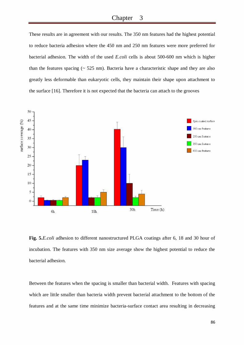
Results………………………………………………………….. 79
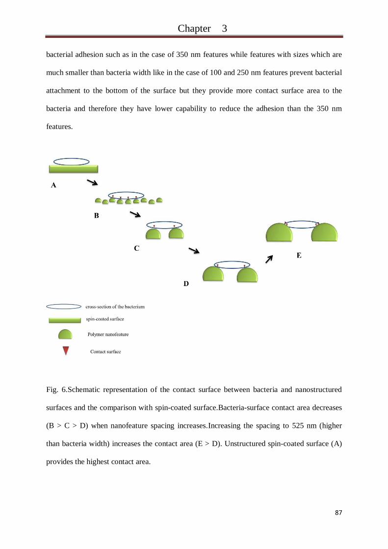
Discussion………………………………………………………. 83
Conclusion……………………………………………………… 88
Acknowledgement……………………………………………… 88
References………………………………………………………. 89
Chapter 4
A novel Method for Designing Nanostructured Polymer
Surfaces for Reduced Bacteria Adhesion…… 93
Abstract…………………………………………………………. 94
Introduction……………………………………………………... 95
Material and methods…………………………………………… 97
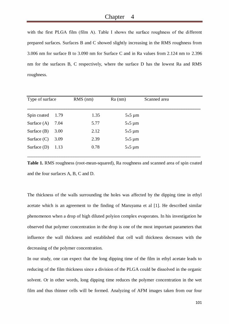
Results and Discussion…………………………………………. 99
Conclusion……………………………………………………… 105
Acknowledgement……………………………………………… 105
References………………………………………………………. 106
Page 10
III
Chapter 5
New Antibacterial, Antiadhesive Films Based on
Self-assemblies ofModified Tetraetherlipid……….. 107
Abstract…………………………………………………………… 108
Introduction………………………………………………………. 109
Material and methods…………………………………………….. 111
Results and Discussion…………………………………………… 113
Conclusion……………………………………………………….. 119
Acknowledgement……………………………………………….. 119
References………………………………………………………... 120
Chapter 6
Self-assembled N-succinyl-chitosan Nanofibers
for Reduced Protein Adhesion………………... 121
Abstract…………………………………………………………… 122
Introduction………………………………………………………. 123
Material and methods…………………………………………….. 124
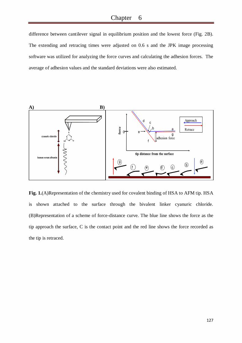
Results and Discussion…………………………………………… 127
Summary………………………………………………………….. 131
Acknowledgement………………………………………………… 131
References………………………………………………………… 132
Page 11
IV
Chapter 7
Nanostructured Medical Device Coatings
Based on Self-assembled Poly(lactic-co-glycolic acid)
Nanoparticles…………………………………. 133
Abstract…………………………………………………………... 134
Introduction………………………………………………………. 135
Material and methods…………………………………………….. 138
Results and Discussion…………………………………………… 144
Conclusion……………………………………………………….. 153
References………………………………………………………... 154
Chapter 8
Summary and perspectives………………………..... 157
Zusammenfassung und Ausblick…………………… 163
Appendices…………………………………………… 170
Page 13
Chapter 1
2
Biofilm
Formation and Architecture
Growth of bacteria is characterized by two forms of life, one being as single cells (planktonic)
and the other being in sessile aggregates [1]. These aggregates are called Biofilms. Like many
other communities, the existing of bacteria in groups offers the members of the population
advantages they would not achieve when they are in a single form. Examples of other
sociobiology existingare easy to find innature; Herds of mammals, flocks of birds, schools of
fish, and colonies of insects are prime examples where life of members becomes simpler with
protection of the groups [2].
Bacterial biofilms are described as cells bound together by extracellular polymeric substances
(EPS) and attached together and to biotic or abiotic surface, the extracellular matrix consists
of different kinds of substances such as protein, DNA and polysaccharides [1,3,4]. In nature,
probably 99% of the bacteria exist in biofilm form. Bacteria attach to surface and secrete
extracellular matrix that protect the bacteria from environment dangers like white blood cells,
antibodies and therapeutic antibiotics [5]. Most of biofilms have water channels which are
employed as distribution systems for water and nutrients [1].
Biofilm formation is not a random process; it is an ancient and integral componentof the
prokaryotic life cycle [6,7] (Fig 1). In the firstphase, bacteria are transported to surfaces by
sedimentation, liquid flow,brownian motionand active motion. In this phase, motile
bacteriacan use their appendages such as flagella and fimbriae (or pilli) for active swimming
[6]. This transport provides direct contact of bacteria with the surface. The attachment of
bacteria in this stage depends mainly on favorable bacteria-surface interaction to overcome
the repulsive forces occur between bacteria surface and the surface to be colonized [8] and
therefore the structure of bacterial surface plays major roll in this stage. In the case of
flagellated bacteria, flagellum is not only responsible for motility which may be necessary to
Page 14
Chapter 1
3
reach the surface; it can also promote recognition and initial adhesion to surface.Studies
compared between various bacterial species and have shown that flagella are either
completely necessary for, or quicken initial attachment [6,9-12]. The explanation depends on
the hypothesis that flagella help the bacteria to overcome the repulsive forces between
bacterial surface and substratum [13]. In addition to flagellum, bacterial fimbriae/pili can also
promote and accelerate surface attachment [14,15].
Fig. 1. Steps of biofilm formation (ref: http://biotuesdays.com/2010/10/19/innovotech-targets-
personalized-medicine-for-bacteria/).
Further key parameters that influence bacterial initial adhesion are the bacterial membrane
molecules such as lipopolysaccharide, lipoproteins, membrane protein, adhesins, etc [16] and
the interactions between these molecules and the colonized surface. When membrane
molecules come in contact with the conditioning film, short and long range forces like
electrostatic, hydrophobic and van der Waals forces in addition to hydrogen bonds, dipole-
dipole and coulomb interactions can take place [17]. In aquatic environment, organic
Page 15
Chapter 1
4
materials attach to the substratum surface before the bacteria, these organic substances form a
conditioning film which covers the original surface. Indeed the initial adhesion of bacteria
depends on the nature of the conditioning film and not of the original one [18].
If the forces are attractive, a weak and reversible attachment occurs.The influence of the
environment must be considered since some factors like ionic strength and pH of the medium
can alter surface charge of both bacteria and substratum surfaces resulting in changes of the
interactions between the two [19-23]. At the end of this phase, bacteria form monolayer
weakly attached to the surface.
In the second step, irreversible attachment is constructed and the bacteria undergo significant
changes and initiate lifestyle switch. In the case of non-motile species, the secretion of
adhesins increases which promote the cell-cell and cell-surface adherence [24] while in the
case of motile species; extracellular matrix is produced to hold the cells together and
strengthening the adhesion to surface [25]. These EPS are not only associated with cell
surface, they are also excreted in the bacterial growth medium and therefore, they can be
presented on the surface to be colonized when secreted from bacteria [26]
It is believed that stimulation of sensory protein bound to membrane leads to producing of
EPS [27]. The process continues resulting in three dimensional architecture. In this step,
bacteria of the biofilm can interact with their neighbor in the local environment by releasing
small diffusible molecules, this system is called Quorum-sensing [28]. It depends on self-
generated molecules which are used as signals to coordinate gene expression in correlation to
population density [29].
The next step is the surface colonization, bacteria grow and divide inside the biofilm;
entrapment of other planktonic cells can also take place leading to the formation of a biofilm
[30].The composition of this biofilm is complicated, it differs between the various species of
bacteria, it mainly consists of biofilm bacteria entrapped in EPS with high preamble water
channels which carry nutrients and waste products [31]. EPS differ between gram-positive
Page 16
Chapter 1
5
and gram negative bacteria as it contains different EPS polysaccharide. These polysaccharides
are neutral or polyanionic in the gram-negative bacteria while gram negative bacteria have
cationic polysaccharide. The biofilm structure and architecture can also be affected by
parameters like available nutrition in the environment [32] and hydrodynamic conditions [33].
The last step is the detachment of bacteria from biofilm, this happens in the case of
unfavorable environment conditions like nutrition limitation [6], the released bacteria attached
on other surface and begin again to form another biofilm.
Page 17
Chapter 1
6
Biofilm on Implant Surfaces
The Food and Drug Administration (FDA) defines an implant as a "device that is placed into a
surgically or naturally formed cavity of the human body if it is intended to remain there for a
period of 30 days or more." [34]. Implants can be classified in regards to the application site,
they also vary due to materials they made from.
Dental implants, neural, orthopedic and urologic prostheses, vascular graft, venous and
urinary catheter are well-known examples for medical implants. The used materials vary
between polyethylene terephthalate (PET or Dacron), polytetrafluoroethylene (PTFE),
polyurethane (PUR), polyimide, silicon and titanium. According to implant type, implant
surface encounter one or more body fluids like saliva, urine, blood and gastrointestinal
secretions. Human tissues come also in contact with the implant resulting in different kinds of
interactions.
Biocompatibility of the implant depends on the tissue-implant and body fluid-implant
interactions.Undesired interactions canadversely affect body and cell functions, the effect can
appear like inflammation, cell proliferation,coagulation, encrustation and biofilm formation.
Biofilms and the associated infections at the site of implantation present a serious problem for
the patients. Bacteria come in contact with implant surface and construct biofilm, the formed
biofilms are highly resistant to both immune system of the host and systematic antibiotic [35].
When the bacteria exist in biofilm, they become 10-1000 times more resistance against
antibacterial agents [36,37]. Hypotheses suggested mechanisms resistance developments; the
first theory suggested that the biofilm glycocalyx prevents diffusion of the antibiotic in the
film[38] the second hypotheses depends on the altering of bacterial growth rate which dictate
the response to antimicrobial agents [39] while the third one supposed that microenvironment
in the biofilm has influence on the antimicrobial activity [40]. The bacteria get into human
body and reach implant surface through different ways; the possible sources are the ambient
Page 18
Chapter 1
7
atmosphere, surgical tools, clothes, bacteria on the patient`s skin and bacteria already in the
body [41]
In spite of theefforts, made to develop new implants materials for reduced bacterial adhesion,
the rate of implant-associated infections is still high. In the United State, 2.6 million
orthopedic implants are inserted in humans annually and 4.3% of them become infected,the
medical costs for implant-associated infections exceed about $ 3 billion yearly in the USA
alone [42]. In addition to the human pain and suffering because of the infections, these
infections can sometimes only be treated by removal of the implant.
To reduce or prevent biofilm formation, effective strategies were followed. They depend on
i) preventing or reduction of bacterial adhesion by physiochemical modifying of implant
surface ii) systematical or local controlled release of antibiotic.
Page 19
Chapter 1
8
Strategies to Resist Biofilm
Between the four steps of biofilm formation, the initial adhesion is a determining step. If the
initial adhesion is prevented, the bacteria fail to build biofilm and are less capable to cause
infections since bacterial attachment is the first step of infection development. The planktonic
bacteria are more easily killed by antibacterial agents or host immune system than the bacteria
in biofilm as described previously.
Characteristics of surfaces of both bacteria and substratum exert significant influence over the
tendency of bacteria to attach to different surfaces [43,44] since the forces that affect bacterial
adhesion are the interaction forces at the interfaces between the two surfaces.
There have been a number of studies concerning the influence of substratum surface
characteristics on bacterial attachment.
Some of these studies have concentrated on the influence of micro- and nanostructured
surfaces on the bacterial adhesion and they showed evidences that bacteria can response to
micro- and nanoscale surface features[45-50]. The mechanisms that regulate this response are
still less well understood. The effect of this factor is extensively discussed in the next section.
Other studies concentrated on the chemical structure of the surface. Biofuctionalization,
coating and chemical modifying of the implant surface showed interesting potential to resist
bacterial adhesion and biofilm formation [51-56]. Plasma treatment of the surface results in
changing of surface free energy. Upon this fact, the treated surface can decrease the tendency
of bacteria to adhere and form biofilm [57,58].
Localized administration of antimicrobial agents is also desirable choice to resist the risk of
bacterial adhesion and biofilm formation. As described previously, bacteria within biofilm can
develop high resistance against antibacterial agents; therefore, the admisitration of
antibacterial agents locally rather than systematically can prevent or reduce the biofilm
formation which in turn inhibits creation of antibacterial-resistant strains in the film. In
Page 20
Chapter 1
9
addition to the last advantage,The high required over-kill dosage in the case of systematical
application exposures human body to different risks like side-effects and develompemt of
resistant strains in the body in addition to the need to take the drug more than one time daily
and the pain caused by intravenous application in some cases.
Different antibiotic like aminoclycosides, cyphalosporines, penicillins and quinolones in
addition to inorganic antibacterial agents like silver and nitric oxide were incorporated in
coatings or implant materials to be released in the site of implantation [59-66]. More
explanation is described in the next sections.
Page 21
Chapter 1
10
Surface Morphology
The fast development of biomedical industry and bio-interfaces analyzing techniques
accompanied with large increasing of studies concerning on the enhancement of implant
biocompatibility. These new techniques allow more understanding of biological response to
implants and its mechanisms at micro-, nano- and molecular scale.The advances in micor- and
nanotechnology have allowed the fabrication of appropriate structured substrates and the aim
was controlling the biological response by altering the unfavorable human-implant
interactions.
It has been shown that surface topography is an important key to modulate human cell
response to this surface. Cells react to macro, micro- and nanostructures. Human cells attach
to surface using different molecules; the most common ones are integrins [67], clusters of
integrins link the cell to extracellular matrix (ECM). The clustering of integrins is essential in
the formation of mutant focal adhesions [68,69]. The cells can expand and bend their
membrane when they adhere to surfaces.
Recently, increasing number of scientist investigated the mechansims by which human cells
adhere to surface. Comparisons were also done between bacteria and human cells regarding
their adhesion to surface; but, till now less is known about the capability of bacteria to sense
the surface and thedriving mechanisms than that of eukaryotic cells [67].
Bacterial cells are more rigid and can‘t change their form which is partly due to the external
layer of peptidoglycan; this layer is thick in gram-positive bacteria while gram-negative
bacteria have thin layer which is covered by additional polysaccharide outer layer [70]. They
also have variety of surface structures and different outer membrane proteins; some of them
express flagella or pili depending on the strain and species [71,72] which renders them very
motile and promote surface attachment as described previously.
Page 22
Chapter 1
11
Bacteria vary significantly in size and shape, their size range between under 1µm to several
tens of micrometers and their shape can be spherical, twisted or rod one [70].
This variety of membrane structures, rigidity, shape, motility resulted in different bacterial
reaction to topography of the surface they colonized. In contrast, surface topography is
characterized by surface roughness, feature shapes (holes, graves, tubes, fibers, micro- and
nanoparticles), feature size and distance between the features.
Roughness may influences surface properties like water contact angle [73,74]. Measured zeta
potential may also depend on surface roughness; electrical forces at peaks are different than
that at valleys [75]. Upon these factors one can expect dissimilar response of bacteria to rough
and flat surface.
Evaluation of surface geometry is very complicated; for accurate description, many
parameters must be considered. Surface roughness parameters are defined in three groups:
amplitude, spacing and hybrid parameters.Among the three, amplitude parameters are the
most important one for characterization of surface topography, amplitude includes many
parameters such as: arithmetic average height (Ra), root mean square roughness (Rq or RMS),
ten-point height (Rz), maximum height of peaks (Rp), maximum height of valleys (Rv) and
many other parameters. Ra, also known as centre line average (CLA), is the most universally
employed parameter for control of general quality andamongst the used parameters, Rais
widely used to characterize surface roughness.
Rais the average of absolute deviation of the roughness irregularities over one sampling length
(Fig 2); it offers good description regarding height variations but its main disadvantages is the
low sensitivity to small changes in the profile and that it doesn‘t provide any information
about the wavelength [76].
Page 23
Chapter 1
12
Rais defined mathematically as:
(seeFig 2)
Fig. 2. Definition of arithmetic average height [ref:76]
In addition to arithmetic average height, Rqis frequently used in literatures to measure surface
roughness. It is defined as standard deviation of surface height distribution.Comparison to Ra,
it shows more sensibility to large deviation from the mean line [76]. Its mathematical
definition is:
[ref:76]
Recently, atomic force microscopy (AFM) is extensively employedto visualization of surface
topography; it is also utilized to determine three-dimensional topographical parameters at the
micrometer and nanometer range [77].
Surface roughening has typically used to reduce bacterial adhesion to implant materials [78].
some reports described positive correlation between bacterial adhesion and surface roughness,
the explanations are the higher contact surface for the attachment, the protection from shear
forces and the increasing in convection mass transport[79-83], this explanation could be right
in the case of microstructured surface and not nanostructured one where the contact surface
Page 24
Chapter 1
13
issmaller and there is no protection from shear forces.Other reports showed negative
correlation between surface roughness and bacterial adhesion [84,85], or they didn‘t find any
significant correlation[86].One possible reason for the conflict in some researches is that most
of these studies considered only one or two roughness parameters, another failure resource
may be the measurement of surface roughness of very limited area (pair of microns), some
surfaces have micro-features and these features are nanostructured. In such cases, the bacteria
are exposed to the influence of micro-roughness (protection of shear force) and nano-
roughness (reduced contact surface).Therefore, more research is required to understand the
relation between bacteria and surface roughness by analyzing the underlying factors for
bacterial behavior on rough surfaces.
Recently, many investigations of the effect of textured and patterned surfaces on bacterial
adhesion were done. Accurate patterned and structured surface were utilized and the
influences of feature size, shape and distance between the features were examined. In these
researches micro, sub-micro and nano-features were constructed. It is evident that bacteria
react to topography that is larger than the bacteria for example they prefer to adhere to the
bottom of crevices than to the top [79]. Similar effect was also noticed on surface with
scratches. When scratches with different micro-sizes were prepared, the bacterial tended to
localize in the bottom of the bigger scratches which provide more bacteria-surface contact and
more generically favorable for the adhesion [80]. Under flow conditions, non-motile bacteria
adhere less than motile ones suggesting that transport from bulk phase to substratum,
especially transport due to motility,plays a predominate roll in initial adhesion. Flagella help
bacteria to transport into grooves and/or to recognize the feature topography [81]. Sub-
micrometer textures (pillars sizes 400 and 500 nm) significantly reduced bacterial adhesion up
to 90% compared to smooth surface of the same chemical nature [87]. The authors described
this phenomenon as the effect of reduced surface contact area which accessed 27.5% and
24.5% for the 400 and 500 nm pillars, respectively.
Page 25
Chapter 1
14
It could be useful to compare the effect of the previous feature with each other. A work done
by Puckett et al [85] investigated the effect of nanofeature shape and organization on bacterial
adhesion. The used methods enabled the production of nanorough, nanotextured and
nanotubular titanium surfaces. Bacterial adhesion tests on the three surfaces were done and
compared with unmodified titanium surface. The results showed different bacterial adhesion
behavior depending on feature shape. Nanorough surface had more potential to inhibit
bacterial adhesion than unmodified surface while nanotextured and nanotubular were more
colonized than unmodified surface. However,analyzing and charachterization of the surface
energy, contact angle and chemical analysis showed clear differences among the surfaces due
to the different steps of modification methods (electron beam evaporation and anodization).
Therefore, the chemical nature of the surface must be more considered [67].
The behavior of bacteria on textured and patterned surface needs more investigation to
understand the factual and effective factors that drive this behavior and enable designing of
ideal anti-bacterial adhesion surfaces.
Page 26
Chapter 1
15
Coatings for Release of Anti-bacterial Agents
Coating of implants is a versatile method for controlled local delivery of therapeutic agents.
Implantation process is mostly associated with a number of complications resulting from
undesired reactions of the body at the interfaces. Releasing of active molecules locally at the
implantation site improves implant longevity and integration into the body. These molecules
can encourage implant acceptance by the body and reduce accompanying rejection responses.
The coating act as reservoir for the drugs and allows drug release after the implantation. In
order to achieve an optimal effect of the drugs, the coating must have the potential to release
operative concentrations of the drugs during the implantation time so many factors that
influence the release rate and durations must be considered. Drug/coating affinity and
interaction are the most important factors.Coating material, chemical nature, porosity,
thickness, homogeneity and preparation methods are also influencing factors. In addition to
these factors, drug solubility in water, molecular weight and drug loading affect its release
from the coating. Release mechanisms varied due to the used coating material and drugs.
Most of the coatings provide diffusive release of the drugs. However,in many coatings that
are diffusion-based, degradation, swelling or erosion of coating material allow and/or enhance
drug diffusion through the coating matrix. [88-93]. For accurate and regulated release, pH and
temperature-sensitive polymer and polymer-blends coating provide the ability to control
release profile according to environment parameters [94-96].
Antibiotic delivery systems from implant coating have found increasing interest for inhibition
of bacterial adhesion and local therapy of implant-related infections,it is one of the oldest
choices used to avoid or alleviate the accompanying infections. As previously described,
implant/bacteria interaction process is determining factor for bacterial attachment on implant
surface and dealing with this problem requires the development of newimplant materials that
are unfavorable for bacterial attachment or coating the implants with anti-adhesive films.
Page 27
Chapter 1
16
Nevertheless releasing of anti-bacterial agents from the surface is an alternative opportunity
which has its advantages over the option of altering implant/bacteria interfaces.
Characteristics of bacterial surface and the mechanisms they used to attach to surfacesare
diverse due to the variety of bacteria strain and spices andso designing of new surfaces or
coatings that resist the adhesion of the different bacteria strains requires taking into account
the surface properties and adhesions mechanisms of all probable colonizing bacteria. The
construct of such a surface is very hard to achieve and in many cases, the prepared surface
were adhesion-resistant against certain kind to bacteria while other strains could survive and
form biofilm. In the case of anti-bacterial release coating, one or more wide-spectrum anti-
bacterial molecules can be loaded and release for targeting of wide range of bacteria.
The released molecules have not only the potential to kill the bacteria that attempt to attach to
the surface, but they can also acts as therapeutic drugs for treatment of the possible infections
in the surrounding tissues.
Large number of antibiotics can be incorporated in a coating and applied on implant surfaces.
For example: ciprofloxacin, norfloxacin, vancomycin, tobramycin, gentamycin, carbenicillin,
amoxicillin, cephalothin, cephamandol, rifampin were loaded in films to release after the
implantation. Anti-bacterial agents and antiseptics were also employed such as: chlorhexidine,
nitric oxide, silver ions etc. The method used to prepare the films varied between: dipping,
spray, solvent evaporation, layer-by-layer (LBL), precipitation and sol-gel methods and the
shape of the yielded films were smooth, porous, nanoparticle-containing, fiber-containing or
multi-layered films [97-103].
Page 28
Chapter 1
17
Protein Adsorption on Implant Surfaces
Proteins are an essential component of the human body, they play critical role for the building
of the muscles and organs in the human body and they are required for the growth and repair
the cells. Their structure consists of hundreds or thousands of amino-acids; the sequence of
these amino-acids varies between the different types of proteins and determines their
functions. Proteins perform different functions: they are main component of the body;
enzymes are proteins that promote chemical reactions in the body. Some proteins from an
important part of immune system like antibodies. Due to their complex structure, the proteins
can also bind various kinds of molecules so that they play a role as transporting molecules.
Hormones are also proteins which regulate the functions of some organs. Many other
functions are also known for the proteins.
When an implant is inserted in the body, within seconds, conditioning film of organic
components is formed on its surface.Largepart of this film consists ofproteins adsorbed from
body fluids. The protein film alters the physiochemical properties of the implants surface like,
roughness, charge density and surface tension [104] which in turn, can impact the biological
response like cell and bacterial adhesion because these films create the interfaces and affect
the subsequence adhesion of human cells and bacteria. Upon this fact, protein adhesion is of
crucial importance for designing biocompatible implants.
Fibrinogen, fibronectin, vitronectin, collagen, albumin and immunoglobulin are the most-
known proteins adsorbed to implant surface. Fibronectin can regulate cell adhesion and tissue
attachment to implant surface and this can promote tissue regeneration [105], vitronectin was
found to be able to enhance cell adhesion and the reorganization of the actin microfilaments
[106,107]. The adhesion of such proteins could be useful for bone or tooth implant where cell
adhesion is required for the growth of the bones and forming strong binding between implant
and bones. This phenomenon is undesired for catheter or contact lenses where low adhesion
Page 29
Chapter 1
18
of the tissues and cells is required [108]. Studies also showed that protein adsorption on
biomaterials is the first step of serial events which lead to thrombosis or failure of the
biomaterials [109,110].
While some proteins enhance cell adhesion, other proteins inhibit the adhesion like albumin.
Its anti-adhesive properties against osteoblast cells were investigated and found to be crucial
problem for bone implants [111]. The adverse effect of protein adsorption was particularly
noticed in the implants which are in contact with blood.The adhesion of plasma proteins was
found to be the initial phase for sequent adhesion of platelet and for coagulation and
complement activation [112]. Another disadvantage of protein adsorption is the adsorption of
tear protein on contact lenses which cause discomfort to the patients. Adhesion of protein on
implant surface alters the surface topography so that the biological responses will depend on
the morphology of the new adsorbed layer.
Protein adsorption on implant surface can limit their efficiency and biocompatibility and
therefore theinvestigations of this phenomenon gain more concern.
Page 30
Chapter 1
19
Factors Influence Protein Adsorption
Proteins are small colloids with complex structure; they are composed of sequences of
different kinds of amino-acids. Their interactions with surfaces depend mainly on their
structure, surface characteristics and environment parameters. Here many interaction forces
are known like ionic, van der Waals, solvation and donor-receptor interactions. These forces
play major role in protein tendency to attach at solid/liquid interfaces [113].
Molecular properties of proteins determine the adsorption activity of their surface.
Hydrophobic forces were reported to be one of the most important forces driving adsorption
process, hydrophobic surfaces were considered to be more favorable for protein adsorption
than hydrophilic surfaces. [114,115]. In the case of charged surfaces like metals, protein and
surface charges are critical for adsorption process. pH value and ionic strength of aqueous
medium are determined for charge of protein and surface and so that they influence protein
adsorption to surfaces [116,117]. Van der Waals, steric hindering, and donor receptor
interactions have showed impact on protein adsorption. The role, these forces play, varies
widely between the different proteins and surfaces and so each case must be lonely
considered.
Attention must be also given to the composition and conformation of the adsorbed protein
film because of the fact that the interaction of cells and other biological component can be
governed by the nature and composition of the protein layer [118].
Surface nanotopography is a key factor influencing thickness of the formed layer; it can also
control the conformation and orientation of adsorbed proteins and therefore it is critical for
cell integrins and adhesion [119-121]. Coating of surfaces with nanostructured films is an
innovative method to modulate protein adsorption for improved biocompatibility of implant;
this will be discussed in the next section.
Page 31
Chapter 1
20
Strategies to Resist Protein Adsorption
Resistance of protein adsorption was the aim of many investigations in the last decades.
Literatures describe two main strategies to enhance the anti-fouling properties of biomaterials
against protein adsorption. The first one depends on structuring of the biomaterial surface to
gain nanostructured surface while the second one exploits the benefits provided by the
advanced chemical techniques to modify the biomaterial surface with molecules that repel
proteins and reduce their adhesion.
Poly (ethylene oxide) (PEO) is one of the most effective polymers used to control protein
adsorption. It is widely used as anti-fouling coatings for implants and biomaterials [123-124].
Its anti-fouling properties against proteins have been attributed to the high mobility of the
molecules resulting in steric repulsion and to its neutral charge which minimalizes the
electrostatic interactions [125-126]. It can also bind water through hydrogen bonds, this leads
to barrier and reduced protein adsorption [127]. Take advantages of polymers that bind water,
another kind of polymers were synthesized and used as anti-fouling coating [128]. Increasing
of surface wettability by polymer coatings is another possible method to minimize protein
adsorption [129]. Examination of new synthesized polymer regards their ability to reduce
protein adhesion showed some promising polymers like dextran-based graft copolymers [130]
and many other polymers.
Recently, increasing number of studies concern on the impact of surface topography at
nanoscale on protein adsorption. The results showed clear evidences that proteins react to
nanostructured surface with sizes comparable to protein dimensions [120-122]. In spite of the
fact that rough surfaces presents more contact area to protein, decreasing protein adsorption
on nanostructured surfaces was noticed [131,119] interactions between nanoscaled surface
and proteins are complex because of the combination of attractive and repulsive forces
Page 32
Chapter 1
21
administered by local changes of surface properties[119] and more investigation in this field
must be done for deep understanding of these interactions.
Page 33
Chapter 1
22
References
1. Bjarnsholt T. Introduction to biofilms. in:Bjarnsholt T, Moser C, Jensen PØ, Høiby N.
Biofilm infections. New York Dordrecht Heidelberg London: Springer.P 1-11.
2. Pseudomonas biofilm matrix composition and niche biology, Ethan E. Mann and Daniel J.
Wozniak. FEMS Microbiology Reviews 2011.Accepted article.
3.Karunakaran E, Mukherjee J, Ramalingam B, Biggs CA. ―Biofilmology‖: a
multidisciplinary review of the study of microbial biofilms. Appl Microbiol Biotechnol
2011;90:1869–1881.
4. Davey ME, O‘Toole GA. Microbial biofilms: from ecology to molecular genetics.
Microbiol Mol Biol Rev 200;64:847-867.
5.Wolcott R, Dowd S. The role of biofilms: Are we hitting the right target. Plastic and
ReconstructiveSurgery2010;127: 28–35.
6. Kierek-Pearson K, Karatan E.Biofilm Development in Bacteria ,Adv Appl Microbiol
2005;57:79-104.
7. Zhang X, Wyss UP, Pichora D, Goosen MF. Biodegradable controlled antibiotic release
devices for osteomyelitis: Optimization of release properties. J. Pharm. Pharmacol
1994;46:718 – 724 .
8. Geesey GG. Bacterial behavior at surfaces.Curr Opin Microbiol 2001;4:296–300.
9. Piette JP, Idziak ES. Role of flagella in adhesion of Pseudomonas fluorescens to tendon
slices.Appl. Environ. Microbiol 1991;57:1635-1639.
10. Prigent-Combaret C, Prensier G, Le Thi TT, Vidal O, Lejeune P, Dorel C. Developmental
pathway for biofilm formation in curli-producing Escherichia coli strains: Role of flagella,
curli and colanic acid. Environ. Microbiol 2000;2:450–464.
11. O‘Toole GA, Kolter R. Flagellar and twitching motility are necessary for Pseudomonas
aeruginosa biofilm development. Mol. Microbiol 1998;30:295–304.
Page 34
Chapter 1
23
12. O‘Toole, GA, Kolter R. Initiation of biofilm formation in Pseudomonas fluorescens
WCS365 proceeds via multiple, convergent signalling pathways: A genetic analysis. Mol.
Microbiol 1998;28:449–461.
13. Lejeune P. Contamination of abiotic surfaces: What a colonizing bacterium sees and how
to blur it. Trends Microbiol 2003;11:179–184.
14.Pratt LA, Kolter R. Genetic analysis of Escherichia coli biofilm formation: Roles ofm
flagella, motility, chemotaxis and type I pili. Mol. Microbiol 1998;30:285–293.
15. Meibom KL, Li XB, Nielsen AT, Wu CY, Roseman S, Schoolnik GK.The Vibrio cholerae
chitin utilization program. Proc. Natl. Acad. Sci. USA 2004;101:2524–2529.
16. Sutherland IW. Bacterial exopolysaccharides. Adv. Microb. Physiol 1972;8:143–212.
17. Hannig C, Hannig M. The oral cavity: a key system to understand substratumdependent
bioadhesion on solid surfaces in man. Clin Oral Invest 2009;13: 123–39.
18. Hwang G, Kang S, Gamal El-Din M, Liu Y.Impact of conditioning films on the initial
adhesion of Burkholderia cepacia. Colloids and Surfaces B: Biointerfaces 2012;91:181– 188.
19. Fang B, Gon S, Park M, Kumar KN, Rotello VM, Nusslein K, Santore MM. Bacterial
adhesion on hybrid cationic nanoparticle–polymer brush surfaces:Ionic strength tunes capture
from monovalent to multivalent binding. Colloids and Surfaces B: Biointerfaces 2011;87:
109– 115.
20. Katsikogianni MG, Missirlis YF.Interactions of bacteria with specific biomaterial surface
chemistries under flow conditions. Acta Biomaterialia 2010;6:1107–1118.
21. Kalasina S, Dabkowskic J, Nüssleinb K, Santorec MM,The role of nano-scale
heterogeneous electrostatic interactions in initial bacterial adhesion from flow: A case study
with Staphylococcus aureus. Colloids and Surfaces B: Biointerfaces 2010;76:489–495.
22. Mi L, Bernards MT, Cheng G, Yu Q, Jiang S. pH responsive properties of non-fouling
mixed-charge polymer brushes based on quaternary amine and carboxylic acid monomers, ,
Biomaterials2010;31:2919–2925.
Page 35
Chapter 1
24
23. Sheng X , Ting YP , Pehkonen SO.The influence of ionic strength, nutrients and pH on
bacterial adhesion to metals.Journal of Colloid and Interface Science 2008;321:256–264.
24. Gotz F. Staphylococcus and biofilms. Mol Microbiol 2002;43:1367-1378.
25.Lemon KP, Earl AM, Vlamakis HC, Aguilar C, Kolter R.Biofilm Development with an
Emphasison Bacillus subtilis. In: Romeo T. Bacterial Biofilms. Berlin Heidelberg: Springer-
Verlag; 2008. P 1-16.
26. Jain A, Bhosle NB, Biochemical composition of the marine conditioning film:implications
for bacterial adhesion, Biofouling: The Journal of Bioadhesion andBiofilm Research 2009;
25:13–19.
27. Boyd A,Chakrabarty AM. Pseudomonas aeruginosa biofi lms: role of the alginate
exopolysaccharide.J. Indust. Microbio 1995;15:162 – 168 .
28. Davies DG.Understanding biofilm resistance to antibacterial agents.Nature Reviews Drug
Discovery 2003;2:114 – 122 .
29. Shunmugaperumal T, Introdcution and overwniew of biofilm. In:Biofilm eradication and
prevention. Hoboken, New Jersey: John Wiley & Sons; 2010. p 3-35.
30. Costerton JW, Lewandowski Z, De Beer D, Caldwell D, Korber D, James G. Biofilms,
the customised microniche.J. Bacteriol 1994;76:2137–2142 .
31. Stoodley P, De Beer D, Lewandowski Z. Liquid fl ow in biofilm Systems. Appl. Environ.
Microbiol 1994;60:2711–2716 .
32 Heydorn A, Ersboll B, Kato J, Hentzer M, Parsek MR, Tolker-Nielsen T, Givskov M,
Molin S. Statistical analysis of Pseudomonas aeruginosa biofilm development: Impact of
mutations in genes involved in twitchingmotility, cell-to-cell signaling, and stationary-phase
sigma factor expression. Appl. Environ. Microbiol2002;68:2008–2017.
33. Purevdorj B, Costerton JW, Stoodley P. Influence of hydrodynamics and cell signaling on
the structure and behavior of Pseudomonas aeruginosa biofilms. Appl. Environ. Microbiol
2002;68:4457–4464.
Page 36
Chapter 1
25
34. U.S. food and drug administration, code of federal regulations, title 21, volume 8.
35. Hetrick EM, Schoenfisch MH.Reducing implant-related infections: active release
strategies.Chem. Soc. Rev;2006:35:780–789.
36.Prosser BL,Taylor D,Dix BA, Cleeland R. Method of evaluatingeffects of antibiotics on
bacterial biofilm.Antimicrob. Agents Chemother 1987;31:1502–1506
37. Nickel JC,Ruseska I,Wright J B, Costerton JW. Tobramycin resistance ofPseudomonas
aeruginosa cells growing as abiofilm on urinary tract catheter. Antimicrob. Agents
Chemother 1985;27:619–624
38. John GT, Litton I, Rinde H. Economic Impact ofBiofilms on TreatmentCosts. in: Pace JL,
Rupp ME, Finch RG. Biofilms, Infection, and Antimicrobial Therapy. Florida, USA: Taylor
& Francis Group; 2006. P 21-38.
39. Eng RH, Padberg FT, Smith SM, Tan EN, Cherubin CE. Bactericidal effects of antibiotics
on slowly growing and nongrowing bacteria.Antimicrob.Agents Chemother 1991;35:1824-
1828.
40. Dunne WMJr. Bacterial adhesion: seen any good biofilms lately? Clin.Microbiol.
Rev2002;15:155–166.
41. An YH, FriedmanRJ. Prevention of sepsis in total joint arthroplasty. J. Hosp. Infect
1996;33:93-108.
42.Rabih O, Darouiche MD. New Engl. J. Med 2004;350:1422-1429.
43. Riedewald F. Bacterial adhesion to surfaces: the influence of surface roughness. PDA J
Pharma Sci Technol 2006;60:164–171.
44. Shellenberger K, Logan BE. Effect of molecular scaleroughness of glass beads on
colloidal and bacterial deposition.Environ Sci Technol 2002;36:184–189.
45. Colon G, Ward BC, Webster TJ. Increased osteoblast and decreasedStaphylococcus
epidermidis functions on nanophase ZnO and TiO2. J BiomedMater Res A 2006;78:595–604.
Page 37
Chapter 1
26
46. Ploux L, Anselme K, Dirani A, Ponche A, Soppera O, Roucoules V. Oppositeresponses of
cells and bacteria to micro/nanopatterned surfaces prepared bypulsed plasma polymerization
and UV-irradiation. Langmuir 2009;25:8161–8169.
47. Bruinsma GM, Rustema-Abbing M, de Vries J, Stegenga B, van der Mei HC, vander
Linden ML, et al. Influence of wear and overwear on surface properties ofEtafilcon a contact
lenses and adhesion of Pseudomonas aeruginosa. InvestOphthalmol Vis Sci 2002;43:3646–
3652.
48. Whitehead KA, Colligon J, Verran J. Retention of microbial cells in substratumsurface
features of micrometer and sub-micrometer dimensions. ColloidsSurf B Biointerfaces
2005;41:129–138.
49. Campoccia D, Montanaro L, Agheli H, Sutherland DS, Pirini V, Donati ME, Arciola CR.
Study of Staphylococcus aureus adhesion on a novel nanostructured surface
bychemiluminometry. Int J Artif Organs 2006;29:622–629.
50. Diaz C, Schilardi PL, Salvarezza RC, Lorenzo Fernandez, de Mele M. Nano/Microscale
Order Affects the Early Stages of Biofilm Formation on Metal Surfaces. Langmuir
2007;23:11206–11210.
51. Lopez AI, Kumar A, Planas MR, Li Y, Nguyen TV, Cai C. Biofunctionalization of
silicone polymers using poly(amidoamine) dendrimersand a mannose derivative for prolonged
interference against pathogen colonization. Biomaterials 2011;32:4336-4346.
52. Wanga H, Wanga L, Zhangb P, Yuana L, Yua Q, Chena H. High antibacterial efficiency
of pDMAEMA modified silicon nanowire arrays Colloids and Surfaces B: Biointerfaces
2011;83:355–359.
53. Yuan S, Wan D, Liang B, Pehkonen SO, Ting YP, Neoh KG, Kang ET. Lysozyme-
Coupled Poly(poly(ethylene glycol)methacrylate)-Stainless Steel Hybrids and Their
Antifoulingand Antibacterial Surfaces. Langmuir 2011;27:2761–2774.
Page 38
Chapter 1
27
54. Belcarz A, Bieniaś J, Surowska B , Ginalska G. Studies of bacterial adhesion on TiN,
SiO2–TiO2 and hydroxyapatite thin layersdeposited on titanium and Ti6Al4V alloy for
medical applications.Thin Solid Films 2010;519:797–803.
55. Sisti L, Cruciania L, Totaroa G, Vanninia M, Berti C , Aloisio I, Di Gioia D. Antibacterial
coatings on poly(fluoroethylenepropylene) films via grafting of 3-hexadecyl-1-
vinylimidazolium bromide. Progress in Organic Coatings 2012;73:257–263.
56. Travan A, Marsich E, Donati I, Benincasa M, Giazzon M , Felisari L, Paoletti S. Silver–
polysaccharide nanocomposite antimicrobial coatings for methacrylic thermosets. Acta
Biomaterialia 2011;7:337–346.
57. TriandafilluaK, BalazsbDJ, AronssoncBO, DescoutscP, Tu QuocdP, van DeldendC,
MathieubHJ, Harms H. Adhesion of Pseudomonas aeruginosa strains to untreated and
oxygen-plasma treated poly(vinyl chloride) (PVC) from endotracheal intubation devices.
Biomaterials 2003;24:1507–1518.
58. KatsikogianniaM, AmanatidesbE, MatarasbD, MissirlisYF. Staphylococcus epidermidis
adhesion to He, He/O2 plasma treated PET films and aged materials: Contributions of surface
free energy and shear rate. Colloids and Surfaces B: Biointerfaces 2008;65:257–268.
59 Simchi A, Tamjid E, Pishbin F, BoccacciniAR.Recent progress in inorganic and composite
coatings with bactericidal capability for orthopaedic applications.Nanomedicine:
Nanotechnology, Biology, and Medicine 2011;7:22–39.
60. Shukla A, Fuller RC, Hammond PT.Design of multi-drug release coatings targeting
infection and inflammation.J Control Release 2011;155:159–166.
61. Ewald A , Hösel D , Patel S , Grover LM, Barralet JE , Gbureck U.Silver-doped calcium
phosphate cements with antimicrobial activity. Acta Biomaterialia 2011;7:4064–4070.
Page 39
Chapter 1
28
62.Popata KC, Eltgrothc M, LaTempad TJ, Grimesd CA, Desai TA.Decreased
Staphylococcus epidermis adhesion and increased osteoblast functionality on antibiotic-
loaded titania nanotubes, Biomaterials 2007;28:4880–4888.
63.Minelli EB, Bora TD, Benini A.Different microbial biofilm formation on
polymethylmethacrylate (PMMA) bone cement loaded with gentamicin and vancomycin.
Anaerobe 2011;17:380-383.
64. Shukla A, Fleming KE, Chuang HF, Chau TM, Loose CR, Stephanopoulos GN,
Hammond PT. Controlling the release of peptide antimicrobial agents from surfaces.
Biomaterials 2010;31:2348–2357.
65.StigteraM, BezemeraJ, de GrootbK, LayrolleP, Incorporation of different antibiotics into
carbonated hydroxyapatite coatings on titanium implants, release and antibiotic efficacy. J
Control Release 2004;99127–137.
66. Malcher M, Volodkin D, Heurtault B, Andre P, Schaaf P , Mohwald H , Voegel JC,
Sokolowski A, Ball V, Boulmedais F, Frisch B.Embedded Silver Ions-Containing Liposomes
in Polyelectrolyte Multilayers: Cargos Films for Antibacterial Agents,Langmuir
2008;24:10209–10215.
67. Anselme K, Davidson P, Popa AM, Giazzon M, Liley M, Ploux L. The interaction of cells
and bacteria with surfaces structured at the nanometre scale. Acta Biomaterialia 2010;6:3824–
3846.
68. Hersel U, Dahmen C, Kessler H. RGD modified polymers:biomaterials for stimulated cell
adhesion and beyond. Biomaterials 2003;24:4385–415.
69. Spatz PS. Cell-nanostructure interactions. Nanobiotechnology.Weinheim: Wiley-VCH;
2004. p. 53–65.
70. Srivastava S, Srivastava PS. Understanding bacteria. Dordrecht: Kluwer Academic; 2003.
71.Proft T, Baker EN. Pili in Gram-negative and Gram-positive bacteria structure, assembly
and their role in disease. Cell Mol Life Sci 2009;66:613–635.
Page 40
Chapter 1
29
72. Van Houdt R, Michiels CW. Role of bacterial cell surface structures in Escherichia coli
biofilm formation. Res Microbiol 2005;156:626–633.
73. Cassie ABD, Baxter S,Wettability of porous surfaces, Transactions of the Faraday Society
1944;40:546–550.
74. WenzelRN. Resistance of solid surfaces to wetting by water, Industrial and Engineering
Chemistry 1936;28:988–994.
75. BowenWR, DonevaTA.Atomic force microscopy studies ofmembranes: effect of surface
roughness on double-layer interactions and particle adhesion, J Colloid Interface Sci
2000;229:544–549.
76. Gadelmawla ES, koura MM, Maksoud TMA, Elewa IM, Soliman HH. Roughness
parameters.journal of material processing technology 2002;123:133-145.
77. De Chi1reL.Industrial Survey on ISO surface texture parameters.Annals of the CIRP
1999;48:74–77.
78. Wu Y, Zitelli JP, Kevor S. TenHuisen KS, Yu X, Libera MR. Differential response of
Staphylococci and osteoblasts to varying titanium surface roughness Biomaterials 2011;32:
951-960.
79. Characklis WG. Fouling biofilm development: a process analysis. Biotechnol Bioeng
1981;23:1923–60.
80. Edwards KJ, Rutenberg AD. Microbial response to surface microtopography: the role of
metabolism in localized mineral dissolution. Chem Geol 2001;180:19–32.
81.Scheuerman TR, Camper AK, Hamilton MA. Effects of substratum topography on
bacterial adhesion. J Colloid Interface Sci 1998;208:23–33.
82. Whitehead KA, Verran J. The effect of surface topography on the retention of
microorganisms. Food Bioprod Process 2006;84:253–9.
83. Flint SH, Brooks JD, Bremer PJ. Properties of the stainless steel substrate, influencing the
adhesion of thermo-resistant streptococci 2000;43:235-242.
Page 41
Chapter 1
30
84.Truong VK, Lapovok R, Estrin YS, Rundell S, Wang JY, Fluke CJ, Crawford RG. Ivanova
IP.The influence of nano-scale surface roughness on bacterial adhesion to ultrafine-grained
titanium. Biomaterials 2010;31:3674–3683.
85. Puckett SD , Taylor E, Raimondo T, Webster TJ.The relationship between the
nanostructure of titanium surfaces and bacterial attachment, , Biomaterials 2010;31:706–713.
86. Hilberta LR, Bagge-Ravnb D, Koldc J, Gramb L.Influence of surface roughness of
stainless steel on microbial adhesion and corrosion resistance. International Biodeterioration
& Biodegradation 2003;52:175 – 185.
87.Xu LC, Siedlecki CA.Submicron-textured biomaterial surface reduces staphylococcal
bacterial adhesion and biofilm formation. Acta Biomaterialia 2012;8:72–81.
88. Abarrategi A, Civantos A, Ramos V, Casado JVS, López-Lacomba JL. Chitosan film as
rhBMP2 carrier: delivery properties for bone tissue
application.Biomacromolecules2008:2:711–718.
89. Wang LC, Chen XG, Zhong DY, Xu QC. Study on poly(vinyl alcohol)/carboxymethyl-
chitosan blend film as local drug delivery system. J Mater Sci Mater Med 2007; 18:1125-
1133.
90. Grant J, Blicker M, Piquette-Miller M,Allen C. Hybrid films from blends of chitosan and
egg phosphatidylcholine for localized delivery of paclitaxel. J Pharm Sci 2005;94:1512–1527.
91. Price JS, Tencer AF, Arm DM,Bohach GA. Controlled release of antibiotics from coated
orthopedic implants.J Biomed Mater Res 1996;30 281–286.
92. Klugherz BD, Jones PL, Cui X, Chen X, Meneveau NF, DeFelice S,Jeanne Connolly J,
Wilensky RL, Levy RJ. Gene delivery from a DNA controlledrelease stent in porcine
coronary arteries.Nature 2000;18 1181-1184.
93. C. J. Pan, J. J. Tang, Y. J. Weng, J. Wang und N. Huang. Preparation and characterization
of rapamycin-loaded PLGA coating stent.J Mater Sci Mater Med 2007;18:2193-219.
Page 42
Chapter 1
31
94. Burke SE, Barrett CJ.pH-Dependent Loading and Release Behavior of Small Hydrophilic
Molecules in Weak Polyelectrolyte Multilayer Films. Macromolecules2004;37:5375–5384.
95. Chung AJ, Rubner MF.Methods of Loading and Releasing Low Molecular Weight
Cationic Molecules in Weak Polyelectrolyte Multilayer Films.Langmuir2002;18:1176–1183.
96. Yoshizawa T, Shin-ya Y, Hong KJ, Kajiuchi T. pH- and temperature-sensitive release
behaviors from polyelectrolyte complex films composed of chitosan and PAOMA
copolymer.Eur J Pharm Biopharm 2005;59:307-313.
97. Verraedt E, Braem A, Chaudhari A, Thevissen K, Adams E, Van Mellaert L, Cammue
BPA, Duyck J, Anné J, Vleugels J, Martens JA. Controlled release of chlorhexidine antiseptic
from microporous amorphous silica applied in open porosity of an implant surface, Int J
Pharm2011;419:28– 32.
98. ParkPIP , Makoid M, Jonnalagadda S. The design of flexible ciprofloxacin-loaded PLGA
implants using a reversedphase separation/coacervation method, Eur J Pharm Biopharm
2011;77:233–239.
99. Ravelingien M, Mullens S, Luyten J, D‘Hondt M , Boonen J, De Spiegeleer B , Coenye T,
Vervaet C, Remon JP. Vancomycin release from poly(D,L-lactic acid) spray-coated
hydroxyapatite fibers, Eur J Pharm Biopharm2010;76:366–370.
100. Shukla A, Fuller RC, Hammond PT. Design of multi-drug release coatings targeting
infection and inflammation.J Control Release2011;155:159–166.
101. Stigtera M, BezemeraJ, de GrootbK, LayrolleP. Incorporation of different antibiotics into
carbonated hydroxyapatite coatings on titanium implants, release and antibiotic efficacy.J
Control Release2004;99:127–137.
102. StigteraM, de GrootaK, LayrolleP. Incorporation of tobramycin into biomimetic
hydroxyapatitecoating on titanium, Biomaterials 2002;23:4143–4153.
103. Nablo BJ, Rothrock AR, Schoenfisch MH.Nitric oxide-releasing sol–gels as antibacterial
coatings for orthopedic implants. Biomaterials 2005;26:917–924.
Page 43
Chapter 1
32
104. Xu LC, LoganBE. Interaction forces between colloids and protein-coatedsurfaces
measured using an atomic force microscope, Environmental Science & Technology
2005;39:3592–3600.
105. Wan L. Cytological study on the effect of fibronectin in promoting periodontal
regeneration. J Stomatology 1994;29:175-177.
106. Degasne I, Basle MF, Demais V, Hure G, Lesourd M, Grolleau B, Mercier L, Chappard
D. Effects of roughness, "bronectin and vitronectin on attachment, spreading and proliferation
of human osteoblast-like cells (Saos-2) on titanium surfaces. Calcif Tissue Int 1999;64:499-
507.
107. Grinnell F, Feld MK. Fibronectin adsorption on hydrophilic and hydrophobic surfaces
detected by antibody binding and analysed during cell adhesion in serum-containing medium.
J Biol Chem 1982;257:4888-4893.
108. Galli C, Coen M C, Hauert R, KatanaevV L, Wymann M P, Groning P, Schlapbach L.
Protein adsorption on topographically nanostructured titanium. Surface Science
2001;474:L180-L184.
109. Horbett TA. Principles underlying the role of adsorbed plasma proteins in blood
interactions with foreign materials. Cardiovasc Pathol 1993;2:1375-1485.
110. Andrade JD,Nagaoka S, Cooper S, Okano T, Kim SW. Surfaces and blood
complatibility: current hypotheses. Am Soc Artif Inter Org J 1987;10:75-84.
111. Klinger A, Steinberg D, Kohavi D, Sela MN. Mechanism of adsorption of human
albumin to titanium in vitro, J. Biomed.Mater.Res 1997;36:387–392.
112. Brash JL. Exploiting the current paradigm of blood-material interactions for the rational
design of blood-compatible materials.J Biomater Sci Polym Edn 2000;11:1135–1146.
113. Haynes CA, Norde W. Globular proteins at solid/liquid interfaces. Colloids Surf B
1994;2:517–566.
Page 44
Chapter 1
33
114. Krisdhasima V,Mcguire J, Sproull R. Surface hydrophobic influences on β-lactoglobulin
adsorption kinetics. J Colloid Interface Sci1992;154:337-350.
115. Buijs J, Hlady VV. Adsorption Kinetics, Conformation, and Mobility of the Growth
Hormone and Lysozyme on Solid Surfaces, Studied with TIRF. J Colloid Interface Sci.
1997;190:171-81.
116. Pasche S, Vörös J, Griesser HJ, Spencer ND, Textor M. Effects of Ionic Strength and
Surface Charge on Protein Adsorption at PEGylated Surfaces. J. Phys. Chem. B
2005:109:17545–17552.
117. Burns NL,Holmberg K, Brink C. Influence of Surface Charge on Protein Adsorption at
an Amphoteric Surface: Effects of Varying Acid to Base Ratio. J Colloid
Interface1996;178:116–122.
118. Lynch I, Dawson KA. Are there generic mechanisms governing interactions between
nanoparticles and cells? Random epitope mapping for the outer layer of the protein–material
interface.Physica A 2007;373:511-520.
119. Lorda MS, Fossb M, Besenbacherb F. Influence of nanoscale surface topography on
protein adsorption and cellular response. Nano Today 2010;5:66-78.
120. DeligianniDD, KatsalaN, LadasS, SotiropoulouD, AmedeeJ, MissirlisYF. Efect of
surface roughness of the titanium alloy Ti-6Al-4V on human bone marrow cell response and
on protein adsorption. Biomaterials 2001;22:1241-1251.
121. Denis FA, Hanarp P, Sutherland DS, Gold J,‡ Mustin C, Rouxhet PG, Dufrêne YF.
Protein Adsorption on Model Surfaces with Controlled Nanotopography and Chemistry.
Langmuir2002;18:819–828.
122. Galli C,Coen MC, Hauert R, Katanaev VL, Gröning P, Schlapbach L. Creation of
nanostructures to study the topographical dependency of protein adsorption. Colloids Surf B
Biointerfaces2002;26:255–267.
Page 45
Chapter 1
34
123. Hamilton-Brown P, Gengenbach T, Griesser H,Meagher L. End Terminal, Poly(ethylene
oxide) Graft Layers: Surface Forces and Protein Adsorption. Langmuir 2009;25:9149–9156.
124. Sofia SJ, Premnath V, Merrill EW. Poly(ethylene oxide) Grafted to Silicon Surfaces:
Grafting Density and Protein Adsorption. Macromolecules1998;31:5059–5070.
125. Jeon SI, Andrade JD.Protein—surface interactions in the presence of polyethylene
oxide: II. Effect of protein size. J. Colloid Interface Sci. 1991;142:159–166.
126. Jeon SI, Lee JH, Andrade JD, de Gennes PG. Protein—surface interactions in the
presence of polyethylene oxide: I. Simplified theoryJ. Colloid Interface Sci. 1991;142:149-
158.
127. Gombotz WR, Wang G, Horbett TA, Hoffman AS.Protein adsorption topoly(ethylene
oxide) surfaces, J. Biomed. Mater.Res. A 1991;25:1547–1562.
128. Liua PS, Chena Q, Wua SS, Shena J, Lina SC. Surface modification of cellulose
membranes with zwitterionic polymers for resistance to protein adsorption and platelet
adhesion. Journal of Membrane Science 2010;350:387–394.
129. Huang B, Wu H, Kim S, Zare N. Coating of poly(dimethylsiloxane) with n-dodecyl-b-D-
maltoside to minimize nonspecific protein adsorption. Lab Chip 2005;5:1005–1007.
130. Perrino C,Lee S,Choi SW,Maruyama A,Spencer ND. A Biomimetic Alternative to
Poly(ethylene glycol) as an Antifouling Coating: Resistance to Nonspecific Protein
Adsorption of Poly(L-lysine)-graft-dextran. Langmuir2008;24:8850-8856.
131. KocY,.de Mello J, McHale G, Newton I, Roach P,Shirtcliffe NJ. Nano-scale
superhydrophobicity: suppression of protein adsorption and promotion of flow-induced
detachment. Lab on a Chip 2008;8:582-586.
Page 46
2Anti-bacterial and Anti-encrustation Hydrophobic
Biodegradable Polymer Coating for Urinary Catheter
In preparation for Journal of Controlled Release
Page 47
Chapter 2
36
Abstract
Bacterial biofilm and crystalline deposits are the common causes of failure of long-term
indwelling urinary catheter. Bacteria colonize the catheter surface causing serious infections
in the urinary tract and encrustations that can block the catheter and induce trauma in patients.
In this study, the strategy used to resist bacterial adhesion and encrustation represents a
combination of the anti-bacterial effects of norfloxacin and silver nanoparticles and the
PLGA-based neutralization of alkalic products of urea hydrolysis gained through the
degradation of the polymer in an aqueous milieu. Silver nanoparticles were coated with
Tetraether lipids (TEL) to avoid the aggregation when dispersed in acetone and during the
film formation. The polymer films loaded with the two anti-bacterial agents were applied on
glass which was used as catheter surface model. It was shown that the release of norfloxacin
from the films was diffusion-controlled and lasted over ~2 months. We also demonstrated the
anti-bacterial and anti-adhesion effectiveness of the coatings whereby glass, unloaded
polymer films and copper were used as a control. Using artificial urine and a new in vitro-
encrustation model, it was shown that the coatings resist the encrustation for at least 2 weeks.
This combination of a degradable polymer and wide-range anti-bacterial agents represents a
potentially attractive biocompatible coating for urinary catheters.
Page 48
Chapter 2
37
Introduction
Indwelling urinary catheters are medical devices employedin both hospital and nursing home
settings to allow the drain of patient‘s urine in case of urinaryretention and to relieve the
urinary incontinence [1]. These catheters are one of the most commonly used medical devices
in urology [2]. More than 30 million urinary catheters were utilized in the United States
yearly and a quarter of the hospitalized patients receive an indwelling urinary catheter [3].
The main serious complication related to urinary catheterisation is the catheter-associated
urinary tract infections (CUTI). Millions of CUTI happen per annum, two million nosocomial
infections happen yearly in the United States and 40% involve the urinary tract infection [4]
and the costs are averagely 3,000 US $ to 4,000 US $ each [5]. Up to 50% of short-term
urinary catheterization cases (7 days) and virtually all long-term catheterization cases (28-30
days) lead to the development of CUTI. These CUTI are the most significantly notable
nosocomial infections in hospitals and nursing homes [6].
After catheterisation, the bacteria capture in the urinary tract through three main routes: A)
Bacteria which colonise the distal urethra can be picked up on the catheter‘s tip and pushed
into the bladder through the insertion of the catheter. B) Bacteria of distal urethra can ascend
the outside of the catheter through growth or motility. C) Bacteria may contaminate and
colonise the catheter bag, which can lead to contamination of catheter lumen and due to the
junction between catheter and catheter bag, bacteria can also grow in the urine residual in the
bladder. Urine can fill the bladder until it reaches the eye-hole above the catheter balloon and
then it drains which means that there is constant volume of urine in the bladder. This urine
pool may provide a reservoir in which bacteria can grow [7].
In order to cause an infection, the bacteria must first adhere to the urinary tract or/and catheter
surface. For adhesion on the epithelia that line the urinary tract, they use specific adhesions.
Most likely this adhesion is the prerequisite to initiate and continue the infection [8].
Page 49
Chapter 2
38
Adhesion of bacteria on catheter surface can also take place on the host-derived protein and
other molecules adsorbed on the catheter surface after catheterisation and the adhered bacteria
form biofilm [9]. This biofilm provides protection for the bacteria against antibiotics,
antibodies and defences of the human body [10].
Encrustation of urinary catheter is another common problem combined with CUTI [11, 12].
Among the bacteria related to CUTI, proteus mirabilis has a dominant role in the encrustation
process [13], other urease producing bacteria like pseudomonas aeruginusa, klebsiella
pneumoniae, morganella morganii, proteus species, some providencia species and some
strains of staphylococcus aureus and coagulates-negative staphylococci are also responsible
for crystalline biofilm [14, 15]. This crystalline biofilm generally consists of two main types
of crystals, struvite (magnesium ammonium phosphate) and apatite (hydroxylated form of
calcium phosphate) [16]. Urease producing bacteria can hydrolyze urea in the residual bladder
urine resulting in two molecules of ammonia to every molecule of carbon dioxide which leads
to rise in pH of the urine and this, in turn, causes the crystallization of magnesium and
calcium phosphate [17]. These crystalline deposits can scratch the urethral mucosa when the
catheter is withdrawn causing pain and haematuria in the patient [2]. It can also block the
catheter which is a major problem in patients undergoing long-term indwelling bladder
catheterisation since these bacteria have the ability to colonise all available types of
indwelling catheter and generate alkaline urine [18].
Due to these complications related to urethral catheters, scientists, clinical investigators and
manufactures are attempting for more than 50 years to optimize the development process of
the catheters and to modify their surfaces to reduce the crystalline film formation and
bacterial adhesion onto catheter surface [19, 20]. These attempts have focused on combining
the catheter with antimicrobial agents. A simple method includes immersion of the catheter
into an antimicrobial solution prior to catheterization. This method provides only a short-term
Page 50
Chapter 2
39
protection against infection since the antimicrobial agent is loosely adsorbed to/or absorbed in
the catheter surface and the release is rapid [20-22]. It is common knowledge that the most
effective choice is coating of catheter surface with antimicrobial agents or polymer film
loaded with antimicrobial agents. Silver and it salts have been the most commonly applied
antimicrobial agents for coating of catheter surface [23-29]. In the USA three antimicrobial
catheters, coated with a silver alloy, were launched to the market [30]. The ionised form of
silver is well-known as broad-spectrum antibacterial agents against both gram-positive and
gram-negative strains. It can attack broad sites within the bacterial cell and therefore it is
improbable that bacteria can develop resistance against it. On the other hand the large
increase of antibiotic-resistant strains of bacteria leads to a great interest in using silver as an
antibacterial agent [31, 32].
The antibacterial effectiveness of silver imbedded into coatings was found to be higher than
the silver coating alone since surface silver can be rapidly de-activated by protein anions [33]
and the impregnation of silver facilitates continuous release of silver ions [32], researchers
investigated numerous numbers of methodologies to construct silver impregnated coatings.
These trials involve the use of silver nanoparticles distributed in a hydrogel coating [34-36]
and silver nanoparticles embedded in a polyelectrolyte multilayer [37, 38].
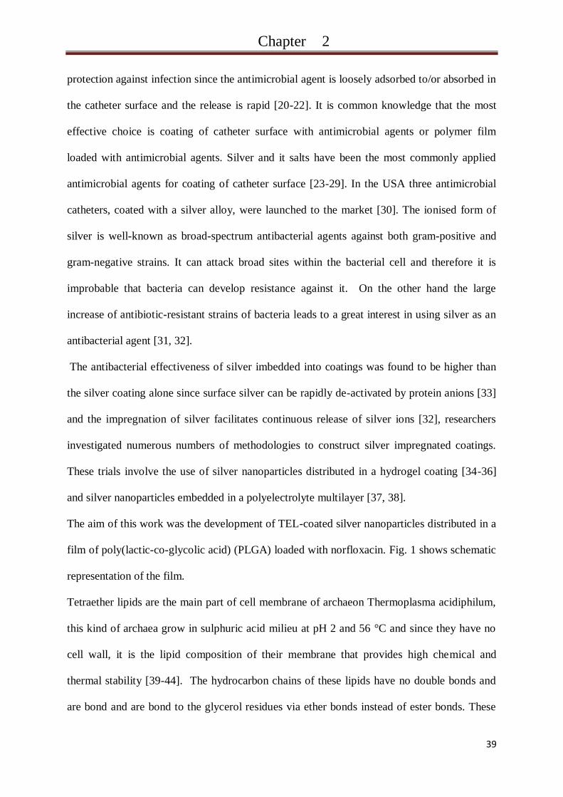
The aim of this work was the development of TEL-coated silver nanoparticles distributed in a
film of poly(lactic-co-glycolic acid) (PLGA) loaded with norfloxacin. Fig. 1 shows schematic
representation of the film.
Tetraether lipids are the main part of cell membrane of archaeon Thermoplasma acidiphilum,
this kind of archaea grow in sulphuric acid milieu at pH 2 and 56 °C and since they have no
cell wall, it is the lipid composition of their membrane that provides high chemical and
thermal stability [39-44]. The hydrocarbon chains of these lipids have no double bonds and
are bond and are bond to the glycerol residues via ether bonds instead of ester bonds. These
Page 51
Chapter 2
40
properties provides long-term resistance against both hydrolytic and oxidative agents and
(bio)chemical degradation [39].
Norfloxacin (1-ethyl-6-fluoro-1,4-dihydro-4-oxo-7-(1-piperazinyl)-3-quinolinecarboxylic acid
(NF) is broad-spectrum fluoroquinolone antibacterial agent which is frequently used for the
treatment of urinary tract infections (UTI) caused by both gram-positive and gram-negative
bacteria [45, 46]. This bactericidal agent builds a complex with enzyme DNA-gyrase enzyme
which is required for synthesis of the bacterial DNA [50].
It is of high importance to select the optimum coating formulation by choosing the compatible
and suitable polymers which have the potential to control the release rate over the whole
catheterization period. Various studies focused on producing hydrogel coatings for urinary
catheter [34-36]. Hydrogel coatings can significantly decrease the damage of the urethral
mucosa and the trauma when the catheter is withdrawn [48-50], it also unlikely to cause
discomfort to the patient due to its soft and lubricant nature. However, it is still not evident
that they promote the anti-encrustation properties [51-53].
In this work, incorporating of the above mentioned anti-bacterial agents was achieved by
employment of PLGA film. PLGA is an FDA-approved, biocompatible and biodegradable
polymer [54-56]. It degrades in water via chemical hydrolysis of the ester bonds resulting in
oligomers with carboxyl end groups or lactic and glycolic acids [57]. The yielded acids have
the ability to decrease the pH in the surrounding microenvironment [58]. This effect can be
exploited to neutralize the alkaline products produced from urea hydrolysis and upgrade the
coating effectiveness against encrustation.
In this study, we developed a new methodology to design anti-bacterial and anti-encrustation
coating for urinary catheter. Glass slides were chosen as a model for catheter surface. Since
the films must be still attached to the surface during the release and bacterial adhesion
experiments, further modification of the glass surface was needed to improve the glass-PLGA
Page 52
Chapter 2
41
interaction. The films were loaded with both norfloxacin and TEL-coated silver nanoparticles.
The release rate of norfloxacin in phosphate buffered saline (PBS) was assessed. The anti-
encrustation potent of the films was tested in synthetic human urine. Finally, quantitative
assays of both dead and live adhered bacteria (five strains) in an in vitro urinary tract infection
model were performed.
Fig. 1Schematic representation of PLGA-NF-Ag construction
Page 53
Chapter 2
42
Materials and Methods
Materials
Poly(D,L-lactide-co-glycolide) (PLGA), Types Resomer® RG 503H was purchased from
Boehringer Ingelheim, Ingelheim, Germany. (3-Aminopropyl)triethoxysilane (APTES), ≥
98%, Norfloxacin, sodium dodecyl sulfate (95%) and urease (type II from jack beans) were
obtained from Sigma-Aldrich (Sigma-Aldrich Chemie GmbH, Germany). Silver nitrate and
glass slides (76 x 26 mm) were purchased from Carl Roth, Germany. Escherichia coli (E.coli)
(DSMZ Nr. 498), Staphylococcus aureus (DSMZ Nr. 20231), Staphylococcus epidermides
(DSMZ Nr. 3269), Enterococcus faecalis (DSMZ Nr. 2570) and Pseudomonas aeruginosa
DSMZ Nr. 1117) were purchased from the DSMZ (Braunschweig, Germany). All other
chemicals and solvents were of high analytical grade and commercially available.
Methods
TEL Extraction and Activation
Extraction and purifying of TEL were done according to the method described elsewhere
[40,43]. For this purpose two-step chromatography with DEAE-cellulose and silica columns
eluted with chloroform and methanol (2:1, v:v) was used. The lipids were then lyophilized at
10-2
torr and stored at -20 °C. TEL were activated by cyanuric chloride, equimolar amount of
cyanuric chloride and TEL were allowed to react overnight at 40 °C in chloroform and N,N-
diisopropylethylamine was employed as catalyzer. Activated lipids were purified with thin
layer chromatography and then stored at 4 °C.
Preparation of Lipid coated Silver Nanoparticle
Page 54
Chapter 2
43
Silver nanoparticles were prepared as described elsewhere [59]. Briefly: 4 ml of sodium
hydroxide solution (0.1 molar) were added to 0.8 ml of ethylenediaminetetracetic acid
(EDTA) 0.1 molar. Distilled water was added to the mixture to get a final volume of 100 ml.
1.3 ml silver nitrate (0.1 molar) and 0.3 ml HCL (0.1 molar) were added to the mixture when
the mixture started to boil. After 90 seconds of boiling the mixture was cooled down to room
temperature and the silver nanoparticles were stored under light exclusion.
Coating of the silver nanoparticles with TEL was done according to the method described in
our previous work [39], briefly: 15 ml of the previous silver nanoparticle dispersion was
diluted up to 50 ml with distilled water. The dispersion was centrifuged (2000 rpm, 5 min)
followed by re-dispersion in 15 ml distilled water and then transferred to a flask.
2 mg of activated TEL were dissolved in 1 ml of chloroform in a flask; the chloroform was
allowed to evaporate and the TEL formed a thin film on the bottom of the flask. The 15 ml
purified silver nanoparticle dispersion and 100 µl sodium dodecyl sulfate (SDS) (30%) were
added to TEL in the flask. Ultrasonic treatment was applied on the mixture and TEL films
were formed surrounding the silver nanoparticles. SDS was eliminated by dialysis the
dispersion in water for 12 h, the coated nanoparticles were then stored at 4 °C under light
exclusion.
Polymer Film Preparation
Aminolysis on Substrate Surface
Glass slides were washed with chloroform, acetone and distilled water and dried in flow
nitrogen. Glass surface was modified by APTES to get an amino-terminated layer as
described by Emoto at al [60]. Briefly, 1 ml APTES were dissolved in 100 ml toluene under
stirring, the glass slides were incubated in this solution for 4 h and then washed with toluene
Page 55
Chapter 2
44
to remove the loosely physisorbed APTES from the surface. The modified glass slides were
heated at 160 °C for 20 h in vacuum and then stored at 4 °C.
Coating of aminated Surface with PLGA
First, unloaded polymer films were prepared. 1,000 mg of PLGA was dissolved in 100 ml
acetone under stirring overnight, APTS-coated glass slides were dipped in the solution for 40
min and air dried at room temperature.
For films containing TEL-coated silver nanoparticles (PLGA/TEL-Ag), the 10 mg/ml
PLGA/acetone solution was used and 1 ml (~1m molar) of the suspension of TEL-coated
silver nanoparticles was added and the same dipping and drying steps were followed as
previous. The same procedure was run for PLGA/NF/TEL-Ag film preparation and the same
concentrations of PLGA and TEL-Ag were used and the only modification is the addition of
100 or 200 mg of norfloxacin to the dipping solution. The coated slides were stored at -20 °C
under light exclusion.
Film Characterization
Water contact angle measurement
Surface wettability was evaluated by water contact angle measurement using the sessile drop
method and Laplace-approach (OCA20, DataPhysics Instruments GmbH, Filderstadt/
Germany). Contact angle of distilled water was determined by dropping 3µl of water onto the
surface followed by measuring the angle within the first 10 s after dropping. The contact
angle was the mean of fifteen readings from three different parts of the surface (± the standard
division). All measurements were performed at ambient conditions.
Page 56
Chapter 2
45
Atomic force microscopy
Atomic force microscopy was performed on a NanoWizard (JPK instruments, Berlin,
Germany) as described elsewhere [40]. Commercial pyramidal silicon tips (NSC16AlBS,
Micromasch, Estonia) mounted to cantilevers (length 230 µm, resonance frequency 170 kHz
and nominal force constant ~40 N/m) were used. AFM was used to measure surface
topography of the films and all measurements were performed in intermittent contact mode to
avoid damaging of the surfaces. The scan speed was proportional to the scan size where the
scan frequency was between 0.5 and 1 Hz. Images were captured by displaying the height,
phase and amplitude signals in the trace direction (512 x 512 pixel).
Film Stability
The stability of the films upon exposing to PBS was investigated. All prepared Polymer films
were incubated in PBS with 0.01% sodium azide under gentle shaking at 37 °C for 60 days.
Surface morphology of the films was imaged by AFM before and after PBS incubation and
the effects of PBS incubation were noticed.
Norfloxacin Release
For measuring the release of norfloxacin, the norfloxacin loaded PLGA films were incubated
in PBS (pH 7.4) with 0.01% sodium azide under gentle shaking (20 rpm) (Rothaterm®, Gebr.
Liebisch, Bielefeld, Germany) at 37 °C.
At predetermined time points, 3ml samples were taken and replaced with 3 ml fresh PBS. The
samples were then stored at 4°C under light exclusion. To determine the Norfloxacin
concentration in the probes, fluorescence intensity was measured using a fluorescent plate
reader (Saphire II; Tecan, Austria) at wavelength 330 nm excitation/440 nm emission.
Cumulative release amount and percentage were calculated at each time point.
Page 57
Chapter 2
46
Artificial Urine
The synthesized urine was based on a proposal by Griffith et al. [60]; two aqueous solutions A
and B were prepared and added to the bioreactor device to avoid the precipitation of brushite
(CaHPO4.2H2O) [61]. Solution A was composed of CaCl2.H2O, MgCl2.6H2O, NaCl, Na2SO4,
Na2Oxalate, Na3Citrate·2H2O, KCl and urease. Solution B was composed of K2PO4,
Na2C2O4,NH4Cl and urea. Creatinine was also added to the urine. A volume of 1.5 l of
artificial urine solution were sterilized by filtration and then used for the in vitro encrustation
model.
In vitro Encrustation Model
For encrustation assays, bioreactor devices (reaction vessels) coupled with temperature and
pH measurement system were employed as described by Jones et al. [61]. A laboratory
bioreactor (Rettberg, Germany) was filled with 1.5 l artificial urine and sterilized prior to the
experiments. The temperature was adjusted to 37 °C and the samples were placed vertically
and fixed inside the vessels. During the experiments, the urine inside the vessels was gently
stirred (150 rpm). This model allows a simultaneous testing of up to sixteen samples. Both
uncoated and PLGA coated glass substrate were analyzed. It is known, that the urinary tract
infection is induced above all by urease-producing bacteria. Consequently, in the in vitro –
crystallization model the pH increase was triggered by addition of urease to the artificial
urine. During a following period of 14 days the pH was enhanced slowly from 5.7 to 8.8, this
allowed salt formation over an incubation time of up to 14 days. After the incubation, the
samples were imaged by Scanning Electron Microscopy (SEM). The samples were treated
with 1N HCl to dissolve the crystalline deposits. The encrustation of Ca, Mg, Phosphate and
Oxalate on the sample surfaces was investigated by quantitative and qualitative analysis. The
Page 58
Chapter 2
47
amount of Na+, K
+ and Ca
+2 was quantified by means of atomic adsorption spectroscopy
while photochemical detection was employed to analyze Phosphate, Oxalate and Ureate. All
experiments were done in triplicate.
Bacterial Adhesion
E.coliwas used for the initial adhesion experiments. The bacteria were grown in Trypic Soy
Broth (TSB) over night at 37 °C under gentle stirring. The suspension was centrifuged and the
bacteria were collected and washed with PBS followed by the next centrifugation step. The
bacteria were then resuspended in artificial urine and their concentration was adjusted to 108
cells/ml. A 24-well multiwell culture plate was filled with bacterial suspension and the
samples were incubated in the suspension for 24 h at 37 °C. The samples were then removed
and extensively washed with PBS and treated in ultrasonic path to remove the non-adherent
bacteria. Cell viability assay was applied to quantify both dead and live bacterial by the use of
LIVE/DEAD BacLight kits. Imaged were captured by confocal laser scanning microscopy
(CLSM). Evaluation of adhered bacteria was confirmed by SEM.
Investigations of bacterial adhesion were done on unmodified glass and PLGA coated glass.
Four different kind of PLGA coating were also tested: unloaded PLGA coating, 10% NF
loaded PLGA coating, 20% NF loaded PLGA coating and PLGA films loaded with NF (20%)
and TEL-Ag (1mmolar/1g).
Additionally, in vitro–bioadhesion experiments based on a new microbiological model of
urinary tract infection were performed. In this model the antibacterial and antiadhesive
effectiveness of unmodified Glass vs. PLGA films loaded with NF (20%) and TEL-Ag
(1mmolar/1g) was tested. This infection model comprised five bacteria strains:
Staphylococcus aureus, Staphylococcus epidermides, Escherichia coli, Enterococcus faecalis
Page 59
Chapter 2
48
and Pseudomonas aeruginosa cultivated in artificial urine. The experimental procedure of this
adhesion experiment corresponds to the method for the E.coli monoculture described above.
Page 60
Chapter 2
49
Results and Discussion
TEL coated Silver Nanoparticles
In our previous work [38] we described the method to prepare TEL coated silver
nanoparticles. Chemical reduction method was used to synthesize of silver nanoparticles; the
particles had a size of 19.2±2.5 nm. After particle preparation, a thin layer of silver oxide is
formed on the particle surface and the surface becomes –OH rich in aqueous solution.
The TEL were activated with cyanuric chloride and the covalent coupling process were
confirmed by FTIR spectroscopy where new peaks at 1509 cm-1
and 1541 cm-1
were visible
which corresponds to the C=N valence oscillation of cyanuric chloride.
Thin film of the activated TEL was formed on the bottom of the flask after evaporation of the
solvent. After adding the silver nanoparticles and the aqueous solution of SDS to the film, the
mixture was ultrasound treated. The energy gained from ultrasound treatment leads to the
formation of TEL incorporated SDS micelles.
The activated TEL are covalently bonded to hydroxyl groups on the surface of silver
nanoparticles and a TEL film is formed on the surface.
Polymer Film Preparation and Characterization
PLGA was used as coating matrix for controlled release of both norfloxacin and silver ions
and for improved anti-encrustation properties. Dipping method was chosen for the coating
process. This method enables the fabrication of films with thicknesses ranging from a couple
of nanometres up to millimetres. The ease and simplicity of the employed method makes it
applicable for coating of several catheter materials. After immersion of the samples in the
polymer solution, a polymer film is formed on the surface. The film thickness depends on
various parameters like polymer concentration and incubation time. After film drying, a thin
Page 61
Chapter 2
50
layer of the polymer is created. The formed film is only adsorbed to the sample surface and
different kinds of polymer-surface interactions are responsible for this adhesion. These
interactions depend on chemical and physical properties of the sample surface and on the
polymer nature.
It is evident that PLGA with free carboxylic acid possesses a good affinity to surfaces with –
OH groups (like glass) due to hydrogen bonds that are formed between the hydroxyl groups
and the –COOH groups of PLGA [62]. Nevertheless when it comes in contact with body
fluids these bonds are weak and they fail to keep the polymer attached to the surface over the
whole catheterization time.
PLGA chains contain carboxyl groups and therefore modification of glass with amino-
terminated layer can upgrade the stability of the PLGA film in aqueous milieu. Modifying of
surfaces is widely used to enhance the polymer-surface interactions and amino-terminated
layer was constructed on the surface when polymers with negatively charged groups were
used to coat these surfaces [37, 59, 63].
In this work, APTES was used to produce amino-terminated layer on the glass surface. The
electrostatic interactions between the negatively-charged carboxyl groups on the polymer and
the positively-charged amine groups on glass surface are essential for film stability.
Fig 2shows AFM images of cleaned and APTES-coated glass. Unmodified glass shows an
unstructured surface, its roughness average (Ra) was 0.19±0.11 nm and the root mean square
value (RMS) of its roughness was 0.24±0.11 nm. After APTES coating, both Ra and RMS
increased to reach the values of 1.05±0.24 nmand 1.28±0.26 nm respectively.
Page 62
Chapter 2
51
Fig. 2AFM imaging of unmodified glass surface (A) and APTES modified surface (B).
By immersion of the APTES-coated glass in PLGA/acetone solution, a homogenous and
smooth film of PLGA was formed (Fig 3). When the aqueous suspension of TEL-coated
silver nanoparticles was added to the PLGA/acetone solution, the yielded films were less
homogenous (Fig 4). It is unlikely that the silver nanoparticles affected significantly the
homogeneity of the polymer films as a very low amount of the particles was added. Our
explanation is based on the fact that PLGA is water-insoluble polymer. When the aqueous
suspension of silver nanoparticles was added to thePLGA/acetone solution, the water was
dissolved in the acetone and a mixture of the two components was formed. The solubility of
PLGA was not affected as the ratio water/acetone was too low (about 1% (v/v)). Using this
mixture, films were prepared and then air dried at room temperature. During the drying
process, acetone evaporates more rapidly than water as it has a much lower boiling point (56
°C). This leads to an increasing water/acetone ratio and the polymer solubility decreases.
Then a continuing evaporation of acetone could lead to phase separation.
Silver nanoparticles were distributed inside the film and only some particles could be seen on
the top of the film surface. Fig 4 presents low density of the particles on the surface. The
upper-right side of the image (A) shows a phase image of the surface where silver
nanoparticles appear like black spots spreaded on the film surface without having aggregated.
Adding norfloxacin to the mixture didn‘t considerably change the film topography (Fig 5).
Page 63
Chapter 2
52
Fig. 3AFM images of PLGA coating, (A) film prepared on ATPES modified glass (B) film
prepared on ATPES modified glass after 53 days of incubation in PBS and (C) film prepared
on unmodified glass after 53 days of incubation in PBS.
Surface wettability of the film was investigated. Fig6 shows that surface wettability was
determined by the type of the anti-bacterial loaded in the film which suggested that the films
were successfully loaded with these agents. As expected the glass surface had the lowest
contact angle (14.3±1.3°) due to its high hydrophilicity. After deposition of PLGA film, the
surface became more hydrophobic because of the hydrophobic nature of the polymer and the
contact angel was 80.4±0.7°. In Fig 6, it can be seen that PLGA/TEL-Ag surface shows the
highest value of water contact angle (106.1±2.1°). In our previous work, we suggested that
coating of cellulose membrane with TEL-Ag enhances the surface hydrophobicity and the
contact angle raises significantly (27° to 93°) [38]. The presence of particles on the polymer
surface provides a good explanation of the increasing contact angel. In addition to surface
chemistry, surface topography must also be considered. Li et al [64] introduced micropatterns
on Thermanox surfaces by laser-pattering. Both patterned and not patterned Thermanox were
coated with collagen. Contact angle of the patterned surface differed from that of not
patterned surface. Despite of the fact that both surfaces were collagen-coated and had the
same chemical nature, their microstructure resulted in a change of surface wettability.
Page 64
Chapter 2
53
Fig. 4AFM images of PLGA/TEL-Ag on APTES modified glass, (A) after the preparation
and (B) after 53 days of incubation in PBS.
Adding of norfloxacin to the PLGA reduced the contact angle value (62.9±2.0°). Norfloxacin
has very low water solubility and one can expect that its adding can increase the contact
angel. The possible explanation is based on the chemical properties of norfloxacin.
Norfloxacin hydrates have higher water solubility than the anhydrates [65] and norfloxacin
molecules on the film surface can be hydrated with water molecules from ambient air. Thus
hydration effect can be detected through enhancement of surface wettability.
Film stability and Norfloxacin release
To demonstrate the utility of the coating for urinary catheter, the films were incubated in PBS
at 37 °C and the surface morphology was examined before and after the incubation. The
effectiveness of catheter coating depends highly on the film stability in body fluids during the
indwelling time and a complete or partial detachment of the film from the surface is one of the
most common reasons of coating failure.
Page 65
Chapter 2
54
Fig. 5 AFM imaging of PLGA/TEL-Ag/NF film on APTES modified glass surface, (A) after
the preparation and (B) after 53 days of incubation in PBS.
In our study, APTES was tested and found to be essential for coating stability. PLGA film,
attached on unmodified glass surface, was partially detached from the surface after 4 days
exposure to buffer solution and the surface coverage with the film reduced to only 30% (Fig
3). The hydrogen bonds between carboxyl groups on PLGA and hydroxyl groups on glass
surface are not strong enough to keep the film attached to the surface as described previously.
APTES modification of glass leads to better attachment of PLGA. This modification produces
amine groups providing a positive charge of glass surface in moderate medium. When PLGA
film is formed on this surface, carboxyl-group/amine-group electrostatic interaction insures a
better attachment of the film to the surface. Fig 3, 4 and 5represent AFM imaging of the films
prepared on modified glass revealing morphology changes before and after incubation in
buffer. Despite the long incubation period (~
2months) and the shear forces induced by
shaking, the films remained attached to the surface.
AFM images show little effect of polymer degradation on the film after incubation of the
samples in buffer solution, the surfaces became rougher and a limited ―bulk erosion‖ was
detectable.
Page 66
Chapter 2
55
Fig. 6 Water contact angel of Glass, unloaded PLGA film, PLGA/TEL-Ag film and
PLGA/NF film.
In order to investigate the feasibility of employment of PLGA as polymer film matrix for
norfloxacin controlled release, in vitro release study was performed in PBS (pH 7.4) at 37 °C.
Fig 7 shows cumulative release profiles of norfloxacin from the film. A fast release rate of
about 60% was observed in the first few days (burst-release) followed by slow release in the
next 50 days (Fig. 7B). Fig. 7A shows that the burst release amount in the first day was
103.59 ng/cm² followed by slow release of 70.39 ng/cm² in the next 52 days. The film must
release a daily amount of norfloxacin in the aqueous environment surrounding the catheter
that is equal to or exceed the overkill dose (400µg/l). The dipping method used for the surface
coating enables the film to get controlled thickness as well as desired antibiotic loading level
to reach the overkill dose. In this work the loading and film thickness provided potential
protection against bacterial adhesion.
Norfloxacin is a fluoroquinolone with hydrophobic nature which was used as anti-bacterial
agent for long-term catheterization [66]. The fast release in the first few days might have been
Page 67
Chapter 2
56
due to the drug diffusion from the film, the release rate decreased in the next days up to the
day 10 because of the hydrophobic interaction between the drug and the polymer chain.
Increasing of release rate was noticed after the 10 day, this might be due to the drop of pH
value within the film. Degradation of PLGA ester bond results in oligomers with carboxyl end
groups or lactic and glycolic acids, this phenomenon is called autocatalysis and it seems to be
responsible for the faster internal degradation of the film when the acids within the film
cannot be set free [67]. Norfloxacin is more soluble in acidic milieu than in mediated one
[68]. The low pH value within the film enhanced the drug solubility in water and decreased
the drug/polymer hydrophobic interaction which resulted in faster release from the film.
Fig. 7.Cumulative percentage release of norfloxacin from PLGA film in PBS at 37 °C.
Bacterial Adhesion and Encrustation
The adhesion of E.coli on unmodified and coated glass was investigated in artificial urine
during 24 h at 37° C. E.coli showed more potential to adhere to the uncoated glass than
hydrophobic surface of PLGA (Fig 8). The hydrophlilc nature of E.coli [69,70] and the
physiochemical properties of the surface influence the adhesion process. Bacterial adhesion to
Page 68
Chapter 2
57
biomaterials is a complex process and depends on chemical compositions of the material
used, hydrophobicity and surface roughness and other factors [63, 69-73].
Four different types of PLGA coating were also tested: unloaded PLGA, 10% norfloxacin
loaded PLGA, 20% norfloxacin loaded PLGA and PLGA loaded with norfloxacin (10%) and
TEL-Ag (1 m molar/1 g Ag/PLGA). Copper discs were used as standard test to be able to
reference the anti-bacterial properties of the different coatings. The anti-bacterial
effectiveness of copper is exploited centuries ago and its broad-spectrum anti-microbial
capacity was intensively investigated in the last decades.
Loading of PLGA with norfloxacin enhanced the anti-adhesive and anti-bacterial properties of
the films. The number of adhered bacteria was significantly decreased after the loading (Fig.
9). It can also be seen that most of the bacteria adhered to unloaded PLGA were still alive
while significantly higher ratio of dead/live bacteria (~50%) was observed on norfloxacin
loaded PLGA coating. Rising of norfloxacin concentration from 10% to 20% resulted in drop
of bacterial adhesion. Adding of TEL coated silver nanoparticles to the coating enhanced the
anti-adhesive property while the ratio of dead/live bacteria was still constant. A comparison
between the four coatings led to the conclusion, that the copper wafer showed a high anti-
bacterial activity where most of the bacteria attached to copper surface were dead and a
negligible number of live bacteria was detectable. Nevertheless, our polymer coating loaded
with silver and norfloxacin showed a higher potential to prevent bacteria adhesion than copper
wafer.
Page 69
Chapter 2
58
Fig. 8 Masse and of Ca, Mg, Phosphate and Oxalate in the crystalline deposits formed on
unmodified glass and PLGA film after 14 days of incubation in artificial urine. The figure
shows also Scanning Electron microscopy images of the crystalline deposits.
Fig 10 represents the results of in vitro-bioadhesion experiments in urinary tract infection
model using five strains of bacteria. A successful validation of further statements for simple
infection culture was reached. In contrast to the uncoated glass surface, a strongly reduced
number of adhered cells (decrease of 45%) was shown on the PLGA/NF/TEL-Ag film.
Scanning microscopic images confirmed the successful anti-adhesive effect.
PLGA was widely used as polymer matrix coating for controlled release of drugs and
biomolecules. The function of PLGA in our system was not limited to control the release but
it also neutralized the alkali products on the coating surface.
Page 70
Chapter 2
59
Fig 8 shows a considerable reduce of encrustation on PLGA coated glass compared to
uncoated glass after 2 weeks of sample incubation in synthesized urine. The total mass of
crystalline deposition was reduced by approximately 40%.
Fig. 9 Adhesion of both live and dead E.coli on unmodified glass, PLGA film, PLGA film
loaded with 10% norfloxacin (PLGA-NF(l)), PLGA film loaded with 20% norfloxacin
(PLGA-NF(h)), PLGA film loaded with both 20% norfloxacin and TEL-Ag and copper. All
samples were incubated in E.coli suspension for 24 h at 37 °C.
The urease catalyzed the urea hydrolysis producing ammonia which increased the pH value of
urine. The pH value of urine was 8.8 and when uncoated glass was incubated in the urine, the
alkalic milieu leaded to the precipitation of crystalline deposits onto the glass. When glass
samples were coated with PLGA, the free carboxylic acid groups on the surface of polymer
coating reduced amount of crystalline deposited on the surface. PLGA undergoes
degradationby hydrolysis of its ester bond in water resulting in acidic monomers and
Page 71
Chapter 2
60
oligomers. It is evident that increasing the pH of the environment promotes the polymer
degradation
Fig. 10 Adhesion of both live and dead bacteria (five bacteria strains) on unmodified glass
and PLGA film loaded with both 20% norfloxacin and TEL-Ag after 24h in synthesized urine
and Scanning electron microscopy images of the two films showing the adhered bacteria.
producing more acids [74]. On the other hand, hydrolysis of urea, which is catalyzed by
urease, produces ammonia and carbon dioxide. Ammonia is a base which dissolves in water
producing hydroxide ions [75]. These hydroxide ions can neutralize the acidic products of
PLGA degradation. When the number of the available hydroxide ions is higher than that of
H+, the urine becomes more alkali and this promotes the polymer degradation producing
Page 72
Chapter 2
61
more acids and neutralizing more hydroxide ions. This process protects the coating surface
from the increasing deposition of crystals during the contact with human urine.
Page 73
Chapter 2
62
Conclusion
In this work, we have introduced a newly developed PLGA film with anti-bacterial and anti-
encrustation functionalities to be used as coating for urinary catheters. Two anti-bacterial
agents, norfloxacin and tetraether lipid coated silver nanoparticles were successfully
incorporated in the film.
Film was deposited on aminated glass by means of dipping-method. The modification of glass
surface with amino-terminated layer supported the film stability in aqueous environment.
From the atomic force microscopy images, it was demonstrated that surface homogeneity of
the films was depended on the antibacterial agent loaded in the film; the silver nanoparticles
were uniformly distributed within the film when they were coated with TEL.
The films released the loaded norfloxacin in about two months through diffusion of the drug
in aqueous milieu. The films effectively inhibited the in vitro adhesion of bacteria compared
to glass and copper surface and unloaded polymer films. It is of value to point out, that the
films did not only reduce the number of adhered bacteria but also decreased the bacterial
viability on the surface.
The results obtained from encrustation experiments in artificial urine demonstrated that the
polymer coating could efficiently reduce the crystalline formation on the surface as the alkali
products of urea hydrolysis were neutralized by acidic products of polymer degradation. This
phenomenon resulted in resistance of the coating against encrustation compared to uncoated
glass. The in vitro bacteria and encrustation tests suggest promising use of these coating to
promote the biocompatibility and biofunctionality of urethral catheters.
Page 74
Chapter 2
63
Acknowledgement
The authors would like to thank Novoplast Schlauchtechnik GmbH (Halberstadt,
Germany), Primed Halberstadt Medizintechnik GmbH (Halberstadt, Germany), MAT
PlasMATec GmbH (Dresden Germany), TRUMPF Medizin Systeme GmbH Deutschland,
(Saalfeld Germany), JPK Instruments Berlin (Germany) and BMWI/AiF (AZ IGF-05/05-
AiF-Nr. 15090 BG/2) for the support. Christian Hobler (research group M. Keusgen) is
gratefully acknowledged for performing the spectrophotometry measurements and Maria
Solovey for the helpful discussion.
Page 75
Chapter 2
64
References
[1]Ramakrishnan K, Mold J.W. Urinary Catheters: A Review. The Internet Journal of family
Practice 2005;3.
[2] Park HJ, Cho YW, Kwon IC, Jeong SY, Bae YH. Assessment of PEO/PTMO
multiblockcopolymer/segmented polyurethane blends as coating materials for urinary
catheters: in vitro bacterial adhesion and encrustation behaviour. Biomaterials 2002;23:3991–
4000.
[3] Jarvis WR. Selected aspects of the socioeconomic impact of nosocomial infection:
morbidity, mortality, cost, and prevention. Infect Control Hosp Epidemiol 1996;17:552-7.
[4] Cho YW, Park JH, Kim SH, Cho Y-H, Choi JM, Shin HJ et al. Gentamicin-releasing
urethral catheter for short-term catheterization. J Biomater Sci Polym Ed 2003;14:963–72.
[5] Kunin CM. Urinary Tract Infections: Detection, Prevention and management. 4th ed.
Philadelphia: Lea & Febiger, 1987. P. 248-51.
[6] Barford JMT, Coates ARM.The pathogenesis of catheter-associated urinary tract
infection.Journal of Infection Prevention2009;10:50-6.
[7] Daifuku R , Stamm WE. Bacterial adherence to bladder uroepithelial cells in catheter-
associated urinary tract infection. New England Journal of Medicine 1986;314:1208–13.
[8] Thomas WE , Nilsson LM , Forero M , Sokurenko EV , Vogel V. Shear-dependent ‗stick-
and-roll‘ adhesion of type 1 fimbriated Escherichia coli . Molecular
Microbiology2004;53:1545–57 .
[9] Kulik E, Ikada Y. In vitro platelet adhesion to non-ionic and ionic hydrogels with
different water contents. J Biomed Mater Res 1996;30:295-304.
[10] Talja M, Korpela A, J.arvi K. Comparison of urethral reaction to full silicone, hydrogel-
coated and siliconised latex catheters. Br J Urol 1990;66:652–7.
Page 76
Chapter 2
65
[11] Denstedt JD, Wollin TA, Reid G. Biomaterials used in urology: current issues of
biocompatibility, infection, and encrustation. J Endourol 1998;12:493–500.
[12]Liedl B. Catheter-associated urinary tract infections. Curr Opin Urol 2001;11:75-9.
[13] Mobley HLT, Warren JW. Urease-positive bacteriuria and obstruction of long-term
catheters. J Clin Microbiol 1987;25:2216–17.
[14] Kunin CM. Blockage of urinary catheters: role of microorganisms and the constituents
of the urine on formation of encrustations. J Clin Epidemiol 1989;42:835–42.
[15] Cox AJ, Hukins DWL. Morphology of mineral deposits on encrusted urinary catheters
investigated by scanning electron microscopy. J Urol 1989;142:1347–50.
[16] Morris NS, Stickler DJ, McLean RJC. The development of bacterial biofilms on
indwelling catheters. World J Urol 1999;17:345–50.
[17] Stickler DJ, Feneley RCL. The encrustation and blockage of long-term indwelling
bladder catheters: a way forward in prevention and control. Spinal Cord 2010;48:784–90.
[18] Kunin CM. Nosocomial urinary tract infections and the indwelling catheter: what is new
and what is true?. Chest 2001;120:10-2.
[19] Wu P, Grainger DW. Drug/device combinations for local drug therapies and infection
prophylaxis. Biomaterials 2006;27:2450–67.
[20] Cormio L, La Forgia P, Siitonen A, Ruutu M, Tormala P, Talja M. Immersion in
antibiotic solution prevents bacterial adhesion onto biodegradable prostatic stents. Br J Urol
1997;79:409–13.
[21] Cormio L, La Forgia P, La Forgia D, Siitonen A, Ruutu M. Bacterial adhesion to urethral
catheters: role of coating materials and immersion in antibiotic solution. Eur Urol
2001;40:354–9.
[22] Cormio L, La Forgia P, La Forgia D, Sittonen A, Ruutu M. is it possible to prevent
bacterial adhesion onto ureteric stents?. Urol Res 1997;25:213-6.
Page 77
Chapter 2
66
[23]Cormio L, La Forgia P, La Forgia D, Sittonen A, Ruutu M. Bacterial Adhesion to
Urethral Catheters: Role of Coating Materials and Immersion in Antibiotic Solution. Eur
Urol 2001;40:354–9.
[24] Ahearn DG, Grace DT, Jennings MJ, Borazjani RN, Boles KJ, Rose LJ et al. Effects of
Hydrogel/Silver Coatings on In Vitro Adhesion to Catheters of Bacteria Associated with
Urinary Tract Infection. Curr Microbiol 2000;41:120-5.
[25] Liedberg H, Lundeberg T. Silver coating of urinary catheters prevents adherence and
growth of Pseudomonas aeruginosa. Urol Res 1989;17:357-8.
[26] Roe D, Karandikar B, Bonn-Savage N, Gibbins B, Roullet JB. Antimicrobial surface
functionalization of plastic catheters by silver nanoparticles.Journal of Antimicrobial
Chemotherapy 2008;61:869–76.
[27] Stobie N, Duffy B, McCormackDE , Colreavy J, Hidalgo M, McHaleP et al. Prevention
of Staphylococcus epidermidisbiofilm formation using a low-temperature processed silver-
doped phenyltriethoxysilanesol–gel coating. Biomaterials 2008;29:963-9.
[28] Melaiye A, Youngs WJ. Silver and its application as an antimicrobial agent.Expert
Opin.Ther. Patents 2005;15:125–30.
[29] Johnson JR, Kuskowski MA, Wilt TJ. Systematic Review: Antimicrobial Urinary
Catheters To Prevent Catheter-Associated Urinary Tract Infection in Hospitalized Patients
Annals of Internal Medicine, Ann Intern Med 2006;144:116-26.
[30] Melaiye A, Youngs WJ. Silver and its application as an antimicrobial agent.Expert
Opin.Ther. Patents 2005;15:125–30.
[31]Rai M , Yadav A, Gade A. Silver nanoparticles as a new generation of antimicrobials.
Biotechnology Advances 2009;27:76–83.
Page 78
Chapter 2
67
[32] Furno F, Morley KS, Wong B, Sharp BL, Arnold PL, Howdle SM et al. Silver
nanoparticles and polymeric medical devices: a new approach to prevention of infection?. J
Antimicrob Chemother 2004;54:1019–24.
[33] Mohan YM, Lee K, Premkumar T, Geckeler KE.Hydrogel networks as nanoreactors: A
novel approach to silver nanoparticles for antibacterial applications. Polymer 2007;48:158-64.
[34] Vimala K, Sivudu KS, Mohan YM, Sreedhar B, K. Raju KM. Controlled silver
nanoparticles synthesis in semi-hydrogel networks of poly(acrylamide) and carbohydrates: A
rational methodology for antibacterial application. Carbohydrate Polymers 2009;75:463–71.
[35] Thomas V, Yallapu MM, Sreedhar B, Bajpai SK.A versatile strategy to fabricate
hydrogel–silver nanocomposites and investigation of their antimicrobial activity.Journal of
Colloid and Interface Science 2007;315:389–95.
[36]Yu DG, Lin WC, Yang MC. Surface Modification of Poly(l-lactic acid) Membrane via
Layer-by-Layer Assembly of Silver Nanoparticle-Embedded Polyelectrolyte Multilayer.
Bioconjugate Chem 2007;18:1521–9.
[37] FuJ, JiJ, FanD, ShenJ. Construction of antibacterial multilayer films containing
nanosilver via layer-by-layer assembly of heparin and chitosan-silverions complex. J Biomed
Mater Res A 2006;79:665-74.
[38] Dayyoub E, Sitterberg J, Rothe U, Bakowsky U. New antibacterial, antiadhesive films
based on self assemblies ofmodified Tetraetherlipids. Advances in Science and Technology
2008;57:188-94.
[39] E. Antonopoulos, U. Bakowsky, H. J. Freisleben, U. Rothe. Tetraether lipids and
liposomes containing said lipids, and use of the same. European Patent No.EP19970905135,
2002.
[40] Ozcetin A, Mutlu S, Bakowsky U. Methods Mol. Biol 2010;605:87–96.
Page 79
Chapter 2
68
[41] Frant M, Stenstad P, Johnsen H, Dölling K, Rothe U, Schmid R et al. Anti-infective
surfaces based on tetraether lipids for peritoneal dialysis catheter systems. Mat.-wiss. u.
Werkstofftech 2006;37:538-45.
[42] VidawatiS, SitterbergJ, BakowskyB, RotheU. AFM and ellipsometric studies on LB
films of natural asymmetric and symmetric bolaamphiphilic archaebacterial tetraether lipids
on silicon wafers. Colloids and Surfaces B: Biointerfaces 2010;78:303–9.
[43]OzcetinA, Dayyoub E, HoblerC, KeusgenM, Bakowsky U. Selective interactions of
concanavalin A-modified tetraether lipid liposomes. Phys. Status Solidi 2011;8:1985–9.
[44] Appelbaum PC, Hunter PA. The fluoroquinolone antibacterials: past, present and future
perspectives. Int. J. Antimicrob. Agents2000;16:5-15.
[45] Hooton TM.2003. The current management strategies for community-acquired urinary
tract infection. Infect. Dis. Clin. N. Am 2003;17:303–32.
[46] Oliphant CM, Green GM. Quinolones: A Comprehensive Review. Am. Fam.
Physician2002;65:455-65.
[47] Nickel JC, Olson ME, Costerton JW. In vivo coefficient of kinetic friction: study of
urinary catheter biocompatibility. Urology 1987;29:501–03.
[48] Nacey JN, Delahunt B. Toxicity study of first and second generation hydrogel-coated
latex urinary catheters. Br J Urol 1991;67:314–6.
[49] Stensballe J, Looms D, Nielsen PN, Tvede M. Hydrophilic-coated catheters for
intermittent catheterisation reduce urethral micro trauma: a prospective, randomised,
participant-blinded, crossover study of three different types of catheters. Eur Urol
2005;48:978–83.
[50] Cox AJ, Millington RS, Hukins DWL, Sutton TM. Resistance of catheters coated with a
modified hydrogel to encrustation during an in vitro test. Urol Res 1989;17:353–6.
Page 80
Chapter 2
69
[51] Cox AJ, Hukins DWL, Sutton TM. Comparison of in vitro encrustation on silicone and
hydrogel-coated latex catheters. Br J Urol 1988;61:156–61.
[52] Tunney MM, Keane PF, Jones DS, Gorman SP. Comparative assessment of ureteral stent
biomaterial encrustation. Biomaterials 1996;17:1541–6.
[53] Anderson JM, Shive MS. Biodegradation and biocompatibility of PLA and PLGA
microspheres. Adv Drug Deliv Rev 1997;28:5–24.
[54] Athanasiou KA, Niederauer GG, Agrawal CM. Sterilization, toxicity, biocompatibility
and clinical applications of polylactic acid polyglycolic acid copolymers.Biomaterials
1996;17:93–102.
[55] Emerich DF, Tracy MA, Ward KL, Figueiredo M, Qian RL, Henschel C et al.
Biocompatibility of poly (DL-lactide-co-glycolide) microspheres implanted into the brain.
Cell Transplant 1999;8:47–58.
[56] Kitchell JP, Wise DL. Poly(lactic/glycolic acid) biodegradable drug-polymer matrix
systems. Meth Enzymol 1985;112:436-48.
[57] Xie S, Zhu Q, Wang B, Gu H, Liu W, Cui L et al. Incorporation of tripolyphosphate
nanoparticles into fibrous poly(lactide-co-glycolide) scaffolds for tissue engineering.
Biomaterials 2010;31:5100-9.
[58] Abid Jean-Pierre. Laser induced synthesis and nonlinear optical properties of metal
nanoparticles, PHD. Thesis S. 117ff 2003.
[59] Emoto K, Nagasaki Y, Kataoka K. Coating of Surfaces with Stabilized Reactive
Micelles from Poly(ethylene glycol)-Poly(DL-lactic acid) Block Copolymer.
Langmuir1999;15:5212-8.
[60] Griffith DP, Musher DM, Itin C. Urease: the preliminary cause of infection-induced
urinary stones. Invest Urol 1976;13:346-50.
Page 81
Chapter 2
70
[61] Jones DS, Djokic J, Gorman SP. Characterization and optimization of experimental
variables within a reproducible bladder encrustation model and in vitro evaluation of the
efficacy of urease inhibitors for the prevention of medical device-related encrustation.
Biomed Mater Res B Appl Biomater 2006;76:1-7.
[62] Li DX, Yamamoto H, Takeuchi H, Kawashima Y. A novel method for modifying AFM
probe to investigate the interaction between biomaterial polymers (Chitosan-coated PLGA)
and mucin film. Eur J Pharm Biopharm 2010;75:277–83.
[63] Fu J, Ji J, Yuan W, Shen J. Construction of anti-adhesive and antibacterial multilayer
films via layer-by-layer assembly of heparin and chitosan. Biomaterials 2005;26: 6684–92.
[64] Li P, Bakowsky U, Yu F, Loehbach C, Muecklich F, Lehr CM. Laser Ablation
Patterning by Interference Induces Directional Cell Growth. IEEE Trans Nanobiosceince
2003;2:138-45.
[65] Roy S, Goud NR, Babu NJ, Iqbal J, Kruthiventi A. Crystal structures of norfloxacin
hydrates. Cryst Growth Des 2008;8:4343-6.
[66] Park JH, Cho YW, Cho YH, Choi JM, Shin HJ, Bae YH et al. Norfloxacin-releasing
urethral catheter for long-term Catheterization. J. Biomater. Sci. Polymer Edn 2003;14:951-
62.
[67] Li S. Hydrolytic degradation characteristics of aliphatic polyesters derived from lactic
and glycolic acids. J Biomed Mater Res 1999;48:342-53.
[68]ReddyJS, GaneshSV, NagalapalliR, DandelaR, SolomonKA, KumarKA et al.
Fluoroquinolone salts with carboxylic acids. J Pharm Sci 2011;100:3160-76.
[69]Ong YL, Razatos A, Georgiou G, Sharma MM. Adhesion Forces between E. coli Bacteria
and Biomaterial Surfaces. Langmuir1999;15:2719-25.
[70] Li B, Logan BE. Bacterial adhesion to glass and metal-oxide surfaces. Colloids and
Surfaces B: Biointerfaces 2004;36:81–90.
Page 82
Chapter 2
71
[71]Dayyoub E, Belz E, Dassinger N, Keusgen M, Bakowsky U. A novel method for
designing nanostructured polymer surfaces for reduced bacteria adhesion.Phys. Status Solid
2011;208:1279-83.
[72] Truong VK, Lapovok R, Estrin YS, Rundell S, Wang JY, Fluke CJ et al.The influence of
nano-scale surface roughness on bacterial adhesion to ultrafine-grained titanium. Biomaterials
2010;31:3674–83.
[73] Ploux L, Anselme K, Dirani A, Ponche A, Soppera O, Roucoules V. Opposite Responses
of Cells and Bacteria to Micro/Nanopatterned Surfaces Prepared by Pulsed Plasma
Polymerization and UV-Irradiation. Langmuir 2009;25:8161–9.
[74] Burkersroda FV, Schedl L, Göpferich A.Why degradable polymers undergo surface
erosion or bulk erosion. Biomaterials 2002;23:4221–31.
[75] Bibby JM, Hukins DW. Acidification of Urine is not a Feasible Method for Preventing
Encrustation of Indwelling Urinary Catheters. Scand J Urol Nephrol 1993;27:63-5.
Page 83
3Highly Ordered Self-Organized Polymer Coatings for
Reduced Bacteria Adhesion
In preparation for Acta Biomaterialia
Page 84
Chapter 3
73
Abstract
Nanostructuring of implant surfaces has emerged as promising way to control biological
responses especially bacterial adhesion. Here, poly(lactic-co-glycolic acid) (PLGA) was used
to create nanostrtuctured films consisting of polymer features on polyurethane (PUR) surface.
For this purpose, dipping method was employed by using acetone solution of PLGA as
dipping solution and adding non-solvent (water) to this solution. The yielded polymer
nanofeatures were half-sphere shaped and their size was tunable by changing manufacturing
parameters. Film stability under shear forces was investigated and it was found that the
polymer films were still attached to PUR surface after 30 days of phosphate buffered saline
(PBS) incubation. Invitro bacterial adhesion tests showed different potential of the
nanostructured films to reduce bacteria attachment. Features with spacing smaller than
bacteria width inhibited the bacterial adhesion in comparison to flat spin-coated PLGA film,
reverse relationship was found between feature spacing and the number of the adhered
bacteria. In contrast, rising of features spacing over the bacterial width decreased their
capacity to reduce bacterial adhesion. These results highlight the importance of designing
nanofeature with accurate size and shaping when reduction of bacterial adhesion is aimed.
This study also pointed the significance of development of new implant coating that show
nanostructured topography and have the ability to be loaded with active agents by choosing
the suitable method for film preparation and the appropriate polymer.
Page 85
Chapter 3
74
Introduction
Textured surfaces of nanoscale topography are of growing importance for several fields such
as medicine and biology. The use of nanostructured materials in these Fields is innovative [1].
It is well-recognized that surface topography at the macro- and microscale has strong effect
on 1.Cell orientation and grow direction [2-7] 2.Cell proliferation [8] and 3.Cell functions [9]
and also on bacteria behavior [10-11]. However, in recent years the bacteria-nanostructured
man-made substrate interactions gained more attention and were studied intensively. These
interactions are of high importance for implant applications, since the biological performance
of biomedical implants strongly depends on the first interaction happening when implant
surfaces come into contact with a biological environment [12]. Microbial cells can foul
implant surface and form biofilm after the body implantation. This biofilm consists of
microorganisms attaching irreversibly to the surfaces and they form a congregate of single or
multiple populations [13], this biofilm become resistant against the immune system and
conventional treatments with antibiotic, therefore new approaches are needed for the
inhibition of biofilm growth during its development [14]. The formation and development of
the biofilm follows steps that may differ depending on the biological characteristics of the
bacteria present. However, four common steps are generally distinguished. In the first step, a
bacterium is brought into contact with the surface by the gravitational forces, Brownian
motion or hydrodynamic forces, in some bacterial species flagella can also induce motion.
The second step of biofilm is the adhesion of the bacteria to the surface. Usually this adhesion
is described as two separate stages: reversible adhesion and irreversible adhesion. These two
stages involve different physio-chemical and chemical bacteria-surface interactions. These
interactions are significantly dependent on the properties of the surface on which the biofilm
is forming [15]. Many researchers found that the second step is the step most influenced by
topographical and chemical properties of the surface [16] and it is a crucial step in the process
Page 86
Chapter 3
75
of biofilm development [17].In principle, it should be possible to retard, if not prevent, the
formation of biofilms on substrates by using materials to which bacteria cannot attach initially
[18]. Recently many approaches were used to arrange nanostructured surfaces by structural
pattering of the surfaces at nanometre scale to control or prevent the initial bacteria adhesion
[19, 20]. Many techniques were employed for topographical pattering of surfaces at
nanoscale like photolithography [21], self-assembled polymers [22], polymer demixing [23,
24], colloidal lithography [21], surface roughening [25] metal oxidation [26] and
electrospinning [27]. A variety of these methods were used to improve the biocompatibility of
implants, but most of these methods are limited to very small area [28,29] or the shape of the
resulted features and the space between them is hard to control and in some cases the method
depended on chemical modification of the implant surface which changes the surface
chemical nature and this may negatively alter the biological response [30]. The main
advantage of our presented method is the ease to construct nanostructured coating using
dipping method which can be applied on unlimited area. In this study, we used PLGA to
design the films. PLGA is FDA-approved polymer, it degrades by hydrolysis of its ester
bonds to lactic and glycolic acid, these acids are removed from human body through
metabolic pathways and therefore it is biocompatible [31,32]. It is widely used for tissue
engineering scaffolds and pharmaceutical products like drug delivery and targeting [28,29,33-
38]. It is also used as coating matrix to control the release of drugs and active agents from
implant surfaces [39,40]. Coating of implant surface with drug-loaded PLGA films can inhibit
the undesired implant-human body interactions by releasing therapeutic agents that reduce the
negative reaction of the body after the implantation. However these interactions are not the
only factors that influence the effectiveness of implants. Bacteria colonization and biofilm
formation on implant surface are counted as serious problems which can lead to implant
failure and/or implant removal. An Ideal coating must have the potential to release drugs in
Page 87
Chapter 3
76
effective concentration during implantation time and to reduce or prevent implant-associated
biofilm and infections. Nanostructuring of the coating is an innovative choice to inhibit
bacterial biofilm. Since the local or systematical administration of antibiotics can lead to
antibiotic resistant strains, surface structuring with nanofeatures introduces a safe and
effective way to avoid the side-effects of antibiotics and appearance of resistant bacteria
strains. In this work, PLGA nanostructured films were prepared on polyurethane surface by
simple dipping method. This method is a useful technique to produce drug-loaded polymer
films by dissolving the drug and the polymer in a the same solvents and incubation the
substratum in the polymer/drug solution, the formed film can then be dried to be used as drug
release coating. Drying parameters like temperature and air humidity in addition to solvent
and polymer nature influence the structure and the shape of the yielded film. To produce
nanostructured films we added non-solvent(water) to PLGA/acetone solution and the resulted
films were nanostructured. Our method doesn‘t differ from the known dipping method where
drug-loaded polymer film can be constructed and the only modification was adding of PLGA-
insoluble solvent (water) to change surface structuring. The new morphology of the films
increased their ability to reduce bacterial adhesion compared to flat and unstructured PLGA
film.
Page 88
Chapter 3
77
Materials and methods
Materials
Poly(D,L-lactide-co-glycolide) (PLGA), lactide/glycolide ratio 53:47 was a gift from Purac
Biochem (Purac, Netherlands). Safranin was purchased from Sigma-Aldrich (Sigma-Aldrich
chemie GmbH, Germany). PUR wafers were a gift from Primed Halberstadt Medizintechnik
GmbH (Halberstadt, Germany).
All other chemicals and solvents were of high analytical grade and commercially available.
Nanostructured Film preparation
PUR wafers were washed with Acetone, 2-propanol and double distilled water and then dried
in nitrogen flow. 100 mg PLGA was dissolved in 100 ml acetone under gentle stirring at room
temperature overnight. The solution was then slowly injected in desired amount of double
distilled water at constant rate of 10 ml/min by the use of injection needle (Neopoint® 0.90 ×
70 mm; Servopharma GmbH, Wesel, Germany) under magnetic stirring (300 rpm). Cleaned
PUR wafers were then directly incubated in the Acetone-PLGA/water mixture for different
times. After the incubation, the wafers were then allowed to dry at ambient conditions (23 °C
and 55% relative humidity) and then stored at -20 °C. All the films were prepared at the same
ambient condition.
Spin-coated PLGA films were prepared on glass slides. Glass slides (round, 20mm in
diameter) were cleaned as previously and dried by exposing to nitrogen flow. The slides were
then spin-coated from 5% (w/v) PLGA/Ethyl acetate solution by using spin coater
(Novocontrol Technologies, Germany) at rate of 2000 rpm for 60 s at ambient condition. The
coated slides were then stored at -20 °C to use later for AFM and bacterial adhesion
measurements.
Page 89
Chapter 3
78
Surface morphology
Morphology of the PUR wafers before and after the coating was analyzed by atomic force
microscopy (AFM). The measurements were performed on a JPK NanoWizard™ (JPK
Instruments, Berlin). Commercially available silicon cantilevers (NSC 16 AIBS, Micromasch,
Estonia) with ultra-sharp pyramidal tips (radius of the tip curvature <10 nm), resonance
frequency between 150-200 kHz and a nominal force constant of ~40 N/m were used for the
AFM imaging. To avoid damaging of the surfaces, intermittent contact (air) mode was
chosen. The scan speeds were proportional to the scan sizes. Images were taken by displaying
the amplitude, height and phase reflection signals of the cantilever in the trace direction. For
each surface the Root-Mean-Squared roughness (RMS) and the arithmetic average roughness
(Ra) were calculated by jpk software. The software was also used to calculate the height of
polymer features and the distance between them.
Film stability
The coated PUR wafers were incubated in PBS with 1% sodium azid at 37 °C in a rotary
shaker for 60 days. Surface morphology of the films was examined before and after the
incubation by the use of AFM. Feature density onto the surface was calculated by the use of
image j software. Changes of feature size and number before and after the incubation were
investigated.
Bactrial adhesion
For bacterial measurement, spin-coated film and nanostructured PLGA films were employed.
E.coli (BL21 strain) was grown overnight in lysogeny broth agar plates (10 g/l Bacto-Trypton,
5 g/l Bacto-Yeast Extract, 5 g/l NaCl, 1,5 (w/v) Bacto-Agar) at 37 C° in CO2-inkubator. One
colony was used to inoculate a 3ml of lysogeny broth medium (LB, 10 g/l Bacto-Trypton; 5
Page 90
Chapter 3
79
g/l Bacto-Yeast Extract; 5 g/l NaCl), the medium with bacteria was then incubated under
constant shaking (250 rpm) for 10 hours at 37 C°. To harvest the bacteria, the suspension was
centrifuged (3000 rpm, 10 min) and the bacteria were washed three times with PBS to remove
the bacteria nutrition, finally the bacteria were resuspended in phosphate buffered saline
(PBS)to reach a concentration of. Bacteria concentration in the suspension was adjusted to 5 x
109 cell/ml. All experiments were done by taking the desired volume from the same bacteria
suspension to avoid variation of cell density.
Each film was incubated in 1 ml of the bacteria suspension at room temperature for 6, 18 and
30 h. The loosely adhered bacteria were eliminated from the surface by washing with large
amount of PBS and distilled water.
Bacteria were stained with Safranin. One drop of the stain was placed on the surface for 3 min
and then washed extensively with distilled water. Surfaces were the imaged by camera (1.3 M
pixel) mounted to light microscopy (Müller, Germany). The pictures were then analyzed by
image j software to calculate the surface coverage with the E.coli and determine bacterial
colonization.
Page 91
Chapter 3
80
Results
Film characterization
The uncoated PUR surface as purchased possessed ununiformed nanometer features as shown
in Fig 1. After incubation in the dipping mixture, half-sphere polymer nanofeatures were
formed on the surface. The topography as a function of water/acetone ratio and incubation
times was measured using AFM. After coating with PLGA (1mg/ml concentration, 10 min
dipping time, 1/1 water/acetone ratio) the surface possessed half-sphered nanofeatures with
diameter of 155± 23 nm. Raising the water/acetone ration to 5/4 resulted in increasing of the
nanofeatures to 270 ± 55 nm in diameter while employment of 6/4 ratio leaded to increasing
the size up to 370 ± 73 nm. Amplitude images of the prepared nanostructured PLGA films are
presented in Fig 1.
Fig 2 represented the films when different incubation times were used. Images show uniform
structures of PLGA, the size of the features increased by increasing the incubation time. 250 ±
48 nm features were obtained when the wafters were incubated for 15 min and increasing of
size was noticed when incubation time was increased to 30 min and feature of 275 ± 50 nm
size were obtained. For incubation times above 30 min the films were random and rising the
incubation times resulted in increasing the feature sizes. RMS and Ra of all surfaces were also
calculated by JPK software from five images (each 10x10 µm), the averages are presented in
table 1.
Page 92
Chapter 3
81
water/acetone
ratio
Incubation
time (min)
Ra (nm) RMS (nm) Feature
size(nm)
4/4 10 6.66±1.15 8.85±1.51 155± 23
5/4 10 19.70±2.79 23.70±3.59 270 ± 55
6/4 10 35.35±4.04 42.14±5.78 370 ± 73
1/1 15 23.28±1.68 27.56±1.93 250 ± 48
1/1 30 20.38±2.29 24.64±2.43 275 ± 50
1/1 60 31.00±4.36 37.14±4.88 ___
1/1 120 37.90±8.21 45.18±10.62 ___
Table 1. Feature sizes and roughness values of the nanostructured surfaces prepared with
different incubation times and water/acetone ratios.
Film stability
The stability of the nanostructured films were investigated by examination the films
morphology before and after PBS exposure for 30 days. The PLGA features were still
attached to the surface and insignificant change of their sizes were observed. Fig 4 shows
feature surface before and after the incubation. Features exhibited negligible changes of their
surface roughness, this expected results are due to the degradation of PLGA in water. Since
the bulk erosion of the used PLGA begins first after two months of PBS incubation, little
changes of surface roughness after PBS incubation can be expected.
Page 93
Chapter 3
82
Fig. 1.Surface morphology of coated and uncoated PUR: (A) (1/1) water/acetone mixture was
used, (B) (5/4), (C) (6/4) and (D) the uncoated PUR. Particle sizes increase with the
increasing of acetone ratio (PLGA amount) in the mixture.
Bacterial Adhesion
Light microscope was used to estimate the bacterial adhesion on nanostructured PLGA
surface the spin-coated glass surface. After staining with safranin, surface coverage with the
bacteria was calculated with image J software. Bacteria attachment to spin-coated surface,
surfaces with different PLGA feature sizes and is represented in Fig 5.
Page 94
Chapter 3
83
Fig. 2.PUR nanostructured coating. 1mg/ml PLGA concentration was used: (A) PUR was
incubated for 15 min, (B) 30 min, (C) for 60 min and (D) for 120 min. Films were examined
under AFM.
No significant increase of bacterial adhesion onto 100 nm diameter features was seen in
comparison to spin-coated surface. The 250 nm features had the ability to inhibit bacterial
attachment and their efficiency was up to four times higher than the 250 nm features and spin-
coated surface. Bacterial response to other feature sizes varied due to the size of the features.
350 nm features were able to reduce bacteria attachment and the surface coverage was
2.0±0.8% after 30 h of incubation. These features had the highest potential to reduce the
bacterial adhesion whereas bigger and smaller features had lower ability.
Page 95
Chapter 3
84
Discussion
AFM investigation of the polymer features showed that increasing the incubation times and
water/acetone ratios leaded to increase of the polymer features. These features were uniform
up to size 450 nm and they had half-sphere shape while bigger features were ununiformed and
not half-sphere shaped. One explanation of the features formation on PUR surface is the
forming of PLGA films when PUR chips were incubated in the PLGA-Acetone/water
mixture. The thickness of these films depends on the incubation time so that more polymer
chains are able to stick to the surface when incubation times are increased. Exposure of these
films to air can change the shape of the films due to the phase separation and leads to the
forming of polymer features. Acetone has a boiling point of about 56 °C. When the film is
exposed to air, acetone evaporates faster than water due to its low boiling point, this leads to
decrease the acetone/water ratio in the film and only little amount of acetone is available in
the film. The process continues until the complete evaporation of acetone. Decreasing of
acetone amount in the film reduces the polymer solubility in the acetone/water mixture and at
this point the film changes its shape from continues to intermittierend film and when acetone
completely evaporated, the polymerforms nanofeatures on PUR surface.
Similar effect of water/acetone-polymer ratio was found and increasing of the ratio leaded to
increase of the feature sizes (Table 1).
Page 96
Chapter 3
85
Fig. 3.Nanostructured film stability. (A) before PBS incubation, (B) after 30 days of PBS
incubation.
Analysing the uniform features (up to 450 nm) with JPK software showed that the spacing
between the features (the distance between the centres of two neighbour features) depends on
the feature size and it was about the 3/2 of the feature diameter. The features with sizes higher
than 450 nm were uniform and there was no relationship between their sizes and the spacing.
Employment of the uniform features provides the ability to investigate bacterial adhesion on
high-ordered surface with spacing of good regularity and to study the influence of features
spacing on bacterial adhesion and therefore the features with sizes over than 450 nm were
precluded from bacterial adhesion studies. Edwards et al [41] constructed microfeatures on
metal sulphide surfaces by scratching the surface and studied the influence of the pit depth
and cross-sectional shape on bacterial adhesion. They found that bacteria prefer to adhere to
the bottom of the pits, they attach in such a manner as to maximize bacteria-surface contact
area. They found also that 500 nm pits hada low contact area for bacteria, which may be less
energetically favourable for bacterial adhesion than the other pit sizes which explain the poor
bacteria adhesion.
Page 97
Chapter 3
86
These results are in agreement with our results. The 350 nm features had the highest potential
to reduce bacteria adhesion where the 450 nm and 250 nm features were more preferred for
bacterial adhesion. The width of the used E.coli cells is about 500-600 nm which is higher
than the features spacing (~ 525 nm). Bacteria have a characteristic shape and they are also
greatly less deformable than eukaryotic cells, they maintain their shape upon attachment to
the surface [16]. Therefore it is not expected that the bacteria can attach to the grooves
Fig. 5.E.coli adhesion to different nanostructured PLGA coatings after 6, 18 and 30 hour of
incubation. The features with 350 nm size average show the highest potential to reduce the
bacterial adhesion.
Between the features when the spacing is smaller than bacterial width. Features with spacing
which are little smaller than bacteria width prevent bacterial attachment to the bottom of the
features and at the same time minimize bacteria-surface contact area resulting in decreasing
Page 98
Chapter 3
87
bacterial adhesion such as in the case of 350 nm features while features with sizes which are
much smaller than bacteria width like in the case of 100 and 250 nm features prevent bacterial
attachment to the bottom of the surface but they provide more contact surface area to the
bacteria and therefore they have lower capability to reduce the adhesion than the 350 nm
features.
Fig. 6.Schematic representation of the contact surface between bacteria and nanostructured
surfaces and the comparison with spin-coated surface.Bacteria-surface contact area decreases
(B > C > D) when nanofeature spacing increases.Increasing the spacing to 525 nm (higher
than bacteria width) increases the contact area (E > D). Unstructured spin-coated surface (A)
provides the highest contact area.
Page 99
Chapter 3
88
When the spacing value is a littlehigher than bacteria width like in the case of 450 nm
features, little more contact area is available for the attachment and the anti-adhesive
properties of the film decreases again. Fig 6 shows schematic representation of the bacteria-
surface contact area, the differences between the features and the comparison with flat spin-
coated surface.
The spin-coated surface has theoretically the highest bacteria-surface contact area and
therefore the lowest potential to inhibit bacteria attachment. After exposing the nanostructured
films to PBS, the features were still attached to the surface. The hydrophobic nature to the
used PLGA may is responsible to the high stability of the films for at least 30 days. The use of
such films as anti-adhesion implants coating are highly depended on the stability of the film
after exposing to blood or body fluids. After 30 days of incubation in PBS, no significantly
differences of surface coverage with polymer and feature shapes were found. Slightly changes
of feature surface roughness were noticed because of the degradation of PLGA in water as
result of ester bond splitting.
Page 100
Chapter 3
89
Conclusion
High-ordered polymer nanofeatures with different sizes were successfully prepared on PUR
surface. It has been found that adding nonsolvent (water) to PLGA/acetone solution is a useful
way to design nanostructured film on PUR wafer. Dipping method was used to achieve this
goal and altering of incubation time and water/acetone ratio resulted in varying sizes of the
yielded features. The thickness of the adsorbed polymer film after the incubation depended on
the incubation time which explain the direct proportion between feature size and incubation
time. The hydrophobic interactions between the features and PUR were the dominant forces
which kept the features attached to the surface after 30 days of PBS incubation. When
bacterial adhesion testes on the different sized polymer features and spin-coated polymer
films were done, the films were found to have resistance properties against adhesion of E.coli
in comparison to the spin-coated films. Analysing of surface roughness, feature size and
spacing leaded to the final conclusion that the available surface contact area for bacterial
attachment influences the bacteria favourability to attach to the surface and that, in our study,
no significant correlation between the number of the adhered bacteria and surface roughness
were found.
Acknowledgement
The authors would like to thank Novoplast Schlauchtechnik GmbH (Halberstadt,
Germany), Primed Halberstadt Medizintechnik GmbH (Halberstadt, Germany), MAT
PlasMATec GmbH (Dresden Germany), TRUMPF Medizin Systeme GmbH Deutschland,
(Saalfeld Germany), JPK Instruments Berlin (Germany) and BMWI/AiF (AZ IGF-05/05-
AiF-Nr. 15090 BG/2) for the support.
Page 101
Chapter 3
90
References
1.Ploux L, Anselme K, Dirani A, Ponche A, Soppera O, Roucoules V. Opposite Responses of
Cells and Bacteria to Micro/Nanopatterned Surfaces. Langmuir 2009;25:8161-8169.
2. Li P, Bakowsky U, Yu F, Loehbach C, Muecklich F, and Lehr CM. Laser Ablation
Patterning by Interference Induces Directional Cell Growth. IEEE Trans Nanobioscience
2003;2:138-145.
3. Anselme K, Bigerelle M,Noël B,Iost A, Hardouin P. Effect of grooved titanium substratum
on human osteoblastic cell growth 2002;60:529–540.
4. Y Fayou, Mücklicha F, Li P, Shenc H, Mathurc S, Lehr CM, Bakowsky U. In Vitro Cell
Response to Polymer Surface Micropatterned by Laser Interference
Lithography.Macromolecules 2005;6:1160-1167.
5. den Braber ET, de Ruijter JE, Ginsel LA, von Recum AF, Jansen JA. Quantitative analysis
of fibroblast morphology on microgrooved surfaces with various groove and ridge
dimensions. Biomaterials 1996;17:2037-2044.
6. Rajnicek AM, Britland S, McCaig CD. Contact guidance of CNS neurites on grooved
quartz: Influence of groove dimensions, neuronal age and cell type. J. Cell Science 1997;110:
2905-2913.
7. Clark P, Connolly P, Curtis AS, Dow JA, Wilkinson CD. Topographical control of cell
behavior: I. Simple step cues. Development 1987;99:439-448.
8. van Kooten T G, Whitesides JF, von Recum AF. Influence of Silicone (PDMS) Surface
Texture on Human Skin Fibroblast Proliferation as Determined by Cell Cycle
Analysis.JBiomed Mater Res B Appl Biomater 1998;43:1–14.
9. Ito Y. Surface micropatterning to regulate cell functions. Biomaterials 1999;20: 2333-2342.
10. Scheuerman TR, Camper AK, Hamilton MA. Effects of Substratum Topography on
Bacterial Adhesion. J Colloid Interface Sci 1998;208:23–33.
Page 102
Chapter 3
91
11. Ploux L, Beckendorff S, Nardin M, Neunlist S. Colloids Surf., B 2007;57:174–181.
12.Cai K , Bossert J, Jandt KD. Does the nanometre scale topography of titanium influence
protein adsorption and cell proliferation? Colloids Surf B Biointerfaces 2006;49:136–144.
13Tamilvanan S, Venkateshan N, Ludwig A. The potential of lipid- and polymer-based
drug delivery carriers for eradicating biofilm consortia on device-related nosocomial
infections. J. Control. Release 2008;128:2-22.
14. Annich GM, Meinhardt JP, Mowery KA, Ashton BA, Merz SI, Hirschal RB, Meyerhoff
ME, Bartlett RH. Reduced platelet activation and thrombosis in extracorporeal circuits coated
with NO-release polymers. Crit. Care Med 2000;28, 915.
15. Ploux L, Ponche A, Anselme K. Bacteria/material interfaces: role of the material and cell
wall properties. J Adhesion Sci Technol 2010;24:2165-2201.
16. Anselme K, Davidson P, Popa A M, Giazzon M, Liley M, Ploux L. The interaction of
cells and bacteria with surfaces structured at the nanometre scale. Acta Biomaterialia
2010;6:3824-3846.
17. Razatos A, Ong Y, Sharma MM, Georgious G. Molecular determinants of bacterial
adhesion monitored by atomic force microscopy. PNAS 1998;15:11059–11064.
18. SateeshA, VogelJ, DayssE, FrickeB, DöllingK, RotheU. Surface modification of medical-
grade polyurethane by cyanurchloride-activated tetraether lipid (a new approach for bacterial
antiadhesion). J Biomed Mater Res A 2007;84A:672-681.
19. Ivanova EP, Truong VK, Wang JY, Berndt CC, Jones RT, Yusuf II, Peake I, W. Schmidt
HW, Fluke C, Barnes D, Crawford RJ. Impact of Nanoscale Roughness of Titanium Thin
Film Surfaces on Bacterial Retention.Langmuir 2010; 26:1973–1982.
20.Truong VK, Lapovok R, Estrin YS, Rundell S, Wang JY, Fluke CJ, Crawford RJ, Ivanova
EP. The influence of nano-scale surface roughness on bacterial adhesion to ultrafine-grained
titanium. Biomaterials 2010;31:3674–3683.
Page 103
Chapter 3
92
21. Blättler T, Huwiler C, Ochsner M, Städler B, Solak H, Vörös J, Michelle GH.
Nanopatterns with biological function. J Nanosci Nanotechnol 2006;6:2237-2264.
22. Meli MV, Badia A, Grütter P, Lennox RB. Self-assembled masks for the transfer of
nanometer-scale patterns into surfaces: characterization by AFM and LFM. Nano Lett
2002;2:131–5.
23. Dalby MJ, Riehle MO, Johnstone H, Affrossman S, Curtis AS. In vitro reaction of
endothelial cells to polymer demixed nanotopography. Biomaterials 2002;23:2945–54.
24. Dalby MJ, Pasqui D, Affrossman S. Cell response to nano-islands produced by polymer
demixing: a brief review. IEEE Proc Nanobiotechnol 2004;151:53–61.
25. Zinger O, Anselme K, Denzer A, Habersetzer P, Wieland M, Jeanfils J, Hardouin P,
Landolt D. Time-dependent morphology and adhesion of osteoblastic cells on titaniummodel
surfaces featuring scale dependant morphology. Biomaterials2004;25:2695–711.
26. Park J, Bauer S, Schlegel KA, Neukam FW, von der Mark K, Schmuki P. TiO2 Nanotube
surfaces: 15 nm – an optimal length scale of surface topography for cell adhesion and
differentiation. Small 2009;5:666–71.
27. Megelski S, Stephens JS, Chase DB, Rabolt JF. Micro- and Nanostructured Surface
Morphology on Electrospun Polymer Fibers. Macromolecules 2002;35:8456–8466.
28.Xu LC, Siedlecki CA. Submicron-textured biomaterial surface reduces staphylococcal
bacterial adhesion and biofilm formation. Acta Biomaterialia 2012;8:72–81.
29. Krsko P, Kaplan JB, Libera M. Spatially controlled bacterial adhesion using surface-
patterned poly(ethylene glycol) hydrogels, , Acta Biomaterialia 2009;5:589–596.
30. Puckett SD, Taylor E, Raimondo T, Webster TJ. The relationship between the
nanostructure of titanium surfaces and bacterial attachment. Biomaterials 2010;31:706–713.
31. Jain R, Shah NH, Malick AW, Rhodes CT. Controlled drug delivery by biodegradable
poly(ester) devices: different preparative approaches. Drug Dev. Ind. Pharm1998;24:703-727.
Page 104
Chapter 3
93
32.Kunou N, Ogura Y, Yasukawa T, Kimura H, Miyamoto H, Honda Y, Ikada Y. Long-term
sustained release of ganciclovir from biodegradable scleral implant for the treatment of
cytomegalovirus retinitis. J. Control Rel 2000;68:263:271.
33. Jain RA. The manufacturing techniques of various drug loaded biodegradable poly
(lactide-co-glycolide) (PLGA) devices. Biomaterials 2000;21:2475-2490.
34.Kim DH, Martin DC. Sustained release of dexamethasone from hydrophilic matrices using
PLGA nanoparticles for neural drug delivery. Biomaterials 2006;27:3031-3037.
35. Brigger I, Dubernet C, Couvreur P. Nanoparticles in cancer therapy and diagnosis. Adv
Drug Deliv Rev 2002;54:631-651.
36.Anderson JM, Shive MS. Biodegradation and biocompatibility of PLA and PLGA
microspheres. Adv Drug Deliv Rev 1997;28:5-24.
37. Cai C, Bakowsky U, Rytting E, Schaper AK, Kissel T. Charged nanoparticles as protein
delivery systems: A feasibility study using lysozyme as model protein. Eur J Pharm Biopharm
2008;69:31–42.
38.Preparation and characterization of cationic PLGA nanospheres as DNA carriers. Kumar
MNVR, Bakowsky U, Lehr CM. Biomaterials 2004;25:1771–1777.
39.Wang X, Venkatraman SS, Boey FYC, Loo JSC, Tan LP. Controlled release of sirolimus
from a multilayered PLGA stent matrix. Biomaterials 2006;27:5588–5595.
40. Dorta MJ, Santoven˜a A, Llabre´s M, Farin˜a JB. Potential applications of PLGA film
implants in modulating in vitro drugs release, Int J Pharm 2002;248:149-156.
41.Edwards KJ, Rutenberg AD, Microbial response to surface microtopography: the role of
metabolism in localized mineral dissolution.Chemical Geology 2001;180:19–32.
Page 105
4A novel Method for Designing Nanostructured Polymer
Surfaces for Reduced Bacteria Adhesion
Published in Physica Status Solidi (a)
Volume 208, issue 6, pages 1279-1283, June 2011
Page 106
Chapter 4
95
Abstract
Bacteria adhesion on implant surfaces is the major reason for local and systemic infections
after implantation. In order to establish an anti-adhesion material, we constructed self-
assembly nanostructured surfaces by wetting of poly(lactic-co-glycolic acid) (PLGA) films
in ethyl acetate followed by a next step of dewetting under wet conditions. The resulting
films had nanostructured surfaces with pores at nanoscale range between 200 and 500 nm.
E.coli adhesion was examined on both flat spin coated and nanostructured PLGA films.
The observations revealed that the bacterial adhesion onto the nanostructured surfaces was
reduced in compared to the flat surfaces. Pore sizes affected the bacteria adhesion
significantly. Due to its high biocompatibility and effectiveness against bacterial adhesion,
these surfaces are ideal for biomedical device coatings.
Page 107
Chapter 4
96
Introduction
Bacterial infection on biomedical devices is a serious clinical problem; it is related to bacterial
adhesion and biofilm formation [5]. Since the bacteria within the biofilm are highly resistant
to antibiotics [6] and the elimination of the biofilm is hard to achieve, the prevention of initial
bacterial adhesion has been mostly chosen as the optimal control strategy [7].
Surface topography at nanoscale range has strong effect on the bacteria adhesion. Several
investigators have revealed that nanopattering of the surface can influence bacteria response
to the surfaces [8-10]. In the last decades, many attempts have been made to manufacture
ordered nanostructured surfaces and to evaluate the bacteria adhesion on it. Most of these
attempts are based on designing nanopatterned surfaces using techniques which are limited to
a very small area [8,9]. The aim of this study was to develop a new versatile technique to
construct ordered PLGA films with nanoscale features and to investigate the adhesion of
E.coli onto these films
Breath figure pattering of polymer surfaces, firstly reported by François et al [11] could be
used to achieve surfaces of potential interest as model surface for biomedical application
[4,9]. The overcoming of the limitation of this technique is of high importance. This work
focused on creating new method which is based on the conventional breath figure pattering
process and applicable for a wide area.
PLGA was used to construct new surfaces. It was chosen due to its biocompatibility which is
an essential advantage for man-made biomedical devices and implants.
Page 108
Chapter 4
97
(A) (B)
Fig. 1.Schematic representation of the process formation of the honeycomb-like structures (A)
side sight, (B) top sight
Page 109
Chapter 4
98
Material and methods
Film preparation
Glass slides (76x26 mm) were washed with chloroform, isopropanol respectively and rinsed
with a large amount of distilled water and then dried in nitrogen flow. Nanostructured films
were prepared onto the cleaned glass slides using new technique. Briefly, 100 mg PLGA
(57:43 lactide/glycolide) were dissolved in 100 ml ethyl acetate under gentle stirring
overnight. Dipping method was used to arrange primary films on the glass slides by
immersing the slides in the PLGA solution for 30 min, the consisting films were then dried in
a vacuum and stored at 4 ºC for further modifications. In the next step, the primary films were
wetted in ethyl acetate by dipping the coated slides in ethyl acetate for 1, 3, 6 and 9s (surfaces
A, B, C and D respectively) and then pulled and allowed to dry under ambient conditions. To
avoid different influences resulted from the use of different ambient parameters on the
resulting films; all the films were constructed under the same ambient condition (relative
humidity 45% and temperature 21 ºC). In the case of flat films, glass slides (round, 20mm in
diameter) were cleaned as previously and dried in nitrogen flow. The flat films were prepared
by spin coating from 1% PLGA/ethyl acetate (w/v) solution at rate of 2000 rpm for 60 s ( spin
coater: Novocontrol Technologies, Germany).
AFM Measurements
Atomic force microscopy (AFM) measurements were carried out on a JPK NanoWizard™
(JPK Instruments, Berlin), the instrument was used to measure the surface morphologyof the
films. AFM cantilevers (NSC 16 AIBS, Micromasch, Estonia) with ultra-sharp tips, a length
of about 125 µm, resonance frequency of 220 kHz and a nominal force constant of 36 N/m
were chosen for all the measurement. To avoid the damage of the surfaces, intermittent
contact (air) mode was preferred. The scan speed was proportional to the scan size.
Page 110
Chapter 4
99
Bacteria culture
E.coli (BL21 strain) was grown overnight in lysogeny broth agar plates (10 g/l Bacto-Trypton,
5 g/l Bacto-Yeast Extract, 5 g/l NaCl, 15 (w/v) Bacto-Agar) in CO2-inkubator at 37 ºC. One
colony was used to inoculate a 3ml of lysogeny broth medium (LB, 10 g/l Bacto-Trypton; 5
g/l Bacto-Yeast Extract; 5 g/l NaCl), which was incubated under constant shaking (250 rpm)
at 37 ºC for 10 hours. The bacteria were then harvested by centrifugation (3000 rpm, 10 min)
and then washed three times with distilled water. Finally the bacteria were resuspended in
PBS (phosphate buffered saline pH 7.4) to reach a concentration of 5 x 10⁹cell/ml.
Bacterial adhesion and quantification of biofilm
The adhesion of E.coli was evaluated under static conditions. Bacterial suspensions with a
concentration of 5 x 10⁹ cell/ml were used. Each coated sample was incubated in the bacteria
suspension by dropping 100 µl onto the surface and the bacteria were allowed to adhere at
37 C°. After 8, 18 and 28 h of incubation, the samples were rinsed twice with fresh PBS to
eliminate the nonadherent bacteria. Safranin was used for staining both live and dead bacteria
and the adhering bacteria were observed with a Camera (1.3 M pixel) mounted on a
microscope (Müller, Germany). Images were collected and then analyzed using Image J
software to determine the surface coverage with bacteria. The surface coverage (%) was
calculated by dividing the surface area covered with bacteria to the total area of the surface
which imaged (time 100 to convert to %). The captured images were converted into black and
white images by the use of Image J. In these images the bacteria appear like black spots while
the uncovered surface looks white. The surface coverage (%) was calculated by dividing the
number of black pixels to the number of the total pixels of the image (time 100). The adhesion
experiments were carried out three times.
Page 111
Chapter 4
100
Results and Discussion
Formation mechanism of the honeycomb-like structures
Ordered honeycomb-like features of polymer assemblies formed by a similar mechanism were
investigated by several researchers [1, 2, 4]. As shown in Fig 1, when a drop of highly diluted
solution of water-insoluble polymer is allowed to dry under wet conditions (ambient
conditions), the organic solvent (ethyl acetate) starts to evaporate. This leads to a cooling of
the solution and water droplets condense onto the ethyl acetate-air interface. The droplets are
transported to the three-phase line and are packed by capillary force produced at solution
front. After complete evaporation of both the ethyl acetate and the water droplets, arranged
holes are stamped in the place where the water droplets condensed. Our films have some
differences compared to the honeycomb-like surfaces which were usually constructed using
the so called ``breath figure`` methodology. In our work, primary films of PLGA were
prepared on glass slides, and then wetted in ethyl acetate and allowed to dry under wet
conditions. This differs from the ``classical `` methodology used to construct honeycomb-like
polymer films in which a drop of highly diluted solution of water-insoluble polymer forms
hexagonally porous film when the solvent evaporate. And thus some differences in pore sizes
and film shape could be expected.
Nanostructured surface morphology
The uncoated glass showed smooth and unstructured surface when it was measured with
AFM. It has a surface roughness (Ra) of 0.353 nm and RMS roughness (root-mean-squared)
of 0.494 nm for a scan region of about 5x5 µm. The spin coated films demonstrate smooth
surfaces (RMS 1.79 nm and Ra 1.35 nm). Regarding AFM measurement, the roughness of the
surface Ra and RMS increased to 5.776 nm and 7.049 nm respectively when it was coated
Page 112
Chapter 4
101
with the first PLGA film (film A). Table І shows the surface roughness of the different
prepared surfaces. Surfaces B and C showed slightly increasing in the RMS roughness from
3.006 nm for surface B to 3.090 nm for Surface C and in Ra values from 2.124 nm to 2.396
nm for the surfaces B, C respectively, where the surface D has the lowest Ra and RMS
roughness.
Type of surface RMS (nm) Ra (nm) Scanned area
___________________________________________________________________________
Spin coated 1.79 1.35 5x5 µm
Surface (A) 7.04 5.77 5x5 µm
Surface (B) 3.00 2.12 5x5 µm
Surface (C) 3.09 2.39 5x5 µm
Surface (D) 1.13 0.78 5x5 µm
___________________________________________________________________________
Table 1. RMS roughness (root-mean-squared), Ra roughness and scanned area of spin coated
and the four surfaces A, B, C and D.
The thickness of the walls surrounding the holes was affected by the dipping time in ethyl
acetate which is an agreement to the finding of Maruyama et al [1]. He described similar
phenomenon when a drop of high diluted polyion complex evaporates. In his investigation he
observed that polymer concentration in the drop is one of the most important parameters that
influence the wall thickness and established that cell wall thickness decreases with the
decreasing of the polymer concentration.
In our study, one can expect that the long dipping time of the film in ethyl acetate leads to
reducing of the film thickness since a division of the PLGA could be dissolved in the organic
solvent. Or in other words, long dipping time reduces the polymer concentration in the wet
film and thus thinner cells will be formed. Analyzing of AFM images taken from our four
Page 113
Chapter 4
102
surfaces shows slimming down of the wall thickness from 20 nm for surface A to 15, 13 nm
for the surfaces B and C respectively (Fig 2)
Solvent evaporation rate must be also considered. As described above, when the wetting times
in ethyl acetate increase, the thickness of the wet film decrease and the ethyl acetate
evaporates very fast from the wet film, while high PLGA concentration in the wet film leads
to increasing of ethyl acetate evaporation temperature and slow evaporation rate takes a place.
Slow evaporation of the solvent produces large holes because the condense water droplets
have more time to coalesce and grow during the self-organization [2] this could provide a
possible explanation of the growing sizes of the cells in our study. Surfaces A and B had pore
sizes of 643±158 nm and 278±67 nm respectively, while surface C had smaller pore size
(168±58 nm) and surface D had the smallest pore size (50±12 nm). As shown in fig. 2, the
higher the size of the pores, the lower its density onto the surface.
The equilibrium contact angle of the nanostructured surfaces was measured as described
elsewhere [13]: one drop of 1 µl water (MilliQ, pH 5.5) was dropped on each of the
nanostructured surfaces and the equilibrium contact angles were determined by the use of
contact angle goniometer (Erma, Tokyo, Japan).
Our nanostructured surfaces didn‘t exhibit superhyrophobicity. The contact angles of water
were less than 90°.
Zhai et al prepared polyelectrolyte honeycomb-like structures and then coated them with
silica particles [3]. Their surfaces were not superhydophobe due to the hydrophilic nature of
the polyelectrolyte and the silica. Theyachieved superhydrophobic surfaces only when they
coated the surfaces with semifluorinated silane. Since PLGA has low hydrophobicity, it is
logically not expectant to get superhyrophobic surfaces as long as PLGA without further
modification is used.
Page 114
Chapter 4
103
Figure. 2. AFM imaging and line profiles of surface topology of (A) surface A, (B) surface B,
(C and D) for surfaces C and D respectively.
Page 115
Chapter 4
104
Bacterial adhesion
To determine the effect of the topology on the bacterial adhesion, bacteria suspensions were
centrifuged to eliminate the LB medium which may cover the film surface and change its
topography during the incubation time. The adhesion measurements aim to elucidate the
relationship between the bacteria adhesion and the order and sizes of the pores. Also
nonporous flat films of PLGA were employed to compare with the nanostructured films.
Surface roughness was widely determined as an important key for bacterial adhesion. In the
present study, surface roughness of our films didn‘t play the major role which affected the
adhesion (Table 1) whereas pore sizes and shape had the perceptible effect on the adhesion.
Fig 3 shows high potential of the surface A to reduce E.coli adhesion in compared to the flat
and the other three surfaces. It has the largest pore size of 643±158 nm while the other three
surfaces had decreasing pore sizes and they showed also decreasing anti-adhesive properties
against E.coli.Nevertheless the flat surface reduced the adhesion weakly. Consequently, this
finding confirms that surface features are the main factor responsible for the bacteria
attachment which is an agreement with the investigations of Truong et al [12].
Page 116
Chapter 4
105
Figure. 3.Surface coverages (%) of adhered E.coli onto the flat and the other four
nanostructured surfaces (A,B,C and D). Surface coverages were determined after 8,18 and 28
hour of E.coli incubation for each surface.
Page 117
Chapter 4
106
Conclusion
In this paper, we demonstrated the ability to construct PLGA nanostructured films applicable
for a coating of wide surface area. The pore sizes of the films could be tuned to achieve the
desired structures by adjustment the preparation parameters. The bacterial adhesion
measurements show interest behavior of the bacteria on the five surfaces and point out the
necessity of the surface features when antiadhesive coatings are designed. The ease of the
preparation and the biocompatibility and the excellent anti-adhesion properties of the
presented films are the significant advantages of this novel coating and this finding may allow
the produce of anti-fouling coatings for medical implants.
Acknowledgements
The authors would like to thank JPK Instruments Berlin (Germany) and BMBF/AiF (AZ IGF-
05/05-AiF-Nr. 15090 BG/2) for the support.
Page 118
Chapter 4
107
References
[1] N. Maruyama, O. Karthaus, K. Ijiro and M. Shimomura, Supramolecular Science 5,
331(1999).
[2] P. Tang and J. Hao, J Colloid Interface Sci 333, 1 (2009).
[3] L. Zhai, R. E Cebecci and M. F. Rubner, Nano Lett 4, 1349 (2004).
[4] A. Munoz-Bonilla, E Ibarboure, V. Bordege, M. Fernandez-Garcia and J. Rodriguez-
Hernandez, Langmuir.
[5] J.W. Costerton, K.J. Cheng, G.G. Geesey, T.I. Ladd, J.C. Nickel, M. Dasgupta and T.J.
Marie, Ann. Rev. Microbiol 41, 435 (1987).
[6] H. Anwar, J.L. Strap and J.W. Costerton, Antimicrob. Agents Chemother 36, 1347
(1992).
[7] B. Gottenbos, H.C. Van Der Mei and H.J. Busscher, J. Biomed. Mater.Res 50, 208
(2000).
[8] C. Satriano, G.M.L. Messina, S. Carnazza, S. Guglielmino and G. Marletta, Materials
Science and Engineering C 26, 942 (2006).
[9] E. Sohn, J. Kim, B. Gak Kim, J. Kang, J. Chung, J. Ahn, J. Yoon and J. Lee, Colloids
and Surfaces B: Biointerfaces, (2010).
[10] L. Ploux, K. Anselme, A. Dirani, A. Ponche, O. Soppera and V. Roucoules, Langmuir
25, 8161 (2009).
[11] G. Widawski, M. Rawisco and B. François, Nature 369, 387 (1994).
[12] V. K. Truong, R. Lapovok, Y. S. Estrin, S. Rundell, J. Y. Wang ,C. J. Fluke, R. J.
Crawford and E. P. Ivanova, Biomaterials 31, 3674 (2010).
[13] P. Li, U. Bakowsky, F. Yu, C. Loehbach, F. Muecklich and CM. Lehr, IEEE
TRANSACTIONS ON NANOBIOSCEINCE 2, 138 (2003).
Page 119
5New antibacterial, Antiadhesive Films Based on Self-
assemblies ofModified Tetraetherlipids
Published in Advances in Science and Technology
Volume 57, pages 188-184, September 2008
Page 120
Chapter 5
109
Abstract
We report the surface modification of a cellulose dialysis membrane by tetraether lipids and
silver colloids to improve the antiadhesive and antibacterial properties of the biopolymer. The
lipid was covalently attached to the membrane via the bivalent linker cyanuric chloride. The
biologically evaluation show that the adhesion of serum components as well as bacteria, was
decreased by this novel coating.
Page 121
Chapter 5
110
Introduction
The common medical therapy by use of hemodialysis and peritoneal catheters is still
associated with infection problems. Infections of the catheter and peritonitis are serious
complications, responsible for substantial morbidity and sometimes even mortality [1-3]. In
addition, bacterial infections of catheters are one of the most frequent problems in
applications of biomaterials to the urogenital system [3]. The bacterial germs originate from
the intestinal flora as well as from the flora of the skin and the mucous [2, 4]. Recent
operation techniques and hygienic standards can minimize the infections of the intracorporal
parts of the catheter, but some problems remain unsolved. Especially fatal is the ability of
some bacterial species to grow on a great variety of plastic materials and to metabolize those.
Furthermore, most of the coagulase active bacteria species are resistant to antibiotics and
represent a hazardous germ reservoir. One of the potential methods to protect biomaterials
from bacterial adhesion is the surface modification with thin films of anti-microbial and anti-
adhesive substances such as silver ions [3, 5], chlorhexidine [5] and phosphaditylcholines etc.
The major innovation of the presented study is the development of a ultrathin but stable
antiadhesive and anti-bacterial barrier of a thin composite film on different biomaterials based
on tetraether lipids and silver colloids. The tetraether lipids are the major part of the cell -
membrane of the archaeon Thermoplasma acidiphilum which is grown in an environmental
milieu of sulfuric acid at pH 2 and 56°C [6, 7]. The absence of double bonds in the
hydrocarbon chain and the ether bonds to the glycerols guarantee the resistance towards
hydrolytic, oxidative and other (bio)chemical attack. Because of the high chemical and
thermal stability of these membrane forming molecules, the tetraether lipids might be useful
for the sealing of all types of vulnerable surfaces. It could be shown that stable, biological
inert and compatible surface coatings can be produced on (bio)materials, such as dialysis
membranes made of cellulose. The lipids could be attached covalently to the surface and are
Page 122
Chapter 5
111
constituted in a highly ordered impermeable and antiadhesive monolayer. The polymers as
well as the colloids can additionally be applied to the medical device surface via self-
assembly and dipping techniques. The physicochemical and biological properties of the
supported film were examined.
Page 123
Chapter 5
112
Materials and methods
Tetraether Lipids Extraction and Activation
The tetraether lipids (TL, see Figure 1) were extracted and purified as described in [6],
modified by a two-step chromatography with DEAE-cellulose and silica columns eluted with
chloroform and methanol (2:1, v:v). The lipid was lyophilized at 10-2 torr and stored at –20°C
under nitrogen. To activate the TL, equimolar amounts of cyanur chlorid and TL were
allowed to react at 40°C overnight in chloroform with N,Ndiisopropylethylamineas catalyzer.
The activated lipid was isolated with thin layer chromatography.
Silver Colloid Preparation
The silver colloids were prepared according to Abid [7]. 0.8ml ethylene diamine tetraacetic
acid (EDTA) 0.1molar was added to 4 ml sodium hydroxide solution (0.1molar) and the
mixture was diluted to a final volume of 100ml by distilled water under heating. Then, 1.3 ml
silver nitrate (0.1molar) and 0.3ml HCL (0.1molar) were added to the solution. After 90
seconds of boiling the mixture was cooled down to room temperature. The resulting silver
colloid dispersion was stored under light exclusion.
TL Coating of Silver Colloids
1 ml of the silver colloid dispersion (1.3mmolar Ag) was diluted to 15 ml with distilled water.
The colloids were purified by centrifugation of the dispersion (2000rpm, 5 min) followed by
washing and re-dispersion. 2mg of the activated TL were dissolved in 1ml chloroform. During
the chloroform evaporation, a TL film was formed on the surface of a glass flask. The 15ml
silver colloid dispersion were transferred to the flask, 100μl sodium dodecyl sulfate (SDS)
30% were added. After ultrasonic treatment, a TL film was formed surrounding the silver
Page 124
Chapter 5
113
colloids. The mixture was dialysed in water for 12h to remove the SDS. The colloid was
collected and stored in dark container at 4C°.
Cellulose Surface Modification and Characterization
The supported TL monolayers were formed by self-assembly. The cellulose dialysis
membranes were treated with NaOH solution(0.1molar) for 5 minutes at 20°C. The
membranes were thoroughly washed with deionized water and dried. The hydrolysed
cellulose was then incubated with cyanuric chloride activated TL (1mmolar in chloroform).
The covalentely coupled TL monofilm was formed within 6 hours. The cyanuric chloride as
bivalent linker facilitated the lipid coupling to the surface. After washing the biomaterials
with chloroform/methanol 2:1 (v:v, 25°C) the membrane was dried in a dry nitrogen stream.
For the silver colloid adsorption, the TL modified cellulose membrane was further incubated
with the silver colloid dispersion (1mmolar, chloroform). In dependence on time, the density
of colloids on the surface could be adjusted. The membranes were characterized by the use of
the equilibrium contact angle, atomic force microscopy (Bioscope IV, Veeco Instruments
Darmstadt,Germany), FTIR and light microscopy measurements.
Biological Evaluation
To determine the antiadhesive properties of the lipid and silver colloid modified cellulose
membrane, different adhesion models were used: i) the modified surfaces were immersed with
human serum over a period of 72 hours and ii) the adhesion of a suspension of E. coli under
static conditions (72 h) was detected. The content of adhesion was evaluated byvisualization
the biomaterial surface by AFM or light microscopy.
Page 125
Chapter 5
114
Results and Discussion
The present study shows that antiadhesive and antibacterial coatings consisting of monolayers
of modified TL could be prepared on cellulose membrane surfaces.
Figure. 1.The scheme shows the chemical structure of the used TL. The activation with
cyanuric acid, in which one of the primary hydroxyl groups was substituted by the bivalent
linker, allows the chemical coupling of the lipid to various surfaces with free hydroxyl or
amino groups such as cellulose or polyurethane. The total thickness of the TL layer is about 4-
5nm, depending on the molecular orientation.
A new process for coating of cellulose monolayers has been developed with special emphasis
on applicability for the coating of other medical devices. Figure 2 shows the modification of a
cellulose membrane surface with TL. The surface morphology changed from a relatively
rough surface to a smoother one after modification. The lipid formed a highly ordered
monomolecular film without visible inhomogenities or gaps. The film thickness was
determined by AFM to 3.2nm 0.8nm. From the molecular structure of the TL [8,9], it can be
concluded that the lipid is organised mainly in an uprightstanding conformation. The covalent
coupling process could be confirmed by fourier transform infrared spectroscopy(FTIR), where
new peaks at 1509 cm-1
and 1541 cm-1
, which corresponds to the C=N valence oscillations of
cyanuric chloride, became visible. Contact angle measurements (water) have demonstrated an
Page 126
Chapter 5
115
increase of hydrophobicity caused by lipid coating on the cellulose membrane. The contact
angle eq increased from 27° to 79°. The monomolecular lipid film is densely coated over
the whole surface and is relatively impermeable for water, which explains the higher
hydrophobicity. The uprightstanding conformation with the second hydrophilic headgroup
orientated outside, limited the increase of hydrophobicity.
The silver colloids adsorbed spontaneously from the chloroformic dispersion onto the TL
modified cellulose membrane (Figure 3). The silver colloids have a size of 19.2 2.5nm and
were of uniform round shape. After 10 min, 414 68 silver nanoparticles (1x1µm) adsorbed
onto the surface. The density of nanoparticles increased with time and reached a maximum
after three hours (1589 215 nanoparticle,s respectively).The contact angle increased from
79° (TL coating), 87° (silver nanoparticle low density) to 93° (silver nanoparticle high
density).
Page 127
Chapter 5
116
Figure. 2.AFM images of the surface modification of dialysis membranes. A) unmodified
membrane, insert 125 x 125nm B) modified with TL, insert 125 x 125nm. The typical TL
morphology on the cellulose surface could be visualized.
The results of adhesion studies with human serum are shown in Figure 4. After the adsorption
time of 12 h, the unmodified cellulose membranes were completely covered by serum
components. The underlying morphology could not be visualized after adsorption any more.
The thickness of the formed biofilm is about 200 nm (AFM). The surface became more
hydrophilic which was indicated by a decreased contact angle (45°). For lipid modified
membranes, the adhesion of serum components was decreased. Some lymphocytes and
proteins were adhered onto the surface, but still free areas were visible. The basic membrane
structure could still be seen (Figure 4).The addition of TL coated silver colloids leads to a
further improvement of the antiadhesive surface properties. As shown in Figure 3, the
nanoparticles are organized in a nanostructure on the celluluse surface.
Page 128
Chapter 5
117
Figure. 3.AFM images of the surface modification of the dialysis membrane with silver
colloids after TL pre-activation. A) low density, B) high density.
The nano roughness increased, whereas the microscopically visible roughness decreased.
These structures can be discussed as the important key for the better antiadhesivity of the
surface. Figure 5 shows the time dependence of the serum adhesion.
Page 129
Chapter 5
118
Figure. 4. AFM images of A) unmodified and B) TL modified dialysis membrane after serum
adhesion.
The same effect of a decreased adhesion in the case of TL coated membranes was found by
the use of suspensions of E.coli. Especially, the beginning of the biofilm formation in the first
24 h is slowed down (data not shown). This is in agreement to previous investigations by
Frant et al. [10]. When the silver colloids were attached, the surface was protected against
E.coli growth for more than 14 days.
Page 130
Chapter 5
119
Figure. 5. The diagram shows the quantification of the serum adhesion. Covered surface area
vs. adsorption time.
Page 131
Chapter 5
120
Conclusion
It could be shown that a highly ordered monomolecular and covalently coupled tetraether
lipid film is formed by the chemical modification of a commercial available cellulose dialysis
membrane. The film is resistant against oxidative or enzymatic degradation and prevents the
membrane surface for protein adhesion and biofilm formation. It was found that it is possible
to control the surface morphology in the nanometer scale by choice of the process parameters.
The further modification of the surface by adsorption of tetraether lipid coated silver colloids
leads to an improvement of the antadhesive and antibacterial properties of the biomedical
membrane. The developed method may be adapted to other polymers than cellulose (e.g.
polyurethane, silicon) and even to the coupling of other biomolecules, e.g. sugars, proteins,
peptides and nucleic acids, and also non-biological coatings, such as hydrogels and
dendrimers. Compared to the state-of-the-art, the new process has the advantage to produce a
stable, covalent linkage of the lipid.
Acknowledgement
This work was supported by DFG Forschergruppe Nanohale 627, DFG Forschergruppe
Biohybride 495, JPK Instruments Berlin (Germany) and SIT Surface and Interface
Technologies GmbH, Heiligenstadt (Germany).
Page 132
Chapter 5
121
References
[1] B. R. Di Iorio, V. Bellizzi, N. Cillo, M. Cirillo, F. Avella, V. E. Andreucci, N. G. De
Santo: JNephrol Vol. 17 (2004), p. 19
[2] R. P. Wenzel, M. B. Edmond: N Engl J Med Vol. 340 (1999), p. 48
[3] J. Brosnahan, A. Jull, C. Tracy: Cochrane Database Syst Rev CD004013 (2004).
[4] Danese: Chemistry & Biology Vol. 9(2002), p. 873
[5] R. O. Darouiche, Raad, II, S. O. Heard, J. I. Thornby, O. C. Wenker, A. Gabrielli, J. Berg,
N. Khardori, H. Hanna, R. Hachem, R. L. Harris, G. Mayhall: N Engl J Med Vol. 340(1999),
p. 1
[6] a.PCT/EP97/01011, 1997, E. Antonopoulos, U. Bakowsky, H. J. Freisleben, U. Rothe, b.
PCT/DE2005/001162, 2006, U. Bakowsky, C. Kneuer, U. Rothe, K. Liefeith, M. Frant, K.
Dölling, R. Schmid, H. Johnsen, P. Stenstad.
[7] Abid, Jean-Pierre 2003; PHD. Thesis S. 117ff, Laser induced synthesis and nonlinear
optical properties of metal nanoparticles,
[8] Bakowsky U, Rothe U, Antonopoulos E, Martini T, Henkel L, Freisleben HJ: Chem Phys
Lipids Vol. 105, Mar 2000, p. 31
[9] Mirghani Z, Bertoia D, Gliozzi A, De Rosa M, Gambacorta: Chem Phys Lipids Vol. 55,
Aug 1990, p. 85
[10]M. Frant, P. Stenstad, H. Johnsen, K. Doelling U. Rothe, R. Schmid, K. Liefeith: Mat.-
wiss. u. Werkstofftech Vol. 37, No. 6 (2006), p. 538
Page 133
6 Self-assembled N-succinyl-chitosan Nanofibers for
Reduced Protein Adhesion
Published in Advances in Science and Technology
Volume 76, pages 36-41, October 2010
Page 134
Chapter 6
123
Abstract
Protein adhesion on biomaterial surfaces plays a major role in determining their
biocompatibility and cell responses. The goal of this study was to produce chitosan-based
coatings of implant material polyurethane (PUR) for reduced human serum albumin (HSA)
adhesion. Succinic anhydride was employed for modifying chitosan and synthesis N-succinyl-
chitosan (NSCS) which was used as a matrix coating of PUR. NSCS showed self-assembly
behaviour as nanofiber structures onto PUR surface. Atomic force Microscopy (AFM) has
emerged as useful instrument for the molecular force measurements and thus it has chosen to
investigate the adhesion properties of Human serum albumin (HSA) on the new matrix
coatings and other three implant materials PUR, Silicon and Titanium.
HSA molecules were covalently bound to the AFM tip by the use of cyanuric chloride as
bivalent linker. Analyzing of the force curves demonstrated the anti-adhesive properties of the
NSCS films in comparison with the uncoated PUR, Silicon and Titanium.
Page 135
Chapter 6
124
Introduction
Protein adsorption on implant surfaces occurs when they are inserted into the human body. As
a layer of the protein is formed, biological response like cellular interaction [2,3], bacterial
adhesion [5] and platelet adhesion and thrombus formation [4] will be influenced by the
produced layer. Many attempts to develop new biomaterials have focused on designing
adhesion-resistant interfaces to promote the biocompatibility of these biomaterials by finding
a solution to protein adsorption on the interfaces [8].
Human blood plasma comprises abundance of Human serum albumin (HSA), it consists of
about 50% of HSA [6] and therefore it has to be carefully considered when the man-made
implants/protein adsorption is studied [7].
Chitosan is a biocompatible and biodegradable polymer [9] it is derived from chitin by
deacetylation process [1]. It is a versatile bio-polysaccharide commonly used in medical and
pharmaceutical fields and widely used as coatings for surface modification of biomedical
devices [11,12]. The aim of this study was to manufacture a chitosan derivate N-succinyl-
chitosan as biomedical coating matrix and to investigate its effectiveness to reduce HSA
adhesion in comparison with implants materials.
Page 136
Chapter 6
125
Materials and Methods
N-Succinyl-chitosan (NSCS) Synthesis
The NSCS was synthesized according to the method of Aiping et al [1]. Briefly, one gram of
chitosan was dissolved in 1 wt % HAc solution under gentle stirring overnight. 0.2 gram
succinic anhydride was dissolved in 20 ml acetone and then added to the chitosan solution by
drop-wise for 30 min at room temperature. The components were then allowed to react for 4h
at 40°C. Finally, the reaction mixture was cooled down to room temperature and then stored
at 4°C.
N-Succinyl-chitosan Nanofibers Coating
Polyurethane (PUR) slides were washed with acetone and isopropanol, then rinsed with
distilled water and dried in nitrogen flow. The slides were incubated in the previous NSCS
solution for 30 min and then rinsed with distilled water and dried by exposing the samples for
nitrogen flow for 3 min and then stored at 4 C°.
AFM Imaging
Atomic force microscopy (AFM) measurements were performed on a JPK NanoWizard™
(JPK Instruments, Berlin). The instrument was used for the visualisation as well as to measure
the nanoindentation of the films. Silicon nitride AFM cantilevers (NSC 16 AIBS,
Micromasch, Estonia) with ultra-sharp pyramidal tips (radius of the tip curvature less than 10
nm), resonance frequency of ~172 kHz and a nominal force constant of ~45 N/m were used
for the topography measurement. To avoid damaging of the surfaces, intermittent contact (air)
mode was chosen. The scan speeds were proportional and so the scan sizes. Images were
taken by displaying the amplitude and height reflection signals of the cantilever in the trace
direction.
Page 137
Chapter 6
126
Tip Functionalization
For force measurements, AFM triangular cantilevers (CSC21/NoAl) with a length of about
290 µm and spring constant of 0.086 ± 0.01N/m were functionalized. The Tips were treated
with Piranha solution [1:2, H2O2 (30%, v/v):H2SO4 (98%, v/v)] for 20 min to remove the
organic contamination and increase the number of –OH groups onto the surface and then
rinsed extensively with distilled water and dried under nitrogen. The clean tips were incubated
in 0.1 mol cyanuric chloride/chloroform overnight and N,N-diisopropylethylamine was used
as catalyzer for the reaction. Human serum albumin (HSA) was immobilized on the
cantilevers by dipping the cyanuric chloride activated tips in 1% w/v solution of HSA in
borate buffer (pH 8) for 1h. The functionalized tips were then washed with distilled water and
dried in nitrogen flow to use for force measurements. To demonstrate the protein coating,
force curves of tip adhesion to cleaned glass surface, were captured before and after the tip
modification.
Force Spectroscopy
Adhesion measurements of HAS modified AFM tips were carried out on PUR coated with
NSCS nanofibers, PUR, and commercially available silicon and titanium chips. The force
curves were recorded by moving the cantilever down from a starting point to reach the surface
and allow the tip to interact with it and then retracing to the start position. The spring constant
and the sensitivity of the cantilever had to be determined first. The spring constant was
defined from the tracing force curve as the slope of section of the force curve captured
through the tip-inelastic surface contact. Mica surface was used for this purpose. For each
measured surface 192 force curves taken from three 5X5 µm force scan maps were recorded
under ambient conditions (relative humidity 56% temperature 21 C°) and the distorted curves
were not quantified. The adhesion force was determined from the retracing curve as the
Page 138
Chapter 6
127
difference between cantilever signal in equilibrium position and the lowest force (Fig. 2B).
The extending and retracing times were adjusted on 0.6 s and the JPK image processing
software was utilized for analyzing the force curves and calculating the adhesion forces. The
average of adhesion values and the standard deviations were also estimated.
A) B)
Fig. 1.(A)Representation of the chemistry used for covalent binding of HSA to AFM tip. HSA
is shown attached to the surface through the bivalent linker cyanuric chloride.
(B)Representation of a scheme of force-distance curve. The blue line shows the force as the
tip approach the surface, C is the contact point and the red line shows the force recorded as
the tip is retraced.
Page 139
Chapter 6
128
Results and Discussion
Synthesis and Self-assembly of NSCS
Succinic anhydride was used to synthesis NSCS from chitosan. Aiping et al [1]. reported on
the chemical structure of NSCS. They demonstrated the formation of -NH-CO- bond
structures when NSCS was synthesized and exhibited that the intramolecular hydrogen bonds
(H-bonds) of chitosan were greatly reduced after the modification.
It is well-known that the intramolecular hydrogen bonds are responsible for the low water
solubility of chitosan with high N-deacetylation degree because of the amine (-NH2) and
hydroxyl (-OH) groups along the chitosan chains. Nevertheless the weak intramolecular
hydrogen bonds within the NSCS promote its water solubility due to the transformation of the
amine (–NH2) group into –NH-CO- bonds and enable the formation of NSCS nanoparticles
when NSCS is dispersed in water. When PUR chips are dipped in the NSCS/HAc solution, a
NSCS films on PUR surface will be formed by the attraction forces between the polymer and
PUR surface.
After the film formation, the coated PUR chips were washed extensively with water and self-
assembled NCSC nanofibers were constructed. These nanostructured films showed high
stability onto the PUR surfaces after more than 14 days of incubation in Phosphate buffered
saline (PBS) as no differences in the shape and sizes of the nanofibers could be noticed before
and after the incubation. This leads to a possible explanation of the self-assembly of NSCS on
PUR and its high stability onto the surface in neutral medium. Since the amino groups of
chitosan were transformed into -NH-CO-, very little groups can be protonated and so the
electrostatic interactions are not the expected forces which induce the formation of nanofibers
and maybe are not responsible for its adsorption on PUR surfaces.
Page 140
Chapter 6
129
The alternative elucidation could depend on the hydrophobic domains within the NSCS and
its amphiphlicity.
After cross linking of chitosan with succinic anhydride, moieties of (-CH2 CH2-) are formed.
The hydrophobic nature of theses moieties as well as the acetyl groups helps NSCS to attach
to the hydrophobic surface of PUR because of the hydrophobic forces while the H bonds and
hydrophobic interaction within NSCS nanofibers are accountable for the assembly as
nanofibers.
Fig. 3. AFM imaging and line profile of surface topography of (A) NSCS nanofiber coated
PUR, (B) uncoated PUR.
Nanostructured Surface Morphology
Fig. 3.compares AFM image of the bare surface of PUR with the image of the surface after
NSCS coating. The uncoated PUR shows rough and irregular surface while the film obtained
Page 141
Chapter 6
130
by NSCS coating presents regular nanofiber structure. This structure consists of assemblies of
nanofibers and each assembly contains some nanofibers which are parallel to each other. The
fiber width varied between 50 and 200 nm while they were between 25 and 75 nm in height.
Line profile of the measured surface is shown in Fig. 3.
Tip Functionalization
Schematic representation of the tip modification chemistry is shown in Fig. 1. HSA
immobilization on AFM tip was achieved by the use of cyanuric chloride as bivalent linker.
Briefly, cyanuric chloride reacted with the free hydroxyl (-OH) group produced after Piranha
treatment on the tip surface which resulted in a covalent bond. HSA was tethered to the tip
through the reaction of cyanuric chloride with one of the free amine groups of the protein.
To confirm the presence of HSA molecules on the tip, the adhesions of blank and HSA
modified tip to cleaned glass surface were examined. The force curves show significant
differences of the adhesion forces of the unmodified tip (5.2 ± 0.4) compared to the modified
one (7.41 ± 0.8 nN) which assures the existence of HSA on the tip.
HSA-Surface Interactions
AFM is a useful tool to measure forces between the tip and sample surfaces, these forces
include van der Waals forces, electrostatic forces, specific forces and many other forces.
During the AFM measurements, the cantilever is approached to the surface, and after the tip-
surface contact it is retraced. The retraction curve represents the force needed to separate the
tip from the surface or to overcome the adhesion forces between the tip and the surface (Fig.
2B)
In this study, AFM was used to measure the adhesion forces between HSA and nanostructured
NSCS films. These forces describe the affinity of HSA for the surface of NSCS film after
Page 142
Chapter 6
131
contact. Adhesion measurements were also done on uncoated PUR and commercially
available silicon and titanium surfaces to compare the anti-adhesion effectiveness of NSCS
nanofibers to the other implant materials.
The quantifications of HSA adhesion force to the different surfaces are shown in Fig. 4. It
shows that NSCS has the lowest affinity to HSA. Protein adhesion at the interfaces is a
complex process. It is regulated by many factors like chemical composition, surface
roughness, hydrophobicity, etc [13].
The results presented in Fig .4 indicate that NSCS nanofibers have the highest effectiveness
to reduce HSA adhesion (adhesion force 4.2 ± 0.4 nN) in comparison with PUR, Silicon and
Titanium. This may due to the physicochemical and structural properties of the NSCS
nanofibers.
Fig. 4.HSA adhesion forces on NSCS film, PUR, Silicon and Titanium measured by modified
AFM tips. NSCS films had the lowest protein affinity.
Page 143
Chapter 6
132
Summary
In this work, we demonstrated the ability to construct NSCS nanofibers films applicable for a
coating of implant material PUR. The fibers are highly ordered and are at the nanoscale.
AFM was utilized to investigate the HSA-NSCS interaction by functionalizing the tip with
HSA which was covalently bond to the tip surface.
The HSA adhesion measurements showed interest reduced adhesion properties of the NSCS
compared to the other implant materials which make NSCS an important candidate when anti-
adhesive coatings are designed.
Acknowledgement
The authors would like to thank JPK Instruments Berlin (Germany) for the support. This work
was also partially supported by the AiF/EFDS 15090BG.
Page 144
Chapter 6
133
References
[1] Z. Aiping, C. Tian, Y. Lanhua, W. Hao, L. Ping: Carbohydrate Polymers Vol. 66 (2006),
p. 274
[2] B.P. Swain: Appl. Surf. Sci Vol. 253 (2006), p. 2310
[3] P. Ying, Y. Yu, G. Jin, Z. Tao: Colloids Surf Vol. B 32 (2003), p. 1
[4] A. Zhu, T. Chen: Colloids Surf Vol. B 50 (2006), p. 120
[5] J. Strauss, Y. Liuand T.A. Camesano: Journal of minerals, metals, and materials society
Vol. 61 (2009), p. 1543
[6] W. Faser, J.C. Chamberlain,Blood and Transport Proteins, in: J. Baynes, M.H.
Dominiczak (Eds.), Medical Biochemistry, Mosby, Philadelphia, 1999, pp.21–29
[7] T. Indesta, J. Laine, K.S. Kleinschek, L.F. Zemlji: Colloids and Surfaces A. (2010)
[8]M.S. Wang, L.B. Palmer, J.D. Schwartz, A. Razatos: Langmuir Vol. 20 (2004), p. 7753
[9] Z. Orhan, E. Cevher, L. Mülazimoglu, D. Gürcan, M. Alper, A. Araman, Y. Özsoy: J
Bone Joint Surg Vol. 88 (2006), p. 270
[10]B. Fei, H. Lu, J. H. Xin: PolymerVol. 47 (2006), p. 947
[11] J Fu, J. Ji, W. Yuan, J. Shen: BiomaterialsVol. 26 (2005), p. 6684
[12] J. Fu, J. Ji, D. Fan, J. Shen: Journal of Biomedical Materials Research part A Vol. 79A
(2006), p. 665
[13] K. Mitsakakis, S. Lousinian, S. Logothetidis: Biomol. Eng Vol. 24 (2007), p.119
Page 145
7Nanostructured Medical Device Coatings Based on
Self-assembled Poly(lactic-co-glycolic acid) Nanoparticles
Submitted to Colloids and Surfaces B: Biointerfaces
Page 146
Chapter 7
135
Abstract
Biological responses to implanted biomaterials often determine the biocompatibility and
effectiveness of the implant. The physiochemical properties and drug loading of implant
surface are the main factors which influence these responses. Here we present a new method
for providing nanostructured drug-loaded polymer films which enable controlling the surface
morphology and deliver therapeutic agents.
Silicon wafers were employed as model for implanted biomaterials and poly(lactic-co-
glycolic acid) (PLGA) nanoparticles were assembled to silicon surface by electrostatic
interaction. For the assembly process, modifying of silicon surface with amino-terminated
layer was essential to produce positive charge on the surface. The prepared particles were
negative charged due to the carboxyl end-group of PLGA. Mono layer of these particles
attached to silicon surface when it was incubated in aqueous particle suspension. Particle
density and surface coverage of silicon wafers were varied by altering particle concentration,
incubation time in particle suspension and ionic strength of the suspension. Dye loaded
nanoparticles were prepared and assembled to silicon surface to form nanoparticle films. After
two weeks of incubation of these particle films in phosphate buffered saline (PBS),
fluorescence intensity measurements showed diffusion-controlled release of the dye over 2
weeks and atomic force microscopy (AFM) observation revealed that these particles remained
attached to the surface during the incubation time. Our work suggests that these nanoparticle
coatings are versatile technique towards drug releasing from implant surface and modulation
of surface morphology.
Page 147
Chapter 7
136
Introduction
Coating of biomedical devices is among the popular and efficient strategies to enhance
biocompatibility and effectiveness of these devices and deliver therapeutic cargos. The films
can act as reservoir for local or systemic drug delivery from the implant surface. Various
studies focused on designing of drug-loaded polymer films, the used polymers varied between
PLA, PLGA and their derivate [1-5], Chitosan and its blends [6-10] and other synthesized
polymers.
The films can also be specifically designed to modulate surface properties in order to regulate
biological responses. Numerous of studies showed that physiochemical properties of medical
devices at interfaces influence the biological responses. Upon this fact, modification of device
surfaces with biocompatible films can potentially reduce the undesired interactions associated
with the implantation. For example, surface topography can be controlled through fabrication
of micro- and nanostructured films, such surfaces potentially influenced protein adsorption,
cell adhesion [11-14] and bacterial adhesion [14-17].
Colloidal lithography, based on self-assembly of colloidal particles onto surface, is a versatile
method for surface pattering. This method has many advantages over the other methods which
are used for producing patterned surfaces. i) It enables the fabrication of well-defined pattered
surface over large surface area [18] ii) particle size and surface coverage can be varied and so
the surface morphology [19] iii) the method provides low coast and high throughput
producing process for designing structured surfaces. This technique can be applied to coat
surfaces with 2D polymer particle and therefore it presents the advantages of being suitable
for both i) modulate of surface morphology by controlling the particle size and organization
and surface coverage ii) release biomolecules from the particles on the surface.
Some reports described the behaviour of cells on particles deposited on surfaces.
Page 148
Chapter 7
137
Kunzler et al. [20] demonstrated on the fabrication of gradient of negatively charged silica
nanoparticles which were electrostatically adsorbed onto positively charged poly(ethylene
imine) (PEI)-coated silicon wafers. After particle sintering, cell experiments with rat calvarial
osteoblasts (RCO) showed that surface coverage with the particles considerably influenced
cell proliferation and spreading and after seven days of seeding, the number of cells was eight
times higher on the particle-free surface compared to the position with maximum particle
coverage.
Gradients of microparticles were also investigated by Li et al. [21] in their work they used
electrospray technique to construct density gradient of PLGA microparticles onto glass slides.
After the deposition, the coated slide has varying surface roughness which enabled studying
the effect of physical cues on neurite outgrowth from dorsal root ganglia. By optimising the
surface roughness, the neuron adhesion and neurite extension were promoted.
Release of biomolecules from surface is another application of particles deposited on surfaces
of medical devices. Lo et al. [22,23] presented in their works new method for coating of
neural devices based on electrostatically attachment of negatively charged PLGA
nanoparticles onto poly(L-lysine) (PLL) coated silicon surface. This particle coating showed
high potential to release multiple agents simultaneously in addition to the high efficiency to
deliver therapeutic agents and plasmid DNA.
Since Implantation process is associated with several of complexities resulted from undesired
human body/implant-surface interactions, an ideal implant should be carefully designed to
overcome these reactions. Tow strategies have shown promising results in this field: surface
pattering at nanoscale and implant coating with drug loaded polymer. The combination of
these two strategies can provide more advantages and promote the implant compatibility.
To the best of our knowledge, until now, no work focused on construction of highly ordered
nanostructured polymer coating which controls the release of drugs or biomolecules.
Page 149
Chapter 7
138
This work aim to present novel coating based on polymer nanoparticles assembled onto
silicon surface for surface topography modulation and active agent release. Therefore, we
investigated surfactant-free PLGA nanoparticles attached electrostatically to silicon surface.
For this purpose, we prepared negative charged PLGA nanoparticles and fabricated films
made of nanoparticles electrostatically assembled to negative charged silicon surface.
To control surface coverage with the particles, ionic strength and particle concentration in the
aqueous suspension were tuned. To demonstrate the effectiveness of this coating for implants
which are in contact with body fluids, morphological characterization of the films was
investigated after PBS incubation. Fluorescence dye as model drug was also loaded into the
particles to test the dye release from the films.
Page 150
Chapter 7
139
Materials and methods
Materials
Poly(D,L-lactide-co-glycolide) (PLGA), Type Resomer®
RG 752H, lactide/glycolide ratio
75:25 was purchased from Boehringer Ingelheim , Ingelheim, Germany. 5-Aminofluorescein
(AF) and (3-Aminopropyl)triethoxysilane (APTES), ≥ 98% and Phenylthrimethoxysilane
(PTMS) were obtained from Sigma-Aldrich (Sigma-Aldrich chemie GmbH, Germany). All
other chemicals and solvents used in this study were of high analytical grade and
commercially available.
Nanoparticle preparation
AF loaded nanoparticles were formed according to the method described elsewhere [24].
Briefly, 160 mg PLGA were dissolved in 20 ml acetone at 25 °C under continuous stirring.
Desired amount of AF was dissolved in 5 ml acetone and the solution was mixed vigorously
with the polymer solution. The resulting solution was slowly added to 50 ml of filtered and
double distilled water at constant flow rate of 10 ml/min and under magnetic stirring (360
rpm). For this purpose a syringe with injection needle (Neopoint® 0.90 × 70 mm;
Servopharma GmbH, Wesel, Germany) was used. The resulting colloidal suspension was
stirred for 4h under reduced pressure to evaporate off the organic solvent. The PLGA
nanoparticles suspension was centrifuged at 10,000 rpm, resuspended in 50 ml double
distilled water and then centrifuged a second time. The nanoparticles were collected and used
for coating and release experiments.
Nanoparticles of 1% and 2% AF theoretical loading were prepared as described above by
adding 1.6 mg and 3.2 mg to the polymer/acetone solution and the same steps were followed
to make blank nanoparticles but without adding AF.
Page 151
Chapter 7
140
Nanoparticle characterization
Particle size measurement
The mean size and the size distribution of the particles were determined by photon correlation
spectroscopy (PCS) using a Zetasizer NanoZS/ZEN3600 (Malvern Instruments, Herrenberg,
Germany) at 25°C. To avoid multiscattering, the particles were suspended in filtrated and
double distilled water to yield a concentration of (32µg/ml). Particle mean diameter (Z-Ave)
and also the width of the fitted Gaussian distribution, which is displayed as the polydispersity
index (PDI) were calculated using the DTS V. 5. 02 software. Each size measurement was
carried out with at least 10 runs for more accuracy.
ζ -Potential measurement
The ζ -potential was measured by the use of NanoZS/ZEN3600 (Malvern Instruments,
Herrenberg, Germany) at 25°C. Each sample was diluted in 1molar (1m) and 0.3molar (0.3m)
Phosphat buffered saline (PBS) solutions and the ζ –potential was measured in these solutions
of different ionic strength .The DTS V. 5.02 software was used to calculate the average ζ -
potential values obtained from the data of 100 runs. All ζ –potential measurements were
carried out in triplicate.
Encapsulation efficiency
The encapsulation efficiency is defined as the percentage of drug associated with the
nanoparticles relative to the total amount of drug added during the nanoparticle preparation
[25], while the theoretical drug loading is defined as the mass of drug added during the
particle preparation relative to the total mass of the nanoparticles (polymer + drug).
Encapsulation efficiency = drug associated with nanoparticles x 100
total amount of the added drug
Page 152
Chapter 7
141
Theoretical drug loading = total amount of the added drug x 100
mass of nanoparticles (polymer + drug)
The encapsulation efficiency of the model drug AF was determined by calculating the mass of
drug associated with 5 mg nanoparticles (experimental loading) by dissolving 5 mg of the AF
in 5 ml DMSO at room temperature and diluting the solution in DMSO and then measuring
the fluorescence absorbance. Plate reader (Saphire II; Tecan, Austria) was employed to
determine the fluorescence intensities of the AF in the DMSO solutions at the wavelengths:
353 nm excitation/426 nm emission and glass plate was used for this purpose. The
concentration of AF in the diluted solution and the mass of AF in 5 mg AF loaded
nanoparticles were calculated. The theoretical load of the particles used in this study was 2%
(w/w) (high dose-loaded nanoparticles) and 1 % (w/w) (low dose-loaded nanoparticles).
Silicon surface modification
Silicon wafers were washed with acetone, isopropanol and large amount of double distilled
water and dried in nitrogen flow and then surface modified. Two kinds of surface
modifications were prepared and examined regarding their effectiveness to produce self-
assembly coating of PLGA nanoparticles. Surface modification with APTES was achieved by
incubation the clean silicon wafers in 2µl/ml APTES/chloroform solution for 1h at room
temperature. The wafers were washed with chloroform to remove loosely physisorbed APTES
and then heated at 110 °C for 1h and stored at 4 °C for the coating experiments. Surface
modification with PTMS was done by using 2µl/ml PTMS/chloroform by following the same
steps. These two modifications were examined regarding their effectiveness to produce self-
assembly coating of PLGA nanoparticles.
Page 153
Chapter 7
142
Nanoparticle self-assembly on modified silicon surface
APTES modified silicon wafers were incubated in blank nanoparticle suspensions in PBS at
room temperature. After particle assembly, the wafer surfaces were extensively washed with
distilled water to eliminate the unattached particle and then dried under vacuum overnight.
Density of the particles and surface coverage on modified silicon surface was compared when
different ionic strength, particle concentrations and incubation times were applied. Particle
density was counted using ImageJ software. Each image was adjusted on 8-bit type and the
particles were calculated by choosing the option ―analyze particles‖. The software was able to
give accurate count of the nanoparticles onto the surface. Five AFM images of each sample
were taken and the average and standard deviation were calculated for each sample. Particle
density was defined as the number of the nanoparticles attached to the surface relative the
surface area. The surface coverage (%) was estimated from AFM images of phase contrast
where nanoparticles appear like bright spots and the background as dark surface. ImageJ
analysis was used to count the pixels of different colours. Surface coverage was calculated as
percentage of pixels of the bright colour divided the pixels of the dark colour.
To determine the influence of nanoparticle concentrations on the assembly process,
0.32mg/ml, 0.16 mg/ml or 0.08 mg/ml suspensions of blank nanoparticle in 1m PBS were
employed to construct nanoparticle coatings and the results of the different concentrations
were examined. The wafers were incubated in particle suspensions for 5 min at room
temperature.
To define the best ionic strength conditions for nanoparticle attachment, blank nanoparticles
were suspended in PBS (1m) to yield a concentration of 0.32 mg/ml and then allowed to
attach to the probe surface. The same experiments were repeated by the use of 0.3 m of PBS
to reduce the ionic strength.
Page 154
Chapter 7
143
Keeping particle concentration and ionic strength constant (0.32 mg/ml particles in 0.3m
PBS), the incubation time was increased from 2 to 20 min and both surface coverage and
particle density on the surface were calculated.
For release studies, APTES modified silicon probes were incubated with 0.32mg/ml AF-
loaded PLGA nanoparticle in 1m PBS for 5 min, followed by intensive washing with PBS and
then dried in vacuum overnight. The release studies from nanoparticle coating were
performed with coatings containing nanoparticles with 1% and 2 % (w/w) theoretical AF
loading.
In vitro release studies
Nanoparticles for release experiments had a theoretical AF load of 1% and 2% (w/w).
The release of AF from the nanoparticle attached to silicon surface was carried out in 1m PBS
at 37 °C for two weeks. The silicon probes were incubated in 40 ml PBS on a rotary shaker
(20 rpm) (Rothaterm®, Gebr. Liebisch, Bielefeld, Germany). Samples of 3 ml were taken at
predetermined time points and replaced with fresh medium of equivalent volume. The
samples were then stored at 4 °C under light exclusion. AF concentrations in the samples
were quantified by measuring the fluorescence intensity at the wavelengths 493 nm
excitation/516 nm emission using plate reader and the cumulative release percentage was
calculated at each time point.
In parallel, 5 mg AF loaded nanoparticles in 5 ml of PBS were incubated under the same
conditions to compare the release profile from free particles with that of the nanoparticles
attached to silicon surface. At predetermined time points the nanoparticle suspensions were
centrifuged, the supernatants were collected and replaced with fresh medium. The particles
were then resuspended in the fresh buffer for the next time point. Fluorescence intensities of
Page 155
Chapter 7
144
the supernatants were quantified and the cumulative release percentage was calculated at each
time point.
Surface morphology of coatings
Morphology of the coatings was analyzed by AFM. The measurements were performed on a
JPK NanoWizard™ (JPK Instruments, Berlin). Commercially available silicon cantilevers
(NSC 16 AIBS, Micromasch, Estonia) with ultra-sharp pyramidal tips (radius of the tip
curvature <10 nm), resonance frequency between 150-200 kHz and a nominal force constant
of ~40 N/m were used for the AFM imaging. To avoid damaging of the surfaces, intermittent
contact (air) mode was chosen. The scan speeds were proportional to the scan sizes. Images
were taken by displaying the amplitude, height and phase reflection signals of the cantilever
in the trace direction.
Nanoparticle coatings were visualized by AFM before and after the release studies and the
effects of PBS incubation on both particle size and density were tested.
The JPK software was used to calculate the particle sizes and ImageJ software was used to
estimate the particle density and surface coverage as described above. Five images of 5x5 µm
size were captured of each sample.
Page 156
Chapter 7
145
Results and discussion
Nanoparticle characterization
The solvent displacement method was chosen to prepare the PLGA nanoparticles. This
method is based on the Marangoni-effect phenomenon caused by the difference in the
interfacial tension [26,27]. This technique provides many advantages over the commonly used
methods such as the ease of scale up, good reproducibility and no requirement for
homogenization during the preparation process. It is also suitable for the preparation of
stabilizer-free PLGA nanoparticles.
The measured physiochemical properties of the nanoparticles are summarized in table 1. No
significant changes of particles sizes before and after AF encapsulation were observed and the
mean particle size ranged between 136 and 166 nm as confirmed by PCS. This indicates that
AF encapsulation had no considerable effect on the particle size. The distribution of size was
not wide and the polydispersity index was between 0.19 and 0.23.
Zeta potential of both blank and AF-loaded particles was measured in PBS (1 m and 0.3 m).
Zeta potential measurements are shown in table 2.
Surface chemical properties of nanoparticles play a major role in the measured zeta potential.
The prepared nanoparticles were strongly negatively charged due to the PLGA chains which
contain carboxyl-end groups. This negative charge of the particle was desired for the
attachment to the amino-terminated films on silicon surface by electrostatic attractions .Since
the used PLGA leaded to negatively charged nanoparticles, surface modification of the
particles or the use of surfactants of negative charged polymers were unnecessary for particle
assembly on silicon surface.
Zeta potential of particle suspended in aqueous solution is a function of surface charge
density, shear plane location and surface structure. When the ion concentrations in this
Page 157
Chapter 7
146
solution are changed, the location of the shear plane changes, this leads to change of the zeta
potential value [28]. For both blank and AF-Loaded nanoparticles, the absolute zeta potential
increased when the buffer was diluted. This increasing was due to the reduction in ion
concentrations in the buffer. PBS contains high concentration of cations like Na+ and K+
which can adsorb to the oppositely charged surface of the particle. When the PBS was diluted,
less cations were available to adsorb to the surface and a shift in the shear plane took place.
The particles exhibited higher absolute values of zeta potential after AF loading (table 2)
The nanoparticles were loaded with AF. AF has low water solubility and relatively high
solubility in acetone; it was used as model for hydrophobic drugs. The particles had high
encapsulation efficiency varied between 87% and 97% for the 2% AF loaded nanoparticles
and 1% AF loaded nanoparticles, respectively (table1). The encapsulation efficiency
decreased as the amount of loaded AF increased. Higher loading resulted in lower
encapsulation efficiency due to higher concentration gradients of the dye which promote the
diffusion of the fluorescence dye out of the PLGA/acetone droplets to the water during the
formation process of the nanoparticles.
Table 1
Properties of blank and AF loaded nanoparticles.
________________________________________________________________________________
Nanoparticles Size (nm)a Polydispersity index Experimental loading Encapsulation efficiency (%)
_______________________________________________________________________________________________________
Balnk PLGA 142 ± 23 0.23 ± 0.04 __ __
AF-Loaded nanoparticles 136 ± 60 0.19 ± 0.02 1% 97%
AF-Loaded nanoparticles 166 ± 21 0.23 ± 0.05 2% 87%
an= 12
Page 158
Chapter 7
147
Nanoparticle self-assembly on modified silicon surface
Silicon probes were modified with APTES or PTMS. Nanoparticle coatings were prepared on
APTES or PTMS modified silicon surfaces by immersing the silicon probe in 0.32 mg/ml
nanoparticles suspension in 1m PBS at room temperature for 5 min and the effect of the used
silane agent on nanoparticle coverage was investigated. The amino-terminated APTES surface
was extensively covered with nanoparticles while the nanoparticle density on PMTS modified
surface was extremely low. This indicates the role of the amino groups of APTES in self-
assembly process compared to PTMS which has hydrophobic nature and no charged groups;
this suggests that the electrostatic attraction is predominant for self-assembly of PLGA
nanoparticles where hydrophobic interaction does not play a major role. The PMTS modified
silicon wafers were therefore eliminated from further coating experimentations.
The effect of several parameters including the particles concentration, incubation time and
buffer ionic strength on the nanoparticle coatings was tested by the use of blank nanoparticles.
Surface coverage and particle density is considered to be associated with the particle
concentration in the solution and therefore the relationship between particle concentration and
surface coverage was investigated.
Fig 1 shows the AFM imaging of coatings obtained from 0.32 mg/ml, 0.16 mg/ml and 0.08
mg/ml nanoparticle suspensions in 1m PBS by 5 min incubation time.
Evaluation of particle density by image analysis showed increasing of surface coverage and
particle density when particle concentration was increased. Particle density of images
captured from coatings were found to be 16,200 ± 990 particles/mm2 (0.08 mg/ml), 22,080 ±
1800 particles/mm2 (0.16 mg/ml) and 38,720 ± 1550 particles/mm2 (0.32 mg/ml ) and the
surface coverage were estimated to be 13.44 ± 2%, 35.8 ± 4% and 66.5 ± 3% respectively
Similar effect of incubation time was observed. Keeping constant the other preparation
parameters, the raise of incubation time yielded higher surfaces coverage and particle density.
Page 159
Chapter 7
148
Fig 1 shows nanoparticle coatings from 0.32 mg/ml suspension in two different incubation
times: 5 and 30 min. The coating reached its maximal coverage after 30 min of incubation
where the surface was almost completely covered with the nanoparticles. Since these
nanoparticles have low zeta potential in magnitude, the hydrophobic effects dominates over
the electrostatic repulsion which explains the closed packing of the particles.
Surface coverage with charged nanoparticles and the morphology of the resulted layer varied
with the ionic strength of the medium [22,29]. Therefore we investigated the effect of ionic
strength on the particle monolayer. The particles were negative charged in 1m PBS (ζ –
Potential -15 ± 2 mV) whereas the magnitude of zeta potential raised up to -52 ± 7 mV in
diluted PBS [table 2] which related to the difference in ionic concentration in the buffer as
described above.
Table 2
ζ –Potentialmeasurements of Blank and AF loaded nanoparticles in 1 and 0.3 m PBS.
Nanoparticles Buffer ζ -Potentiala(mV)
_____________________________________________________________________________________________
Blank PLGA 1m PBS -15 ± 2
Blank PLGA 0.3m PBS -52 ± 7
1% AF-Loaded nanoparticles 1m PBS -23 ± 2
1% AF-loaded nanoparticles 0.3m PBS -53 ± 5
2% AF-loaded nanoparticles 1m PBS -25 ± 4
2% AF-loaded nanoparticles 0.3m PBS -54± 5
an= 100
The electrostatic repulsion between particles with large zeta potential in magnitude limits
surface coverage as the particles repel their neighbors preventing closed packing of the
Page 160
Chapter 7
149
particles [22,29,30]. This observation is consistent with the results in our study. Fig 1 shows
very low density of nanoparticles onto the surface due to their high zeta potential value in
0.3m PBS (600 ± 90 particles/mm2 and surface coverage of 2 ± 1%) whereas the particles
were densely attached to the surface when they were less charged in 1m PBS.
Depended on these results, the coating conditions were optimized to obtain required
nanoparticle density and surface coverage for release studies. The nanoparticles exhibited
increasing magnitude of zeta potential in PBS when AF was loaded [table 2]. Because of the
high zeta potential value, suspensions of AF-loaded nanoparticle with high concentrations
were used for the release experimentation. The concentration was set at 0.32 mg/ml to achieve
high surface loading. The incubation time was kept on 5 min and all the coating experiments
were done in undiluted PBS. Under these conditions the obtained particle density was 16,240
± 900 nanoparticles/mm2 and 28,800 ± 2200 nanoparticles/mm2 for the 2% and 1% AF-
loaded nanoparticles, respectively.
The effectiveness of coating can depend on the loading capability of the active agent in this
coating. The coating must have the capacity to release the active agent in the desired level and
poor loading my limit this capacity.
Lo et al [22]. demonstrated the fabrication of PLGA nanoparticle coating onto poly(L-lysine)
(PLL) coated silicon oxide wafers. One limitation of this study is the low of the surface with
particles where the maximum surface coverage was ~ 13%. When multilayering was
attempted to improve the nanoparticle density, heavy aggregates of nanoparticles were
observed on the surface. Attempt was also made by Jiao et al [31] to construct layer-by-layer
assembly of poly(lactic acid) nanoparticles. Despite the higher number of the nanoparticle
layers, the amount of the used model drug in the film was extremely low.
In comparison to Lo and Jiao studies, the unique advantage of our nanoparticle coating is the
high loading capability of the surface with nanoparticles up to ~100% and the ability to
Page 161
Chapter 7
150
control the surface coverage by adjustment the coating parameters.
Fig. 1.Surface morphologies of nanoparticle coatings were observed under AFM. For self-
assembly process, APTES modified silicon wafers were incubated in particle suspension in
1m PBS for 5 min and the particle concentration in the suspension was 0.32mg/ml (A), 0.16
mg/ml (B) and 0.08 mg/ml (C). Keeping the preparation parameters of surface (A) constant
and raising the incubation time from 5 to 20 min resulted in higher surface coverage (D). Film
Page 162
Chapter 7
151
(E) was prepared using the same parameters like A but the ionic strength was reduced using
0.3m PBS.
In vitro release studies
For release studies AF was chosen as hydrophobic model drug. PLGA nanoparticles are
suitable candidate for encapsulation of hydrophobic agents due to the hydrophobic nature of
PLGA. Release kinetics of AF from our coatings was compared with the release profile from
PLGA nanoparticles in PBS for 14 days at 37 °C. Fluorescence measurements of the released
dye demonstrated typical release profile of PLGA nanoparticles. Fig 3 shows the release of
AF from coated silicon and nanoparticles at different theoretical loading. An initial burst
release in the first few days followed by sustained release at the next days was observed. The
release efficiencies of the coatings were similar to that of the nanoparticles at the same
theoretical loading. It can also be seen that the release rate at 1% theoretical loading was
faster than that at 2% theoretical loading. The increased amount of loaded AF decreased the
release rate. One possible explanation is the increasing dye-dye interaction when dye loading
in the particle increases. Due to its hydrophobic nature, the dye may build small aggregates
inside the polymer particle when acetone diffuse from the particle to water phase during
particle formation process, these aggregates can slow the release rate.
The potential of these coating as drug delivery coatings depends strongly on the nanoparticle
stability onto the surface after exposing to blood or other body fluids. Therefore, the
nanoparticle coatings were examined in regards their stability on the surface before and after
release studies. Surface morphology of the coating was imaged by AFM before and after PBS
incubation. The particles were still attached to the surface after 14 days of incubation under
mild stirring (20 rpm), the surface coverage remained almost the same and insignificant
differences of particle size and shape were observed (Fig 2). PLGA degrades in water via
Page 163
Chapter 7
152
chemical hydrolysis of the ester bonds resulting in oligomers with carboxyl end groups or
lactic and glycolic acids [32]. The initially yielded acids catalyze the further hydrolysis of
other ester bonds. This phenomenon is called autocatalysis and it is responsible for the faster
internal degradation of the nanoparticles when the acids within the particles cannot be freed
Fig. 2.AF loaded nanoparticles assembled to APTES modified silicon using 1% and 2% AF
theoretical-loaded nanoparticles (A) and (B) respectively. Surface morphology after
incubation of surface B in PBS for 2 weeks (C).
which induces internal morphological and compositional changes [33] were surface erosion of
PLGA nanoparticles in not detectable in the first 2 weeks [22].
As expected Fig 2 shows no considerable changes of surface morphology of the particles
whereas slightly increasing of particles sizes was noticed. This is because of the internal
degradation of the particles leading to increase the hydrophilicity of the inner part and particle
swelling in water. The assembly and release experiments suggest that the particles can be
assembled to the surface with high density; they have also the ability to release active agents
and remain attached to the surface.
Page 164
Chapter 7
153
Release from self-assembled nanoparticles
Release from free nanoparticles
Fig. 3. Percentage cumulative release of AF from both free and surface-assembled
nanoparticles. The AF theoretical loading was 1% (A) and 2% (B).
Page 165
Chapter 7
154
Conclusion
Herein, we demonstrated that mono layer of stabilizer-free PLGA nanoparticles can
successfully construct onto silicon surface. The used particles were uniform and negative
charged in PBS and the absolute value of their zeta potential depended on the ion
concentration in the puffer. This negative charge enabled the attachment of the particle to
silicon wafers by electrostatic interaction when the wafers were modified with amino-
terminated layer. The particle attached in 1m PBS better than in 0.3m because diluting of PBS
raises their absolute value of zeta potential and the attached particle repel their neighbour
preventing further particle attachment. Increasing of particle concentration enhanced the
attachment rate where more particles are available in the suspension and raising the
incubation time provided higher surface coverage with the particles. These three factors
enabled altering particle density and surface coverage of the coating. The coatings were
proved regarding their effectiveness to release hydrophobic fluorescence dye (AF). The
release profiles from attached particle were compared with that from free particles in 1m PBS.
The two profiles were similar and the coatings were capable to release the dye over a period
of two weeks. The particle/silicon-surface interactions were found to be strong enough to
keep the particle attached to the surface after the two week of PBS incubation which makes
these coating suitable for implants and biomaterials which are in contact with body fluids.
These results suggests that our technique for implant coating can be promising approach for
eliciting desired biological responses by delivery of drugs and controlling surface
morphology.
Page 166
Chapter 7
155
References
[1] M. Ravelingien, S. Mullens, J. Luyten, M. D‘Hondt, J. Boonen c, B. D. Spiegeleer, T.
Coenye, C. Vervaet, J. P. Remon, Eur J Pharm Biopharm, 76 (2010) 366.
[2] J. K. Jackson, J. Smith, K. Letchford, K. A. Babiuk, L.Machan, P. Signore, W. L. Hunter,
K. Wang, H. M. Burt, Int J Pharm, 283 (2004) 97.
[3] U.Westedt, M. Wittmar, M. Hellwig, P. Hanefeld, A. Greiner, A. K. Schaper, T.Kissel, J
Control Release, 111 (2006) 235.
[4] A. Finkelstein, D McClean, S. Kar, K. Takizawa, K. Varghese, N. Baek, K. Park, M. C.
Fishbein, R. Makkar, F. Litvack, N. L. Eigler, Circulation, 107 (2003) 777.
[5] T. Xi, R. Gao, B. Xu, L. Chen, T. Luo, J. Liu, Y. Wei, S. Zhong, Biomaterials 31 (2010)
5151.
[6] N. Bhattarai, J. Gunn, M. Zhang, Adv Drug Deliv Rev, 62 (2010) 83.
[7] S. P. Noel, H. Courtney, J. D. Bumgardner, W. O. Haggard, Clin Orthop Relat Res 466
(2008) 1377.
[8] L. B. Rodrigues, H. F. Leite, M. I. Yoshida, J. B. Saliba, A. S. Cunha Junior, A.A.G.
Faraco, Int J Pharm, 368 (2009) 1.
[9] J. Nunthanid, M. Luangtana-anan, P. Sriamornsak, S. Limmatvapirat, K. Huanbutta,
S. Puttipipatkhachorn ; Eur J Pharm Biopharm, 71 (2009 356.
[10] J. K. Smith, J. D. Bumgardner, H. S. Courtney, M. S. Smeltzer, W. O. Haggard, J
Biomed Mater Res B Appl Biomater, 94B (2010) 203.
[11] KY. Suha, J. Seonga, A. Khademhosseinib, PE. Laibinisa, R. Langer, Biomaterials, 25
(2004) 557.
[12]R. Iwata, P. Suk-In, V. P. Hoven, A. Takahara, K. Akiyoshi, Y. Iwasaki,
Biomacromolecules, 5 (2004) 2308.
Page 167
Chapter 7
156
[13] P. Kim, D. H. Kim, B. Kim, S. K. Choi, S. H. Lee, A. Khademhosseini, R. Langer, K. Y.
Suh, Nanotechnology, 16(2005) 2420.
[14] L. Ploux, K. Anselme, A. Dirani, A. Ponche, O. Soppera, V. Roucoules, Langmuir, 25
(2009) 8161.
[15] E. Dayyoub, E. Belz, N. Dassinger, M. Keusgen, U. Bakowsky, Phys. Status Solidi A,
208 (2011) 1279.
[16] S. Hou, H. Gu, C. Smith, D. Ren, Langmuir, 27 (2011) 2686.
[17] C. Satriano, G.M.L. Messina, S. Carnazza, S. Guglielmino, G. Marletta, Mater Sci Eng
C, 26 (2006) 942.
[18] P. Hanarp, D. Sutherland, J. Gold, B. Kasemo, Nanostruct. Mater.12 (1999) 429.
[19] P. Hanarp, D. Sutherland, J. Gold, B. Kasemo, Colloids SurfA,214 (2003) 23.
[20] T. P. Kunzlera, C. Huwiler, T. Drobek, J. Vörös, N. D. Spencer, Biomaterials, 28 (2007)
5000.
[21] X. Li, M. R. Macewan, J. Xie, D. Siewe, X. Yuan, Y. Xia, Adv. Funct. Mater. 20 (2010)
1632.
[22] C. T. Lo, P. R. Van Tassel, W. M. Saltzman, Biomaterials, 30 (2009) 4889.
[23] C. T. Lo, P. R. Van Tassel, W. M. Saltzman, Biomaterials, 31 (2010) 3631.
[24] C. Cai, U. Bakowsky, E. Rytting, A. K. Schaper, T. Kissel, Eur J Pharm Biopharm, 69
(2008) 31.
[25] E. Rytting, M. Bur , R. Cartier, T. Bouyssou, X. Wang, M. Krüger , CM. Lehr, T. Kissel,
J Control Release, 141 (2010) 101.
[26] C.V Sternling, L.E Scriven, AIChE J, 5 (1959) 514.
[27] B. Dimitrova, I. Ivanov, E. Nakache, J. Dispers. Sci. Technol. 9 (1988) 321.
[28] M. D. Marco, C. Sadun, M. Port, I. Guilbert, P. Couvreur, C. Dubernet, Int J
Nanomedicine, 2 (2007) 609.
Page 168
Chapter 7
157
[29] K. Yamaguchi, T. Taniguchi, S. Kawaguchi, K. Nagai, Colloid Polym Sci, 282 (2004)
684.
[30] IU. Vakarelski, CE. McNamee, K. Higashitani, Colloids Surf A Physicochem Eng Asp,
295 (2007) 16.
[31] YH. Jiao, Y. Li, S. Wang, K. Zhang, YG Jia, Y. Fu, Langmuir, 26 (2010) 8270.
[32]JP. Kitchell, DL. Wise, Meth Enzymol, 112 (1985) 436.
[33]S. Li, J Biomed Mater Res, 48 (1999) 342.
Page 169
8Summary and perspectives
Page 170
Chapter 8
159
Summary
In this thesis, new coatings against bacterial adhesion and protein adsorptionwere developed,
characterized and their effectiveness against bacterial adhesion and protein adsorption was
investigated.
Two strategies were followed to resist the bacterial adhesion. The first one is based on
designing of high-ordered nanostructured polymer features while the second one concern with
construction of polymer loaded with anti-bacterial agents.
Reduction of protein adsorption is the second aim of this thesis. To achieve this goal, ultra-
thin films with nano-scaled topography were manufactured.
Chapter 1 introduces the formation of biofilm. It describes detailed the formation steps and
the factors that influence each phase during this process. Focusing on the biofilm associated
with implant, risks of bacteria releasing from biofilm and the strategies to avoid bacterial
adhesion were discussed. Since protein adsorption is mostly accompanying with biofilm
formation, special concern was given to the driving factor that inhibit protein attachment on
implant surfaces like nano-topography and surface chemistry.
Chapter 2 deals with thewell-known complications of urinary catheters, bacterial attachment
to the surface and crystal encrustation. In this chapter, we suggested new way to solve these
problems. In our developed method, antibiotic and silver ions-released film of biodegradable
polymer (PLGA) was prepared and characterized to be deposited on catheter surface as anti-
bacterial and anti-encrustation coating. The films had the ability to release the wide-spectrum
antibiotic (norfloxacin) in an effective concentration over more than 50 days. In addition to
norfloxacin, silver ions were released from silver nanoparticles incorporated in the films and
the two showed high potential to inhibit the attachmentof four strains of urinary tract infection
Page 171
Chapter 8
160
bacteria in addition to E.coli. this due to the effect of the two antibacterial agents in addition
to the anti-adhesive properties of tetraether lipids which were used to construct thin layer
surrounding silver nanoparticles. Low viability of the adhered bacteria was noticed and so low
number of the adhered bacteria has the capability to cause urinary tract infection. Crystal
depositions resulted from increasing urine pH were reduced, this was achieved by choosing
the suitable polymer for the films. The acidic products of PLGA degradation could neutralize
the alkali resultant of urea hydrolysis on the film surface which can increase the
biocompatibility of urinary catheter by reducing the complications and the pain associated
with the encrustation.
Chapter 3 presents the development of nanostructured PLGA features using dipping method.
The resulting films were characterized regarding their morphology using atomic force
microscopy and their stability under flow conditions. It was shown that changing preparation
parameters affect feature size and spacing. The half-sphere shaped features ranged between
100 and 450 nm in diameter, and this could be controlled by changing dipping time, and
water/acetone content in the PLGA solution. The adhesion of E.colion the coating surface for
30 h was investigated. Result analysing suggested significant relationship between both
feature size and spacing and bacterial adhesion. It was concluded that features with spacing
smaller than bacteria dimension inhibit bacterial attachment by reducing the available
attachment area. Increasing of the spacing decreased the bacteria-surface contact area and
bacterial attachment as expected and that was the case for spacing smaller than bacteria
dimensions. When the spacing was raised up to values that are higher than bacteria
dimension, surprising results showed decreased efficiency to prevent bacterial attachment.
The results highlight the importance of feature size and spacing at nanoscale with respect to
bacterial adhesion.
Page 172
Chapter 8
161
Chapter 4 includes explanation of new technique to construct polymer honeycomb-like films.
Characterization of the topography of the yielded film was done via atomic force microscopy.
The films were all similar in shape but variation of pore sizes were noticed when
manufacturing parameters were changed, the size ranging was between 650 and 50 nm. Water
contact angel of the film surfaces were investigated showing little differences between the
films where no one of the nanostructured films showed superhydrophobic properties, this was
discussed in the chapter. The bacterial adhesion results of this section also confirm that
bacteria-surface contact area is key factor that influence bacterial adhesion and this was in
agreement with the results of chapter 3. Rising of pore size reduced the contact area resulted
in lower bacterial adhesion. Comparative experiments with smooth spin-coated PLGA films
confirmed this hypothesis. The smooth film had the lowest anti-adhesive properties because
provides the highest contact area.
Chapter 5 describes method for coating of cellulose dialysis membrane with tetraether lipid-
coated silver nanoparticles. Fouling of this membrane with bacteria and proteins raises the
risks of infection and membrane failure. Combination between silver nanoparticles as depot
for silver ions and teteraether lipids was the aim of this study to present anti-bacterial and
anti-adhesive film. Silver ions have wide popularity as antibacterial agent. Coating of silver
nanoparticles with tetraether lipids provide extra anti-adhesive properties to the particles as
described in this chapter. The lipid coated silver nanoparticles were self-assembled to
cellulose dialysis membrane. Variation of particle concentration in the coating solution and
incubation times resulted in different surface coverage with the particles. The different films
were examined regarding their sufficiency to reduce protein adsorption.
Page 173
Chapter 8
162
Chapter 6presents the construction of N-succinyl-chitosan and its assembly as nanofibres on
implant material, polyurethane. The topography of the assembled film was characterized by
atomic force microscopy which showed fiber-shaped nanostructured surface. The same tool,
atomic force microscopy, was utilized to estimate protein adhesion on the prepared surface.
Cyanuric chloride was used as bilinker for covalently binding of human serum albumin to the
tip of atomic force microscopy cantilever. The chapter includes Schematic presentation of the
chemistry we used for the covalent binding. Force measurements with the modified tips were
applied on the N-succinyl-chitosan surface and the results were compared with the adhesion
values of the protein on uncoated polyurethane and commercially available implant materials
(silicon and titanium). Calculation of the adhesion forces showed anti-adhesive properties of
the N-succinyl-chitosan film.
Chapter 7 describes colloidal lithography-based technique to alter surface topography of
silicon by deposition of drug-loaded PLGA nanoparticles. The negative charged particles
were allowed to attach to modified positively charged silicon. Manufacturing parameters were
optimized to obtain high surface coverage with the particle up to about 100%. Low surface
coverage was also possible to achieve. The results gained from chapter 6 confirmed that
feature spacing play major roll in bacterial adhesion and therefor this study aimed to design
films with different spacing between the particles which was achieved by changing
manufacturing parametes. This technique provides more adavantages since the deposited
particles can be loaded with active agents or drugs and allowed to attach to the surface to
release their cargo. The combination of the two mechanisms (nanostructuring of the surface
and release therapeutic molecules) is a promising way to reduce bacterial adhesion by
controlling surface topography and releasing antibacterial agents from the particle to decrease
bacterial viability on the surface.
Page 174
Chapter 8
163
Perspectives
Development of medicine and medical technology leaded to the invention of implants. In last
decades, new types of implant with more functions and improved biocompatibility appeared
on the market. In spite of the fast development of implant material, functions and
performance, the biofilm and protein adsorption are still the unsolved problems associated
with implantation process. In this thesis, one step towards solving this problem was done. We
present here varieties of method to reduce the bacterial adhesion and protein adsorption.
Bacterial adhesion were inhibited by three ways: using of materials that resist bacterial
adhesion like tetraether lipids, construction of nanostructured polymer films, and designing of
polymer films loaded with antibacterial agents. The efficiency of each method was
investigated separately and showed promosing results. Here, it must be considered that this
war against bacterial adhesion and the associated infection is endless. Bacteria success to
develop new strains and speices with higher virulence and capacity to adhere to wide range of
surfaces and therefore a combination of the three strategies we used in one active method is
more likely to inhibit bacterial adhesion than each method alone.
Reduction of protein adsorption was achieved by construction of nanostructured films and
materials which repel the proteins and reduce the adhesion.The phenomenon of reduced
protein adsorption of nanostructured surfaces was widely investigated but the reason is still
not completely known. More focus on the adhesion process of proteins at molecular scale can
improve our knowledge and lead to more understanding of this phenomenon towards
producing new biomaterials with advanced properties against protein adsorption.
.
Page 175
Zusammenfassung und Ausblick
.
Page 176
Chapter 8
165
Zusammenfassung
Im Rahmen der vorliegenden Dissertation wurden neuartige Beschichtungen von
Kathetersystemen entwickelt, charakterisiert und ihre Effektivität gegenüber bakterieller
Adhäsion und Proteinadsorption getestet.
Als erstes Ziel wurden zwei Strategien zur Verhinderung bakterieller Adhäsion verfolgt. Die
Erste basiert auf der Entwicklung hochorganisierter nanostrukturierter Polymerfilme, während
bei der zweiten Methode antibakterielle Wirkstoffe in die Polymerfilme eingebettet werden.
Das zweite Ziel dieser Arbeit setzte sich mit der Reduzierung der Proteinadsorption
auseinander. Um dies zu erreichen, wurden ultra-dünne Filme mit nanostrukturierter
Oberfläche entwickelt.
Kapitel 1 beschreibt die Grundlagen der Bildung und des Aufbaus von Biofilmen. Detailliert
werden die einzelnen Schritte und die beeinflussenden Faktoren bei der Bildung des Biofilms
dargestellt. Der Schwerpunkt wurde dabei auf Biofilme an Implantaten und die damit
einhergehenden Probleme gelegt. Das Risiko einer Herauslösung von Bakterien aus einem
Biofilm und Strategien zur Vermeidung der bakteriellen Adhäsion werden diskutiert. Ein
weiterer wichtiger Punkt ist die Proteinadsorption, die normalerweise mit der Bildung eines
Biofilms einher geht. Hier wurde besonderes Augenmerk auf Faktoren gelegt, die die
Proteinadsorption auf Implantatoberflächen vermindern (Oberflächenmorphologie und
Oberflächenchemie).
Kapitel 2 beschäftigt sich mit den bekannten Problemen die bei der Verwendung von
Urogenitalkathetern auftreten: (i) bakterielle Adhäsion und (ii) Kristallablagerung auf der
Katheteroberfläche. Hier werden von uns neue Wege zur Lösung dieser Probleme
vorgeschlagen und praktisch umgesetzt. Unsere Methode verwendet bioabbaubare
Page 177
Chapter 8
166
Polymerfilme (PLGA), die mit klassichen Antibiotika und Silbernanopartikeln
(Nanotoxikologie und Silberwirkung) beladen sind. Aus diesen Filmen werden Silberionen
und die Antibiotika gezielt freigesetzt, um die Bakterienadhäsion wie auch die
Kristallablagerung zu vermindern. Eine Freisetzung des Breitband-Antibiotikums
(Norfloxacin) aus den Filmen war über einen Zeitraum von mehr als 50 Tagen in wirksamer
Konzentration möglich. Zusätzlich zu Norfloxacin werden Silberionen aus den inkorporierten
Silbernanopartikeln freigesetzt. Die Kombination der beiden Wirkstoffe zeigte eine große
Wirksamkeit gegen die Adhäsion von E.coli. und der verwendeten Bakterienstammmischung.
Der antibakterielle Effekt der beiden Wirkstoffe wird verstärkt durch die anti-adhäsiven
Eigenschaften von archealen Tetraetherlipiden, die verwendet wurden, um eine dünne
Schicht um die Silbernanopartikel aufzubauen. Die Ausbildung einer Urogenitalinfektion
konnta dadurch signifikant unterdrückt werden. Eine Ablagerung von Kristallen,
hervorgerufen durch pH-Erhöhung im Urin, wurde durch das Auswählen eines geeigneten
Polymers für den Film vermindert. Die beim PLGA Abbau entstehen sauren Produkte
neutralisieren die basischen Reste der Harnstoffhydrolyse an der Oberfläche der Filme.
Dadurch wird die Biokompatibilität der Urogenitalkatheter zusätzlich verbessert.
Kapitel 3 beschreibt die Entwicklung von nanostrukturierten PLGA-Filmen mittels
Eintauchmethode. Die morphologischen Eigenschaften der Filme mittels AFM charakterisiert
und bezüglich ihrer Stabilität unter Durchfluss-Bedingungen getestet. Die Herstellung der
halbkugligen Polymerstrukturen mit Durchmessern zwischen 100 nm und 450 nm konnte
durch Veränderung der Eintauchzeit und des Aceton/Wasser-Verhältnisses der PLGA-Lösung
gesteuert werden. Die Adhäsion von E. coli auf der Oberfläche der neuartigen PLGA Filme
wurde für 30 h getestet. Bei der Auswertung der Ergebnisse zeigte sich eine starke
Abhängigkeit der Bakterienadhäsion von der Größe und den Abständen der
Page 178
Chapter 8
167
Polymerstrukturen. Daraus konnte der Schluss gezogen werden, dass Polymerstrukturen, mit
Abständen kleiner als die Breite der Bakterien, die Adhäsion dieser durch Verkleinerung der
Adhäsionsfläche vermindern.
Kapitel 4 stellt die Entwicklungeiner neuen Technik zur Herstellung honigwaben-artiger
Polymerfilme vor. Die Charakterisierung der erhaltenen Filme erfolgte mit dem AFM. Die
Filme zeigten alle eine ähnliche Gestalt. Durch Veränderung der Herstellungsparameter
konnten Porengrößen zwischen 50 und 650 nm erzeugt werden. Die Kontaktwinkel
gegenüber Wasser zeigten keine signifikanten Unterschiede. In Übereinstimmung zu Kapitel
3 konnte auch hier gezeigt werden, dass die Kontaktfläche der Bakterien entscheidend ist für
die Verminderung der Bakterienadhäsion. Ein Ansteigen der Porengröße führt zu einer
Verkleinerung der Bakterienkontaktfläche und somit zu einer Verminderung der
Bakterienadhäsion. Vergleichsexperimente mit glatten spin-gecoateten PLGA-Oberflächen
unterstützen diese Hypothese. Der glatte Film hatte die schlechtesten anti-adhäsiven
Eigenschaften, da er die größte Kontaktfläche bot.
Kapitel 5 beschreibt eine Methode, Dialysemembranen aus Cellulose mit thetraetherlipid-
beschichteten Silbernanopartikeln zu überziehen. Eine Ablagerung von Bakterien und
Proteinen auf diesen Membranen führt zu einem erhöhten Infektionsrisiko und einer erhöhten
Rate von Membranfehlfunktionen. Ein Ziel dieser Arbeit ist die Herstellung antibakterieller
und antiadhäsiver Schichten unter Verwendung einer Kombination von Silbernanopartikeln
als Silberionendepot und Tetraetherlipiden. Silberionen sind als antibakterieller Wirkstoff
etabliert. In diesem Kapitel wird beschrieben, wie die Beschichtung der Silbernanopartikel
mit Tetraetherlipiden zu einer erhöhten antiadhäsiven Wirkung gegen Proteine führt. Die
Tetraetherlipid-beschichteten Silbernanopartikel lagern sich selbständig auf der
Page 179
Chapter 8
168
Dialysemembran an. Unterschiede in der Partikelkonzentration und den Inkubationszeiten
führten zu verschiedenen Oberflächenbedeckungen. Die verschiedenen Filme wurden
bezüglich ihrer Effektivität der Reduktion der Proteinadsorption untersucht.
Kapitel 6 zeigt die Nutzung von N-succinyl-chitosan zum Aufbau von Nanofasern als
Implantatmaterialbeschichtungen aus Polyurethan. Die Topographie der Filme wurde mit
Raster-Kraft-Mikroskopie untersucht, wobei die Organisation der faser-artigen
Nanostrukturen sichtbar wurde. Die Proteinadsorption und Wechselwirkung auf diesen
Oberflächen wurde mit der Raster-Kraft-Mikroskopie / Kraftspektroskopie analysiert. Zur
Herstellung eines Nanosensors konnte mit dem bivalenten Linker Cyanurchlorid humanes
Serumalbumin an die AFM-Spitze gebunden werden. Kraftmessungen mit der modifizierten
Spitze wurden auf den N-succinyl-chitosan Oberflächen durchgeführt. Die antiadhäsiven
Eigenschaften der N-succinyl-chitosan Filme wurden im Vergleich zu reinem Polyurthan,
Silicon und Titanoberflächen charakterisiert. Die berechneten Adhäsionskräfte zeigen die
deutlich verminderte molekulare Adhäsivität der Schichten.
Kapitel 7 beschreibt eine Technik der kolloidalen Lithographie zur Veränderung der
Oberflächentopographie von Silizium durch Deposition von wirkstoffbeladenen PLGA-
Nanopartikeln. Die negativ geladenen Partikel konnten auf die positiv geladene
Siliziumoberfläche gebunden werden. Die Herstellungsparameter wurden so lange optimiert,
bis eine Oberflächenbedeckung von bis zu 100 % erreicht wurde. Wie aus Kapitel 6 bekannt,
spielt der Abstand der Oberflächenstrukturen eine entscheidende Rolle bei der
Bakterienadhäsion. Daher wird in diesem Kapitel durch Veränderung der
Herstellungsparameter versucht, Filme mit verschiedenen Partikelabständen herzustellen. Die
dabei verwendete Technik beinhaltet entscheidende Vorteile, da die Partikel mit Wirkstoffen
Page 180
Chapter 8
169
beladen als „controlled release system― dienen konnen. Diese Kombination von zwei
Mechanismen (Nanostrukturierung der Oberflächen und Freisetzung von Wirkstoffen) ist eine
vielversprechende Möglichkeit die Bakterienadhäsion durch die Oberflächeneigenschaften der
Katheter bzw. Implantatmaterialien zu reduzieren und gleichzeitig die Bakterien durch
Freisetzung von Wirkstoffen zu verringern.
Page 181
Chapter 8
170
Ausblick
In den letzten Jahren führte die Entwicklung der Medizin und der Medizintechnik zu neuen
Implantattypen mit mehr Funktionen und verbesserter Biokompatibilität. Trotz der schnellen
Entwicklung von Materialien, Funktionen und Eigenschaften der Implantate sind die
Adsorption von Biofilmen und Proteinen immer noch ungelöste Probleme im Bereich der
Implantate. In dieser Arbeit wurde versucht Lösungen für diese Problem zu finden. Es
konnten verschiedene Methoden zur Reduktion der Bakterien Adhäsion und Proteinadsorption
vorgestellt werden. Die Bakterienadhäsion wurde mit drei unterschiedlichen Strategien
vermindert: (i) es wurden Materialien wie Tetraetherlipide verwendet, die der Adhäsion von
Bakterien entgegenwirken; (ii) nanostrukturierte Filme wurden hergestellt und (iii) außerdem
Polymerfilme mit antibakteriellen Wirkstoffen beladen. Die Effektivität jeder Methode wurde
einzeln mit vielversprechenden Ergebnissen untersucht. An dieser Stelle muss allerdings
gesagt werden, dass durch unsere Entwicklungen die Bakterien Adhäsion und damit
verbundene mögliche Infektion nur zeitlich begrenzt verhindert werden können, da die
Eigenschaft der Bakterien neue Stämme zu entwickeln und Spezies mit hoher „Virulenz― und
gesteigerter Adhäsionsfähigkeit zu verschiedenen Materialien zu schaffen, sehr groß ist.
Daher sollte eine Kombination der drei von uns beschriebenen Methoden zu einem besseren
Ergebnis führen, als jede Methode allein dies vermag.
Die Reduktion der Proteinadsorption wurde durch Konstruktion nanostrukturierter Filme und
Verwendung Proteinabwehrender Materialien erreicht. Das Phänomen das eine
nanostrukturierte Oberfläche die Proteinadsorption reduziert konnte deutlich nachgewiesen
werden, der mechanistische Grund dafür ist bisher nicht vollständig geklärt. Ein stärkerer
Fokus auf die molekularen Vorgänge der Proteinadsorption könnte unser Wissen auf diesem
Gebiet vermehren und die Produktion neuer Biomaterialien mit verbesserten Eigenschaften
gegen Proteinadsorption fördern.
Page 182
Chapter 8
171
Appendices
Page 183
Chapter 8
172
Abbreviations
APTES (3-Aminopropyl)triethoxysilane
AF 5-Aminofluorescein
AFM Atomic force microscopy
CUTI Catheter-associated urinary tract infections
CLA Centre line average
CLSM Confocal laser scanning microscopy
EDTA Ethylenediaminetetracetic acid
ECM Extracellular matrix
EPS Extracellular polymeric substances
FDA Food and Drug Administration
FTIR Fourier transform infrared spectroscopy
HAS Human serum albumin
LBL Layer-by-layer
Rp Maximum height of peaks
Rv Maximum height of valleys
NF Norfloxacin
NSCS N-succinyl-chitosan
PTMS Phenylthrimethoxysilane
PBS Phosphate buffered saline
PCS Photon correlation spectroscopy
PDI Polydispersity index
PEI Poly(ethylene imine)
PEO Poly (ethylene oxide)
PET Polyethylene terephthalate
PLGA Poly(lactic-co-glycolic acid)
Page 184
Chapter 8
173
PLL Poly(L-lysine)
PTFE Polytetrafluoroethylene
PUR Polyurethane
Ra Arithmetic average height
RCO Rat calvarial osteoblasts
Rq Root mean square roughness
Rz Ten-point height
SEM Scanning Electron Microscopy
SDS Sodium dodecyl sulfate
TEL Tetraether lipids
TSB Trypic Soy Broth
Page 185
Chapter 8
174
List of publications
1. Eyas Dayyoub, Johannes Sitterberg, Ulrich Rothe, Udo Bakowsky.New Antibacterial,
Antiadhesive Films Based on Self Assemblies of Modified Tetraetherlipids.Advances in
Science and Technology 2008;57:188-194.
2. Eyas Dayyoub, Udo Bakowsky. Self-Assembled N-Succinyl-Chitosan Nanofibers for
Reduced Protein Adhesion.Advances in Science and Technology 2010;76:36-41.
3. Eyas Dayyoub, Elvira Belz, Nina Dassinger, Michael keusgen, Udo Bakowsky. A novel
method for designing nanostructured polymer surfaces for reduced bacteria adhesion. Physica
Status Solidi (a) 2011;28:1279-1283.
4.Eyas Dayyoub, Marion Frant, Klaus Liefeith, Udo Bakowsky. Anti-bacterial and Anti-
encrustation Hydrophobic Biodegradable Polymer Coating for Urinary Catheter.In
preparation for journal of controlled release.
5.Eyas Dayyoub, Christian Hobler, Pierina Nonnweiler, Michael Keusgen, Udo Bakowsky.
Nanostructured Medical Device Coatings Based on Self-assembled Poly(lactic-co-glycolic
acid) Nanoparticles. Submitted to Colloids and Surfaces B: Biointerfaces.
6. Eyas Dayyoub, Elvira Belz, Nina Dassinger, Jens Schäfer, Johannes Sitterberg, Michael
Keusgen, Udo Bakowsky. Highly ordered self-organized polymer coatings for reduced
bacteria adhesion. In preparation for Acta Biomaterialia.
7. Juliane Nguyen, Regina Reul, Thomas Betz, Eyas Dayyoub, Thomas Schmehl, Tobias
Gessler, Udo Bakowsky, Werner Seeger, Thomas Kissel. Nanocomposites of lung surfactant
and biodegradable cationic nanoparticles improve transfection efficiency to lung cells. Journal
od Controlled Release 2009;140:47-54.
8. Juliane Nguyen, Regina1 Reul, Susanne Roesler, Eyas Dayyoub, Thomas Schmehl, Tobias
Gessler, Werner Seeger, Thomas Kissel. Amine-Modified Poly(Vinyl Alcohol)s as Non-viral
Page 186
Chapter 8
175
Vectors for siRNA Delivery: Effects of the Degree of Amine Substitution on Physicochemical
Properties and Knockdown Efficiency. Pharmaceutical Research 2010;27:2670-2682.
9. Aybike Ozcetin, Eyas Dayyoub, Christian Hobler, Michael Keusgen, Udo Bakowsky.
Selective interactions of concanavalin A-modified tetraether lipid liposomes.. Physica Status
Solidi (c) 2011;8:1985-1989.
10. Nina Dassinger, Doru Vornicescu , Stefan Merkl, Marcus Kehrel, Eyas Dayyoub, Udo
Bakowsky , Michael Keusgen. A fusion protein for regenerative surfaces.Submitted toPhysica
Status Solidi (a).
Page 187
Chapter 8
176
Curriculum Vitae
Name: Eyas Dayyoub
Geboren am 29.11.1979 in Latakia/Syrien
Staatsangehörigkeit: Syrisch
Familienstand: ledig
Schulische Ausbildung:
1985-1991 Grundschule in Latakia
1991-1994 Mittelschule in Latakia
1994-1998 Sekundarschule in Latakia und Abitur mit dem Prädikat ―ausgezeichnet‖und einer
Durschschnittsnote von 88%.
Studium:
1998- 2003 Studium der Pharmazie an der Tischrin-Universität Latakia, Bachelor-Abschluss
mit dem Prädikat ―gut‖.
Berufstätigkeit:
2003-2005 Tätigkeit als Landapotheker in meiner eigenen Apotheke in Latakia/Syrien.
März 2006 – März 2012 Anfertigung der vorliegenden Dissertation unter Leitung von Herren
prof. Dr. Udo Bakowsky, Institut für pharmazeutische Technologie und Biopharmazie.
Marburg 02.02.2012
Page 188
Chapter 8
177
E R K L Ä R U N G
Ich versichere, daß ich meine Dissertation
„Anti-bacterial and Anti-adhesive Nanostructured Coatings for Improved
Implant Biocompatibility―
selbständig ohne unerlaubte Hilfe angefertigt und mich dabei keiner anderen als der von mir
ausdrücklich bezeichneten Quellen bedient habe.
Die Dissertation wurde in der jetzigen oder einer ähnlichen Form noch bei keiner anderen
Hochschule eingereicht und hat noch keinen sonstigen Prüfungszwecken gedient.
Marburg, den 14.02.2012
Eyas Dayyoub