1521-0103/365/3/652–663$35.00 https://doi.org/10.1124/jpet.117.244368 THE JOURNAL OF PHARMACOLOGY AND EXPERIMENTAL THERAPEUTICS J Pharmacol Exp Ther 365:652–663, June 2018 Copyright ª 2018 by The American Society for Pharmacology and Experimental Therapeutics Anti-inflammatory Properties of Cannabidiol, a Nonpsychotropic Cannabinoid, in Experimental Allergic Contact Dermatitis Stefania Petrosino, Roberta Verde, Massimo Vaia, Marco Allarà, Teresa Iuvone, and Vincenzo Di Marzo Endocannabinoid Research Group, Istituto di Chimica Biomolecolare, Consiglio Nazionale delle Ricerche, Pozzuoli, Napoli, Italy (S.P., R.V., M.A., V.D.); Epitech Group SpA, Saccolongo, Padova, Italy (S.P., M.A.); and Dipartimento di Farmacologia Sperimentale, Università di Napoli “Federico II”, Napoli, Italy (M.V., T.I.) Received July 29, 2017; accepted March 6, 2018 ABSTRACT Phytocannabinoids modulate inflammatory responses by reg- ulating the production of cytokines in several experimental models of inflammation. Cannabinoid type-2 (CB 2 ) receptor activation was shown to reduce the production of the monocyte chemotactic protein-2 (MCP-2) chemokine in polyinosinic- polycytidylic acid [poly-(I:C)]–stimulated human keratinocyte (HaCaT) cells, an in vitro model of allergic contact dermatitis (ACD). We investigated if nonpsychotropic cannabinoids, such as cannabidiol (CBD), produced similar effects in this experi- mental model of ACD. HaCaT cells were stimulated with poly- (I:C), and the release of chemokines and cytokines was measured in the presence of CBD or other phytocannabinoids (such as cannabidiol acid, cannabidivarin, cannabidivarinic acid, cannabichromene, cannabigerol, cannabigerolic acid, can- nabigevarin, tetrahydrocannabivarin, and tetrahydrocannabivarinic acid) and antagonists of CB 1 , CB 2 , or transient receptor potential vanilloid type-1 (TRPV1) receptors. HaCaT cell via- bility following phytocannabinoid treatment was also mea- sured. The cellular levels of endocannabinoids [anandamide (AEA), 2-arachidonoylglycerol] and related molecules (palmi- toylethanolamide, oleoylethanolamide) were quantified in poly-(I:C)–stimulated HaCaT cells treated with CBD. We show that in poly-(I:C)–stimulated HaCaT cells, CBD elevates the levels of AEA and dose-dependently inhibits poly-(I:C)– induced release of MCP-2, interleukin-6 (IL-6), IL-8, and tumor necrosis factor-a in a manner reversed by CB 2 and TRPV1 antagonists 6-iodopravadoline (AM630) and 59-iodio-resiniferatoxin (I-RTX), respectively, with no cytotoxic effect. This is the first demonstration of the anti-inflammatory properties of CBD in an experimental model of ACD. Introduction Allergic contact dermatitis (ACD), a form of delayed-type hypersensitivity, is a typical T-cell-mediated skin inflamma- tory response that occurs after cutaneous exposure to an allergen. In particular, following first application to the skin, epidermal Langerhans cells (LCs) take up the allergen, pro- cess it, and migrate toward the regional lymph nodes, where the antigen is presented to naïve T cells, which, once activated, migrate toward peripheral tissues. During this process, known as “the sensitization phase,” LCs convert from a “quiescent” into an “activated” functional state. This activa- tion of LCs is initiated by keratinocytes, which secrete in- flammatory cytokines such as interleukin (IL)-6, IL-2, tumor necrosis factor-a (TNF-a), and granulocyte-macrophage colony-stimulating factor (GM-CSF), which in turn contribute to LC activation and migration (Enk and Katz, 1992; Becker and Knop, 1993). The subsequent allergen application induces “the elicitation phase” of ACD that involves the degranulation of mast cells, vasodilatation and influx of neutrophils, followed by substantial leukocyte infiltration into tissue and edema formation peaking between 24 and 48 hours. This late-phase response has the same direct effects on the skin as the first allergen contact during sensitization (i.e., proinflammatory effects, LC activation), but T cell activation is subjected to the release of cytokines produced by T lymphocytes, which amplify the inflammatory response by generating a process that leads to further accumulation of infiltrating cells and resulting S.P. and M.A. are employees of Epitech Group SpA. V.D. is the recipient of research grants, provides consultancy services, and performs sponsored research for GW Research Ltd. No other authors have conflicts of interests. This work was partly supported by a research grant from GW Research Ltd to V.D. https://doi.org/10.1124/jpet.117.244368. ABBREVIATIONS: ACD, allergic contact dermatitis; AEA, anandamide (N-arachidonoyl-ethanolamine); 2-AG, 2-arachidonoylglycerol; AM251, 1- (2,4-dichlorophenyl)-5-(4-iodophenyl)-4-methyl-N-(1-piperidyl)pyrazole-3-carboxamide; AM630, 6-iodopravadoline; APCI, atmospheric pressure chemical ionization; CB 1 and CB 2 , cannabinoid receptors of type-1 and -2; CBC, cannabichromene; CBD, cannabidiol; CBDA, cannabidiol acid; CBDV, cannabidivarin; CBDVA, cannabidivarinic acid; CBG, cannabigerol; CBGA, cannabigerolic acid; CBGV, cannabigevarin; DMSO, dimethylsulfoxide; DNFB, 2,4-dinitrofluorobenzene; G-CSF, granulocyte colony-stimulating factor; GM-CSF, granulocyte-macrophage colony- stimulating factor; HaCaT, human keratinocytes; IL, interleukin; I-RTX, 59-iodio-resiniferatoxin; LC, Langerhans cell; MCP, monocyte chemotactic protein; MTT, 3-(4,5-dimethylthiazol-2yl)-2,5-diphenyl tetrazolium bromide; OEA, oleoylethanolamide; PEA, palmitoylethanolamide; poly-(I:C), polyinosinic-polycytidylic acid; THC, D 9 -tetrahydrocannabinol; THCV, tetrahydrocannabivarin; THCVA, tetrahydrocannabivarinic acid; TNF-a, tumor necrosis factor-a; TRPV1, transient receptor potential vanilloid type-1 channel; URB597, [3-(3-Carbamoylphenyl)phenyl] N-cyclohexylcarbamate. 652 at ASPET Journals on November 15, 2021 jpet.aspetjournals.org Downloaded from
Transcript
1521-0103/365/3/652–663$35.00 https://doi.org/10.1124/jpet.117.244368THE JOURNAL OF PHARMACOLOGY AND EXPERIMENTAL THERAPEUTICS J Pharmacol Exp Ther 365:652–663, June 2018Copyright ª 2018 by The American Society for Pharmacology and Experimental Therapeutics
Anti-inflammatory Properties of Cannabidiol, a NonpsychotropicCannabinoid, in Experimental Allergic Contact Dermatitis
Stefania Petrosino, Roberta Verde, Massimo Vaia, Marco Allarà, Teresa Iuvone,and Vincenzo Di MarzoEndocannabinoid Research Group, Istituto di Chimica Biomolecolare, Consiglio Nazionale delle Ricerche, Pozzuoli, Napoli, Italy(S.P., R.V., M.A., V.D.); Epitech Group SpA, Saccolongo, Padova, Italy (S.P., M.A.); and Dipartimento di FarmacologiaSperimentale, Università di Napoli “Federico II”, Napoli, Italy (M.V., T.I.)
Received July 29, 2017; accepted March 6, 2018
ABSTRACTPhytocannabinoids modulate inflammatory responses by reg-ulating the production of cytokines in several experimentalmodels of inflammation. Cannabinoid type-2 (CB2) receptoractivation was shown to reduce the production of themonocytechemotactic protein-2 (MCP-2) chemokine in polyinosinic-polycytidylic acid [poly-(I:C)]–stimulated human keratinocyte(HaCaT) cells, an in vitro model of allergic contact dermatitis(ACD). We investigated if nonpsychotropic cannabinoids, suchas cannabidiol (CBD), produced similar effects in this experi-mental model of ACD. HaCaT cells were stimulated with poly-(I:C), and the release of chemokines and cytokines wasmeasured in the presence of CBD or other phytocannabinoids(such as cannabidiol acid, cannabidivarin, cannabidivarinicacid, cannabichromene, cannabigerol, cannabigerolic acid, can-nabigevarin, tetrahydrocannabivarin, and tetrahydrocannabivarinic
acid) and antagonists of CB1, CB2, or transient receptorpotential vanilloid type-1 (TRPV1) receptors. HaCaT cell via-bility following phytocannabinoid treatment was also mea-sured. The cellular levels of endocannabinoids [anandamide(AEA), 2-arachidonoylglycerol] and related molecules (palmi-toylethanolamide, oleoylethanolamide) were quantified inpoly-(I:C)–stimulated HaCaT cells treated with CBD. We showthat in poly-(I:C)–stimulated HaCaT cells, CBD elevates thelevels of AEA and dose-dependently inhibits poly-(I:C)–induced release of MCP-2, interleukin-6 (IL-6), IL-8, and tumornecrosis factor-a in a manner reversed by CB2 and TRPV1antagonists 6-iodopravadoline (AM630) and 59-iodio-resiniferatoxin(I-RTX), respectively, with no cytotoxic effect. This is the firstdemonstration of the anti-inflammatory properties of CBD in anexperimental model of ACD.
IntroductionAllergic contact dermatitis (ACD), a form of delayed-type
hypersensitivity, is a typical T-cell-mediated skin inflamma-tory response that occurs after cutaneous exposure to anallergen. In particular, following first application to the skin,epidermal Langerhans cells (LCs) take up the allergen, pro-cess it, and migrate toward the regional lymph nodes, wherethe antigen is presented to naïve T cells, which, once activated,migrate toward peripheral tissues. During this process,known as “the sensitization phase,” LCs convert from a
“quiescent” into an “activated” functional state. This activa-tion of LCs is initiated by keratinocytes, which secrete in-flammatory cytokines such as interleukin (IL)-6, IL-2, tumornecrosis factor-a (TNF-a), and granulocyte-macrophagecolony-stimulating factor (GM-CSF), which in turn contributeto LC activation and migration (Enk and Katz, 1992; Beckerand Knop, 1993). The subsequent allergen application induces“the elicitation phase” of ACD that involves the degranulationofmast cells, vasodilatation and influx of neutrophils, followedby substantial leukocyte infiltration into tissue and edemaformation peaking between 24 and 48 hours. This late-phaseresponse has the same direct effects on the skin as the firstallergen contact during sensitization (i.e., proinflammatoryeffects, LC activation), but T cell activation is subjected to therelease of cytokines produced by T lymphocytes, which amplifythe inflammatory response by generating a process thatleads to further accumulation of infiltrating cells and resulting
S.P. and M.A. are employees of Epitech Group SpA. V.D. is the recipient ofresearch grants, provides consultancy services, and performs sponsoredresearch for GW Research Ltd. No other authors have conflicts of interests.
This work was partly supported by a research grant from GW Research Ltdto V.D.
in clinically manifested ACD (van Loveren et al., 1983;Watanabe et al., 2002). However, the production of cytokines(such as IL-1, IL-6, and IL-8) from keratinocytes, as well as theinduction of adhesion molecules (such as intercellular adhe-sion molecule) in keratinocytes, is also required for T cellactivation, chemotactic activity, and adhesion in the epider-mis, indicating that these cells have a crucial role in ACD(Barker, 1992).Although many studies reported anti-inflammatory proper-
ties for two major cannabinoids present in marijuana, such asthe psychoactive compound D9-tetrahydrocannabinol (THC)and the nonpsychotropic compound cannabidiol (CBD)(Burstein and Zurier, 2009; Burstein, 2015), the first evidenceof the anti-inflammatory effects of cannabinoids in an animalmodel of ACD was reported by Oka et al. (2006), and soonthereafter by Karsak et al. (2007) in collaboration with ourgroup (Karsak et al., 2007). In particular, it was demonstratedthat both subcutaneous and topical application of THCattenuated ACD in 2,4-dinitrofluorobenzene (DNFB)–treatedwild-type mice (Karsak et al., 2007). THC significantly de-creased ear swelling and reduced the recruitment ofGr-positive granulocytes in comparison with untreated mice(Karsak et al., 2007). Intriguingly, the cannabinoid type-2(CB2) receptor antagonist SR144528 was reported, on the onehand, to counteract DNFB-induced (Ueda et al., 2005) andoxazoline-induced (Oka et al., 2006) ACD in mice, and on theother hand, to inhibit the anti-inflammatory effect of THC onthis condition (Karsak et al., 2007). Nevertheless, Karsaket al. (2007) also demonstrated that the synthetic cannabinoidagonistHU-210wasable to reduce theproduction of themonocytechemotactic protein-2 (MCP-2) chemokine in polyinosinic-polycytidylic acid [poly-(I:C)]–stimulated human keratinocytes(HaCaT) cells, an in vitro model of the first phase of ACD. Morerecently, THCwas suggested to inhibitDNFB-induceddermatitisalso via non-CB1, non-CB2–mediated pathways (Gaffal et al.,2013). Indeed, we reported that the anti-inflammatory com-pound palmitoylethanolamide (PEA), which belongs to thesame chemical class as the endocannabinoid anandamide(AEA) but is unable to directly activate cannabinoid recep-tors (Petrosino and Di Marzo, 2017), also reduced theproduction of MCP-2 in poly-(I:C)–stimulated HaCaT cells,as well as the DNFB-induced ear skin edema in mice(Petrosino et al., 2010). Moreover, we also demonstratedthat, although PEA is known to directly activate theperoxisome proliferator-activated receptor-a (Lo Vermeet al., 2005), only the selective antagonism of transientreceptor potential vanilloid type-1 (TRPV1) channelsreversed the effects of PEA on MCP-2 production in poly-(I:C)–stimulated HaCaT cells and on the first, keratinocyte-mediated stage of DNFB-induced ear skin edema in mice(Petrosino et al., 2010), whereas CB2 receptors were in-volved in PEA effects in the late, mast cell–mediated stage ofthis in vivo model of ACD (Vaia et al., 2016). These previousdata are in agreement with the indirect stimulatory actionsof PEA on TRPV1 and CB2 and with the role of TRPV1 andCB2 in ACD (De Petrocellis et al., 2001; Petrosino et al.,2016).On the basis of this background, and in view of the fact that
it has been demonstrated that CBD also stimulates anddesensitizes TRPV1 channels (De Petrocellis et al., 2011;Iannotti et al., 2014), the aim of the present study was toinvestigate the pharmacological effects of this and other
Fig. 1. CBD reduces MCP-2 levels in poly-(I:C)–stimulated HaCaT cells.Enzyme-linked immunosorbent assay for MCP-2 release in the superna-tants of poly-(I:C)–stimulated HaCaT cells (100 mg/ml) in the presence ofvehicle or CBD (1, 5, 10, and 20 mM) for 6 (A), 12 (B), and 24 hours (C) at37°C in 5% CO2. Data represent the mean 6 S.E.M. of three independentexperiments performed in triplicate. xxxP, 0.001 vs. vehicle; ***P, 0.001vs. poly-(I:C). Assay range for MCP-2, 0.8–200 pg/ml.
(.99.9% purity) were provided by GW Research Ltd. (Cambridge,UK). 1-(2,4-dichlorophenyl)-5-(4-iodophenyl)-4-methyl-N-(1-piperidyl)pyrazole-3-carboxamide (AM251), 6-iodopravadoline (AM630) 59-iodio-resiniferatoxin (I-RTX), AEA, and [3-(3-Carbamoylphenyl)phenyl]N-cyclohexylcarbamate (URB597) were purchased from Tocris Biosci-ence (Space Import-Export Srl, Milano, Italy). The human MCP-2enzyme-linked immunosorbent assay kit was purchased from RayBio-tech, Inc. (Tebu-Bio Srl,Milano, Italy). The Bio-Plex Pro human cytokineassay was purchased fromBio-Rad (Life Science, Segrate, Milano, Italy).Deuterated standards—[2H]8-AEA, [
2H]5-2-arachidonoylglycerol (2-AG),[2H]4-PEA, and [2H]2-oleoylethanolamide (OEA)—were purchased fromCayman Chemical (Cabru SAS, Arcore, Italy).
Cell Culture. The HaCaT cell line (item number: 300493;mycoplasma-specific polymerase chain reaction: negative) was pur-chased from CLS Cell Lines Service (Eppelheim, Germany) and
Fig. 2. CBC, CBG, THCV, and CBGV reduce MCP-2 levels in poly-(I:C)–stimulated HaCaT cells. Enzyme-linked immunosorbent assay for MCP-2release in the supernatants of poly-(I:C)–stimulated HaCaT cells (100 mg/ml, 6 hours, 37°C) in the presence of vehicle or CBC (A), CBG (B), THCV (C),and CBGV (D) (all tested at 5, 10, and 20 mM). Data represent the mean6 S.E.M. of three independent experiments performed in triplicate. xxxP, 0.001vs. vehicle; **P , 0.01; ***P , 0.001 vs. poly-(I:C). Assay range for MCP-2, 0.8–200 pg/ml.
cultured in Dulbecco’s modified Eagle’s medium supplemented withglutamine (2mM), penicillin (400U/ml), streptomycin (50mg/ml), and10% fetal bovine serum at 37°C in humidified 5% CO2.
Poly-(I:C)–Induced ACD in HaCaT Cells. HaCaT cells wereplated into 24-well culture plates at a cell density of 2 � 105 cells perwell, and after 1 day were stimulated with 100 mg/ml poly-(I:C)(Petrosino et al., 2010) or vehicle (water) and incubated for 6, 12, and24 hours at 37°C in 5% CO2. To study the effect of CBD, poly-(I:C)–stimulatedHaCaT cells were treatedwithCBD (1, 5, 10, and 20mM) orvehicle (methanol) for the indicated times. To study the effect of CB1,CB2, and TRPV1 antagonists, poly-(I:C)–stimulatedHaCaT cells weretreated with AM251 (1, 2.5, and 5 mM), AM630 (0.01, 0.1, and 1 mM),and I-RTX (0.01, 0.1, and 1 mM), respectively, in the presence orabsence of CBD (20 mM) for the indicated times. AM251 was dissolvedin methanol, AM630 and I-RTX in dimethylsulfoxide (DMSO). Theeffects of the other phytocannabinoids (all dissolved inDMSO), such asCBDA, CBDV, CBDVA, CBC, CBG, CBGA, CBGV, THCV, andTHCVA (all tested at 5, 10, and 20 mM), on MCP-2 production inpoly-(I:C)–stimulatedHaCaT cells were also investigated. Finally, theeffects of the endocannabinoid AEA (dissolved in methanol) and ofURB597 (dissolved in methanol) (a selective inhibitor of the majorenzyme responsible for AEA inactivation, fatty acid amide hydrolase)on MCP-2 production in poly-(I:C)–stimulated HaCaT cells were alsostudied. After 6, 12, and 24 hours, the supernatants were used forMCP-2 enzyme-linked immunosorbent assay and for Bio-Plex Proassay [IL-1b, IL-2, IL-6, IL-8, granulocyte colony-stimulating factor(G-CSF), GM-CSF, TNF-a], according to the manufacturer’s instruc-tions. Results are expressed as picograms per milliliter of releasedMCP-2 and cytokines.
Cell Viability. Cell viability was measured after 6, 12, and24 hours in HaCaT cells treated with CBD, CBC, CBG, THCV, CBGV(all tested at 10 and 20 mM), or vehicle by using the MTT colorimetricassay. In brief, after 6, 12, and 24 hours, HaCaT cells were incubatedwith MTT (5 mg/ml) for 3 hours at 37°C in 5% CO2. After 3 hours,HaCaT cells were lysed with DMSO, and absorbance wasmeasured at630 nm. Results are expressed as percentage of cell viability, whereoptical density values from vehicle-treated cells were defined as 100%of cell viability.
Analysis of Endocannabinoids and Related N-Acylethanolamines. HaCaT cells were plated into six-well cultureplates at a cell density of 9 � 105 cells per well, and after 1 day werestimulated with poly-(I:C) (100 mg/ml) and treated with CBD (20 mM)or vehicle, and incubated for 6, 12, and 24 hours at 37°C in 5% CO2.After the indicated times, the resulting cells and supernatants weresubjected to measurement of endocannabinoids such as AEA and2-AG, and N-acylethanolamines related to AEA, i.e., PEA and OEA.
Cells and supernatants were homogenized in a solution of chlor-oform/methanol/Tris-HCl 50 mM, pH 7.4 (2:1:1 by vol.), containing10 pmol of [2H]8-AEA and 5 pmol of [2H]5-2-AG, [2H]4-PEA, and [2H]2-OEA as internal deuterated standards. The lipid-containing organicphase was prepurified by open-bed chromatography on silica gel(Bisogno et al., 1997; Di Marzo et al., 2001), and fractions obtained byeluting the column with a solution of chloroform/methanol (90:10 byvol.) were analyzed by liquid chromatography–atmospheric pressurechemical ionization (APCI)–mass spectrometry by using a Shimadzuhigh-performance liquid chromatography apparatus (LC10ADVP)coupled to a Shimadzu (LCMS-2020) quadrupole mass spectrometervia a Shimadzu APCI interface. Liquid chromatography–APCI–massspectrometry analyses of AEA, 2-AG, PEA, and OEA were carried outin the selected ionmonitoringmode (Marsicano et al., 2002), usingm/zvalues of molecular ions 11 for deuterated and undeuterated com-pounds, respectively, as follows: 356 and 348 (AEA), 384.35 and 379.35(2-AG), 304 and 300 (PEA), and 328 and 326 (OEA). AEA, 2-AG, PEA,andOEA levelswere calculated on the basis of their area ratiowith theinternal deuterated standard signal areas, and their amounts (pico-mole) were normalized per milliliter of volume.
Statistics. Statistical analyses were performed using GraphPadPrism version 5.0 (GraphPad Software Inc., San Diego, CA). The data
Fig. 3. CBD reduces IL-6, IL-8, and TNF-a levels in poly-(I:C)–stimulatedHaCaT cells after 6 hours. Bio-Plex Pro assay for IL-6 (A), IL-8 (B), andTNF-a (C)release in the supernatants of poly-(I:C)–stimulated HaCaT cells (100 mg/ml,6 hours, 37°C) in the presence of vehicle or CBD (1, 5, 10, and 20 mM). Datarepresent the mean 6 S.E.M. of three independent experiments performed intriplicate. xxx P , 0.001 vs. vehicle; *P , 0.05; ***P , 0.001 vs. poly-(I:C). Assayrange for IL-6, 37.68 pg/ml; for IL-8, 42.15 pg/ml; and for TNF-a, 64.80 pg/ml.
are expressed as means6 S.E.M. Student’s t test or one-way analysisof variance followed by Newman-Keuls multiple comparison test wereused for analysis. P values ,0.05 were considered statisticallysignificant.
ResultsCBD Reduces MCP-2 Protein Levels in Poly-(I:C)–
Stimulated HaCaT Cells. We investigated the effects ofCBD, CBDA, CBDV, CBDVA, CBC, CBG, CBGA, CBGV,THCV, and THCVA on MCP-2 protein levels in poly-(I:C)–stimulated HaCaT cells. HaCaT cells stimulated for 6, 12, and24 hours with poly-(I:C) (100 mg/ml) and treated with thevehicle of the phytocannabinoids produced significantlyhigher levels of the MCP-2 chemokine as compared withvehicle-stimulated HaCaT cells (Fig. 1). When HaCaT cellswere costimulated with poly-(I:C) and CBD (1, 5, 10, and20 mM) for 6 hours, we observed a strong concentration-dependent reduction of MCP-2 protein levels as comparedwith poly-(I:C)–stimulated HaCaT cells treated with thevehicle of CBD (Fig. 1A). The maximum effect was observedat the highest concentration of CBD tested (20 mM), ascompared with poly-(I:C)–stimulated HaCaT cells treatedwith the vehicle of CBD (Fig. 1A). Likewise, CBD (1, 5, 10,and 20 mM), in a concentration-dependent manner, was alsoable to strongly reduce MCP-2 production in poly-(I:C)–stimulated HaCaT cells after 12 and 24 hours, and themaximum effect was also observed with 20 mM CBD (Fig. 1,B and C). On the contrary, when HaCaT cells were costimu-lated with poly-(I:C) and CBC or CBG, no effect was observedat low concentrations (5 and 10 mM), although at the highestconcentration tested (20 mM), these two phytocannabinoidswere able to reduce MCP-2 production (Fig. 2, A and B).Likewise, when HaCaT cells were costimulated with poly-(I:C), THCV had no effect at the lowest concentration tested(5 mM), but at 10 and 20 mM, it was able to reduce MCP-2production (Fig. 2C). CBGV was able to reduce MCP-2 pro-duction only at 10 mM (Fig. 2D). No effect was observed onMCP-2 protein levels after treatment of poly-(I:C)–stimulatedHaCaT cells with CBDA, CBDV, CBDVA, CBGA, and THCVAas compared with poly-(I:C)–stimulated HaCaT cells treatedwith the respective vehicles (data not shown). Likewise, nosignificant variation was observed on MCP-2 protein levelsafter HaCaT cells were treated with CBD or the otherphytocannabinoids alone (at highest concentration tested,20 mM), i.e., in the absence of poly-(I:C), as compared withvehicle-treated HaCaT cells (data not shown), indicating thatthis concentration of CBD was not cytotoxic.CBD Reduces IL-6, IL-8, and TNF-a Protein Levels in
Poly-(I:C)–Stimulated HaCaT Cells. We also investigatedthe effects of CBD, CBC, CBG, THCV, and CBGV on theproduction of different cytokines (IL-1b, IL-2, IL-6, IL-8,G-CSF, GM-CSF, TNF-a) in poly-(I:C)–stimulated HaCaT cells.HaCaT cells stimulated for 6 hours with poly-(I:C) (100 mg/ml)and treated with the vehicle of the phytocannabinoids producedsignificantly higher levels of only IL-6, IL-8, and TNF-a, ascompared with vehicle-stimulated HaCaT cells (Fig. 3). CBDFig. 4. CBD reduces IL-6 and TNF-a levels in poly-(I:C)–stimulated
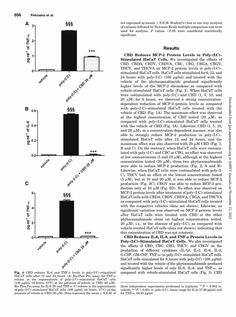
HaCaT cells after 12 and 24 hours. (A) Bio-Plex Pro assay for TNF-arelease in the supernatants of poly-(I:C)–stimulated HaCaT cells(100 mg/ml, 12 hours, 37°C) in the presence of vehicle or CBD (20 mM).Bio-Plex Pro assay for IL-6 (B) and TNF-a (C) release in the supernatantsof poly-(I:C)–stimulated HaCaT cells (100 mg/ml, 24 hours, 37°C) in thepresence of vehicle or CBD (20 mM). Data represent the mean 6 S.E.M. of
three independent experiments performed in triplicate. xxxP , 0.001 vs.vehicle; ***P, 0.001 vs. poly-(I:C). Assay range for IL-6, 37.68 pg/ml; andfor TNF-a, 64.80 pg/ml.
(1, 5, 10, and 20 mM), in a concentration-dependentmanner, wasable to strongly reduce IL-6 and TNF-a protein levels in poly-(I:C)–stimulated HaCaT cells, as compared with poly-(I:C)–stimulated HaCaT cells treated with the vehicle of CBD (Fig. 3,
A and C). IL-8 protein levels were strongly reduced only by thetwo highest concentrations of CBD tested (10 and 20 mM) (Fig.3B).WhenHaCaT cellswere stimulated for 12hourswithpoly-(I:C) and treatedwith the vehicle of the phytocannabinoids,we only
Fig. 5. CBC, CBG, and THCV reduce IL-6 and IL-8 levels in poly-(I:C)–stimulated HaCaT cells after 6 hours. Bio-Plex Pro assay for IL-6 (A, C, and E)and IL-8 (B and D) release in the supernatants of poly-(I:C)–stimulated HaCaT cells (100 mg/ml, 6 hours, 37°C) in the presence of vehicle or CBC, CBG,and THCV (all tested at 5, 10, and 20 mM). Data represent the mean6 S.E.M. of three independent experiments performed in triplicate. xxxP, 0.001 vs.vehicle; *P , 0.05; **P , 0.01; ***P , 0.001 vs. poly-(I:C). Assay range for IL-6, 37.68 pg/ml; and for IL-8, 42.15 pg/ml.
observed a strong increase of TNF-a protein levels (Fig. 4A),which was significantly reduced by the highest concentration ofCBD tested (20 mM) (Fig. 4A). When HaCaT cells werestimulated for 24 hours with poly-(I:C) and treated with thevehicle of the phytocannabinoids, we observed a strong increasein both IL-6 (Fig. 4B) and TNF-a (Fig. 4C) protein levels, whichwere again strongly reduced by treatment with CBD 20mM(Fig.4, B and C). On the contrary, the other phytocannabinoids foundhere to produce anti-inflammatory effects on MCP-2 levels wereable to downregulate only some of these cytokines after 6 hours,i.e., 1) CBC (20 mM) was able to reduce only IL-6 and IL-8 levels(Fig. 5,AandB), 2)CBG (10mM)wasable to reduce only IL-6 andIL-8 levels (Fig. 5, B and C), and 3) THCV (20 mM) was able toreduce only IL-6 levels (Fig. 5D). No effect was observed on thelevels of other cytokines, i.e., IL-1b, IL-2, G-CSF, and GM-CSF,after stimulation ofHaCaT cellswith poly-(I:C) in the presence orabsence of the other phytocannabinoids (data not shown).CBD and Other Phytocannabinoids Are Not Cyto-
toxic to HaCaT Cells. No cytotoxicity was observed aftertreatment of HaCaT cells for 6 hours with CBD, CBC, CBG,THCV, or CBGV at the highest concentrations tested (10 and20 mM) (Fig. 6A). Likewise, no cytotoxicity was observed aftertreatment of HaCaT cells for 12 and 24 hours with 10 and20 mM CBD (Fig. 6B). These results indicate that the de-creasedMCP-2 and/or cytokine levels in poly-(I:C)–stimulatedHaCaT cells were due to the anti-inflammatory effects of thesecompounds.CB1 Receptors Do Not Mediate the Action of CBD in
Poly-(I:C)–Stimulated HaCaT Cells. We investigated theeffect of a CB1 receptor antagonist (AM251; 1, 2.5, and 5 mM)onMCP-2 protein levels in poly-(I:C)–stimulated HaCaT cells,in the presence or absence of CBD (20 mM). Our results showthat when HaCaT cells were costimulated for 6 hours withpoly-(I:C) and low concentrations of AM251 (1 or 2.5 mM),MCP-2 protein levels were comparable to those found in theabsence of the antagonist (Fig. 7A). On the contrary, whenHaCaT cells were costimulated with poly-(I:C) and the highestconcentration of AM251 tested (5 mM), MCP-2 protein levelswere comparable to those observed in the presence of 20 mMCBD (Fig. 7A). In addition, when HaCaT cells were costimu-lated with poly-(I:C), 20 mM CBD and a per se inactiveconcentration of AM251 (2.5 mM), MCP-2 production was
comparable to that observed in poly-(I:C)–stimulatedHaCaT cells treated with 20 mM CBD alone (Fig. 7A). Noeffect was observed onMCP-2 protein levels after treatment ofHaCaT cells with the antagonist AM251 alone (at the highestconcentration tested, 5mM), i.e., in the absence of poly-(I:C), ascompared with vehicle-treated HaCaT cells (data not shown).Likewise, no additive effect was found onMCP-2 protein levelsafter that poly-(I:C)–stimulated HaCaT cells were treatedwith 20 mM CBD and the highest concentration of AM251tested (5 mM), as compared with poly-(I:C)–stimulatedHaCaT cells treated with 20 mM CBD only (data not shown).CB2 and TRPV1 Receptors Mediate the Action of
CBD in Poly-(I:C)–Stimulated HaCaT Cells. We investi-gated the effect of a CB2 receptor antagonist (AM630; 0.01, 0.1,and 1 mM) and a TRPV1 receptor antagonist (I-RTX; 0.01, 0.1,and 1 mM) on MCP-2 protein levels in poly-(I:C)–stimulatedHaCaT cells in the presence or absence of CBD (20 mM). Ourresults show that when HaCaT cells were costimulated for6 hours with poly-(I:C) and low concentrations of AM630 (0.01or 0.1 mM) or high concentration of I-RTX (1 mM), MCP-2protein levels were comparable to those observed in theabsence of the antagonists (Fig. 7B). On the contrary, whenHaCaT cells were costimulated with poly-(I:C) and a higherconcentration of AM630 (1 mM) or lower concentrations ofI-RTX (0.01 or 0.1 mM),MCP-2 protein levels were comparableto those observed in the presence of CBD 20 mM (Fig. 7B).Importantly, when HaCaT cells were costimulated with poly-(I:C), 20 mM CBD, and the highest per se inactive concentra-tions of AM630 or I-RTX (0.1 and 1 mM, respectively), MCP-2chemokine production was comparable to that observed inpoly-(I:C)–stimulated HaCaT cells treated with the vehicle(Fig. 7B). However, no effects of AM630 or I-RTX (0.1 and1 mM, respectively) on MCP-2 protein levels were observedafter 12 and 24 hours in poly-(I:C)–stimulated HaCaT cellstreated with 20 mM CBD, as compared with poly-(I:C)–stimulated HaCaT cells treated with 20 mM CBD alone (datanot shown). In addition, no effect was observed on MCP-2protein levels after that HaCaT cells were treated with theantagonists, AM630 or I-RTX, alone (at highest concentra-tions tested, 1 mM), i.e., in the absence of poly-(I:C), ascompared with vehicle-treated HaCaT cells (data not shown).Likewise, no additive effects were found on MCP-2 protein
Fig. 6. CBD and other phytocannabinoids are not cytotoxic in HaCaT cells. (A) MTT assay in HaCaT cells treated with vehicle (white histogram) orCBD, CBC, CBG, THCV, and CBGV (all tested at 10 and 20 mM) for 6 hours at 37°C in 5% CO2. (B) MTT assay in HaCaT cells treated with vehicle orCBD (10 and 20 mM) for 12 and 24 hours at 37°C in 5%CO2. Data represent the mean6 S.E.M. of three independent experiments performed in triplicate.
levels after that poly-(I:C)–stimulated HaCaT cells weretreated with 20 mM CBD and the highest concentration ofAM630 tested (1 mM) or the lowest concentrations of IRTX
tested (0.01 and 0.1 mM), as compared with poly-(I:C)–stimulated HaCaT cells treated with 20 mM CBD (data notshown). On the basis of these results, we also investigated theeffects of AM630 (0.1 mM) and I-RTX (1 mM) on the productionof cytokines, such as IL-6, IL-8, and TNF-a, in poly-(I:C)–stimulated HaCaT cells in the presence or absence of CBD(20 mM). Our results show that no effect was found withAM630 (0.1 mM) or I-RTX (1 mM) on the inhibitory action ofCBD (20 mM) on the levels of the other three cytokines thatwere elevated after poly-(I:C) stimulation of HaCaT cells (datanot shown).CBD Elevates AEA Levels in Poly-(I:C)–Stimulated
HaCaT Cells. We measured the effect of CBD (20 mM) onAEA, 2-AG, PEA, and OEA levels in poly-(I:C)–stimulatedHaCaT cells. We observed that when HaCaT cells werestimulated for 6 hours with poly-(I:C), AEA levels weresignificantly increased by 3-fold compared with vehicle-treated HaCaT cells, and a nearly statistically significanttrend toward elevation of PEA levels (P 5 0.0633) was alsoobserved (Fig. 8, A and C). When poly-(I:C)–stimulatedHaCaT cells were treated with CBD (20 mM), AEA levels wereincreased by 8-fold compared with vehicle-treated HaCaTcells, and by 2.7-fold compared with poly-(I:C)–stimulatedHaCaT cells (Fig. 8A). These increases were only observedduring the early sensitization phase of ACD, i.e., after 6 hours(Fig. 8A). No consistent effect of CBDwas observed on 2-AGandOEA levels after 6 hours (Fig. 8, B and D), as well as on AEAlevels after 12 and 24 hours (data not shown), in poly-(I:C)–stimulated HaCaT cells.AEA and URB597 Reduce MCP-2, IL-6, and IL-8
Protein Levels in Poly-(I:C)–Stimulated HaCaT Cells.We investigated the effects of AEA and a selective fatty acidamide hydrolase inhibitor, URB597, on the production ofMCP-2 and other cytokines (IL-6, IL-8, and TNF-a) in poly-(I:C)–stimulated HaCaT cells. Our results show that whenHaCaT cells were costimulated with poly-(I:C) and AEA(10 mM), the levels of MCP-2 and IL-8 proteins were reducedas compared with poly-(I:C)–stimulated HaCaT cells treat-ed with the vehicle (Fig. 9, A and C). WhenHaCaT cells werecostimulated with poly-(I:C) and URB597 (10 mM), thelevels of MCP-2, IL-6, and IL-8 proteins were also reducedas compared with poly-(I:C)–stimulated HaCaT cells treat-ed with the vehicle (Fig. 9). No effect was observed on IL-6levels after treatment of poly-(I:C)–stimulated HaCaT cellswith AEA (10 mM) (Fig. 9B). Likewise, no effect wasobserved on TNF-a production after treatment of poly-(I:C)–stimulated HaCaT cells with either AEA (10 mM) orURB597 (10 mM) (data not shown). Thus, the effects of AEAand URB597 in poly-(I:C)–stimulated HaCaT cells weresimilar, although not identical, to those observed with CBD(20 mM) (Fig. 9).
DiscussionIn this study, we demonstrated for the first time that a
nonpsychotropic plant cannabinoid, CBD, inhibits the produc-tion of the MCP-2 chemokine and other proinflammatorycytokines (i.e., IL-6, IL-8, and TNF-a) in poly-(I:C)–stimulatedkeratinocytes more potently/efficaciously/dose-dependentlythan other nonpsychotropic phytocannabinoids tested here(CBDA, CBDV, CBDVA, CBC, CBG, CBGA, CBGV, THCV,
Fig. 7. CB2 and TRPV1 receptors mediate the action of CBD in poly-(I:C)–stimulated HaCaT cells after 6 hours. (A) Enzyme-linked immunosorbentassay for MCP-2 release in the supernatants of poly-(I:C)–stimulatedHaCaT cells (100 mg/ml, 6 hours, 37°C) treated with AM251 (1, 2.5, and5 mM) in the presence or absence of CBD (20 mM). (B) Enzyme-linkedimmunosorbent assay for MCP-2 release in the supernatants of poly-(I:C)–stimulated HaCaT cells (100 mg/ml, 6 hours, 37°C) treated with AM630(0.01 and 0.1 mM) or I-RTX (0.01 and 0.1 mM) in the presence or absence ofCBD (20 mM). Data represent the mean 6 S.E.M. of three independentexperiments performed in triplicate. xxxP, 0.001 vs. vehicle. ***P, 0.001;**P , 0.01 vs. poly-(I:C); °°P , 0.01; °°°P , 0.001 vs. poly-(I:C) + CBD20 mM. Assay range for MCP-2, 0.8–200 pg/ml.
and THCVA). Importantly, we also identified the moleculartargets for some of the actions of CBD on keratinocytes.First, we demonstrated that when HaCaT cells are stim-
ulated for 6 hours and up to 24 hours with an agonist of thetoll-like receptor 3, i.e., poly-(I:C), they produce higher levelsof MCP-2. This chemokine is a proinflammatory mediatorinvolved in the recruitment of macrophages and mast cellsinto inflammatory sites (Taub et al., 1995; de Paulis et al.,2001), and is downregulated by CB1/CB2 agonists, suchas THC and HU-210, in ear keratinocytes of mice withDNFB-induced ACD, as well as in poly-(I:C)–stimulatedHaCaT cells in vitro (Karsak et al., 2007). We previouslydemonstrated that this chemokine can also be downregu-lated by an endogenous lipid mediator, namely PEA, which isknown for its important anti-inflammatory properties and iscurrently used in the clinic against neuropathic and in-flammatory pain but, unlike AEA, is not capable of directlyactivating CB1 or CB2 receptors (Iannotti et al., 2016). Inparticular, we showed that PEA at a concentration of 10 mMwas able to reduce, through a TRPV1-mediated mechanism,both the expression and the production of MCP-2 in poly-(I:C)–stimulated HaCaT cells in vitro (Petrosino et al., 2010).On the contrary, neither the antagonism of CB2 receptors northe antagonism of peroxisome proliferator-activated recep-tor-a receptors reversed the anti-inflammatory effect of PEAin poly-(I:C)–stimulated HaCaT cells (Petrosino et al., 2010).Therefore, here we investigated 1) whether CBD, which, likePEA and unlike THC, is unable to directly activate CB1/CB2
receptors, can also nevertheless reduce MCP-2 protein levelsin poly-(I:C)–stimulated HaCaT cell; and 2) the molecu-lar target(s) through which CBD exerts this putative anti-inflammatory action. We found that CBD, in a concentration-dependent manner, strongly reduces MCP-2 protein levels inHaCaT cells stimulated for 6 hours and up to 24 hours withpoly-(I:C), with the maximum effect being observed at thehighest concentration tested (20 mM), and no significantdifferences in the extent of the effect being noted at differenttime points. This concentration is 2-fold higher than theefficacious (and maximally soluble in the same vehicle)concentration of PEA (10 mM), in the same in vitro model ofACD (Petrosino et al., 2010). Here, we also demonstrated forthe first time that when HaCaT cells are stimulated for6 hours and up to 24 hours with poly-(I:C), they producehigher levels of other proinflammatory cytokines (i.e., IL-6,IL-8, and TNF-a), which, similar to MCP-2, are producedfrom keratinocytes during the sensitization phase (Barker,1992; Enk and Katz, 1992; Becker and Knop, 1993), and thatCBD also reduces these effects of poly-(I:C). However, ofthese three cytokines, only TNF-a exhibited significantincreases at all time points, suggesting their potentiallydifferent roles in different phases of inflammation, at least inthis in vitro model.Since it is well known that 1) the pharmacological blockade
of CB2 receptor attenuates oxazoline-induced contact derma-titis in mice (Oka et al., 2006); 2) the activation of the CB2
receptor, but not the CB1 receptor, is involved in THC-induced
Fig. 8. CBD elevates AEA levels in poly-(I:C)–stimulated HaCaT cells after 6 hours. Concentrations of AEA (A), 2-AG (B), PEA (C), and OEA (D) in poly-(I:C)–stimulated HaCaT cells (100 mg/ml, 6 hours, 37°C) in the presence of vehicle or CBD (20 mM). Data represent the mean 6 S.E.M. of threeindependent experiments performed in triplicate. ***P , 0.001 vs. vehicle; °°°P , 0.001 vs. poly-(I:C) + vehicle.
anti-inflammatory effects (Yang et al., 2015; Shang, et al.,2016; Xie et al., 2016); 3) CB1 or CB2 receptor activation andthe activation/desensitization of TRPV1 channels contributeto the in vitro and in vivo pharmacological actions of CBD(Costa et al., 2004; Arnold et al., 2012; Pazos et al., 2013;Stanley et al., 2015); 4) CBD can directly activate anddesensitize TRPV1 channels (Iannotti et al., 2014); and 5)CB1 and CB2 receptors as well as TRPV1 are targets for thetreatment of the early phase of ACD (Karsak et al., 2007;Petrosino et al., 2010), we investigated whether the anti-inflammatory action of CBD could be mediated by one or moreof these targets in the in vitro model of ACD. Although itis also known that CBD weakly activates the human5-hydroxytryptamine (serotonin) receptor 1A (Russo et al.,2005), we did not investigate this mechanism because it hasbeen demonstrated that HaCaT cells do not express suchreceptor (Slominski et al., 2003). Thus, we studied the effectsof CB1, CB2, and TRPV1 antagonists on MCP-2 and cyto-kine (i.e., IL-6, IL-8, and TNF-a) production in poly-(I:C)–stimulated HaCaT cells in vitro, in the presence or absence of20 mM CBD. Our results show that, similar to CBD, the CB1
antagonist AM251, at the highest concentration tested (5 mM),was able to only reduce MCP-2 protein levels after 6 hours inpoly-(I:C)–stimulated HaCaT cells. The inhibitory effect ofCBD on MCP-2 production was not influenced by the highestper se inactive concentration of AM251 (2.5 mM). Similar toAM251 and CBD, the CB2 antagonist AM630, at the highestconcentration tested (1 mM), was able to only reduce MCP-2protein levels after 6 hours in poly-(I:C)–stimulatedHaCaT cells,but unlike AM251, the highest per se inactive concentration ofAM630 tested (0.1mM)was able to reverse the inhibitory effectof CBD on MCP-2 levels. On the other hand, low concentra-tions of the TRPV1 antagonist I-RTX were able to only reduceMCP-2 protein levels after 6 hours in poly-(I:C)–stimulatedHaCaT cells, whereas the highest per se inactive concentra-tion of this compound tested (1 mM) was able to reverse theinhibitory effect of CBD on MCP-2 levels. These results 1)suggest that, since the effects of CB2 and TRPV1 antagonists,per se or on the effects of CBD, were only observed on MCP-2production and only at 6 hours, this chemokine and thesereceptors are mostly involved in the early sensitization phaseof ACD and in the inhibitory actions thereupon of thephytocannabinoid; the effect of the highest concentrations ofAM251 per semay be non–CB1-mediated, since this compoundat high concentrations acts as an agonist on another potentialanti-inflammatory off-target, that is GPR55 (Ryberg et al.,2007; Cantarella et al., 2011; Montecucco et al., 2016); 2)confirm that CB2 receptors may play also a facilitatory role inACD (Ueda et al., 2005), as indicated here by the anti-inflammatory effect of the highest concentration of AM630;and 3) support the previous finding that TRPV1may play bothfacilitatory and inhibitory roles against ACD (Banvolgyi et al.,2005), as shown here by the fact that low, but not high,concentrations of I-RTX counteracted poly-(I:C)–inducedMCP-2 production in HaCaT keratinocytes. Thus, the anti-inflammatory effect of CBD might be due to both CB2
activation, as previously shown for THC (Karsak et al., 2007;Yang et al., 2015; Shang et al., 2016; Xie et al., 2016), and
Fig. 9. AEA and URB597 reduce MCP-2, IL-6, and IL-8 levels in poly-(I:C)–stimulated HaCaT cells after 6 hours. Enzyme-linked immunosorbentassay for MCP-2 (A) and Bio-Plex Pro assay for IL-6 (B) and IL-8 (C)release in the supernatants of poly-(I:C)–stimulated HaCaT cells(100 mg/ml, 6 hours, 37°C) in the presence of vehicle or CBD (20 mM),AEA (10 mM), or URB597 (10 mM). Data represent the mean 6 S.E.M. ofthree independent experiments performed in triplicate. xxxP , 0.001 vs.
vehicle; *P, 0.05; **P, 0.01; ***P, 0.001 vs. poly-(I:C). Assay range forMCP-2, 0.8–200 pg/ml; for IL-6, 37.68 pg/ml; and for IL-8, 42.15 pg/ml.
TRPV1 activation/desensitization, as suggested here by thefact that this effect was reversed by both a low concentration ofa CB2 receptor antagonist (AM630) and by a high concentra-tion of a TRPV1 antagonist (I-RTX). Although this latter effectis not surprising due to the aforementioned capability of CBDto stimulate and desensitize TRPV1 (Iannotti et al., 2014), thisphytocannabinoid exhibits only low affinity for CB2 (Pertwee,2008). Therefore, we hypothesized that endogenous ligandscould mediate the anti-inflammatory effect of CBD at thisreceptor thus explaining why such effect was antagonized byAM630. Indeed, CBD, at a ∼20 mM concentration (IC50 5 27.5mM) inhibits both AEA cellular uptake and enzymatic hydro-lysis (Bisogno et al., 2001), and these effects could explain boththe present finding of its stimulatory action on AEA levels andindirect activation of CB2 receptors. Consequently, our hy-pothesis that CBD acted via elevation of AEA levels inHaCaT cells was supported by our present finding that bothAEA and a synthetic inhibitor of its degradation, URB597,similar to CBD, were able to reduce the production of bothMCP-2 and other proinflammatory cytokines (i.e., IL-6 andIL-8) produced by poly-(I:C)–stimulated keratinocytes. In-terestingly, the stimulatory action of CBD on AEA levelswas only observed after 6 hours, and this could explain whythe CB2 antagonist here did not attenuate the anti-inflammatory effects of CBD after 12 and 24 hours. Impor-tantly, whereas the effects of AEA and URB597 on MCP-2production were comparable to those of CBD, those on IL-6and IL-8 were statistically significant but less efficacious. Thisobservation supports the aforementioned suggestion thatthese cytokines play different roles in the sensitization phaseof ACD, at least in the in vitro model used here, thus possiblyexplaining why CB2 and TRPV1 antagonists did not attenuatethe effect of CBD on these inflammatory mediators.In conclusion, in the present study, we demonstrated that, in
an in vitromodel of ACD, 1) CBD inhibits the production ofMCP-2 as well as IL-6, IL-8, and TNF-a; 2) the endogenous levels ofAEA are increased after CBD treatment; and 3) the anti-inflammatory effect of CBD during the early sensitization phase(i.e., after 6 hours) is antagonized both by a selective CB2
antagonist—and hence potentially mediated by the endogenousagonist for CB2 receptors, AEA—and a selective TRPV1antagonist—likely because the phytocannabinoid can directlyactivate and desensitize the TRPV1 channel. Given the estab-lished safety profile of CBD in humans (Leweke et al., 2012;Pertwee, 2015), these data warrant further experiments on thepreclinical testing of this compound in animal models of ACD.
Authorship Contributions
Participated in research design: Petrosino, Iuvone, Di Marzo.Conducted experiments: Petrosino, Verde, Vaia, Allarà, Iuvone.Performed data analysis: Petrosino, Di Marzo.Wrote or contributed to the writing of the manuscript: Petrosino, Di
Marzo.
References
Arnold JC, Hone P, Holland ML, and Allen JD (2012) CB2 and TRPV1 receptorsmediate cannabinoid actions on MDR1 expression in multidrug resistant cells.Pharmacol Rep 64:751–757.
Bánvölgyi A, Pálinkás L, Berki T, Clark N, Grant AD, Helyes Z, Pozsgai G, Szolc-sányi J, Brain SD, and Pintér E (2005) Evidence for a novel protective role of thevanilloid TRPV1 receptor in a cutaneous contact allergic dermatitis model. JNeuroimmunol 169:86–96.
Barker JN (1992) Role of keratinocytes in allergic contact dermatitis. Contact Dermat26:145–148.
Becker D and Knop J (1993) Mechanism in allergic contact dermatitis. Exp Dermatol2:63–69.
Bisogno T, Hanus L, De Petrocellis L, Tchilibon S, Ponde DE, Brandi I, Moriello AS,Davis JB, Mechoulam R, and Di Marzo V (2001) Molecular targets for cannabidioland its synthetic analogues: effect on vanilloid VR1 receptors and on the cellularuptake and enzymatic hydrolysis of anandamide. Br J Pharmacol 134:845–852.
Bisogno T, Sepe N, Melck D, Maurelli S, De Petrocellis L, and Di Marzo V (1997)Biosynthesis, release and degradation of the novel endogenous cannabimimeticmetabolite 2-arachidonoylglycerol in mouse neuroblastoma cells. Biochem J 322:671–677.
Burstein S (2015) Cannabidiol (CBD) and its analogs: a review of their effects oninflammation. Bioorg Med Chem 23:1377–1385.
Burstein SH and Zurier RB (2009) Cannabinoids, endocannabinoids, and relatedanalogs in inflammation. AAPS J 11:109–119.
Cantarella G, Scollo M, Lempereur L, Saccani-Jotti G, Basile F, and Bernardini R(2011) Endocannabinoids inhibit release of nerve growth factor by inflammation-activated mast cells. Biochem Pharmacol 82:380–388.
Costa B, Giagnoni G, Franke C, Trovato AE, and Colleoni M (2004) VanilloidTRPV1 receptor mediates the antihyperalgesic effect of the nonpsychoactivecannabinoid, cannabidiol, in a rat model of acute inflammation. Br J Pharmacol143:247–250.
de Paulis A, Annunziato F, Di Gioia L, Romagnani S, Carfora M, Beltrame C, MaroneG, and Romagnani P (2001) Expression of the chemokine receptor CCR3 on humanmast cells. Int Arch Allergy Immunol 124:146–150.
De Petrocellis L, Davis JB, and Di Marzo V (2001) Palmitoylethanolamide enhancesanandamide stimulation of human vanilloid VR1 receptors. FEBS Lett 506:253–256.
De Petrocellis L, Ligresti A, Moriello AS, Allarà M, Bisogno T, Petrosino S, Stott CG,and Di Marzo V (2011) Effects of cannabinoids and cannabinoid-enriched Cannabisextracts on TRP channels and endocannabinoid metabolic enzymes. Br J Phar-macol 163:1479–1494.
Di Marzo V, Goparaju SK, Wang L, Liu J, Bátkai S, Járai Z, Fezza F, Miura GI,Palmiter RD, Sugiura T, et al. (2001) Leptin-regulated endocannabinoids are in-volved in maintaining food intake. Nature 410:822–825.
Enk AH and Katz SI (1992) Early events in the induction phase of contact sensitivity.J Invest Dermatol 99:39S–41S.
Gaffal E, Cron M, Glodde N, and Tüting T (2013) Anti-inflammatory activity oftopical THC in DNFB-mediated mouse allergic contact dermatitis independent ofCB1 and CB2 receptors. Allergy 68:994–1000.
Iannotti FA, Di Marzo V, and Petrosino S (2016) Endocannabinoids andendocannabinoid-related mediators: targets, metabolism and role in neurologicaldisorders. Prog Lipid Res 62:107–128.
Iannotti FA, Hill CL, Leo A, Alhusaini A, Soubrane C, Mazzarella E, Russo E,Whalley BJ, Di Marzo V, and Stephens GJ (2014) Nonpsychotropic plant can-nabinoids, cannabidivarin (CBDV) and cannabidiol (CBD), activate and de-sensitize transient receptor potential vanilloid 1 (TRPV1) channels in vitro:potential for the treatment of neuronal hyperexcitability. ACS Chem Neurosci 5:1131–1141.
Karsak M, Gaffal E, Date R, Wang-Eckhardt L, Rehnelt J, Petrosino S, Starowicz K,Steuder R, Schlicker E, Cravatt B, et al. (2007) Attenuation of allergic contactdermatitis through the endocannabinoid system. Science 316:1494–1497.
Leweke FM, Piomelli D, Pahlisch F, Muhl D, Gerth CW, Hoyer C, Klosterkötter J,Hellmich M, and Koethe D (2012) Cannabidiol enhances anandamide signalingand alleviates psychotic symptoms of schizophrenia. Transl Psychiatry 2:e94.
Lo Verme J, Fu J, Astarita G, La Rana G, Russo R, Calignano A, and Piomelli D(2005) The nuclear receptor peroxisome proliferator-activated receptor-alpha me-diates the anti-inflammatory actions of palmitoylethanolamide.Mol Pharmacol 67:15–19.
Maione S, Piscitelli F, Gatta L, Vita D, De Petrocellis L, Palazzo E, de Novellis V,and Di Marzo V (2011) Non-psychoactive cannabinoids modulate the descendingpathway of antinociception in anaesthetized rats through several mechanisms ofaction. Br J Pharmacol 162:584–596.
Marsicano G, Wotjak CT, Azad SC, Bisogno T, Rammes G, Cascio MG, Hermann H,Tang J, Hofmann C, Zieglgänsberger W, et al. (2002) The endogenous cannabinoidsystem controls extinction of aversive memories. Nature 418:530–534.
Montecucco F, Bondarenko AI, Lenglet S, Burger F, Piscitelli F, Carbone F, Roth A,Liberale L, Dallegri F, Brandt KJ, et al. (2016) Treatment with the GPR55 an-tagonist CID16020046 increases neutrophil activation in mouse atherogenesis.Thromb Haemost 116:987–997.
Oka S, Wakui J, Ikeda S, Yanagimoto S, Kishimoto S, Gokoh M, Nasui M,and Sugiura T (2006) Involvement of the cannabinoid CB2 receptor and its en-dogenous ligand 2-arachidonoylglycerol in oxazolone-induced contact dermatitis inmice. J Immunol 177:8796–8805.
Pazos MR, Mohammed N, Lafuente H, Santos M, Martínez-Pinilla E, Moreno E,Valdizan E, Romero J, Pazos A, Franco R, et al. (2013) Mechanisms of cannabidiolneuroprotection in hypoxic-ischemic newborn pigs: role of 5HT(1A) and CB2 re-ceptors. Neuropharmacology 71:282–291.
Pertwee RG (2008) The diverse CB1 and CB2 receptor pharmacology of three plantcannabinoids: delta9-tetrahydrocannabinol, cannabidiol and delta9-tetrahy-drocannabivarin. Br J Pharmacol 153:199–215.
Pertwee RG (2015) Endocannabinoids and their pharmacological actions. Handb ExpPharmacol 231:1–37.
Petrosino S, Cristino L, Karsak M, Gaffal E, Ueda N, Tüting T, Bisogno T, De FilippisD, D’Amico A, Saturnino C, et al. (2010) Protective role of palmitoylethanolamidein contact allergic dermatitis. Allergy 65:698–711.
Petrosino S and Di Marzo V (2017) The pharmacology of palmitoylethanolamide andfirst data on the therapeutic efficacy of some of its new formulations. Br J Phar-macol 174:1349–1365.
Petrosino S, Schiano Moriello A, Cerrato S, Fusco M, Puigdemont A, De Petrocellis L,and Di Marzo V (2016) The anti-inflammatory mediator palmitoylethanolamideenhances the levels of 2-arachidonoyl-glycerol and potentiates its actions at TRPV1cation channels. Br J Pharmacol 173:1154–1162.
Russo EB, Burnett A, Hall B, and Parker KK (2005) Agonistic properties of canna-bidiol at 5-HT1a receptors. Neurochem Res 30:1037–1043.
Ryberg E, Larsson N, Sjögren S, Hjorth S, Hermansson NO, Leonova J, Elebring T,Nilsson K, Drmota T, and Greasley PJ (2007) The orphan receptor GPR55 is anovel cannabinoid receptor. Br J Pharmacol 152:1092–1101.
Shang VC, Kendall DA, and Roberts RE (2016) D9-Tetrahydrocannabinol reversesTNFa-induced increase in airway epithelial cell permeability through CB2re-ceptors. Biochem Pharmacol 120:63–71.
Slominski A, Pisarchik A, Zbytek B, Tobin DJ, Kauser S, and Wortsman J (2003)Functional activity of serotoninergic and melatoninergic systems expressed in theskin. J Cell Physiol 196:144–153.
Stanley CP, Hind WH, Tufarelli C, and O’Sullivan SE (2015) Cannabidiol causesendothelium-dependent vasorelaxation of human mesenteric arteries via CB1 ac-tivation. Cardiovasc Res 107:568–578.
Taub DD, Proost P, Murphy WJ, Anver M, Longo DL, van Damme J, and OppenheimJJ (1995) Monocyte chemotactic protein-1 (MCP-1), -2, and -3 are chemotactic forhuman T lymphocytes. J Clin Invest 95:1370–1376.
Ueda Y, Miyagawa N, Matsui T, Kaya T, and Iwamura H (2005) Involvement ofcannabinoid CB(2) receptor-mediated response and efficacy of cannabinoid CB(2)receptor inverse agonist, JTE-907, in cutaneous inflammation in mice. Eur JPharmacol 520:164–171.
Vaia M, Petrosino S, De Filippis D, Negro L, Guarino A, Carnuccio R, Di Marzo V,and Iuvone T (2016) Palmitoylethanolamide reduces inflammation and itch in amouse model of contact allergic dermatitis. Eur J Pharmacol 791:669–674.
van Loveren H, Meade R, and Askenase PW (1983) An early component of delayed-typehypersensitivity mediated by T cells and mast cells. J Exp Med 157:1604–1617.
Watanabe H, Unger M, Tuvel B, Wang B, and Sauder DN (2002) Contact hyper-sensitivity: the mechanism of immune responses and T cell balance. J InterferonCytokine Res 22:407–412.
Xie J, Xiao D, Xu Y, Zhao J, Jiang L, Hu X, Zhang Y, and Yu L (2016) Up-regulation ofimmunomodulatory effects of mouse bone-marrow derived mesenchymal stem cellsby tetrahydrocannabinol pre-treatment involving cannabinoid receptor CB2.Oncotarget 7:6436–6447.
Yang L, Li FF, Han YC, Jia B, and Ding Y (2015) Cannabinoid receptor CB2 isinvolved in tetrahydrocannabinol-induced anti-inflammation against lipopolysac-charide in MG-63 cells. Mediators Inflamm 2015:362126.
Address correspondence to: Dr. Vincenzo Di Marzo, Istituto di ChimicaBiomolecolare, Consiglio Nazionale delle Ricerche, Via Campi Flegrei 34,80078 Pozzuoli, Napoli, Italy. E-mail: [email protected]