Vol. 38, No. 5 APPLIED AND ENVIRONMENTAL MICROBIOLOGY, Nov. 1979, p. 821-826 0099-2240/79/11-0821/06$02.00/0 Antibacterial Action of Lactoperoxidase-Thiocyanate- Hydrogen Peroxide on Streptococcus agalactiae M. N. MICKELSON National Animal Disease Center, Agricultural Research, Science and Education Administration, U.S. Department of Agriculture, Ames, Iowa 50010 Received for publication 20 August 1979 Antibacterial activity of lactoperoxidase (LP)-thiocyanate (SCN)-hydrogen peroxide (H202) on Streptococcus agalactiae requires that the three reactants must be in contact with the cells simultaneously. Small but assayable amounts of LP adsorb to the cell surface and are not removed by washing. A diffusible antibacterial product of LP-SCN-H202 reaction was not found under our experi- mental conditions. Incubation of S. agalactiae cells with LP-H202 and "C-labeled sodium SCN resulted in the incorporation of SCN into the bacterial protein. Most of the LP-catalyzed, incorporated SCN was released from the bacterial protein with dithiothreitol. Cells that had their membrane permeability changed by treatment with Cetab or 80% ethanol incorporated more SCN than did untreated cells, i.e., approximately 1 mol of SCN for each mol of sulfhydryl group present in the reaction mixture. Alteration of membrane permeability caused protein sulfhydryls, normally protected by the cytoplasmic membrane, to become exposed to oxidation. The results suggest the LP-H202-catalyzed incorporation of SCN into the proteins of S. agalactiae by a mechanism similar to that reported for bovine serum albumin. Removal of reactive protein sulfhydryls from a functional role in membrane transport and in glucolysis is a likely cause of the antibacterial effect for S. agalactiae. The mode of action of the lactoperoxidase (LP)-thiocyanate (SCN)-hydrogen peroxide (H202) antibacterial system for the streptococci has been of interest since the three components of the system were identified (9, 19, 24). The reversibility of the antibacterial effect with re- ducing agents such as thioglycolate, glutathione, and catalase suggested an oxidative function for LP-H202 and the possible involvement of sulfhy- dryl groups in the antibacterial effect (11). A role for SCN was unexplained, although studies on the LP-catalyzed oxidation of SCN suggested that a "short-lived" intermediate oxidation product might be involved in growth inhibition (16). Reports on the LP-catalyzed incorporation of SCN into sulfhydryl groups of bovine serum albumin to form sulfenylthiocyanate derivatives of the protein sulfhydryls offered, for the first time, an explanation of a definite role for SCN in the antibacterial action (3; T. M. Aune and E. L. Thomas, Fed. Proc. 35:1386, 1976). The fur- ther studies on LP-catalyzed SCN oxidation showed an accumulation of hypothiocyanate (OSCN-) ion, which was stable in conditions existing in bacterial growth media and would oxidize protein sulfhydryl groups to sulfenylthio- cyanate derivatives (1). Hoogendoorn et al. (8) suggested that OSCN- was the growth inhibitor. This present report deals with studies to show the LP-catalyzed incorporation of SCN into the proteins of Streptococcus agalactiae. It also deals with experiments to show the need for the simultaneous presence of all components in im- mediate contact with the bacteria for an anti- bacterial effect. MATERUILS AND METHODS Culture and medium. The culture of S. agalactiae and the chemically defined culture medium used were described in an earlier report (12). For growth inhibition studies, the inoculum was grown in the chemically defined medium containing 5 /g of L-cystine per ml. Four milliliters of medium in screw-capped tubes (13 by 100 mm) was inoculated with 0.1 ml of the stock broth culture, maintained in 1% yeast extract-0.5% Casitone-0.5% glucose-0.1 M phosphate (pH 7), and incubated for 3 to 3.5 h at 37°C until the optical density at 610 nm in a Bausch & Lomb Spectronic 20 colorimeter was 0.55 to 0.60. The cells were sedimented by centrifugation, washed once, and suspended in 4 ml of sterile water. This suspension was diluted 1 to 10, and 0.2 ml (equivalent to about 20 jAg of dry cells) was used to inoculate 4 ml of culture medium. In tests to show binding of LP to bacteria, cells (equivalent to about 40 ,ug of cells [dry weight]) were suspended in 4 ml of medium containing (per milliliter) 1 U of LP, 0.7 ,umol of sodium SCN (NaSCN), and 0.25 821 on January 18, 2020 by guest http://aem.asm.org/ Downloaded from
Transcript
Vol. 38, No. 5APPLIED AND ENVIRONMENTAL MICROBIOLOGY, Nov. 1979, p. 821-8260099-2240/79/11-0821/06$02.00/0
Antibacterial Action of Lactoperoxidase-Thiocyanate-Hydrogen Peroxide on Streptococcus agalactiae
M. N. MICKELSON
National Animal Disease Center, Agricultural Research, Science and Education Administration, U.S.Department ofAgriculture, Ames, Iowa 50010
Received for publication 20 August 1979
Antibacterial activity of lactoperoxidase (LP)-thiocyanate (SCN)-hydrogenperoxide (H202) on Streptococcus agalactiae requires that the three reactantsmust be in contact with the cells simultaneously. Small but assayable amounts ofLP adsorb to the cell surface and are not removed by washing. A diffusibleantibacterial product of LP-SCN-H202 reaction was not found under our experi-mental conditions. Incubation of S. agalactiae cells with LP-H202 and "C-labeledsodium SCN resulted in the incorporation ofSCN into the bacterial protein. Mostof the LP-catalyzed, incorporated SCN was released from the bacterial proteinwith dithiothreitol. Cells that had their membrane permeability changed bytreatment with Cetab or 80% ethanol incorporated more SCN than did untreatedcells, i.e., approximately 1 mol of SCN for each mol of sulfhydryl group presentin the reaction mixture. Alteration of membrane permeability caused proteinsulfhydryls, normally protected by the cytoplasmic membrane, to become exposedto oxidation. The results suggest the LP-H202-catalyzed incorporation of SCNinto the proteins of S. agalactiae by a mechanism similar to that reported forbovine serum albumin. Removal of reactive protein sulfhydryls from a functionalrole in membrane transport and in glucolysis is a likely cause of the antibacterialeffect for S. agalactiae.
The mode of action of the lactoperoxidase(LP)-thiocyanate (SCN)-hydrogen peroxide(H202) antibacterial system for the streptococcihas been of interest since the three componentsof the system were identified (9, 19, 24). Thereversibility of the antibacterial effect with re-ducing agents such as thioglycolate, glutathione,and catalase suggested an oxidative function forLP-H202 and the possible involvement of sulfhy-dryl groups in the antibacterial effect (11). Arole for SCN was unexplained, although studieson the LP-catalyzed oxidation ofSCN suggestedthat a "short-lived" intermediate oxidationproduct might be involved in growth inhibition(16). Reports on the LP-catalyzed incorporationof SCN into sulfhydryl groups of bovine serumalbumin to form sulfenylthiocyanate derivativesof the protein sulfhydryls offered, for the firsttime, an explanation of a definite role for SCNin the antibacterial action (3; T. M. Aune and E.L. Thomas, Fed. Proc. 35:1386, 1976). The fur-ther studies on LP-catalyzed SCN oxidationshowed an accumulation of hypothiocyanate(OSCN-) ion, which was stable in conditionsexisting in bacterial growth media and wouldoxidize protein sulfhydryl groups to sulfenylthio-cyanate derivatives (1). Hoogendoorn et al. (8)suggested that OSCN- was the growth inhibitor.
This present report deals with studies to showthe LP-catalyzed incorporation of SCN into theproteins of Streptococcus agalactiae. It alsodeals with experiments to show the need for thesimultaneous presence of all components in im-mediate contact with the bacteria for an anti-bacterial effect.
MATERUILS AND METHODSCulture and medium. The culture of S. agalactiae
and the chemically defined culture medium used weredescribed in an earlier report (12).
For growth inhibition studies, the inoculum wasgrown in the chemically defined medium containing 5/g of L-cystine per ml. Four milliliters of medium inscrew-capped tubes (13 by 100 mm) was inoculatedwith 0.1 ml of the stock broth culture, maintained in1% yeast extract-0.5% Casitone-0.5% glucose-0.1 Mphosphate (pH 7), and incubated for 3 to 3.5 h at 37°Cuntil the optical density at 610 nm in a Bausch &Lomb Spectronic 20 colorimeter was 0.55 to 0.60. Thecells were sedimented by centrifugation, washed once,and suspended in 4 ml of sterile water. This suspensionwas diluted 1 to 10, and 0.2 ml (equivalent to about 20jAg of dry cells) was used to inoculate 4 ml of culturemedium.
In tests to show binding of LP to bacteria, cells(equivalent to about 40 ,ug of cells [dry weight]) were
suspended in 4 ml ofmedium containing (per milliliter)1 U of LP, 0.7 ,umol of sodium SCN (NaSCN), and 0.25
,umol of H202 or in medium containing LP withoutNaSCN or H202. After incubation for 30 min at 24°C,the cells were recovered by centrifugation and resus-pended to 4 ml in distilled water. The supernatantmedium was carefully removed to avoid any loss ofbacterial cells. Portions of the suspension and super-natant liquor were assayed for LP activity (15).
Experiments were conducted to determine the fol-lowing: (i) whether intimate contact of LP with bac-teria was necessary for growth inhibition, (ii) whetherH202 without LP would oxidize a cell component(s) sothat subsequent reaction with SCN would causegrowth inhibition, and (iii) whether a low-molecular-weight (diffusible) toxic product of the LP-SCN-H202reaction would inhibit growth.
For (ii), the inoculum was incubated for 20 min at25°C in 2 ml of medium containing 1 tLmol of H202,followed by the addition of 2 ml of medium containingSCN. For (iii), the LP complex was separated from theinoculated portion of the medium with a semiperme-able membrane reported to retain substances with amolecular weight in excess of 50,000. LP has a molec-ular weight of 78,000. No LP activity has been detectedin filtrates from these membranes. The membranepermitted rapid transfer of LP-free filtrate from theLP-SCN-H202 mixture into the inoculated mediumwith the use of centrifugal force. About 10 min elapsedfrom the mixing of the reactants until the filtrate hadbeen transferred into the inoculated portion of themedium. The membrane cone, membrane cone sup-port, and centrifuge tube assembly were sterilizedbefore use, the membrane cone with ethylene oxideand the cone support and centrifuge tube by exposurefor 15 min at 100°C in an autoclave. The inoculum in2 ml of medium was incubated for 20 min at 25°C with1 ,umol of H202, and the other 2 ml of medium con-taining the LP-SCN-H202 mixture was rapidly filteredthrough the membrane cone and into the inoculatedmedium.Measurement ofLP-catalyzed incorporation of
SCN into proteins of S. agalactiae. The effect ofLP-H202 on the incorporation ofSCN into the proteinsof S. agalactiae cells was measured with the proceduredescribed by Aune et al. (3) for the incorporation ofSCN into bovine serum albumin.
Cells were grown in the stock culture medium.Phosphate buffer solution was sterilized separatelyand combined with the medium after cooling. A 4-mlculture was grown at 37°C for 3 to 4 h, until it reachedan optical density at 610 nm of about 0.90. This culture,held overnight at 40C, was used to inoculate 50 ml ofmedium in a 125-ml, loosely capped Erlenmeyer flaskand incubated on a rotary shaker (New BrunswickScientific Co., New Brunswick, N.J.) at 120 oscillationsper min for about 2 h or until the optical density wasabout 0.90. The cells were harvested by centrifugation(yield, 0.36 to 0.38 g of wet paste), washed twice indistilled water, and then resuspended to a concentra-tion of about 33 to 35 mg of dry cells per ml. Sulfhydryl,protein, and cell dry weight were determined on thesuspension before the cells were used. Cell dry weightwas determined by drying 0.2 ml of suspension at1050C to a constant weight on tared, stainless-steelplanchets (300-mm diameter by 2 mm deep). Cellprotein was measured on 3-,Il samples of the suspen-
sion after digestion at 100°C for 10 min with 0.05 mlof 1 N NaOH (10). Sulfhydryl groups were estimatedby the procedure of Ellman (5) as applied to bacterialcells by Thomas and Aune (22).
After a 15-min incubation of the reaction mixture(see Table 3) at 37°C, 10 ml of cold 20% trichloroaceticacid was added, and the precipitate was removed bycentrifugation. The precipitated cell material waswashed further by centrifuging and resuspending fivetimes in 5-ml portions of 5% trichloroacetic acid; eachtime, the supernatant liquor was filtered through a0.45-,um membrane filter (Millipore Corp., Bedford,Mass.), and the precipitate was finally transferred tothe filter with four successive portions of 2.5 ml of 5%trichloroacetic acid. The Millipore disks were trans-ferred to scintillation vials and dried ovemight at37°C. The vials were then filled with Cab-O-Sil and 15ml of XDC scintillation fluid (4), and the radioactivitywas determined in a Packard 3380 scintillationcounter. The 14C counting efficiency was 90%.
Glutathione content of the cells was measured withthe procedure described by Fahey et al. (6).
Sulfenyl derivatives were estimated by their reac-tion with ['4C]thiourea to yield a radioactive mixeddisulfide (18). The SCN incorporation reaction wascarried out with unlabeled SCN, LP, and H202 asdescribed in Table 3. Two micromoles of [14C]thioureacontaining 0.5 ,uCi of radioactivity and 10 mg of sodiumdodecyl sulfate were then added. After incubation for1 h at 37°C, the cells were precipitated with trichlo-roacetic acid, washed, and filtered, and their radioac-tivity was determined as described above.
Chemicals and materials. LP was prepared frombovine skim milk by the procedure of Morrison andHultquist (14); further details relating to purity andpotency have been published previously (12). 5,5'-Di-thiobis (2-nitrobenzoic acid) was purchased fromSigma Chemical Co., St. Louis, Mo.; reduced nicotin-amide adenine dinucleotide phosphate, dithiothreitol(DTT), and glutathione reductase were from P-L Bio-chemicals, Inc., Milwaukee, Wis.; Na2S406 was fromTridom Chemicals, Inc., Hauppauge, N.Y.; Cetab (cet-yltrimethylammonium bromide) was from Mann Re-search Laboratories, New York, N.Y.; "4C-labeledNaSCN (NaS'4CN) and [14C]thiourea were from ICNIsotope and Nuclear Div., Cleveland, Ohio; Cab-O-Silwas from Packard Instrument Co., Inc., DownersGrove, Ill.; and the CF50A membrane cones, supports,and centrifuge tubes were from Amicon Corp., Lexing-ton, Mass.
RESULTSS. agalactiae cells suspended in a culture
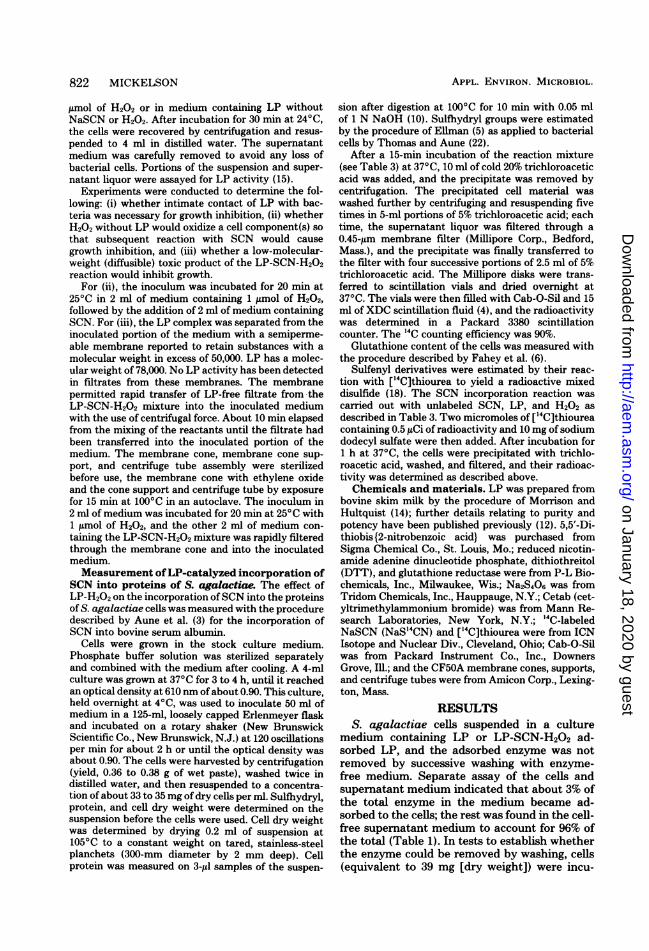
medium containing LP or LP-SCN-H202 ad-sorbed LP, and the adsorbed enzyme was notremoved by successive washing with enzyme-free medium. Separate assay of the cells andsupernatant medium indicated that about 3% ofthe total enzyme in the medium became ad-sorbed to the cells; the rest was found in the cell-free supernatant medium to account for 96% ofthe total (Table 1). In tests to establish whetherthe enzyme could be removed by washing, cells(equivalent to 39 mg [dry weight]) were incu-
TABLE 1. Adsorption of lactoperoxidase to S.agalactiae
LP
Total LP activ- % ofity total(U)
Initially in mediuma 2.690 100.00Adsorbed to cells" 0.084 3.12In supernatant medium 2.490 92.70
after adsorption withbacteria
Recovered in cells plus 2.574 95.82supernatant medium
a Chemically defined medium with 5 yIg of L-cystineper ml.
b Cells from a 3-h culture (equivalent to 47 ,g [dryweight]) were suspended in 4 ml of medium containing2.69 LP activity units, 2.86,umol of NaSCN, and 0.5Mmol of H202. After 30 min at 24°C, the cells wererecovered by centrifugation and suspended in distilledwater; the final volume was 4 ml. Portions of cellsuspension and supernatant medium were assayed forLP activity.
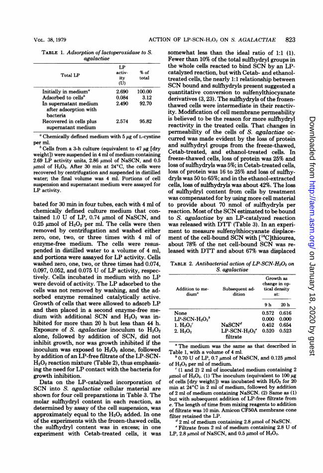
bated for 30 min in four tubes, each with 4 ml ofchemically defined culture medium that con-tained 1.0 U of LP, 0.74 ,umol of NaSCN, and0.25 t,mol of H202 per ml. The cells were thenremoved by centrifugation and washed eitherzero, one, two, or three times with 4 ml ofenzyme-free medium. The cells were resus-pended in distilled water to a volume of 4 ml,and portions were assayed for LP activity. Cellswashed zero, one, two, or three times had 0.074,0.097, 0.052, and 0.075 U of LP activity, respec-tively. Cells incubated in medium with no LPwere devoid of activity. The LP adsorbed to thecells was not removed by washing, and the ad-sorbed enzyme remained catalytically active.Growth of cells that were allowed to adsorb LPand then placed in a second enzyme-free me-dium with additional SCN and H202 was in-hibited for more than 20 h but less than 44 h.Exposure of S. agalactiae inoculum to H202alone, followed by addition of SCN, did notinhibit growth, nor was growth inhibited if theinoculum was exposed to H202 alone, followedby addition of an LP-free filtrate ofthe LP-SCN-H202 reaction mixture (Table 2), thus emphasiz-ing the need for LP contact with the bacteria forgrowth inhibition.Data on the LP-catalyzed incorporation of
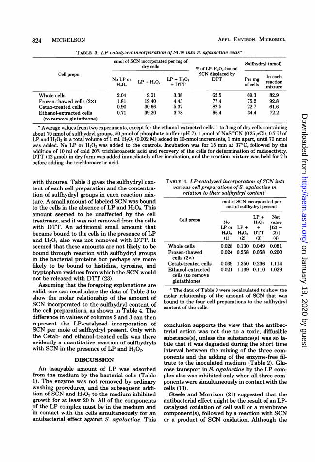
SCN into S. agalactiae cellular material areshown for four cell preparations in Table 3. Themolar sulfihydryl content in each reaction, asdetermined by assay of the cell suspension, wasapproximately equal to the H202 added. In oneof the experiments with the frozen-thawed cells,the sulfhydryl content was in excess; in oneexperiment with Cetab-treated cells, it was
somewhat less than the ideal ratio of 1:1 (1).Fewer than 10% of the total sulfhydryl groups inthe whole cells reacted to bind SCN by an LP-catalyzed reaction, but with Cetab- and ethanol-treated cells, the nearly 1:1 relationship betweenSCN bound and sulfhydryls present suggested aquantitative conversion to sulfenylthiocyanatederivatives (3, 23). The sulfhydryls of the frozen-thawed cells were intermediate in their reactiv-ity. Modification of cell membrane permeabilityis believed to be the reason for more sulfhydrylreactivity in the treated cells. That changes inpermeability of the cells of S. agalactiae oc-curred was made evident by the loss of proteinand sulfhydryl groups from the freeze-thawed,Cetab-treated, and ethanol-treated cells. Infreeze-thawed cells, loss of protein was 25% andloss of sulfhydryls was 5%; in Cetab-treated cells,loss of protein was 16 to 25% and loss of sulfhy-dryls was 50 to 65%; and in the ethanol-extractedcells, loss of sulfhydryls was about 42%. The lossof sulfhydryl content from cells by treatmentwas compensated for by using more cell materialto provide about 70 nmol of sulfhydryls perreaction. Most ofthe SCN estimated to be boundto S. agalactiae by an LP-catalyzed reactionwas released with DTT (Table 3). In an experi-ment to measure sulfenylthiocyanate displace-ment of the cell-bound SCN with ['4C]thiourea,about 78% of the net cell-bound SCN was re-leased with DTT and about 67% was displaced
filtratea The medium was the same as that described in
Table 1, with a volume of 4 ml."0.70 U of LP, 0.7 umol of NaSCN, and 0.125 ,umol
of H202 per ml of medium.c (1 and 2) 2 ml of inoculated medium containing 2
,umol of H202. (1) The inoculum (equivalent to 100 ,ugof cells [dry weight]) was incubated with H202 for 20min at 24°C in 2 ml of medium, followed by additionof 2 ml of medium containing NaSCN. (2) Same as (1)but with subsequent addition of LP-free filtrate frome. The length of time from mixing reagents to additionof filtrate was 10 min. Amicon CF50A membrane conefilter retained the LP.
d 2 ml of medium containing 2.8 Mmol of NaSCN.e Filtrate from 2 ml of medium containing 2.8 U of
(to remove glutathione)a Average values from two experiments, except for the ethanol-extracted cells. 1 to 3 mg of dry cells containing
about 70 nmol of sulfhydryl groups, 50 ,umol of phosphate buffer (pH 7), 1,mol of NaS'4CN (0.25 tiCi), 0.7 U ofLP and H202 in a total volume of 1 ml. H202 (0.002 M) added in 10-nmol increments, 1 min apart, until 70 nmolwas added. No LP or H202 was added to the controls. Incubation was for 15 min at 370C, followed by theaddition of 10 ml of cold 20% trichloroacetic acid and recovery of the cells for determination of radioactivity.DTT (12 ,mol) in dry form was added immediately after incubation, and the reaction mixture was held for 2 hbefore adding the trichloroacetic acid.
with thiourea. Table 3 gives the sulfhydryl con-
tent of each cell preparation and the concentra-tion of sulfhydryl groups in each reaction mix-ture. A small amount of labeled SCN was boundto the cells in the absence of LP and H202. Thisamount seemed to be unaffected by the celltreatment, and it was not removed from the cellswith DTT. An additional small amount thatbecame bound to the cells in the presence of LPand H202 also was not removed with DTT. Itseemed that these amounts are not likely to bebound through reaction with sulfhydryl groupsin the bacterial proteins but perhaps are morelikely to be bound to histidine, tyrosine, andtryptophan residues from which the SCN wouldnot be released with DTT (23).Assuming that the foregoing explanations are
valid, one can recalculate the data of Table 3 toshow the molar relationship of the amount ofSCN incorporated to the sulfhydryl content ofthe cell preparations, as shown in Table 4. Thedifference in values of columns 2 and 3 can thenrepresent the LP-catalyzed incorporation ofSCN per mole of sulfhydryl present. Only withthe Cetab- and ethanol-treated cells was thereevidently a quantitative reaction of sulfhydrylswith SCN in the presence of LP and H202.
DISCUSSIONAn assayable amount of LP was adsorbed
from the medium by the bacterial cells (Table1). The enzyme was not removed by ordinarywashing procedures, and the subsequent addi-tion of SCN and H202 to the medium inhibitedgrowth for at least 20 h. All of the componentsof the LP complex must be in the medium andin contact with the cells simultaneously for an
antibacterial effect against S. agalactiae. This
TABLE 4. LP-catalyzed incorporation ofSCN intovarious cell preparations of S. agalactiae in
relation to their sulfhydryl contentamol of SCN incorporated permol of sulfhydryl present
LP + NetCell prepn No H202 valueLP or LP + + [(2)-H202 H202 DTT (3)](1) (2) (3) (4)
a The data of Table 3 were recalculated to show themolar relationship of the amount of SCN that wasbound to the four cell preparations to the sulfhydrylcontent of the cells.
conclusion supports the view that the antibac-terial action was not due to a toxic, diffusiblesubstance(s), unless the substance(s) was so la-bile that it was degraded during the short timeinterval between the mixing of the three com-ponents and the adding of the enzyme-free fil-trate to the inoculated medium (Table 2). Glu-cose transport in S. agalactiae by the LP com-plex also was inhibited only when all three com-ponents were simultaneously in contact with thecells (13).
Steele and Morrison (21) suggested that theantibacterial effect might be the result of an LP-catalyzed oxidation of cell wall or a membranecomponent(s), followed by a reaction with SCNor a product of SCN oxidation. Although the
oxidative roles ofLP and H202 were understand-able, the role of SCN in the antibacterial effectwas less clear. A role for SCN was suggested byreports of the LP-catalyzed incorporation ofSCN into crystalline bovine serum albumin inwhich the protein sulfhydryls were oxidized tosulfenylthiocyanate derivatives (3; T. M. Auneand E. L. Thomas, Fed. Proc. 35:1386, 1976).Recent work on the products ofthe LP-catalyzedoxidation of SCN (1, 2, 8) and their role in theformation of sulfenylthiocyanate derivatives ofprotein sulfhydryls offered for the first time thedefinition of a specific role for SCN in the anti-bacterial effect of the LP complex.The LP-catalyzed incorporation of SCN into
the cell proteins of S. agalactiae herein reportedsuggested that the role of SCN in the antibac-terial effect may be explained by a similar mech-anism. With the four cell preparations used, theamount of SCN incorporated was apparentlyrelated to the exposure of sulfhydryls to LP-catalyzed oxidation. The LP-catalyzed incorpo-ration of SCN into whole cells exceeded thecontrols by 3- to 5-fold, the frozen-thawed cellsby 8- to 14-fold, and the Cetab-treated andethanol-extracted cells by 30- to 50-fold. Most ofthe SCN incorporated into cellular material byLP catalysis was displaced from the combinationwith DTT. The amount displaced ranged from77 to 96% with the treated cells. With wholecells, when the total SCN incorporation was onlythree- to fivefold greater with LP catalysis thanwithout LP, the portion of cell-bound SCN re-leased with DTT was about 63%. The nearly 1:1 ratio ofSCN incorporated to sulfhydryl groupspresent in the Cetab- and ethanol-treated cellsindicated a nearly quantitative exposure tosulfhydryl groups in the bacterial protein to LP-catalyzed oxidation. That ratio also suggestedthat the protein sulfhydryls were oxidized pri-marily to sulfenylthiocyanate derivatives. Therelease of cell-bound thiocyanate with DTT andreplacement of it by thiourea also support theview that thiocyanate incorporated by LP catal-ysis was bound primarily as a sulfenylthiocya-nate derivative of cell sulfhydryl groups. Thesodium dodecyl sulfate (0.75%) in the reagentsused for determining the total sulfhydryl contentof the bacterial cells would be as likely to exposeprotein sulfhydryl groups, the free sulfhydryls,and those masked by the configuration of theprotein (7) as effectively as did the Cetab andethanol treatments. The displacement of theSCN incorporated by LP catalysis into sulfen-ylthiocyanate derivatives by DTW (23), the in-hibition of glucose transport in S. agalactiae bythe LP complex and its reversibility with DTT,and the inhibition of glucolysis and its reversi-bility with DTT (13) all suggested that the func-
tional protein sulfhydryls involved in the trans-port of glucose and the enzymes of substratedegradation were sites where the LP complexcan exert antibacterial effects. Recently, it hasbeen shown (M. Adamson and K. Pruitt, Fed.Proc. 76:1579, 1978) that LP catalyzed the in-corporation ofSCN into purified hexokinase andwas accompanied by enzyme inactivation. Inac-tivation of triosephosphate dehydrogenase incell-free extracts from Streptococcus pyogenesby the LP complex and its reversal with cysteineand glutathione have also been shown (11). WithEscherichia coli, the antibacterial effect causedby OSCN-, a product of the LP-catalyzed oxi-dation of SCN, was due to the oxidation ofprotein sulihydryls to sulfenylthiocyanate deriv-atives, which resulted in inhibition of bacterialrespiration. The effect could be reversed withDTT or by the removal of the OSCN- from thecells (23).The hot-ethanol extraction used for the re-
moval of glutathione from the bacterial cellsevidently exposed reactive sulfihydryl groups aseffectively as did the treatment with Cetab.About 1 mol of SCN per mol of sulfhydryl wasincorporated, in the presence of LP and H202,by cells treated with either procedure. The in-creased incorporation ofSCN into S. agalactiaeproteins after treatment with Cetab or afterextraction with 80% ethanol is believed to resultfrom changes in membrane permeability, caus-ing exposure of protein sulfhydryls, normallyprotected by the cell membrane, to the oxidativeaction of LP and H202. Cetab treatment of S.pyogenes caused the cell membrane to becomepermeable to citrulline and carbamyl phosphate(20). No observable morphological changes werenoted in electron microscopic examination of S.pyogenes, nor were cell structure changes notedin S. agalactiae when the cells were examinedwith a scanning electron microscope (x 20,000magnification) after freeze-thawing or Cetabtreatment. Freeze-thawing of the bacteria sev-eral times also increased the exposure of proteinsulfhydryls but less effectively than did Cetab orethanol treatment.The low SCN incorporation into whole cells
in the presence of LP and H202 suggested thatonly protein sulfhydryls on the exterior of thecell membrane were accessible to oxidation. Per-haps, they are the sulfhydryls that are functionalin membrane transport, and their modificationmay be all that is necessary for the antibacterialeffect. Incorporation of SCN into the proteinsulfhydryls of S. agalactiae would remove thereacted sulfhydryls from a functional role inglucose transport and glucolysis, and this couldaccount for the antibacterial effect. The inhibi-tion of glucolysis would be of secondary impor-
tance because without a functioning glucosetransport system, the glucolytic enzymes wouldhave no substrate to act upon. Paigen and Wil-liams have estimated that in bacteria with adoubling time of 60 min and an internal glucoseconcentration of 1 mM at any one time, enoughglucose would be present to sustain growth forslightly longer than 1 s (17). S. agalactiae has adoubling time of about 21 min during rapidgrowth; therefore, cessation of growth would bealmost immediate when glucose transport wasstopped. Results in recent experiments at thislaboratory have shown that L-valine transportwas also strongly inhibited in S. agalactiae cellsthat had been exposed to LP-SCN-H202. Likethe inhibition of glucose transport, the inhibitionof valine transport was reversible with DTT (J.W. Moran, unpublished data).The incorporation of SCN into the sulfhydryl
groups of the bacterial proteins defimes a specificrole for the SCN of the LP complex in thegrowth inhibition of S. agalactiae.
ACKNOWLEDGMENTSGrateful appreciation is expressed to Joseph E. Gallagher
for the scanning electron microscope examination of S. aga-lactiae.
LITERATURE CITED1. Aune, T. M., and E. L. Thomas. 1977. Accumulation of
hypothiocyanite ion during peroxidase-catalyzed oxi-dation of thiocyanate ion. Eur. J. Biochem. 80:209-214.
2. Aune, T. M., and E. L. Thomas. 1978. Oxidation ofprotein sulfhydryls by products of peroxidase-catalyzedoxidation of thiocyanate ion. Biochemistry 17:1005-1010.
3. Aune, T. M., E. L. Thomas, and M. Morrison. 1977.Lactoperoxidase-catalyzed incorporation of thiocyanateion into a protein substrate. Biochemistry 16:4611-4616.
4. Bruno, A., and J. E. Christian. 1961. Determination ofcarbon-14 in aqueous bicarbonate solutions by liquidscintillation counting techniques. Anal. Chem. 33:1216-1218.
5. Ellman, G. L. 1959. Tissue sulfhydryl groups. Arch. Bio-chem. Biophys. 82:70-77.
6. Fahey, R. C., S. Brody, and S. D. Mikolajczyk. 1975.Changes in the glutathione thiol-disulfide status ofNeu-rospora crassa conidia during germination and aging.J. Bacteriol. 121:144-151.
7. Fraenkel-Conrat, H. 1957. Methods for investigating theessential groups for enzyme activity. Methods Enzymol.4:247-269.
8. Hoogendoorn, H., J. P. Piessens, W. Scholtes, and L.
A. Stoddard. 1977. Hypothiocyanite ion: the inhibitorformed by the system lactoperoxidase-thiocyanate-hy-drogen peroxide. I. Identification of the inhibiting com-pound. Caries Res. 11:77-84.
9. Jago, G. R., and M. Morrison. 1962. Antistreptococcalactivity of lactoperoxidase III. Proc. Soc. Exp. Biol.Med. 111:585-588.
10. Lowry, 0. H., N. J. Rosebrough, A. L. Farr, and R. J.Randall. 1951. Protein measurement with the Folinphenol reagent. J. Biol. Chem. 193:265-275.
11. Mickelson, M. N. 1966. Effect of lactoperoxidase andthiocyanate on the growth of Streptococcus pyogenesand Streptococcus agalactiae in a chemically definedculture medium. J. Gen. Microbiol. 43:3143.
12. Mickelson, M. N. 1976. Effects of nutritional character-istics of Streptococcus agalactiae on inhibition ofgrowth by lactoperoxidase-thiocyanate-hydrogen per-oxide in a chemically defined culture medium. Appl.Environ. Microbiol. 32:238-244.
13. Mickelson, M. N. 1977. Glucose transport in Streptococ-cus agalactiae and its inhibition by lactoperoxidase-thiocyanate-hydrogen peroxide. J. Bacteriol. 132:541-548.
14. Morrison, M., and D. E. Hultquist. 1963. Lactoperoxi-dase. II. Isolation. J. Biol. Chem. 238:3847-3849.
15. Oram, J. D., and B. Reiter. 1966. The inhibition ofstreptococci by lactoperoxidase, thiocyanate and hydro-gen peroxide. The effect of the inhibitory system onsusceptible and resistant strains of group N strepto-cocci. Biochem. J. 100:373-381.
16. Oram, J. D., and B. Reiter. 1966. The inhibition ofstreptococci by lactoperoxidase, thiocyanate, hydrogenperoxide. The oxidation of thiocyanate and the natureof the inhibitory compound. Biochem. J. 100:382-388.
17. Paigen, K., and B. Williams. 1970. Catabolite repressionand other control mechanisms on carbohydrate utiliza-tion. Adv. Microb. Physiol. 4:252-324.
18. Park, D. J., and W. S. Allison. 1969. The mechanism ofinactivation of glyceraldehyde-3-phosphate dehydro-genase by tetrathionite, O-iodobenzoate and iodinemonochloride. J. Biol. Chem. 244:180-189.
19. Reiter, B., A. Pickering, J. D. Oram, and G. D. Pope.1963. Peroxidase-thiocyanate inhibition of streptococciin raw milk. J. Gen. Microbiol. 33:12.
20. Slade, H. D. 1957. Studies on Streptococcus pyogenes.III. The effect of trypsin and a cationic detergent onthe structure, permeability and metabolism of the cell.J. Gen. Physiol. 41:63-76.
21. Steele, W. F., and M. Morrison. 1969. Antistreptococcalactivity of lactoperoxidase. J. Bacteriol. 97:635-639.
22. Thomas, E. L., and T. M. Aune. 1978. Susceptibility ofEscherichia coli to bactericidal action of lactoperoxi-dase, peroxide, and iodide or thiocyanate. AntimicrobiolAgents Chemother. 13:261-265.
23. Thomas, E. L., and T. M. Aune. 1978. Lactoperoxidase,peroxide, thiocyanate antimicrobial system: correlationof sulflydryl oxidation with antimicrobial action. Infect.Immun. 20:456-463.
24. Wright, R. C., and J. D. Tramer. 1958. Factors influ-encing the activity of cheese starters. The role of milkperoxidase. J. Dairy Sci. 25:104-118.