1 Antibacterial Activity of Naturally Occurring Compounds from Selected Plants Olgica Stefanović, Ivana Radojević, Sava Vasić and Ljiljana Čomić Labaratory of Microbiology, Department of Biology and Ecology, Faculty of Science, University of Kragujevac, Kragujevac Serbia 1. Introduction Man is in constant contact with a large number of different bacteria which temporarily or permanently inhibit his body creating temporary or permanent community. Relations which are thus established are various and very complex, from those positive to those whose consequences for man are extremely negative. Very often, both on and in man’s body, bacteria which have the ability to cause an infection are present. This ability of pathogenic bacteria is reflected in possession of certain pathogenicity factors. A set of factors which enable successful invasion and damage of the host are: toxins, surface structures and enzymes. Between the host and the pathogen very complex relations are established whose income depends on host’s characteristics as well as on pathogen’s characteristics. Infections caused by bacteria can be prevented, managed and treated through anti-bacterial group of compounds known as antibiotics. Antibiotics are natural, semi-synthetic or synthetic compounds that kill or inhibit the growth of bacteria. When bacteria are exposed to an antibiotic, they doubly respond: i) they are sensitive what cause the inhibition of their growth, division and death or ii) they can remain unaffected or resistant. The resistance of bacteria to antibiotics can be natural (intrinsic) or acquired. Natural resistance is achieved by spontaneous gene mutation. The acquired resistence occurs after the contact of bacteria with an antibiotic as a result of adaptation of a species to adverse environmental conditions. In such population, an antibiotic as a selective agent, acts on sensitive individuals, while resistant survive and become dominant. Bacteria gain antibiotic resistance due to three reasons namely: (i) modification of active site of the target resulting in reduction in the efficiency of binding of the drug, (ii) direct destruction or modification of the antibiotic by enzymes produced by the organism or, (iii) efflux of antibiotic from the cell (Sheldon, 2005). The evolution of antibacterial resistance in human pathogenic and commensal microorganisms is the result of the interaction between antibiotic exposure and the transmission of resistance within and between individuals. It is especially interesting the phenomenon of horizontally gene transfer. Extrachromosomal DNA material, so-called plasmids, often carry genes of resistance and can transfer information within and between the individuals of the same or related bacterial species, thus also spreading the resistance. Transformation, transduction and conjugation represent the horizontal gene transfer mechanisms of resistance between the bacteria.
Transcript
1
Antibacterial Activity of Naturally Occurring Compounds from Selected Plants
Olgica Stefanović, Ivana Radojević, Sava Vasić and Ljiljana Čomić Labaratory of Microbiology, Department of Biology and Ecology,
Faculty of Science, University of Kragujevac, Kragujevac Serbia
1. Introduction
Man is in constant contact with a large number of different bacteria which temporarily or permanently inhibit his body creating temporary or permanent community. Relations which are thus established are various and very complex, from those positive to those whose consequences for man are extremely negative. Very often, both on and in man’s body, bacteria which have the ability to cause an infection are present. This ability of pathogenic bacteria is reflected in possession of certain pathogenicity factors. A set of factors which enable successful invasion and damage of the host are: toxins, surface structures and enzymes. Between the host and the pathogen very complex relations are established whose income depends on host’s characteristics as well as on pathogen’s characteristics.
Infections caused by bacteria can be prevented, managed and treated through anti-bacterial group of compounds known as antibiotics. Antibiotics are natural, semi-synthetic or synthetic compounds that kill or inhibit the growth of bacteria. When bacteria are exposed to an antibiotic, they doubly respond: i) they are sensitive what cause the inhibition of their growth, division and death or ii) they can remain unaffected or resistant.
The resistance of bacteria to antibiotics can be natural (intrinsic) or acquired. Natural resistance is achieved by spontaneous gene mutation. The acquired resistence occurs after the contact of bacteria with an antibiotic as a result of adaptation of a species to adverse environmental conditions. In such population, an antibiotic as a selective agent, acts on sensitive individuals, while resistant survive and become dominant. Bacteria gain antibiotic resistance due to three reasons namely: (i) modification of active site of the target resulting in reduction in the efficiency of binding of the drug, (ii) direct destruction or modification of the antibiotic by enzymes produced by the organism or, (iii) efflux of antibiotic from the cell (Sheldon, 2005). The evolution of antibacterial resistance in human pathogenic and commensal microorganisms is the result of the interaction between antibiotic exposure and the transmission of resistance within and between individuals. It is especially interesting the phenomenon of horizontally gene transfer. Extrachromosomal DNA material, so-called plasmids, often carry genes of resistance and can transfer information within and between the individuals of the same or related bacterial species, thus also spreading the resistance. Transformation, transduction and conjugation represent the horizontal gene transfer mechanisms of resistance between the bacteria.
Antimicrobial Agents
2
Having in mind the current progress of resistance spreading and resilience of larger and larger number of bacteria to traditional antibiotics as well as a way of transmitting the gene of resistance, above all via plasmids, one can conclude that the ability of obtaining bacterium resistance to antibiotics represents a very dynamic and unpredictable phenomenon. For that reason, bacterial resistance to antibiotics represents a major health problem. Solving this problem and search for new sources of antimicrobial agents is a worldwide challenge and the aim of many researches of scientific and research teams in science, academy institutions, pharmaceutical companies. One of the approaches in solving this issue is testing the biologically active compounds of plant origin.
1.1 Plants as potential antibacterial agents
Healing potential of plants has been known for thousands of years. Medicinal use of plants and their products was passed down from generation to generation in various parts of the world throughout its history and has significantly contributed to the development of different traditional systems of medicine. Even today, the World Health Organization (WHO) has estimated that approximately 80% of the global population relies on traditional herbal medicines as part of standard healthcare (Foster et al., 2005). Many drugs presently prescribed by physicians are either directly isolated from plants or are artificially modified versions of natural products. In Western countries, approximately 25% of the drugs used are of natural plant origin (Payne et al., 1991).
Plants produce a whole series of different compounds which are not of particular significance for primary metabolism, but represent an adaptive ability of a plant to adverse abiotic and biotic environmental conditions. They can have a remarkable effect to other plants, microorganisms and animals from their immediate or wider environment. All these organic compounds are defined as biologically active substances, and generally represent secondary metabolites, given the fact that they occur as an intermediate or end products of secondary plant metabolism. These secondary metabolites, apart from determining unique plant traits, such as: colour and scent of flowers and fruit, characteristic flavour of spices, vegetables, they also complete the functioning of plant organism, showing both biological and pharmacological activity of a plant. Therefore, medicinal properties of plants can be attributed to secondary metabolites (Hartmann, 2008).
1.1.1 Antibacterial secondary metabolites
It is known and proved by in vitro experiments that plants produce a great number of secondary metabolites that have antimicrobial activity (Iwu et al., 1999; Cowan, 1999; Rios & Recio, 2005; Cos et al., 2006). These plant-formed antibiotics have been classified as phytoanticipins, which are preformed inhibitory compounds, or phytoalexins, which are derived from precursors via de novo synthesis in response to microbial attack (VanEtten et al., 1994). Antibacterial secondary metabolites are usually classified in three large molecule families: phenolics, terpenes and alkaloids.
The phenolics and polyphenols are one of the largest groups of secondary metabolites that have exhibited antimicrobial activity. Important subclasses in this group of compounds include phenols, phenolic acids, quinones, flavones, flavonoids, flavonols, tannins and coumarins. Phenols are a class of chemical compounds consisting of a hydroxyl functional
Antibacterial Activity of Naturally Occurring Compounds from Selected Plants
3
group (-OH) attached to an aromatic phenolic group. The site(s) and number of hydroxyl groups on the phenol group are thought to be related to their relative toxicity to microorganisms, with evidence that increased hydroxylation results in increased toxicity (Geissman, 1963, as cited in Cowan, 1999). Quinones have aromatic rings with two ketone substitutions. Quinones have a potential to form irreversible complex with nucleophilic amino acids in proteins (Stern et al., 1996, as cited in Cowan, 1999). This could explain their antibacterial properties. Probable targets in the microbial cell are surface-exposed adhesins, cell wall polypeptides and membrane-bound enzymes. Flavones are phenolic structures containing one carbonyl group. The addition of a 3-hydroxyl group yields a flavonol. Flavonoids are also hydroxylated phenolic substances but occur as a C6-C3 unit linked to an aromatic ring. Flavones, flavonoids and flavonols have been known to be synthesized by plants in response to microbial infection so it is not surprising that they have been found, in vitro, to be effective antimicrobial substances against a wide array of microorganisms (Dixon et al., 1983, as cited in Cowan, 1999). Their activity is probably due to their ability to complex with extracellular and soluble proteins and to complex with bacterial cell walls. Lipophilic flavonoids may also disrupt microbial membranes (Tsuchiya et al., 1996, as cited in Cowan, 1999). Tanins, a group of polymeric phenolic substances, are found in almost every plant part: bark, wood, leaves, fruits and roots. They are divided into two groups, hydrolyzable and condensed tannins. In plant tissue, tannins have been synthesized and accumulated after microbial attack. Their mode of antimicrobial action may be related to their ability to inactivate microbial adhesins, enzymes, cell envelope transport proteins, because of a property known as astringency. Coumarins are benzo-α-pyrones and could be categorised as: simple coumarins and cyclic coumarins (furanocoumarins and pyranocoumarins) (Ojala, 2001). Coumarins have been found to stimulate macrophages, which could have an indirect negative effect on infections (J. R. Casley-Smith & J. R.Casley-Smith, 1997, as cited in Cowan, 1999).
Terpenes are a large and varied class of organic compounds built up from isoprene subunits, while the terpenoids are oxygen-containing analogues of the terpenes. According to number of isoprene subunits, there are monoterpenes (C10), sesquiterpenes (C15), diterpenes (C20), triterpenes (C30), tetraterpenes (C40) and polyterpenes (Kovacevic, 2004). Monoterpenes, diterpenes and sesquiterpenes are the primary constituents of the essential oils. The mechanism of antibacterial action of terpenes is not fully understood but is speculated to involve membrane disruption by the lipophilic compounds. Accordingly, Mendoza et al., 1997 found that increasing the hydrophilicity of kaurene diterpenoids by addition of a methyl group drastically reduced their antimicrobial activity. (Mendoza et al., 1997, as cited in Cowan, 1999).
Alkaloids, one of the earliest isolated bioactive compounds from plants, are heterocyclic nitrogen compounds. They are derived from amino acids, and the nitrogen gives them alkaline properties. The mechanism of antibacterial action is attributed to their ability to intercalate with DNA, inhibition of enzymes (esterase, DNA-, RNA-polymerase), inhibition of cell respiration (Kovacevic, 2004).
1.1.2 Plant extracts as potential antibacterial agents
The screening of plant extracts has been of great interest to scientists for the discovery of new compound effective in the treatment of bacterial infection. Plant extracts exhibit:
Antimicrobial Agents
4
• direct antibacterial activity showing effects on growth and metabolism of bacteria • indirect activity as antibiotic resistance modifying compounds which, combined with
antibiotics, increase their effectiveness.
A great number of reports concerning the antibacterial screening of plant extracts have appeared in the literature. Examples of such articles include studies of medicinal plants from different geographical regions: Brazil (Alves et al., 2000), Argentina (Salvat et al., 2000), Columbia (López et al., 2001); India (Perumal et al., 1998; Ahmad & Beg, 2001), China (Zuo et al., 2008), Turkey (Sokman et al., 1999; Uzun et al., 2004); Greece (Skaltsa et al., 2003), Spain (Ríos et al., 1987; Recio et al., 1989), Serbia (Stefanovic et al., 2009a; Stefanovic et al., 2009b, Stanojevic et al., 2010a; Stanojevic et al., 2010b; Stojanovic-Radic et al., 2010; Stefanovic et al., 2011; Stefanovic et al., 2012); Africa (Atindehou et al., 2002; Konning et al., 2004; Chah et al., 2006); Australia (Palombo & Semple, 2001). According to published data, the plant extracts exhibited the activity against a great number of bacterial species (Gram positive and Gram negative strains; sensitive and resistant, pathogens and opportunistic pathogens). The experiments involved the standard strains as well as the clinical isolates of pathogenic bacteria, as more realistic test organisms for estimation of antibacterial activity. Plant extracts were prepared from fresh or dried plant material using conventional extraction methods (Soxhlet extraction, maceration, percolation). Extraction is process of separation of active compounds from plant material using different solvents. During extraction, solvents diffuse into the plant material and solubilise compounds with similar polarity. At the end of the extraction, solvents have been evaporated, so that an extract is a concentrated mixture of plant active compounds. Successful extraction is largely dependent on the type of solvent used in the extraction procedure. The most often tested extracts are: water extract as a sample of extract that primarily used in traditional medicine and extracts from organic solvents such as methanol, ethanol as well as ethyl acetate, acetone, chloroform, dichlormethane (Ncube et al., 2008).
Diffusion and dilution method are two types of susceptibility test used to determine the antibacterial efficacy of plant extracts. Diffusion method is a qualitative test which allows classification of bacteria as susceptible or resistant to the tested plant extract according to size of diameter of the zone of inhibition. In dilution method, the activity of plant extracts is determined as Minimum Inhibitory Concentration (MIC). MIC is defined as the lowest concentration able to inhibit bacterial growth. In broth-dilution methods, turbidity and redox-indicators are most frequently used for results reading. Turbidity can be estimated visually or spectofotometrically while change of indicator colour indicate inhibition of bacterial growth (Cos et al., 2006).
2. Evaluation of antibacterial activity of selected plant species
Considering a large number of still insufficiently or incompletely examined species in the plant world; in inexhaustible possibilities of modifying natural substances, there is a need for systematic research and even greater affirmation of plant antimicrobial compounds. In this study the in vitro antibacterial activity of selected plant species was tested and evaluated the potential use of their extracts as a source of antibacterial compounds. The experiment involved water, ethanol, ethyl acetate and acetone extracts from 10 plant species belonging to 4 the most important plant families. The following plants were used: Cychorium intybus L. (Asteraceae); Salvia officinalis L., Melissa officinalis L., Clinopodium vulgare L. (Lamiaceae); Torilis anthriscus L. (Gmel), Aegopodium podagraria L. (Apiaceae); Cytisus nigricans L., Cytisus capitatus Scop.,
Antibacterial Activity of Naturally Occurring Compounds from Selected Plants
5
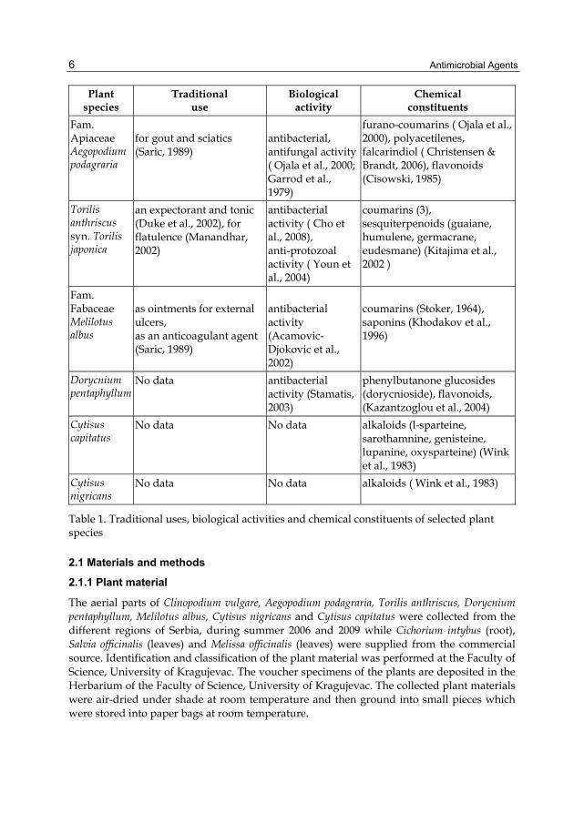
Melilotus albus Medic., Dorycnium pentaphyllum Vill. (Fabaceae). In general, the plants are annual or perennial, herbaceous or shrubby, widespread in Europe. They are rich in secondary metabolites from a group of phenols, flavonoids, coumarins, tannins, terpenes. It is known that different solvents extracted different groups of secondary metabolites, hence different types of extracts were prepared. The plants, as potential candidates for antibacterial agents, were selected on the base of three criteria: i) use in traditional medicine as antiseptic agents, ii) random selection followed by chemical screening (ii) insufficient antibacterial scientific data. Detailed description of known traditional uses, in vitro found biological activities and the chemical constituent is shown in Table 1.
Plant species
Traditional use
Biological activity
Chemical constituents
Fam. Asteraceae Cichorium intybus
improving digestion, for a diarrhea, as diuretic, for cleansing the liver and benefiting the gallbladder (Saric, 1989)
antibacterial, antifungal activity (Petrovic et al., 2004; Rani & Khullar, 2004; Mares et al., 2005)
for disorders of the digestive system, as antiseptic for sore throats, ulcers, to treat insect bites, mouth and gum infections and vaginal discharge for night sweats (Saric, 1989)
antibacterial, antifungal, antiviral activity ( Velickovic et al., 2003; Nolkemper et al., 2006; Horiuchi et al., 2007; Weckesser et al., 2007)
to reduce indigestion and flatulence, as a mild sedative, to treat headache, migraine, nervous tension and insomnia, to treat cold, fever and cough (Saric, 1989)
antibacterial, antifungal, antiviral activity ( Iauk et al., 2003; Ertürk, 2006; Nolkemper et al., 2006)
flavonoids ( Herodež et al., 2003; Patora & Klimek , 2002), phenolic acids (Herodež et al., 2003; Canadanović –Brunet et al., 2008), simple phenols, tannins (Hohmann et al., 1999)
Clinopodium vulgare
as a heart tonic, an expectorant, as a diuretic (Saric, 1989) as an antiseptic for wounds and injuries (Opalchenova and Obreshkova, 1999).
as ointments for external ulcers, as an anticoagulant agent (Saric, 1989)
antibacterial activity (Acamovic-Djokovic et al., 2002)
coumarins (Stoker, 1964), saponins (Khodakov et al., 1996)
Dorycnium pentaphyllum
No data antibacterial activity (Stamatis, 2003)
phenylbutanone glucosides (dorycnioside), flavonoids, (Kazantzoglou et al., 2004)
Cytisus capitatus
No data No data alkaloids (l-sparteine, sarothamnine, genisteine, lupanine, oxysparteine) (Wink et al., 1983)
Cytisus nigricans
No data No data alkaloids ( Wink et al., 1983)
Table 1. Traditional uses, biological activities and chemical constituents of selected plant species
2.1 Materials and methods
2.1.1 Plant material
The aerial parts of Clinopodium vulgare, Aegopodium podagraria, Torilis anthriscus, Dorycnium pentaphyllum, Melilotus albus, Cytisus nigricans and Cytisus capitatus were collected from the different regions of Serbia, during summer 2006 and 2009 while Cichorium intybus (root), Salvia officinalis (leaves) and Melissa officinalis (leaves) were supplied from the commercial source. Identification and classification of the plant material was performed at the Faculty of Science, University of Kragujevac. The voucher specimens of the plants are deposited in the Herbarium of the Faculty of Science, University of Kragujevac. The collected plant materials were air-dried under shade at room temperature and then ground into small pieces which were stored into paper bags at room temperature.
Antibacterial Activity of Naturally Occurring Compounds from Selected Plants
7
2.1.2 Extraction
Dried, ground plant material was extracted by direct maceration with water, ethanol, ethyl acetate and acetone. Briefly, 30g of plant material was soaked with 150ml of solvent for 24h at room temperature. During 24 hours, targeted compounds from plant material were extracted by the solvent. After that the resulting extract was filtered through filter paper (Whatman no.1). The residue from the filtration was extracted again, twice, using the same procedure. The filtrates obtained were combined and then evaporated to dryness using a rotary evaporator at 40°C, for water extracts heating on a water bath. The crude plant extracts are stored at -20°C. Before the testing, stock solutions of the crude extracts were obtained by dissolving in dimethyl sulfoxide (DMSO) and then diluted into nutrient liquid medium to achieve a concentration of 10% DMSO. The groups of secondary metabolites which are expected in prepared plant extracts are given in Table 2.
Table 2. The expected groups of plant secondary metabolites (according to Kovacevic, 2004)
2.1.3 Microorganisms
The following bacteria were used: Staphylococcus aureus ATCC 25923, Escherichia coli ATCC 25922, Pseudomonas aeruginosa ATCC 27853 and clinical isolate of Staphylococcus aureus (PMFKg-B30), Bacillus subtilis (PMFKg-B2), Enterococcus faecalis (PMFKg-B22), Enterobacter cloaceae (PMFKg-B23), Klebsiella pneumoniae (PMFKg-B26), Escherichia coli (PMFKg-B32), Pseudomonas aeruginosa (PMFKg-B28) and Proteus mirabilis (PMFKg-B29). All clinical isolates were a generous gift from the Institute of Public Health, Kragujevac. Bacteria are stored in microbiological collection at the Laboratory of Microbiology (Faculty of Science, University of Kragujevac).
Bacterial suspension were prepared from overnight cultures by the direct colony method. Colonies were taken directly from the plate and suspent into 5ml of sterile 0,85% saline. The turbidity of initial suspension was adjusted comparing with 0,5 Mc Farland standard (0,5 ml 1,17% w/v BaCl2 × 2H2O + 99,5 ml 1% w/v H2SO4) (Andrews, 2001). When adjusted to the turbidity of a 0,5 Mc Farland standard, a suspension of bacteria contains about 108 colony forming units (CFU)/ml. Ten-fold dilutions of initial suspension were additionally prepared into sterile 0,85% saline to achieve 106 CFU/ml.
2.1.4 Microdilution method
Antibacterial activity was tested by determining the minimum inhibitory concentration (MIC) using microdilution plate method with resazurin (Sarker et al., 2007). Briefly, the 96-well
Antimicrobial Agents
8
microplate was prepared by dispensing 100 μL of Mueller-Hinton broth (Torlak, Belgrade) into each well. A 100 μL from the stock solution of tested extract (concentration of 40mg/ml) was added into the first row of the plate. Then, two-fold, serial dilutions were performed by transferring 100 μl of solution from one row to another, using a multichannel pipette. The obtained concentration range was from 20 mg/ml to 0.156 mg/ml. Ten microlitres of each 106
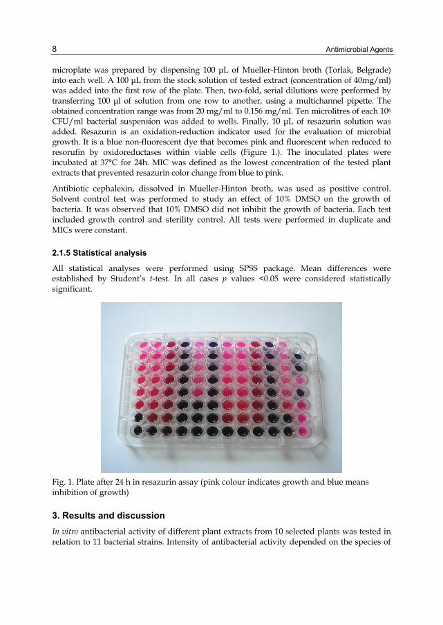
CFU/ml bacterial suspension was added to wells. Finally, 10 μL of resazurin solution was added. Resazurin is an oxidation-reduction indicator used for the evaluation of microbial growth. It is a blue non-fluorescent dye that becomes pink and fluorescent when reduced to resorufin by oxidoreductases within viable cells (Figure 1.). The inoculated plates were incubated at 37°C for 24h. MIC was defined as the lowest concentration of the tested plant extracts that prevented resazurin color change from blue to pink.
Antibiotic cephalexin, dissolved in Mueller-Hinton broth, was used as positive control. Solvent control test was performed to study an effect of 10% DMSO on the growth of bacteria. It was observed that 10% DMSO did not inhibit the growth of bacteria. Each test included growth control and sterility control. All tests were performed in duplicate and MICs were constant.
2.1.5 Statistical analysis
All statistical analyses were performed using SPSS package. Mean differences were established by Student’s t-test. In all cases p values <0.05 were considered statistically significant.
Fig. 1. Plate after 24 h in resazurin assay (pink colour indicates growth and blue means inhibition of growth)
3. Results and discussion
In vitro antibacterial activity of different plant extracts from 10 selected plants was tested in relation to 11 bacterial strains. Intensity of antibacterial activity depended on the species of
Antibacterial Activity of Naturally Occurring Compounds from Selected Plants
9
bacteria, plant species and the type of extract. The MIC values were in range from 0.019 mg/ml to >20mg/ml. In relation to positive control (cephalexin MIC 0. 00156 - >1mg/ml), the extracts showed lower activity. In general, according to obtained results, the following remarks could be made:
• Detectable MICs were noticed in 100% of tested bacteria for Cichorium intybus, 70% for Salvia officinalis, 90% for Melissa officinalis, 83, 33% for Clinopodium vulgare, 100% for Torilis anthriscus, 33, 33% for Aegopodium podagraria, 96, 67% for Cytisus nigricans, 76, 67% for Cytisus capitatus, 60% for Melilotus albus and 96, 67% for Dorycnium pentaphyllum.
• Among tested plants, the best inhibitory effects showed acetone extract from Salvia officinalis, ethyl acetate and acetone extract from Cichorium intybus and ethanol extract from Aegopodium podagraria. Moderate antibacterial activity exhibited Melissa officinalis, Clinopodium vulgare (ethyl acetate and acetone extract), Torilis anthriscus, Cytisus nigricans, Cytisus capitatus and Dorycnium pentaphyllum while low activity showed Clinopodium vulgare (ethanol extract), Melilotus albus and Aegopodium podagraria (water and ethyl acetate extract).
• Water extracts were less active than ethanol, ethyl acetate and acetone extracts. • The antibacterial activity of the tested extracts was closely associated with present
secondary metabolites. • The Gram-positive bacteria were more sensitive than the Gram-negative bacteria. The
reason for higher sensitivity of the Gram-positive bacteria than Gram-negative bacteria could be ascribed to their differences in cell wall constituents and their arrangement. The Gram-positive bacteria contain a peptidoglycan layer, which is an ineffective permeability barrier while Gram-negative bacteria are surrounded by an additional outer membrane carrying the structural lipopolysaccharide components, which makes it impermeable to lipophilic solutes and porins constitute a selective barrier to the hydrophilic solutes (Nikaido, 2003).
• Among tested bacteria, the most sensitive bacteria were Gram positive bacteria: B. subtilis and S. aureus ATCC 25923. Susceptibility of E. cloaceae, Ent. faecalis, K. pneumoniae, S. aureus, Ps. aeruginosa ATCC 27853 was moderate. Clinical isolate of Gram negative bacteria, Ps. aeruginosa, P. mirabilis, E. coli, exhibited low susceptibility or resistance.
3.1 Antibacterial activity of Cichorium intybus
The results of antibacterial activity of ethanol, ethyl acetate and acetone extract from Cichorium intybus are presented on the Figure 2. Extracts showed different activity. Ethanol extract acted at concentrations of 2.5 mg/ml to 20mg/ml; ethyl acetate from 1.09 mg/ml to 8.75 mg/ml; acetone extract from 2.5 mg/ml to 5 mg/ml. Antibacterial activity of ethyl acetate and acetone extract is more pronounced than ethanol extract. Similar results were obtained by Nandagopal & Ranjitha Kumari, 2007, among tested extracts, ethyl acetate was one of the more active. Statistic analysis confirms the presented results. Activity of ethyl acetate (pEtAc=0.001) and acetone (pAcOH=0.002) extract was statistically significantly higher that the activity of ethanol extract. Acetone extract was the most active (p=0.018).
The tested bacteria manifested different sensitivity level to the tested extracts. MIC values were ranged from 1.09 mg/ml to 20 mg/ml. The most sensitive bacterium to the tested
Antimicrobial Agents
10
Fig. 2. Antibacterial activity of Cichorium intybus extracts expressed as MIC values (mg/ml)
extracts was Pseudomonas aeruginosa ATCC 27853. MIC values for this bacterium were 2.5 mg/ml for ethanol and acetone extract, and 1.09 mg/ml for ethyl acetate extract. Bacteria Bacillus subtilis, Enterobacter cloacae, Staphylococcus aureus ATCC 25923 with 2.18 mg/ml, 2.5 mg/ml and 5 mg/ml values also manifested significant sensitivity. Proteus mirabilis, Escherichia coli ATCC 25922, according to ethanol extract, have showed the least sensitivity (MIC = 20 mg/ml), while they were more sensitive to the other two extracts. Similar results were obtained by other scientists. Petrović et al., 2004 noted the inhibitory effect of chicory extract also on phypopathogenic and human pathigenic bacteria. Acroum et al., 2009 showed the effect of methanol extract on Gram-positive bacteria (Bacillus subtilis, Bacillus cereus, Staphylococcus aureus) with the MIC values of 0.010 mg/ml and 0.075 mg/ml while the extract did not act on Gram-negative bacteria Escherichia coli, Pseudomonas aeruginosa, Klebsiella pneuminiae.
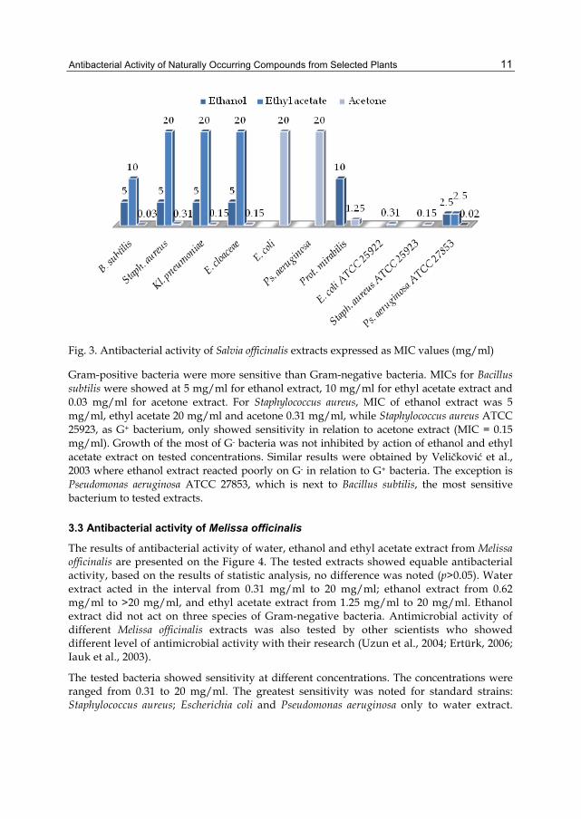
3.2 Antibacterial activity of Salvia officinalis
The acetone extract was the most reactive extract at this testing (pEtOH=0,004; pEtAc=0,001), while there was no statistically significant difference between ethanol and ethyl acetate extracts in acting. Ethanol and ethyl acetate extract showed the activity at concentrations from 2.5 mg/ml to >20 mg/ml, and acetone extract from 0.02 mg/ml to 20 mg/ml (Figure 3.). The reason for good results which acetone extract showed can also be saught in the fact that acetone is a good extractant, of low toxicity and high extractional capacity (Eloff, 1998). Similar results for acetone extract were also obtained by (Horiuchi et al., 2007), MIC values were from 256 µg/ml tо 512 µg/ml.
The tested bacteria, except for Pseudomonas aeruginosa and Escherichiа coli, showed a significant sensitivity in relation to acetone extract. MIC values were below 1 mg/ml.
Antibacterial Activity of Naturally Occurring Compounds from Selected Plants
11
Fig. 3. Antibacterial activity of Salvia officinalis extracts expressed as MIC values (mg/ml)
Gram-positive bacteria were more sensitive than Gram-negative bacteria. MICs for Bacillus subtilis were showed at 5 mg/ml for ethanol extract, 10 mg/ml for ethyl acetate extract and 0.03 mg/ml for acetone extract. For Staphylococcus aureus, MIC of ethanol extract was 5 mg/ml, ethyl acetate 20 mg/ml and acetone 0.31 mg/ml, while Staphylococcus aureus ATCC 25923, as G+ bacterium, only showed sensitivity in relation to acetone extract (MIC = 0.15 mg/ml). Growth of the most of G- bacteria was not inhibited by action of ethanol and ethyl acetate extract on tested concentrations. Similar results were obtained by Veličković et al., 2003 where ethanol extract reacted poorly on G- in relation to G+ bacteria. The exception is Pseudomonas aeruginosa ATCC 27853, which is next to Bacillus subtilis, the most sensitive bacterium to tested extracts.
3.3 Antibacterial activity of Melissa officinalis
The results of antibacterial activity of water, ethanol and ethyl acetate extract from Melissa officinalis are presented on the Figure 4. The tested extracts showed equable antibacterial activity, based on the results of statistic analysis, no difference was noted (p>0.05). Water extract acted in the interval from 0.31 mg/ml tо 20 mg/ml; ethanol extract from 0.62 mg/ml to >20 mg/ml, and ethyl acetate extract from 1.25 mg/ml to 20 mg/ml. Ethanol extract did not act on three species of Gram-negative bacteria. Antimicrobial activity of different Melissa officinalis extracts was also tested by other scientists who showed different level of antimicrobial activity with their research (Uzun et al., 2004; Ertürk, 2006; Iauk et al., 2003).
The tested bacteria showed sensitivity at different concentrations. The concentrations were ranged from 0.31 tо 20 mg/ml. The greatest sensitivity was noted for standard strains: Staphylococcus aureus; Escherichia coli and Pseudomonas aeruginosa only to water extract.
Antimicrobial Agents
12
Fig. 4. Antibacterial activity of Melissa officinalis extracts expressed as MIC values (mg/ml)
The most tested bacteria showed sensitivity at 10 mg/ml for ethyl acetate extract. Escherichia coli, Pseudomonas aeruginosa and Proteus mirabilis showed resistance to ethanol extract. Weaker activity of lemon balm to mentioned Gram-negative bacterium was also noted in the work of Canadanović-Brunet et al., 2008.
3.4 Antibacterial activity of Clinopodium vulgare
The extracts manifested different level of antibacterial activity, the results are shown on the Figure 5. The ethanol extract acted in concentrations from 1.25 mg/ml tо >20 mg/ml, ethyl acetate and acetone extract from 0.62 mg/ml tо 20 mg/ml. Ethyl acetate and acetone extract acted better than ethanol extract. Statistically significant difference was noted (pЕtAc=0.015 и pAcOH=0.018). Between ethyl acetate and acetone extract no statistically significant difference was noted in the activity (p=0.756).
The tested bacteria in most of the cases showed sensitivity at 10 mg/ml and 20 mg/ml. Gram-positive bacteria Bacillus subtilis and Staphylococcus aureus ATCC 25923 were the most sensitive to the action of Clinopodium vulgare extracts. MIC values were in the interval from 0.62 mg/ml tо 2.5 mg/ml. Klebsiella pneumoniae, Staphylococcus aureus, Enterococcus faecalis, Escherichia coli and Pseudomonas aeruginosa manifested the resistance to the tested concentrations of ethanol extract. Opalchenova & Obreshkova, 1999 showed the action of ethanol extract to G+ and G- bacteria but only at 5% of extract’s concentration, while Sarac & Ugur, 2007 did not notice the action of ethanol extract to the tested bacteria. These results are in accordance with the shown activity of ethanol extract in this work.
Antibacterial Activity of Naturally Occurring Compounds from Selected Plants
13
Fig. 5. Antibacterial activity of Clinopodium vulgare extracts expressed as MIC values (mg/ml)
3.5 Antibacterial activity of Torilis anthriscus
Among the tested extracts, the most active was ethanol extract, then ethyl acetate, and the weakest water extract. Statistically significant difference was noted between the activity of ethanol extract, on one hand, and ethyl acetate and water extract on the other hand (pH20 = 0.008; pEtAc = 0.032). MIC values were ranged in the interval from 1.25 mg/ml to 20 mg/ml. The bacteria showed different level of sensitivity to the tested extracts (Figure 6.). They were the most sensitive to ethanol extract (MIC for most of the bacterium was 5 mg/ml), and then to ethyl acetate extract (MIC for most of the bacterium was 10 mg/ml). Most of the bacteria showed weak sensitivity to water extract with MIC values of 20 mg/ml. For Staphylococcus aureus ATCC 25923, Pseudomonas aeruginosa ATCC 27853 and Staphylococcus aureus, water extract acted at lower concentrations (MIC = 2.5 and 10 mg/ml). The most sensitive bacterium was Staphylococcus aureus ATCC 25923, growth inhibition of this bacterium was noted at 1.25 mg/ml and 2.5 mg/ml.
Antibacterial activity of T. anthriscus extracts is not explored enough. Inhibitory effect was tested on phytopathogenic bacteria and especially the action of water, ethanol and ethyl acetate extract was shown on Pseudomonas glycinea (Brkovic et al., 2006). Methanol extract of fruits slowed germination of spores down and inhibited the growth of vegetative cells of Bacillus subtilis (Cho et al., 2008).
3.6 Antibacterial activity of Aegopodium podagraria
The results of antibacterial activity of water, ethanol and ethyl acetate extract from Aegopodium podagraria are presented on the Figure 7. The extracts showed low antibacterial
Antimicrobial Agents
14
Fig. 6. Antibacterial activity of Torilis anthriscus extracts expressed as MIC values (mg/ml)
Fig. 7. Antibacterial activity of Aegopodium podagraria extracts expressed as MIC values (mg/ml)
Antibacterial Activity of Naturally Occurring Compounds from Selected Plants
15
activity. Only the ethanol extract activity stands out in relation to water extract, which was confirmed by statistis analysis (p=0.035). Ethanol extract inhibited the growth of most of the bacteria. Water extract only acted on two bacteria, and ethyl acetate extract on four bacteria. Ethanol turned the best extractant of active compounds from this plant.
The tested bacteria showed significant sensitivity to ethanol extract, exceptions were Escherichia coli ATCC 25922 and Pseudomonas aeruginosa ATCC 27853. Growth inhibition occurred at concentrations from 0.62 mg/ml tо 5 mg/ml. All bacteria, except Pseudomonas aeruginosa ATCC 27853 and Staphylococcus aureus ATCC 25923, were resistant to water extract, while Escherichia coli ATCC 25922 was resistant to all three extracts. Bacteria did not show any significant sensitivity to ethyl acetate extract. Ethyl acetate extract acted on Enterobacter cloacae, Klebsiella pneumoniae, Pseudomanas aeruginosa ATCC 27853 and Staphylococcus aureus ATCC 25923. Staphylococcus aureus ATCC 25923 was the most sensitive bacterium to A. podagraria extracts.
Ojala et al., 2000 tested methanol extract of A. podagraria on G+, G- bacteria, yeasts and molds and only partial effect on Bacillus subtilis, Staphylococcus aureus, Staphylococcus epidermidis and Pseudomonas aeruginosa was noted as well as on Fusarium culmorum and Heterobasidion annosum, phytopathogenic fungi. Brkovic et al., 2006 also noted the effect on phytopathogenic bacteria. Similar results were obtained for methanol extract (Ojala et al., 2000) and for ethanol extract in this study were expected since the methanol and ethanol are solvents of similar polarity and that there are similar groups of secondary metabolites isolated in extracts.
3.7 Antibacterial activity of Cytisus nigricans
Cytisus nigricans extracts showed weaker antibacterial activity than other tested plants. They mostly acted at the highest tested concentration (20 mg/ml). Acetone extract did not act on Escherichia coli. There is no statistically significant action difference between extracts (p<0.05).
Bacteria sensitivity to tested extracts is shown on the Figure 8. The most significant results showed the following bacteria: Bacillus subtilis (2.5 mg/ml, 5 mg/ml), Staphylococcus aureus ATCC 25923 (2.5 mg/ml, 5 mg/ml) and Pseudomonas aeruginosa ATCC 27853 (1.25 mg/ml, 5 mg/ml, 10 mg/ml). Growth of other bacteria was inhibited at approximately same concentration (20 mg/ml), and Escherichia coli also showed the resistance to Cytisus nigricans extracts. Antibacterial activity of this plant was tested for the first time in this study.
3.8 Antibacterial activity of Cytisus capitatus
Cytisus capitatus extracts showed equable antibacterial activity. MICs were in the interval from 5 mg/ml tо >20 mg/ml for ethanol extract, from 1.25 mg/ml tо >20 mg/ml for ethyl acetate extract and from 1.25 mg/ml to >20 mg/ml for acetone extract. Based on statistic analysis no difference was noted in acting between extracts (p<0.05). The obtained results are presented on the Figure 9.
The most sensitive bacteria to tested extracts were Bacillus subtilis and Staphylococcus aureus ATCC 25923. MIC for Bacillus subtilis showed at 5 mg/ml for ethanol extract, 2.5 mg/ml for ethyl acetate extract and 1.25 mg/ml for acetone extract. For Staphylococcus aureus ATCC 25923, MIC of ethanol extract was 10 mg/ml, ethyl acetate 1.25 mg/ml and acetone extract 2.5 mg/ml. Escherichia coli showed resistance to all three extracts. The results for
Antimicrobial Agents
16
Fig. 8. Antibacterial activity of Cytisus nigricans extracts expressed as MIC values (mg/ml)
Fig. 9. Antibacterial activity of Cytisus capitatus extracts expressed as MIC values (mg/ml)
Antibacterial Activity of Naturally Occurring Compounds from Selected Plants
17
Staphylococcus aureus and Enterococcus faecalis are unexpected since Gram-positive bacteria were more sensitive than Gram-negative bacteria, and in this case they showed resistance or lowered sensitivity. Growth of other bacteria was inhibited at approximately the same concentration. Antibacterial activity of Cytisus capitatus was tested for the first time in this study. Based on literary data and other species from Cytisus genus are less explored as potential antimicrobial agents. Benaiche, 2007 tested antibacterial activity of Cytisus purgans methanol extract in relation to Staphylococcus aureus, Escherichia coli and Pseudomonas aeruginosa. The biggest zones of inhibition showed at 80 mg/ml.
3.9 Antibacterial activity of Melilotus albus
Melilotus albus extracts showed weaker antibacterial activity than other tested plants. Between extracts, the most active one was acetone extract, then ethyl acetate and ethanol extract which was confirmed by statistic analysis (pEtOH = 0,018; pEtAc=0,029). Action interval of extracts was from 1.25 mg/ml tо 20 mg/ml (Figure 10). In most of the cases ethanol extracts did not act at tested concentrations.
Fig. 10. Antibacterial activity of Melilotus albus extracts expressed as MIC values (mg/ml)
Bacillus subtilis, Staphylococcus aureus ATCC 25923 and Pseudomonas aeruginosa ATCC 27853 showed sensitivity according to tested extracts where Bacillus subtilis and Staphylococcus aureus ATCC 25923 were the most sensitive (MIC=1.25; 2.5 mg/ml). Escherichia coli showed resistance to all three extracts, while Klebsiella pneumoniae, Staphylococcus aureus and Enterococcus faecalis showed resistance to ethanol and ethyl acetate extract. Pseudomonas aeruginosa, Proteus mirabilis and Escherichia coli ATCC 25922 were resistant only to ethanol extract. Aćamović-Đoković et al., 2002 tested antibacterial activity of petrol ether and ethyl acetate extract of Melilotus officinale, Melilotus albus and Melitis melissophyllum in relation to
Antimicrobial Agents
18
Escherichia coli, Proteus mirabilis, Salmonella enteritidis, Pseudomonas aeruginosa, Streptococcus-haemoliticus A, Staphylococcus aureus and Candida albicans. Melilotus albus extracts were less efficient than other tested plants.
3.10 Antibacterial activity of Dorycnium penthaphyllum
Dorycnium penthaphyllum extracts showed different level of antibacterial activity. Ethanol extract acted in the interval from 2.5 mg/ml tо 20 mg/ml, ethyl acetate from 1.25 mg/ml tо >20 mg/ml, and acetone extract from 1.25 mg/ml tо 20 mg/ml (Figure 11.). Between the extracts there was no statistically significant difference in action (p<0.05).
Fig. 11. Antibacterial activity of Dorycnium penthaphylum extracts expressed as MIC values (mg/ml)
The most significant results were obtained for Bacillus subtilis, Staphylococcus aureus ATCC 25923, Pseudomonas aeruginosa and Proteus mirabilis. MIC values were between 1.25 mg/ml - 10 mg/ml. Other bacteria showed sensitivity at approximately the same concentrations (20 mg/ml). The exception was Escherichia coli which was resistant to ethylaacetate extract. Sensitivity of tested bacteria to the extracts of D. penthaphylum was presented for the first time in this study. A group of scientists tested anti - Helicobacter pilory effect of medicinal plants of Greek traditional medicine, among which is also D. penthaphylum, although they did not note this plant’s effect (Stamatis et al., 2003).
4. Conclusion
Plant extracts represent very interesting source of bioactive compounds which provide unlimited opportunities for new antibacterial agents. The results obtained in this study
Antibacterial Activity of Naturally Occurring Compounds from Selected Plants
19
confirm this statement. Significant activities of ethanol extract from Aegopodium podagraria and ethyl acetate and acetone extract from Cichorium intybus, one of insufficiently explored plants, indicate their use as potential, new antibacterial agents. These results, also, offer a scientific basis for the traditional use of extracts of Salvia officinalis and Melissa officinalis. The extracts from Clinopodium vulgare, Torilis anthriscus, Cytisus nigricans, Cytisus capitatus and Dorycnium pentaphyllum showed interesting activity against certain pathogenic bacteria. Mostly, the most sensitive bacteria were B. subtilis and S. aureus ATCC 25923. Susceptibility of E. cloaceae, Ent. faecalis, K. pneumoniae, S. aureus, Ps. aeruginosa ATCC 27853 was moderate while Ps. aeruginosa, P. mirabilis, E. coli were resistant. This study represents the first preliminary report on antibacterial activity of the extracts from Cytisus nigricans, Cytisus capitatus and Dorycnium pentaphyllum and contributes to overall examine antibacterial activity of plant species. Since, the compounds and mechanisms of action responsible for the antibacterial activities of these extracts are currently unclear; the further work will be performed on the isolation and identification of the active compounds and understanding of mechanisms of action.
5. Acknowledgment
This work was supported by the Ministry of Science and Education of the Republic of Serbia (grant number OI173032).
6. References
Alves, T.M., Silva, A.F., Brandão, M., Grandi, T.S., Smânia, E., Smânia Júnior, A. & Zani, C. (2000). Biological Screening of Brazilian Medicinal Plants. Memórias do Instituto Oswaldo Cruz, Vol. 95, No. 3, (May-Jun 2000), pp. 367-373, ISSN 0074-0276
Ahmad, I. & Beg, A.Z. (2001). Antimicrobial and phytochemical studies on 45 Indian medicinal plants against multi-drug resistant human pathogens. Journal of Ethnopharmacology, Vol. 74, No. 2, (February 2001), pp. 113–123, ISSN 0378-8741
Atindehou, K.K., Koné, M., Terreaux, C., Traore, D., Hostettmann, K. & Dosso, M. (2002). Evaluation of the antimicrobial potential of medicinal plants from the Ivory Coast. Phytotherapy Research, Vol. 16, No. 5, (August 2002), pp. 497–502, ISSN 0951-418X
Andrews, J.M. (2001). Determination of minimum inhibitory concentrations. Journal of Antimicrobial Chemotherapy, Vol. 48, Suppl. 1, pp. 5-16, ISSN 0305-7453.
Akroum, S., Satta, D. & Lalaoui, K. (2009). Antimicrobial, Antioxidant, Cytotoxic Activities and Phytochemical Screening of Some Algerian Plants. European Journal of Scientific Research, Vol. 31, No. 2, pp. 289-295, ISSN 1450-216X
Acamovic-Djokovic, G., Djukic, D., Mandic, L., Kalinic, S. & Boskovic, T. (2002). Antimicrobial activity of the petrol-ether and ethyl-acetate extracts of Melilotus officinalis (L.) Pall, Melilotus albus Medic. and Melitis melissophyllum L. Lekovite sirovine, Vol. 22, pp. 59-63, ISSN 0455-6224
Brkovic, D.L., Comic, Lj. & Solujic-Sukdolak, S. (2006). Antibacterial activity of some plants from family Apiaceae in relation to selected phytopathogenic bacteria. Kragujevac Journal of Science, Vol. 28, pp. 65-72, ISSN 1450-9636
Benaiche G. (2007). Extraction and GC/MS Analysis of major Alkaloids found in the Family of Fabaceae. Academic Dissertation, Department of Chemistry, Faculty of Sciences and Engineer Sciences, University of M’sila, Algeria, Africa
Cos, P., Vlietinck, A.J., Berghe, D.V. & Maes, L. (2006). Anti-infective potential of natural products: How to develop a stronger in vitro „proof-of-concept“. Journal of Ethnopharmacology, Vol. 106, No. 3, (July 2006), pp. 290-302, ISSN 0378-8741
Chah, K.F., Eze, C.A., Emuelosi, C.E. & Esimone, C.O. (2006). Antibacterial and wound healing properties of methanolic extracts of some Nigerian medicinal plants. Journal of Ethnopharmacology, Vol. 104, No. 1-2, (March 2006), pp. 164–167, ISSN 0378-8741
Christensen, L.P. & Brandt, K. (2006). Bioactive polyacetylenes in food plants of the Apiaceae family: Occurrence, bioactivity and analysis. Journal of Pharmaceutical and Biomedical Analysis, Vol. 41, No. 3, (Jun 2006), pp. 683-693, ISSN 0731-7085
Cisowski, W. (1985). Flavonoid Compounds in the Herb Aegopodium podagraria. Herba Polonica, Vol. 31, pp. 135-40, ISSN 0018-0599
Canadanović-Brunet, J., Cetković, G., Djilas, S., Tumbas, V., Bogdanović, G., Mandić, A., Markov, S., Cvetković, D., Canadanović, V. (2008). Radical scavenging, antibacterial and antiproliferative activities of Melissa officinalis L. extracts. Journal of Medicinal Food, Vol. 11, No. 1, (March 2008 ), pp. 133-143, ISSN 1096-620X
Cho, W.I., Choi, J.B., Lee, K., Chung, M.S. & Pyun, Y.R. (2008). Antimicrobial activity of torilin isolated from Torilis japonica fruit against Bacillus subtilis. Journal of Food Science, Vol. 73, No. 2, (March 2008), pp. 37-46, ISSN 0022-1147
Durling, N.E., Catchpole, O.J., Grey, J.B., Webby, R.F., Mitchell, K.A., Foo, L.Y. & Perry, N.B. (2007). Extraction of phenolics and essential oil from dried sage (Salvia officinalis) using ethanol-water mixtures. Food Chemistry, Vol. 101, No. 4, pp. 1417-1424, ISSN 0308-8146
Duke, J.A. (2002). Handbook of Medicinal Herbs, 2nd ed. CRC Press, ISBN 0849312841, Florida Eloff, J.N. (1998). Which extractant should be used for the screening and isolation of
antimicrobial components from plants? Journal of Ethnopharmacology, Vol. 60, No. 1, (February 1998), pp. 1-8, ISSN 0378-8741
Ertürk O. (2006). Antibacterial and antifungal activity of ethanolic extracts from eleven spice plants. Biologia, Vol. 61, No. 3, pp. 275-278, ISSN: 0006-3088
Foster, B.C., Arnason, J.T. & Briggs, C.J. (2005). Natural health products and drug disposition. Annual review of pharmacology and toxicology, Vol. 45, pp. 203-226, ISSN 0362-1642
Garrod, B., Lea, E. & Lewis, G.B. (1979). Studies on the mechanism of action of the antifungal compound falcarindiol. New Phytologist, Vol. 83, No. 2, (September 1979), pp. 463-471, ISSN 0028-646X
Hartmann, T. (2008). The lost origin of chemical ecology in the late 19th century. Proceedings of the National Academy of Sciences of the United States of America, Vol. 105, No. 12, (March 2008), pp. 4541-4546, ISSN 0027-8424
Herodež, Š.S., Hadolin, M., Škerget, M. & Knez, Ž. (2003). Solvent extraction study of antioxidants from Balm (Melissa officinalis L.) leaves. Food Chemistry, Vol. 80, No. 2, (February 2003), pp. 275–282, ISSN 0308-8146
Hohmann, J., Zupkó, I., Rédei, D., Csányi, M.C., Falkay, G., Máthé, I. & Janicsák, G. (1999). Protective effects of the aerial parts of Salvia officinalis, Melissa officinalis and
Antibacterial Activity of Naturally Occurring Compounds from Selected Plants
21
Lavandula angustifolia and their constituents against enzyme-dependent and enzyme-independent lipid peroxidation. Planta Medica, Vol. 65, No. 6, (August 1999), pp. 576-578, ISSN 0032-0943
Horiuchi, K., Shiota, S., Hatano, T., Yoshida, T., Kuroda, T. & Tsuchiya, T. (2007). Antimicrobial activity of oleanolic acid from Salvia officinalis and related compounds on vancomycin-resistant Enterococci (VRE). Biological & pharmaceutical bulletin, Vol. 30, No. 6, (Jun 2007), pp. 1147-1149, ISSN 0918-6158
Iwu, M.W., Duncan, A.R. & Okunji, C.O. (1999). New antimicrobials of plant origin, In: Perspectives on new crops and new uses, J. Janick, (Ed.), pp. 457–462, ASHS Press, ISBN 0-9615027-0-3, Alexandria, VA
Iauk, L., Lo Bue, A.M., Milazzo, I., Rapisarda, A. & Blandino, G. (2003). Antibacterial Activity of Medicinal Plant Extracts Against Periodontopathic Bacteria. Phytotherapy Research, Vol. 17, No. 6, (Jun 2003), pp. 599–604, ISSN 0951-418X
Kovacevic, N. (2004). Osnovi farmakognozije, Srpska školska knjiga, ISBN 86-83565-19-X, Beograd
Konning, G.H., Agyare, C. & Ennison, B. (2004). Antimicrobial activity of some medicinal plants from Ghana. Fitoterapia, Vol. 75, No. 1, (January 2004), pp. 65-67, ISSN 0367-326X
Kratchanova, M., Denev, P., Ciz, M., Lojek, A. & Mihailov, A. (2010). Evaluation of antioxidant activity of medicinal plants containing polyphenol compounds. Comparison of two extraction systems. Acta Biochimica Polonica, Vol. 57, No. 2, pp. 229-234, ISSN 0001-527X
Kitajima, J., Suzuki, N., Satoh, M. & Watanabe, M. (2002). Sesquiterpenoids of Torilis japonica fruit. Phytochemisrty, Vol. 59, No. 8, (April 2002), pp. 811-815, ISSN 0031-9422
Khodakov, G.V., Akimov, Y.A., Shashkov, A.S., Kintia, P.K. & Grishkovets, V.I. (1996). Triterpene and steroid saponins isolated from two Melilotus species. Advance in Experimental Medicine and Biology, Vol. 405, pp. 211-222, ISSN 0065-2598
Kazantzoglou, D., Magiatis, P., Panoutsopoulos, G. & Skaltsounis, A.L. (2004). Dorycnioside, a New Phenylbutanone Glucoside from Dorycnium pentaphyllum subsp. herbaceum. Zeitschrift für Naturforschung B, Vol. 59c, pp. 23-26, ISSN 0932-0776
López, A., Hudson, J.B. & Towers, G.H. (2001). Antiviral and antimicrobial activities of Colombian medicinal plants. Journal of Ethnopharmacology, Vol. 77, No. 2-3, (October 2001), pp. 189–196, ISSN 0378-8741
Lu, Y. & Foo L.Y. (1999). Rosmarinic acid derivatives from Salvia officinalis. Phytochemistry, Vol. 51, No. 1, (May 1999), pp. 91-94, ISSN 0031-9422
Lu, Y. & Foo, L.Y. (2002). Polyphenolics of Salvia - a review. Phytochemistry, Vol. 59, No. 2, (January 2002), pp. 117-40, ISSN 0031-9422
Miyase T. & Matsushima Y. (1997). Saikosaponin homologues from Clinopodium spp. The structures of clinoposaponins XII-XX. Chemical and Pharmaceutical Bullten, Vol. 45, No. 9, (September 1997), pp. 1493-1497, ISSN 0009-2363
Manandhar, N. P. & Manandhar, S. (2002). Plants and People of Nepal, Timber Press, ISBN 0881925276, Portland, OR
Mares, D., Romagnoli, C.B., Tosi, B., Andreotti, E., Chillemi, G. & Poli, F. (2005). Chicory extracts from Cichorium intybus L. as potential antifungals. Mycopathologia, Vol. 160, No 1, (August 2005), pp. 85-92, ISSN 0301-486X
Ncube, N.S., Afolayan, A.J. & Okoh, A.I. (2008). Assessment techniques of antimicrobial properties of natural compounds of plant origin: current methods and future
Antimicrobial Agents
22
trends. African Journal of Biotechnology, Vol. 7, No. 12, (June 2008), pp. 1797-1806, ISSN 1684-5315
Nikaido, H. (2003). Molecular Basis of Bacterial Outer Membrane Permeability Revisited. Microbiology and Molecular Biology Reviews, Vol. 67, No. 4, (December 2003), pp. 593-656, ISSN 1092-2172
Nandagopal S. & Ranjitha Kumari, B.D. (2007). Phytochemical and Antibacterial Studies of Chicory (Cichorium intybus L.) - A Multipurpose Medicinal Plant. Advances in Biological Research, Vol. 1, No. 1-2, (January-April 2007), pp. 17-21, ISSN 1992-0067
Nolkemper, S., Reichling, J., Stintzing, F.C., Carle, R. & Schnitzler, P. (2006). Antiviral effect of aqueous extracts from species of the Lamiaceae family against Herpes simplex virus type 1 and type 2 in vitro. Planta Medica, Vol. 72, No. 15, (December 2006), pp. 1378-1382, ISSN 0032-0943
Ojala, T., Remes, S., Haansuu, P., Vuorela, H., Hiltunen, R., Haahtela, K. & Vuorela, P. (2000). Antimicrobial activity of some coumarin containing herbal plants growing in Finland. Journal of Ethnopharmacology, Vol. 73, No. 1-2, (November 2000), pp. 299-305, ISSN 0378-8741
Ojala, T. (2001). Biological Screening of Plant Coumarins. Academic Dissertation, Department Of Pharmacy, Faculty of Science, University of Helsinki, Finland
Opalchenova, G. & Obreshkova, D. (1999). Antibacterial action of extracts of Clinopodium vulgare L. curative plant. Drug development and industrial pharmacy, Vol. 25, No. 3, (March 1999), pp. 323-328, ISSN 0363-9045
Obreshkova, D.,Tashkov, W. & Ilieva, I. (2001). Phenolcarboxylic acids in Clinopodium vulgare L. Comptes Rendus de l'Academie Bulgare des Sciences, Vol. 54, No. 1, pp. 57-58, ISSN 0366-8681
Payne, G.F., Bringi, V., Prince, C. & Shuler, M.L. (1991). Plant Cell and Tissue Culture in Liquid Systems, Hanser Publishers, ISBN 9783446158306
Perumal Samy, R., Ignacimuthu, S. & Sen, A. (1998). Screening of 34 Indian medicinal plants for antibacterial properties. Journal of Ethnopharmacology, Vol. 62, No. 2, (September 1998), pp. 173–182, ISSN 0378-8741
Palombo, E.A. & Semple, S.J. (2001). Antibacterial activity of traditional Australian medicinal plants. Journal of Ethnopharmacology, Vol. 77, No. 2-3, (October 2001), pp. 151–157, ISSN 0378-8741
Patora, J. & Klimek, B. (2002). Flavonoids from lemon balm (Melissa officinalis L., Lamiaceae). Acta Poloniae Pharmaceutica, Vol. 59, No. 2, (March-April 2002), pp. 139-143, ISSN 0001-6837
Petrovic, J., Stanojkovic, A., Comic, Lj. & Curcic, S. (2004). Antibacterial activity of Cichorium intybus. Fitoterapia, Vol. 75, No 7-8, (December 2004), pp. 737-739, ISSN 0367-326X
Rios, J.L. & Recio, M.C. (2005). Medicinal plants and antimicrobial activity. Journal of Ethnopharmacology, Vol. 100, No. 1-2, (August 2005), pp. 80-84, ISSN 0378-8741
Ríos, J.L., Recio, M.C. & Villar, A. (1987). Antimicrobial activity of selected plants employed in the Spanish Mediterranean area. Journal of Ethnopharmacology, Vol. 21, No. 2, (November 1987), pp. 139–152, ISSN 0378-8741
Recio, M.C., Ríos, J.L. & Villar, A. (1989). Antimicrobial activity of selected plants employed in the Spanish Mediterranean area. Part II. Phytotherapy Research, Vol. 3, No. 3, pp. 77–80, ISSN 0951-418X
Antibacterial Activity of Naturally Occurring Compounds from Selected Plants
23
Rani, P. & Khullar, N. (2004). Antimicrobial evaluation of some medicinal plants for their anti-enteric potential against multi-drug resistant Salmonella typhi. Phytotherapy Research, Vol. 18, No. 8, (August 2004), pp. 670-673, ISSN 0951-418X
Sheldon, A.T. (2005). Antibiotic resistance: a survival strategy. Clinical laboratory science: Journal of the American Society for Medical Technology, Vol. 18, No. 3, (summer 2005), pp. 170-180, ISSN 0894-959X
Salvat, A., Antonacci, L., Fortunato, R.H., Suárez, E.Y. & Godoy, H.M. (2004). Antimicrobial activity in methanolic extracts of several plant species from northern Argentina. Phytomedicine : international journal of phytotherapy and phytopharmacology, Vol. 11, No. 2-3, (Feb 2004), pp. 230–234, ISSN 0944-7113
Sokmen, A., Jones, B.M. & Erturk, M. (1999). The in vitro antibacterial activity of Turkish medicinal plants. Journal of Ethnopharmacology, Vol. 67, No. 1, (October 1999), pp. 79-86, ISSN 0378-8741
Skaltsa, H.D., Demetzos, C., Lazari, D. & Sokovic, M. (2003). Essential oil analysis and antimicrobial activity of eight Stachys species from Greece. Phytochemistry, Vol. 64, No. 3, (October 2003), pp. 743–752, ISSN 0031-9422
Stefanović, O., Čomić, Lj. & Stanojević, D. (2009). Inhibitory effect of Torilis anthriscus on growth of microorganisms. Central European Journal of Biology, Vol. 4, No. 4, (September 2009), pp. 493-498, ISSN 189-104X
Stefanović, O., Čomić, Lj., Stanojević, D. & Solujić-Sukdolak, S. (2009). Antibacterial activity of Aegopodium podagraria L. extracts and interaction between extracts and antibiotics. Turkish Journal of Biology, Vol. 33, No. 2, pp. 145-150, ISSN 1300-0152
Stanojević, D., Čomić, Lj. & Stefanović, O. (2010). In vitro synergy between Salvia officinalis L. and some preservatives. Central European Journal of Biology, Vol. 5, No. 4, (August 2010), pp. 491-495, ISSN 1895-104X
Stanojević, D., Čomić, Lj., Stefanović, O. & Solujić-Sukdolak, S. (2010). In vitro synergistic antibacterial activity of Melissa officinalis L. and some preservatives. Spanish Journal of Agricultural Research, Vol. 8, No. 1, pp. 109-115, ISSN 1695-971X
Stojanović-Radić, Z., Čomić, Lj., Radulović, N., Dekić, M., Ranđelović, V. & Stefanović O. (2010). Chemical composition and antimicrobial activity of Erodium species: E. ciconium L., E. cicutarium L., and E. absinthoides Willd. (Geraniaceae). Chemical papers, Vol. 64, No. 3, (November 2009), pp. 368-377, ISSN 0366-6352
Stefanovic, O., Stanojevic, D. & Comic, Lj. (2012). Synergistic antibacterial activity of Salvia officinalis and Cichorium intybus extracts and antibiotics. Acta Poloniae Pharmaceutica - Drug Research, Vol. 3, ISSN 0001-6837 (in press)
Stefanovic, O., Stankovic, M.S. & Comic, Lj. (2011). In vitro antibacterial efficacy of Clinopodium vulgare L. extracts and their synergistic interaction with antibiotics. Journal of Medicinal Plants Research, Vol. 5, (September 2011), pp. xxx-xxx, ISSN 1996-0875 (in press)
Sareedenchai, V. & Zidorn, C. (2010). Flavonoids as chemosystematic markers in the tribe Cichorieae of the Asteraceae. Biochemical Systematics and Ecology, Vol. 38, No. 5, (October 2010), pp. 935-957, ISSN 0305-1978
Stoker, J.R. (1964). The biosynthesis of coumarin in Melilotus alba. Biochemical and Biophysical Research Communications, Vol. 14, No. 1, (December 1964), pp. 17-20, ISSN 0006-291X
Sarker, S.D., Nahar, L. & Kumarasamy, Y. (2007). Microtitre plate-based antibacterial assay incorporating resazurin as an indicator of cell growth, and its application in the in
Antimicrobial Agents
24
vitro antibacterial screening of phytochemicals. Methods, Vol. 42, No. 4, (August 2007), pp. 321-324, ISSN 1046-2023
Sarac, N. & Ugur, A. (2007). Antimicrobial activities and usage in folkloric medicine of some Lamiaceae species growing in Mugla, Turkey. EurAsian Journal of BioSciences, Vol. 1, No.4, pp. 28-34, ISSN 1307-9867
Stamatis, G., Kyriazopoulos, P., Golegou, S., Basayiannis, A., Skaltsas S. & Skaltsa H. (2003). In vitro anti-Helicobacter pylori activity of Greek herbal medicines. Journal of Ethnopharmacology, Vol. 88, No. 2-3, (October 2003), pp. 175–179, ISSN 0378-8741
Saric, M. (1989). Medicinal Plants of SR Serbia, Serbia Academy of Science and Arts, Belgrade
VanEtten, H.D., Mansfield, J.W., Bailey, J.A. & Farmer, E.E. (1994). Two Classes of Plant Antibiotics: Phytoalexins versus "Phytoanticipins". Plant Cell, Vol. 6, No. 9, (September 1994), pp. 1191–1192, ISSN 1191-1192
Uzun, E., Sariyar, G., Adsersen, A., Karakoc, B., Otük, G., Oktayoglu, E. & Pirildar, S. (2004). Traditional medicine in Sakarya province (Turkey) and antimicrobial activities of selected species. Journal of Ethnopharmacology, Vol. 95, No. 2-3 (December 2004), pp. 96-287, ISSN 0378-8741
Velickovic, D., Randjelovic, N., Ristic, M., Velickovic, A. & Smelcerovic, A. (2003). Chemical constituents and antimicrobial activity of the ethanol extracts obtained from the flower, leaf and stem of Salvia officinalis L. Journal of Serbian Chemical Society, Vol. 68, No. 1, pp. 17-24, ISSN 0352-5139
Wink, M., Witte, L., Hartmann, T., Theuring, C. & Volz, V. (1983). Accumulation of Quinolizidine Alkaloids in Plants and Cell Suspension Cultures: General Lupinus, Cytisus, Baptisia, Genista, Laburnum, and Sophora. Planta Medica, Vol. 48, No. 8, (August 1983), pp. 253-257, ISSN 0032-0943
Weckesser, S., Engel, K., Simon-Haarhaus, B., Wittmer, A., Pelz, K. & Schempp, C.M. (2007). Screening of plant extracts for antimicrobial activity against bacteria and yeasts with dermatological relevance. Phytomedicine, Vol. 14, No. 7-8, (August 2007), pp. 508-516, ISSN 0944-7113
Youn, H.J., Lakritz, J., Rottinghaus, G.E., Seo, H.S., Kim, D.Y., Cho, M.H. & March, A.E. (2004). Anti-protozoal efficacy of high performance liquid chromatography fractions of Torilis japonica and Sophora flavescens extracts on Neospora caninum and Toxoplasma gondii. Veterinary Parasitology, Vol. 125, No. 3-4, (November 2004), pp. 409-414, ISSN 0304-4017
Zidorn, C. (2008). Sesquiterpene lactones and their precursors as chemosystematic markers in the tribe Cichorieae of the Asteraceae. Phytochemistry, Vol. 69, No. 12, (September 2008), pp. 2270-2296, ISSN 0031-9422
Zuo, G.Y., Wang, G.C., Zhao, Y.B., Xu, G.L., Hao, X.Y., Han, J. & Zhao, Q. (2008). Screening of Chinese medicinal plants for inhibition against clinical isolates of methicillin-resistant Staphylococcus aureus (MRSA). Journal of Ethnopharmacology, Vol. 120, No. 2, (November 2008), pp. 287–290, ISSN 0378-8741