Page 1
Antibacterial and anti-inflammatory activity of a Temporin
B peptide analogue on a in vitro model of cystic fibrosis
Journal: Journal of Peptide Science
Manuscript ID: PSC-14-0084.R1
Wiley - Manuscript type: Research Article
Date Submitted by the Author: n/a
Complete List of Authors: Bezzerri, Valentino; Università di Verona, Dipartimento di Patologia e Diagnostica Avitabile, Concetta; Università di Napoli "Federico II", Dipartimento di Farmacia Dechecchi, Maria; Università di Verona, Dipartimento di Patologia e Diagnostica Lampronti, Ilaria; Università di Ferrara, Dipartimento di Scienze della Vita e Biotecnologie Borgatti, Monica; Università di Ferrara, Dipartimento di Scienze della Vita e Biotecnologie Montagner, Giulia; Università di Ferrara, Dipartimento di Scienze della Vita e Biotecnologie Cabrini, Giulio; Università di Verona, Dipartimento di Patologia e Diagnostica Gambari, Roberto; Università di Ferrara, Dipartimento di Scienze della Vita e Biotecnologie Romanelli, Alessandra; Università di Napoli "Federico II", Dipartimento di Farmacia
Keywords: temporin, antimicrobial, anti-inflammatory, Pseudomonas aeruginosa, pro-inflammatory, peptide
http://mc.manuscriptcentral.com/jpsc
Journal of Peptide Science
Page 2
Antibacterial and anti-inflammatory activity of a Temporin B peptide analogue on a in vitro
model of cystic fibrosis
Valentino Bezzerri,1 Concetta Avitabile,
2 Maria Cristina Dechecchi,
1 Ilaria Lampronti,
3 Monica
Borgatti,3 Giulia Montagner,
3 Giulio Cabrini,
1 Roberto Gambari
3 and Alessandra Romanelli
2
1 Università di Verona, Dipartimento di Patologia e Diagnostica, 37134 – Verona
2 Università di Napoli “Federico II” , Dipartimento di Farmacia, 80134 – Napoli
3 Università di Ferrara, Dipartimento di Scienze della Vita e Biotecnologie, 44100 –
Ferrara
Corresponding author: Alessandra Romanelli
e-mail: [email protected]
tel: +39 0812532037
Abstract
Natural peptides with antimicrobial properties are deeply investigated as tools to fight bacteria
resistant to common antibiotics. Small peptides, as those belonging to the temporin family, are very
attractive since their activity can easily be tuned after small modification to their primary sequence.
Structure-activity studies previously reported by us allowed the identification of one peptide,
analogue of temporin B, TB_KKG6A, showing, unlike temporin B, antimicrobial activity against
both Gram positive and Gram negative bacteria. In this paper we investigated the antimicrobial and
anti-inflammatory activity of the peptide TB_KKG6A against Pseudomonas aeruginosa.
Interestingly we found that the peptide exhibits antimicrobial activity at low concentrations, being
able to down-regulate the pro-inflammatory chemokines and cytokines IL-8, IL-1β, IL-6 and TNF-
α produced downstream in infected human bronchial epithelial cells. Experiments were carried out
also with Temporin B, which was found to show pro-inflammatory activity. Details on the
interaction between TB_KKG6A and the P. aeruginosa LPS were obtained by CD and fluorescence
studies.
Page 1 of 25
http://mc.manuscriptcentral.com/jpsc
Journal of Peptide Science
123456789101112131415161718192021222324252627282930313233343536373839404142434445464748495051525354555657585960
Page 3
1. Introduction
Cystic fibrosis (CF) is a severe and diffuse recessive genetic disease due to defects of the CF
Transmembrane conductance Regulator (CFTR) gene.1 CF affects several organs, with the chronic
pulmonary disease being the major cause of reduction of the quality and expectancy of life. The
hallmark of CF lung disease is chronic infection generally sustained by the Gram-negative
bacterium Pseudomonas aeruginosa (P.aeruginosa) and excessive lung inflammation with a huge
infiltrate of neutrophils in the bronchial lumen, mainly due to the release of the chemokine
interleukin IL-8.2-5
The identification of innovative drugs, exhibiting strong antibacterial activity
and thereby able to reduce the excessive lung inflammation in CF patients, is considered a
promising therapeutic strategy to prevent the progressive lung tissue deterioration. Unfortunately,
many of the known antibacterial molecules targeting P. aeruginosa have important undesired side
effects. The ability of bacteria as P. aeruginosa to adapt themselves to the CF pulmonary
environment and to form biofilms resistant to commonly used antibiotics renders the research of
new molecules against such bacteria compelling.6
To this aim, antimicrobial peptides have been proposed as a tool to overcome bacterial
insusceptibility. The search of drugs able to kill strains resistant to common antibiotics led to the
discovery and design of several peptides, derived from natural fonts, with improved antibacterial
and anti-inflammatory activities. Peptides derived from thrombin were found able to inhibit the
inflammatory response and reduce mortality in a mouse model of P. aeruginosa induced sepsis7; the
peptide Api88 derived from aepidaecin shows strong antibacterial activity against Gram negative
bacteria including several isolates of P. aeruginosa, without evidences of immunomodulatory
activity.8 Several peptides between those isolated from frog skin have been tested and found active
against P. aeruginosa strains9-12
: the esculentin derivative Esc(1-21) was reported to be highly
active against P. aeruginosa isolated from CF patients, having the ability to prolong the survival of
animals in models of P. aeruginosa infections.13
In order to identify novel molecules active against P. aeruginosa, we have focused our
attention on antimicrobial peptides belonging to the temporin family. Temporins are short peptides
secreted by the granular glands of the European frog Rana Temporaria, mainly active against Gram
positive bacteria14, 15
. The only member of the temporin family showing activity also against Gram
negative bacteria is temporin L, whose activity and structure in membrane like environment has
been deeply investigated16, 17
. The antimicrobial activity of the peptide Temporin-1Tb (TB) has
been investigated on multidrug resistant clinical isolates of P. aeruginosa and on a C. elegans
model9: it has been found that although TB promotes the survival of infected nematodes, it does not
Page 2 of 25
http://mc.manuscriptcentral.com/jpsc
Journal of Peptide Science
123456789101112131415161718192021222324252627282930313233343536373839404142434445464748495051525354555657585960
Page 4
display antimicrobial activity in vitro. Studies focused on Temporin B analogues demonstrated that
subtle changes of the peptide primary structure result in new and interesting biological properties:
addition of a tripeptide KKY at the N-terminus of temporin B produces the peptide TB-KK which
acts in synergy with temporin A against Gram positive and Gram negative bacteria also in vivo18
.
TB-KK in combination with an analog of royal jellein I (RJI-C), an antimicrobial peptide isolated
from the bee jelly, is strongly active against S. epidermidis; the combination of TB-KK and RJI-C
does not kill probiotic bacteria and in vivo, in cells stimulated with LPS, down regulates the level of
the pro-inflammatory cytokines TNF−α and IFN-γ while enhancing the expression of the anti-
inflammatory cytokine IL-10, to an extent comparable with gentamicin19
.
The recently developed TB analogue TB_KKG6A, unlike TB, shows activity against Gram
positive and Gram negative bacteria at low concentrations20
. Compared to TB, this peptide has
glycine 6 replaced by alanine and two extra lysines at the N-terminus. Fluorescence, CD and NMR
data demonstrated that this peptide strongly interacts with the E. coli LPS and folds into a helix
upon binding. Unlike TB, TB_KKG6A does not aggregate on LPS, probably due to the high
number of positive charges and interestingly does not show hemolytic activity. The features
discovered for TB_KKG6A encouraged us to explore the antimicrobial activity of this peptide
against microorganisms as P. aeruginosa.
The aim of the present study was to determine the activity of TB_KKG6A on P. aeruginosa
growth and downstream biological effects on the cystic fibrosis IB3-1 cell line (see Table 1 for
peptide sequences). This cell line, after exposure to P. aeruginosa activates several pro-
inflammatory cytokines and chemokines, as published by some of us.21, 22
We investigated the
antimicrobial activity of TB_KKG6A against P. aeruginosa strain PAO1 and the amount of IL-8,
IL-1β, IL-6 and TNF-α produced in IB3-1 cells in different experimental conditions. Interactions of
TB_KKG6A with bacterial LPS were investigated by Circular Dichroism with the aim to determine
the secondary structure assumed by the peptide on bacterial cells and by fluorescence to gain
information on the peptide-LPS binding.
2. Material and Methods
2.1 Peptide synthesis
Peptides were synthesized on solid phase by Fmoc chemistry on the MBHA (0.54 mmol/g)
resin by consecutive deprotection, coupling and capping cycles. 19
20
Deprotection: 30% piperidine
Page 3 of 25
http://mc.manuscriptcentral.com/jpsc
Journal of Peptide Science
123456789101112131415161718192021222324252627282930313233343536373839404142434445464748495051525354555657585960
Page 5
in DMF, 5 min (2×). Coupling: 2.5 equivalents of amino acid+2.49 equivalent of HOBT/ HBTU
(0.45 M in DMF)+3.5 equivalents NMM, 40 min. Capping: acetic anhydride/DIPEA/DMF 15/15/70
v/v/v, 5 min. Peptides were cleaved off the resin and deprotected by treatment of the resin with a
solution of TFA/TIS/H2O 95/2.5/2.5 v/v/v, 90 min. TFA was concentrated and peptides were
precipitated in cold ethylic ether.
Analysis of the crudes was performed by LC–MS using a gradient of acetonitrile (0.1% TFA) in
water (0.1% TFA) from 30 to 70% in 30 min. Purifications were performed by semipreparative RP-
HPLC using a gradient of acetonitrile (0.1% TFA) in water (0.1% TFA) from 30 to 70% in 30 min.
The conjugation of the peptide to NBD was carried out on solid phase, on the peptide derivatized
with a 6 amino-hexanoic acid (Ahx) linker at the N-terminus. NBD-Cl was reacted with the free
amino group of Ahx in the presence of NMM.20
. NBD-Cl (5 eq.) was dissolved in DMF, NMM (7
eq) was added; the solution was reacted with the peptide 3 hours at r.t. and double couplings were
performed.
Peptides were cleaved off the resin and deprotected by treatment of the resin with a solution of
TFA/TIS/H2O 95/2.5/2.5 v/v/v, 90 minutes. TFA was concentrated and the peptides were
precipitated in cold ethylic ether. Analysis of the crudes was performed by LC-MS using a gradient
of acetonitrile (0.1% TFA) in water (0.1% TFA) from 30 to 70% in 30 minutes. Purification was
performed by semipreparative RP-HPLC using a gradient of acetonitrile (0.1% TFA) in water
(0.1% TFA) from 30 to 70% in 30 minutes. Characterization of the peptides by LC-MS confirmed
previously reported results 20
. Peptides sequences and names are reported in Table 1.
2.2. Cell lines and bacteria
IB3–1 cells, derived from a CF patient with a ∆F508/W1282X mutant genotype and immortalized
with adeno12/SV40, were grown in LHC-8 supplemented with 5% FBS in the absence of
gentamicin, at 37 °C/ 5% CO2.23
The effects of active principles were analyzed as elsewhere
described.24
The nonmucoid laboratory strain of P. aeruginosa, PAO1, has been donated by A.
Prince (Columbia University, New York, NY). Bacteria colonies from overnight cultures on
trypticase soy agar (Difco, Detroit, MI) plates were grown with shaking in 20 ml trypticase soy
broth (Difco) at 37°C until an OD (A660 nm wavelength), corresponding to 1.5 x 10⁷ CFU/ml, was
reached. Bacteria were washed twice with PBS and diluted in each specific serum-free medium
before infection and added to cells at the concentration indicated as CFUs per cell.
2.3. Anti-microbial activity assay
Page 4 of 25
http://mc.manuscriptcentral.com/jpsc
Journal of Peptide Science
123456789101112131415161718192021222324252627282930313233343536373839404142434445464748495051525354555657585960
Page 6
The anti-microbial activity of peptides was determined following the procedure for the Minimum
Inhibitory Concentration (MIC) of the National Committee for Clinical Laboratory Standards. In
brief, P. aeruginosa was cultured on plates of Tryptic Soy Agar (TSA) overnight at 37 °C. The
colonies were harvested, suspended in sterile saline, and adjusted to a concentration of a 0.5
McFarland standard. The range of TB and derivatives concentrations tested (as indicated in the
figure) was prepared in 15 ml tubes containing 5 ml of Tryptic Soy Broth (TSB) starting from a
1000-fold concentrated of each compound stock solution. McFarland 0.5 standard of P. aeruginosa
(20 µl) was added to each tube, and samples were incubated at 37 °C for 24 h. MIC is defined as the
lowest concentration of compound at which there is no visible growth of the organism. In addition,
the samples were read at 660 nm wavelength for quantitative analysis with a Beckman DU 640
spectrophotometer.
2.4. Quantification of mRNA content
Total RNA was extracted using TRIzol Reagent (Sigma, St. Louis, MO) following the
manufacturer’s instructions. Reverse transcription (RT) was performed using Reverse Transcription
System kit (Promega, Madison, WI): 1 µg of total RNA was reverse transcribed in the presence of
5 mM MgCl2, 1× Reverse transcription Buffer (10 mM Tris–HCl, 50 mM KCl, 0.1% Triton X-100),
1 mM each dNTPs, 20 U recombinant Rnasin Ribonuclease Inhibitor, 15 U AMV Reverse
Transcriptase, 0.5 µg Oligo(dT)15 primers in a total volume of 20 µl for 10 min at 70 °C and 60 min
at 42 °C. The resulting cDNA was quantified by relative quantitative real-time PCR (real-time
qPCR). For the Real-time qPCR, 5 µl of cDNA were used for each Sybr Green real-time PCR to
quantify the relative IL-8 expression. The cDNA (5 µl) was then amplified for 40 PCR cycles using
the SYBR Green PCR Master Mix (Applied Biosystems) in a 25 µl reaction using 7900HT Fast
Real-Time PCR apparatus (Applied Biosystems, Foster City, CA). In order to perform the PCR
reaction QuantiTect Primer assays (Qiagen, Hilden, Germany) for IL-8 (Hs_IL8_1_SG,
NM_000584), IL-1β (Hs_IL1B_1_SG, NM_000576), IL-6 (Hs_IL6_1_SG, NM_000600), TNF-α
(Hs_TNF_1_SG, NM_000594) and Actin-beta (ACTB) (Hs_ACTB_1_SG, NM_001101) were
purchased. The quantified real-time PCRs were performed in duplicates for both target and
normalizer genes. Relative quantification of gene expression was performed utilizing the
comparative threshold (CT) method. Changes in mRNA expression level were calculated following
normalization with the ACTB calibrator gene and expressed as fold change over untreated samples.
2.5. Statistics
Page 5 of 25
http://mc.manuscriptcentral.com/jpsc
Journal of Peptide Science
123456789101112131415161718192021222324252627282930313233343536373839404142434445464748495051525354555657585960
Page 7
Results are expressed as mean ± standard error (SEM). Comparisons between groups were made by
using paired Student's t test and a one-way analysis of variance (ANOVA). Differences were
considered significant when p<0.05 and highly significant when p<0.01.
2.6. Bio-Plex-analysis
IL-8 in tissue culture supernatants released from the cells under analysis, was measured by Bio-Plex
cytokine assay (Bio-Rad Laboratories, Hercules, CA).25, 26
IL-8 standards or samples (supernatants
recovered from treated cells) were incubated with anti-IL-8 conjugated beads in 96-well filter plates
for 30 min at RT with shaking. Plates were then washed with Bio-Plex wash buffer, diluted
detection antibody was added and were incubated for 30 min at RT with shaking. After washes,
streptavidin-phycoerythrin was added and the plates were incubated for 10 min at RT with shaking.
Finally, plates were washed by vacuum filtration three times, beads were suspended in Bio-Plex
assay buffer, and samples were analyzed on a Bio-Rad 96-well plate reader using the Bio-Plex
Suspension Array System and Bio-Plex Manager software (Bio-Rad Laboratories).
2.7. Circular dichroism
Circular dichroism (CD) spectra were recorded at 25 °C using a 1 cm quartz cell with the Jasco-715
spectropolarimeter, a 260–198 nm measurement range, 100 nm/min scanning speed, 1 nm
bandwidth, 4 s response time, 1.0 nm data pitch. LPS from P. aeruginosa 10 (Sigma, purified by
phenol extraction) was employed for the experiments. The peptide TB_KKG6A was dissolved in
phosphate buffer 10 mM pH 7.0 at a 5 µM concentration; the LPS was dissolved in phosphate
buffer 10 mM pH 7.0 at a 0.75µg/µL concentration, before use it was subjected to temperature
cycles between 4° and 70 °C, interrupted by vortexing (10 min). The sample was stored at 4 °C
overnight. LPS titrations were carried out recording the CD spectra of the peptide in the presence
of increasing concentrations of LPS at 25 °C, adding aliquots of 18.7 µg LPS.
2.8. Fluorescence studies
LPS from P. aeruginosa 10 (Sigma, purified by phenol extraction) was employed for the
experiments. The peptide TB_KKG6A-NBD was dissolved in phosphate buffer 10 mM pH 7.0 at a
0.5 µM concentration, the LPS was dissolved in phosphate buffer 10 mM pH 7 at a 0.075µg/µL
concentration. LPS titrations were carried out monitoring the fluorescence intensity at 550 nm of
the peptide in the presence of increasing concentrations of LPS, from 1.8 to 28 µg. The excitation
wavelength was set at 487 nm. The maximum emission wavelength vs the excitation wavelength
were monitored for the peptide TB_KKG6A-NBD and for the mixture TB_KKG6A-NBD +18.7 µg
Page 6 of 25
http://mc.manuscriptcentral.com/jpsc
Journal of Peptide Science
123456789101112131415161718192021222324252627282930313233343536373839404142434445464748495051525354555657585960
Page 8
of P. aeruginosa LPS to detect possible red edge excitation shift effects. All experiments were
repeated in duplicate.
3. Results and discussion
3.1. Inhibition of P. aeruginosa cell growth after exposure to Temporin derivatives.
P. aeruginosa strain PAO1 was exposed for 24 hours to increasing amounts of the peptides TB,
TB_G6A , TB_KKG6A and gentamicin (Figure 1) as positive control, since this compound is one
of the gold standards in the current antibiotic therapy on cystic fibrosis patients.27
As clearly
evident, no inhibitory effects on PAO1 were displayed by TB and TB_G6A, even when
administered at 25-50 µM concentrations (Figures 1A and 1B). Interestingly, TB_KKG6A
displayed anti-PAO1 activity at 5 µM concentrations (Figure 1C), confirming the ability of the
modified peptide to kill Gram negative bacteria; gentamicin was more active (full PAO1
suppression obtained at 1 µM concentration) (Figure 1D), as elsewhere reported.27
3.2. Effects of Temporin analogues on P. aeruginosa induced upregulation of IL-8 gene
expression.
The results of this experiment are shown in Figure 2. Cystic fibrosis IB3-1 cells were exposed to the
analyzed compound for 24 hours before infection with PAO1 for 4 hours and RNA isolation for IL-
8 mRNA content analysis. We found that TB and TB_G6A were inactive (Figure 2, A and B). On
the contrary, TB_KKG6A displayed inhibitory activity, but only at high concentrations (10 and 50
µM) (Figure 2C). As expected, gentamycin displayed inhibitory activity at 5-10 µM concentration
(Figure 2D). These data indicate that TB_KKG6A, as gentamicin, exerts anti-inflammatory activity,
by inhibiting the PAO1 induced up-regulation of IL-8 gene expression.
3.3. Effects of pre-incubation of PAO1 with Temporin analogues on IL-8 gene expression.
When PAO1 cells were pre-incubated for 24 hours with the peptides and then mixed with IB3-1
cells, only TB_KKG6A exhibited strong inhibitory effects on accumulation of IL-8 mRNA (Figure
3, panels A-C). Very strong effects were found at 5 µM concentration. As expected, gentamicin
displayed inhibitory effects with higher efficiency (Figure 3D).
Page 7 of 25
http://mc.manuscriptcentral.com/jpsc
Journal of Peptide Science
123456789101112131415161718192021222324252627282930313233343536373839404142434445464748495051525354555657585960
Page 9
3.4. Effects of temporin analogues on expression of IL-1ββββ, IL-6 and TNF-αααα pro-inflammatory
genes.
The effects of pre-treatment of IB3-1 cells with the synthetic peptides before 4 hours PAO1
infection were also determined by RT-PCR on other pro-inflammatory genes, such as IL-1β, IL-6
and TNF-α. Surprisingly, TB was found to up-regulate the expression of two pro-inflammatory
genes, IL-1β and IL-6 (Figure 4, A and B), while was inactive on TNF-α (Figure 4C). On the
contrary, TB_G6A, as found for IL-8 (see Figure 2) exhibited no effect on the expression of IL-1β
and TNF-α and only minor effects on IL-6 (Figure 4, D-F). By sharp contrast TB_KKG6A was
found to significantly inhibit the expression of IL-1β, IL-6 and TNF-α (Figure 4, G-I). This
suggests that TB_KKG6A is a strong inhibitor of PAO1 induced expression of pro-inflammatory
genes. Fully in agreement with the results shown in Figure 3C, TB_KKG6A was found to abolish
PAO1 induction of IL-1β, IL-6 and TNF-α gene expression when pre-incubated with PAO1, before
infection of IB3-1 cells (Figure 5). This suggests that TB_KKG6A is a strong inhibitor of PAO1
induced expression of pro-inflammatory genes, working with an efficiency similar to that exhibited
by gentamicin, extensively used as antibacterial drug on cystic fibrosis patients.
3.5. Effects of TB_KKG6A on IL-8 release.
In order to confirm that the effects of TB_KKG6A measured with RT-PCR (and therefore
measuring IL-8 mRNA levels, see Figures 2 and 3) are accompanied by inhibition of IL-8 protein
secretion, the levels of IL-8 protein were analyzed in the medium of cultured IB3-1 cells, following
the protocols described for Figure 6A in the legends of Figures 2 and 4 and for Figure 6B in the
legends of Figures 3 and 5. Fully in agreement with the RT-PCR data, Figure 6 shows that
TB_KKG6A inhibits IL-8 secretion either when added to IB3-1 before PAO1 infection (Figure 6A)
or when added to PAO1 before treatment of IB3-1 cells (Figure 6B).
3.6. Effects of TB_KKG6A on expression of IL-1ββββ, IL-6 and TNF-αααα pro-inflammatory genes
in uninfected IB3-1 cells.
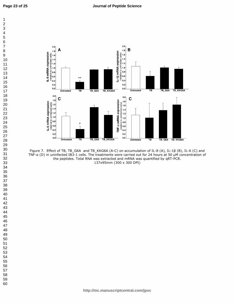
In order to exclude an effect of TB_KKG6A on the basic levels of IL-8, IL-1β, IL-6 and TNF-α, the
experiment reported in Figure 7 was performed. IB3-1 cells were treated for 24 hours with 50 µM
TB, TB_G6A and TB_KKG6A and the expression of the pro-inflammatory genes IL-8, IL-1β, IL-6
and TNF-α was determined by RT-PCR. The results obtained demonstrated that the peptides
Page 8 of 25
http://mc.manuscriptcentral.com/jpsc
Journal of Peptide Science
123456789101112131415161718192021222324252627282930313233343536373839404142434445464748495051525354555657585960
Page 10
TB_G6A and TB_KKG6A do not affect the expression of pro-inflammatory genes in uninfected
IB3-1 cells. The only inhibitory effect was found when TB was employed and the expression of IL-
8 and IL-6 genes analyzed (Figure7, A and C). When these data are considered together with the
results shown in Figures 2-5, they suggests that TB_KKG6A is a strong inhibitor of PAO1 induced
expression of pro-inflammatory genes and its activity is mainly due to antibacterial effects without
alteration of cellular pathways involved in inflammatory processes.
3.7 CD and fluorescence studies
As the antibacterial activity of peptides belonging to the temporin family is supposed to be
mediated by the interactions of the peptides with the bacterial outer membrane, we performed CD
and fluorescence studies of TB_KKG6A in the presence of P. aeruginosa LPS. The peptide
TB_KKG6A was titrated with the lipopolysaccharide from P. aeruginosa in phosphate buffer at pH
7.0 . CD spectra of the peptide in buffer show one minimum at 200 nm which clearly indicates that
the peptide is in an unordered conformation in buffer, while in the presence of LPS two minima
around 207 and 224 nm appear, suggesting that the peptide assumes the conformation of a helix
upon binding to LPS. (Figure 8) The interaction of the peptide with LPS was also assessed by
fluorescence, monitoring the intensity of the fluorescence emission of the NBD labeled
TB_KKG6A, TB_KKG6A_NBD, titrated with increasing amounts of LPS. A sigmoidal curve was
obtained plotting the fluorescence intensity at 550 nm vs µg of LPS added.(Figure 9) This result is
very similar to what reported in the literature for titrations carried out for this and other NBD
labeled peptides with LPS from other bacteria, as E.coli.20, 28
The binding of the peptide to LPS
causes an increase in the hydrophobicity of the peptide environment which is sensed by the NBD
probe. As it is reported in the literature that the LPS from P. aeruginosa has a high degree of
heterogeneity29
, we could not calculate its molar concentration and therefore we could not express
the binding constant of the peptide to LPS. In order to detect the position of the fluorophore with
respect to the LPS, we also monitored how the maximum in the emission wavelength varies with
the excitation wavelength. It has been observed, in fact, that membrane bound peptides show red
edge excitation shift (REES), due to the location of the fluorophore in a membrane region which is
motionally restricted30
. No REES was detected for the peptide in solution and for the peptide in the
presence of LPS (data not shown), suggesting that the N-terminus of the peptide is solvent exposed.
CD and fluorescence data demonstrate that the peptide interacts with the bacterial membrane and
that its folding is mediated by the interaction with the bacterial LPS. It has recently been
demonstrated that CD spectra recorded for antimicrobial peptides in the presence of LPS recall CD
Page 9 of 25
http://mc.manuscriptcentral.com/jpsc
Journal of Peptide Science
123456789101112131415161718192021222324252627282930313233343536373839404142434445464748495051525354555657585960
Page 11
spectra recorded in the presence of cells31
; based on this we might hypothesize that the peptide
TB_KKG6A also assumes an helical structure upon interaction with P.aeruginosa cells.
Conclusions
The major conclusion of this work is that TB_KKG6A exhibits strong antimicrobial activity on
P.aeruginosa PAO1 cells, likely mediated by the interactions of the peptide with the bacterial
membranes. Experiments aimed to evaluate the antimicrobial activity of TB_KKG6A were carried
out in parallel also on TB, TB_G6A and gentamycin. Interestingly TB_KKG6A was found active
against P. aeruginosa at 10 µM concentration, unlike the other peptides which were found inactive.
Gentamicin shows activity at lower concentration. These results confirm the ability of TB_KKG6A
to kill Gram negative bacteria at low concentrations. In addition, TB_KKG6A was found to
strongly inhibit the PAO1 induced upregulation of the pro inflammatory genes IL-8, IL-1β, IL-6
and TNF-α in IB3-1 cystic fibrosis cells infected by P. aeruginosa PAO1 in different conditions.
This effect is mainly associated with the antibacterial activity of TB_KKG6A, and in fact it is
particularly evident when PAO-1 was pre-treated with TB_KKG6A before infection of the IB3-1
cells (Figure 5); moreover no inhibitory effects of TB_KKG6A were found on the expression of IL-
8, IL-1b, IL-6 and TNF-a genes in uninfected IB3-1 cells (Figure 7). In any case inhibitory effects
of TB_KKG6A are clearly detectable in IB3-1-infected cells in the protocol mimicking the
pathological situation and based on pre-treatment of IB3-1 cells with the inhibitory peptide before
infection. TB_KKG6A. The effects of TB_KKG6A on IL-8 gene expression are of relevance in
consideration of the key role of this protein in cystic fibrosis inflammatory process. On the other
hand TB was found to be inactive on IL-8 and TNF-α gene expression, but to exert induction
effects on pro-inflammatory IL-1β and IL-6 genes, suggesting that TB should be considered as a
potential pro-inflammatory compound.
Acknowledgments
We are grateful to Alice Prince for donating the P. aeruginosa strain PAO1, to Valentina Lovato
for excellent technical support. This work was supported by grants from the Italian Cystic Fibrosis
Research Foundation (grants # 15/2004 and # 17/2010 to R.G; grant # 14/2012 to IL as external
collaborator). VB is a fellow of Italian Cystic Fibrosis Research Foundation. CA is granted by
Programma Merit RBNE08YFN3.
Page 10 of 25
http://mc.manuscriptcentral.com/jpsc
Journal of Peptide Science
123456789101112131415161718192021222324252627282930313233343536373839404142434445464748495051525354555657585960
Page 12
Page 11 of 25
http://mc.manuscriptcentral.com/jpsc
Journal of Peptide Science
123456789101112131415161718192021222324252627282930313233343536373839404142434445464748495051525354555657585960
Page 13
References
1. Welsh MJ, Tsui LC, Boat TF, Beaudet AL. Cystic fibrosis. The Metabolic and Molecular
Bases of Inherited Disease
1995: 3799-876.
2. Becker MN, Sauer MS, Muhlebach MS, Hirsh AJ, Wu Q, Verghese MW, et al. Cytokine
secretion by cystic fibrosis airway epithelial cells. Am J Respir Crit Care Med. 2004; 169: 645-
53.
3. Black HR, Yankaskas JR, Johnson LG, Noah TL. Interleukin-8 production by cystic
fibrosis nasal epithelial cells after tumor necrosis factor-alpha and respiratory syncytial virus
stimulation. Am J Respir Cell Mol Biol. 1998; 19: 210-5.
4. Bonfield TL, Panuska JR, Konstan MW, Hilliard KA, Hilliard JB, Ghnaim H, et al.
Inflammatory cytokines in cystic fibrosis lungs. Am J Respir Crit Care Med. 1995; 152: 2111-8.
5. Tamanini A, Borgatti M, Finotti A, Piccagli L, Bezzerri V, Favia M, et al.
Trimethylangelicin reduces IL-8 transcription and potentiates CFTR function. Am J Physiol
Lung Cell Mol Physiol. 2011; 300: L380-90.
6. Hancock RE, Speert DP. Antibiotic resistance in Pseudomonas aeruginosa: mechanisms
and impact on treatment. Drug Resist Updat. 2000; 3: 247-55.
7. Kalle M, Papareddy P, Kasetty G, Morgelin M, van der Plas MJ, Rydengard V, et al. Host
defense peptides of thrombin modulate inflammation and coagulation in endotoxin-mediated
shock and Pseudomonas aeruginosa sepsis. PLoS One. 2012; 7: e51313.
8. Czihal P, Knappe D, Fritsche S, Zahn M, Berthold N, Piantavigna S, et al. Api88 is a novel
antibacterial designer peptide to treat systemic infections with multidrug-resistant Gram-
negative pathogens. ACS Chem Biol. 2012; 7: 1281-91.
9. Uccelletti D, Zanni E, Marcellini L, Palleschi C, Barra D, Mangoni ML. Anti-Pseudomonas
activity of frog skin antimicrobial peptides in a Caenorhabditis elegans infection model: a
plausible mode of action in vitro and in vivo. Antimicrob Agents Chemother. 2010; 54: 3853-
60.
10. Conlon JM, Sonnevend A, Patel M, Al-Dhaheri K, Nielsen PF, Kolodziejek J, et al. A family
of brevinin-2 peptides with potent activity against Pseudomonas aeruginosa from the skin of
the Hokkaido frog, Rana pirica. Regul Pept. 2004; 118: 135-41.
11. Simmaco M, Mignogna G, Barra D, Bossa F. Antimicrobial peptides from skin secretions
of Rana esculenta. Molecular cloning of cDNAs encoding esculentin and brevinins and
isolation of new active peptides. J Biol Chem. 1994; 269: 11956-61.
12. Giacometti A, Cirioni O, Barchiesi F, Fortuna M, Scalise G. In-vitro activity of cationic
peptides alone and in combination with clinically used antimicrobial agents against
Pseudomonas aeruginosa. J Antimicrob Chemother. 1999; 44: 641-5.
13. Luca V, Stringaro A, Colone M, Pini A, Mangoni ML. Esculentin(1-21), an amphibian skin
membrane-active peptide with potent activity on both planktonic and biofilm cells of the
bacterial pathogen Pseudomonas aeruginosa. Cell Mol Life Sci. 2013; 70: 2773-86.
14. Simmaco M, Mignogna G, Canofeni S, Miele R, Mangoni ML, Barra D. Temporins,
antimicrobial peptides from the European red frog Rana temporaria. Eur J Biochem. 1996;
242: 788-92.
15. Wade D, Silveira A, Silberring J, Kuusela P, Lankinen H. Temporin antibiotic peptides: A
review and derivation of a consensus sequence. Protein Peptide Lett. 2000; 7: 349-57.
16. Rinaldi AC, Mangoni ML, Rufo A, Luzi C, Barra D, Zhao H, et al. Temporin L:
antimicrobial, haemolytic and cytotoxic activities, and effects on membrane permeabilization
in lipid vesicles. Biochem J. 2002; 368: 91-100.
17. Bhunia A, Saravanan R, Mohanram H, Mangoni ML, Bhattacharjya S. NMR structures
and interactions of temporin-1Tl and temporin-1Tb with lipopolysaccharide micelles:
Page 12 of 25
http://mc.manuscriptcentral.com/jpsc
Journal of Peptide Science
123456789101112131415161718192021222324252627282930313233343536373839404142434445464748495051525354555657585960
Page 14
mechanistic insights into outer membrane permeabilization and synergistic activity. J Biol
Chem. 2011; 286: 24394-406.
18. Capparelli R, Romanelli A, Iannaccone M, Nocerino N, Ripa R, Pensato S, et al.
Synergistic antibacterial and anti-inflammatory activity of temporin A and modified temporin
B in vivo. PLoS One. 2009; 4: e7191.
19. Romanelli A, Moggio L, Montella RC, Campiglia P, Iannaccone M, Capuano F, et al.
Peptides from Royal Jelly: studies on the antimicrobial activity of jelleins, jelleins analogs and
synergy with temporins. J Pept Sci. 2011; 17: 348-52.
20. Avitabile C, Netti F, Orefice G, Palmieri M, Nocerino N, Malgieri G, et al. Design,
structural and functional characterization of a Temporin-1b analog active against Gram-
negative bacteria. Biochim Biophys Acta. 2013; 1830: 3767-75.
21. Bezzerri V, Borgatti M, Nicolis E, Lampronti I, Dechecchi MC, Mancini I, et al.
Transcription factor oligodeoxynucleotides to NF-kappaB inhibit transcription of IL-8 in
bronchial cells. Am J Respir Cell Mol Biol. 2008; 39: 86-96.
22. Gambari R, Borgatti M, Bezzerri V, Nicolis E, Lampronti I, Dechecchi MC, et al. Decoy
oligodeoxyribonucleotides and peptide nucleic acids-DNA chimeras targeting nuclear factor
kappa-B: inhibition of IL-8 gene expression in cystic fibrosis cells infected with Pseudomonas
aeruginosa. Biochem Pharmacol. 2010; 80: 1887-94.
23. Nicolis E, Lampronti I, Dechecchi MC, Borgatti M, Tamanini A, Bezzerri V, et al.
Modulation of expression of IL-8 gene in bronchial epithelial cells by 5-methoxypsoralen. Int
Immunopharmacol. 2009; 9: 1411-22.
24. Gambari R, Borgatti M, Lampronti I, Fabbri E, Brognara E, Bianchi N, et al. Corilagin is a
potent inhibitor of NF-kappaB activity and downregulates TNF-alpha induced expression of
IL-8 gene in cystic fibrosis IB3-1 cells. Int Immunopharmacol. 2012; 13: 308-15.
25. de Jager W, te Velthuis H, Prakken BJ, Kuis W, Rijkers GT. Simultaneous detection of 15
human cytokines in a single sample of stimulated peripheral blood mononuclear cells. Clin
Diagn Lab Immunol. 2003; 10: 133-9.
26. Penolazzi L, Lambertini E, Tavanti E, Torreggiani E, Vesce F, Gambari R, et al.
Evaluation of chemokine and cytokine profiles in osteoblast progenitors from umbilical cord
blood stem cells by BIO-PLEX technology. Cell Biol Int. 2008; 32: 320-5.
27. Hodson ME, Penketh AR, Batten JC. Aerosol carbenicillin and gentamicin treatment of
Pseudomonas aeruginosa infection in patients with cystic fibrosis. Lancet. 1981; 2: 1137-9.
28. Rosenfeld Y, Sahl HG, Shai Y. Parameters involved in antimicrobial and endotoxin
detoxification activities of antimicrobial peptides. Biochemistry. 2008; 47: 6468-78.
29. Pier GB. Pseudomonas aeruginosa lipopolysaccharide: a major virulence factor,
initiator of inflammation and target for effective immunity. Int J Med Microbiol. 2007; 297:
277-95.
30. Raghuraman H, Chattopadhyay A. Orientation and dynamics of melittin in membranes
of varying composition utilizing NBD fluorescence. Biophys J. 2007; 92: 1271-83.
31. Avitabile C, D'Andrea LD, Romanelli A. Circular Dichroism studies on the interactions of
antimicrobial peptides with bacterial cells. Sci Rep. 2014; 4: 4293.
Page 13 of 25
http://mc.manuscriptcentral.com/jpsc
Journal of Peptide Science
123456789101112131415161718192021222324252627282930313233343536373839404142434445464748495051525354555657585960
Page 15
Table 1. Name and sequences of the peptides employed.
Sequence Name
LLPIVGNLLKSLL TB
LLPIVANLLKSLL TB_G6A
KKLLPIVANLLKSLL TB_KKG6A
NBD-Ahx-KKLLPIVANLLKSLL TB_ KKG6A _NBD
Page 14 of 25
http://mc.manuscriptcentral.com/jpsc
Journal of Peptide Science
123456789101112131415161718192021222324252627282930313233343536373839404142434445464748495051525354555657585960
Page 16
Legend to Figures
Figure 1. Anti-microbial assay of Temporin B and its derivatives against P. aeruginosa. Effects of
increasing amounts of TB (A), TB_G6A (B), TB_KKG6A (C) and Gentamicin (positive control)
(D) on P. aeruginosa growth in TSB after 24 hours at 37°C.
Figure 2. Effects of pre-incubation of Temporin B and its derivatives in human bronchial epithelial
cells before infection with P. aeruginosa. IB3-1 cells were pre-incubated with increasing amounts
of TB, TB derivatives or Gentamicin for 24 hrs before P. aeruginosa (PAO1 strain) infection
sustained for further 4 hours. Total RNA was finally extracted and qRT-PCR was performed in
order to quantify IL-8 mRNA expression. Effects of pre-incubation of TB (A), TB_G6A (B),
TB_KKG6A (C), Gentamicin (D).
Figure 3. Effect of pre-incubation of Temporin B and its derivatives with P. aeruginosa on PAO1-
mediated IL-8 mRNA expression in human bronchial epithelial cells. Equal amounts of P.
aeruginosa, PAO1 strain, were pre-incubated for 24 hours in TSB culture medium containing
increasing amounts of TB, TB derivatives or Gentamicin. Then, the resulting bacterial suspension
was washed with sterile PBS and IB3-1 cells were infected with the bacterial suspension obtained
after PBS washes for 4 hours. Total RNA was extracted and mRNA was quantified by qRT-PCR.
Effect of pre-incubation of TB (A), TB_G6A (B), TB_KKG6A (C), Gentamicin ( D).
Figure 4. Effect of pre-incubation of Temporin B and its derivatives in human bronchial epithelial
cells before infection with P. aeruginosa: expression of pro-inflammatory IL-1β, IL-6 and TNF-α
genes. IB3-1 cells were pre-incubated with increasing amounts of TB and TB derivatives for 24 hrs
before PAO1infection sustained for further 4 hours. Total RNA was finally extracted and qRT-PCR
was performed in order to quantify IL-1β, IL-6 and TNF-α mRNAs. A-C. Effect of pre-incubation
with TB on IL-1β (A), IL-6 (B) and TNF-α (C); D-F. Effect of TB_G6A on IL-1β (D), IL-6 (E) and
TNF-α (F); G-I) effect of TB_KKG6A on IL-1β (G), IL-6 (H) and TNF-α (I).
Page 15 of 25
http://mc.manuscriptcentral.com/jpsc
Journal of Peptide Science
123456789101112131415161718192021222324252627282930313233343536373839404142434445464748495051525354555657585960
Page 17
Figure 5. Effect of pre-incubation of P.aeruginosa with TB_KKG6A (A-C) or Gentamycin (D-F)
on PAO1-mediated IL-1β (A,D), IL-6 (B,E) and TNF-α (C,F) mRNA expression in IB3-1 cells.
Equal amounts of P. aeruginosa, PAO1 strain, were pre-incubated for 24 hours in TSB culture
medium containing increasing amount of TB_KKG6A or Gentamicin. Then, the resulting bacterial
suspension was washed with sterile PBS and IB3-1 cells were infected with the bacterial suspension
obtained after PBS washes for 4 hours. Total RNA was extracted and mRNA was quantified by
qRT-PCR.
Figure 6. Effects of TB_KKG6A of IL-8 secretion. A. Effects of pre-incubation of human
bronchial epithelial IB3-1 cells with the peptide before infection with P.aeruginosa (for details on
the experimental protocol, see legend to Figs. 2 and 4). B. Effects of pre-incubation of the peptide
with P. aeruginosa before infection of IB3-1 cells (for details on the experimental protocol, see
legend to Figs. 3 and 5). IL-8 was quantified by Bio-plex analysis. Open symbols: control
uninfected cells.
Figure 7. Effect of TB, TB_G6A and TB_KKG6A (A-C) on accumulation of IL-8 (A), IL-1β (B),
IL-6 (C) and TNF-α (D) in uninfected IB3-1 cells. The treatments were carried out for 24 hours at
50 µM concentration of the peptides. Total RNA was extracted and mRNA was quantified by qRT-
PCR.
Figure 8. Superimposition of CD spectra obtained titrating P. aeruginosa LPS into the
TB_KKG6A (5µM) solution, in phosphate buffer pH7. The direction of the arrow indicates
increasing LPS concentration.
Figure 9. Plot of the fluorescence intensity at 550 nm vs µg P.aeruginosa LPS obtained titrating the
LPS into the TB_KKG6A_NBD (0.5 µM) solution.
Page 16 of 25
http://mc.manuscriptcentral.com/jpsc
Journal of Peptide Science
123456789101112131415161718192021222324252627282930313233343536373839404142434445464748495051525354555657585960
Page 18
Figure 1. Anti-microbial assay of Temporin B and its derivatives against P. aeruginosa. Effects of increasing amounts of TB (A), TB_G6A (B), TB_KKG6A (C) and Gentamicin (positive control) (D) on P. aeruginosa
growth in TSB after 24 hours at 37°C. 126x86mm (600 x 600 DPI)
Page 17 of 25
http://mc.manuscriptcentral.com/jpsc
Journal of Peptide Science
123456789101112131415161718192021222324252627282930313233343536373839404142434445464748495051525354555657585960
Page 19
Figure 2. Effects of pre-incubation of Temporin B and its derivatives in human bronchial epithelial cells before infection with P. aeruginosa. IB3-1 cells were pre-incubated with increasing amounts of TB, TB derivatives or Gentamicin for 24 hrs before P. aeruginosa (PAO1 strain) infection sustained for further 4
hours. Total RNA was finally extracted and qRT-PCR was performed in order to quantify IL-8 mRNA expression. Effects of pre-incubation of TB (A), TB_G6A (B), TB_KKG6A (C), Gentamicin (D).
133x95mm (300 x 300 DPI)
Page 18 of 25
http://mc.manuscriptcentral.com/jpsc
Journal of Peptide Science
123456789101112131415161718192021222324252627282930313233343536373839404142434445464748495051525354555657585960
Page 20
Figure 3. Effect of pre-incubation of Temporin B and its derivatives with P. aeruginosa on PAO1-mediated IL-8 mRNA expression in human bronchial epithelial cells. Equal amounts of P. aeruginosa, PAO1 strain, were pre-incubated for 24 hours in TSB culture medium containing increasing amounts of TB, TB derivatives or Gentamicin. Then, the resulting bacterial suspension was washed with sterile PBS and IB3-1 cells were
infected with the bacterial suspension obtained after PBS washes for 4 hours. Total RNA was extracted and mRNA was quantified by qRT-PCR. Effect of pre-incubation of TB (A), TB_G6A (B), TB_KKG6A (C),
Gentamicin ( D). 152x124mm (600 x 600 DPI)
Page 19 of 25
http://mc.manuscriptcentral.com/jpsc
Journal of Peptide Science
123456789101112131415161718192021222324252627282930313233343536373839404142434445464748495051525354555657585960
Page 21
Figure 4. Effect of pre-incubation of Temporin B and its derivatives in human bronchial epithelial cells before infection with P. aeruginosa: expression of pro-inflammatory IL-1β, IL-6 and TNF-α genes. IB3-1 cells were pre-incubated with increasing amounts of TB, TB derivatives or Gentamicin for 24 hrs before PAO1infection
sustained for further 4 hours. Total RNA was finally extracted and qRT-PCR was performed in order to quantify IL-1β, IL-6 and TNF-α mRNAs. A-C. Effect of pre-incubation with TB on IL-1β (A), IL-6 (B) and TNF-α (C); D-F. Effect of TB_G6A on IL-1β (D), IL-6 (E) and TNF-α (F); G-I) effect of TB_KKG6A on IL-1β (G),
IL-6 (H) and TNF-α (I). 126x86mm (600 x 600 DPI)
Page 20 of 25
http://mc.manuscriptcentral.com/jpsc
Journal of Peptide Science
123456789101112131415161718192021222324252627282930313233343536373839404142434445464748495051525354555657585960
Page 22
Figure 5. Effect of pre-incubation of P.aeruginosa with TB_KKG6A (A-C) or Gentamycin (D-F) on PAO1-mediated IL-1β (A,D), IL-6 (B,E) and TNF-α (C,F) mRNA expression in IB3-1 cells. Equal amounts of P. aeruginosa, PAO1 strain, were pre-incubated for 24 hours in TSB culture medium containing increasing
amount of TB_KKG6A or Gentamicin. Then, the resulting bacterial suspension was washed with sterile PBS and IB3-1 cells were infected with the bacterial suspension obtained after PBS washes for 4 hours. Total
RNA was extracted and mRNA was quantified by qRT-PCR. 88x42mm (600 x 600 DPI)
Page 21 of 25
http://mc.manuscriptcentral.com/jpsc
Journal of Peptide Science
123456789101112131415161718192021222324252627282930313233343536373839404142434445464748495051525354555657585960
Page 23
Figure 6. Effects of TB_KKG6A of IL-8 secretion. A. Effects of pre-incubation of human bronchial epithelial IB3-1 cells with the peptide before infection with P.aeruginosa (for details on the experimental protocol, see
legend to Figs. 2 and 4). B. Effects of pre-incubation of the peptide with P. aeruginosa before infection of
IB3-1 cells (for details on the experimental protocol, see legend to Figs. 3 and 5). IL-8 was quantified by Bio-plex analysis. Open symbols: control unifected cells.
85x43mm (600 x 600 DPI)
Page 22 of 25
http://mc.manuscriptcentral.com/jpsc
Journal of Peptide Science
123456789101112131415161718192021222324252627282930313233343536373839404142434445464748495051525354555657585960
Page 24
Figure 7. Effect of TB, TB_G6A and TB_KKG6A (A-C) on accumulation of IL-8 (A), IL-1β (B), IL-6 (C) and TNF-α (D) in uninfected IB3-1 cells. The treatments were carried out for 24 hours at 50 µM concentration of
the peptides. Total RNA was extracted and mRNA was quantified by qRT-PCR. 137x95mm (300 x 300 DPI)
Page 23 of 25
http://mc.manuscriptcentral.com/jpsc
Journal of Peptide Science
123456789101112131415161718192021222324252627282930313233343536373839404142434445464748495051525354555657585960
Page 25
Figure 8. Superimposition of CD spectra obtained titrating P. aeruginosa LPS into the TB_KKG6A (5µM) solution, in phosphate buffer pH 7. The direction of the arrow indicates increasing LPS concentration.
6x5mm (600 x 600 DPI)
Page 24 of 25
http://mc.manuscriptcentral.com/jpsc
Journal of Peptide Science
123456789101112131415161718192021222324252627282930313233343536373839404142434445464748495051525354555657585960
Page 26
Figure 9. Plot of the fluorescence intensity at 550 nm vs µg P.aeruginosa LPS obtained titrating the LPS into the TB_KKG6A_NBD (0.5 µM) solution.
57x41mm (600 x 600 DPI)
Page 25 of 25
http://mc.manuscriptcentral.com/jpsc
Journal of Peptide Science
123456789101112131415161718192021222324252627282930313233343536373839404142434445464748495051525354555657585960


![Characterization of Garcinia Mangostana Linn. …anti-inflammatory, antibacterial, astringent, antitumor and antioxidative activities [12]. According to [11], seeds of Garcinia Mangostana](https://static.documents.pub/doc/80x56/5ea4a8220ada486f87166614/characterization-of-garcinia-mangostana-linn-anti-inflammatory-antibacterial.jpg)


