Antibacterial effects and biocompatibility of titanium surfaces with graded silver incorporation in titania nanotubes Shenglin Mei a, b, d,1 , Huaiyu Wang b, c,1 , Wei Wang a , Liping Tong b , Haobo Pan c , Changshun Ruan c , Qianli Ma a , Mengyuan Liu a , Huiling Yang a , Liang Zhang a , Yicheng Cheng a , Yumei Zhang a, * , Lingzhou Zhao a, * , Paul K. Chu b, * a School of Stomatology, The Fourth Military Medical University, Xi’an 710032, China b Department of Physics & Materials Science, City University of Hong Kong, Tat Chee Avenue, Kowloon, Hong Kong, China c Center for HumanTissues and Organs Degeneration, Shenzhen Institutes of Advanced Technology, Chinese Academy of Sciences, Shenzhen 518055, China d Department of Stomatology, The 518th Military Hospital, Xi’an 710043, China article info Article history: Received 18 December 2013 Accepted 4 February 2014 Available online 22 February 2014 Keywords: Titania nanotubes Silver Plasma immersion ion implantation Antibacterial effects Biocompatibility abstract Most commercial dental implants are made of titanium (Ti) because Ti possesses excellent properties such as osseointegration. However, many types of Ti products still suffer from insufficient antibacterial capability and bacterial infection after surgery remains one of the most common and intractable com- plications. In this study, a dual process encompassing anodization and silver plasma immersion ion implantation (Ag PIII) is utilized to produce titania nanotubes (TiO 2 -NTs) containing Ag at different sites and depths. The concentration and depth of the incorporated Ag can be tailored readily by changing the PIII parameters. The Ag-embedded TiO 2 -NTs which retain the nanotubular morphology are capable of sterilizing oral pathogens as opposed to pure Ti plates and pristine TiO 2 -NTs. Biological assays indicate that the in vitro and in vivo biocompatibility of the sample plasma-implanted at a lower voltage of 0.5 kV (NT-Ag-0.5) is significantly compromised due to the large amount of surface Ag. On the other hand, the sample implanted at 1 kV (NT-Ag-1.0) exhibits unimpaired effects due to the smaller surface Ag accu- mulation. Sample NT-Ag-1.0 is further demonstrated to possess sustained antibacterial properties due to the large embedded depth of Ag and the technique and resulting materials have large potential in dental implants. Ó 2014 Elsevier Ltd. All rights reserved. 1. Introduction Titanium (Ti) and Ti alloys which have good mechanical prop- erties, high corrosion resistance, excellent biocompatibility [1,2] are commonly found in orthopedic prostheses, orthodontics, joint re- placements, and so on [1,3e5]. Although Ti products account for a large proportion of the commercial dental market, their antibac- terial properties are inadequate in many applications and despite thorough disinfection in advance, microbial infection remains one of the intractable problems [6,7]. Microorganisms are abundant in the oral environment and the trans-gingival abutment of the im- plants is an important portal for bacterial infection [8]. The surgical trauma after implantation can also compromise the host defense consequently facilitating bacterial invasion. Implant failure can be a direct consequence of serious microbial attack and hence, new Ti- based biomaterials with the desirable antibacterial properties are essential for high quality dental care. There have been studies about incorporating antibacterial agents into the surface of biomedical implants [9e12]. However, most of these attempts fail to deliver sustained antibacterial effects as the surface layers and coatings are prone to rapid degradation in the oral environment. In fact, fast release of antibacterial agents causes safety concerns and the emergence of resistant strains is another issue when excessive antibiotics are administered [8]. In contrast to antibiotics, silver (Ag) is a non-specific bactericide that acts against a broad spectrum of bacterial and fungal species. Ag is attractive on account of the good stability in the physiological environment and the difficulty to develop resistant strains [6e8,13e16], although it has been documented that the therapeutic window of Ag is rather small [17,18] and Ag is cytotoxic above a certain dose. Nevertheless, * Corresponding authors. E-mail addresses: [email protected](Y. Zhang), zhaolingzhou1983@ hotmail.com (L. Zhao), [email protected](P.K. Chu). 1 These 2 authors contributed equally to this work. Contents lists available at ScienceDirect Biomaterials journal homepage: www.elsevier.com/locate/biomaterials http://dx.doi.org/10.1016/j.biomaterials.2014.02.005 0142-9612/Ó 2014 Elsevier Ltd. All rights reserved. Biomaterials 35 (2014) 4255e4265
Transcript
Antibacterial effects and biocompatibility of titanium surfaces withgraded silver incorporation in titania nanotubes
Shenglin Mei a,b,d,1, Huaiyu Wang b,c,1, Wei Wang a, Liping Tong b, Haobo Pan c,Changshun Ruan c, Qianli Ma a, Mengyuan Liu a, Huiling Yang a, Liang Zhang a,Yicheng Cheng a, Yumei Zhang a,*, Lingzhou Zhao a,*, Paul K. Chu b,*
a School of Stomatology, The Fourth Military Medical University, Xi’an 710032, ChinabDepartment of Physics & Materials Science, City University of Hong Kong, Tat Chee Avenue, Kowloon, Hong Kong, ChinacCenter for Human Tissues and Organs Degeneration, Shenzhen Institutes of Advanced Technology, Chinese Academy of Sciences, Shenzhen 518055, ChinadDepartment of Stomatology, The 518th Military Hospital, Xi’an 710043, China
a r t i c l e i n f o
Article history:Received 18 December 2013Accepted 4 February 2014Available online 22 February 2014
Keywords:Titania nanotubesSilverPlasma immersion ion implantationAntibacterial effectsBiocompatibility
a b s t r a c t
Most commercial dental implants are made of titanium (Ti) because Ti possesses excellent propertiessuch as osseointegration. However, many types of Ti products still suffer from insufficient antibacterialcapability and bacterial infection after surgery remains one of the most common and intractable com-plications. In this study, a dual process encompassing anodization and silver plasma immersion ionimplantation (Ag PIII) is utilized to produce titania nanotubes (TiO2-NTs) containing Ag at different sitesand depths. The concentration and depth of the incorporated Ag can be tailored readily by changing thePIII parameters. The Ag-embedded TiO2-NTs which retain the nanotubular morphology are capable ofsterilizing oral pathogens as opposed to pure Ti plates and pristine TiO2-NTs. Biological assays indicatethat the in vitro and in vivo biocompatibility of the sample plasma-implanted at a lower voltage of 0.5 kV(NT-Ag-0.5) is significantly compromised due to the large amount of surface Ag. On the other hand, thesample implanted at 1 kV (NT-Ag-1.0) exhibits unimpaired effects due to the smaller surface Ag accu-mulation. Sample NT-Ag-1.0 is further demonstrated to possess sustained antibacterial properties due tothe large embedded depth of Ag and the technique and resulting materials have large potential in dentalimplants.
� 2014 Elsevier Ltd. All rights reserved.
1. Introduction
Titanium (Ti) and Ti alloys which have good mechanical prop-erties, high corrosion resistance, excellent biocompatibility [1,2] arecommonly found in orthopedic prostheses, orthodontics, joint re-placements, and so on [1,3e5]. Although Ti products account for alarge proportion of the commercial dental market, their antibac-terial properties are inadequate in many applications and despitethorough disinfection in advance, microbial infection remains oneof the intractable problems [6,7]. Microorganisms are abundant inthe oral environment and the trans-gingival abutment of the im-plants is an important portal for bacterial infection [8]. The surgical
trauma after implantation can also compromise the host defenseconsequently facilitating bacterial invasion. Implant failure can be adirect consequence of serious microbial attack and hence, new Ti-based biomaterials with the desirable antibacterial properties areessential for high quality dental care.
There havebeen studies about incorporating antibacterial agentsinto the surface of biomedical implants [9e12]. However, most ofthese attempts fail to deliver sustained antibacterial effects as thesurface layers and coatings are prone to rapid degradation in theoral environment. In fact, fast release of antibacterial agents causessafety concerns and the emergence of resistant strains is anotherissue when excessive antibiotics are administered [8]. In contrast toantibiotics, silver (Ag) is a non-specific bactericide that acts against abroad spectrum of bacterial and fungal species. Ag is attractive onaccount of the good stability in the physiological environment andthe difficulty to develop resistant strains [6e8,13e16], although ithas been documented that the therapeutic window of Ag is rathersmall [17,18] and Ag is cytotoxic above a certain dose. Nevertheless,
hotmail.com (L. Zhao), [email protected] (P.K. Chu).1 These 2 authors contributed equally to this work.
Contents lists available at ScienceDirect
Biomaterials
journal homepage: www.elsevier .com/locate/biomater ia ls
http://dx.doi.org/10.1016/j.biomaterials.2014.02.0050142-9612/� 2014 Elsevier Ltd. All rights reserved.
Biomaterials 35 (2014) 4255e4265
by introducing the proper amount of Ag into the implants andcontrolling its release, excellent antibacterial properties can beattained without compromising the original biological functions ofthe substrate [14e16].
Titania nanotubes (TiO2-NTs) have attractedmuch attention dueto their low elastic modulus [19] and unique topography [20e22].The dimensions of TiO2-NTs can be controlled and TiO2-NTs withcertain dimensions do not impair and even promote the biologicalproperties of the Ti substrate [20e22]. Furthermore, the “half-open” structure of TiO2-NTs can serve as the reservoir to storeantibacterial agents such as Ag [7,23] to produce sustained anti-bacterial effects. In this study, TiO2-NTs are fabricated on Ti byanodization and low-voltage Ag plasma immersion ion implanta-tion (PIII) is conducted to embed Ag at different depths withoutbreaking the nanotubular structure. The different antibacterialcharacteristics rendered by the different Ag depth distributions andsubsequent biocompatibility are assessed systematically bothin vitro and in vivo.
2. Materials and methods
2.1. Sample preparation
Commercial pure Ti plates (10 mm � 10 mm � 1 mm) were polished and ul-trasonically cleaned in acetone, ethanol, and deionized water. Table 1 lists thedifferent processing parameters together with sample designations. PT denotes theuntreated pure Ti as the control. Sample NT underwent anodization at a constantvoltage of 20 V for 30min [24] and samples NT-Ag-0.5 and NT-Ag-1.0were subjectedto Ag PIII at different voltages. Ag PIII was conducted on a plasma ion implanterequipped with a filtered cathodic arc plasma source made of 99.99% pure Ag. Acurved magnetic duct was inserted between the plasma source and main chamberto remove macro-particles produced from the cathodic arc [16] and during Ag PIII,the pulse of cathodic arc was synchronized with that of the target voltage.
2.2. Surface characterization
Scanning electron microscopy (SEM, S-4800, Hitachi High Technologies) andatomic force microscopy (AFM, NanoScope V MultiMode system, Veeco) wereemployed to evaluate the surface morphology. The samples were dried and sputter-coated with platinum prior to SEM examination and the surface roughness wasdetermined by AFM. X-ray photoelectron spectroscopy (XPS) was conducted on aPhysical Electronics PHI 5802 equipped with a monochromatic Al Ka source. Aconstant pass energy (11.75 eV) was employed and all the data were collected at atake-off angle of 45� . The static contact angles were measured by the static sessiledrop method on the EasyDrop Standard instrument (KRUSS) at ambient humidityand temperature. Distilled water, formamide, and diiodomethane were used as themedia (10 ml per drop) and 6 measurements were performed on each specimen forstatistical accountability.
2.3. Bacteria culture
Porphyromonas gingivalis (Pg, ATCC33277) and Actinobacillus actino-mycetemcomitans (Aa, ATCC29523) were cultivated in the brain heart infusion (BHI,Oxoid) broth medium supplemented with blood. Both kinds of bacteria wereinvolved in the assays of various samples. The specimens were placed on 24-wellculture plates and separately incubated in 1 ml of the bacteria-containing me-dium (106 CFUml�1) under standard anaerobic conditions (80% N2, 10% H2, 10% CO2,at 37 �C) [25] for different time durations. Before bacteria incubation, the substrateswere sterilized with 75% alcohol overnight and rinsed three times with a sterilephosphate buffered saline (PBS) solution.
2.3.1. Fluorescence stainingAfter exposure to bacteria for 1 day, the specimens were rinsed three times with
PBS, stained with SYTO 9 and propidium iodide dyes (LIVE/DEAD� BacLight� Bac-terial Viability Kits, Molecular Probes) for 15 min in darkness, and examined by laser
scanning confocal microscopy (FV1000, Olympus). The fluorescence intensity wasmonitored by the Olympus software kit (OLYMPUS FLUOVIEW Ver.3.1 Viewer).
2.3.2. SEM observationAfter bacteria incubation for 1 day, the substrates were rinsed thrice with PBS,
fixed with 3% glutaraldehyde at 4 �C for 2 h, and dehydrated sequentially in a seriesof ethanol solutions for 10 min each. Prior to SEM observation, the specimens weredried and sputter coated with platinum.
2.3.3. Antibacterial assayAfter culturing for 1, 4, and 7 days, the bacteria on the various specimens were
gently rinsed with PBS and ultrasonically detached in 1 ml of the PBS solution for5 min. The bacteria suspensions were re-cultivated on agar plates for colonycounting. The antibacterial rates were determined by the following relationship:Antibacterial rate (%) ¼ (CFU of control � CFU of experimental groups)/CFU ofcontrol � 100%, where PT served as the control and NT, NT-Ag-0.5, and NT-Ag-1.0constituted the experimental groups.
2.4. Ag release test
The 28-day Ag release from NT-Ag-0.5 and NT-Ag-1.0 was monitored in 5 ml ofPBS solution at 37 �C under agitation (60 revolutions per minute) and periodic re-placements of the PBS. The Agþ concentration of each harvested specimen wasquantitatively determined by inductively-coupled plasma mass spectrometry (ICP-MS, Thermo X series 2).
2.5. Cell culture
Both the epithelia-like cell line Tca-8113 and fibroblast-like cell line HT1080were used. Tca-8113 and HT1080 were cultured in RPMI-1640 medium (Sigma) andDulbecco’s modified eagle medium (DMEM, Gibco) supplemented with 10%newborn bovine serum, respectively, in a humidified atmosphere of 5% CO2 at 37 �C.The cells were seeded onto the specimens at a density of 2 � 104 cells per sample byusing 24-well tissue culture plates as the holders. Before cell seeding, the substrateswere sterilized with 75% alcohol overnight and rinsed three times with sterile PBS.
2.5.1. Cell viability assayAfter incubation for 1, 4, and 7 days, the samples were rinsed three times with
sterile PBS and incubated with a 1 mg/mL 3-(4,5-dimethylthiazol-2-yl)-2,5-diphenyltetrazolium bromide (MTT, Sigma) solution at 37 �C for 4 h to allow for-mazan formation. The formazanwas dissolved by dimethyl sulfoxide and the opticaldensity (OD) was determined spectrophotometrically at 570 nm.
2.5.2. Morphological determinationAfter culturing for 1 day, the substrates were rinsed three times with PBS, fixed
with 3% glutaraldehyde at 4 �C for 2 h, and dehydrated in a series of ethanol solu-tions for 10 min each. The dehydrated samples were dried, platinum coated, andexamined by SEM.
2.5.3. Real-time polymerase chain reaction (RT-PCR)After culturing with cells for 7 days, the specimens were rinsed three times with
PBS, and the total RNA of the cultured cells was extracted by a TRIZOL reagent(Invitrogen). One mg of RNA from each specimen was reverse transcribed into cDNAby a PrimeScript� RT reagent kit (TaKaRa) according to the manufacturer’s pro-tocols. RT-PCRwas performed on a Bio-Rad iQ5 real time PCR system using amixtureof SYBR� Premix Ex Taq II (TaKaRa), cDNA templates, and the primers. Vascularendothelial growth factor (VEGF) and fibronectin (FN) of the cultured epithelial cells,and type I collagen (Coll-I) and intercellular adhesion molecule-1 (ICAM-1) of thecultured fibroblasts were analyzed with glyceraldehyde-3-phosphate dehydroge-nase (GAPDH) serving as an endogenous house-keeping gene for normalization. Theprimer sequences of the genes are shown in Table 2. Quantification of the geneexpressions was based on the comparative cycle-threshold method.
2.6. Animals and surgery
The animal experiments were approved by Institutional Animal Care and UseCommittee of the FourthMilitaryMedical University, and a total of 12male Sprague-Dawley rats (weighing 200e250 g) were used. The animals were housed under a
Table 1Main parameters of the anodization and plasma immersion ion implantation processes.
Sample name Anodization Ag plasma immersion ion implantation
Voltage (V) Treatment time (min) Pulse duration (ms) Pulse frequency (Hz) Pressure (Pa) Treatment time (min) Voltage (kV)
S. Mei et al. / Biomaterials 35 (2014) 4255e42654256
12 h light/dark cycle with free access to water and food. Prior to surgery, the ratswere anesthetized by inhaling 1.25e2.5% isoflurane with 100% oxygen. Thereafter, 2parallel incisions (8 mm long) were made through the shaved and cleaned back skinof each rat, and then 4 small openings in themuscle sheath were made by dissectionon both sides of the spine. All 4 types of samples were inserted into each rat and theimplants were placed randomly in the openings with the modified surface placedtoward the muscle tissue. The surgical wounds were closed with two sutures tosecure the position of each implant. After surgery, the animals received intramus-cular injections of gentamicin (2 � 105 U) to avoid postoperation infection.
2.6.1. MorphometryAt 12 days after implantation, the rats were sacrificed by an overdose of
pentobarbital and the implants with the surrounding tissue attached were removeden bloc. After rinsing thrice in PBS, frozen at �80 �C, and immersing in 4% para-formaldehyde for 24 h, the specimens were dehydrated in a series of ethanol andembedded in paraffin. The embedded tissue was sectioned (6 mm thick) and stainedby hematoxylin and eosin for light microscopy (DMI6000B, Leica). The thickness ofthe fibrotic capsule around each implant was determined at five equally locatedpoints on the side of modified surface for statistical accountability.
2.6.2. ImmunohistochemistryAt 12 days after implantation, the rats were sacrificed and the tissue section
(6 mm thick) of each implant was prepared following the above procedures. Formacrophages staining, non-specific binding was initially removed by incubation ingoal serum for 30min and the sections were incubatedwith themouse anti-rat cd68monoclonal antibody (AbD Serotec) at 4 �C for 12 h. After threewashes with PBS, thebiotinylated secondary antibody (goal anti-mouse IG-FITC) was added and thesections were incubated at room temperature for 2 h in darkness. The specimenswere washed thrice with PBS, counterstained by 40 ,6-diamidino-2-phenylindol(DAPI), and mounted on slides for confocal microscopy (FV1000, Olympus). AnImage-Pro Plus 6.0 software was utilized to determine the ratio of cd68-positivecells to total cells in each fibrotic capsule.
2.7. Statistics
The in vitro assays were performed in triplicate and values were expressed asmean � standard deviation. Each in vitro experiment was repeated three times withdata from a typical experiment shown. Six animals were involved in each in vivoassay with the data expressed as mean � standard deviation. A one-way ANOVAcombined with a Student-Newman-Keuls (SNK) post hoc test was utilized todetermine the level of significance. In all the statistical evaluations, p < 0.05 wasconsidered significant and p < 0.01 was considered highly significant.
3. Results
3.1. Surface characterization
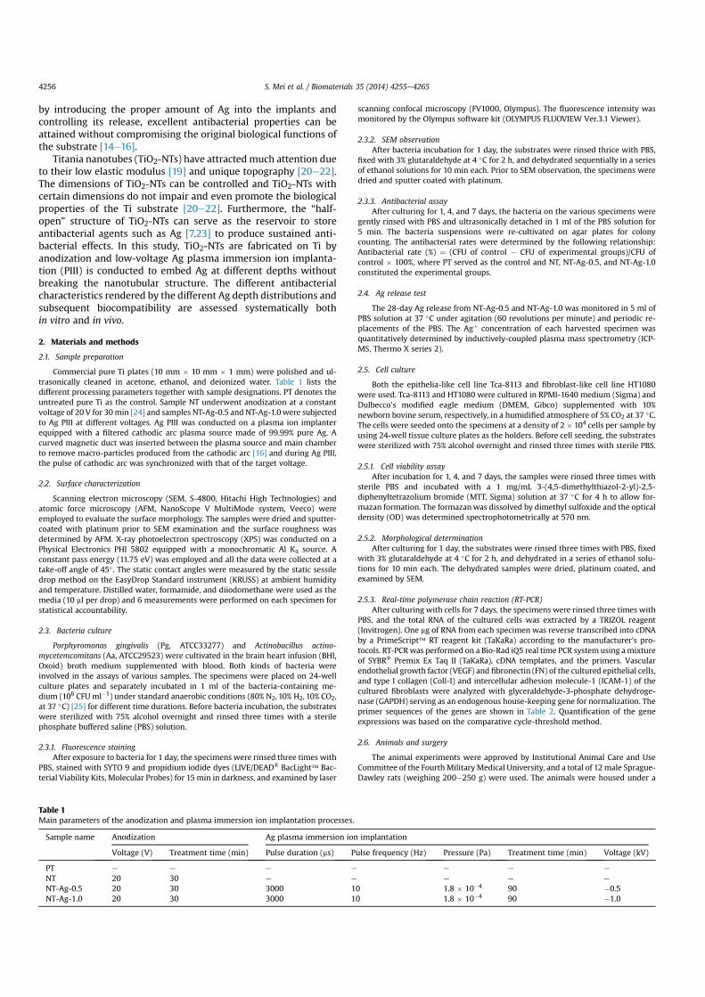
The control, as-anodized, anodized and Ag PIII at 0.5 kV, andanodized and Ag PIII at 1 kV samples are designated as PT, NT, NT-Ag-0.5, and NT-Ag-1.0, respectively (Table 1). As shown in the SEMimages in Fig. 1a, the typical nanotube array structure has adiameter of 80 nm and length of 270 nm. After Ag PIII at 0.5 and1.0 kV, the nanotubular structure is preserved although the featuresare different. Ag is randomly deposited on the surface of NT-Ag-0.5(Fig. 1b) but is found mainly on the inner nanotube wall of NT-Ag-1.0 (Fig.1c). As a result, the nanotubular morphology of NT-Ag-0.5 ismore obscure and the tube thickness of NT-Ag-1.0 increases. The 3-dimensional AFM images acquired from an area of 2.0 mm � 2.0 mmare displayed in Fig. 1deg. In contrast to PT which has a planar
microtopography, the samples after anodization are more ruggedand the size of the granular structures (Fig. 1eeg) follows thefollowing order: NT-Ag-0.5 > NT-Ag-1.0 > NT. The results areconsistent with the SEM images that Ag is deposited randomly onNT-Ag-0.5 but mainly on the inner nanotube wall of NT-Ag-1.0. Thesample surface roughness is also evaluated by AFM and the resultsare shown in Table 3. The roughness depends on the size of thegranular structures and has the same order of NT-Ag-0.5 > NT-Ag-1.0 > NT. The difference in roughness originates from the absenceas well as different locations of Ag.
XPS is performed to determine the Ag chemical states and depthprofiles. As shown in Fig. 1h and i, the Ag3d peaks (368.25 eV and374.25 eV) do not shift with depths and can be assigned to 3d5/2 and3d3/2 of metallic Ag0, indicating that the embedded Ag has themetallic (Ag0) state. Ag is mainly located in the top 20 nm (about 76at%) in NT-Ag-0.5 and the concentration diminishes rapidly toabout 5 at% at a depth of 50 nm and less than 1 at% beyond 100 nm(Fig.1h and j). On the other hand, the Ag concentration in NT-Ag-1.0is 27 at% at 20 nm and decreases slowlywith depth (Fig.1i and j). Agis detected from NT-Ag-1.0 throughout the entire nanotube length(270 nm). XPS, SEM, and AFM furnish evidence that Ag is incor-porated at different depths without breaking the nanotubularstructure. There is a larger amount of surface Ag on NT-Ag-0.5 thanNT-Ag-1.0 but Ag penetrates more deeply in the latter sample.
The surface hydrophilicity affects bio-functions such as bacteria/cell adhesion and spreading. Here, it is evaluated by the staticsessile drop method using distilled water, formamide and diiodo-methane. As shown in Fig. 2, NT is more hydrophilic than PT, but AgPIII undermines the surface hydrophilicity with the largest contactangles observed from NT-Ag-1.0. Among the 4 types of samples,only NT-Ag-1.0 is hydrophobic with a water contact angle >90�
(Fig. 2).
3.2. Effects on oral pathogens
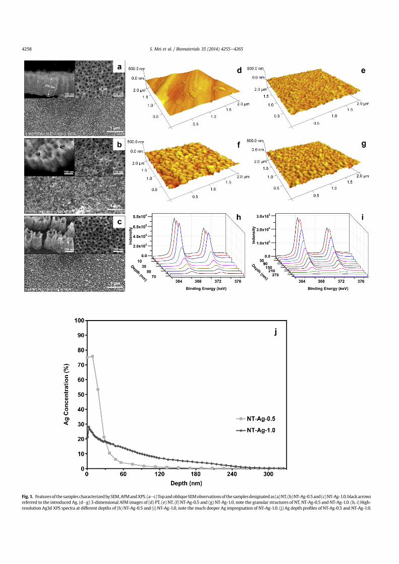
Dental implants are subjected to a variety of microbial strains inthe oral environment, especially anaerobes and facultative anaer-obes. In this study, two of these oral pathogens, Pg (Gram-positivebacteria) and Aa (Gram-negative bacteria) are utilized in the anti-bacterial evaluation. It has been documented that implants aremost susceptible to surface bacteria colonization during the initial6 h after implantation [26,27] and so the antimicrobial effects in thefirst day are critical to implant success. Fig. 3a depicts the repre-sentative images of re-cultivated bacteria after culturing for 1 day.The amounts of viable Pg and Aa on PT and NT are large, but thoseon NT-Ag-1.0 are few and those on NT-Ag-0.5 are even fewer.During the same time frame, the samples with bacteria incubationare evaluated by SYTO 9 and propidium iodide staining. The green-fluorescent SYTO 9 and red-fluorescent propidium iodide differ intheir ability to penetrate bacterial cells. When both dyes areemployed, the live bacteria with intact membranes fluoresce ingreen (stained by SYTO 9) and the dead bacteria with damagedmembranes fluoresce in red (stained by propidium iodide). Asshown in Fig. 3b, large amounts of live bacteria are detected fromPT and NT. On the contrary, the vast majority of bacteria on NT-Ag-0.5 and NT-Ag-1.0 are dead as the viable bacteria that fluoresce ingreen are very few. The fluorescence intensities of the live,dead, and total (live and dead) bacteria are presented together inTable 4. In spite of the strongest sterilizing effect of NT-Ag-0.5, thesmallest amount of total bacteria is observed from NT-Ag-1.0instead of NT-Ag-0.5, possibly because the hydrophobic surface ofNT-Ag-1.0 is less favorable to bacteria attachment.
SEM is performed to examine the morphology of the attachedbacteria. Fig. 3c shows that Pg and Aa maintain their normal shapeon PT and NT after incubation for 1 day. In the meantime, the
Table 2Sequences of the primers for cell RT-PCR.
Gene Primers
VEGF 50-GAGCCTTGCCTTGCTGCTCTAC-30
50-CACCAGGGTCTCGATTGGATG-30
FN 50-ACCTACGGATGACTCGTGCTTTGA-30
50-CAAAGCCTAAGCACTGGCACAACA-30
Coll-I 50-TCTAGACATGTTCAGCTTTGTGGAC-30
50-TCTGTACGCAGGTGATTGGTG-30
ICAM-1 50-TGAGCAATGTGCAAGAAGATAGC-30
50-CCCGTTCTGGAGTCCAGTACA-30
GAPDH 50-GCACCGTCAAGGCTGAGAAC-30
50-TGGTGAAGACGCCAGTGGA-30
S. Mei et al. / Biomaterials 35 (2014) 4255e4265 4257
Fig. 1. Featuresof the samplescharacterizedbySEM,AFMandXPS. (aec)TopandobliqueSEMobservationsof the samplesdesignatedas (a)NT, (b)NT-Ag-0.5and(c)NT-Ag-1.0, blackarrowsreferred to the introduced Ag. (deg) 3-dimensional AFM images of (d) PT, (e) NT, (f) NT-Ag-0.5 and (g) NT-Ag-1.0, note the granular structures of NT, NT-Ag-0.5 and NT-Ag-1.0. (h, i) High-resolution Ag3d XPS spectra at different depths of (h) NT-Ag-0.5 and (i) NT-Ag-1.0, note the much deeper Ag impregnation of NT-Ag-1.0. (j) Ag depth profiles of NT-Ag-0.5 and NT-Ag-1.0.
S. Mei et al. / Biomaterials 35 (2014) 4255e42654258
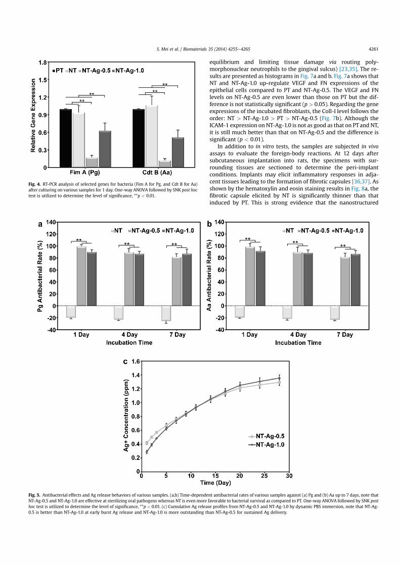
adherent bacteria on NT-Ag-0.5 and NT-Ag-1.0 are rarely intact,indicating that most of these pathogens are inactivated. The seededsamples are further investigated by RT-PCR. The fimbrillin(encoding the distinct fimbriae of Pg, playing a key role in peri-odontal and peri-implant Pg invasion) [8,28] gene of Pg and cyto-lethal distending toxin B (a heat-labile protein cytotoxin producedby several Gram-negative bacteria including Aa, regulating themorphology of cultured bacteria) [29] gene of Aa are down-regulated by both NT-Ag-0.5 and NT-Ag-1.0 after 1 day (Fig. 4).The bactericidal activity of NT-Ag-0.5 and NT-Ag-1.0 is confirmed.
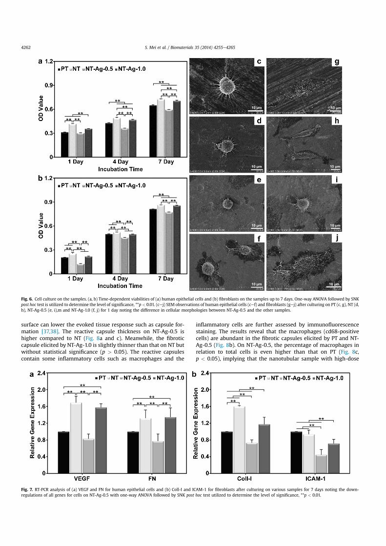
To monitor the sustained antibacterial effects, the bacteria in-cubation time is extended to 7 days. After different culturing timedurations of 1, 4, and 7 days, the viable bacteria are detached fromthe samples and re-cultivated on agar plates to count the colonies.The antibacterial rates of NT, NT-Ag-0.5, and NT-Ag-1.0 are quan-titatively referenced to PT and as shown in Fig. 5a and b, NT is betterthan PT in supporting bacterial survival. It is probably due to thenanotubular structure and excellent hydrophilicity of NT. On theother hand, NT-Ag-0.5 and NT-Ag-1.0 exhibit antibacterial effects toPg and Aa, and NT-Ag-0.5 is more effective than NT-Ag-1.0 after 1and 4 days. However, the antimicrobial activity of NT-Ag-0.5 de-creases gradually with incubation time but that of NT-Ag-1.0 isnearly constant. Eventually, NT-Ag-1.0 is more antibacterial thanNT-Ag-0.5 after 7 days.
As aforementioned, NT-Ag-0.5 and NT-Ag-1.0 are antibacterialbut NT is not. The antimicrobial properties of NT-Ag-0.5 and NT-Ag-1.0 originate from the Ag introduced by PIII. It is generally acceptedthat the bactericidal activity of Ag-incorporated materials stemsfrom leaching of Ag ions from the surface. NT-Ag-0.5 and NT-Ag-1.0are separately immersed in a PBS solution and the concentration ofleached Ag is determined by ICP-MS. The results in Fig. 5c indicate
Table 3Surface roughness (mean � SD) of the various samples determined by AFM. In thesame column, values with different lowercase letter superscripts mean significantdifferences.
Fig. 2. (a) Water, formamide, and diiodomethane contact angles measured on various samples, note the excellent hydrophilicity of NT and the hydrophobicity of NT-Ag-1.0. One-way ANOVA followed by SNK post hoc test is utilized to determine the level of significance, **p < 0.01. (b) Typical water droplet images on various samples.
S. Mei et al. / Biomaterials 35 (2014) 4255e4265 4259
that NT-Ag-0.5 is better than NT-Ag-1.0 initially. However, the cu-mulative Ag release from NT-Ag-1.0 rises faster with time. Whenthe immersion time is 14 days or longer, the total Ag delivery fromNT-Ag-1.0 is more dominant. In other words, although Ag releasefrom NT-Ag-1.0 is not as substantial as that from NT-Ag-0.5 in theearly stage, NT-Ag-1.0 fares better in terms of sustained Ag delivery.
3.3. In vitro and in vivo evaluation
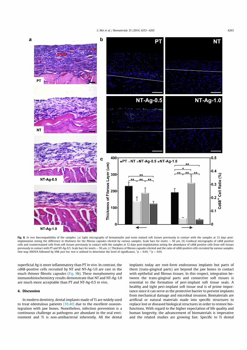
The interactions between biomaterials and surrounding tissuesare crucial to post-surgery recovery. With regard to dental im-plants, the trans-gingival parts face both epithelial and fibroustissues and tight and healthy integration with these soft tissues isdesirable. The samples are incubated with epithelial cells and fi-broblasts to assess the roles in supporting cell functions. A colori-metric assay is first employed to measure the time-dependentviability of cells after 1, 4, and 7 days. As shown in Fig. 6a and b, NTis the most positive sample to both cell types whereas NT-Ag-0.5 ismore unfavorable to the cells than PT. In spite of the hydrophobicsurface which is generally considered to be negative to mammaliancells, NT-Ag-1.0 is better than PT in supporting epithelial cellviability. Proliferation of the seeded fibroblasts on NT-Ag-1.0 after 7
days is improved as well. After 1 day, the attached cells areobserved by SEM and as shown in Fig. 6cef, the epithelial cellsadhere tightly to the substrates except NT-Ag-0.5 with dense filo-podia. The cells on NT-Ag-1.0 even secrete abundant extracellularmatrix to benefit cell proliferation. Fig. 6g, h, and j reveal that thefibroblasts on PT, NT, and NT-Ag-1.0 spread extensively and have aspindle shape. On the contrary, NT-Ag-0.5 is unfavorable to thespreading of fibroblasts for the adherent cells are still spherical inshape (Fig. 6i).
The gene expressions of cultured cells are sensitive to the un-derlying materials [30,31]. The change in cellular genes is objectiveand may take place earlier than other cellular alterations. The cellsafter culturing for 7 days are quantitatively determined by RT-PCRfor genes of epithelial cells as VEGF (a glycoprotein inducing themicrovascular permeability and angiogenesis during the woundhealing stage) [32] and FN (a major component of serum and theperiodontal tissues, playing an important role in the early woundhealing events such as cells migration and adhesion) [33] and forgenes of fibroblasts as Coll-I (the main component in gingival tis-sues which are associated with wound healing and peri-implantsoft tissue sealing) [23,34] and ICAM-1 (an adhesion moleculebelonging to the Ig super family, maintaining local hosteparasite
Fig. 3. Short-term bacteria incubation on the samples. (a) Typical images of re-cultivated Pg and Aa colonies from the samples after 1 day incubation, note that the re-cultivatedbacteria colonies from PT and NT are numerous but those from NT-Ag-0.5 and NT-Ag-1.0 are scarce. (b) Confocal micrographs of bacteria cultured on various samples for 1 day withthe green fluorescence referred to live bacteria and red fluorescence referred to dead bacteria and noting that the vast majority of bacteria on PT and NT are viable but those on NT-Ag-0.5 and NT-Ag-1.0 are inactivated. Scar bars for insets ¼ 25 mm. (c) SEM images of bacteria on samples after incubation for 1 day noting that the bacteria on PT and NT havemainly the normal shape but those on NT-Ag-0.5 and NT-Ag-1.0 are rarely intact. (For interpretation of the references to color in this figure legend, the reader is referred to the webversion of this article.)
Table 4The green, red and total fluorescence intensities (Mean values� SD) of Pg and Aa bacteria on various samples after culturing for 1 day. In the same column, values with differentlowercase letter superscripts mean significant differences.
S. Mei et al. / Biomaterials 35 (2014) 4255e42654260
equilibrium and limiting tissue damage via routing poly-morphonuclear neutrophils to the gingival sulcus) [23,35]. The re-sults are presented as histograms in Fig. 7a and b. Fig. 7a shows thatNT and NT-Ag-1.0 up-regulate VEGF and FN expressions of theepithelial cells compared to PT and NT-Ag-0.5. The VEGF and FNlevels on NT-Ag-0.5 are even lower than those on PT but the dif-ference is not statistically significant (p > 0.05). Regarding the geneexpressions of the incubated fibroblasts, the Coll-I level follows theorder: NT > NT-Ag-1.0 > PT > NT-Ag-0.5 (Fig. 7b). Although theICAM-1 expression on NT-Ag-1.0 is not as good as that on PTand NT,it is still much better than that on NT-Ag-0.5 and the difference issignificant (p < 0.01).
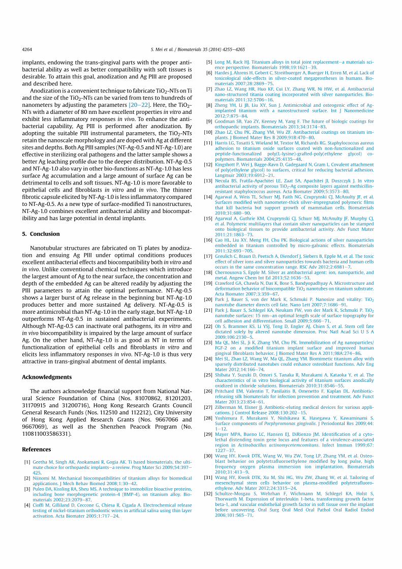
In addition to in vitro tests, the samples are subjected in vivoassays to evaluate the foreign-body reactions. At 12 days aftersubcutaneous implantation into rats, the specimens with sur-rounding tissues are sectioned to determine the peri-implantconditions. Implants may elicit inflammatory responses in adja-cent tissues leading to the formation of fibrotic capsules [36,37]. Asshown by the hematoxylin and eosin staining results in Fig. 8a, thefibrotic capsule elicited by NT is significantly thinner than thatinduced by PT. This is strong evidence that the nanostructured
Fig. 5. Antibacterial effects and Ag release behaviors of various samples. (a,b) Time-dependent antibacterial rates of various samples against (a) Pg and (b) Aa up to 7 days, note thatNT-Ag-0.5 and NT-Ag-1.0 are effective at sterilizing oral pathogens whereas NT is even more favorable to bacterial survival as compared to PT. One-way ANOVA followed by SNK posthoc test is utilized to determine the level of significance, **p < 0.01. (c) Cumulative Ag release profiles from NT-Ag-0.5 and NT-Ag-1.0 by dynamic PBS immersion, note that NT-Ag-0.5 is better than NT-Ag-1.0 at early burst Ag release and NT-Ag-1.0 is more outstanding than NT-Ag-0.5 for sustained Ag delivery.
Fig. 4. RT-PCR analysis of selected genes for bacteria (Fim A for Pg, and Cdt B for Aa)after culturing on various samples for 1 day. One-way ANOVA followed by SNK post hoctest is utilized to determine the level of significance, **p < 0.01.
S. Mei et al. / Biomaterials 35 (2014) 4255e4265 4261
surface can lower the evoked tissue response such as capsule for-mation [37,38]. The reactive capsule thickness on NT-Ag-0.5 ishigher compared to NT (Fig. 8a and c). Meanwhile, the fibroticcapsule elicited by NT-Ag-1.0 is slightly thinner than that on NT butwithout statistical significance (p > 0.05). The reactive capsulescontain some inflammatory cells such as macrophages and the
inflammatory cells are further assessed by immunofluorescencestaining. The results reveal that the macrophages (cd68-positivecells) are abundant in the fibrotic capsules elicited by PT and NT-Ag-0.5 (Fig. 8b). On NT-Ag-0.5, the percentage of macrophages inrelation to total cells is even higher than that on PT (Fig. 8c,p < 0.05), implying that the nanotubular sample with high-dose
Fig. 6. Cell culture on the samples. (a, b) Time-dependent viabilities of (a) human epithelial cells and (b) fibroblasts on the samples up to 7 days. One-way ANOVA followed by SNKpost hoc test is utilized to determine the level of significance, **p < 0.01. (cej) SEM observations of human epithelial cells (cef) and fibroblasts (gej) after culturing on PT (c, g), NT (d,h), NT-Ag-0.5 (e, i),m and NT-Ag-1.0 (f, j) for 1 day noting the difference in cellular morphologies between NT-Ag-0.5 and the other samples.
Fig. 7. RT-PCR analysis of (a) VEGF and FN for human epithelial cells and (b) Coll-I and ICAM-1 for fibroblasts after culturing on various samples for 7 days noting the down-regulations of all genes for cells on NT-Ag-0.5 with one-way ANOVA followed by SNK post hoc test utilized to determine the level of significance, **p < 0.01.
S. Mei et al. / Biomaterials 35 (2014) 4255e42654262
superficial Ag is more inflammatory than PT in vivo. In contrast, thecd68-positive cells recruited by NT and NT-Ag-1.0 are rare in themuch thinner fibrotic capsules (Fig. 8b). These morphometry andimmunohistochemistry results demonstrate that NT and NT-Ag-1.0are much more acceptable than PT and NT-Ag-0.5 in vivo.
4. Discussion
In modern dentistry, dental implants made of Ti are widely usedto treat edentulous patients [39,40] due to the excellent osseoin-tegration with jaw bones. Nonetheless, infection prevention is acontinuous challenge as pathogens are abundant in the oral envi-ronment and Ti is non-antibacterial inherently. All the dental
implants today are root-form endosseous implants but parts ofthem (trans-gingival parts) are beyond the jaw bones in contactwith epithelial and fibrous tissues. In this respect, integration be-tween the trans-gingival parts and connective soft tissues isessential to the formation of peri-implant soft tissue seals. Ahealthy and tight peri-implant soft tissue seal is of prime impor-tance since it can serve as the protective barrier to prevent implantsfrom mechanical damage and microbial invasion. Biomaterials areartificial or natural materials made into specific structures toreplace lost or diseased biological structures in order to restore bio-functions. With regard to the higher expectation of life quality andhuman longevity, the advancement of biomaterials is imperativeand the related studies are growing fast. Specific to Ti dental
Fig. 8. In vivo biocompatibility of the samples: (a) Light micrographs of hematoxylin and eosin stained soft tissues previously in contact with the samples at 12 days post-implantation noting the difference in thickness for the fibrous capsules elicited by various samples. Scale bars for insets ¼ 50 mm. (b) Confocal micrographs of cd68 positivecells and counterstained cells from soft tissues previously in contact with the samples at 12 days post-implantation noting the abundance of cd68 positive cells from soft tissuespreviously in contact with PT and NT-Ag-0.5. Scale bars for insets ¼ 50 mm. (c) Thickness of fibrous capsules elicited and the ratio of cd68 positive cells recruited by various samples.One-way ANOVA followed by SNK post hoc test is utilized to determine the level of significance, *p < 0.05, **p < 0.01.
S. Mei et al. / Biomaterials 35 (2014) 4255e4265 4263
implants, endowing the trans-gingival parts with the proper anti-bacterial ability as well as better compatibility with soft tissues isdesirable. To attain this goal, anodization and Ag PIII are proposedand described here.
Anodization is a convenient technique to fabricate TiO2-NTs onTiand the size of the TiO2-NTs can be varied from tens to hundreds ofnanometers by adjusting the parameters [20e22]. Here, the TiO2-NTs with a diameter of 80 nm have excellent properties in vitro andexhibit less inflammatory responses in vivo. To enhance the anti-bacterial capability, Ag PIII is performed after anodization. Byadopting the suitable PIII instrumental parameters, the TiO2-NTsretain the nanoscalemorphology and are dopedwith Ag at differentsites and depths. BothAg PIII samples (NT-Ag-0.5 andNT-Ag-1.0) areeffective in sterilizing oral pathogens and the latter sample shows abetter Ag leaching profile due to the deeper distribution. NT-Ag-0.5and NT-Ag-1.0 also vary in other bio-functions as NT-Ag-1.0 has lesssurface Ag accumulation and a large amount of surface Ag can bedetrimental to cells and soft tissues. NT-Ag-1.0 is more favorable toepithelial cells and fibroblasts in vitro and in vivo. The thinnerfibrotic capsule elicited byNT-Ag-1.0 is less inflammatory comparedto NT-Ag-0.5. As a new type of surface-modified Ti nanostructures,NT-Ag-1.0 combines excellent antibacterial ability and biocompat-ibility and has large potential in dental implants.
5. Conclusion
Nanotubular structures are fabricated on Ti plates by anodiza-tion and ensuing Ag PIII under optimal conditions producesexcellent antibacterial effects and biocompatibility both in vitro andin vivo. Unlike conventional chemical techniques which introducethe largest amount of Ag to the near surface, the concentration anddepth of the embedded Ag can be altered readily by adjusting thePIII parameters to attain the optimal performance. NT-Ag-0.5shows a larger burst of Ag release in the beginning but NT-Ag-1.0produces better and more sustained Ag delivery. NT-Ag-0.5 ismore antimicrobial than NT-Ag-1.0 in the early stage, but NT-Ag-1.0outperforms NT-Ag-0.5 in sustained antibacterial experiments.Although NT-Ag-0.5 can inactivate oral pathogens, its in vitro andin vivo biocompatibility is impaired by the large amount of surfaceAg. On the other hand, NT-Ag-1.0 is as good as NT in terms offunctionalization of epithelial cells and fibroblasts in vitro andelicits less inflammatory responses in vivo. NT-Ag-1.0 is thus veryattractive in trans-gingival abutment of dental implants.
Acknowledgments
The authors acknowledge financial support from National Nat-ural Science Foundation of China (Nos. 81070862, 81201203,31170915 and 31200716), Hong Kong Research Grants CouncilGeneral Research Funds (Nos. 112510 and 112212), City Universityof Hong Kong Applied Research Grants (Nos. 9667066 and9667069), as well as the Shenzhen Peacock Program (No.110811003586331).
References
[1] Geetha M, Singh AK, Asokamani R, Gogia AK. Ti based biomaterials, the ulti-mate choice for orthopaedic implantsea review. Prog Mater Sci 2009;54:397e425.
[2] Niinomi M. Mechanical biocompatibilities of titanium alloys for biomedicalapplications. J Mech Behav Biomed 2008;1:30e42.
[3] Puleo DA, Kissling RA, Sheu MS. A technique to immobilize bioactive proteins,including bone morphogenetic protein-4 (BMP-4), on titanium alloy. Bio-materials 2002;23:2079e87.
[4] Cioffi M, Gilliland D, Ceccone G, Chiesa R, Cigada A. Electrochemical releasetesting of nickel-titanium orthodontic wires in artificial saliva using thin layeractivation. Acta Biomater 2005;1:717e24.
[5] Long M, Rack HJ. Titanium alloys in total joint replacementea materials sci-ence perspective. Biomaterials 1998;19:1621e39.
[6] Hardes J, Ahrens H, Gebert C, Streitbuerger A, Buerger H, Erren M, et al. Lack oftoxicological side-effects in silver-coated megaprostheses in humans. Bio-materials 2007;28:2869e75.
[7] Zhao LZ, Wang HR, Huo KF, Cui LY, Zhang WR, Ni HW, et al. Antibacterialnano-structured titania coating incorporated with silver nanoparticles. Bio-materials 2011;32:5706e16.
[8] Zheng YH, Li JB, Liu XY, Sun J. Antimicrobial and osteogenic effect of Ag-implanted titanium with a nanostructured surface. Int J Nanomedicine2012;7:875e84.
[9] Goodman SB, Yao ZY, Keeney M, Yang F. The future of biologic coatings fororthopaedic implants. Biomaterials 2013;34:3174e83.
[10] Zhao LZ, Chu PK, Zhang YM, Wu ZF. Antibacterial coatings on titanium im-plants. J Biomed Mater Res B 2009;91B:470e80.
[11] Harris LG, Tosatti S, Wieland M, Textor M, Richards RG. Staphylococcus aureusadhesion to titanium oxide surfaces coated with non-functionalized andpeptide-functionalized poly(L-lysine)-grafted-poly(ethylene glycol) co-polymers. Biomaterials 2004;25:4135e48.
[12] Kingshott P, Wei J, Bagge-Ravn D, Gadegaard N, Gram L. Covalent attachmentof poly(ethylene glycol) to surfaces, critical for reducing bacterial adhesion.Langmuir 2003;19:6912e21.
[13] Necula BS, Fratila-Apachitei LE, Zaat SA, Apachitei JI, Duszczyk J. In vitroantibacterial activity of porous TiO2-Ag composite layers against methicillin-resistant staphylococcus aureus. Acta Biomater 2009;5:3573e80.
[14] Agarwal A, Weis TL, Schurr MJ, Faith NG, Czuprynski CJ, McAnulty JF, et al.Surfaces modified with nanometer-thick silver-impregnated polymeric filmsthat kill bacteria but support growth of mammalian cells. Biomaterials2010;31:680e90.
[15] Agarwal A, Guthrie KM, Czuprynski CJ, Schurr MJ, McAnulty JF, Murphy CJ,et al. Polymeric multilayers that contain silver nanoparticles can be stampedonto biological tissues to provide antibacterial activity. Adv Funct Mater2011;21:1863e73.
[16] Cao HL, Liu XY, Meng FH, Chu PK. Biological actions of silver nanoparticlesembedded in titanium controlled by micro-galvanic effects. Biomaterials2011;32:693e705.
[17] Greulich C, Braun D, Peetsch A, Diendorf J, Siebers B, Epple M, et al. The toxiceffect of silver ions and silver nanoparticles towards bacteria and human cellsoccurs in the same concentration range. RSC Adv 2012;2:6981e7.
[18] Chernousova S, Epple M. Silver as antibacterial agent: ion, nanoparticle, andmetal. Angew Chem Int Ed 2013;52:1636e53.
[19] Crawford GA, Chawla N, Das K, Bose S, Bandyopadhyay A. Microstructure anddeformation behavior of biocompatible TiO2 nanotubes on titanium substrate.Acta Biomater 2007;3:359e67.
[20] Park J, Bauer S, von der Mark K, Schmuki P. Nanosize and vitality: TiO2nanotube diameter directs cell fate. Nano Lett 2007;7:1686e91.
[21] Park J, Bauer S, Schlegel KA, Neukam FW, von der Mark K, Schmuki P. TiO2nanotube surfaces: 15 nmean optimal length scale of surface topography forcell adhesion and differentiation. Small 2009;5:666e71.
[22] Oh S, Brammer KS, Li YSJ, Teng D, Engler AJ, Chien S, et al. Stem cell fatedictated solely by altered nanotube dimension. Proc Natl Acad Sci U S A2009;106:2130e5.
[23] Ma QL, Mei SL, Ji K, Zhang YM, Chu PK. Immobilization of Ag nanoparticles/FGF-2 on a modified titanium implant surface and improved humangingival fibroblasts behavior. J Biomed Mater Res A 2011;98A:274e86.
[24] Mei SL, Zhao LZ, Wang W, Ma QL, Zhang YM. Biomimetic titanium alloy withsparsely distributed nanotubes could enhance osteoblast functions. Adv EngMater 2012;14:166e74.
[25] Shibata Y, Suzuki D, Omori S, Tanaka R, Murakami A, Kataoka Y, et al. Thecharacteristics of in vitro biological activity of titanium surfaces anodicallyoxidized in chloride solutions. Biomaterials 2010;31:8546e55.
[27] Zilberman M, Elsner JJ. Antibiotic-eluting medical devices for various appli-cations. J Control Release 2008;130:202e15.
[28] Yoshimura F, Murakami Y, Nishikawa K, Hasegawa Y, Kawaminami S.Surface components of Porphyromonas gingivalis. J Periodontal Res 2009;44:1e12.
[29] Mayer MPA, Bueno LC, Hansen EJ, DiRienzo JM. Identification of a cyto-lethal distending toxin gene locus and features of a virulence-associatedregion in Actinobacillus actinomycetemcomitans. Infect Immun 1999;67:1227e37.
[30] Wang HY, Kwok DTK, Wang W, Wu ZW, Tong LP, Zhang YM, et al. Osteo-blast behavior on polytetrafluoroethylene modified by long pulse, highfrequency oxygen plasma immersion ion implantation. Biomaterials2010;31:413e9.
[31] Wang HY, Kwok DTK, Xu M, Shi HG, Wu ZW, Zhang W, et al. Tailoring ofmesenchymal stem cells behavior on plasma-modified polytetrafluoro-ethylene. Adv Mater 2012;24:3315e24.
[32] Schultze-Mosgau S, Wehrhan F, Wichmann M, Schlegel KA, Holst S,Thorwarth M. Expression of interleukin 1-beta, transforming growth factorbeta-1, and vascular endothelial growth factor in soft tissue over the implantbefore uncovering. Oral Surg Oral Med Oral Pathol Oral Radiol Endod2006;101:565e71.
S. Mei et al. / Biomaterials 35 (2014) 4255e42654264
[33] Jones RA, Nicholas B, Mian S, Davies PJA, Griffin M. Reduced expression oftissue transglutaminase in a human endothelial cell line leads to changes incell spreading, cell adhesion and reduced polymerisation of fibronectin. J CellSci 1997;110:2461e72.
[34] Shioya K, Sawada T, Miake Y, Inoue S, Yanagisawa T. Ultrastructural study oftissues surrounding replanted teeth and dental implants. Clin Oral ImplantsRes 2009;20:299e305.
[35] Tonetti MS, Imboden MA, Lang NP. Neutrophil migration into the gingivalsulcus is associated with transepithelial gradients of interleukin-8 and ICAM-1. J Periodontol 1998;69:1139e47.
[36] Remes A, Williams DF. Immune-response in biocompatibility. Biomaterials1992;13:731e43.
[38] Rosengren A, Wallman L, Danielsen N, Laurell T, Bjursten LM. Tissue reactionsevoked by porous and plane surfaces made out of silicon and titanium. IEEETrans Bio-Med Eng 2002;49:392e9.
[39] Arvidson K, Bystedt H, Frykholm A, von Konow L, Lothigius E. Five-yearprospective follow-up report of the Astra Tech Dental Implant System in thetreatment of edentulous mandibles. Clin Oral Implants Res 1998;9:225e34.
[40] Astrand P, Ahlqvist J, Gunne J, Nilson H. Implant treatment of patientswith edentulous jaws: a 20-year follow-up. Clin Implant Dent Res2008;10:207e17.
S. Mei et al. / Biomaterials 35 (2014) 4255e4265 4265