Antibody binding loop insertions as diversity elements Csaba Kiss, Hugh Fisher, Emanuele Pesavento, Minghua Dai, Rosa Valero, Milan Ovecka, Rhiannon Nolan, M. Lisa Phipps, Nileena Velappan, Leslie Chasteen, Jennifer S. Martinez, Geoffrey S. Waldo, Peter Pavlik and Andrew R.M. Bradbury* HCDR3s as diversity elements, Los Alamos National Laboratory, Los Alamos, NM, USA Received August 31, 2006; Accepted September 3, 2006 ABSTRACT In the use of non-antibody proteins as affinity reagents, diversity has generally been derived from oligonucleotide-encoded random amino acids. Although specific binders of high-affinity have been selected from such libraries, random oligonuc- leotides often encode stop codons and amino acid combinations that affect protein folding. Recently it has been shown that specific antibody binding loops grafted into heterologous proteins can confer the specific antibody binding activity to the created chimeric protein. In this paper, we examine the use of such antibody binding loops as diversity ele- ments. We first show that we are able to graft a lysozyme-binding antibody loop into green fluores- cent protein (GFP), creating a fluorescent protein with lysozyme-binding activity. Subsequently we have developed a PCR method to harvest random binding loops from antibodies and insert them at predefined sites in any protein, using GFP as an example. The majority of such GFP chimeras remain fluorescent, indicating that binding loops do not disrupt folding. This method can be adapted to the creation of other nucleic acid libraries where divers- ity is flanked by regions of relative sequence conservation, and its availability sets the stage for the use of antibody loop libraries as diversity elements for selection experiments. INTRODUCTION It is believed that a new suite of technologies, generically termed the ‘display’ technologies will overcome many of the disadvantages associated with the generation of antibodies by immunization. In particular, they avoid animals, provide monoclonal reagents and since genes are cloned simultan- eously with selection, can be easily manipulated to provide novel downstream reagents with additional properties. Although antibody fragments were originally most com- monly used as scaffolds, many other proteins have also been used successfully (1,2), with the most widely pursued being single domains based on the immunoglobulin fold: e.g. single VH (3) or VL (4) chains, camel VHH domains (5), CTLA4 (6) or fibronectin (7) domains. In general these tend to be relatively well expressed (1–10 mg/l) with affinities in the nanomolar range, although expression in intracellular com- partments can be difficult due to the presence of disulfide bonds. Beyond immunoglobulin domains, nanomolar binders have also been selected from libraries based on a three helix bundle domain from protein A [Affibodies (8,9)], lipocalins [termed anticalins (10,11)], cysteine rich domains (12) and ankyrins [termed DARPINS (13,14)], with X-ray crystallo- graphy (13,15) of anticalins and ankyrins showing that the mutated residues undergo structural changes, when compared to the parent molecule, to accomodate binding. Transformation of a protein into a binding scaffold requires the introduction of diversity at the site targeted to become the binding site. This has generally been either replacement diversity (3–6,8–11,13)—where amino acids present in the scaffold of interest, within the chosen loops or surfaces, are randomized—or insertional diversity, where a specific inser- tional site is chosen and stretches of random amino acids are inserted. The latter has been carried out both in antibody binding loops (16–19) and other proteins (20–24), with diversity derived from random peptides encoded by degener- ate oligonucleotides or in rare cases by trinucleotide codons (25). Recently, antibodies with high affinities have also been selected from libraries where the introduced comple- mentarity determining region (CDR) diversity is limited to only four (tyrosine, alanine, aspartate and serine) (26) or two (tyrosine and serine) (27) different amino acids at spe- cific sites in multiple CDRs. Nature provides a potential source of functional and well folding binding elements in the form of the binding loops which make up the antibody binding site. Antibodies contain six such binding loops, termed CDRs, which are involved in forming the antibody binding site. The first and second CDRs in both light and heavy chains are encoded by the germline V genes and subsequent mutation, while CDR3 is created as a result of recombination between V and J genes in the case *To whom correspondence should be addressed. Tel: +505 665 0281; Fax: +1 505 665 3024; Email: [email protected]Ó 2006 The Author(s). This is an Open Access article distributed under the terms of the Creative Commons Attribution Non-Commercial License (http://creativecommons.org/licenses/ by-nc/2.0/uk/) which permits unrestricted non-commercial use, distribution, and reproduction in any medium, provided the original work is properly cited. Published online 5 October 2006 Nucleic Acids Research, 2006, Vol. 34, No. 19 e132 doi:10.1093/nar/gkl681 Downloaded from https://academic.oup.com/nar/article-abstract/34/19/e132/3111976 by guest on 31 January 2018
Transcript
Antibody binding loop insertions as diversity elementsCsaba Kiss, Hugh Fisher, Emanuele Pesavento, Minghua Dai, Rosa Valero,
Milan Ovecka, Rhiannon Nolan, M. Lisa Phipps, Nileena Velappan, Leslie Chasteen,
Jennifer S. Martinez, Geoffrey S. Waldo, Peter Pavlik and Andrew R.M. Bradbury*
HCDR3s as diversity elements, Los Alamos National Laboratory, Los Alamos, NM, USA
Received August 31, 2006; Accepted September 3, 2006
ABSTRACT
In the use of non-antibody proteins as affinityreagents, diversity has generally been derivedfrom oligonucleotide-encoded random amino acids.Although specific binders of high-affinity have beenselected from such libraries, random oligonuc-leotides often encode stop codons and amino acidcombinations that affect protein folding. Recently ithas been shown that specific antibody binding loopsgrafted into heterologous proteins can confer thespecific antibody binding activity to the createdchimeric protein. In this paper, we examine the useof such antibody binding loops as diversity ele-ments. We first show that we are able to graft alysozyme-binding antibody loop into green fluores-cent protein (GFP), creating a fluorescent proteinwith lysozyme-binding activity. Subsequently wehave developed a PCR method to harvest randombinding loops from antibodies and insert them atpredefined sites in any protein, using GFP as anexample. The majority of such GFP chimeras remainfluorescent, indicating that binding loops do notdisrupt folding. This method can be adapted to thecreation of other nucleic acid libraries where divers-ity is flanked by regions of relative sequenceconservation, and its availability sets the stage forthe use of antibody loop libraries as diversityelements for selection experiments.
INTRODUCTION
It is believed that a new suite of technologies, genericallytermed the ‘display’ technologies will overcome many ofthe disadvantages associated with the generation of antibodiesby immunization. In particular, they avoid animals, providemonoclonal reagents and since genes are cloned simultan-eously with selection, can be easily manipulated to providenovel downstream reagents with additional properties.
Although antibody fragments were originally most com-monly used as scaffolds, many other proteins have also beenused successfully (1,2), with the most widely pursued beingsingle domains based on the immunoglobulin fold: e.g. singleVH (3) or VL (4) chains, camel VHH domains (5), CTLA4 (6)or fibronectin (7) domains. In general these tend to berelatively well expressed (1–10 mg/l) with affinities in thenanomolar range, although expression in intracellular com-partments can be difficult due to the presence of disulfidebonds. Beyond immunoglobulin domains, nanomolar bindershave also been selected from libraries based on a three helixbundle domain from protein A [Affibodies (8,9)], lipocalins[termed anticalins (10,11)], cysteine rich domains (12) andankyrins [termed DARPINS (13,14)], with X-ray crystallo-graphy (13,15) of anticalins and ankyrins showing that themutated residues undergo structural changes, when comparedto the parent molecule, to accomodate binding.
Transformation of a protein into a binding scaffold requiresthe introduction of diversity at the site targeted to become thebinding site. This has generally been either replacementdiversity (3–6,8–11,13)—where amino acids present in thescaffold of interest, within the chosen loops or surfaces, arerandomized—or insertional diversity, where a specific inser-tional site is chosen and stretches of random amino acids areinserted. The latter has been carried out both in antibodybinding loops (16–19) and other proteins (20–24), withdiversity derived from random peptides encoded by degener-ate oligonucleotides or in rare cases by trinucleotide codons(25). Recently, antibodies with high affinities have alsobeen selected from libraries where the introduced comple-mentarity determining region (CDR) diversity is limited toonly four (tyrosine, alanine, aspartate and serine) (26) ortwo (tyrosine and serine) (27) different amino acids at spe-cific sites in multiple CDRs.
Nature provides a potential source of functional and wellfolding binding elements in the form of the binding loopswhich make up the antibody binding site. Antibodies containsix such binding loops, termed CDRs, which are involved informing the antibody binding site. The first and second CDRsin both light and heavy chains are encoded by the germline Vgenes and subsequent mutation, while CDR3 is created as aresult of recombination between V and J genes in the case
*To whom correspondence should be addressed. Tel: +505 665 0281; Fax: +1 505 665 3024; Email: [email protected]
� 2006 The Author(s).This is an Open Access article distributed under the terms of the Creative Commons Attribution Non-Commercial License (http://creativecommons.org/licenses/by-nc/2.0/uk/) which permits unrestricted non-commercial use, distribution, and reproduction in any medium, provided the original work is properly cited.
Published online 5 October 2006 Nucleic Acids Research, 2006, Vol. 34, No. 19 e132doi:10.1093/nar/gkl681
Downloaded from https://academic.oup.com/nar/article-abstract/34/19/e132/3111976by gueston 31 January 2018
of the light chain, and V, D and J genes for the heavy chain(28,29). Further diversity is created by the addition and lossof nucleotides at the junctions between the recombinedgene segments (30,31) and somatic hypermutation (32).Structurally, each class of CDRs is similar in size and struc-ture, with each adopting one or a few distinct or ‘canonical’conformations (33–35). HCDR3 is an exception, showingwide variations in length, structure, shape and sequence(36,37), as well as intrinsic conformational diversity(38–40), reflecting the importance of HCDR3s in antibodybinding specificity (41,42). Given this data, and the factthat HCDR3s also contain very few stop codons, they appearto represent a very effective form of diversity. This conclu-sion is bolstered by the structural conservation found at theends of HCDR3s, revealed by the finding that the fourN-terminal and six C-terminal residues from differentHCDR3 regions demonstrate <2.75 s r.m.s.d for >99.7% ofall pair-wise comparisons examined (37). As a result,HCDR3s would be expected to be less disruptive to proteinstructure than random peptides of the same length. Further-more, if a scaffold is able to accept a single HCDR3 at a spe-cific site, it is likely that many different HCDR3s can also beaccommodated at that same site.
Although libraries of HCDR3s have never been assessed fortheir effects on protein structure, a number of examples of theuse of specific antibody CDRs as diversity elements able totransplant binding activity to a heterologous protein havebeen described. An HCDR3 from an integrin binding antibodyhas been inserted into an exposed loop in tissue-type plasmino-gen activator, conferring integrin binding activity tothe plasminogen activator, without eliminating its normalenzymatic function (43). Similarly, a CDR3 from a camelidVHH recognizing lysozyme has been transplanted to neocar-zinostatin, a bacterial chromoprotein with a beta sheet structure,allowing the chimeric molecule to recognize lysozyme (44). Ascamelid VHH CDR3s are very similar to traditional antibodyHCDR3s, these two examples indicate the potential for usinglibraries of HCDR3s as diversity/binding elements, if meansfor harvesting that diversity can be developed. More recently,an HCDR1 loop from a CD4 binding antibody was insertedinto three exposed loops of the protein inhibitor of neuronalnitric oxide synthase and each construct was shown to exhibitCD4 binding (45). This work was based on earlier work show-ing that peptides derived from five of the six CDRs of the anti-CD4 antibody, and not other regions of the variable region wereable to bind CD4 as soluble, circularized peptides (46).
While these experiments show that in these specificexamples, HCDR3s can be inserted into heterologous pro-teins without disruption of protein function, it does not dem-onstrate that this can be carried out generally.
In this paper we explore the possibility of using HCDR3s asa source of insertional diversity. Using the green fluorescentprotein (GFP), which is not fluorescent unless correctly folded(47), as a reporter protein, we first show that the VHH CDR3described above is also functional when inserted into two sitesin GFP. Subsequently we describe a novel PCR method to
harvest HCDR3 diversity, based on the fact that the N- andC-terminal HCDR3 amino acids (CXX . . .XXWG) areextremely well conserved at the DNA, protein and structurallevels. We examine the effects of inserting antibody bindingloops amplified using this method into GFP, and show thatfor most sites, and most HCDR3s, there is relatively little dis-ruption to GFP function, validating HCDR3s as a potentialsource of diversity. These experiments set the stage for furtherexploration of the use of HCDR3s as diversity elements in avariety of different scaffold proteins.
MATERIALS AND METHODS
pET-CK3 expression vector construction
Four BpmI sites, one SphI and BssHII site were eliminatedfrom pET-C6His (48), a pET-28 derivative. The SphI sitewas eliminated by digesting pETC6-His with SphI. The linearDNA fragment was treated with T4 DNA polymerase andre-ligated with T4 DNA ligase. The ligation was digestedwith SphI and transformed into DH5aFT cells. The BpmIsites and the BssHII site were mutated using the Stratagenemutagenesis kit (Stratagene, La Jolla, CA) according to themanufacturer’s recommendation. Briefly, 100 ng of pETC-His6-DSphI template DNA was amplified in a 25 ml reactionusing 1 mM of the primers indicated in Table 1. A total of 1 mlof dNTP mix, 0.75 ml of QuikSolution and 1 ml ofQuikChange� Multi enzyme blend with the following tem-perature cycle: 95�C for 1 min followed by 30 cycles of95�C for 1 min, 55�C for 1 min, 65�C for 10 min. ThePCR product was digest with DpnI, BpmI and BssHII for1 h and the mixture was transformed into XL-10 Gold�
ultracompetent cells. The resulting pET-CK3 vector waschecked by restriction mapping and sequencing.
SacB insertion into the GFP loops
SacB is a negative selectable marker, which can be used tokill bacteria bearing by growth on sucrose (49). The SacBgene was inserted into superfolder GFP (50) at each of thedifferent identified loop sites (Table 2) in such a way that itwas flanked by two type BpmI restriction sites. These allowedthe removal of the sacB gene and the creation of an appropri-ate cloning site for CDR3 sequences, which were also flankedwith compatible BpmI sites. After BpmI cleavage, the N andC portions of a generic CDR3 were exposed, allowing thereassembly of a full CDR3 after ligation of amplifiedCDR3 inserts (see Figure 1). Since the sacB gene disruptsthe GFP coding sequence, clones are not fluorescent unlesspermissive CDR3s have been inserted. These vectors werecreated by amplifying the full pET-CK3-sfGFP plasmidusing pairs of primers flanking each insertion site. This cre-ated the following structure (illustrated for the insertion atloop 2), with the portion in green corresponding to GFP,the portion in red representing the primer encoded sequenceswhich complement the cloned HCDR3, and the underlinedbases the cleavage site for the indicated BpmI sites:
F K D S G D G
50..TTC AAA GAT TCT GGC GAG GAA TAC TAA CTC CAG AGT AGA CCC TAA TGA TGA GCT GGA GCC TAA AGA CCC GGG GGC GAC GGG..
30..AAG TTT CTA AGA CCG CTC CTT ATG ATT GAG GTC TCA TCT GGG AGG ACT ACT CGA CCT CGG ATT TCT GGG CCC CCG CTG CCC..
Downloaded from https://academic.oup.com/nar/article-abstract/34/19/e132/3111976by gueston 31 January 2018
For each of the different loops, the primers in Table 1 wereused. These PCR products were cleaved with BpmI and lig-ated to SacB amplified with sacB.2-50 and sacB-30 alsocleaved with BpmI (Table 1). These SacB primers placedBpmI sites at equivalent positions, allowing the SacB geneto be removed by cleavage with BpmI in the ligated clone.After cloning, bacteria were tested for their inability togrow on both liquid and agar media containing 2–5% sucrose,as well as by restriction digestion.
HCDR3 amplification
Total RNA was prepared from 40 different samples of humanperipheral blood lymphocytes purified by Ficoll Hypaque(Amersham Pharmacia Biotech, UK). Pathogens were deemedto be inactivated by the use of Trizol to purify RNA. This workwas carried out under the auspices of the LANL IRB. cDNAwas synthesized using random hexamers and reverse tran-scriptase following standard protocols. HCDR3s were ampli-fied by nested PCR using the IgM for forward primer and amixture of VH primers (4–6,10,12,14,22,51) with the follow-ing temperature cycle: 94�C, 60 s followed by 30 cycles of94�C, 30 s, 55�C, 30 s, 72�C, 1 min followed by 72�C for7 min. One microliter of the first PCR after gel purificationwas used as template in the second PCR. Biotinylated primersin Tables 2–5 were used to amplify the CDR3 sequences withthe following temperature cycle: 94�C, 60 s followed by30 cycles of 94�C, 30 s, 50�C, 30 s, 72�C, 1 min followedby 72�C for 7 min. The PCR product was phenol/chloroformextracted and ethanol precipitated. It was dissolved in 90 ml ofwater and digested with 50 U of BpmI for 2 h. The enzyme
was heat inactivated at 65�C for 20 min. A total of 100 ml ofM-280 Streptavidin Dynabeads (Dynal, Norway) was washedthree times with TE and the beads were resuspended in theBpmI digested PCR products. The beads were mixed atroom temperature for 30 min and collected with a magnet.The supernatant, which contains digested HCDR3s was useddirectly in the ligation reactions.
Library construction
pET-CK3-sfGFP-SacB in eight different loops were digestedwith BpmI, treated with Antarctic phosphatase and gel puri-fied using the Qiagen gel purification kit. The concentrationof the vector was measured by spectrofluorometer and liga-tions were set up with CDR3 fragment with a vector:insertratio of 1:3 overnight at 4�C in 20 ml volume using 800 Uof T4 DNA ligase (NEB). tRNA (1 mg) was added to the reac-tions and the total ethanol precipitated and redissolved in50 ml of water. A total of 2 ml of each of the ligation reactionswere electroporated into BL21 (DE3) Gold (Novagen) cellsand plated on nitrocellulose filters on Luria–Bertani (LB)plates containing 50 mg/ml kanamycin/2% glucose/2% suc-rose. Cells were grown overnight at 37�C. The filters weretransferred onto kanamycin LB plates containing 1 mg/mlisopropyl-b-D-thiogalactopyranoside (IPTG) and induced for4 h at 30�C.
Determination of c-lys affinity by flow cytometry
Streptavidin coated beads (50 ml) (Spherotec) were incubatedwith either 1 ng of biotinylated (Pierce Biotechnology) lyso-zyme (Sigma) or 15 ng of biotinylated 9E10 (anti-myc,
Table 1. The sequences of the primers used to create the different recipient GFP vectors
Loop 1 (23/24) and loop 1a (22/24)TAATGATGAGCTGGAGCCTAAAGACCCGGGGGCGGGCACAAATTTTCTGTCAGAGGAG pDAN5-GFP-loop1-30
The portion in green corresponds to GFP and the portion in red, the conserved portion of the HCDR3. Underlined bases represent restriction sites (BpmI and SmaI).
Downloaded from https://academic.oup.com/nar/article-abstract/34/19/e132/3111976by gueston 31 January 2018
Upstate, New York) antibody in a final volume of 100 mlphosphate-buffered saline (PBS), for 1 h at room temperature.A total of 100 ml of 5% BSA was added to block the beadsurface and incubated for a further hour at room temperature.Beads were washed once in PBS and resuspended in 150 ml ofPBS. GFP containing the anti-lysozyme CDR3 with an initial
concentration of 0.6 mg/ml was 2-fold serially diluted and 5ml of antigen coated beads added to 50 ml diluted protein perwell. After 1 h incubation at room temperature the supernat-ant was removed by washing once in PBS and the beads wereanalyzed by flow cytometry using a FACSAria instrument(BD Biosciences). For the determination of affinity at each
(a)
(b)
Figure 1. (a) The genetic rearrangement which creates human VH genes, and the PCR strategy used to amplify, digest and purify HCDR3s is shown on the left,while on the right is shown the general scheme used to create a template for simple insertion of HCDR3s exploiting type IIs restriction sites and a negativeselectable marker, such as SacB. (b) The detailed sequence of a recipient vector containing SacB inserted into loop 2, and the cloning strategy used to insertHCDR3s. The letters depicted in green represent GFP sequences, black are HCDR3 sequences, and red are junctional sequences which come together during theligation procedure. As a result of the cloning procedure, the conserved cysteine present in the HCDR3 is converted into a serine. BpmI R (underlined) identifiesthe BpmI recognition site, while BpmI C identifies the cleavage site.
Downloaded from https://academic.oup.com/nar/article-abstract/34/19/e132/3111976by gueston 31 January 2018
dilution, the mean fluorescence intensity of the non-specificbinding of c-lys to the beads coated with an irrelevant targetwas subtracted from the specific fluorescence of the lysozymecoated ones. The resulting fluorescence values at each dilu-tion were fitted to a logistic function using Origin (MicrocalSoftware, Inc., Northampton, MA) and the affinity determ-ined as the concentration at which half maximal fluorescencewas obtained (52).
Flow cytometric analysis of bacterial fluorescence
Bacterial libraries expressing GFP clones were inoculatedin minimal medium (53) and grown overnight at 37�C. Thefollowing day 1 ml of autoinduction medium (53) was inocu-lated with 10 ml of each library and grown at 30�C overnight.The cells were diluted in PBS and analyzed using a BD LSRII flow cytometer (BD Biosciences), using the 488 nm laser toexcite GFP).
Protein expression, purification and characterization
All plasmids were transformed into Escherichia coli BL 21Gold, plated on 2XTY/Kan/10% glucose and grown overnightat 37�C. Individual colonies were picked and grown overnightin liquid 2XTY/Kan/Glucose at 37�C. Confluent culture (1 ml)
was used to inoculate 50 ml 2XTY/Kan/IPTG in 250 ml sha-ker flasks for expression at 30�C overnight.
Proteins were purified by low-salt immobilized metal affin-ity chromatography. Cultures were harvested by centrifuga-tion, sonicated and resuspended in 10 mM Tris.HCl (pH 8.0)and recentrifuged at 3000 g for 30 min at 4�C. The supernatantwas applied to IMAC columns pre-equilibrated with Tris forinitial adhesion. The flow-through was reapplied three addi-tional times and washed with 20 bed volumes Tris. An addi-tional wash of 20 bed volumes of 10 mM Tris/300 mMNaCl/10% glycerol was performed preceding a final Triswash before elution in 600 mM Imidazole. The buffers wereexchanged from the eluted proteins using three passes ofspin filtration with 10 000 MWCO Amicon Ultra centrifugalfiltration devices at 4�C. The desalted proteins were dilutedin prechilled Tris and stored at 4�C preceding further evalu-ation. Protein samples for SDS–PAGE comparison werediluted for equivalent fluorescence utilizing a Tecan Spectra-fluor Plus plate fluorometer equipped with 485 nm excitationand 535 nm emission filters prior to standard denaturationand gel loading.
Absorption spectra were collected on a ThermoSpectronicGenesys2 and exported to Microsoft Excel for comparison.Excitation and emission spectra were generated by either aQuantumMaster 6SE (Photon Technologies Incorporated;Edison, NJ) or a SPEX Fluorolog spectrofluorometer utilizing1 cm2 cuvettes. Excitation scans were evaluated with 509 nmemission wavelength. Emission scans were generated with 488nm excitation. All emission scans were normalized to themaximum value obtained at the main emission peak for eachsample.
Sequences of those portions of the 50 and 30 primers corresponding to the VH or JH genes suitable for the amplification of HCDR3s are shown. These were analyzedby using only the portion of the primer which recognizes the V or J gene and searching against the database of 5646 rearranged V genes or the 49 germline V genes or6 JH genes. These analyses are stringent (100%), so it is likely that in real experimental situations, more sequences are likely to be amplified, as in each case the30 primer sequences are extremely well conserved. Under ‘Rearranged V’, the total numbers of the 5646 rearranged VH genes downloaded from IMGT withabsolute homology to each of the primers are given. In brackets are given the number of VH genes recognized by each of the individual primers making up thedegenerate pool. Under ‘Germline V’, the number of germline VH genes with 100% homology to the primers is given.
The sites at which HCDR3 libraries were inserted into GFP are indicated. Forloops 1, 2 and 3, two different insertion points were used as shown in the table.In loop 1a the underlined asparagine was deleted and the HCDR3s insertedbetween the valine and the glycine.
Downloaded from https://academic.oup.com/nar/article-abstract/34/19/e132/3111976by gueston 31 January 2018
biotinylated lysozyme was used as ligand on the Streptavidinchip (flow cells 1,2,3), and our biotinylated myoglobin wasused as the negative control (non-specific binding control)ligand on the same chip (flow cell 4). Approximately 4000RUs of both ligands were bound to the chip, under whichconditions specific binding could be demonstrated.
RESULTS
Inserting a defined CDR3 into GFPto confer binding activity
The HCDR3 from a VHH recognizing lysozyme has beentransplanted to neocarzinostatin, a bacterial chromoproteinwith a beta sheet structure, with the chimeric molecule recog-nizing lysozyme with an affinity of 500 nM (44). We attemp-ted to replicate this finding, by transferring the same HCDR3to two surface exposed loops in ‘Superfolder’ GFP (sfGFP)(50), a GFP mutant selected to be resistant to the destabilizingeffects of poorly folding proteins fused to its N-terminus, andhence more stable than other forms of GFP. In order to effect-ively use HCDR3s as diversity elements, both structural andsequence conservation must exist at the N- and C-terminalends of the isolated HCDR3. Structural conservation isrequired to ensure that once a permissive site has been cho-sen, different HCDR3s can be inserted equally effectivelyat the same site, while sequence conservation is required toallow effective cloning of the isolated HCDR3s. Within thefour N-terminal and six C-terminal amino acids from differ-ent HCDR3 regions found to be structurally similar by Moreaet al. (37), the DNA sequences encoding the N-terminalcysteine and C-terminal tryptophan and glycine are extremelyconserved. As the cysteine 104 [IMGT numbering (54)] usu-ally forms a double hydrogen bond with the glycine 119 (37),these two amino acids were chosen to be the limits of thecloned HCDR3. However, to avoid the presence of anunpaired cysteine (the HCDR3 N-terminal cysteine normallydisulfide bonds with another cysteine in framework one), thiscodon was mutated to a serine. This is identical to cysteine,except for the replacement of sulfur by oxygen, and so isable to form the same hydrogen bonds. In order to createrecipient GFPs which could be used for cloning HCDR3 lib-raries, as well the specific anti-lysozyme HCDR3, we inserted(see Figure 1 and below) a SacB gene at each targeted inser-tion site flanked by BpmI sites. The SacB gene is a negativeselector able to reduce vector background by 105-fold by plat-ing bacteria on sucrose after transformation (49,55). BpmI isa type IIs restriction site which cleaves 14/16 bp away fromits recognition site. The cleavage sites were designed toinclude conserved 50- and 30-HCDR3 sequences, whichwere exposed after digestion by BpmI, allowing the recon-struction of full-length HCDR3s within the GFP from eitherannealed oligonucleotides or amplified PCR fragments.
The anti-lysozyme HCDR3 described above was synthes-ized as a pair of overlapping phosphorylated oligonucleotides.These were annealed and ligated directly into two of the BpmIcut vectors (Figure 1). Loops 1 and 3 (see Table 2 for nomen-clature) were both independently targeted. Both clones, namedc-lys1 and c-lys3, depending upon the loop insertion site,yielded fluorescent proteins. These were expressed in BL21using either 100 mM IPTG or autoinduction media (53), andsubsequently purified by immobilized metal affinity chroma-tography using the C-terminal His6 tag.
Binding between GFP containing this HCDR3 was demon-strated in an enzyme-linked immunosorbent assay (ELISA)format in which the lysozyme was biotinylated and interactedwith the modified GFP prior to capture on a neutravidin coatedplate (Figure 2A). Specific binding could also be demonstratedusing a flow cytometric bead based method, in which detection
The sequences shown represent the full diversity found in the germline V genescentered around the conserved cysteine (TGT). All six JH sequences areindicated. For both VH and JH, the region of primer recognition is given,and the cut site when BpmI is used is underlined. The IMGT numbering systemis used.
Table 5. HCDR3 amplification primer sequences
50 primer setsVR35-1.450Biotin-AACGTGCTGGAGGCCACRTATTACTGTGVR35-2.450Biotin-AACGTGCTGGAGGCCATNTATTACTGTGVR35-3.450Biotin-AACGTGCTGGAGGCCGTHTATTACTGTGVR35-4.450Biotin-AACGTGCTGGAGGCCTTGTATTACTGTGVR35-5.450Biotin-AACGTGCTGGAGGCTGTHTATTACTGTGVR35-6.450Biotin-AACGTGCTGGAGGCYGTGTATTACTGTGVR35-7.450Biotin-AACGTGCTGGAGGCYGTVTATTATTGTGVR35-8.450Biotin-AACGTGCTGGAGGCYGTNTATTTCTGTG30 primer setsJH1.4-3050Biotin-TGAGGAGACTGGAGCCAGGGTGCCCTGGCCCCAJH2.4-3050Biotin-TGAGGAGACTGGAGCCAGGGTGCCACGGCCCCAJH3.4-3050Biotin-TGAAGAGACTGGAGCCATTGTCCCTTGGCCCCAJH4.4-3050Biotin-TGAGGAGACTGGAGCCAGGGTTCCCTGGCCCCAJH6.4-3050Biotin-TGAGGAGACTGGAGCCGTGGTCCCTTGGCCCCA
The 50 and 30 primer sequences used are shown. Each primer is biotinylated atthe 50 end. The biotin is followed by four bases to assist in recognition andcleavage by the type IIs enzyme, BpmI (recognition sequence CTGGAG16/14). The cut site is underlined.
Downloaded from https://academic.oup.com/nar/article-abstract/34/19/e132/3111976by gueston 31 January 2018
was carried out by measuring the fluorescence of streptavidincoated beads to which biotinylated lysozyme and GFP contain-ing the anti-lys HCDR3 had bound (Figure 2B). Unlike ELISA,this method relies on the intrinsic fluorescence of the binder,demonstrating that binding activity and fluorescence reside inthe same protein, and that, at least in this case, HCDR3 insertionhas not disrupted GFP function. This technique can also be usedto determine affinity (52) by incubating microspheres coatedwith antigen with increasing concentrations of fluorescent bin-der. As concentration increases, the bead bound fluorescencereaches a plateau, as all target sites on the microspheres arebound. By subtracting background binding and determiningthe concentration of fluorescent binder at which half maximumfluorescence is obtained, we were able to estimate the affinity ofthis interaction to be 1.34 mM for c-lys1 (Figure 2C), similar tothe estimate, obtained by isothermal calorimetry, for neocar-zinostatin containing the same HCDR3 (44). Binding wasalso examined using surface plasmon resonance. Although,specific binding could be demonstrated when chips were den-sely coated with lysozyme, similar to the neocarzinostatin res-ults (44), affinity was not high enough to show binding when thelower levels of coating required to determine affinity were used(data not shown).
These results indicate that the orientation and structure ofthe HCDR3 is maintained at both insertion sites and is similar
to that in the original VHH, suggesting that HCDR3s are avalid potential source of diversity, and may alone providesufficient binding energy to yield micromolar binders.
Analyzing human HCDR3 flanking sequences
In order to determine the best way to clone HCDR3s, 5669human heavy chain variable genes were downloaded fromthe IMGT web site (56), using ‘human heavy chain variablegenes of any specificity’ as search criteria. These were pareddown to 5646 full-length VH genes representing a wide spec-trum of different V genes, and encompass the full range ofmutations found at all different sites within the V genes,including potential primer sites flanking the CDR3. Ofthese 5646 VH genes, 4842 can be accounted for by search-ing for the following motifs (all based on the 10 bases finish-ing with the extremely conserved cysteine and the base whichfollows it—TGT G) found at the 30 end of framework region3 just before the CDR3 (number of times found):
TATTACTGTG (4061)TATTATTGTG (462)TATTTCTGTG (319)As amplification usually requires more than 10 bases of
homology, the 4842 sequences described above were extrac-ted from the database and analyzed for homology upstream of
Figure 2. (A) ELISA was carried out by first interacting biotinylated antigen with the GFP clone of interest and then capturing on a neutravidin coated plate. GFPbinding was revealed using SV5, a monoclonal antibody (73), which recognizes a tag appended to the C-terminus. The non-specific clone was GFP containingthe myc epitope (74) at the loop 3 position, and indicates the level of binding due to a similarly disrupted GFP molecule. clyslp1 and clyslp3 have the lysozymebinding HCDR3 inserted into loop 1 and loop 3, respectively. (B) Streptavidin coated beads were incubated with either biotinylated lysozyme or myoglobin,(which serves as the negative control for non-specific binding to coupled beads) and subsequently with GFP or GFP containing the anti-lysozyme HCDR3inserted at loop 1 (c-lys1). Analysis was carried out using a FACSCalibur. (C) As in (B), except that different concentrations of c-lys1 were used and the meanfluorescence of the non-specific (myoglobin) beads was subtracted from the fluorescence of the lysozyme coated beads and plotted. The affinity is calculatedfrom the concentration of c-lys1, which gives half maximal fluorescence.
Downloaded from https://academic.oup.com/nar/article-abstract/34/19/e132/3111976by gueston 31 January 2018
these 10 bp sequences. Based on the homology found, thesequences described in Table 3 were designed. A similar pro-cedure was carried out for the 30 end of the HCDR3 in the JHgene sequences. However, in this case, the sequences werecentered around the highly conserved CTGGGGCC sequencefound in all JH genes. The alignment of these sequences withgermline V and J genes is given in Table 4.
Seven of the thirteen primer sequences were degenerate,with each component sequence recognizing V genes.Table 3 shows the numbers of the 5646 rearranged and germ-line VH genes recognized by each of these sequences, assum-ing 100% homology. In experimental use, it is likely thatmany more genes will be recognized by each individualprimer, since single mismatches do not usually preventPCR amplification, especially when found upstream of ninehomologous bases.
Cloning design
The strategy used to clone HCDR3s amplified from B cellsinto GFP is described in Figure 1a and detailed inFigure 1b. The GFP recipient vectors contain the SacBgene flanked by the highly conserved 50 and 30 portion ofthe HCDR3. GFP containing SacB at the different insertsites is non-fluorescent, and the only way fluorescence can berestored is by removal and replacement of the sacB negativeselector with a sequence that encodes a peptide permissivefor GFP folding at the targeted insertion site. In order toisolate these sequences, without flanking frameworksequences, and to recreate full-length HCDR3s, a type IIsrestriction site, BpmI, was used. This cuts 16/14 basesaway from its recognition site, allowing it to be placedupstream of an amplifying oligonucleotide in such a waythat the majority of the oligonucleotide sequence can beremoved after PCR, leaving a 2 bp 30 extension for ligation(see Figure 1b). Based on the sequences described inTables 4 and 5, the BpmI site in the 50 HCDR3 primer wasplaced to cleave across the conserved cysteine and adjacentcodon (TGTG) (Tables 4 and 5), while at the 30 end it wasplaced to cleave within the conserved tryptophan codon(TGGGGC). Overhanging bases are underlined in bothcases. Altogether, eight vectors containing SacB insertionsat eight different sites, comprising five different loops, werecreated (Table 2 and below).
In order to eliminate the primer ends removed by BpmIcleavage (corresponding to the framework sequences), theoligonucleotides were biotinylated at their 50 ends (Table 5),allowing their removal with streptavidin Dynabeads.
PCR amplification and cloning
Non-biotinylated primers were tested by amplifying HCDR3sfrom the peripheral blood lymphoctye cDNA of 40 donorsusing non-biotinylated primers. Using each individual primerwith a pool of the complementary primers yielded a 75–150bp smear for all primers (Figure 3A and B), which is morevisible for the primers recognizing more rearranged Vgenes (e.g. VR-35–36.4). When the length of the primers istaken into account, this corresponds to HCDR3s rangingfrom �20–95 bp, similar to the previously published rangeof HCDR3 lengths (36) (24–90 bp and Figure 5).
Biotinylated versions of the primers were created andpooled for amplification before purification and cloning.Unfortunately, primer JH4.4-3 had to be omitted, due to thesub-optimal quality of the biotinylated primer. Lane 1 inFigure 3C shows the HCDR3 smear prior to digestion withBpmI, ranging from 75 to 150 bp (arrow A). After digestion(lane 2), the smear is reduced in size by 55 bp (range 25–95 bp,arrow B), corresponding to the primer portions removed byBpmI, which can be seen as an additional sharp lower band(arrow C). Lane 3 shows the final purified HCDR3 preparationafter removal of the biotinylated primers using magnetic strep-tavidin beads (Dynabeads). The extent and intensity of thesmear is essentially identical to that in lane 2, except thatthe lower band is eliminated, indicating the efficiency of theuse of streptavidin to remove the biotinylated external primerportions.
HCDR3 libraries
In order to assess the effects of the insertion of many differentHCDR3s on protein folding in general, and GFP folding andfunction in particular, five different loops in GFP were tar-geted for the insertion of HCDR3 libraries (Table 2). Theinsertion sites were identified by an examination of the struc-ture of GFP (57), with the goal of placing the HCDR3s at thetips of the loops, and so hopefully continue the GFP betastrand structure into the first part of the HCDR3. In addition,one alternative site, differing by a single amino acid, andconsequently slightly off the tip center, was also targetedfor three of the loops (1, 2 and 3 in Table 2), to see whetherinsertion within loops had to be precisely localized, orwhether it was sufficient to target a loop, without concernfor the exact insertion sites.
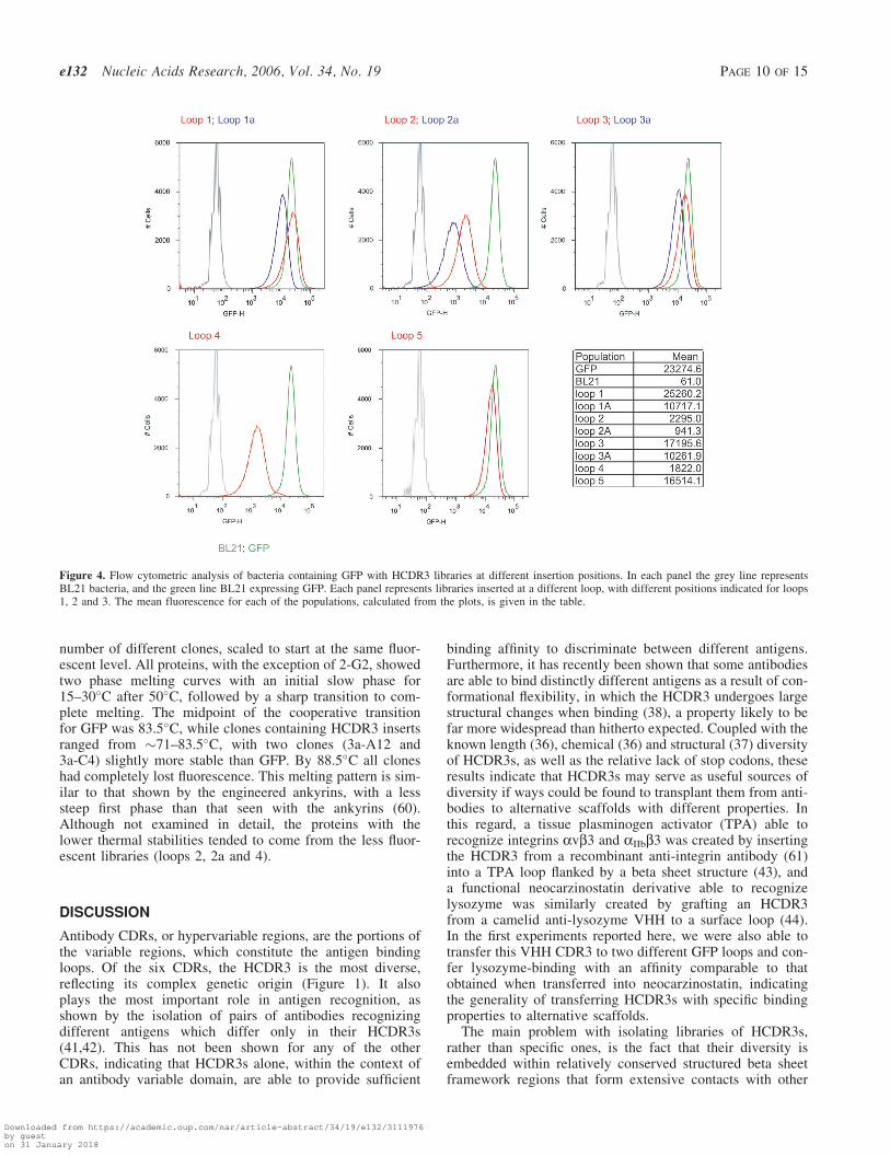
The HCDR3 fragments were gel purified and cloned into theeight recipient GFP vectors. The libraries were then inducedand analyzed by flow cytometry. Each of the libraries showedsignificant numbers of fluorescent bacteria (Figure 4) wheninduced with IPTG, with loops 1, 1a, 3, 3a and 5 providingthe greatest number, many of which overlapped with the fluor-escence of bacteria expressing GFP. The mean fluorescencefor these libraries ranged from 10 711–25 260, while GFPhad a mean fluorescence of 23 274. The remaining loops (2,2a, 4) were less fluorescent (941–2295), although still signific-antly more fluorescent than BL21 (mean 61). All the fluores-cent profiles for bacteria containing GFP with inserts werebroader than GFP with a slightly longer tail on the low fluor-escence portion of the curve. The differences between librariesinserted at different loops sites differed by as much as 27-fold(loop 2a compared to loop 1), while the greatest differencebetween insertions in the same loop differing by a singleamino acid were in no case greater than 2.5. This suggeststhat the primary determinant of fluorescence is the targetedloop, with the site within the loop playing a lesser role.
Sequence analysis of HCDR3s
302 random clones containing inserts were sequenced to fur-ther analyze the nature of the cloned diversity. The length dis-tributions of the HCDR3s (Figure 5) showed a slight increasein shorter HCDR3s (14–16 amino acid), and a reduction inlonger ones (>21 amino acid) compared to the HCDR3 lengthdistributions reported in the literature (36). All inserts were in
Downloaded from https://academic.oup.com/nar/article-abstract/34/19/e132/3111976by gueston 31 January 2018
frame with GFP, and nucleotide blast searches showed homo-logy to HCDR3s (Figure 6a shows representative sequences).Only 1.3% of the sequences contained stop codons, and 4.8%lacked the characteristic C(S) . . . . WG HCDR3 sequence.In addition there were 18 sequences repeated more than once,corresponding to 5.9% of all sequences. Interestingly, two ofthese duplicated HCDR3 sequences were each found in threedifferent loops (Figure 6b) indicating duplication was presentin the source cDNA, rather than a cloning artifact. This hasbeen previously observed in sequencing human peripheralB cell V genes (58), and reflects different VH gene poolswith different representations, such as those derived fromrecent infections, rather than the saturation of diversity.
A number of clones from each library were picked atrandom and analyzed for their fluorescence, and correlatedwith the length of the HCDR3 insert. Figure 6c shows thefluorescence of individual clones expressed as a percentageof GFP fluorescence, correlated with the length of the inser-ted HCDR3. As can be seen, the spread of HCDR3s rangesfrom 40 to 75 bp, with little correlation between lengthand fluorescence, except beyond 80 bp, where fluorescenceis reduced. This indicates that providing the HCDR3 lengthis within the normal range (40–75 bp), larger HCDR3s donot reduce fluorescence more frequently than smallerHCDR3s.
In order to determine whether there were any differencesbetween HCDR3s found in strongly fluorescent clones com-pared to weakly fluorescent ones, bacteria containing thelibraries were flow sorted for fluorescence, and an additional434 sequences analyzed. Although the length distributions ofthe HCDR3s in the unsorted and sorted libraries wereextremely similar (see Figure 5), the percentages of repeatedsequences (1.6%), non-characteristic HCDR3 sequences (2%)and sequences containing stop codons (0.7%) were allreduced.
Examination of protein properties
A number of fluorescent clones containing HCDR3 insertswere expressed and purified to study their properties com-pared to GFP. The expected size differences between GFPand clones containing inserts were apparent for all clones(Figure 7a). The expression levels of the different clonesranges from 5.2–138 mg/l, 1.3–30% the level of GFP.Normalized absorption/emission spectra were essentiallyidentical to GFP (data not shown).
The stability of some of these proteins was studied usinga real time PCR machine in which protein fluorescence wasmonitored as temperature was gradually raised (0.1�C/s)(59). Figure 7d shows fluorescence levels for GFP and a
Figure 3. PCR amplification of lymphocyte cDNA using a mixture of the 30 J region primers and individual 50 V gene primers (a), or a mixture of the 50 V geneprimers and the individual J region primers (b). In (c), the pooled PCR product prior to digestion is shown in lane 1. In lane 2 the PCR product after digestionwith BpmI, and in lane 3 the product after the biotinylated primers are removed. In each case the arrows show the amplified HCDR3s except C in 3c.
Downloaded from https://academic.oup.com/nar/article-abstract/34/19/e132/3111976by gueston 31 January 2018
number of different clones, scaled to start at the same fluor-escent level. All proteins, with the exception of 2-G2, showedtwo phase melting curves with an initial slow phase for15–30�C after 50�C, followed by a sharp transition to com-plete melting. The midpoint of the cooperative transitionfor GFP was 83.5�C, while clones containing HCDR3 insertsranged from �71–83.5�C, with two clones (3a-A12 and3a-C4) slightly more stable than GFP. By 88.5�C all cloneshad completely lost fluorescence. This melting pattern is sim-ilar to that shown by the engineered ankyrins, with a lesssteep first phase than that seen with the ankyrins (60).Although not examined in detail, the proteins with thelower thermal stabilities tended to come from the less fluor-escent libraries (loops 2, 2a and 4).
DISCUSSION
Antibody CDRs, or hypervariable regions, are the portions ofthe variable regions, which constitute the antigen bindingloops. Of the six CDRs, the HCDR3 is the most diverse,reflecting its complex genetic origin (Figure 1). It alsoplays the most important role in antigen recognition, asshown by the isolation of pairs of antibodies recognizingdifferent antigens which differ only in their HCDR3s(41,42). This has not been shown for any of the otherCDRs, indicating that HCDR3s alone, within the context ofan antibody variable domain, are able to provide sufficient
binding affinity to discriminate between different antigens.Furthermore, it has recently been shown that some antibodiesare able to bind distinctly different antigens as a result of con-formational flexibility, in which the HCDR3 undergoes largestructural changes when binding (38), a property likely to befar more widespread than hitherto expected. Coupled with theknown length (36), chemical (36) and structural (37) diversityof HCDR3s, as well as the relative lack of stop codons, theseresults indicate that HCDR3s may serve as useful sources ofdiversity if ways could be found to transplant them from anti-bodies to alternative scaffolds with different properties. Inthis regard, a tissue plasminogen activator (TPA) able torecognize integrins avb3 and aIIbb3 was created by insertingthe HCDR3 from a recombinant anti-integrin antibody (61)into a TPA loop flanked by a beta sheet structure (43), anda functional neocarzinostatin derivative able to recognizelysozyme was similarly created by grafting an HCDR3from a camelid anti-lysozyme VHH to a surface loop (44).In the first experiments reported here, we were also able totransfer this VHH CDR3 to two different GFP loops and con-fer lysozyme-binding with an affinity comparable to thatobtained when transferred into neocarzinostatin, indicatingthe generality of transferring HCDR3s with specific bindingproperties to alternative scaffolds.
The main problem with isolating libraries of HCDR3s,rather than specific ones, is the fact that their diversity isembedded within relatively conserved structured beta sheetframework regions that form extensive contacts with other
Figure 4. Flow cytometric analysis of bacteria containing GFP with HCDR3 libraries at different insertion positions. In each panel the grey line representsBL21 bacteria, and the green line BL21 expressing GFP. Each panel represents libraries inserted at a different loop, with different positions indicated for loops1, 2 and 3. The mean fluorescence for each of the populations, calculated from the plots, is given in the table.
Downloaded from https://academic.oup.com/nar/article-abstract/34/19/e132/3111976by gueston 31 January 2018
VH region amino acids. In this paper we overcome thisproblem with a PCR based method that uses flanking typeIIs restriction sites to remove the framework regions afteramplification. This relies on the structural, DNA and aminoacid sequence conservation found at either end of humanHCDR3s: cysteine 104 and tryptophan 119 (IMGT number-ing) are essentially 100% conserved at the amino acid and
nucleotide levels. Structurally these amino acids are joinedby two hydrogen bonds and so very close to one another, pro-viding further justification for their use as diversity elements.This allowed us to design 13 primers annealing within theflanking framework regions which were able to amplify alarge percentage of rearranged VH gene CDR3s. By addingBpmI sites at the ends of the primers, these framework
Figure 5. The length distribution of cloned HCDR3s derived from bacterial libraries, which were unsorted (blue), or sorted for fluorescence (red), with publisheddistributions (36) in green.
Figure 6. (a) Shows the sequence of random HCDR3s cloned into the different loops. In (b) are shown two HCDR3 sequences whose sequences were found indifferent loops. (c) Shows the correlation between HCDR3 length and bacterial fluorescence as expressed as a percentage of the fluorescence of bacteriaexpressing GFP.
Downloaded from https://academic.oup.com/nar/article-abstract/34/19/e132/3111976by gueston 31 January 2018
regions could be removed, leaving two base pair overhangsresiding within the conserved amino acids at either end ofthe HCDR3. These overhangs were ligated to sequencesencoding the remaining portion of the HCDR3 (with thecysteine exchanged for serine), exposed when the negativeselector (SacB) gene was removed from GFP by digestionwith BpmI (Figure 1). This arrangement facilitated the clon-ing of a diverse set of HCDR3s, independently of knowledgeof their sequences.
By inserting HCDR3 libraries into the tips of five loops onone end of GFP, we were able to assess the degree of disturb-ance these HCDR3s caused to folding, since GFP must foldcorrectly to become fluorescent (47). Of the eight sitesexamined, five were very permissive, giving mean bacterialfluorescence profiles within 2.3-fold of GFP. The remainingthree sites, although up to 25-fold less fluorescent than GFP,were nevertheless significantly fluorescent. The mean fluores-cence per cell is a combination of the number of fluorescentproteins per cell and the intrinsic fluorescence of those
fluorescent proteins. In Figure 7a, the amount of each fluores-cent protein loaded on the polyacrylamide gel was normalizedfor fluorescence. The similar intensity of the coomassie bluestaining suggests that the greatest variability between differentbacterial clones is in the amount of expressed fluorescentprotein, rather than any difference in intrinsic fluorescence.This is supported by the differing levels of protein whichcan be purified from each clone.
For three of the sites (loops 1, 2 and 3) two insertion siteswere tested. These differed by a single amino acid, and therationale was to determine whether it was sufficient toplace the HCDR3 within a permissive loop, or whether theexact site within that loop was critical. Although there wasa small (<2.5-fold) difference in the fluorescence at the twosites for each loop, this was far less than the differenceobserved between different loops (up to 27-fold), suggesting,at least within this small set, that the loop targeted is moreimportant than the precise position within the loop. We didnot examine insertion at other sites, and it is possible that
(a)
(b)
Figure 7. (a) Shows a polyacrylamide gel of different purified clones containing HCDR3 inserts at different positions. (b) Shows the normalized fluorescentemission of different clones gradually heated up at the rate of 0.1�C/second. For each clone, the first figure indicates the loop insertion site.
Downloaded from https://academic.oup.com/nar/article-abstract/34/19/e132/3111976by gueston 31 January 2018
insertion of HCDR3s into secondary structures which are notloops, or perhaps at the loop extremities, may be significantlymore disruptive, especially since loops tend to be less wellconserved than more structured elements.
We sequenced over 300 cloned HCDR3s. Over 90% hadthe characteristic HCDR3 sequence: C(S)XX . . .XXWG.The sequence characteristics of HCDR3s more permissivefor GFP fluorescence were identified by flow sorting bacteriaexpressing GFP containing HCDR3 libraries in differentloops. We found that HCDR3s in the more fluorescent cloneswere less likely to contain stop codons, non-characteristic orrepeated HCDR3 sequences. There was no bias in favor ofeither bulged or non-bulged HCDR3s (37), with the propor-tion remaining constant before and after sorting. As bothnon-characteristic and repeated HCDR3s are more likely tobe derived from heavily mutated clones, this may explaintheir detrimental effect on GFP fluorescence.
GFP is known to be an extremely stable protein. The GFPwe used, superfolder (50), was selected to be particularlyresistant to the effects of poorly folding proteins fused toits N-terminus. This also confers improved stability to theprotein generally (50). This was confirmed in our thermal sta-bility studies, in which the cooperative transition midpointwas 83.5�C, not dissimilar to GFP containing loops whichranged from 71 to 83.5�C. This minimal disturbance likelyreflects both the stability of GFP as well as the relativelynon-perturbing nature of the HCDR3 inserts, which wasalso shown by the fact that the spectral properties of theseproteins were essentially identical to GFP.
This study was carried out with human HCDR3s becauseof the great deal of sequencing information available. Thismade the design of appropriate framework primers relativelystraightforward. However, with sufficient information on thesequences of appropriate flanking regions, this approachcould be applied to the harvesting of diversity from the anti-body genes of other species. This may be of more than aca-demic interest, since the means by which antibody diversityhas evolved in different species, although sharing some com-monalities, has tended to be species-specific (62,63). As a res-ult, different CDRs have different properties, lengths andamino acid distributions in different species, with tendenciesto bind to different classes of antigens. Cow (64) and drom-edary heavy chain genes (65,66), e.g. have far longerHCDR3s than humans. In camels, these have been shownto be important in the mediation of enzyme inhibition by dir-ect insertion into active sites (67,68), as well the recognitionof conserved cryptic epitopes of infectious agents, perhaps bypenetration into conserved receptor binding sites (69).Although less dramatically different, murine HCDR3s tendto be shorter than human HCDR3s, with different aminoacid compositions (36), resulting in more HCDR3s whichhave stabilized hydrogen bond ladder structures, as opposedto human HCDR3s which contain more prolines, preventingthe formation of such ladders.
The method we describe here can also be adapted to otherantibody CDRs, as well as to other immunological proteins,such as T cell receptors, which share similar primary struc-tures: variable regions flanked by relatively conserved frame-work regions. g and d TCRs have CDR3s which resembleimmunoglobulin heavy chain CDR3s in their length variabil-ity, while a and b TCRs have CDR3s, which are extremely
homogenous in length (8–9 amino acids), in common withthe other antibody CDRs (70). The common component ofall such diversity elements is that they have evolved tobind, and so are likely to be more functional than randompeptides, which generally contain more stop codons and aremore likely to contain destabilizing inserts.
An alternative to the use of completely random aminoacids, has been the use of restricted amino acid sets in thegeneration of antibody libraries (26,27,71,72). In theseexperiments it has been shown that different amino aciddiversities at specific sites significantly affect the successfuloutcome of selection experiments, and in one of the most sur-prising results, specific high affinity antibodies can be selec-ted from libraries in which heavy chain diversity is limited toonly two amino acids (27). These careful studies of the rolesof different amino acids in functional diversity, are similar tothose which nature has been conducting over evolutionarytime in the different molecules involved in immune recogni-tion in many different species. The method described hereenables the harvesting of such diversity for transplantationinto heterologous proteins, setting the stage for the explora-tion of the use of libraries containing such sequences forselection experiments.
ACKNOWLEDGEMENTS
This work was funded by a DOE GTL pilot grant, and LANLLDRD-DR funds, to A.R.M.B. The authors are grateful to theNational Flow Cytometry Resource at LANL for help withsorting, and to Prof. C. Kado (UC Davis) for the SacB gene.Funding to pay the Open Access publication charges for thisarticle was provided by the DOE GTL grant.
Conflict of interest statement. None declared.
REFERENCES
1. Skerra,A. (2000) Engineered protein scaffolds for molecularrecognition. J. Mol. Recognit., 13, 167–187.
2. Nygren,P.A. and Skerra,A. (2004) Binding proteins from alternativescaffolds. J. Immunol. Meth., 290, 3–28.
3. Davies,J. and Riechmann,L. (1995) Antibody VH domains as smallrecognition units. Biotechnology (N Y), 13, 475–479.
4. van den Beucken,T., van Neer,N., Sablon,E., Desmet,J., Celis,L.,Hoogenboom,H.R. and Hufton,S.E. (2001) Building novel bindingligands to B7.1 and B7.2 based on human antibody single variable lightchain domains. J. Mol. Biol., 310, 591–601.
5. Arbabi Ghahroudi,M., Desmyter,A., Wyns,L., Hamers,R. andMuyldermans,S. (1997) Selection and identification of single domainantibody fragments from camel heavy-chain antibodies. FEBS Lett.,414, 521–526.
6. Nuttall,S.D., Rousch,M.J., Irving,R.A., Hufton,S.E., Hoogenboom,H.R.and Hudson,P.J. (1999) Design and expression of soluble CTLA-4variable domain as a scaffold for the display of functional polypeptides.Proteins, 36, 217–227.
Downloaded from https://academic.oup.com/nar/article-abstract/34/19/e132/3111976by gueston 31 January 2018
characterization of HER2/neu-binding affibody ligands. Protein Eng.Des. Sel., 17, 455–462.
10. Beste,G., Schmidt,F.S., Stibora,T. and Skerra,A. (1999) Smallantibody-like proteins with prescribed ligand specificities derived fromthe lipocalin fold. Proc. Natl Acad. Sci. USA, 96, 1898–1903.
11. Vogt,M. and Skerra,A. (2004) Construction of an artificial receptorprotein (‘anticalin’) based on the human apolipoprotein D.Chembiochem., 5, 191–199.
12. Silverman,J., Lu,Q., Bakker,A., To,W., Duguay,A., Alba,B.M.,Smith,R., Rivas,A., Li,P., Le,H. et al. (2005) Multivalent avimerproteins evolved by exon shuffling of a family of human receptordomains. Nat. Biotechnol., 23, 1556–1561.
13. Binz,H.K., Amstutz,P., Kohl,A., Stumpp,M.T., Briand,C., Forrer,P.,Grutter,M.G. and Pluckthun,A. (2004) High-affinity binders selectedfrom designed ankyrin repeat protein libraries. Nat. Biotechnol., 22,575–582.
14. Kohl,A., Binz,H.K., Forrer,P., Stumpp,M.T., Pluckthun,A. andGrutter,M.G. (2003) Designed to be stable: crystal structure of aconsensus ankyrin repeat protein. Proc. Natl Acad. Sci. USA, 100,1700–1705.
15. Korndorfer,I.P., Beste,G. and Skerra,A. (2003) Crystallographicanalysis of an ‘anticalin’ with tailored specificity for fluoresceinreveals high structural plasticity of the lipocalin loop region. Proteins,53, 121–129.
16. Parhami-Seren,B., Viswanathan,M. and Margolies,M.N. (2002)Selection of high affinity p-azophenyarsonate Fabs from heavy-chainCDR2 insertion libraries. J. Immunol. Meth., 259, 43–53.
17. Knappik,A., Ge,L., Honegger,A., Pack,P., Fischer,M., Wellnhofer,G.,Hoess,A., Wolle,J., Pluckthun,A. and Virnekas,B. (2000) Fullysynthetic human combinatorial antibody libraries (HuCAL) based onmodular consensus frameworks and CDRs randomized withtrinucleotides. J. Mol. Biol., 296, 57–86.
18. Lamminmaki,U., Pauperio,S., Westerlund-Karlsson,A., Karvinen,J.,Virtanen,P.L., Lovgren,T. and Saviranta,P. (1999) Expanding theconformational diversity by random insertions to CDRH2 resultsin improved anti-estradiol antibodies. J. Mol. Biol., 291,589–602.
19. Moroncini,G., Kanu,N., Solforosi,L., Abalos,G., Telling,G.C.,Head,M., Ironside,J., Brockes,J.P., Burton,D.R. and Williamson,R.A.(2004) Motif-grafted antibodies containing the replicative interface ofcellular PrP are specific for PrPSc. Proc. Natl Acad. Sci. USA, 101,10404–10409.
20. Scalley-Kim,M., Minard,P. and Baker,D. (2003) Low free energy costof very long loop insertions in proteins. Protein Sci., 12, 197–206.
21. Minard,P., Scalley-Kim,M., Watters,A. and Baker,D. (2001) A ‘loopentropy reduction’ phage-display selection for folded amino acidsequences. Protein Sci., 10, 129–134.
22. Bessette,P.H., Rice,J.J. and Daugherty,P.S. (2004) Rapid isolation ofhigh-affinity protein binding peptides using bacterial display. ProteinEng. Des. Sel., 17, 731–739.
23. Camaj,P., Hirsh,A.E., Schmidt,W., Meinke,A. and von Gabain,A.(2001) Ligand-mediated protection against phage lysis as a positiveselection strategy for the enrichment of epitopes displayed on thesurface of E. coli cells. Biol. Chem., 382, 1669–1677.
24. Lu,Z., Murray,K.S., Van Cleave,V., LaVallie,E.R., Stahl,M.L. andMcCoy,J.M. (1995) Expression of thioredoxin random peptide librarieson the Escherichia coli cell surface as functional fusions to flagellin:a system designed for exploring protein protein interactions.Biotechnology, 13, 366–372.
25. Virnekas,B., Ge,L., Pluckthun,A., Schneider,K.C., Wellnhofer,G. andMoroney,S.E. (1994) Trinucleotide phophoramidites: ideal reagents forthe synthesis of mixed oligonucleotides for random mutagenesis.Nucleic Acids Res., 22, 5600–5607.
26. Fellouse,F.A., Wiesmann,C. and Sidhu,S.S. (2004) Synthetic antibodiesfrom a four-amino-acid code: a dominant role for tyrosine in antigenrecognition. Proc. Natl Acad. Sci. USA, 101, 12467–12472.
27. Fellouse,F.A., Li,B., Compaan,D.M., Peden,A.A., Hymowitz,S.G. andSidhu,S.S. (2005) Molecular recognition by a binary code. J. Mol.Biol., 348, 1153–1162.
28. Early,P., Huang,H., Davis,M., Calame,K. and Hood,L. (1980) Animmunoglobulin heavy chain variable region gene is generated fromthree segments of DNA: VH, D and JH. Cell, 19, 981–992.
29. Tonegawa,S. (1983) Somatic generation of antibody diversity. Nature,302, 575–581.
30. Lieber,M.R. (1991) Site-specific recombination in the immune system.FASEB J., 5, 2934–2944.
31. Schatz,D.G., Oettinger,M.A. and Schlissel,M.S. (1992) V(D)Jrecombination: molecular biology and regulation. Annu. Rev. Immunol.,10, 359–383.
32. French,D.L., Laskov,R. and Scharff,M.D. (1989) The role of somatichypermutation in the generation of antibody diversity. Science, 244,1152–1157.
33. Chothia,C. and Lesk,A.M. (1987) Canonical structures for thehypervariable regions of immunoglobulins. J. Mol. Biol., 196,901–917.
34. Chothia,C., Lesk,A.M., Gherardi,E., Tomlinson,I.M., Walter,G.,Marks,J.D., Llewelyn,M.B. and Winter,G. (1992) The structuralrepertoire of the human VH segments. J. Mol. Biol., 227, 799–817.
35. Chothia,C., Lesk,A.M., Tramontano,A., Levitt,M., Smith,G.S., Air,G.,Sheriff,S., Padlan,E.A., Davies,D., Tulip,W.R. et al. (1989)Conformations of immunoglobulin hypervariable regions [seecomments]. Nature, 342, 877–883.
36. Zemlin,M., Klinger,M., Link,J., Zemlin,C., Bauer,K., Engler,J.A.,Schroeder,H.W., Jr and Kirkham,P.M. (2003) Expressed murine andhuman CDR-H3 intervals of equal length exhibit distinct repertoiresthat differ in their amino acid composition and predicted range ofstructures. J. Mol. Biol., 334, 733–749.
37. Morea,V., Tramontano,A., Rustici,M., Chothia,C. and Lesk,A.M.(1998) Conformations of the third hypervariable region in the VHdomain of immunoglobulins. J. Mol. Biol., 275, 269–294.
38. James,L.C., Roversi,P. and Tawfik,D.S. (2003) Antibodymultispecificity mediated by conformational diversity. Science,299, 1362–1367.
39. James,L.C. and Tawfik,D.S. (2003) The specificity of cross-reactivity:promiscuous antibody binding involves specific hydrogen bonds ratherthan nonspecific hydrophobic stickiness. Protein Sci., 12, 2183–2193.
41. Davis,M.M. (2004) The evolutionary and structural ‘logic’ of antigenreceptor diversity. Semin. Immunol, 16, 239–243.
42. Xu,J.L. and Davis,M.M. (2000) Diversity in the CDR3 region of V(H)is sufficient for most antibody specificities. Immunity, 13, 37–45.
43. Smith,J.W., Tachias,K. and Madison,E.L. (1995) Protein loop graftingto construct a variant of tissue-type plasminogen activator that bindsplatelet integrin alpha IIb beta 3. J. Biol. Chem., 270, 30486–30490.
44. Nicaise,M., Valerio-Lepiniec,M., Minard,P. and Desmadril,M. (2004)Affinity transfer by CDR grafting on a nonimmunoglobulin scaffold.Protein Sci., 13, 1882–1891.
45. Bes,C., Troadec,S., Chentouf,M., Breton,H., Dominique Lajoix,A.,Heitz,F., Gross,R., Pluckthun,A. and Chardes,T. (2006) PIN-bodies:a new class of antibody-like proteins with CD4 specificity derived fromthe protein inhibitor of neuronal nitric oxide synthase. Biochem.Biophys. Res. Commun., 343, 334–344.
46. Bes,C., Briant-Longuet,L., Cerruti,M., De Berardinis,P.,Devauchelle,G., Devaux,C., Granier,C. and Chardes,T. (2001) EfficientCD4 binding and immunosuppressive properties of the 13B8.2monoclonal antibody are displayed by its CDR-H1-derived peptideCB1. FEBS Lett., 508, 67–74.
47. Reid,B.G. and Flynn,G.C. (1997) Chromophore formation in greenfluorescent protein. Biochemistry, 36, 6786–6791.
48. Pedelacq,J.D., Cabantous,S., Tran,T., Terwilliger,T.C. and Waldo,G.S.(2006) Engineering and characterization of a superfolder greenfluorescent protein. Nat. Biotechnol., 24, 79–88.
49. Gay,P., Le Coq,D., Steinmetz,M., Berkelman,T. and Kado,C.I. (1985)Positive selection procedure for entrapment of insertion sequenceelements in gram-negative bacteria. J. Bacteriol., 164, 918–921.
50. Cabantous,S., Terwilliger,T.C. and Waldo,G.S. (2005) Protein taggingand detection with engineered self-assembling fragments of greenfluorescent protein. Nat. Biotechnol., 23, 102–107.
51. Sblattero,D. and Bradbury,A. (1998) A definitive set of oligonucleotideprimers for amplifying human V regions. Immunotechnology, 3,271–278.
Downloaded from https://academic.oup.com/nar/article-abstract/34/19/e132/3111976by gueston 31 January 2018
54. Lefranc,M.P., Pommie,C., Ruiz,M., Giudicelli,V., Foulquier,E.,Truong,L., Thouvenin-Contet,V. and Lefranc,G. (2003) IMGT uniquenumbering for immunoglobulin and T cell receptor variable domainsand Ig superfamily V-like domains. Dev. Comp. Immunol., 27,55–77.
55. Siegel,R.W., Velappan,N., Pavlik,P., Chasteen,L. and Bradbury,A.(2004) Recombinatorial cloning using heterologous lox sites. GenomeRes., 14, 1119–1129.
56. Giudicelli,V., Chaume,D. and Lefranc,M.P. (2005) IMGT/GENE-DB:a comprehensive database for human and mouse immunoglobulin andT cell receptor genes. Nucleic Acids Res., 33, D256–D261.
57. Ormo,M., Cubitt,A.B., Kallio,K., Gross,L.A., Tsien,R.Y. andRemington,S.J. (1996) Crystal structure of the Aequorea victoria greenfluorescent protein. Science, 273, 1392–1395.
58. Huang,S.C., Jiang,R., Glas,A.M. and Milner,E.C. (1996)Non-stochastic utilization of Ig V region genes in unselected humanperipheral B cells. Mol. Immunol., 33, 553–560.
59. Utermark,J. and Karlovsky,P. (2006) Quantification of greenfluorescent protein fluorescence using real-time PCR thermal cycler.Biotechniques, 41, 150–154.
60. Binz,H.K., Stumpp,M.T., Forrer,P., Amstutz,P. and Pluckthun,A.(2003) Designing repeat proteins: well-expressed, soluble and stableproteins from combinatorial libraries of consensus ankyrin repeatproteins. J. Mol. Biol., 332, 489–503.
61. Barbas,C.F., 3rd, Languino,L.R. and Smith,J.W. (1993) High-affinityself-reactive human antibodies by design and selection: targeting theintegrin ligand binding site. Proc. Natl Acad. Sci. USA, 90,10003–10007.
62. Cohn,M. (2006) What are the commonalities governing the behavior ofhumoral immune recognitive repertoires? Dev. Comp. Immunol., 30,19–42.
63. Marchalonis,J.J., Adelman,M.K., Schluter,S.F. and Ramsland,P.A.(2006) The antibody repertoire in evolution: chance, selection, andcontinuity. Dev. Comp. Immunol., 30, 223–247.
64. Zhao,Y., Jackson,S.M. and Aitken,R. (2006) The bovine antibodyrepertoire. Dev. Comp. Immunol., 30, 175–186.
65. De Genst,E., Saerens,D., Muyldermans,S. and Conrath,K. (2006)Antibody repertoire development in camelids. Dev. Comp. Immunol.,30, 187–198.
66. Conrath,K.E., Wernery,U., Muyldermans,S. and Nguyen,V.K. (2003)Emergence and evolution of functional heavy-chain antibodies inCamelidae. Dev. Comp. Immunol., 27, 87–103.
69. Stijlemans,B., Conrath,K., Cortez-Retamozo,V., Van Xong,H.,Wyns,L., Senter,P., Revets,H., De Baetselier,P., Muyldermans,S. andMagez,S. (2004) Efficient targeting of conserved cryptic epitopes ofinfectious agents by single domain antibodies. African trypanosomes asparadigm. J. Biol. Chem., 279, 1256–1261.
70. Rock,E.P., Sibbald,P.R., Davis,M.M. and Chien,Y.H. (1994) CDR3length in antigen-specific immune receptors. J. Exp. Med., 179,323–328.
71. Lee,C.V., Liang,W.C., Dennis,M.S., Eigenbrot,C., Sidhu,S.S. andFuh,G. (2004) High-affinity human antibodies from phage-displayedsynthetic Fab libraries with a single framework scaffold. J. Mol. Biol.,340, 1073–1093.
72. Sidhu,S.S., Li,B., Chen,Y., Fellouse,F.A., Eigenbrot,C. and Fuh,G.(2004) Phage-displayed antibody libraries of synthetic heavychain complementarity determining regions. J. Mol. Biol., 338,299–310.
73. Hanke,T., Szawlowski,P. and Randall,R.E. (1992) Construction ofsolid matrix-antibody-antigen complexes containing simianimmunodeficiency virus p27 using tag-specific monoclonal antibodyand tag-linked antigen. J. Gen. Virol., 73, 653–660.
74. Evan,G.I., Lewis,G.K., Ramsay,G. and Bishop,J.M. (1985) Isolation ofmonoclonal antibodies specific for human c-myc proto-oncogeneproduct. Mol. Cell. Biol., 5, 3610–3616.