Citation: Nayak V, Kini R, Rao PK, Bhandarkar GP, Kashyap RR, Shetty U, et al. Antiepileptic Drug Induced Pigmentation of Oral Mucosa - A Case Report and Review. J Drug Discov Develop and Deliv. 2017; 4(1): 1029.
Journal of Drug Discovery, Development and Delivery
Open Access
Abstract
Pigmentations in the oral cavity show various clinical appearances which can be physiologic changes, malignant conditions, or associated with systemic diseases. Pigmentations can result in color changes in the oral mucosa because of the deposition of the exogenous or endogenous substances. At times Antiepileptic drugs precipitate oral mucosal pigmentations. Herein is a reported case of drug induced pigmentation with a review regarding oral mucosal pigmentation induced by antiepileptic drugs.
pigmentation of hard palate and left and right buccal mucosa was given. Patient was informed about the findings and referred to respective departments for the needful.
DiscussionPigmentations are commonly found in the oral cavity. Oral
pigmentation can either physiologic or pathologic. They represent in various clinical patterns. Pathologic pigmentation can be due oral manifestations of systemic diseases and malignancies. Pathologic pigmentations can be classified into exogenous pigmentation and endogenous pigmentation based on their causes [7].
Pigmentation of oral mucosa is mainly due to the few pigments present in the body like Melanin, Carotenoids, Reduced HB, and Oxygenated HB [7]. Amongst which melanin contributes maximum in pigmentation physiology [8].
IntroductionEpilepsy is a condition characterized by recurrent, excessive
and unpredictable discharges of neurons within the brain that disturb normal brain function. Epilepsy manifests from various structural brain modifications which increase susceptibility for future seizures [1]. Pigmentation is defined as the process of deposition of pigments in tissues. Various systemic diseases and the medications consumed to cure the disease can lead to change in appearance of oral mucosa. Pigmented lesions of oral cavity are due to, Augmentation of melanin production, Increase in number of melanocytes, Deposition of exogenous materials accidentally [2,3]. Systemic Diseases which precipitate pigmentations in oral cavity are Addison’s disease, Cushing’s syndrome, Peutz-Jeghers syndrome [4] and McCune Albright syndrome [5]. Drugs precipitating oral mucosal pigmentations are Antihypertensive drugs, Antimalarial drugs, Chemotherapeutic agents, Antiepileptic drugs and Antifungal drugs [6].
Case PresentationA 32 year old female patient came to the department of oral
medicine and radiology with a chief complain of missing teeth in the upper right back tooth region since six months. The medical history was remarkable with a history of antiepileptic medications since 5 years and is on medication since then. At present she is consuming oxycarbazepine and phenytoin once daily. Patient also gives history of epileptic attack 2 years ago. Patient gives history of extraction of teeth 6 months ago. Patient was moderately built and nourished with no altered gait. Extra oral examination revealed no significant changes. Intra-oral examination of hard tissues revealed multiple missing teeth and decays with root stumps in relation to upper third molars. Intra-oral soft tissue examination revealed the mucosa of the hard palate with purplish blue colored pigmentation (Figure 1A). The discoloration of the palate observed was uniform, extending symmetrically across the mucosa from the median palatine suture to the gingival margins on the palatal aspect of the teeth. Purplish blue pigmentation was evident on the right and left buccal mucosa along the occlusal line and extending posteriorly till the retro molar region (Figure 1B &1C). A provisional diagnosis of Drug induced
Case Presentation
Antiepileptic Drug Induced Pigmentation of Oral Mucosa - A Case Report and ReviewNayak V*, Kini R, Rao PK, Bhandarkar GP, Kashyap RR, Shetty U and Girish YRDepartment of Oral Medicine and Radiology, A.J. Institute of Dental Sciences, India
*Corresponding author: Vijayendranath Nayak S, Department of Oral Medicine and Radiology, A.J. Institute of Dental Sciences, Kuntikana, NH-66, Mangaluru, Karnataka, India
Received: Mar 03, 2017; Accepted: July 03, 2017; Published: July 12, 2017
Figure 1: A-Purplish blue colored pigmentation in hard palate. B-Pigmentations on the right buccal mucosa. C-Pigmentations on left buccal mucosa.
J Drug Discov Develop and Deliv 4(1): id1029 (2017) - Page - 02
Nayak V Austin Publishing Group
Submit your Manuscript | www.austinpublishinggroup.com
Possible reasons behind pathologic pigmentation are [9,10]:
1. Melanin accumulation.
2. Localized accumulation of drug under layer of skin.
3. Iron accumulation throughout dermis from drug induced inflammatory changes.
4. Some special pigments synthesis due to drug influence.
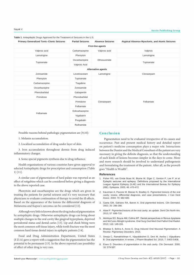
Health organizations of various countries have given approval to selected Antiepileptic drugs for prescription and consumption (Table 1) [11].
A similar case of pigmentation of hard palate was reported as an effect of retigabine which can be considered before giving a diagnosis to the above reported case.
Phenytoin and oxcarbazepine are the drugs which are given in treating the patients for partial seizures and it’s very necessary that physicians re-evaluate continuation of therapy to avoid the ill effects. Based on the appearance of the lesions the differential diagnosis of Melanoma and Kaposi’s sarcoma can be considered [12].
Although very little is known about the drug induced pigmentation by antiepileptic drugs. Otherwise antiepileptic drugs can bring about multiple changes in the oral cavity like gingival hyperplasia, deprived periodontal status and dental caries [13]. Lip and cheek biting were the most common soft tissue injury, while tooth fracture was the most common hard tissue dental injury in epileptic patients [14].
Food and Drug Administration Department, United States (F.D.A) gave a report with a suggestion that the pigmentation has the potential to be permanent [15]. In the above reported case possibility of effect of other drug is very rare.
Table 1: Antiepileptic Drugs Approved for the Treatment of Seizures in the U.S.
ConclusionPigmentation need to be evaluated irrespective of its causes and
occurrence. Past and present medical history and detailed report on patient’s medicine consumption plays a major role. Interactions between the Dentist and the Medical Consultant of the patient are very necessary in giving the definite diagnosis, so that the understanding of such kinds of lesions becomes simpler in the days to come. More and more research should be involved to understand pathogenesis and formulating the treatment of the patient. After all, as the proverb goes “Health is Wealth”.
References1. Fisher RS, van Emde Boas W, Blume W, Elger C, Genton P, Lee P, et al.
Epileptic seizures and epilepsy: Definitions proposed by the International League Against Epilepsy (ILAE) and the International Bureau for Epilepsy (IBE). Epilepsia. 2005; 46: 470-472.
2. Kauzman A, Pavone M, Blanas N, Bradley G. Pigmented lesions of the oral cavity: review, differential diagnosis, and case presentations. J Can Dent Assoc. 2004; 70: 682-683.
J Drug Discov Develop and Deliv 4(1): id1029 (2017) - Page - 03
Nayak V Austin Publishing Group
Submit your Manuscript | www.austinpublishinggroup.com
9. Margoob AA, Schoenbach SP, Kopf AW, Orlow SJ, Nossa R. Pathophysiology of drug induced melanin pigmentation & the risk of developing malignancy: a review of the world literature. J Invest Dermatol. 1995; 104: 563-565.
10. Pack GT, Davis J. Nevus giganticus pigmentosus with malignant transformation. Surgery. 1961; 49: 347-354.
11. Goldenberg MM. Overview of drugs used for epilepsy and seizures: etiology, diagnosis, and treatment. P T. 2010; 35: 392-441.
12. Beacher GN, Brodie MJ, Goodall C. A case report: retigabine induced oral mucosal dyspigmentation of the hard palate. BMC Oral Health. 2015; 15: 122.
13. Cornacchio AL, Burneo JG, Aragon CE. The effects of antiepileptic drugs on oral health. J Can Dent Assoc. 2011; 77: b140.
14. Ghafoor PA, Rafeeq M, Dubey A. Assessment of oral side effects of Antiepileptic drugs and traumatic oro-facial injuries encountered in Epileptic children. J Int Oral Health. 2014; 6: 126–128.
15. F.D.A. FDA Drug Safety Communication: Anti-seizure drug Potiga (ezogabine) linked to retinal abnormalities and blue skin discoloration. FDA. 2013.
Citation: Nayak V, Kini R, Rao PK, Bhandarkar GP, Kashyap RR, Shetty U, et al. Antiepileptic Drug Induced Pigmentation of Oral Mucosa - A Case Report and Review. J Drug Discov Develop and Deliv. 2017; 4(1): 1029.