Citation: Vengala N. Antihyperlipidemic Activity of Flax Lignan Concentrate in Nicotinamide-Streptozotocin Induced Diabetic
Hyperlipidemia Rats. Res Rev Biosci. 2017;12(2):120.
© 2017 Trade Science Inc.
1
Antihyperlipidemic Activity of Flax Lignan Concentrate in Nicotinamide-
Streptozotocin Induced Diabetic Hyperlipidemia Rats
Nikhila Vengala*
Department of Pharmacology, Poona College of Pharmacy, Bharati Vidyapeeth Deemed University, Pune, Maharashtra,
India
*Corresponding author: Nikhila Vengala, Department of Pharmacology, Poona College of Pharmacy, Bharati Vidyapeeth
Deemed University, Pune, Maharashtra, India, Tel: 9767843561; E-mail: [email protected]
Received: June 02, 2017; Accepted: June 27, 2017; Published: July 04, 2017
Abstract
Background: Flax seeds have been shown to have multiple benefits in cardiovascular disorders.
Objective: To evaluate Antihyperlipidemic activity of methanolic fraction of flax lignan concentrate (MF-FLC) obtained from Linum
usitatissium L., Linaceae in nicotinamide-STZ induced diabetic hyperlipidemia rats.
Methods: Male Wistar rats were used for the study. The IAEC of Poona college of Pharmacy approved the proposal. Protocol no.
CPCSEA/01/14. Diabetes was induced in rats by nicotinamide-STZ. Diabetic animals were divided into five groups: Diabetic
atherosclerosis control, Atorvastatin (10 mg/kg), MF-FLC (200 mg/kg), MF-FLC (400 mg/kg), and MF-FLC (800 mg/kg). After the
confirmation of diabetes, rats were treated with cholesterol (4% w/w) and cholic acid (1% w/w) for 45 days. After 15 days of
cholesterol treatment, atorvastatin and MF-FLC treatment was given to respective groups for 30 days. Non-diabetic atherosclerosis
group received cholesterol-cholic acid treatment alone. The control group was given coconut oil as vehicle.
Results: Treatment of MF-FLC (400 mg/kg) and atorvastatin (10 mg/kg) significantly reduced systolic blood pressure (p<0.01),
diastolic blood pressure (p<0.01), mean arterial blood pressure (p<0.001) and left ventricular end diastolic pressure (p<0.001) whereas
there was significant increase in Max dp/dt (p<0.001) and Min dp/dt (p<0.01) indicating the effect on cardiac contractility. These
findings were confirmed in histopathology. MF-FLC (400 mg/kg) showed significant (p<0.01) reduction in the glucose level. MF-FLC
(400 mg/kg) was found to be effective, increase in the dose (800 mg/kg) there showed a ceiling effect.
Conclusion: The treatment with MF-FLC showed significant increase in the both max and min dp/dt. Atorvastatin (10 mg/kg) also
showed significant increase in the both max and min dp/dt. This suggests that MF-FLC provides sufficient contractile reserve to
ameliorate the detrimental effects of diabetes on cardiac contractility. MF-FLC had additional benefit of anti-hyperglycemic effect
when compared to atorvastatin.
Keywords: Flax lignan concentrate; Flaxseed; Streptozotocin; Linum usitatissimum; Hyperlipidemia; Cholic acid
www.tsijournals.com | April-2017
2
Abbreviations
SBP: Systolic Blood Pressure;
DBP: Diastolic Blood Pressure;
MABP: Mean Arterial Blood Pressure;
LVEDP-Left: Ventricular End Diastolic Pressure;
MF-FLC: Methanolic Fraction of Flax Lignan Concentrate
Introduction
Diabetes mellitus is the name given to a group of disorders with different etiologies. It is characterized by disarrangements in
carbohydrates, proteins and fat metabolism caused by the complete or relative insufficiency of insulin secretion and/or insulin
action [1]. Diabetes mellitus is characterized by hyperglycemia, hypercholesterolemia and hypertriglyceridemia resulting
from defects in insulin secretion followed by dysfunction and failure of organs especially the eyes, kidneys, nerves, heart and
arteries. Type 2 diabetes mellitus is a well-recognized risk factor for atherosclerotic and cardiovascular disease (CVD) that
confers a markedly increased risk of coronary heart disease (CHD) and cardiovascular mortality when compared to non-
diabetic patients [2].
Atherosclerosis is a cardiovascular disease characterized by the deposition of lipids in the artery wall and the infiltration of
inflammatory cells, such as monocytes and lymphocytes. Hyperlipidemia is a major risk factor for the premature
development of atherosclerosis and it has been shown to increase the incidence of myocardial ischemia and cardiac events
[3]. Patients with diabetes are at a higher risk for developing atherosclerosis than non-diabetic subjects and have enhanced
susceptibility for myocardial infarction, peripheral artery disease and stroke. Diabetes induces phenotypic changes in vascular
smooth muscle cells (SMC) such as increased contractility, cellular proliferation, migration and extracellular matrix
formation, which may lead to accelerated SMC accumulation in atherosclerotic lesions. The cellular and molecular
mechanisms by which diabetes accelerates atherosclerotic cardiovascular disease are still poorly understood [4].
Linum usitatissimum commonly called as flaxseed or linseed is brown or yellow colored seed harvested from the blue flowers
of flax crop. It has been used as food in India and around the world for a long time. Flaxseed mainly contains omega-3 fatty
acid, α-linolenic acid, dietary fiber and secoisolariciresinol diglucoside (SDG) lignan (Bassett et al., 2009). Flaxseed contains
ten to hundred times more lignan than most other edible plants seeds. It is reported that flaxseed also contains other lignans
like matairesinol, lariciresinol, hinokinin, arctigenin, pinoresinol and demethoxy secoisolariciresinol in small quantity with
several phenolic acid compounds [5,6]. Traditionally, flaxseed has been grown for its oil.
Flaxseed has been taking part in a serious role within the field of diet and disease research because of its potential health
benefits as it is associated to high content of α-linolenic acid (ALA) (57%), that is a necessary omega-3-fatty acid and
additionally as a result of a high lignan, specifically secoisolariciresinol diglucoside (SDG). linseed is an oilseed that contains
regarding 38%-45% oil. ALA content in flax oil is 55%-60% and lignan content in linseed is up to 13 mg/g flaxseed [7-9].
The lignan SDG is converted by bacteria in the colon of humans and other animals to enterodiol and enterolactone, referred
to as mammalian lignans. These exhibit weak estrogenic activity as they can bind to estrogen receptors on cell membranes
[10]. The interest in ALA and lignans has opened opportunities. Flaxseed is also a good source of protein and dietary fibre,
www.tsijournals.com | April-2017
3
accounting for 20% and 28% of the flaxseed, respectively. Many dietary antioxidants and some non-nutrient based
antioxidants from plants, such as sulphur containing compounds in garlic, phyto-oestrogens in soy, green tea, anthocyanins in
red berries, lycopene in tomatoes, red and white wines from grape seeds are increasingly being recognized as potential health
promoters in reducing the risk of cardiovascular disease and atherosclerosis. Flaxseed has gained much importance in recent
times as ethnomedicine due to its wide pharmacological actions [11,12].
Materials and Methods
Experimental research protocol
Male Wistar rats weighing (220 g-250 g) were purchased from National Institute of Biosciences, Pune, India. The animals
were housed at ambient temperature 25 ± 1°C and relative humidity of 45-55% under 12 h day/night conditions during entire
experimental period. Food pellets were purchased from Nav Maharashtra Chakan oil mills Ltd., Sangli) and water ad libitum.
The research proposal was prepared according to the guidelines of the Committee for the Purpose of Control and Supervision
of Experiments of Animals (CPCSEA). The Institutional Animal Ethical Committee (IAEC) of Poona College of Pharmacy
approved the proposal. Protocol no. : CPCSEA/01/14.
Drugs and chemicals
Atorvastatin (Aurobindo Pharma Ltd), Streptozotocin, Nicotinamide (Sigma-Aldrich Corporation, USA), Cholesterol, Cholic
acid, Urethane, (HiMedia Laboratories, Mumbai, India), Anesthetic ether (Narson Pharma, Mumbai), Heparin (Gland Pharma
Ltd., Mumbai) were used for study.
Collection and authentication of plant seeds
Seeds of Linum usitatissimum were obtained from Dr. P. B. Ghorpade, Principal Scientist, Punjabrao Deshmukh Krushi
Vidyapeeth, College of Agriculture, and Nagpur, India. After the authentication of seeds, a voucher specimen was deposited
at the institute. Flaxseeds were stored in cold room before processing. Flaxseeds were processed for oil extraction at our
Indian Council of Agricultural Research under National Agriculture Innovation Project, Omega-3-oil unit, Maharashtra,
India.
Preparation of methanolic fraction of flax lignan concentrates (MF-FLC)
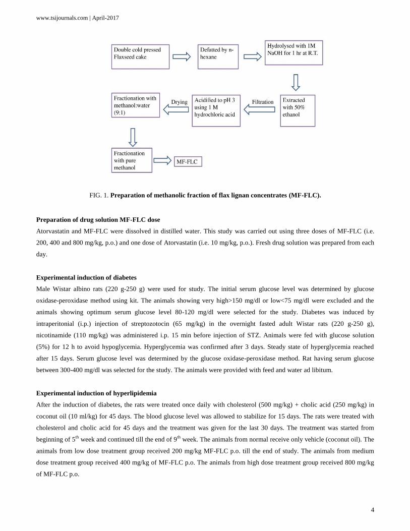
Preparation of flax lignan concentrate was carried out as described previously [13]. Flaxseed cake was defatted by n-hexane
and then hydrolyzed with 1 M aqueous sodium hydroxide for 1 h at room temperature with intermittent shaking followed by
extraction with 50% ethanol. The filtrate was acidified to pH 3 using 1 M hydrochloric acid. The filtrate was dried using rota
vac at 50°C. The dry powder of hydro alcoholic extract was labeled as flax lignan concentrate (FLC). Further enrichment of
active constituent by solvent-solvent fractionation on FLC was carried out as followed. FLC was subjected for solvent-
solvent fractionation. 25 g of FLC was fractionated with Methanol: Water (9:1). Further resultant fraction was again
fractionated with pure methanol to get lignan rich concentrate called MF-FLC (FIG. 1). The yield of MF-FLC was 15% w/w.
The powdered MF-FLC was dissolved in distilled water to prepare the different concentrations of MF-FLC and used for
pharmacological studies.
www.tsijournals.com | April-2017
4
FIG. 1. Preparation of methanolic fraction of flax lignan concentrates (MF-FLC).
Preparation of drug solution MF-FLC dose
Atorvastatin and MF-FLC were dissolved in distilled water. This study was carried out using three doses of MF-FLC (i.e.
200, 400 and 800 mg/kg, p.o.) and one dose of Atorvastatin (i.e. 10 mg/kg, p.o.). Fresh drug solution was prepared from each
day.
Experimental induction of diabetes
Male Wistar albino rats (220 g-250 g) were used for study. The initial serum glucose level was determined by glucose
oxidase-peroxidase method using kit. The animals showing very high>150 mg/dl or low<75 mg/dl were excluded and the
animals showing optimum serum glucose level 80-120 mg/dl were selected for the study. Diabetes was induced by
intraperitonial (i.p.) injection of streptozotocin (65 mg/kg) in the overnight fasted adult Wistar rats (220 g-250 g),
nicotinamide (110 mg/kg) was administered i.p. 15 min before injection of STZ. Animals were fed with glucose solution
(5%) for 12 h to avoid hypoglycemia. Hyperglycemia was confirmed after 3 days. Steady state of hyperglycemia reached
after 15 days. Serum glucose level was determined by the glucose oxidase-peroxidase method. Rat having serum glucose
between 300-400 mg/dl was selected for the study. The animals were provided with feed and water ad libitum.
Experimental induction of hyperlipidemia
After the induction of diabetes, the rats were treated once daily with cholesterol (500 mg/kg) + cholic acid (250 mg/kg) in
coconut oil (10 ml/kg) for 45 days. The blood glucose level was allowed to stabilize for 15 days. The rats were treated with
cholesterol and cholic acid for 45 days and the treatment was given for the last 30 days. The treatment was started from
beginning of 5th
week and continued till the end of 9th
week. The animals from normal receive only vehicle (coconut oil). The
animals from low dose treatment group received 200 mg/kg MF-FLC p.o. till the end of study. The animals from medium
dose treatment group received 400 mg/kg of MF-FLC p.o. The animals from high dose treatment group received 800 mg/kg
of MF-FLC p.o.
www.tsijournals.com | April-2017
5
Experimental design
The rats were randomly divided into seven groups, each containing six rats:
Group I: Vehicle control
Group II: Diabetic atherosclerosis control group
Group III: Non-diabetic atherosclerosis group
Group IV: Atorvastatin treated group (10 mg/kg)
Group V: MF-FLC treated group (low dose i.e. 200 mg/kg)
Group VI: MF-FLC treated group (medium dose i.e. 400 mg/kg)
Group VII: MF-FLC treated group (high dose i.e. 800 mg/kg)
MF-FLC and Atorvastatin were and administered to the rats orally using an oral feeding needle daily for nine consecutive
weeks. The vehicle control rats received vehicle as coconut oil.
Assessment of Hemodynamic Changes
On last day of study, animals were anaesthetized by urethane (1.25 gm/kg) i.p. The right carotid artery of each rat was
cannulated (FIG. 2) for the measurement of systolic blood pressure (SBP), diastolic blood pressure (DBP), mean arterial
blood pressure (MABP). The cannula was filled with heparinized saline and connected to pressure transducer to record all the
parameters mentioned above. After 30 minutes of stabilization hemodynamic parameters were recorded by eight channel
recorder Power lab (AD Instruments, UK) having LABCHART-6 pro software. Followed by their where insertion of Millar
catheter (AD Instruments, UK) having LABCHART-6 pro software, to record the left ventricular function such as left
ventricular end diastolic pressure (LVEDP), dp/dt Max, dp/dt Min respectively.
FIG. 2. Cannulation of carotid artery for measurement of hemodynamic parameters.
Results
Effect of MF-FLC and atorvastatin on serum glucose (mg/dl) by experimentally induced atherosclerosis in diabetic
male Wistar rats.
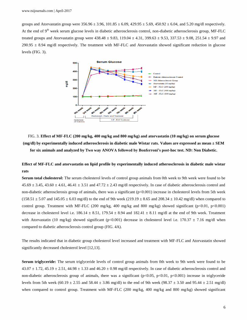
The plasma glucose levels on the 0th
day in control, diabetic atherosclerosis control, non-diabetic atherosclerosis, MF-FLC
treated groups (200 mg/kg, 400 mg/kg and 800 mg/kg) and Atorvastatin group were 99.74 ± 5.95, 109.75 ± 4.86, 102.89 ±
3.95, 109.34 ± 3.58, 105.03 ± 4.56, 107.34 ± 3.59 and 104.45 ± 3.34 mg/dl respectively. After 3 weeks of induction of
diabetes the serum glucose levels in diabetic atherosclerosis control, non-diabetic atherosclerosis group, MF-FLC treated
www.tsijournals.com | April-2017
6
groups and Atorvastatin group were 356.96 ± 3.96, 101.85 ± 6.09, 429.95 ± 5.69, 450.92 ± 6.04, and 5.20 mg/dl respectively.
At the end of 9th
week serum glucose levels in diabetic atherosclerosis control, non-diabetic atherosclerosis group, MF-FLC
treated groups and Atorvastatin group were 438.48 ± 9.83, 119.04 ± 4.31, 399.63 ± 9.53, 337.53 ± 9.08, 251.54 ± 9.97 and
290.95 ± 8.94 mg/dl respectively. The treatment with MF-FLC and Atorvastatin showed significant reduction in glucose
levels (FIG. 3).
FIG. 3. Effect of MF-FLC (200 mg/kg, 400 mg/kg and 800 mg/kg) and atorvastatin (10 mg/kg) on serum glucose
(mg/dl) by experimentally induced atherosclerosis in diabetic male Wistar rats. Values are expressed as mean ± SEM
for six animals and analyzed by Two way ANOVA followed by Bonferroni’s post-hoc test. ND: Non Diabetic.
Effect of MF-FLC and atorvastatin on lipid profile by experimentally induced atherosclerosis in diabetic male wistar
rats
Serum total cholesterol: The serum cholesterol levels of control group animals from 0th week to 9th week were found to be
45.69 ± 3.45, 43.60 ± 4.61, 46.41 ± 3.51 and 47.72 ± 2.43 mg/dl respectively. In case of diabetic atherosclerosis control and
non-diabetic atherosclerosis group of animals, there was a significant (p<0.001) increase in cholesterol levels from 5th week
(158.51 ± 5.07 and 145.05 ± 6.03 mg/dl) to the end of 9th week (219.19 ± 8.65 and 208.34 ± 10.42 mg/dl) when compared to
control group. Treatment with MF-FLC (200 mg/kg, 400 mg/kg and 800 mg/kg) showed significant (p<0.01, p<0.001)
decrease in cholesterol level i.e. 186.14 ± 8.51, 179.54 ± 8.94 and 182.41 ± 8.11 mg/dl at the end of 9th week. Treatment
with Atorvastatin (10 mg/kg) showed significant (p<0.001) decrease in cholesterol level i.e. 170.37 ± 7.16 mg/dl when
compared to diabetic atherosclerosis control group (FIG. 4A).
The results indicated that in diabetic group cholesterol level increased and treatment with MF-FLC and Atorvastatin showed
significantly decreased cholesterol level [12,13].
Serum triglyceride: The serum triglyceride levels of control group animals from 0th week to 9th week were found to be
43.07 ± 1.72, 45.19 ± 2.51, 44.98 ± 1.33 and 46.20 ± 0.98 mg/dl respectively. In case of diabetic atherosclerosis control and
non-diabetic atherosclerosis group of animals, there was a significant (p<0.05, p<0.01, p<0.001) increase in triglyceride
levels from 5th week (60.19 ± 2.55 and 58.44 ± 3.86 mg/dl) to the end of 9th week (98.37 ± 3.50 and 95.44 ± 2.51 mg/dl)
when compared to control group. Treatment with MF-FLC (200 mg/kg, 400 mg/kg and 800 mg/kg) showed significant
www.tsijournals.com | April-2017
7
(p<0.05, p<0.01, p<0.001) decrease in triglyceride level i.e. 84.15 ± 2.99, 79.32 ± 2.66 and 81. 97 ± 2.55 mg/dl at the end of
9th week. Treatment with Atorvastatin (10 mg/kg) showed significant (p<0.01, p<0.001) decrease in triglyceride level i.e.
81.59 ± 4.63 and 78.98 ± 3.13 mg/dl when compared to diabetic group (FIG 4B).
The results indicated that in diabetic group triglyceride level increased and treatment with MF-FLC and Atorvastatin showed
significantly decreased triglyceride level.
High density lipoproteins: The HDL level of normal control group animals was found to be 23.39 ± 0.46 mg/dl. In case of
diabetic atherosclerosis control and non-diabetic atherosclerosis group of animals, HDL levels were 17.16 ± 0.81 mg/dl and
17.09 ± 0.63 mg/dl which showed significant (p<0.001) decrease in HDL level when compared to control group. Treatment
with MF-FLC (200 mg/kg, 400 mg/kg and 800 mg/kg) and Atorvastatin (10 mg/kg) showed significant (p<0.001, p<0.01)
increase i.e. 21.42 ± 0.81 mg/dl, 22.24 ± 0.77 mg/dl, 21.98 ± 0.70 mg/dl and 22.68 ± 0.96 mg/dl, in HDL level when
compared to diabetic atherosclerosis control group (FIG. 4C).
The results indicated that in diabetic atherosclerosis control and group HDL level increased and treatment with MF-FLC and
Atorvastatin showed significantly decreased HDL level [14-16].
Low density lipoproteins: The LDL level of normal control group animals was found to be 38.06 ± 3.04 mg/dl. In case of
diabetic atherosclerosis control and non-diabetic atherosclerosis group of animals, LDL levels were 58.08 ± 2.36 mg/dl and
56.84 ± 2.33 mg/dl which showed significant (p<0.001) increase in LDL level when compared to control group. Treatment
with MF-FLC (200 mg/kg, 400 mg/kg and 800 mg/kg) and Atorvastatin (10 mg/kg) showed significant (p<0.001, p<0.01,
p<0.05) decrease i.e. 42.52 ± 2.09 mg/dl, 39.61 ± 2.98 mg/dl, 20.29 ± 2.29 mg/dl and 35.79 ± 2.13 mg/dl, in LDL level when
compared to diabetic atherosclerosis control group (FIG. 4D).
The results indicated that in diabetic atherosclerosis control and group LDL level increased and treatment with MF-FLC and
Atorvastatin showed significantly decreased LDL level [17].
Very low density lipoproteins: The VLDL level of normal control group animals was found to be 9.24 ± 0.95 mg/dl. In case
of diabetic atherosclerosis control and non-diabetic atherosclerosis group of animals, VLDL levels were 22.81 ± 1.34 mg/dl
and 22.45 ± 1.29 mg/dl which showed significant (p<0.001) increase in VLDL level when compared to control group.
Treatment with MF-FLC (200 mg/kg, 400 mg/kg and 800 mg/kg) and Atorvastatin (10 mg/kg) showed significant (p<0.001,
p<0.01, p<0.05) decrease i.e. 17.83 ± 0.91, 15.86 ± 0.72, 16.45 ± 0.83 and 14.79 ± 0.75 mg/dl in VLDL level when compared
to diabetic atherosclerosis control group (FIG. 4E).
The results indicated that in diabetic atherosclerosis control and group VLDL level increased and treatment with MF-FLC
and Atorvastatin showed significantly decreased VLDL level.
www.tsijournals.com | April-2017
8
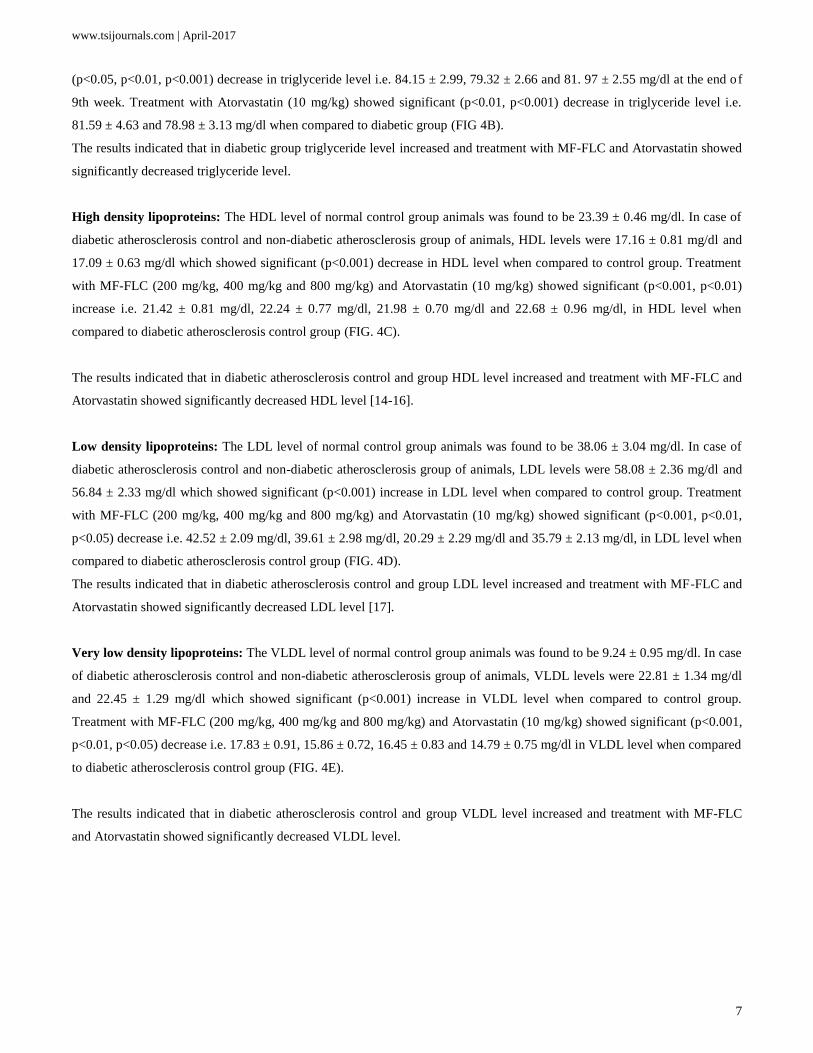
FIG. 4. Effect of MF-FLC and Atorvastatin on lipid profile by experimentally induced atherosclerosis in diabetic male
Wistar rats. Values are expressed as mean ± SEM for six animals and analyzed by one way ANOVA followed by
Bonferroni’s post-hoc test. ###p<0.001 when compared to control animals. ***p<0.001, **p<0.01 when compared to
diabetic atherosclerosis control. ND: Non Diabetic.
Effect of MF-FLC and Atorvastatin on hemodynamic parameters by experimentally induced atherosclerosis in
diabetic male Wistar rats
Systolic blood pressure (SBP): In normal control group of animals it was found to be 108.24 ± 1.48 mmHg. In case of
diabetic atherosclerosis control and non-diabetic atherosclerosis group of animals, SBP was 129.77 ± 2.13 mmHg and 128.14
± 1.47 mmHg which showed significant (p<0.001) increase in SBP (mmHg) when compared to control group. Treatment
with MF-FLC (200 mg/kg, 400 mg/kg and 800 mg/kg) showed significant decrease (p<0.01, p<0.05) in SBP i.e. 117.81 ±
2.31, 113.87 ± 3.18 and 115.98 ± 1.05 mmHg. Treatment with Atorvastatin (10 mg/kg) also showed significant (p<0.01)
increase in SBP i.e. 111.64 ± 2.61 mmHg when compared to diabetic atherosclerosis control group (FIG. 5A).
The results indicated that in diabetic atherosclerosis control group systolic blood pressure (SBP) increased and treatment with
MF-FLC and Atorvastatin showed significant decrease in systolic blood pressure.
Diastolic blood pressure (DBP): In normal control group of animals it was found to be 89.57 ± 2.01 mmHg. In case of
diabetic atherosclerosis control and non-diabetic atherosclerosis group of animals, DBP was 119.44 ± 1.81 mmHg and 118.14
± 2.31 mmHg which showed significant (p<0.001) increase in DBP (mmHg) when compared to control group. Treatment
with MF-FLC (200 mg/kg, 400 mg/kg and 800 mg/kg) showed significant decrease (p<0.01, p<0.05) in DBP i.e. 105.64 ±
2.53, 102.44 ± 2.32 and 103.57± 2.42 mmHg. Treatment with Atorvastatin (10 mg/kg) also showed significant (p<0.01)
increase in DBP i.e. 100.46 ± 2.83 mmHg when compared to diabetic atherosclerosis control group (FIG. 5B).
The results indicated that in diabetic atherosclerosis control group Diastolic blood pressure (DBP) increased and treatment
with MF-FLC and Atorvastatin showed significant decrease in diastolic blood pressure.
Mean Arterial Blood Pressure: The MABP in control group of animals was found to be 95.06 ± 3.04 mmHg. In case of
diabetic atherosclerosis control and non-diabetic atherosclerosis group of animals, MABP was 134.61 ± 2.46 mmHg and
126.41 ± 3.46 mmHg which showed significant (p<0.001) increase when compared to control group. On the other hand
treatment with MF-FLC (400 mg/kg and 800 mg/kg) and Atorvastatin (10 mg/kg) showed significant (p<0.01) decrease in
www.tsijournals.com | April-2017
9
MABP i.e. 114.06 ± 2.95 mmHg, 117.35 ± 3.44 mmHg and 112.48 ± 2.41 mmHg when compared to diabetic atherosclerosis
control group. Whereas MF – FLC (200 mg/kg) showed non-significant decrease i.e. 120.49 ± 4.06 mmHg (FIG. 5C).
The results indicated that in diabetic atherosclerosis control group MABP increased and treatment with MF-FLC (400 mg/kg
and 800 mg/kg) and Atorvastatin (10 mg/kg) significantly decreased and MF-FLC (200 mg/kg) non-significantly decreased
the MABP as compared to that of diabetic atherosclerosis control group.
Left ventricular end diastolic pressure: The LVEDP in control group of animals found to be 6.92 ± 0.49 mmHg. In case of
diabetic atherosclerosis control and non-diabetic atherosclerosis group of animals, LVEDP was 10.77 ± 0.36 mmHg and 9.85
± 0.47 mmHg which showed significant (p<0.001, p<0.01) increase in LVEDP (mmHg) when compared to control group.
Treatment with MF-FLC (200 mg/kg, 400 mg/kg and 800 mg/kg) showed significant decrease (p<0.001, p<0.01, p<0.05) in
LVEDP i.e. 8.57 ± 0.46, 7.99 ± 0.35 and 8.25 ± 0.37 mmHg. Treatment with Atorvastatin (10 mg/kg) also showed significant
(p<0.001) increase in LVEDP i.e. 7.74 ± 0.36 mmHg when compared to diabetic atherosclerosis control group (FIG. 5D).
The results indicated that in diabetic atherosclerosis control group LVEDP increased and treatment with MF-FLC and
Atorvastatin showed significant decrease in end diastolic blood pressure.
Max dp/dt: The Max dp/dt in control group of animals found to be 5537.08 ± 100.19 mmHg. In case of diabetic
atherosclerosis control and non-diabetic atherosclerosis group of animals, Max dp/dt was 2925.27 ± 90.01 mmHg and
2891.93 ± 86.69 mmHg which showed significant (p<0.001) decrease in Max dp/dt (mmHg) when compared to control
group. Treatment with MF-FLC (200 mg/kg, 400 mg/kg and 800 mg/kg) showed significant increase (p<0.001, p<0.01) in
Max dp/dt i.e. 3587.42 ± 127.94, 3665.41 ± 135.42 and 3599.10 ± 119.45 mmHg. Treatment with Atorvastatin (10 mg/kg)
also showed significant (p<0.001) decrease in Max dp/dt i.e. 3720.42 ± 113.16 mmHg when compared to diabetic
atherosclerosis control group (FIG. 5E).
The results indicated that in diabetic atherosclerosis control group Max dp/dt decrease and treatment with MF-FLC and
Atorvastatin showed significant increase in diastolic blood pressure.
Min dp/dt: The Min dp/dt in control group of animals was found to be-2916.67 ± 114.19 mmHg. In case of diabetic
atherosclerosis control and non-diabetic atherosclerosis group of animals, Min dp/dt was 2095.27 ± 119.03 mmHg and-
2067.64 ± 74.76 mmHg which showed significant (p<0.001) decrease when compared to control group. On the other hand
treatment with MF-FLC (400 mg/kg and 800 mg/kg) and Atorvastatin (10 mg/kg) showed significant (p<0.01, p<0.05)
increase in Min dp/dt i.e.-2717.44 ± 144.35 mmHg,-2697.42 ± 113.94 mmHg and-2806.59 ± 171.88 mmHg when compared
to diabetic atherosclerosis control group. Whereas MF – FLC (200 mg/kg) showed non-significant increase i.e.-2378.03 ±
105.45 mmHg (FIG. 5F).
The results indicated that in diabetic atherosclerosis control group Min dp/dt decreased and treatment with MF-FLC (400
mg/kg and 800 mg/kg) and Atorvastatin (10 mg/kg) significantly increased and MF – FLC (200 mg/kg) non-significantly
increased the Min dp/dt as compared to that of diabetic atherosclerosis control group [18-22].
www.tsijournals.com | April-2017
10
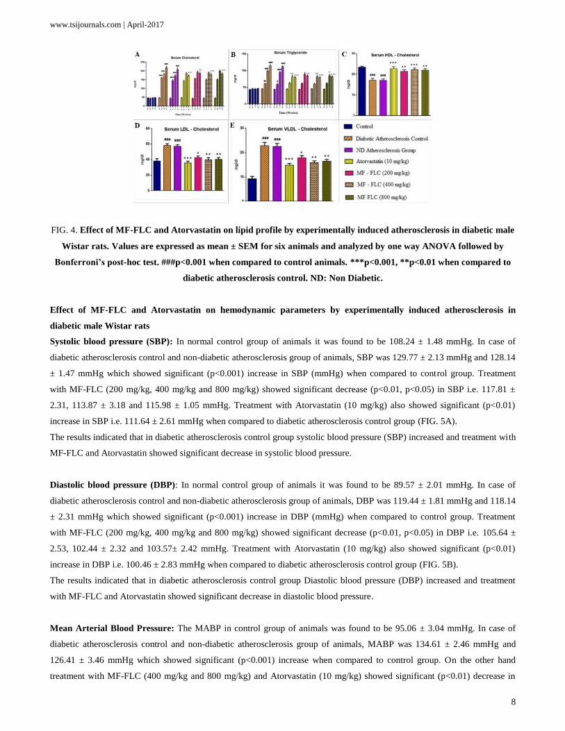
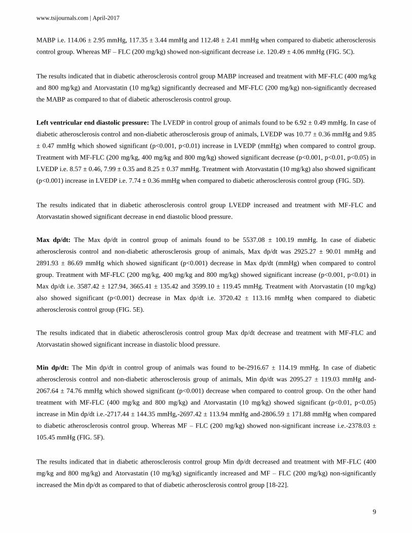
FIG. 5. Effect of MF-FLC and Atorvastatin on hemodynamic parameters by experimentally induced atherosclerosis in
diabetic male Wistar rats. Values are expressed as mean ± SEM for six animals and analyzed by One way ANOVA
followed by Bonferroni’s post – hoc test. ###p<0.001 when compared to control animals. ***p<0.001, **p<0.01 when
compared to Diabetic Atherosclerosis control. ND: Non Diabetic.
Effect of MF-FLC and atorvastatin histopathology of heart by experimentally induced atherosclerosis in diabetic male
Wistar rats
Histopathological observations of the heart from control group revealed normal architecture with normal myocardial fibers
and muscle bundles (FIG. 6A). The hearts from the diabetic atherosclerotic animals (Group II) showed moderate
degenerating cardiomyocytes, the muscle fibers were disrupted and degenerated and there was infiltration of inflammatory
cells (FIG. 6B). In case of atherosclerotic group, heart showed minimal hypertrophy, mild vacuolations and degenerating
cardiomyocytes (FIG. 6C). Treatment of atorvastatin reduced the muscle damage and there was minimal hyperplasia (FIG.
6D). Treatment of the test drug at low and high doses did not showed much effect on the heart sections, there was moderate
to mild degeneration in cardiomyocytes and the muscle fibers were disrupted (FIG. 6E and FIG. 6G). Treatment of test drug
at the medium dose showed minimal areas of damage and there was tendency towards recovery (FIG. 6F).
www.tsijournals.com | April-2017
11
FIG. 6. Effect of MF-FLC and Atorvastatin on heart histopathology by experimentally induced atherosclerosis in
diabetic male Wistar rats. (A) Control (Group-I): nothing abnormal detected. (B) Diabetic atherosclerosis control
(Group-II) showed moderate degenerating cardiomyocytes and infiltration of inflammatory cells. (C) ND
atherosclerosis (Group-III) showed mild vacuolations and degenerating cardiomyocytes. (D) Atorvastatin (10 mg/kg)
(Group-IV) showed Minimal degenerating cardiomyocytes. (E) MF-FLC (200 mg/kg) (Group V) showed Moderate
degeneration in cardiomyocytes. (F) MF-FLC (400 mg/kg (Group VI) Minimal vacuolations in and degeneration of
cardiomyocytes. (G) MF-FLC (800 mg/kg (Group VII) Mild vacuolations in and degeneration of cardiomyocytes.
Discussion
In the earlier study, Zanwar et al reported that there was a reduction in the blood glucose level by MF-FLC in alloxan
induced diabetes in rats. Similar findings were observed in nicotinamide-streptozotocin induced diabetes with hyperlipidemic
rats. In the present study, diabetes was induced by injecting nicotinamide-streptozotocin intraperitoneally. Streptozotocin is
commonly used for experimental induction of type 1 diabetes mellitus which results in rapid reduction in pancreatic islet
pyridine nucleotide concentration and causes subsequent β-cell necrosis. Nicotinamide was given 15 minutes before STZ
administration causes 40% decrease in pancreatic β-cell mass which resembles human type 2 diabetes. In the present study, it
was observed that only MF-FLC in a dose dependent manner reduced blood glucose level. The anti-hyperglycemic effect was
not shown by atorvastatin; on the other hand non-significant increase in the blood glucose level was obtained.
Currently there is a controversy regarding the role of statins, [23] had reported hyperglycemic effect of statins. Similar
findings were observed in [24], in their review of five trials (HPS [simvastatin, 40 mg], LIPID [pravastatin, 40 mg], ASCOT
[Atorvastatin 10 mg], CORONA [rosuvastatin 10 mg] and JUPITER [rosuvastatin, 20 mg]) involving 51,619 participants
among whom 1943 developed diabetes. They concluded that this small but significant increase may related to statin use but
was not drug or dose specific. Treatment with MF-FLC appears to be beneficial as the extract was effective in reducing the
blood glucose level. In this respect MF-FLC differs significantly compared to Atorvastatin. Administration of 4% cholesterol
www.tsijournals.com | April-2017
12
(w/w) and 1% cholic acid (w/w) for 45 days induced hyperlipidemic in rats. Hyperlipidemia was confirmed by measuring
total cholesterol and triglyceride in the serum samples at the end of 9th week.
Diabetes induces both systolic and diastolic dysfunction. Vehicle control diabetic atherosclerosis animals showed reduced
SBP, DBP MABP and LVSP when compared with normal group of animals. This decrease might be due to the myocardial
necrosis or hypertrophy. In the present study MF-FLC (400 mg/kg) and atorvastatin (10 mg/kg) treated animals showed
significant increase in SBP, DBP MABP and LVSP indicating improved cardiac function.
The max dp/dt and min dp/dt are the maximum and minimum rates of pressure development during contraction and
relaxation. In present study diabetic atherosclerosis control rats showed decrease in both max and min dp/dt. These findings
were in accordance with previous findings of Zanwar et al., 2012. The treatment with MF-FLC showed significant increase in
the both max and min dp/dt. Atorvastatin (10 mg/kg) also showed significant increase in the both max and min dp/dt. This
suggests that MF-FLC provides sufficient contractile reserve to ameliorate the detrimental effects of diabetes on cardiac
contractility.
Minimal hypertrophy, mild vacuolations, degenerating cardiomyocytes, severe vacuolation, distorted architecture,
hypertrophy of hepatocytes, necrosis, severe tubular degeneration and degeneration of glomeruli and infiltration of
inflammatory cells were observed in the histopathology signifying the anti-hyperlipidemic, hepatoprotective and anti-
inflammatory effect of MF-FLC adds to the accumulating evidence for therapeutic effect of the extract.
Conclusion
The results indicate that MF-FLC possesses therapeutic property in restoring the diabetes induced insults.
Rats treated with MF-FLC (200 mg/kg, 400 mg/kg and 800 mg/kg) showed significant reduction in glucose levels.
This indicates anti-diabetic activity of MF-FLC. Atorvastatin (10 mg/kg) also showed non-significant increase in blood
glucose level. MF-FLC offers advantage over Atorvastatin in controlling the blood glucose levels.
In the present study MF-FLC (400 mg/kg) and atorvastatin (10 mg/kg) treated animals showed significant increase
in SBP, DBP MABP and LVSP indicating improved cardiac function.
The treatment with MF-FLC showed significant increase in the both max and min dp/dt atorvastatin (10 mg/kg) also
showed significant increase in the both max and min dp/dt. This suggests that MF-FLC provides sufficient contractile reserve
to ameliorate the detrimental effects of diabetes on cardiac contractility.
REFERENCES
1. Srinivasan, Subramani, Pari L. Antihyperlipidemic effect of diosmin: A citrus flavonoid on lipid metabolism in
experimental diabetic rats. J Funct Foods. 2013;5:484-92.
2. Fatima SS, Rajasekhar MD, Kumar KV, et al. Antidiabetic and antihyperlipidemic activity of ethyl acetate:
Isopropanol (1: 1) fraction of Vernonia anthelmintica seeds in Streptozotocin induced diabetic rats. Food Chem
Toxicol. 2010;48:495-501.
www.tsijournals.com | April-2017
13
3. Thiruchenduran M, Vijayan NA, Sawaminathan JK, et al. Protective effect of grape seed proanthocyanidins against
cholesterol cholic acid diet-induced hypercholesterolemia in rats. Cardiovasc Pathol. 2011;20:361-68.
4. Fledderus JO, van Oostrom O, de Kleijn DP, et al. Increased amount of bone marrow-derived smooth muscle-like
cells and accelerated atherosclerosis in diabetic apoE-deficient mice. Atherosclerosis. 2013;226:341-47.
5. Prasad K, Mantha SV, Muir AD, et al. Reduction of hypercholesterolemic atherosclerosis by CDC-flaxseed with
very low alpha-linolenic acid. Atherosclerosis. 1998;136:367-75.
6. Kamal-Eldin A, Peerlkamp N, Johnsson P, et al. An oligomer from flaxseed composed of
secoisolariciresinoldiglucoside and 3-hydroxy-3-methyl glutaric acid residues. Phytochemistry. 2001;58:587-90.
7. Hall C, Tulbek MC, Xu TY. Advances in food and nutrition research, in Flaxseed. 2006, Elsevier Inc, Amsterdam,
Netherlands, 2-3.
8. Nesbitt PD, Lam Y, Thompson LU. Human metabolism of mammalian lignan precursors in raw and processed
flaxseed. Am J Clin Nutr. 1999;69:549-55.
9. Ames BN, Shigenaga MK, Hagen TM. Oxidants, antioxidants, and the degenerative diseases of aging
(cancer/mutation/endogenous DNA adducts/oxygen radicals). Proc Natl Acad Sci USA. 1993;90:7915-22.
10. Prasad K, Mantha SV, Muir AD, et al. Protective effect of secoisolariciresinol diglucoside against streptozotocin-
induced diabetes and its mechanism. Mol. Cell Biochem. 2006;206:141-49.
11. Raygude KS, Kandhare AD, Ghosh P, et al. Anticonvulsant effect of fisetin by modulation of endogenous
biomarkers. Biomed Prev Nutr. 2012a;2: 215-22.
12. Zanwar A, Hegde MV, Bodhankar SL. Ethanolic extract of seeds of Linum ussitattimum (Flax lignan concentrate)
prevents doxorubicin-induced cardiotoxicity in rats. Atherosclerosis. 2011. Suppl. 12:146.
13. Zanwar AA, Hegde MV, Bodhankar SL. Protective role of concomitant administration of flax lignan concentrate
and omega-3-fatty acid on myocardial damage in doxorubicin-induced cardiotoxicity. Food Sci. Hum. Wellness.
2013;2:29-38.
14. Ghule AE, Jadhav SS, Bodhankar SL. Renoprotective effect of Linum usitatissimum seeds through haemodynamic
changes and conservation of antioxidant enzymes in renal ischaemia-reperfusion injury in rats. Arab J Urol. 2011;9:
215-21.
15. Ghule AE, Jadhav SS, Bodhankar SL. Effect of ethanolic extract of seeds of Linum usitatissimum (Linn.) on
hemodynamic changes and left ventricular function in renal artery occluded renovascular hypertension in rats.
Pharmacologia. 2012;3:283-90.
16. Ghule AE, Kandhare AD, Jadhav SS, et al. Omega-3-fatty acid adds to the protective effect of flax lignan
concentrate in pressure overload-induced myocardial hypertrophy in rats via modulation of oxidative stress and
apoptosis. Int Immunopharmacol. 2015;28:751-63.
17. Prasad K. Dietary flax seed in prevention of hypercholesterolemic atherosclerosis. Atherosclerosis. 1997;132: 69-76.
18. Chen J, Stavro PM, Thompson LU. Dietary flaxseed inhibits human breast cancer growth and metastasis and
downregulates expression of insulin-like growth factor and epidermal growth factor receptor. Nutr Cancer. 2002;43:
187-92.
19. Collins TF, Sprando RL, Black TN, et al. Effects of flaxseed and defatted flaxseed meal on reproduction and
development in rats. Food Chem Toxicol. 2003;41:819-34.
www.tsijournals.com | April-2017
14
20. Kinniry P, Amrani Y, Vachani A, et al. Dietary flaxseed supplementation ameliorates inflammation and oxidative
tissue damage in experimental models of acute lung injury in mice. J Nutr. 2006;136:1545-51.
21. Rajesha J, Murthy KN, Kumar MK, et al. Antioxidant potentials of flaxseed by in vivo model. J Agric Food Chem.
2006;54:3794-99.
22. Zanwar AA, Hegde M, Bodhankar SL. In vitro antioxidant activity of ethanolic extract of Linum usitatissimum.
Pharmacol Online. 2010;1:683-96.
23. Sattar N, Taskinen MR. Statins are diabetogenic–Myth or reality? Atheroscler Suppl. 2012;13:1-10.
24. Rajpathak SN, Kumbhani DJ, Crandall J, et al. Statin therapy and risk of developing type 2 diabetes: a meta-
analysis. Diabetes care. 2009;32:1924-29.