Zbl. Bakt. Abt. II, Bd. 131, S. 46-52 (1976) [National Research Centre, Soil Microbiology Unit, Dokki, Cairo, A.R.E.] Antimicrobial Substances in Certain Members of Solanaceae II. Detection of Active Principles in Egg-plant M. S. M. Saber With one figure Summary A paper· chromatographic method for detecting antimicrobial substances in plant extracts is presented. The various extracts of egg.plant produced 95 active spots. The leaves yielded the highest number of active spots, followed by the stems and the roots. Most of the active spots (58.9 %) caused a medium zone of inhibition, 20 % caused a narrow zone, and 2l.1 % a wide zone of inhibition. Zusammenfassung Eine papierchromatographische Methode zur Auffindung antimikrobieller Verbindungen in pflanzlichen Extrakten wird prasentiert. Verschiedene Extrakte einer Auberginenpflanze ergaben eine Ausbeute von 95 Farbflecken. Die Blattextrakte erbrachten die hochste Zahl an aktiven Farbflecken, gefolgt von Stengeln und Wurzeln. Die meisten Farbflecke (58,9 %) produzierten eine mittelbreite Hemmzone, 20 % eine schmale und 20 % eine breite. The presence of antimicrobial substances in the various extracts of egg-plant (Solanum melongena) had been previously evidenced (ZAYED et al. 1971 and SABER. 1975). The extracts that proved to posses the highest antimicrobial potency, i.e., ethyl ether, chloroform, and ethyl acetate extracts of the roots, acetone, chloroform,. and ethyl acetate extracts of the stems, and benzene, chloroform, and ethyl acetate extracts of the leaves (SABER 1975), were further studied in the current work to· detect the active principles present therein. The purpose of this study was to develop a screening technique that would be, able to detect the active principles in a given plant extract. Materials and Methods Plant samples: The whole air-dried mature egg-plant was cut into roots, stems, and leaves. Each plant organ was dried at 70-80 °C for 24 hours, ground to fine powder, and kept cold in the- dark. . Extraction: The active extracts were prepared as previously described (SABER 1975). The- extracts were concentrated in vacuum to such a volume that one ml of the extract represents the soluble fraction in four grammes of the plant material. Chr oma t 0 gr a p hy: The one-dimensional ascending paper-chromatographic technique was used. Whatman No.1 filter paper was cut to 30 X 35 cm sheets with the long axis of the machine direction
Transcript
Zbl. Bakt. Abt. II, Bd. 131, S. 46-52 (1976)
[National Research Centre, Soil Microbiology Unit, Dokki, Cairo, A.R.E.]
Antimicrobial Substances in Certain Members of Solanaceae
II. Detection of Active Principles in Egg-plant
M. S. M. Saber
With one figure
Summary
A paper· chromatographic method for detecting antimicrobial substances in plant extracts is presented. The various extracts of egg.plant produced 95 active spots. The leaves yielded the highest number of active spots, followed by the stems and the roots. Most of the active spots (58.9 %) caused a medium zone of inhibition, 20 % caused a narrow zone, and 2l.1 % a wide zone of inhibition.
Zusammenfassung
Eine papierchromatographische Methode zur Auffindung antimikrobieller Verbindungen in pflanzlichen Extrakten wird prasentiert.
Verschiedene Extrakte einer Auberginenpflanze ergaben eine Ausbeute von 95 Farbflecken. Die Blattextrakte erbrachten die hochste Zahl an aktiven Farbflecken, gefolgt von Stengeln und Wurzeln. Die meisten Farbflecke (58,9 %) produzierten eine mittelbreite Hemmzone, 20 % eine schmale und 20 % eine breite.
The presence of antimicrobial substances in the various extracts of egg-plant (Solanum melongena) had been previously evidenced (ZAYED et al. 1971 and SABER.
1975). The extracts that proved to posses the highest antimicrobial potency, i.e., ethyl ether, chloroform, and ethyl acetate extracts of the roots, acetone, chloroform,. and ethyl acetate extracts of the stems, and benzene, chloroform, and ethyl acetate extracts of the leaves (SABER 1975), were further studied in the current work to· detect the active principles present therein.
The purpose of this study was to develop a screening technique that would be, able to detect the active principles in a given plant extract.
Materials and Methods
Plant samples: The whole air-dried mature egg-plant was cut into roots, stems, and leaves. Each plant organ was dried at 70-80 °C for 24 hours, ground to fine powder, and kept cold in thedark.
. Extraction: The active extracts were prepared as previously described (SABER 1975). Theextracts were concentrated in vacuum to such a volume that one ml of the extract represents the soluble fraction in four grammes of the plant material.
Chr oma t 0 gr a p hy: The one-dimensional ascending paper-chromatographic technique was used. Whatman No.1 filter paper was cut to 30 X 35 cm sheets with the long axis of the machine direction
AntImicrobial Rubstances in Certain Members of Solanaceae. II. 47
of the paper. The extraets were placed as bands 3 em above the lower edge of the sheet with a micropipette. The sheets were exposed to the vapours of the solvent system for 2 hours before being suspended in a glass jar, so that approximately 1 em was dipped in the solvent system. The development was carried out four succesive times in the same solvent system to avoid tailing and to ensure excellent separation. After the solvent front had migrated to 19.4, 19.6, 19.8, and 20.0 cm from the origin, respectively, in the four runs, the chromatograms were dried thouroughly. Thereupon the chromatograms were cut to strips (1 X 20 em) and used in bio-autography and in staining with the various dyeing reagents.
Each extract was analyzed in the following solvent systems:
Bioautography: The antimicrobial principles were detected bioautographically, using StaPhylococcus au reus NRRL-B 313 and Oandida utilis. Each chromatogram (1 X 20 em) was cut in 20 squares (1 cm2) , which were placed on the surface of PeIl80Ssay agar (DIFKO MANUAL 1969), seded with the test microorganism. The plates were kept at 6 DC for 24 hours before being incubated at 30 DC for 24 hours, and the inhibition zones were measured.
Dyeing reagents: To detect the spot location, each of the chromatograms was sprayed with the following reagents: silver nitrate for phenols, Mayer's reagent, Dragendorf's reagent, Bindicti reagent, fluorescein for lipids, and ninhydrin reagent (MERCK 1971).
Results
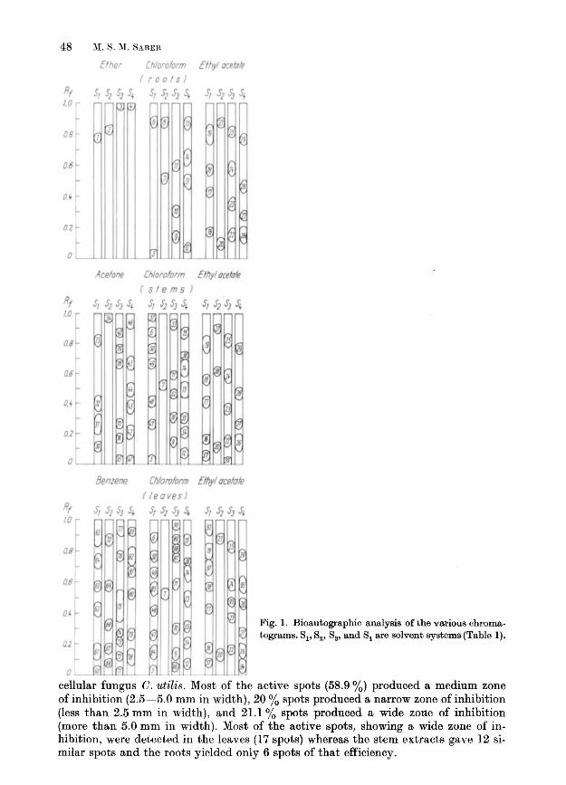
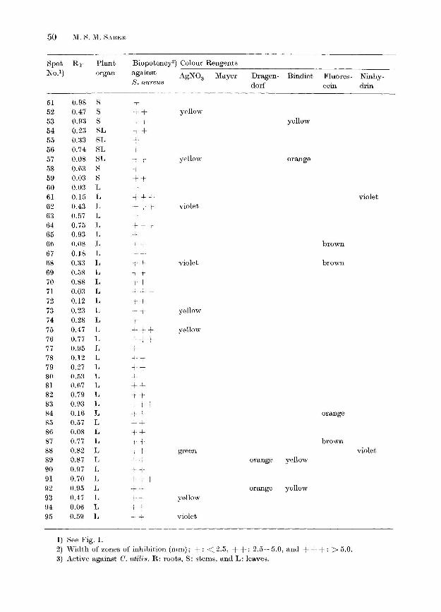
It is apparent from the bioautographic analysis, illustrated in Fig. 1, that the highest number of active substances was detectable in the leaves and the lowest number in the roots. The leave extracts yielded 68 active spots, 24 each in benzene and chloroform extracts and 20 in ethyl acetate extract. The stems yielded 54 active spots; 20 in chloroform extract and 17 each in acetone and ethyl acetate extract. The roots yielded 29 active spots; 14 in ethyl acetate extract, 11 in chloroform extract, and 4 in ethyl ether extract. Generally, chloroform extracts contained higher numbers of active substances than the acetate extracts. The former gave 11, 20, and 24 active spots, compared to 4, 17, and 20 in the latter in roots, stems, and leaves, respectively.
A considerable variation in the distribution of active substances in the various plant organs was observed. In some cases, active substances, were detectable in more than one organ, i.e., 24 spots were present in roots, stems, and leaves, and 8 spots were present in stems and leaves, but absent in roots. In other cases, some substances were detectable in a certain plant organ, i.e., in leaves (36 spots), stems (30 spots), and roots (5 spots).
With regard to the solvent system, it is clear in Fig. 1 that the alkaline system 84 was superior in the number of active spots (45), followed by the acid system 83 (44) and the highly polar system Sl (44), then the weakly-polar system 82 (18). The number of active spots in each solvent system were 19, 17, and 9; 20, 16, and 8; 21, 16 and 7, and 8, 5, and 5 in the extracts of leaves, stems, and roots in the chromatograms prepared, using alkaline, acid, highly-polar and weakly-polar sytems, respectively.
The efficiency of the dyeing reagents in staining the active spots appeared to be in the following decreasing order: silver nitrate (28 spots), ninhydrin (11 spots), fluorescein (9 spots), Dragendorf (7 spots), Bindict (7 spots), and Mayer (1 spot).
Examination of the data presented in Table 1 reveals that the active spots exhibited various degrees of potency towards the test micro-organisms. While all the active spots affected S. aureU8, only 2 spots (i.e. No. 21 and 56) inhibited the uni-
48 M. S. :VI. RABER
Elher
Rf 5, 5Z S3 S 10 ... J \
r-
ae rt ...
06
o -
02 -
0,---
Acelone
Chloroform Elhyl oce/u'p ( rools
S,~S35t S,Sl~s'
6 8 IS " :1g IS Q!
II
Chloroform Elhyl ocelole
(si em s) R lO
5, 525J~ S, Ji5J ~ S, 52 S] S, ...
~ ~ @~ ~ lPl ~ - 6
... ))
~ I ~ 1
~ ,i)
~ -t! l"
I ,9
I!!: I" ~~~ I-
[H, 1 rJ !J ~
.... II 111 ~ n
- ~~ Fi-t 17 ~ ...
fJ: } ~ , • (J if - 1)
'iT M ~
1/ W J ----
08
a6
0. •
02
o
Benzene Chloroform E/lly/ uee/ule
(leaves) Rf 5, Sl S3 5~ :0
S,$1S]~ $, SlS]~
II
ae
a
o J
Fig. 1. Bioautographic analysis of the various chromatograms. 81' 82,83, and S4 are solvent systems (Table I).
cellular fungus C. utilis . Most of the active spots (58.9 %) produced a medium zone of inhibition (2.5-5.0 mm in width), 20 % spots produced a narrow zone of inhibition (less than 2.5 mm in width) , and 21.1 % spots produced a wide zone of inhibition (more than 5.0 mm in width). Most of the active spots, showing a wide zone of inhibition, were detected in the leaves (17 spots) whereas the stem extracts gave 12 similar spots and the roots yielded only 6 spots of that efficiency.
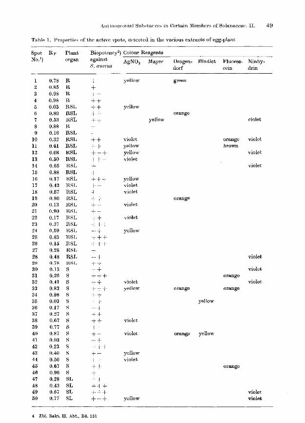
Antimicrobial Substances in Certain Members of Solanaceae. II. 49
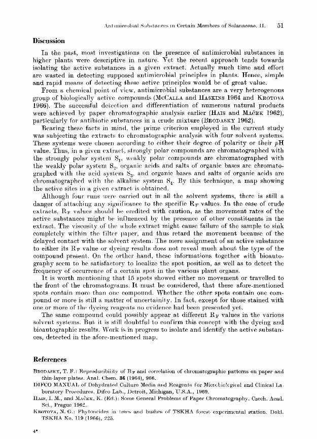
Table l. Properties of the active spots, detected in the various extracts of egg-plant
Spot RF Plant Biopotency2) Colour Reagents No.1) organ against AgN03 Mayer Dragen- Bindict Fluores- Ninhy·
S. aureU8 dorf cein drin
1 0.78 R +- yellow green 2 0.85 R + 3 0.98 R ++ 4 0.98 R ++ 5 0.03 RSL ++ yellow 6 0.89 RSL ++ orange 7 0.53 RSL ++ yellow ciolet 8 0.88 R + 9 0.16 RSL T
No.1) organ against AgN03 Mayer Dragen- Bindict Fluores- Ninhy-8. (pU·j'eUB dorf cein drin
51 0.98 8 T
52 0.47 S + yellow
53 0.93 S T yellow 54 0.23 SL -I 55 0.33 SL 56 0.74 SL 57 0.08 SL yellow orange 58 0.6:3 H 59 0.03 S + 60 0.0:3 L 61 0.15 L i + violet 62 0.43 L ·-;--+-r violet 63 0.57 L 64 0.75 L I- + 65 0.93 L 66 0.08 L brown 67 0.18 L ,+ 68 0.:13 L violet brown 69 0.58 L ,
-,- T
70 0.88 L -\ + 71 0.03 L 72 0.12 L 73 0.23 L -f- ~ yellow 74 0.28 L 75 0.47 L + ~ yellow 76 0.77 L T+ 77 0.95 L 78 0.12 L --.l_ + 79 0.27 L + 80 O.5a L -l--
81 0.67 L -+ -+. 82 0.79 L -r-83 0.93 L "-+ +-84 0.16 L orange 85 0.57 L ++ 86 0.08 L i+ 87 0.77 L ,+ brown 88 0.82 L + -+- green violet 89 0.87 L ++ orange yellow 90 0.97 L ,-91 0.70 L t -+ + 92 0.95 L orange yellow 93 0.47 L yellow 94 0.06 L 95 0.59 L ---!-- ---i- violet
1) See Fig. 1. 2) Width of zones of inhibition (mm); +: <2.5, + +: 2.5-5.0, and + + +: > 5.0. 3) Active against C. utili8. R: roots, S: stems, and L: leaves.
Arlhmicr-ubin,] ;;;llb,tallce~ in Certain Members of Solanaceae. II. 51
Discussion
In the past, most investigations on the presence of antimicrobial substances in higher plants were descriptive in nature. Yet the recent approach tends towards isolating the active substances in a given extract. Actually much time and effort are wasted in detecting supposed antimicrobial principles in plants. Hence, simple and rapid means of detecting these active principles would be of great value.
From a chemical point of view, antimicrobial substances are a very heterogenous group of biologically active compounds (MCCALLA and HASKINS 1964 and KROTOVA 1966). The successful detcction and differentiation of numerous natural products were achieved by paper chromatographic analysis earlier (HAIS and MACEK 1962), particularly for antibiot.ic substances in a crude mixture (BRODASKY 1962).
Bearing these facts in mind, the prime criterion employed in the current study was subjecting the cxtracts to chromatographic analysis with four solvent systems. These systems were chosen according to either their degree of polarity or their pH value. Thus, in a given extract, strongly polar compounds are chromatographed with the strongly polar system 81, weakly polar compounds are chroma to graphed with the weakly polar system 8~, organic acids and salts of organic bases are chromatographed with the acid system 83 , and organic bases and salts of organic acids are chromatographed with the alkaline system 84 , By this technique, a map showing the active sites in a given extract is obtained.
Although foUl' runs were carried out in all the solvent systems, there is still a danger of attaching any significance to the specific RF values. In the case of crude extracts, RF values should he ercdited wit.h eaution, as the movement rates of the active substances might be influenced by the presence of other constituents in the extract. The viscosity of tlw whole extract might cause failure of the sample to sink completely within the filter paper, and thus retard the movement because of the delayed contact with the solvent system. The mere assignment of an active substance to either its RF value or dyeing results does not reveal much about the type of the compound present. On the orther hand, these informations together with bioautography seem to be satisfaetory to localize the spot position, as well as to detect the frequency of occurrence of a certain spot in the various plant organs.
It is worth mentioning that 15 spots showed either no movement or travelled to the front of the chromatograms. It must be considered, that these afore-mentioned spots contain more than one compound. ~Whether the other spots contain one compound or more is still a matter of uncertainity. In fact, except for those stained with one or more of the dyeing reagents no evidence had been presented yet.
The same compound could possibly appear at different Rj,' values in the various solvent systems. But it is st.ill doubtful to confirm this concept with the dyeing and bioautographic results. 'Work is in progress to isolate and identify the active substances, detected in the afore-mentioned map.
References
BRODASKY, T. F.: Reproducibility of RF and correlation of chromatographic patterns on paper and thin-layer plates. Anal. Chern. 36 (1964), 966.
DIFCO MANUAL of Deh ydrated Culture Media and R eagents for Microbiolcgical and Clinical Laboratory Procedures. Difco Lab. , Detroit, Michigan, U.S .A., 1969.
HAIS, I. M., and MACEK, K . (Ed.): Some General Problems of Paper Chromatography. Czech. Acad. Sci., PrBgue 1962.
KROTOVA, N . G.: Phytoneides in trees and bushes of TSKHA forest experimental station. Dokl. TSKHA No. 119 (1966) , 225.
4*
52 M. fl. M. flABER, ,\ntlmicrobial i:lub"tance8 in Certain Members of Solanaceae. II.
MCCALLA, T . .!VI., and HASKINS, F. A.: Phytotoxic substances from soil micro-organisms and crop residues. Bact. Rev. 28 (1964), 181.
MERCK, E.: Dyeing Reagents for Thin-Layer and Paper Chromatography. E. Merck, Darmstadt, Germany, 1971.
SABER, M. S . .!VI.: Antimicrobial substances in certain members of Solanaceae. 1. Preliminary investi. gations. Zbl. Bakt. II 131 (1976), 40-45.
ZAYED, M. N., SABER, M. S. M., ABD-EL-MALEK, Y., and MONIB, M.: Antimicrobial substances in dry residues of certain higher Egyptian plants. Zbl. Bakt. II 126 (1971), 615.
Author's address:
Dr. M. S. M. SABER, National Research Centre, Soil Microbiology Unit, Dokki, Cairo (Egypt).