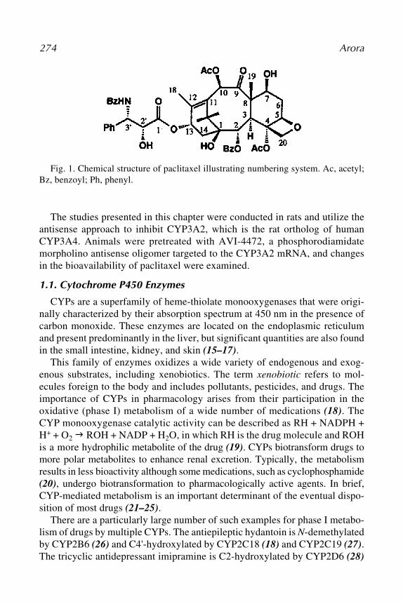
M E T H O D S I N M O L E C U L A R M E D I C I N E TM Antisense Therapeutics Edited by M. Ian Phillips, PhD, DSc Second Edition Antisense Therapeutics Edited by M. Ian Phillips, PhD, DSc Second Edition
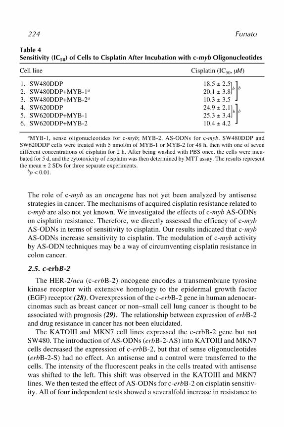
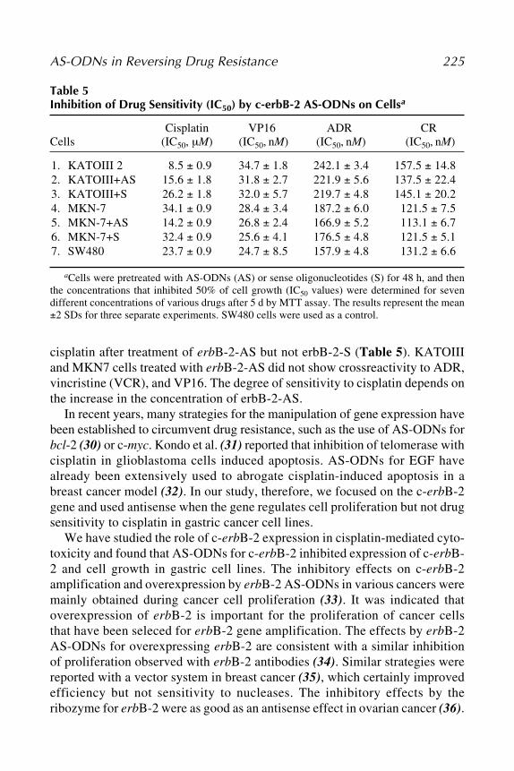
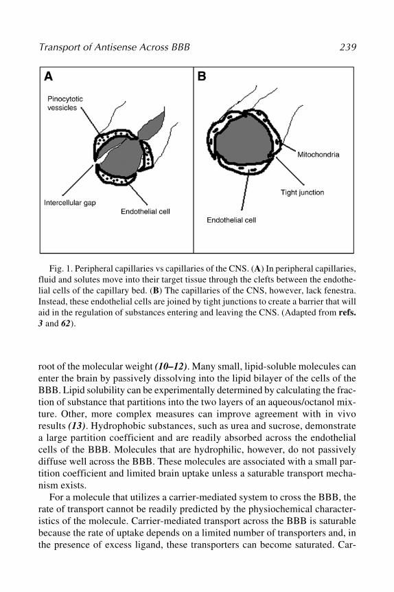
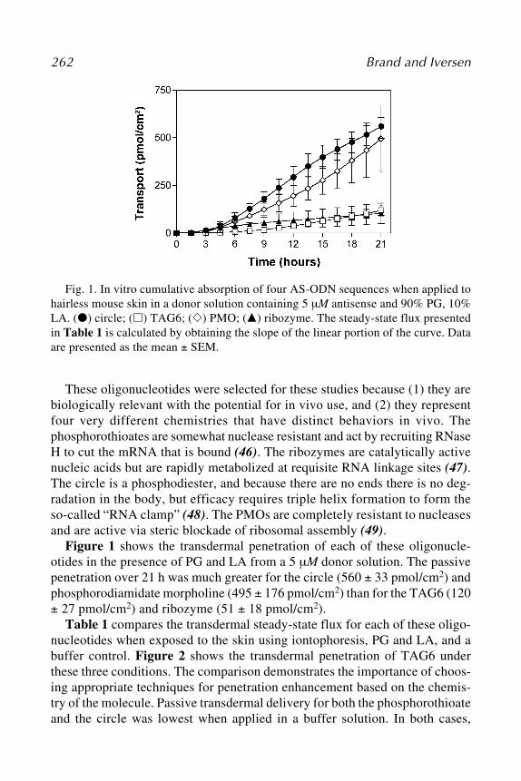
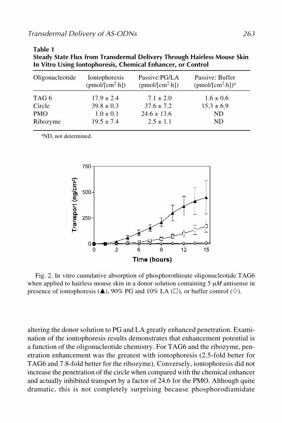
Transcript
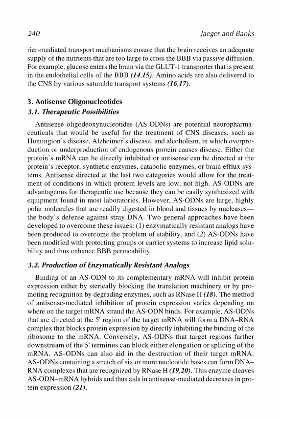
M E T H O D S I N M O L E C U L A R M E D I C I N ETM
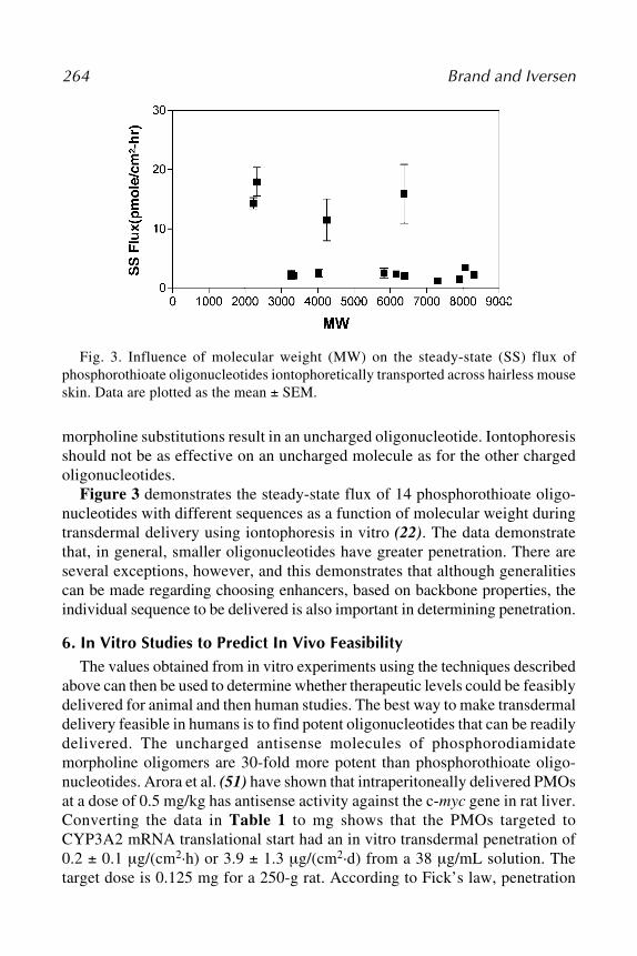
AntisenseTherapeutics
Edited by
M. Ian Phillips, PhD, DSc
Second Edition
AntisenseTherapeutics
Edited by
M. Ian Phillips, PhD, DSc
Second Edition
i
Antisense Therapeutics
M E T H O D S I N M O L E C U L A R M E D I C I N ETM
John M. Walker, SERIES EDITOR
113. Multiple Myeloma: Methods and Protocols,edited by Ross D. Brown and P. Joy Ho, 2005
112. Molecular Cardiology: Methods and Protocols,edited by Zhongjie Sun, 2005
111. Chemosensitivity: Volume 2, In Vivo Models,Imaging, and Molecular Regulators, edited byRosalyn D. Blumethal, 2005
110. Chemosensitivity: Volume 1, In Vitro Assays,edited by Rosalyn D. Blumethal, 2005
109. Adoptive Immunotherapy, Methods andProtocols, edited by Burkhard Ludewig andMatthias W. Hoffman, 2005
108. Hypertension, Methods and Protocols,edited by Jérôme P. Fennell and AndrewH. Baker, 2005
107. Human Cell Culture Protocols, SecondEdition, edited by Joanna Picot, 2005
106. Antisense Therapeutics, Second Edition,edited by M. Ian Phillips, 2005
105. Developmental Hematopoiesis: Methodsand Protocols, edited by Margaret H. Baron,2005
104. Stroke Genomics: Methods and Reviews, editedby Simon J. Read and David Virley, 2005
103. Pancreatic Cancer: Methods and Protocols,edited by Gloria H. Su, 2005
102. Autoimmunity: Methods and Protocols, editedby Andras Perl, 2004
101. Cartilage and Osteoarthritis: Volume 2,Structure and In Vivo Analysis, edited byFrédéric De Ceuninck, Massimo Sabatini,and Philippe Pastoureau, 2004
100. Cartilage and Osteoarthritis: Volume 1,Cellular and Molecular Tools, edited byMassimo Sabatini, Philippe Pastoureau, andFrédéric De Ceuninck, 2004
99. Pain Research: Methods and Protocols, editedby David Z. Luo, 2004
98. Tumor Necrosis Factor: Methods and Protocols,edited by Angelo Corti and Pietro Ghezzi, 2004
97. Molecular Diagnosis of Cancer: Methods andProtocols, Second Edition, edited by Joseph E.Roulston and John M. S. Bartlett, 2004
96. Hepatitis B and D Protocols: Volume 2,Immunology, Model Systems, and ClinicalStudies, edited by Robert K. Hamatake andJohnson Y. N. Lau, 2004
95. Hepatitis B and D Protocols: Volume 1,Detection, Genotypes, and Characterization,edited by Robert K. Hamatake and JohnsonY. N. Lau, 2004
94. Molecular Diagnosis of Infectious Diseases,Second Edition, edited by Jochen Decker andUdo Reischl, 2004
93. Anticoagulants, Antiplatelets, andThrombolytics, edited by Shaker A. Mousa,2004
92. Molecular Diagnosis of Genetic Diseases,Second Edition, edited by Rob Elles andRoger Mountford, 2004
91. Pediatric Hematology: Methods and Protocols,edited by Nicholas J. Goulden and Colin G.Steward, 2003
90. Suicide Gene Therapy: Methods and Reviews,edited by Caroline J. Springer, 2004
89. The Blood–Brain Barrier: Biology andResearch Protocols, edited by Sukriti Nag,2003
88. Cancer Cell Culture: Methods and Protocols,edited by Simon P. Langdon, 2003
87. Vaccine Protocols, Second Edition, edited byAndrew Robinson, Michael J. Hudson, andMartin P. Cranage, 2003
86. Renal Disease: Techniques and Protocols,edited by Michael S. Goligorsky, 2003
85. Novel Anticancer Drug Protocols, edited byJohn K. Buolamwini and Alex A. Adjei, 2003
84. Opioid Research: Methods and Protocols,edited by Zhizhong Z. Pan, 2003
83. Diabetes Mellitus: Methods and Protocols,edited by Sabire Özcan, 2003
82. Hemoglobin Disorders: Molecular Methodsand Protocols, edited by Ronald L. Nagel, 2003
81. Prostate Cancer Methods and Protocols,edited by Pamela J. Russell, Paul Jackson,and Elizabeth A. Kingsley, 2003
iii
Humana Press Totowa, New Jersey
M E T H O D S I N M O L E C U L A R M E D I C I N ETM
Edited by
M. Ian Phillips, PhD, DScVice President for Research
All rights reserved. No part of this book may be reproduced, stored in a retrieval system, or transmitted in anyform or by any means, electronic, mechanical, photocopying, microfilming, recording, or otherwise withoutwritten permission from the Publisher. Methods in Molecular Biology™ is a trademark of The Humana PressInc.
The content and opinions expressed in this book are the sole work of the authors and editors, who havewarranted due diligence in the creation and issuance of their work. The publisher, editors, and authors are notresponsible for errors or omissions or for any consequences arising from the information or opinions presentedin this book and make no warranty, express or implied, with respect to its contents.
This publication is printed on acid-free paper. ∞ANSI Z39.48-1984 (American Standards Institute)
Permanence of Paper for Printed Library Materials.
Cover illustration: “The principle of antisense inhibition,” Figure 1 from chapter 1, Antisense Therapeutics: APromise Waiting to be Fulfilled, by M. Ian Philips
Cover design by Patricia F. Cleary.
For additional copies, pricing for bulk purchases, and/or information about other Humana titles, contact Hu-mana at the above address or at any of the following numbers: Tel.: 973-256-1699; Fax: 973-256-8341; E-mail: [email protected]; or visit our Website: www.humanapress.com
Photocopy Authorization Policy:
Authorization to photocopy items for internal or personal use, or the internal or personal use of specific cli-ents, is granted by Humana Press Inc., provided that the base fee of US $25.00 per copy is paid directly to theCopyright Clearance Center at 222 Rosewood Drive, Danvers, MA 01923. For those organizations that havebeen granted a photocopy license from the CCC, a separate system of payment has been arranged and isacceptable to Humana Press Inc. The fee code for users of the Transactional Reporting Service is: [1-58829-205-3/05 $25.00].
Printed in the United States of America. 10 9 8 7 6 5 4 3 2 1
Library of Congress Cataloging in Publication Data
Antisense therapeutics / edited by M. Ian Phillips.— 2nd ed.p. ; cm. — (Methods in molecular medicine ; 106)
Includes bibliographical references and index. ISBN 1-58829-205-3 (alk. paper); eISBN 1-59259-854-41. Antisense nucleic acids—Therapeutic use. [DNLM: 1. Oligonucleotides, Antisense—therapeutic use. 2. Oligonucleotides, Antisense—pharmacology. QU 57 A6332 2005] I.Phillips, M. Ian. II. Series.RM666.A564A585 2005
615'.31—dc22 2004006680
v
Foreword
v
We are now more than 15 years into a large-scale experiment to deter-mine the viability of antisense technology. The challenges of creating a newpharmacological drug discovery platform are prodigious, requiring sizeableinvestments, long-term commitment, insight, and perseverance. For antisensetechnology to progress, advances in understanding the behavior of the recep-tor, RNA, and the behavior of the drugs, oligonucleotide analogs, were neces-sary. A new medicinal industry, the medicinal industry of oligonucleotides,had to be invented, and numerous drug development challenges—such as creat-ing efficient manufacturing and analytical processes and formulations—had to beovercome. All of those advances then needed to be focused in drug candidatesdesigned to interact with specific targets and to be effective in patients with spe-cific diseases. This has taken time and a good bit of money and although theprogress in the technology has been gratifying, there have, of course, been failuresof individual clinical trials and individual drugs along the way.
What have we learned? Antisense technology works. Oligonucleotideanalogs with a reasonable drug-dependent property can be synthesized and usedto inhibit gene function through a variety of antisense mechanisms. Antisensedrugs distribute to a wide range of tissues and reduce the expression of targets ina dose fashion consistent with the pharmaceutics of the drugs. First-generationantisense drugs are sufficient for relatively severe indications and second-genera-tion drugs are performing significantly better. Moreover, these drugs are effec-tive by a wide variety of routes including intravenous, subcutaneous, intradermal,rectal, and aerosol, and progress in oral delivery has been reported. Today numer-ous clinical trials in a wide range of diseases using a variety of oligonucleotidechemistries and antisense mechanisms are in progress.
In this year alone, positive clinical data in rheumatoid arthritis, diabetes,hyperlipidemia, cancer, and other diseases have been reported.
In this edition of Antisense Therapeutics, a number of approaches to anti-sense and therapeutic areas are discussed, as well as specific diagnostic oppor-tunities. That the breadth of activities presented in this volume is as impressiveas it is and yet does not begin to cover all of the work in progress, underscoresthe range of utility and potential value of antisense technology.
vi
Nevertheless, despite antisense being an accepted tool that has facili-tated better understanding of biological systems, much remains to be donebefore the true potential of the technology for therapeutic purposes can bedefined. What this volume emphasizes, however, is that exponential progressin defining the long-term roles and value of antisense-based therapeutics isbeing made.
We look forward to the continued evolution of the technology.
Stanley T. Crooke, MD, PhD
Foreword
vii
Preface
This is the second edition of Antisense Therapeutics. The first editionwas edited by Sudhir Agrawal and published in 1996. At that time there wasno therapy based on antisense, but plenty of promise for the highly specifictargeting of genes that cause disease. Antisense oligonucleotides were firstreported as viral replication inhibitors by Paul Zamecnik and Mary Stephen-son in 1978. Although this was excellent work, nothing much happened untilnew procedures for synthesizing DNA sequences were developed. Once oli-gonucleotides were easy to make, more and more studies were published inthe 1980s, most of which were directed to cells in culture. In the early 1990santisense oligonucleotides were increasingly tested in vivo. There were manycontroversies and a great deal of concern about backbone modification of thephosphodiester bridges that link the DNA bases. To protect against break-down by nucleases in cells or blood, phosphorothioate oligonucleotides wereadopted. In 1998 a phosphorothioated antisense agent was the first FDA-approved antisense therapy. Vitravene™, developed by Isis Pharmaceuticals,made antisense therapeutics a reality.
Since then, the complete sequencing of the human genome in April, 2003has demonstrated the presence of a vast number of targets for antisense oligo-nucleotides. So we now have thousands of targets, hundreds of preclinicalanimal studies, and some 20 clinical trials ongoing. Any successful trial withan antisense compound will open a floodgate of new therapies for a panoplyof diseases.
This second edition of Antisense Therapeutics deals less with the basicscience of antisense and more with the actual therapeutic applications. Forthat reason it is organized into disease states.
I thank the authors for their patience and their strong contributions. Sincethis book was being edited at a time when I moved from the University ofFlorida to the University of South Florida, I ended up with two secretaries. Iwould like to thank Ms. Gayle Butters at the University of Florida and Mr.Eric J. Wheeler at the University of South Florida for their essential help. Iam also grateful to Craig Adams at Humana Press for his patience.
Preface ........................................................................................................... vii
Contributors ..................................................................................................... xi
PART I. INTRODUCTION
1 Antisense Therapeutics: A Promise Waiting to be FulfilledM. Ian Phillips ......................................................................................... 3
2 Antisense Inhibition: Oligonucleotides, Ribozymes, and siRNAsY. Clare Zhang, Meghan M. Taylor, Willis K. Samson,
and M. Ian Phillips ........................................................................... 11
PART II. CARDIOVASCULAR
3 Local Application of Antisense for Prevention of RestenosisPatrick L. Iversen, Nicholas Kipshidze, Jeffrey W. Moses,
and Martin B. Leon .......................................................................... 374 Antisense Therapeutics for Hypertension:
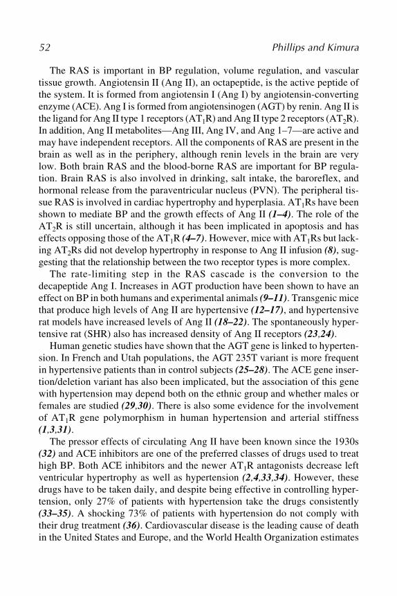
Targeting the Renin–Angiotensin SystemM. Ian Phillips and Birgitta Kimura ................................................... 51
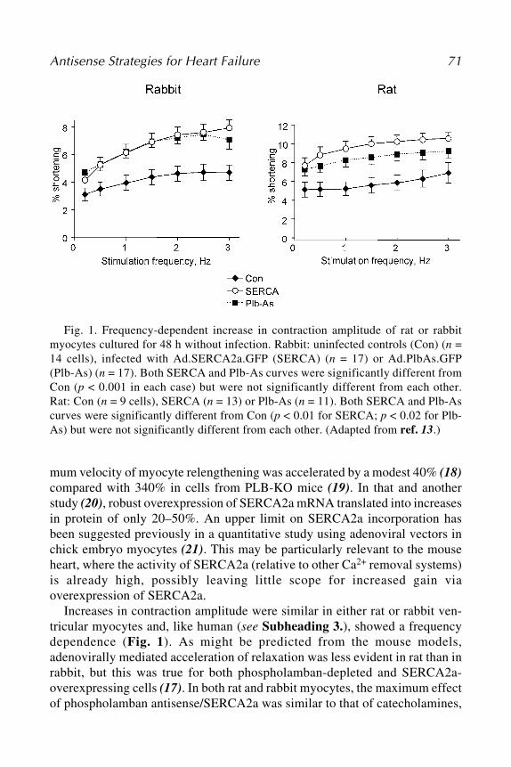
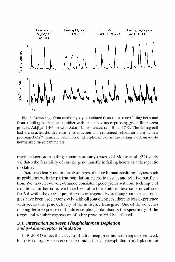
5 Antisense Strategies for the Treatment of Heart FailureSian E. Harding, Federica del Monte, and Roger J. Hajjar ............. 69
PART III. CANCER
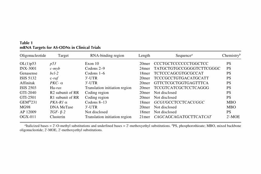
6 Clinical Studies of Antisense Oligonucleotides for Cancer TherapyRosanne M. Orr and F. Andrew Dorr ................................................. 85
7 Antisense Therapy in Clinical Oncology:Preclinical and Clinical Experiences
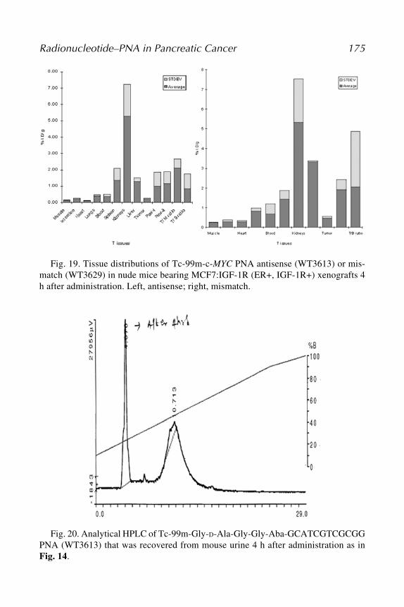
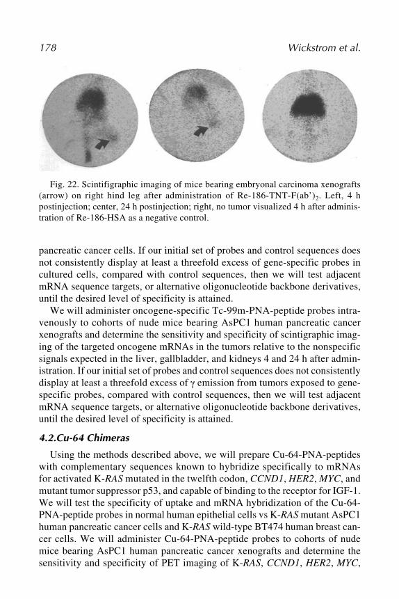
Ingo Tamm .......................................................................................... 1138 Radionuclide–Peptide Nucleic Acid Diagnosis and Treatment
of Pancreatic CancerEric Wickstrom, Xiaobing Tian, Nariman V. Amirkhanov,
Atis Chakrabarti, Mohan R. Aruva, Ponugoti S. Rao,Wenyi Qin, Weizhu Zhu, Edward R. Sauter,and Mathew L. Thakur................................................................... 135
ix
x Contents
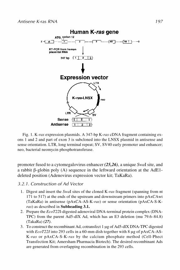
9 Suppression of Pancreatic and Colon Cancer Cellsby Antisense K-ras RNA Expression Vectors
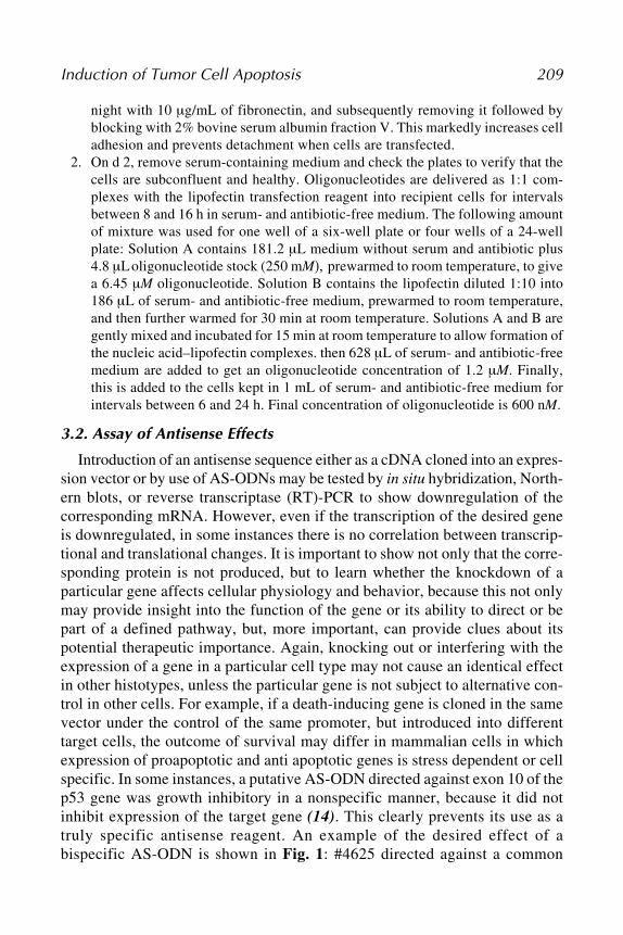
Kazunori Aoki, Shumpei Ohnami, and Teruhiko Yoshida ............ 19310 Induction of Tumor Cell Apoptosis and Chemosensitization
by Antisense StrategiesManuel Rieber and Mary Strasberg-Rieber .................................... 205
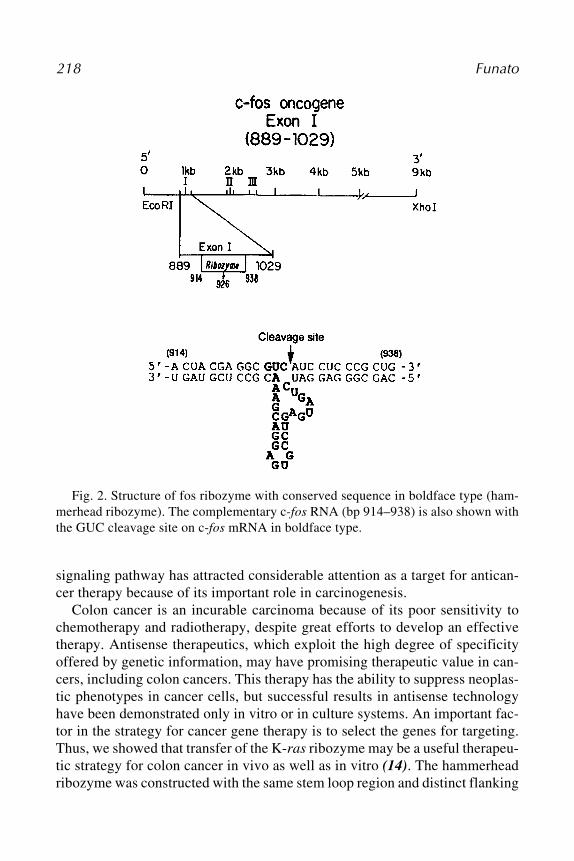
11 Utility of Antioncogene Ribozymes and AntisenseOligonucleotides in Reversing Drug Resistance
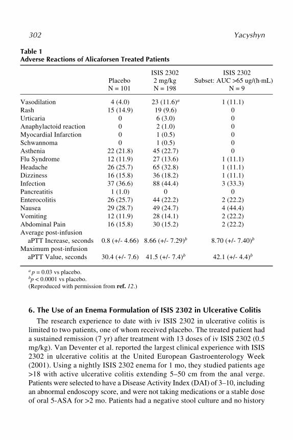
Bruce R. Yacyshyn ............................................................................ 295
PART VIII. HEPATITIS
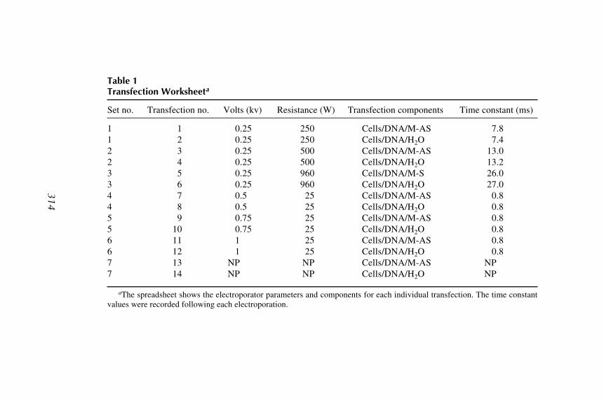
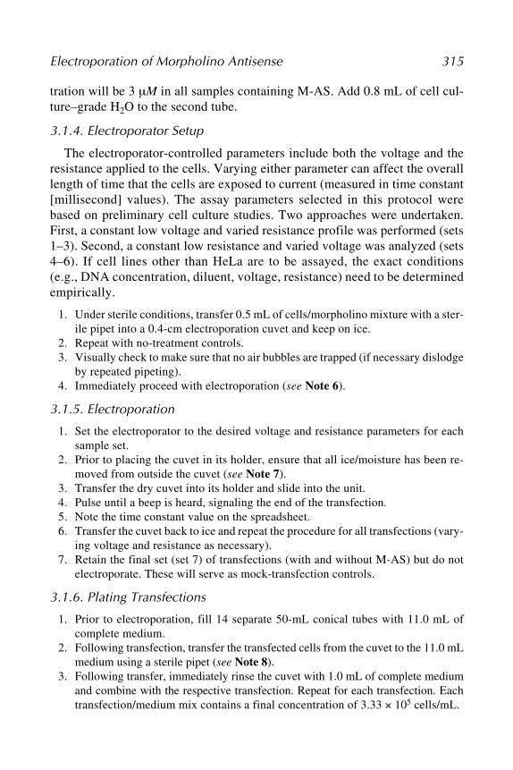
16 Optimizing Electroporation Conditions for the Intracellular Deliveryof Morpholino Antisense Oligonucleotides Directed Against theHepatitis C Virus Internal Ribosome Entry Site
Ronald Jubin ...................................................................................... 309
Index ............................................................................................................ 323
xi
xi
NARIMAN V. AMIRKHANOV • Departments of Biochemistry and MolecularPharmacology, Kimmel Cancer Center, Thomas Jefferson University,Philadelphia, PA
KAZUNORI AOKI • Section for Studies on Host-Immune Response, NationalCancer Center Research Institute, Tokyo, Japan
VIKRAM ARORA • Research and Development, AVI BioPharma, Corvallis, ORMOHAN R. ARUVA • Department of Radiology, Kimmel Cancer Center,
Thomas Jefferson University, Philadelphia, PAWILLIAM A. BANKS • GRECC, VA Medical Center St. Louis, Department of
Internal Medicine, St. Louis University, St. Louis, MORHONDA M. BRAND • Division of Emergency Medicine, Evanston Northwestern
Healthcare, and Department of Medicine, Feinberg School of Medicine,Northwestern University, Evanston, IL
ATIS CHAKRABARTI • Departments of Biochemistry and Molecular Pharmacology,Kimmel Cancer Center, Thomas Jefferson University, Philadelphia, PA
STANLEY T. CROOKE • Chairman and CEO, ISIS Pharmaceuticals Inc.,Carlsbad, CA
FEDERICA DEL MONTE • Cardiovascular Research Center, MassachusettsGeneral Hospital and Harvard Medical School, Boston, MA
F. ANDREW DORR • Salmedix Inc., San Diego, CATADAO FUNATO • Division of Molecular Diagnostics, Tohoku University
School of Medicine, Sendai, JapanROGER J. HAJJAR • Cardiovascular Research Center, Massachusetts General
Hospital and Harvard Medical School, Boston, MASIAN E. HARDING • National Heart and Lung Institute, Imperial College,
London, UKPATRICK L. IVERSEN • AVI BioPharma, Corvallis, ORLAURA B. JAEGER • Department of Pharmacological and Physiological
Science, St. Louis University, St. Louis, MORONALD JUBIN • Department of Antiviral Therapy, Schering Plough Research
Institute, Kenilworth, NJBIRGITTA KIMURA • Department of Anthropology, University of Florida,
Gainesville, FL
Contributors
xii Contributors
NICHOLAS KIPSHIDZE • Lenox Hill Heart and Vascular Institute, CardiovascularResearch Foundation, Lenox Hill Hospital, New York, NY
MARTIN B. LEON • Lenox Hill Heart and Vascular Institute, CardiovascularResearch Foundation, Lenox Hill Hospital, New York, NY
JEFFREY W. MOSES • Lenox Hill Heart and Vascular Institute, CardiovascularResearch Foundation, Lenox Hill Hospital, New York, NY
SHUMPEI OHNAMI • Central RI Laboratory, National Cancer Center ResearchInstitute, Tokyo, Japan
ROSANNE M. ORR • Cancer Research UK Centre for Cancer Therapeutic,The Institute of Cancer Research, Sutton, Surrey, UK
M. IAN PHILLIPS • Vice President for Research, Office of Research, Universityof South Florida, Tampa, FL
WENYI QIN • Department of Surgery, University of Missouri, Columbia, MOPONUGOTI S. RAO • Department of Radiology, Kimmel Cancer Center,
Thomas Jefferson University, Philadelphia, PAMANUEL RIEBER • Tumor Cell Biology Laboratory, Center of Microbiology
and Cell Biology, IVIC, Caracas, VenezuelaWILLIS K. SAMSON • Department of Pharmacological and Physiological
Science, St. Louis University, St. Louis, MOEDWARD R. SAUTER • Department of Surgery, University of Missouri, Columbia, MOMARY STRASBERG-RIEBER • Tumor Cell Biology Laboratory, Center of
Microbiology and Cell Biology, IVIC, Caracas, VenezuelaINGO TAMM • Department of Hematology and Oncology, Charite, Campus
Virchow, Humboldt University of Berlin, Berlin, GermanyMEGHAN M. TAYLOR • Department of Pharmacological and Physiological
Science, St. Louis University, St. Louis, MOMATHEW L. THAKUR • Department of Radiology, Kimmel Cancer Center,
Thomas Jefferson University, Philadelphia, PAXIAOBING TIAN • Departments of Biochemistry and Molecular Pharmacology,
Kimmel Cancer Center, Thomas Jefferson University, Philadelphia, PAERIC WICKSTROM • Departments of Biochemistry and Molecular Pharmacology,
Kimmel Cancer Center, Thomas Jefferson University, Philadelphia, PABRUCE R. YACYSHYN • Louis Stokes VA Hospital and Case Western Reserve
University, Cleveland, OHTERUHIKO YOSHIDA • Genetics Division, National Cancer Center Research
Institute, Tokyo, JapanY. CLARE ZHANG • Department of Pediatrics, University of South Florida, St.
Petersburg, FLWEIZHU ZHU • Department of Surgery, University of Missouri, Columbia, MO
Antisense TherapeuticsA Promise Waiting to Be Fulfilled
M. Ian Phillips
1. IntroductionDuring the past decade, only one antisense-based therapy has received full
Food and Drug Administration (FDA) approval. Vitravene™, developed byIsis Pharmaceuticals, was the first drug based on antisense technology to besuccessfully commercialized and used in treatment (1). The therapeutic area itis used in is a small niche related to the treatment of preventing blindness inacquired immunodeficiency syndrome (AIDS) patients by inhibiting cytome-galovirus-induced retinitis. The success of Vitravene, however, showed thatantisense could be taken all the way through the FDA approval process andprovide those patients taking it with a vitally important effect. With Vitravenewe saw the first breakthrough in antisense therapy, and, yet, euphoria has turnedto disappointment without a second breakthrough. Subsequent trials ofAffinitak (Isis), an antisense inhibitor of protein kinase C , failed to showstatistically significant benefits as an antisense therapy for the treatment ofnon–small cell carcinoma of the lung better than the median survival with con-trol treatments. The results nevertheless proved that antisense was well toler-ated and tended toward greater benefit to the survival of patients (p < 0.054).The promise of antisense therapy is so attractive that some 20 trials continue.
The appeal of antisense is that it potentially provides highly specific, nontoxiceffects for safe and effective therapeutics of an enormous number of diseasesincluding AIDS, Crohn’s disease, pouchitis, psoriasis, cancers, diabetes,mulitiple sclerosis, muscular dystrophy, restenosis, asthma, rheumatoid arthri-tis, hepatitis, skin diseases, polycystic kidney disease, and chronic cardiovas-
4 Phillips
cular disease, such as hypertension, restenosis, and heart failure. Successes inphase I have shown that antisense therapy consistently has excellent safetyresults. With each trial we learn more, and this makes each new antisense drugcandidate more easy to test. We are hampered by a lack of understanding of thetheoretical considerations for optimal antisense inhibition. Failures in the pasthave been the result of incorrect design and use of unmodified backbones caus-ing instability, overly long oligonucleotides leading to unpredictable targeting,and aptermeric or nonantisense effects. However, with each experiment welearned more. For example, high doses of antisense in monkeys triggered car-diovascular collapse (2). This result was a setback until it was found that thereaction could be accounted for by the extremely high doses and a sensitivityto complement activation unique to nonhuman primates (3). Human trials, bycontrast, have shown how well antisense is tolerated and how few side effectsare encountered. The number of trials is increasing, and more than 2000 patientshave received antisense. Isis is the leader with 11 phase I, 7 phase II, and 3 phaseIII trials. Genta is active with Genasense, and antisense to Bcl 2 for antitumorcell treatment is in phase III. AVI Biopharm has a third generation antisenseplatform, and around this it is testing four phase I, five phase II, and two phaseIII trials. Hybridon has conducted two phase I and has two phase II trialsplanned.
2. Mechanism of Antisense Inhibition
Antisense oligonucleotides (AS-ODNs) are designed to bind and inactivatespecific mRNA sequences inside cells. The potential uses for AS-ODNs is vastbecause RNA is so ubiquitous and abundant. With the publication of the humangenome sequence, we now have such a wide open access to the sequences ofgenes that antisense can in theory be applied to almost every known gene toinhibit its mRNA. Inhibiting mRNA prevents specific proteins from being pro-duced. Although routine human therapy may have been difficult to achieve, ata scientific level, antisense gene knockdown has become one of the fastestways to study new therapeutic targets.
AS-ODNs are synthetically made, single-stranded short sequences of DNAbases designed to hybridize to specific sequences of mRNA forming a duplex.This DNA-RNA coupling attracts an endogenous nuclease, RNase H, thatdestroys the bound RNA and frees the DNA antisense to rehybridize withanother copy of mRNA (2). In this way, the effect is not only highly specificbut prolonged because of the recycling of the antisense DNA sequence. Thereduction in mRNA reduces the total amount of protein specified by mRNA. Itis also theorized that hybridization sterically prevents ribosomes from translat-ing the message of the mRNA into protein. Therefore, there are at least two
Antisense Therapeutics 5
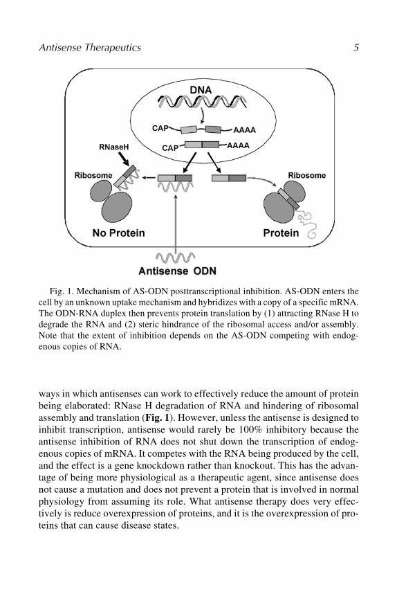
ways in which antisenses can work to effectively reduce the amount of proteinbeing elaborated: RNase H degradation of RNA and hindering of ribosomalassembly and translation (Fig. 1). However, unless the antisense is designed toinhibit transcription, antisense would rarely be 100% inhibitory because theantisense inhibition of RNA does not shut down the transcription of endog-enous copies of mRNA. It competes with the RNA being produced by the cell,and the effect is a gene knockdown rather than knockout. This has the advan-tage of being more physiological as a therapeutic agent, since antisense doesnot cause a mutation and does not prevent a protein that is involved in normalphysiology from assuming its role. What antisense therapy does very effec-tively is reduce overexpression of proteins, and it is the overexpression of pro-teins that can cause disease states.
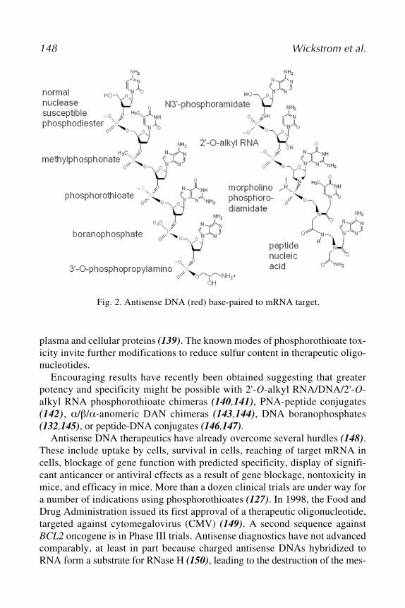
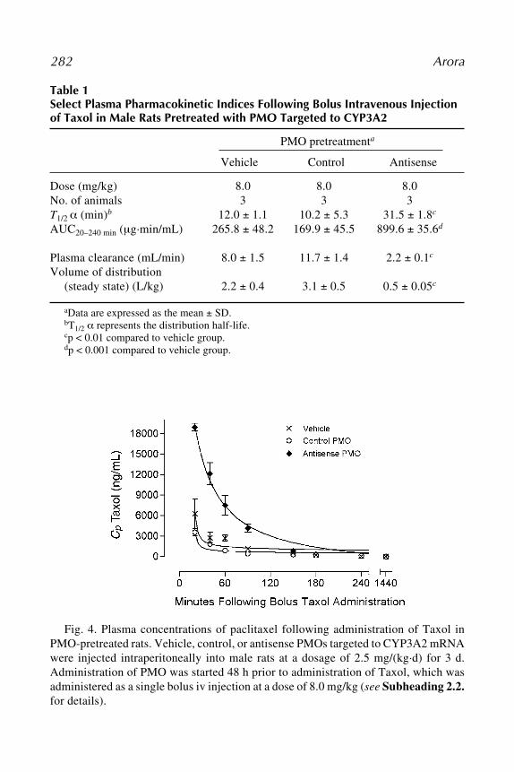
Fig. 1. Mechanism of AS-ODN posttranscriptional inhibition. AS-ODN enters thecell by an unknown uptake mechanism and hybridizes with a copy of a specific mRNA.The ODN-RNA duplex then prevents protein translation by (1) attracting RNase H todegrade the RNA and (2) steric hindrance of the ribosomal access and/or assembly.Note that the extent of inhibition depends on the AS-ODN competing with endog-enous copies of RNA.
6 Phillips
3. StabilityOne of the problems that dogged early attempts to achieve a therapy with
antisense was the question of stability. This is largely being answered by numer-ous ways to modify backbones of the DNA sequence in an AS-ODN. NativeDNA has a phosphodiester bridge between each successive base of the DNAsequence. It was quickly learned that unmodified AS-ODNs were very shortlasting, because they were unprotected from breakdown by nucleases, whichbreak apart the nuclear acids. A very successful modification was phos-phorothioate in which a sulfur atom replaces one oxygen atom in the phospho-rate group of the phosphodiester bond. Phosphorothioate oligonucleotides areresistant to nucleases and are stable. This extends the life of the AS-ODN toseveral days instead of a few hours. Many variations on this theme have beentested and patented so that there is now a range of second- and even third-generation backbone modifications available (2–4). Each company appearsto favor its own particular modification. Isis uses phosphorothivates with2'-O-methyl modification. Hybridon favors its IMO™ backbone modifica-tion, which can increase or decrease immunomodulation. AVI Biopharm hasused NeuGene® as a platform of third-generation antisense for its nine clinicaltrials. A factor in developing backbone modifications such as these and others,including peptide nucleic acid, is the cost.
4. Cellular UptakeAnother area that has required time (and money) to investigate is the opti-
mal conditions for uptake and distribution. This is particularly important whenit comes to systemic injection as opposed to the early experiments in whichantisenses were simply applied to cells in culture. There is both uptake andefflux of intact AS-ODNs in cells (5).The backbone modifications becomeextremely important when systemic injections are used because of nucleasesand the binding of oligonucleotides to proteins. The backbone modificationcan alter cell uptake, distribution, metabolism, and excretion. Nonantisenseeffects are a concern because they may alter the interpretation of whether theantisense effect is truly through an antisense mechanism or not. Mechanismsfor the uptake of oligonucleotides into cells are still not clearly understood.The lack of a theory of the uptake and kinetic effects on oligonucleotides hasrequired a lot of trial-and-error studies. This affects how to determine the opti-mal length of the oligonucleotide, the optimal concentration for effective treat-ment, and the frequency of treatments to maintain constant therapy. Despitethese complications and holes in the study of antisense, phosphorothioated oli-gonucleotides are surprisingly easy to work with. In our own studies, whichwere in vivo applications of AS-ODN, we aimed injections into the brain and
Antisense Therapeutics 7
into the blood at receptor targets involved in cardiovascular disease. We foundhighly significant effects using AS-ODNs of 15–18 bases in length deliveredin the brain without any vehicle (6) and in the blood delivered with liposomes(7). Call it science or dumb luck, we nevertheless were able to show significantphysiological effects of antisense delivery in models of hypertension. Becausehypertension is a chronic disease, the findings were remarkable because of thelong-lasting efficacy of a single antisense treatment. Reductions in bloodpressure lasted weeks with a single systemic injection of antisense targeting
-1 receptors (8).The distribution of AS-ODNs injected systemically is to all parts of the body
except the brain. The lipophobicity and/or negative change appear to preventAS-ODNs from crossing the blood-brain barrier. However, the oligonucle-otides accumulate in liver, kidney, and spleen. The lack of entry into the brainprobably translates into few side effects. With the antisense to -1 receptors,this could be a definite advantage (8). For treating liver or kidney disease,however, AS-ODNs might have a built-in advantage in terms of delivery.
5. The Target
Clearly, the target protein for antisense inhibition is crucially important fora therapeutic effect. To reach the target, the antisense therapy must enter thecell through an uptake mechanism and escape from endosomes and lysosomeswithin the cell in sufficient amounts to avoid intracellular degradation. If thetarget mRNA is shielded or coiled, it may be difficult for AS-ODNs to hybrid-ize. DNA and RNA are folded and studded with regulated proteins. Predictinghow RNA folds and its secondary structures in a living cell is still very diffi-cult. Once again, trial and error must be used. The stability of the oligos alsodepends on the interactions of the G-C proportions because of the three hydro-gen bonds instead of the two hydrogen bonds that are in the A-T interaction.Having sufficient length of bases is necessary to make a specific match, buthaving too long a sequence can overlap the coding regions and inhibit morethan single-target RNA.
Even when everything is successful and there is good uptake—good inhibi-tion of the target—it does not necessarily lead to a therapeutic effect, becausethe target may not be the only player in the disease. If knocking down one geneleads to an increase in a compensatory gene, there may be little or no effect.Alternatively, a target gene may have been involved in starting the disease, butonce the disease is present that target is no longer necessary, and, therefore,inhibiting it does not alter the disease state. Targeting transcription factors orsignaling pathway proteins important in regulating cells may not be specificenough. If the target protein is overexpressed only in the disease state, then
8 Phillips
antisense should be efficacious, but if the target is similarly expressed in bothnormal and malignant cells, antisense treatment may cause both types of cellsto undergo apoptosis. Then the therapy becomes a question of benefit vs risk.Because of the competition for RNA inhibition with antisense vs endogenousproduction of copies of mRNA in a cell, antisense for cancer is not a cell killerand, therefore, will not destroy all cancerous cells. However, it can be usedwith other treatments for cancer, and that is the protocol proposed for Affinitakand for Genasense.
6. Alternative to OligonucleotidesIn recent years, there has been a tremendous increase in interest in mor-
pholinos (9), small inhibitory RNA (siRNA) (10), as well as ribozymes (11).Morpho-linos are assembled from four different morpholino subunits each ofwhich contains one of the four genetic bases linked to a six-sided morpholinering. Morpholinos are supposed to have complete resistance to nucleases,high sequence specificity, and predictable targeting because they invade theRNA secondary structure and are fast and easy to deliver to the nucleus with-out liposome delivery systems. siRNAs are double-stranded RNA (dsRNA)molecules of 21–25 bp in length. They mediate RNA interference, an antiviralresponse initially identified in Caenorhabditis elegans and subsequently foundactive in specific gene silencing in many other organisms including mamma-lian cells. The sense and antisense strands of an siRNA first unwind, and theantisense strand binds to the target mRNA and recruits RNA-induced silencingcomplex (RISC) (Fig. 2). The sense strand is released from RISC, and RISCcatalyzes the mRNA cleavage. The gene silencing efficiency of siRNA hasreportedly been greater than antisense in general, typically reaching 80–90%.However, the maximal effects of optimal AS-ODNs and siRNAs targeting thesame mRNA sequence are comparable. siRNAs are being used because of theirstability and specificity, but it is not clear how effective they will be in sys-temic injections or oral delivery. Vickers et al. (12) conducted a comparativestudy of single-stranded AS-ODNs vs siRNA. Examination of 80 siRNA oli-gonucleotide duplexes designed to bind human RNA showed that both strate-gies are valid in terms of potency, maximal effects, specificity, and duration ofaction, at least in vitro.
The design of AS-ODNs and siRNAs follows different rules. Unlike AS-ODNs, the selection of an effective siRNA does not depend on the secondarymRNA structure or sequence accessibility. Instead, nucleotide composition andthe release rate of the sense strand from RISC seem to play major roles. Sev-eral siRNA molecules targeting the same mRNA can be used in combination toachieve greater effects and to avoid cellular resistance to siRNA. An indepen-dent combinatorial effect of AS-ODNs and siRNAs has also been observed
Antisense Therapeutics 9
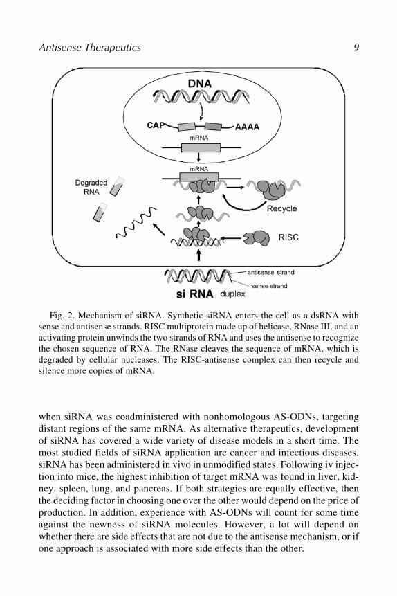
when siRNA was coadministered with nonhomologous AS-ODNs, targetingdistant regions of the same mRNA. As alternative therapeutics, developmentof siRNA has covered a wide variety of disease models in a short time. Themost studied fields of siRNA application are cancer and infectious diseases.siRNA has been administered in vivo in unmodified states. Following iv injec-tion into mice, the highest inhibition of target mRNA was found in liver, kid-ney, spleen, lung, and pancreas. If both strategies are equally effective, thenthe deciding factor in choosing one over the other would depend on the price ofproduction. In addition, experience with AS-ODNs will count for some timeagainst the newness of siRNA molecules. However, a lot will depend onwhether there are side effects that are not due to the antisense mechanism, or ifone approach is associated with more side effects than the other.
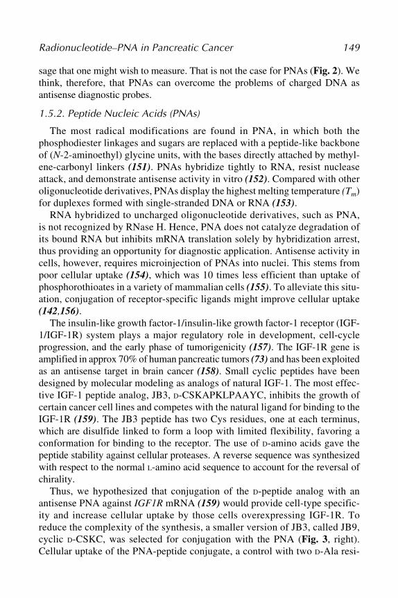
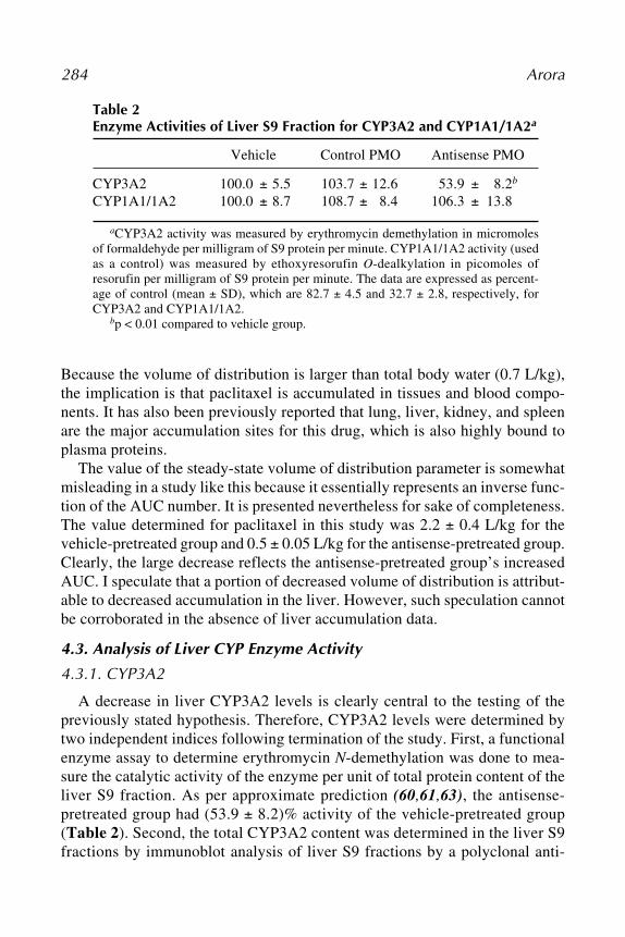
Fig. 2. Mechanism of siRNA. Synthetic siRNA enters the cell as a dsRNA withsense and antisense strands. RISC multiprotein made up of helicase, RNase III, and anactivating protein unwinds the two strands of RNA and uses the antisense to recognizethe chosen sequence of RNA. The RNase cleaves the sequence of mRNA, which isdegraded by cellular nucleases. The RISC-antisense complex can then recycle andsilence more copies of mRNA.
10 Phillips
7. ConclusionThe brief history of antisense therapeutics has been characterized by cycles
of success and disappointment. However, through it all, the promise ofantisense therapy has been so appealing that hope remains for that block-buster breakthrough that will open the doors for so many potential treatments.There are now thousands of targets available with known genomic sequences.There are hundreds of preclinical studies pointing to new treatments withantisense. And there are a score of human trials that are paving the way. Onceone major treatment is accepted, each new antisense therapy will be more eas-ily and quickly brought to those who suffer from diseases that are not yet satis-factorily treated with drugs.
References1. Crooke, S. T. (2004) Progress in antisense technology. Annu. Rev. Med. 55, 61–95.2. Crooke, S. T. (1998) Molecular mechanisms of antisense drugs: RNase H.
Antisense Nucleic Acid Drug Dev. 8(2), 133–134.3. Wickstrom, E. and Smith, J. B. (1998) DNA combination therapy to stop tumor
growth. Cancer J. Sci. Am. 4(Suppl. 1), S43–S47.4. Agrawal, S., Kandimalla, E. R., Yu, D., et al. (2002) GEM 231, a second-genera-
tion antisense agent complementary to protein kinase A alpha subunit, potentiateantitumor activity of irinotecan in human colon, pancreas, prostrate and lung can-cer xenografts. Int. J. Oncol. 21(1), 65–72.
5. Li, B., Hughes, J. A., and Phillips, M. I. (1997) Uptake and efflux of intact antisensephosphorothioate deoxyoligonucleotide directed against angiotensin receptors inbovine adrenal cells. Neurochem. Int. 31(3), 393–403.
6. Gyurko, R., Wielbo, D., and Phillips, M. I. (1993) Antisense inhibition of AT1 recep-tor mRNA and angiotensinogen mRNA in the brain of spontaneously hypertensiverats reduces hypertension of neurogenic origin. Regul. Pept. 49(2), 167–174.
7. Phillips, M. I. (2001) Gene therapy for hypertension: sense and antisense strate-gies. Expert Opin. Biol. Ther. (4), 655–662.
8. Zhang, Y. C., Bui, J. D., Shen, L., and Phillips, M. I. (2000) Antisense inhibitionof beta(1)-adrenergic receptor mRNA in a single dose produces a profound andprolonged reduction in high blood pressure in spontaneously hypertensive rats.Circulation 101(6), 682–688.
9. Summerton, J. (1999) Morpholino antisense oligomers: the case for an RNase H-independent structural type. Biochim. Biophys. Acta. 1489(1), 141–158.
10. Zamore, P. D. and Aronin, N. (2003) siRNAs knocks down hepatitis. Nat. Med.9(3), 266–267.
11. Fedor, M. J. and Westhof, E. (2002) Ribozymes: the first 20 years. Mol. Cell.10(4), 703–704.
12. Vickers, T. A., Koo, S., Bennett, C. F., Crooke, S. T., Dean, N. M., and Baker, B.F. (2003) Efficient reduction of target RNAs by small interfering RNA and RNaseH–dependent antisense agents: a comparative analysis. J. Biol. Chem. 278(9),7108–7118.
Antisense InhibitionOligonucleotides, Ribozymes, and siRNAs
Y. Clare Zhang, Meghan M. Taylor, Willis K. Samson,and M. Ian Phillips
1. IntroductionOver a span of more than two decades, antisense strategies for gene therapy
have expanded from antisense oligonucleotides (AS-ODNs) solely, to theaddition of ribozymes and, more recently, to the inclusion of small interferingRNAs (siRNAs). Antisense therapeutics has also experienced its phases of highexpectation, sudden disappointment, and meticulous rediscovery, whilemaintaining its status as a viable and effective gene therapy approach. Withthe discovery of RNA interference (RNAi) and development in delivery ofthese gene drugs, more preclinical and clinical investigations are anticipatedto take place in the near future to finally fulfill the promise of antisense thera-peutics in humans.
2. Antisense OligonucleotidesAS-ODNs are typically 18–25 bases in length, consisting of sequences that
are complementary to the target RNA. They can be injected directly into tis-sues or delivered systemically. Once delivered into cells, oligonucleotide bindsto its RNA counterpart and suppresses expression of the proteins encoded bytarget RNA. The specificity of this approach is based on the probability thatany sequence longer than a minimal number of nucleotides (nt)—13 for RNAand 17 for DNA—occurs only once within the human genome. The idea ofantisense therapy for inhibiting disease-associated proteins has become par-
12 Zhang et al.
ticularly appealing since Zamecnik and Stephenson (1) first demonstrated in1978 the reduction of Rous sarcoma viral RNA translation by a specific oligo-nucleotide.
2.1. Mechanisms of Antisense Inhibition
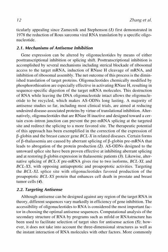
Gene expression can be altered by oligonucleotides by means of eitherposttranscriptional inhibition or splicing shift. Posttranscriptional inhibition isaccomplished by several mechanisms including sterical blockade of ribosomalaccess to the target mRNA, induction of RNase H cleavage of mRNA, andinhibition of ribosomal assembly. The net outcome of this process is the dimin-ished translation of target proteins. Oligonucleotides chemically modified byphosphorothioation are especially effective in activating RNase H, resulting insequence-specific digestion of the target mRNA molecules. This destructionof RNA while leaving the DNA oligonucleotide intact allows the oligonucle-otide to be recycled, which makes AS-ODNs long lasting. A majority ofantisense studies so far, including most clinical trials, are aimed at reducingundesired disease-associated proteins by virtue of translational inhibition. Alter-natively, oligonucleotides that are RNase H inactive and designed toward a cer-tain exon–intron junction can prevent the pre-mRNA splicing at the targetedsite and redirect the splicing to a more favored site. The therapeutic potentialof this approach has been exemplified in the correction of the expression of
-globin and the breast cancer gene BCL-X in related diseases. Certain formsof -thalassemia are caused by aberrant splicing of -globin pre-mRNA thatleads to abrogation of the protein production (2). AS-ODNs designed to theuntoward splice site have been proven effective at inhibiting aberrant splicingand at restoring -globin expression in thalassemic patients (3). Likewise, alter-native splicing of BCL-X pre-mRNA gives rise to two isoforms, BCL-XL andBCL-XS, with opposing antiapoptotic and proapoptotic activities. Targetingthe BCL-XL splice site with oligonucleotides favored production of theproapoptotic BCL-XS protein that enhances cell death in prostate and breasttumor cells (4).
2.2. Targeting Antisense
Although antisense can be designed against any region of the target RNA intheory, different sequences vary markedly in efficiency of gene inhibition. Theaccessibility of oligonucleotides to RNA is considered the most important fac-tor in choosing the optimal antisense sequences. Computational analysis of thesecondary structure of RNA by programs such as mfold or RNAstructure hasbeen used to facilitate selection of target sites for antisense action (5); how-ever, it does not take into account the three-dimensional structures as well asthe instant interaction of RNA molecules with other factors. More commonly
Antisense Inhibition 13
taken routes involve evaluation of accessible sites by use of RNase H mapping(6) or scanning oligonucleotide arrays for the best hybridization signals (7).Nevertheless, in general, targeting the start codon AUG, where mRNA is sup-posedly open for ribosomal entry, has been a successful strategy, although inmany cases other sequences turned out to be more effective. Despite these pre-dictive approaches, the selection of optimal antisense sequences still requirestrial-and-error testing initially and, in the end, needs to be confirmed in vivo.
2.3. Chemical Modifications
Stability and efficient delivery, prerequisites for oligonucleotides to achieveobservable therapeutic effects, have been obstacles due to their macromolecu-lar nature. Numerous chemical modifications and delivery approaches havebeen developed to overcome this problem (Fig. 1). The first generation ofantisense agents contains backbone modifications such as replacement of oxy-gen atom of the phosphate linkage by sulfur (phosphorothioates), methyl group(methylphosphonates), or amines (phosphoramidates). Of these, the phosphor-othioates have been the most successful and used for gene silencing because oftheir sufficient resistance to nucleases and ability to induce RNase H func-tions. However, their profiles of binding affinity to the target sequences, speci-ficity, and cellular uptake are less satisfactory. The second generation ofantisense modifications was aimed at improving these properties, among whichsubstitutions of position 2' of ribose with an alkoxyl group (e.g., methyl ormethoxyethyl groups) were most successful. 2'-O-methyl and 2'-O-methoxyethyl derivatives can be further combined with phosphorothioate link-age (8). The third generation contains structural elements, such as zwitterionicoligonucleotides (possessing both positive and negative charges in the mol-ecule); locked nucleic acids (LNAs)/bridged nucleic acids (BNAs) (9);morpholino (10); peptide nucleic acids (PNAs) (with a pseudopeptide back-bone) (11); and, more recently, hexitol nucleic acids (HNA) (12). All of themodifications enhanced AA-ODNs in terms of nuclease resistance; specificbinding; and with agents such as PNA and morpholino, cellular uptake. How-ever, the ability of oligonucleotides to induce RNase H cleavage was abolishedby these alterations. Therefore, chimeric oligonucleotides with an unmodifiedRNase H–susceptible core flanked by modified nuclease-resistant nucleotideshave recently been proposed to address this issue and applied in a number ofinvestigations (13), including clinical trials.
2.4. Delivery of Antisense
Oligonucleotides are primarily taken up by cells via endocytosis. Only aportion of oligonucleotides are able to escape endosome/lysome, enter thenucleus, and bind to its RNA complement. Because of the hydrophilic and
macromolecular nature, permeation of oligonucleotides across cell membraneis relatively difficult. Even after two decades of research, safe and efficientdelivery of oligonucleotides in vivo still remains a major barrier to the clinicalsuccess of antisense therapies. Cationic liposomes and electroporation are com-monly used carriers. A large variety of liposomal formulas have been devel-oped to facilitate antisense delivery, some of which have entered clinical trials(14). More recently, nanoparticles and oligonucleotide conjugates have shownimproved cellular uptake, biodistribution, and targeted delivery, especially incancer treatment (15,16). A hydrodynamic tail vein injection has proven veryeffective in delivering oligonucleotides into liver of rodents (17). Inhalableand topical applications of oligonucleotides in patients have shown satisfac-tory profiles of uptake and distribution (18,19). However, interestingly, mostAS-ODNs that are therapeutically valuable in animal models and in patientshave been administered in the form of naked compounds, despite the progressin antisense delivery.
2.5. Antisense in Therapies
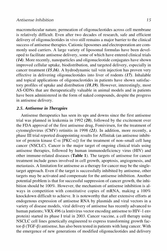
Antisense therapeutics has seen its ups and downs since the first antisensetrial was planned in leukemia in 1992 (20), followed by the excitement overthe FDA approval of the first antisense drug, Fomivirsen, for the treatment ofcytomeglovirus (CMV) retinitis in 1998 (21). In addition, more recently, aphase III trial reported disappointing results for Affinitak (an antisense inhibi-tor of protein kinase C- [PKC- ]) for the treatment of non–small cell lungcancer (NSCLC). Cancer is the major target of ongoing clinical trials usingantisense therapies, followed by human immunodeficiency virus (HIV) andother immune-related diseases (Table 1). The targets of antisense for cancertreatment include genes involved in cell growth, apoptosis, angiogenesis, andmetastasis. A limitation for antisense as a therapy for cancer may be the single-target approach. Even if the target is successfully inhibited by antisense, othertargets may be activated and compensate for the antisense inhibition. Anotherpotential problem is that for successful suppression of cancer growth, the inhi-bition should be 100%. However, the mechanism of antisense inhibition is al-ways in competition with constitutive copies of mRNA, making a 100%knockdown difficult to achieve. It is noteworthy that after extensive efforts atendogenous expression of antisense RNA by plasmids and viral vectors in avariety of disease models, viral delivery of antisense has recently advanced tohuman patients; VRX 496 (a lentivirus vector encoding antisense to HIV-1 envprotein) started its phase I trial in 2003. Cancer vaccine, a cell therapy usingNSCLC cell lines genetically engineered to express transforming growth fac-tor- (TGF- ) antisense, has also been tested in patients with lung cancer. Withthe emergence of new generations of modified oligonucleotides and delivery
16Z
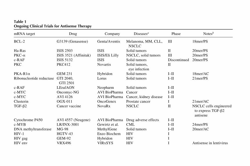
hang et al.Table 1Ongoing Clinical Trials for Antisense Therapy
mRNA target Drug Company Diseasesa Phase Notesb
BCL-2 G3139 (Genasense) Genta/Aventis Melanoma, MM, CLL, III 18mer/PSNSCLC
Ha-Ras ISIS 2503 ISIS Solid tumors II 20mer/PSPKC- ISIS 3521 (Affinitak) ISIS/Eli Lilly NSCLC, solid tumors III 20mer/PSc-RAF ISIS 5132 ISIS Solid tumors Discontinued 20mer/PSPKC PKC412 Novartis Solid tumors, II
GTI 2501c-RAF LErafAON Neopharm Solid tumors I–IIc-MYC Oncomyc-NG AVI BioPharma Cancer I–IIc-MYC AVI 4126 AVI BioPharma Cancer, kidney disease I–IIClusterin OGX-011 OncoGenex Prostate cancer I 21mer/ACTGF- 2 Cancer vaccine NovaRx NSCLC II NSCLC cells engineered
to express TGF- 2antisene
Cytochrome P450 AVI 4557 (Neugene) AVI BioPharma Drug adverse effects I–IIc-MYB LR/INX-3001 Gewirtz et al. CML I–II 24mer/PSDNA methyltransferase MG-98 MethylGene Solid tumors I–II 20mer/ACHIV-1 HGTV-43 Enzo Biochem HIV IHIV gag GEM-92 Hybridon HIV IHIV env VRX496 VIRxSYS HIV I Antisense in lentivirus
Antisense Inhibition
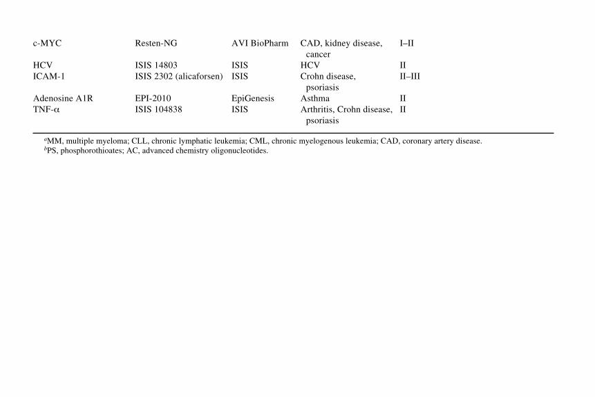
17c-MYC Resten-NG AVI BioPharm CAD, kidney disease, I–II
technologies, antisense therapeutics is closer to fulfilling its promise in theclinic for diseases other than cancer, such as cardiovascular disease, psoriasis,and Crohn’s disease.
3. Ribozymes3.1. What Are Ribozymes?
It was discovered in the early 1980s that some naturally occurring RNAmolecules have enzymatic activity (22,23). These enzymatic RNA moleculeswere termed ribozymes. Ribozymes recognize specific RNA sequences andthen catalyze a site-specific phosphodiester bond cleavage within the targetmolecule. Following cleavage, the ribozyme releases itself and binds to anothertarget molecule, repeating the process. The cellular consequence varies depend-ing on the setting. There are many naturally occurring ribozymes, including inplant viroids, ribosomes, self-splicing introns, and the RNA portion of RNaseP. In plant and animal cells, as well as in viruses, ribozymes are necessary forsome normal cellular processes such as transcription. The goal of most syn-thetic ribozyme usage, however, is reduction in targeted RNA and, thus, lowerlevels of the protein encoded by the target RNA.
Ribozyme substrate recognition occurs in the same manner as antisense pair-ing, through strand complementarity. Therefore, any decrease in target proteinfollowing ribozyme treatment could in part be due to antisense inhibition oftranslation or the recruitment of cellular enzymes to the double-stranded RNA(dsRNA) molecules. However, the ability of each ribozyme molecule to rap-idly cleave multiple target molecules gives this technology an advantage overclassic antisense that can act only on a single RNA molecule. In fact, the rateconstants of ribozyme cleavage reactions can approach and exceed those ofprotein enzymes, including enzymes with similar functions such as RNase A(24,25).
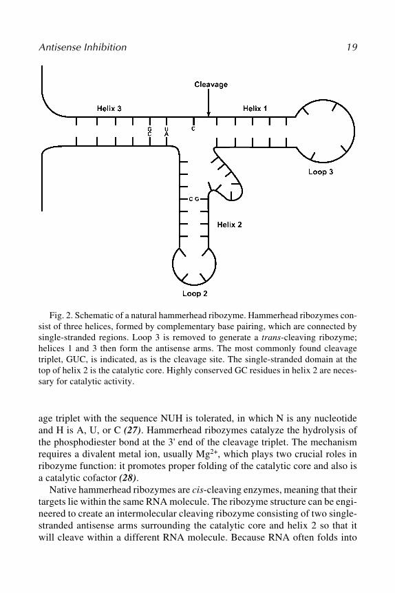
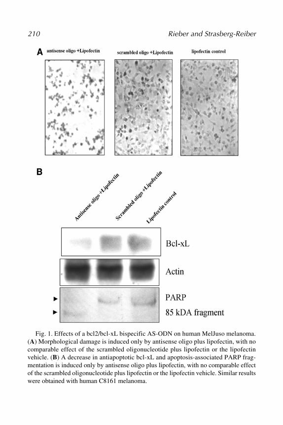
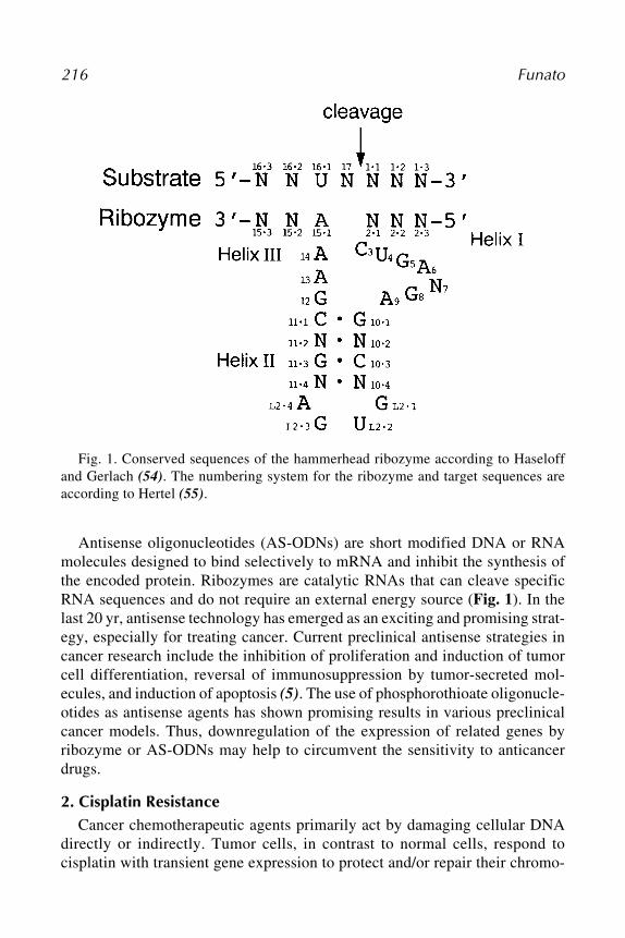
There are multiple types of ribozymes; the two most commonly used forresearch and therapeutic purposes are the hammerhead ribozyme and the hair-pin ribozyme (Figs. 2 and 3). One of the smallest and most well-understoodribozymes, the hammerhead ribozyme, is composed of 30–40 nt and was origi-nally discovered as a common sequence found in plant viroids that undergosite-specific, self-catalyzed cleavage as part of their replication process (26).All hammerhead ribozymes have a common structure consisting of three base-paired helices connected by two invariant single-stranded regions forming thecatalytic core. Helices 1 and 3 contain the antisense arms of the ribozyme.Helix 3 also contains the cleavage triplet, the site that is cut by the catalyticcore. The triplet most commonly found in naturally occurring hammerheadribozymes is GUC; however, mutagenesis studies have shown that any cleav-
Antisense Inhibition 19
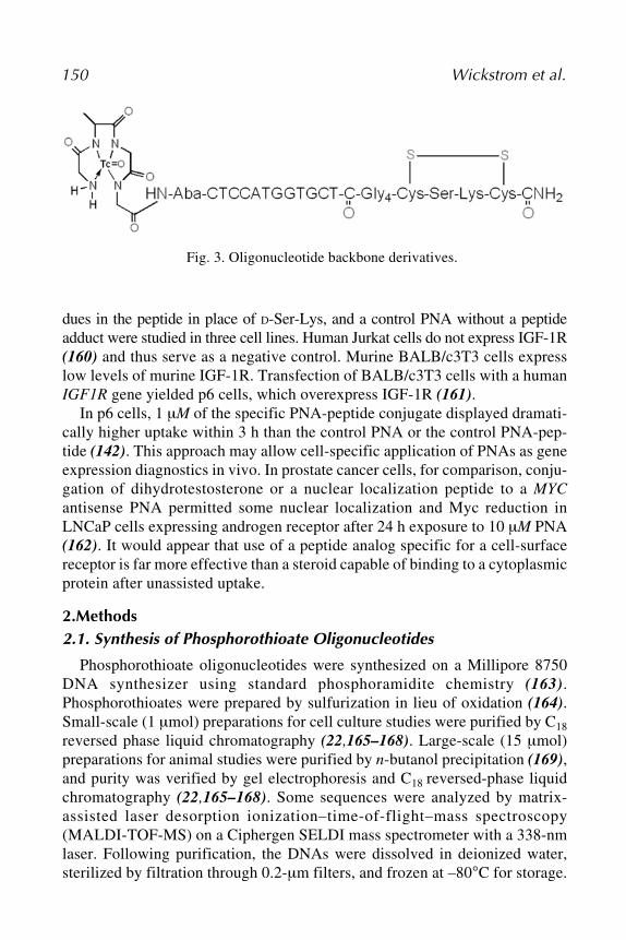
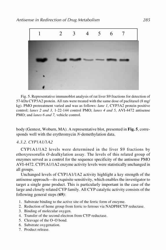
Fig. 2. Schematic of a natural hammerhead ribozyme. Hammerhead ribozymes con-sist of three helices, formed by complementary base pairing, which are connected bysingle-stranded regions. Loop 3 is removed to generate a trans-cleaving ribozyme;helices 1 and 3 then form the antisense arms. The most commonly found cleavagetriplet, GUC, is indicated, as is the cleavage site. The single-stranded domain at thetop of helix 2 is the catalytic core. Highly conserved GC residues in helix 2 are neces-sary for catalytic activity.
age triplet with the sequence NUH is tolerated, in which N is any nucleotideand H is A, U, or C (27). Hammerhead ribozymes catalyze the hydrolysis ofthe phosphodiester bond at the 3' end of the cleavage triplet. The mechanismrequires a divalent metal ion, usually Mg2+, which plays two crucial roles inribozyme function: it promotes proper folding of the catalytic core and also isa catalytic cofactor (28).
Native hammerhead ribozymes are cis-cleaving enzymes, meaning that theirtargets lie within the same RNA molecule. The ribozyme structure can be engi-neered to create an intermolecular cleaving ribozyme consisting of two single-stranded antisense arms surrounding the catalytic core and helix 2 so that itwill cleave within a different RNA molecule. Because RNA often folds into
20 Zhang et al.
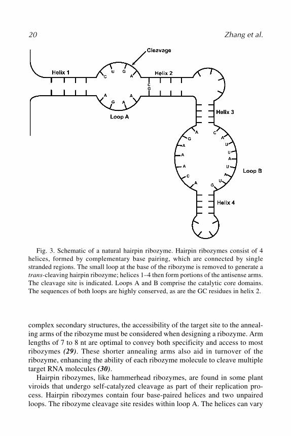
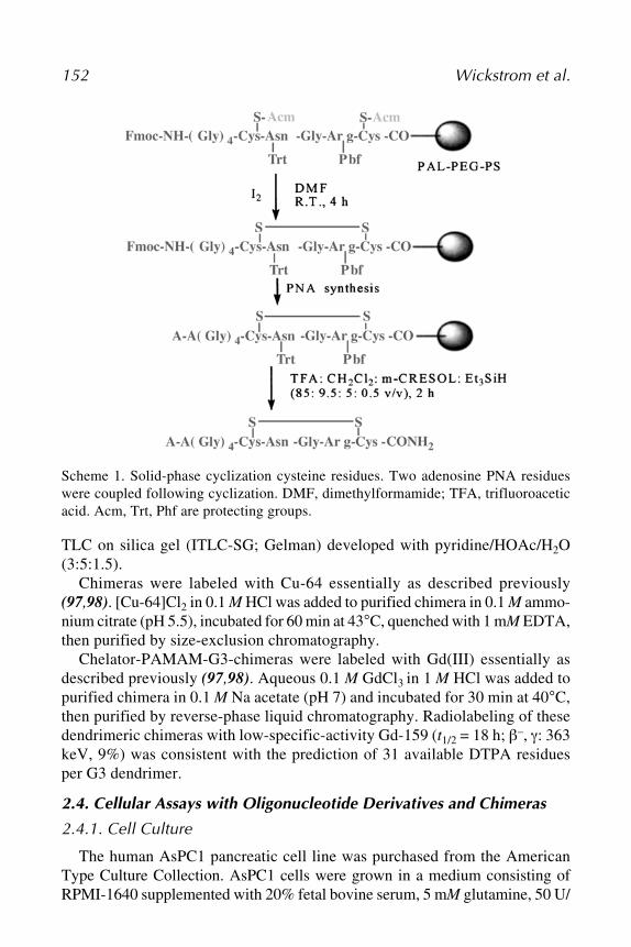
Fig. 3. Schematic of a natural hairpin ribozyme. Hairpin ribozymes consist of 4helices, formed by complementary base pairing, which are connected by singlestranded regions. The small loop at the base of the ribozyme is removed to generate atrans-cleaving hairpin ribozyme; helices 1–4 then form portions of the antisense arms.The cleavage site is indicated. Loops A and B comprise the catalytic core domains.The sequences of both loops are highly conserved, as are the GC residues in helix 2.
complex secondary structures, the accessibility of the target site to the anneal-ing arms of the ribozyme must be considered when designing a ribozyme. Armlengths of 7 to 8 nt are optimal to convey both specificity and access to mostribozymes (29). These shorter annealing arms also aid in turnover of theribozyme, enhancing the ability of each ribozyme molecule to cleave multipletarget RNA molecules (30).
Hairpin ribozymes, like hammerhead ribozymes, are found in some plantviroids that undergo self-catalyzed cleavage as part of their replication pro-cess. Hairpin ribozymes contain four base-paired helices and two unpairedloops. The ribozyme cleavage site resides within loop A. The helices can vary
Antisense Inhibition 21
in length and will tolerate any sequence that maintains complementarity withthe exception of a requirement for a guanine residue located at the beginning ofhelix 2, which is required for cleavage site recognition (31). Nucleotides withinthe catalytic loop regions, however, must be highly conserved to ensure cata-lytic activity of the ribozyme (31).
Hairpin ribozymes catalyze site-specific hydrolysis of the phosphodiesterbond on the complementary strand of RNA that is one base upstream of theconserved guanine in helix 2. Hairpin ribozymes, like hammerhead ribozymes,require an Mg2+ ion to activate proper secondary structure. However, unlikethe hammerhead ribozyme, Mg2+ does not play a direct role in the catalyticprocess (32). The exact catalytic mechanism used by hairpin ribozymes is notyet fully understood. A greater understanding of how both hammerhead andhairpin ribozymes work and of methods to optimize their function will enhancetheir attractiveness as potential therapeutic agents.
3.2. Delivery of Ribozymes
Two major issues in the use of ribozymes for research and therapy are ensur-ing that the ribozyme is delivered to the target tissues and ensuring that the levelsof ribozyme delivered are adequate to produce the desired effect. There aretwo methods for delivering the ribozyme to cells: exogenous delivery of apresynthesized ribozyme or endogenous expression of the ribozyme. Exog-enous delivery is relatively easy and rapid; however, as with antisense, thereare two main problems with this technique; cellular uptake of the ribozyme isoften difficult to achieve, and once the ribozyme is taken up, it is quicklydegraded. Cellular uptake of the ribozyme can be enhanced through the useof cationic liposomes. These cationic lipid micelles have the added benefit ofprotecting the ribozymes from RNase present in serum. To enhance further thelifespan of ribozymes they are frequently chemically modified. The addition ofa 2'-O-methyl moiety on some or all of the bases is the most commonly usedmodification. Work is currently being done to engineer DNAzymes, whichshould be more stable than their ribozyme RNA counterparts (33). One benefitof exogenous ribozyme delivery in vivo is that the immune system is fairlytolerant of foreign RNA molecules (34).
The other method for delivering ribozymes, endogenous expression of theribozyme, is most often accomplished using viral vectors; however, plasmidvectors may also be used. Both retroviral and DNA viral vectors have beenused. Expression cassettes can be designed to carry cell type–specific or condi-tional transcription initiation sites, as well as to include reporter proteins. Thebig advantage of an endogenous ribozyme is that it can be continuously pro-duced, allowing for the compromise of target protein production over a longperiod of time.
22 Zhang et al.
3.3. Research and Therapeutic Uses of Ribozymes
There are four main uses of ribozymes in the medical field: as a researchtool, as a chemotherapeutic agent, as an antiviral agent, and as a method toovercome acquired dominant genetic diseases.
With the recent sequencing of the Drosophila, mouse, and human genomes,there was a surge of newly identified proteins whose role in the organism iscurrently unknown or not fully understood. The use of ribozymes to selec-tively target these new proteins offers an attractive method to rapidly screentheir role in vivo. This method, along with other antisense techniques, offersseveral advantages over traditional methods of screening proteins. First, only apartial cDNA sequence is required to design a ribozyme. Second, ribozymescan be generated very rapidly, whereas both traditional and conditional knock-out animals as well as transgenic overexpression animals require a significantamount of time to generate. Finally, ribozymes can lead to greater effects forlonger periods of time when compared with antibody neutralization of the tar-get protein.
In addition to rapid screening of new proteins, ribozyme technology canalso be used to overcome problems with traditional protein function studies. Forexample, we use ribozymes to target a protein that when knocked out results inembryonic demise in mice and for which conditional knockouts have beenunsuccessful (35). Ribozymes can also be used to locally target a protein thatis made in many tissues, such as to lower targeted protein levels in brainwithout altering protein expression in the periphery.
The specificity of ribozymes makes them very attractive as therapeutics indisease states in which a protein is overexpressed or is malfunctioning.Ribozymes have the capability to specifically recognize single nucleotide dif-ferences in their targets. This special feature has resulted in the development ofribozymes to target oncogenes that are frequently mutated in tumors. Forinstance, the oncogene H-ras is mutated at a high frequency in many cancers;therefore, a ribozyme that recognizes only the mutant H-ras transcript has thepotential to be a very efficacious treatment. Several ribozymes have been devel-oped that can discriminate between H-ras mutants and the normal H-ras tran-script and initial studies have shown that stable expression of H-ras mutantribozymes leads to reduced tumor formation in athymic mice (36,37).
Alternative uses for ribozymes in cancer therapy are to block the elevationof normal gene products, such as c-fos, that occur in transformed cells or toblock angiogenic pathways. One such antiangiogenic ribozyme is targeted toflt-1 mRNA, which encodes for the vascular endothelial growth factor receptor(VEGF-R). The ribozyme has been shown to be well tolerated when adminis-tered daily by sc injection, and this dosing schedule leads to prolonged eleva-
Antisense Inhibition 23
tion in the plasma levels of ribozyme (38). This ribozyme is now in phase IIclinical trials in which therapeutic efficacy in breast and colorectal cancers isbeing examined.
A different set of circumstances in which a ribozyme can offer great thera-peutic potential is the treatment of acquired dominant genetic diseases. Retini-tis pigmentosa is a genetic disease that causes carriers of the dominant P23Hrhodopsin allele to slowly lose their vision. Hauswirth and Lewin have devel-oped a ribozyme that recognizes only the dominant version of the gene tran-script, which differs by two bases from the wild-type gene. Ribozyme treatmenthas resulted in a halt of disease progression in various species including rat,dog, and now monkey (39,40). This treatment is currently being prepared toenter the first phase of clinical trials. Promising results from this study couldopen the door for the development of ribozymes to treat other dominant geneticdisorders.
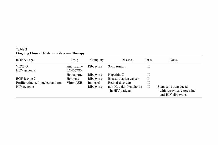
One final area where ribozyme therapy holds much promise is as antiviralagents, particularly in the treatment of retroviral infections. Many RNA virusessuch as HIV have very high mutation rates throughout much of their genomethat renders the mutated viruses resistant to current treatments. However, somesequences, including promoters and slicing signals, are highly conserved inHIV and among other RNA viruses. These regions provide excellent targetsfor ribozymes. In fact, some groups have designed ribozymes against conservedareas of HIV and have shown that ribozyme treatment can provide long-termHIV resistance and decrease HIV replication in infected cells (41). Severalcompanies now have ribozymes directed against HIV in various stages of clini-cal trials. Other viruses for which ribozyme treatments are also being designedinclude hepatitis B, hepatitis C, and the herpes viruses. Table 2 summerizesongoing clinical trials using ribozymes.
The use of ribozymes for the inhibition of gene expression holds great prom-ise in both therapeutics and research; however, we have only begun to under-stand the potential of these molecules. Efforts to improve the stability anddelivery of ribozymes will enhance their usefulness as therapeutic agents andlead to a greater recognition of the role of novel proteins in selected tissuesand the body as a whole.
4. RNA Interference with siRNA4.1. What Is siRNA?
RNAi is a form of antiviral immune response mounted by many higher eu-karyotes—including plants, nematodes, and insects—on exposure to dsRNA.dsRNA molecules are key intermediates in the genomic replication of manyviruses but are not normally found in eukaryotic cells. In contrast to the inter-
24Z
hang et al.
Table 2Ongoing Clinical Trials for Ribozyme Therapy
Heptazyme Ribozyme Hepatitis C IIEGF-R type 2 Herzyme Ribozyme Breast, ovarian cancer IProliferating cell nuclear antigen VitrenASE Immusol Retinal disorders IIHIV genome Ribozyme non-Hodgkin lymphoma II Stem cells transduced
in HIV patients with retrovirus expressinganti-HIV ribozymes
Antisense Inhibition 25
feron responses of mammalian cells in the face of viral infection, RNAi is usedby many other eukaryotes to defend against viruses through dsRNA-induceddegradation of viral RNAs.
The first evidence that dsRNA could suppress gene functions came from thework in Caenorhabditis elegans (42). In 1998, Fire et al. (43) found that senseRNA was as effective as antisense RNA for inhibiting genes. Subsequently,Zamore et al. (44) demonstrated that dsRNA was at least 10-fold more potentas a silencing trigger than was sense or antisense RNA alone. Since then, genesilencing by dsRNA has been termed RNAi, and its mechanisms have beenelucidated vigorously. Our current mechanistic understanding of RNAi deriveslargely from work in the Drosophila system (44,45). The first step of RNAi isto process longer dsRNA into 21- to 23-nt fragments that bear 3' overhangs byan RNase III–like enzyme called Dicer (46). These approx 21 nt dsRNAs,which are termed as siRNA, are essential to form a large (approx 500-kDa)RNA-induced silencing complex (RISC) (47). Through a yet-undefined mecha-nism, RISC cleaves the target mRNA that is complementary to the guidesiRNA, whether the target RNA is a viral mRNA or a cognate gene.
4.2. Application in Mammalian Cells
The key characteristics of RNAi are its remarkable sequence specificity,and it can therefore be used to target gene expression. It was found in Droso-phila that artificial siRNAs can be incorporated into RISC and induce degrada-tion of target mRNA. However, previous efforts to induce RNAi in culturedmammalian cells had largely failed because long dsRNAs (>30 bp) could inducea potent, nonspecific interferon response and activation of the protein kinasePKR and 2',5'-oligoadenylate synthetase (48,49). dsRNAs shorter than 30 bpdo not activate the PKR and interferon pathways. In 2001, a pioneering workby Elbashir et al. (50) demonstrated that transfection of 21 nt synthetic siRNAsinto cultured human cells can effectively inhibit gene expression in a sequence-specific manner with extremely high inhibitory efficiency. The advantages andgreat potential of RNAi technology, including high efficiency (typically>85%), have since raised tremendous interest in this field and generated rap-idly emerging progress (reviewed in refs. 51 and 52). These advances haveexpanded RNAi technology from the use of synthetic siRNA for the endog-enous production of small hairpin RNA (shRNA) by plasmid and viral vectors,and from transient inhibition in vitro to longer-lasting effects in vivo and intransgenic animals. This makes possible the utilization of RNAi in suppressingundesirable genes for human gene therapy.
siRNA-mediated gene silencing is sequence specific and dose dependent.As with antisense oligonucleotides and ribozymes, the efficient delivery ofsiRNA into cells of choice is currently the limiting factor to successful gene
26 Zhang et al.
inhibition. The delivery of siRNA can be in the form of naked compounds(53), or markedly improved by cationic liposomes (54) or electroporation (55),depending on cell types. Structural variations and sequence mutations havebeen made to investigate the structural and sequence requirements for siRNA-induced gene silencing. It was found that the status of the 5' hydroxyl terminusof the antisense strand of an siRNA determines RNAi activity, while a 3' termi-nus block is tolerated (56–58). Sequence mutations, on the other hand, are gen-erally tolerated at the 5' end, but not the 3' end (59). Chemical modificationssuch as phosphorothioation and 2'-O-methylation and 2'-O-allylation weredeveloped to improve the nuclease resistance of synthetic siRNAs (56,59,60).Certain chemical modifications at selected sites prolonged the siRNA activi-ties, whereas others compromised the efficiency. Simultaneous knockdown ofmore than two genes is possible, as illustrated by the double suppression of thenuclear mitotic apparatus protein (NUMA1) and lamin in HeLa cells (61).However, different siRNA species can possibly undergo reversible competi-tion in a sequence-independent manner (58,62), suggesting that the RNAi ma-chinery might be titratable or limited in mammalian cells.
4.3. siRNA as an Antiviral Agent
The ability of RNAi to protect plants and insects from viral infection can beapplied to mammals, although mammalian cells themselves do not possessinherent RNAi mechanisms. siRNAs designed against a variety of viruses,HIV (63–68), hepatitis C virus (HCV) (69–74), hepatitis B virus (75–77),papillomavirus (78), herpesvirus (79), rotavirus (80), and influenza virus (81),have been tested in cell cultures and displayed high efficiency in inhibitingviral infection and replication. In experiments aimed at HIV suppression,siRNAs have been targeted to various regions of the HIV genome includingthe long-terminal repeats and all five encoded genes, and typically a 30- to 50-fold decrease was observed in the viral levels in cell lines as well as in primaryT-lymphocytes (63–68). Similar cases were reported in the siRNA-mediatedHCV suppression (69–74). Silencing of HCV RNAs was dose dependent andspecific, resulting in a dramatic decrease in HCV RNAs and clearance of HCVin hepatoma cell lines bearing an HCV subgenome. The effects lasted for 3 to4 d with synthetic siRNA and for more than 3 wk with expression vectors(70,72). It is noteworthy that siRNA-resistant virus strains might already bepresent in the original viral population and thereby selectively survive the treat-ment with siRNA, as reported in an attempt to inhibit poliovirus with siRNAs(82). These results suggest that a pool of different siRNA sequences should beused to avoid the selection pressure for favoring siRNA-immune virus.
Antisense Inhibition 27
4.4. Comparison of siRNA AS-ODNsDirect comparisons of AS-ODNs and siRNAs to the same targets were made
in cell culture and in vivo (83–87). Dose-response experiments revealed thatthe IC50 value for the siRNA was about 100-fold lower than that of the AS-ODNs (83). The effect of siRNA is also longer lasting than of AS-ODNsbecause siRNAs are more stable in fetal calf serum, human plasma, and cellcultures (84). By contrast, Vickers et al. (87) reported that optimized RNase-H-dependent oligonucleotides and siRNAs are comparable in terms of potency,maximal effectiveness, sequence specificity, and duration of action. An inde-pendent combinatorial effect of AS-ODNs and siRNAs has been observedwhen siRNA was coadministered with nonhomologous antisense oligonucle-otides, targeting distant regions of the same mRNA (88).
4.5. Small Hairpin RNA
The silencing activity of synthetic siRNAs is transient, lasting 3–5 d in cellculture and 10 d in vivo (53,70,84). This phenomenon can be attributed tosiRNA degradation and dilution of siRNA concentrations over cell divisions.To achieve persistent inhibitory effects of RNAi, various plasmid- and virus-based vectors have been developed to express siRNA or shRNA endogenously.RNA polymerase III–dependent promoters such as U6, H1, and tRNA promot-ers are among the most commonly used, followed by RNA polymerase II–dependent promoters such as CMV promoter (68,89–97). Pol III is ideal fortranscribing small RNAs, and its transcripts are not modified posttranscrip-tionally. Vectors based on adenovirus, retrovirus, and lentivirus have been usedto produce functional shRNA species, resulting in persistent and robust genesilencing in vitro and in vivo (97–105). The structure, length, and compositionof hairpins appeared to be crucial in determining siRNA activities; however,they varied greatly in these studies. So far it is unclear what makes the bestshRNA. Furthermore, RNAi has provided a rapid and effective means to func-tionally silence genes in stem cells and transgenic animals (99,100,106,107).
4.6. In Vivo Delivery
Efficient in vivo delivery of siRNAs has been reported in a number of mousemodels (53,54,74,97,108–110). After a rapid systemic injection into tail vein,the target gene expression was effectively inhibited in liver, kidney, spleen,lung, and pancreas (111). Intravenous injection of siRNA targeting Fas spe-cifically reduced Fas protein expression in mouse hepatocytes and protectedmice from fulminant hepatitis induced by concanavalin A or an agonistic Fasantibody (53). Moreover, siRNA is capable of gene silencing when adminis-tered into brain or retina (108,110).
28 Zhang et al.
Although RNAi technology is still in the fledgling stage, exhaustive effortsin the past few years have advanced our knowledge of RNAi from an antiviralmechanism in higher eukaryotes, to a powerful tool in functional genetics inmammalian cells, and then to a promising therapeutic approach for genetherapy. While many mechanistic and functional questions await answers, theadvantages of RNAi in terms of its extraordinary efficiency and specificity,coupled with extensive research for improving its stability, delivery, and dura-tion of action, warrant further preclinical and clinical explorations in a widevariety of diseases.
References
1. Zamecnik, P. C. and Stephenson, M. L. (1978) Inhibition of Pous sarcoma virusreplication and cell transformation by a specific oligodeoxynucleotide. Proc. Natl.Acad. Sci. USA 75, 280–284.
2. Schwartz, E. and Benz, E. (1995) Thalassemia Syndromes. Churchill Livingston,New York.
3. Lacerra, G., Sierakowska, H., Carestia, C., et al. (2000) Restoration of hemoglo-bin A synthesis in erythroid cells from peripheral blood of thalassemic patients.Proc. Natl. Acad. Sci. USA 97, 9591–9596.
4. Mercatante, D. R., Bortner, C. D., Cidlowski, J. A., and Kole, R. (2001) Modifi-cation of alternative splicing of Bcl-x pre-mRNA in prostate and breast cancercells. Analysis of apoptosis and cell death. J. Biol. Chem. 276, 16,411–16,417.
5. Mathews, D. H., Sabina, J., Zuker, M., and Turner, D. H. (1999) Expanded sequencedependence of thermodynamic parameters improves prediction of RNA secondarystructure. J. Mol. Biol. 288, 911–940.
6. Ho, S. P., Bao, Y., Lesher, T., et al. (1998) Mapping of RNA accessible sites forantisense experiments with oligonucleotide libraries. Nat. Biotechnol. 16, 59–63.
7. Milner, N., Mir, K. U., and Southern, E. M. (1997) Selecting effective antisensereagents on combinatorial oligonucleotide arrays. Nat. Biotechnol. 15, 537–541.
8. Manoharan, M. (1999) 2'-carbohydrate modifications in antisense oligonucleotidetherapy: importance of conformation, configuration and conjugation. Biochim.Biophys. Acta. 1489, 117–130.
9. Morita, K, Takagi M, Hasegawa C, et al. (2002) 2'-O,4'-C-ethylene-bridgednucleic acids (ENA): highly nuclease-resistant and thermodynamically stable oli-gonucleotides for antisense drug. Bioorg. Med. Chem. Lett. 12, 73–76.
10. Summerton, J. (1999) Morpholino antisense oligomers: the case for an RNase—independent structure type. Biochim. Biophys. Acta 1489, 141–158.
11. Elayadi, A. N. and Corey, D. R. (2001) Application of PNA and LNA oligomersto chemotherapy. Curr. Opin. Investig. Drugs 2, 558–561.
12. Declercq, R., Van Aerschot, A., Read, R. J., Herdewijn, P., and Van Meervelt, L.(2002) Crystal structure of double helical hexitol nucleic acids. J. Am. Chem. Soc.124, 928–933.
Antisense Inhibition 29
13. Wang, H., Wang, S., Nan, L., Yu, D., Agrawal, S., and Zhang, R. (2002)Antisense anti-MDM2 mixed-backbone oligonucleotides enhance therapeuticefficacy of topoisomerase I inhibitor irinotecan in nude mice bearing humancancer xenografts: in vivo activity and mechanisms. Int. J. Oncol. 20, 745–752.
14. Maurer, N., Fenske, D. B., and Cullis, P. R. (2001) Developments in liposomaldrug delivery systems. Expert Opin. Biol. Ther. 1, 923–947.
15. Brigger, I., Dubernet, C., and Couvreur, P. (2002) Nanoparticles in cancer therapyand diagnosis. Adv. Drug Deliv. Rev. 54, 631–651.
16. Manoharan, M. (2002) Oligonucleotide conjugates as potential antisense drugs withimproved uptake, biodistribution, targeted delivery, and mechanism of action.Antisense Nucleic Acid Drug Dev. 2, 103–128.
17. Lecocq, M., Andrianaivo, F., Warnier, M. T., Wattiaux-De Coninck, S., Wattiaux,R., and Jadot, M. (2003) Uptake by mouse liver and intracellular fate of plasmidDNA after a rapid tail vein injection of a small or a large volume. J. Gene Med. 5,142–156.
18. Sandrasagra, A., Leonard, S. A., et al. (2002) Discovery and development of re-spirable antisense therapeutics for asthma. Antisense Nucleic Acid Drug Dev. 12,177–181.
19. Brand, R. M. (2001) Topical and transdermal delivery of antisense oligonucle-otides. Curr. Opin. Mol. Ther. 3, 244–248.
20. Reynolds, T. (1992) First antisense drug trials planned in leukemia. J. Natl. Can-cer Inst. 84, 288–290.
21. Roehr, B. (1998) Fomivirsen approved for CMV retinitis. J. Int. Assoc. Physi-cians AIDS Care 4, 14–16.
22. Altman, S. (1990) Ribonuclease P. Postscript. J. Biol. Chem. 265, 20,053–20,056.23. Cech, T. R. (1987) The chemistry of self-splicing RNA and RNA enzymes. Sci-
ence 236, 1532–1539.24. Esteban, J. A., Banerjee, A. R., and Burke, J. M. (1997) Kinetic mechanism of the
hairpin ribozyme. J. Biol. Chem. 272, 13,629–13,639.25. Fedor, M. J. and Uhlenbeck, O. C. (1992) Kinetics of intermolecular cleavage by
hammerhead ribozymes. Biochemistry 31, 1242–1254.26. Symons, R. H. (1992) Small catalytic RNAs. Annu. Rev. Biochem. 61, 641–671.27. Birikh, K. E., Heaton, P. A., and Eckstein, F. (1997) The structure, function and
application of the hammerhead ribozyme. Eur. J. Biochem. 245, 1–16.28. Dahm, S. C. and Uhlenbeck, O. C. (1991) Role of divalent metal ions in the ham-
merhead RNA cleavage reaction. Biochemistry 30, 9464–9469.29. Lieber, A. and Strauss, M. (1995) Selection of efficient cleavage sites in target
RNAs by using a ribozyme expression library. Mol. Cell. Biol. 15, 540–551.30. Hendry, P. and McCall, M. (1996) Unexpected anisotropy in substrate cleavage
rates by asymmetric hammerhead ribozymes. Nucleic Acids Res. 24, 2679–2684.31. Fedor, M. J. (2000) Structure and function of the hairpin ribozyme. J. Mol. Biol.
297, 269–291.32. Young, K. J., Gill, F., and Grasby, J. A. (1997) Metal ions play a passive role in
the hairpin ribozyme catalyzed reaction. Nucleic Acids Res. 25, 3760–3766.
30 Zhang et al.
33. Li, Y. and Breaker, R. R. (1999) Deoxyribozymes: new players in the ancientgame of biocatalysis. Curr. Opin. Struct. Biol. 9, 315–323.
34. Riddell, S. R., Elliott, M., Lewinsohn, D. A., et al. (1996) T-cell mediated rejectionof gene-modified HIV-specific cytotoxic T lymphocytes in HIV infected patients.Nat. Med. 2, 216–223.
35. Taylor, M. M. and Samson, W. K. (2002) Ribozyme compromise of adrenomedullinmRNA reveals a physiological role in the regulation of water intake. Am. J. Physiol.282, R1739–R1745.
36. Koizumi, M., Hayase, Y., Iwai, S., et al. (1989) Design of a RNA enzyme distin-guishing a single base mutation in RNA. Nucleic Acids Res. 17, 7059–7071.
37. Koizumi, M., Kmiya, H., and Ohtsuka, D. (1992) Ribozymes designed to inhibit trans-formation of NIH/3T3 cells by the activated c-Ha-ras gene. Gene 117, 179–184.
38. Usman, N. and Blatt, L. M. (2000) Nuclease-resistant synthetic ribozymes: devel-oping a new class of therapeutics. J. Clin. Invest. 106, 1197–1202.
39. Lewin, A. S., Drenser, K. A., Hauswirth, W. W., et al. (1998) Ribozyme rescue ofphotoreceptor cells in a transgenic rat model of autosomal dominant retinitispigmentosa. Nat. Med. 4, 967–971.
40. Lavail, M. M., Yasumura, D., Matthes, M. T., et al. (2000) Ribozyme rescue ofphotoreceptor cells in P23H transgenic rats: long-term survival and late-stagetherapy. Proc. Nat. Acad. Sci. USA 97, 11,488–11,493.
41. Muotri, A. R., Pereira, L. V., Vasques, L. R., et al. (1999) Ribozymes and the anti-gene therapy: how a catalytic RNA can be used to inhibit gene function. Gene237, 303–310.
42. Guo, S. and Kemphues, K. J. (1995) Par-1, a gene required for establishing polar-ity in C. elegans embryos, encodes a putative Ser/Thr kinase that is asymmetri-cally distributed. Cell 81, 611–620.
43. Fire, A., Xu, S., Montgomery, M. K., Kostas, S. A., Driver, S. E., and Mello, C. C.(1998) Potent and specific genetic interference by double-stranded RNA inCaenorhabditis elegans. Nature 391, 806–811.
44. Zamore, P. D., Tuschl, T., Sharp, P. A., and Bartel, D. P. (2000) RNAi: double-stranded RNA directs the ATP-dependent cleavage of mRNA at 21 to 23 nucle-otide intervals. Cell 101, 25–33.
45. Elbashir, S. M., Harborth, J., Lendeckel, W., Yalcin, A., Weber, K., and Tuschl,T. (2001) RNA interference is mediated by 21- and 22-nucleotide RNAs. GenesDev. 15, 188–200.
46. Bernstein, D., Caudy, A. A., Hammond, S. M., and Hannon, G. J. (2001) Role fora bidentate ribonuclease in the initiation step of RNA interference. Nature 409,363–366.
47. Hammond, S. M., Bernstein, F., Beach, D., and Hannon, G. J. (2000) An RNA-directed nuclease mediates post-transcriptional gene silencing in Drosophila cells.Nature 404, 293–296.
48. Stark, G. R., Kerr, I. M., Williams, B. R., et al. (1998) How cells respond tointerferons. Annu. Rev. Biochem. 67, 227–264.
Antisense Inhibition 31
49. Minks, M. A., West, D. K., Benvin, S., and Baglioni, C. (1979) Structural require-ments of double-stranded RNA for the activation of 2',5'-oligo(A) polymeraseand protein kinase of interferon-treated Hela cells. J. Biol. Chem. 254, 10,180–10,183.
50. Elbashir, S. M., Harborth, J., Lendeckel, W., et al. (2001) Duplexes of 21-nucle-otide RNAs mediate RNA interference in cultured mammalian cells. Nature 411,494–498.
51. McManus, M. T. and Sharp, P. A. (2002) Gene silencing in mammals by smallinterfering RNAs. Nat. Rev. 3, 737–747.
52. Shi, Y. (2003) Mammalian RNAi for the masses. Trends Genet. 19, 9–12.53. Song, E., Lee, S. K., Wang, J., et al. (2003) RNA interference targeting Fas pro-
tects mice from fulminant hepatitis. Nat. Med. 9, 347–351.54. Sorensen, D. R., Leirdal, M., and Sioud, M. (2003) Gene silencing by systemic
delivery of synthetic siRNAs in adult mice. J. Mol. Biol. 327, 761–766.55. Herweijer, H. and Wolff, J. A. (2003) Progress and prospects: naked DNA gene
transfer and therapy. Gene Ther. 10, 453–458.56. Czauderna, F., Fechtner, M., Dames, S., et al. (2003) Structural variations and
stabilizing modifications of synthetic siRNAs in mammalian cells. Nucleic AcidRes. 31, 2705–2716.
57. Chiu, Y. L. and Rana, T. M. (2002) RNAi in human cells: basic structural andfunctional features of small interfering RNA. Mol. Cell 10, 549–561.
58. Holen, T., Amarzguioui, M., Wiiger, M. T., Babaie, E., and Prydz, H. (2002)Positional effects of short interfering RNAs targeting the human coagulation trig-ger tissue factor. Nucleic Acids Res. 30, 1757–1766.
59. Amarzguioui, M., Holen, T., Babaie, E., and Prydz, H. (2003) Tolerance for muta-tions and chemical modifications in a siRNA. Nucleic Acids Res. 31, 589–595.
60. Harborth, J., Elbashir, S. M., Vandenburgh, K., et al. (2003) Sequence, chemical,and structural variation of small interfering rnas and short hairpin RNAs and theeffect on mammalian gene silencing. Antisense Nucleic Acid Drug Dev. 13, 83–105.
61. Elbashir SM, Harborth J, Weber K, Tuschl T. Analysis of gene function in somaticmammalian cells using small interfering RNAs. Methods 2002; 26: 199–213.
62. MaManus, M. T., Haines, B. B., Chen, J,. and Sharp, P. A. (2002) SiRNA-medi-ated gene silencing in T-lymphocytes. J. Immunol. 169, 5754–5760.
63. Jacque, J. M., Triques, K., and Stevenson, M. (2002) Modulation of HIV-1 repli-cation by RNA interference. Nature 418, 435–438.
64. Novina, C. D., Murray, M. F., Dykxhoorn, D. M., et al. (2002) siRNA-directedinhibition of HIV-1 infection. Nat. Med. 8, 681–686.
65. Surabhi, R. M. and Gaynor, R. B. (2002) RNA interference directed against viraland cellular targets inhibits human immunodeficiency virus type 1 replication. J.Virol. 76, 12,963–12,973.
66. Capodici, J., Kariko, K., and Weissman, D. (2002) Inhibition of HIV-1 infection bysmall interfering RNA-mediated RNA interference. J. Immunol. 169, 5196–5201.
32 Zhang et al.
67. Coburn, G. A. and Cullen, B. R. (2002) Potent and specific inhibition of humanimmunodeficiency virus type 1 replication by RNA interference. J. Virol. 76,9225–9231.
68. Lee, N. S., Dohjima, T., Bauer, G., Li, H., et al. (2002) Expression of small inter-fering RNAs targeted against HIV-1 rev transcripts in human cells. Nat.Biotechnol. 20, 500–505.
69. Yokota, T., Sakamoto, N., Enomoto, N., et al. (2003) Inhibition of intracellularhepatitis C virus replication by synthetic and vector-derived small interferingRNAs. EMBO Rep. 4, 602–608.
70. Wilson, J. A., Jayasena, S., Khvorova, A., et al. (2003) RNA interference blocksgene expression and RNA synthesis from hepatitis C replicons propagated inhuman liver cells. Proc. Natl. Acad. Sci. USA 100, 2783–2788.
71. Kapadia, S. B., Brideau-Andersen, A., and Chisari, F. V. (2003) Interference ofhepatitis C virus RNA replication by short interfering RNAs. Proc. Natl. Acad.Sci. USA 100, 2014–2018.
72. Randall, G., Grakoui, A., and Rice, C. M. (2003) Clearance of replicating hepati-tis C virus replicon RNAs in cell culture by small interfering RNAs. Proc. Natl.Acad. Sci. USA 100, 235–240.
73. Seo, M. Y., Abrignani, S., Houghton, M., and Han, J. H. (2003) Small interferingRNA-mediated inhibition of hepatitis C virus replication in the human hepatomacell line Huh-7. J. Virol. 77, 810–812.
74. McCaffrey, A. P., Meuse, L., Pham, T. T., Conklin, D. S., Hannon, G. J., and Kay,M. A. (2002) RNA interference in adult mice. Nature 418, 38–39.
75. Hamasaki, K., Nakao, K., Matsumoto, K., Ichikawa, T., Ishikawa, H., and Eguchi,K. (2003) Short interfering RNA-directed inhibition of hepatitis B virus replica-tion. FEBS Lett. 543, 51–54.
76. McCaffery, A. P., Nakai, H., Pandey, K., et al. (2003) Inhibition of hepatitis Bvirus in mice by RNA interference. Nat. Biotechnol. 21, 639–644.
77. Shlomai, A. and Shaul, Y. (2003) Inhibition of hepatitis B virus expression andreplication by RNA interference. Hepatology 37, 764–770.
78. Hall, A. H. and Alexander, K. A. (2003) RNA interference of humanpapillomavirus type 18 E6 and E7 induces senescence in Hela cells. J. Virol. 77,6066–6069.
79. Jia, Q. and Sun, R. (2003) Inhibition of gammaherpesvirus replication by RNAinterference. J. Virol. 77, 3301–3306.
80. Dector, M. A., Romero, P., Lopez, S., and Arias, F. (2002) Rotavirus gene silenc-ing by small interfering RNAs. EMBO Rep. 3, 1175–1180.
81. Ge, Q., McManus, M. T., Nguyen, T., et al. (2003) RNA interference of influenzavirus production by directly targeting mRNA for degradation and indirectly in-hibiting all viral RNA transcription. Proc. Natl. Acad. Sci. USA 100, 2718–2723.
82. Gitlin, L., Karelsky, S., and Andino, R. (2002) Short interfering RNA confersintracellular antiviral immunity in human cells. Nature 418, 430–434.
83. Miyagishi, M., Hayashi, T., and Taira, K. (2003) Comparison of the suppressiveeffects of antisense oligonucleotides and siRNAs directed against the same tar-gets in mammalian cells. Antisense Nucleic Acid Drug Dev. 13, 1–7.
Antisense Inhibition 33
84. Bertrand, J. R., Pottier, M., Vekris, A., et al. (2002) Comparison of antisenseoligonucleotides and siRNAs in cell culture and in vivo. Biochem. Biophys. Res.Commun. 29, 1000–1004.
85. Aoki, Y., Cioca, D. P., Oidaira, H., Kamiya, J., and Kiyosawa, K. (2003) RNAinterference may be more potent than antisense RNA in human cancer cell lines.Clin. Exp. Pharmacol. Physiol. 30, 96–102.
86. Garber, K. (2003) Better blocker: RNA interference dazzles research community.J. Natl. Cancer Inst. 95, 500–502.
87. Vickers, T. A., Koo, S., Bennett, C. F., Crooke, S. T., Dean, N. M., and Baker, B.F. (2003) Efficient reduction of target RNAs by small interfering RNA and RNaseH-dependent antisense agents. J. Biol. Chem. 278, 7108–7118.
88. Hemmings-Mieszczak, M., Dorn, G., Natt, F. J., et al. (2003) Independent combi-natorial effect of antisense oligonucleotides and RNAi-mediated specific inhibi-tion of the recombinant rat P2X3 receptor. Nucleic Acids Res. 31, 2117–2126.
89. Yu, J. Y., DeRuiter, S. L., and Turner, D. L. (2002) RNA interference by expres-sion of short-interfering RNAs and hairpin RNAs in mammalian cells. Proc. Natl.Acad. Sci. USA 99, 6047–6052.
90. Paddison, P. J., Caudy, A. A., Bernstein, E., et al. (2002) Short hairpin RNAs(shRNAs) induce sequence-specific silencing in mammalian cells. Genes Dev.16, 948–958.
91. Brummelkamp, T. R., Bernards, R., and Agami, R. (2002) A system for stableexpression of short interfering RNAs in mammalian cells. Science 296, 550–553.
92. Paul, C. P., Good, P. D., Winer, I., and Engelke, D. R. (2002) Effective expressionof small interfering RNA in human cells. Nat. Biotechnol. 20, 505–508.
93. Miyagishi, M. and Taira, K. (2002) U6 promoter driven siRNAs with four uridine3' overhangs efficiently suppress targeted gene expression in mammalian cells.Nat. Biotechnol. 20, 497–500.
94. Sui, G., Soohoo, C., Affar, B., et al. (2002) A DNA vector-based RNAi technol-ogy to suppress gene expression in mammalian cells. Proc. Natl. Acad. Sci. USA99, 5515–5520.
95. Kawasaki, H. and Taira, K. (2003) Short hairpin type of dsRNAs that are con-trolled by tRNA(Val) promoter significantly induce RNAi-mediated gene silenc-ing in the cytoplasm of human cells. Nucleic Acids Res. 31, 700–707.
96. Zeng, Y., Wagner, E. J., and Cullen, B. R. (2002) Both natural and designedmicroRNAs can inhibit the expression of cognate mRNAs when expressed inhuman cells. Mol. Cell. 9, 1–20.
97. Xia, H., Mao, Q., Paulson, H. L., and Davidson, B. (2002) siRNA-mediated genesilencing in vitro and in vivo. Nat. Biotechnol. 20, 1006–1010.
98. Barton, G. M. and Medzhitov, R. (2002) Retroviral delivery of small interferingRNA into primary cells. Proc. Natl. Acad. Sci. USA 99, 14,943–14,945.
99. Rubinson, D. A., Dillon, C. P., Dwiatkowski, A. U., et al. (2003) A lentivirus-based system to functionally silence genes in primary mammalian cells, stem cellsand transgenic mice by RNA interference. Nat. Genet. 33, 401–406.
34 Zhang et al.
100. Tiscornia, G., Singer, O., Ikawa, M., and Verma, I. M. (2003) A general methodfor gene knockdown in mice by using lentiviral vectors expressing small interfer-ing RNA. Proc. Natl. Acad. Sci. USA 100, 1844–1848.
101. Shen, C., Buck, A. K., Liu, X., Winkler, M., and Reske, S. W. (2003) Gene silenc-ing by adenovirus-delivered siRNA. FEBS Lett. 539, 111–114.
102. Stewart, S. A., Dykxhoorn. D, M., Palliser, D., et al. (2003) Lentivirus-deliveredstable gene silencing by RNAi in primary cells. RNA 9, 493–501.
103. Dirac, A. M. and Bernards, R. (2003) Reversal of senescence in mouse fibro-blasts through lentiviral suppression of p53. J. Biol. Chem. 278, 11,731–11,734.
104. Brummelkamp, T. R., Bernards, R., and Agami, R. (2002) Stable suppression oftumorigenicity by virus-mediated RNA interference. Cancer Cell 2, 243–247.
105. Hasuwa, H., Kaseda, K., Einarsdottir, T., and Okabe, M. (2002) Small interferingRNA and gene silencing in transgenic mice and rats. FEBS Lett. 532, 227–230.
106. Kunath, T., Gish, G., Lickert, H., Jones, N., Pawson, T., and Rossant, J. (2003)Transgenic RNA interference in ES cell-derived embryos recapitulates a geneticnull phenotype. Nat. Biotechnol. 21, 559–561.
107. Reich, S. J., Fosnot, J., Kuroki, A., et al. (2003) Small interfering RNA (siRNA)targeting VEGF effectively inhibits ocular neovascularization in a mouse model.Mol. Vis. 9, 210–216.
108. Verma UN, Surabhi, R. M., Schmalties, A., Becerra, C., and Gaynor, R. B. (2003)Small interfering RNAs directed against beta-catenin inhibits the in vitro and invivo growth of colon cancer cells. Clin. Cancer Res. 9, 1291–300.
109. Makimura, H., Mizunno, T. M., Mastaitis, J. W., Agami, R., and Mobbs, C. V.(2002) Reducing hypothalamic AGRP by RNA interference increases metabolicrate and decreases body weight without influencing food intake. BMC Neurosci.3, 18.
110. Lewis, D. L., Hagstrom, J. E., Loomis, A. G., Wolff, J. A., and Herweijer, H.(2002) Efficient delivery of siRNA for inhibition of gene expression in postnatalmice. Nat. Genet. 32, 107–108.
Local Application of Antisense for Preventionof Restenosis
Patrick L. Iversen, Nicholas Kipshidze, Jeffrey W. Moses,and Martin B. Leon
1. IntroductionIn 1977, percutaneous transluminal coronary angioplasty (PTCA) was first
introduced (1), and it has become an effective treatment for limited coronaryartery disease (2). PTCA treatment has since become more extensive andgained favor as an alternative treatment for coronary artery bypass grafting (3).The artery is injured at the site of PTCA, leading to wound healing responsesincluding thrombosis, smooth muscle proliferation and migration, elastic recoil,and vascular remodeling. Each of these responses may contribute to recurrentobstruction or vessel narrowing, referred to as restenosis.
The clinical applicability of antisense technology, however, has been lim-ited due to a relative lack of target specificity, slow uptake across the cell mem-branes, and rapid intracellular degradation of the oligonucleotide (4). The onlyrandomized study in humans with c-myc antisense demonstrated no reductionin restenosis after stent implantation when arteries were pretreated with thedrug (5). Recently introduced, AVI-4126 belongs to a family of moleculesknown as the phosphorodiamidate morpholino oligomers (PMOs). These oli-gomers comprise (dimethylamino) phosphinylideneoxy-linked morpholinosubunits. The morpholino subunits contain a heterocyclic base recognitionmoiety of DNA (A, C, G, T) attached to a substituted morpholine ring system.In general, PMOs are capable of binding to RNA in a sequence-specific fash-ion with sufficient avidity to be useful for inhibition of the translation of mRNAinto protein in vivo, a result commonly referred to as an “antisense” effect.Although PMOs share many similarities with other substances capable of pro-
38 Iversen et al.
ducing antisense effects, such as DNA, RNA, and their analogous oligonucle-otide analogs such as the phosphorothioates, there are important differences.Most important, PMOs are uncharged and resistant to degradation under bio-logical conditions. The combination of efficacy, potency, and lack of nonspe-cific activities of the PMO chemistry compelled us to reexamine the approachto antisense to c-myc for the prevention of restenosis following balloonangioplasty.
AVI-4126 is an antisense PMO with sequence complementary to the trans-lation initiation start site of the c-myc mRNA. The mechanism of action ofAVI-4126 involves interference with ribosomal assembly, thus preventingtranslation of c-myc, and interference with intron 1–exon 2 splicing of the c-mycpre-mRNA, preventing appropriate translation of the c-myc mRNA. The IC50 forinhibition of c-myc by AVI-4126 is 0.3 µM in cell culture (6). The cellularresponse to AVI-4126 is diminished cell growth associated with arrest of cellsin the G0/G1 phase of the cell cycle. Inhibition of c-myc would also interferewith expression of downstream genes such as those associated with cellularadhesion, the cell cycle, and connective tissue matrix remodeling.
1.1. Designing Treatment Regimens for Preventing Restenosis
Interpretation of antisense data tends to be more complex than for smallmolecule inhibitors. This is due to the fact that the antisense mechanism ofaction involves inhibition of protein synthesis but the rate of protein turnoveris generally not influenced. The equation for inhibition of c-myc expression isfollows:
in which MYC is the protein, c-myc mRNA is the transcript, and c-myc is thegene. The magnitude of injury will determine the magnitude of new transcrip-tion of c-myc mRNA, and the amount of c-myc mRNA is also a balance betweenthe rate of synthesis and rate of decay.
The rate of MYC synthesis depends on the concentration of c-myc mRNA.Equation 1 can be simplified to Eq. 2 as follows:
Critical questions in the development of a delivery tool for AVI-4126 are asfollows: (1) What is the time course for MYC synthesis? (2) Can AVI-4126inhibit MYC expression in the appropriate cells in the vessel wall followingappropriate magnitude and duration of injury? (3) Are the amount and durationof AVI-4126 delivered relative to injury to the vessel wall sufficient? The ex-pected result of a study in which the initial synthesis of MYC is not inhibited isshown in Fig. 1B.
Antisense for Prevention of Restenosis 39
Fig. 1. MYC expression as dependent variable and time in hours as independentvariable. (A) Situation when AVI-4126 is delivered to vessel wall at or near time ofvascular injury. MYC concentration is efficiently prevented by inhibiting expressionof new protein. (B) Situation when AVI-4126 is delivered to vessel wall shortly afterinduced MYC protein synthesis has begun. The MYC protein level is elevated prior toAVI-4126 blockade. In this situation, antisense inhibition is reflected only in creatinga steeper slope in the decline in MYC concentrations in the vessel wall due to inhibi-tion of lingering translation, which occurs approx 3 h after balloon injury.
1.2. MYC Expression Time Course
Considerations important to the evaluation of MYC expression and antisenseinhibition include the stability of the transcript and the translated protein. Thehalf-life of the transcribed mRNA is between 30 min and 1 h (7). The half-lifeof the MYC protein is 20–50 min (8,9). MYC expression is induced within 2–6hours of injury (10).
40 Iversen et al.
The injury caused by stent placement produces a rapid increase in MYCdetected by Western blot within 3 h. The peak MYC expression is followed byreduced but ongoing transcription and translation of c-myc mRNA that occurssimultaneously with the normal degradation of MYC protein. The expected resultof a polymer-coated stent delivering AVI-4126 into the vascular wall will be toreduce the translation of c-myc mRNA. The rate of MYC degradation is not influ-enced, so the result will be observation of an enhanced rate of loss of MYC.
Concern for lingering MYC expression influences duration for antisensepresent in the injured vessels. This combined with a report of MYC expressionat 7 d postinjury prompted further evaluation at 7 d (Table 1). Swine coronaryvessels were injured by balloon overstretch, and then vessels were recovered 2 hlater. The expression of MYC was determined by western blot and a ratio of blotintensity for MYC divided by intensity for -actin, an internal control, to com-pensate for variability in sample preparation.
These data indicate that elevated MYC expression is shorter than 7 d aftervessel injury. This is true for bare stents and polymer-coated stents. Therefore,delivery of AVI-4126 does not require prolonged (>4 d) release.
2. Catheter DeliveryLocal drug delivery was designed to bring the antisense agent to the coro-
nary artery during the period of time corresponding to peak injury response.The earliest attempts to deliver antisense agents for prevention of restenosisinvolved a rat carotid artery model using adventitial (11) or surgical applica-tion (12). The initial clinically applicable devices were catheter-based, provid-ing local delivery as a bolus injection, and then the catheter was withdrawn.The combination of antisense targeting to c-myc with a catheter-based deliveryto coronary arteries of pigs for prevention of restenosis began withphosphorothioate oligonucleotides (13). The bolus injection of phosphor-othioate oligomers produced reduced heart rate, blood pressure (BP), and car-diac output in primate models, which, in some cases, were lethal (14–18). ThePMOs have been evaluated for similar effects after iv bolus injections in bothprimates (GLP studies by Sierra Biomedical) and man (GCP studies at MDS
Table 1Comparison of No Injury with 7 D Postinjurya
Numbers in parentheses indicate the number of vessels evaluated.aIntensity of MYC/ -actin.
Antisense for Prevention of Restenosis 41
Harris). No alterations in heart rate, BP, or cardiac output have been observed. Insummary, bolus injections of PMOs by local catheter-based delivery devices arefeasible.
2.1. Transport Catheter Studies in Rabbit Iliac Vessels
Twenty-five male New Zealand white atherosclerotic rabbits maintained ona diet of 0.25% cholesterol were anesthetized, a Transport Catheter™ wasinserted into the iliac artery, and PTCA was performed (8 atm for 30 s, threetimes). The endoluminal delivery of saline or 0.5 mg of AVI-4126 to the PTCAsite was at 2 atm via the outer balloon for 2 min (19). The area of the intima andmedia were determined by planimetry (Table 2). Quantitative angiographyfrom these animals shows that the maximum lumen diameter (MLD) at thetime of harvest (60 d after PTCA) was significantly greater in the antisense-treated group than in the control animals. The morphometric analysis confirmsthe angiography in demonstrating significantly greater lumen area than in thecontrol The intimal area was also significantly smaller in the AVI-4126 treatedanimals. We also observed positive remodeling of the vessel. Vessel area wassignificantly greater (p < 0.05) in the treated animals.
2.2. Infiltrator Catheter Studies in Swine
We evaluated the long-term influence of intramural delivery of advancedc-myc antisense on neointimal hyperplasia following stenting in a pig model(20). In acute experiments, different doses (from 500 µg to 5 mg) of Resten-NG (n = 11) or saline (n = 14) were delivered prior to the stent implantationsite with the Infiltrator™ delivery system. Animals were sacrificed at 2, 6, and18 h after interventions, and excised vessels were analyzed for c-myc expres-sion by Western blot. In chronic experiments (n = 20), saline or 1, 5, and 5 mgof AVI-4126 was delivered in the same fashion, and animals were sacrificed at28 d following intervention.
Table 2Rapid Bolus Local Delivery in Rabbit Iliac Vessels
Control Treated
MLD 0.8 ± 0.2 1.6 ± 0.3a
Late loss 1.8 ± 0.3 0.9 ± 0.2a
Lumen (mm2) 0.62 ± 0.73 1.89 ± 0.35a
Intima (mm2) 1.67 ± 0.44 0.82 ± 0.32a
ap < 0.05.
42 Iversen et al.
Western blot analysis demonstrated inhibition of c-myc expression and wasdose dependent. Morphometry showed that the intimal area was significantlyreduced relative to the control (Table 3). There was a statistically significantreduction of intimal areas in the 5- and 10-mg groups (2.01 ± 0.66 and 1.95 ±0.91, respectively; p < 0.001) but no significant reduction in the 1-mg group(2.81 ± 0.56; p > 0.5) in comparison with control. This study demonstrated thatintramural delivery of advanced c-myc neutrally charged antisense morpholinocompound completely inhibits c-myc expression and dramatically reducesneointimal formation in a dose-dependent fashion in a porcine coronary stentrestenosis model while allowing for complete vascular healing.
3. Coated Stents3.1. Phosphorylcholine Matrix for Drug Delivery
Phosphorylcholine stents were loaded with AVI-4126 using soak-trap (ST)and dry-trap (DT) methods. Twelve pigs underwent AVI-4126 phosphoryl-choline coronary stent implantation (three stents/animal). Two to 6 hpostprocedure, three pigs were sacrificed, and stented segments were analyzedby Western blot for c-myc expression. In chronic experiments, nine pigs (27stent sites) were sacrificed at 28 d following intervention and vessels wereperfusion fixed. High-performance liquid chromatography (HPLC) analysis ofplasma showed a minimal presence of the antisense oligomer. Western blotanalysis involved determination of both MYC and -actin (an internal controlprotein) band intensities. The ratio of MYC to -actin was 48% lower in theAVI-4126-treated vessels than in the untreated control vessels with stent implan-tation. The concentration of AVI-4126 in those vessels was 52 nM, as deter-mined by HPLC. Quantitative histological morphometry (Table 4) showed thatthe neointimal area was significantly reduced (by 40%) in the ST group com-pared with control (2.2 ± 0.7 vs 3.7 ± 0.7 mm2, respectively; p = 0.0077).Immunostaining and electron microscopy demonstrated complete endothelial-ization, without fibrin deposition, thrombosis, or necrosis, in all groups.
Table 3Rapid Bolus Local Delivery in Swine Coronary Vessels
Control Treated
Lumen area (mm2) 3.26 ± 1.57 5.62 ± 1.40a
Intimal area (mm2) 3.88 ± 1.04 1.95 ± 0.91a
IA/ISb 4.08 ± 0.76 2.13 ± 0.55a
ap < 0.05.bIA/IS = intimal area (IA) divided by injury score (IS).
Antisense for Prevention of Restenosis 43
Control arteries exhibited a substantial neointima consisting mostly of stel-late and spindle-shaped cells, in a loose extracellular matrix. The neointimafrom treated arteries with antisense-loaded stent implantation was significantlysmaller in size. Most important, there was no difference in appearance ofreendothelialization. Transmission electron microscopy revealed a virtually nor-mal appearance of endothelium. A semiquantitative histological grading sys-tem demonstrated similar smooth muscle cell (SMC) colonization in all groupsand minimal residual fibrin deposition for the ST-eluting stents. However, DTand control phosphorylcholine stents had higher intimal fibrin scores.
We also observed less inflammation after implantation of the antisense-loaded stent. In general, the neointima of the ST- and DT-coated stents con-sisted of SMCs, matrix proteoglycans, and minimal focal regions of residualfibrin adjacent to the stent struts. Focal medial necrosis or intimal hemorrhagewas an infrequent observation within any of the control or drug-coated stents.Its favorable influence on hyperplasia (reduction of intima by 40%) in the ab-sence of endothelial toxicity may represent an advantage over more destruc-tive methods such as brachytherapy (21) or cytotoxic inhibitors (22). Indeed,recently local antiproliferative strategies including pharmacological stent coat-ings (e.g., paclitaxel, rapamycin) have demonstrated inhibition of SMC prolif-eration in vitro, reduced neointimal thickening in animal models of restenosis,and produced promising results in pilot human studies (23). However, ques-tions remain about the reendothelialization process after stent implantation withcertain cytotoxic compounds that could put patients at risk for late stent throm-bosis and cause late complications (23). In contrast with other chemotherapeu-tics (paclitaxel, actinomycin D) Resten-NG inhibits the cell cycle in the G1phase. Compounds that inhibit the cell cycle in the early phase are often lesstoxic. Therefore, Resten-NG as well as Rapamycin fits this description.
Table 4Drug Eluting Stent in Swine Coronary Vessels
Control Treated
MLD 2.3 ± 0.6 3.3 ± 0.6a
Late loss 1.1 ± 0.2 0.4 ± 0.5a
Lumen area (mm2) 3.4 ± 0.9 5.2 ± 0.9a
Intima area (mm2) 3.9 ± 0.8 2.3 ± 1.1a
ap < 0.05.
44 Iversen et al.
3.2. Experimental Matrices in Coated Stents
Some polymer coatings induce MYC to greater levels perhaps as a result ofgreater vessel wall injury. The bare stent vessel represents the best measureof the rate of the concentrations of MYC returning to steady state, which isapprox 4 d.
Table 5 shows tabular data describing the rate of MYC loss in the injuredvessels. The column referred to as dx/dt represents the difference inMYC:internal control ratio (both -actin and GAPDH were utilized as internalcontrols) from the 3-h point to the 24-h point. This rate of MYC loss appears tobe dose dependent.
If we assume that the injury does not alter steady-state MYC expression,then the time to recovery in the control group can be determined by (1.51 –0.5)/(0.27/21) = 78.3 h. If synthesis of MYC were inhibited by 100%, then theMYC half-life would bring MYC to steady state in 5 half-lives, or approx 2.5 h. Inthe case of the low-dose stent, we observe (1.68 – 0.5)/(0.86/21) = 28.8 h, whichrepresents a 63% reduction in the time to MYC steady state (IC63). The high-dose stent would be (2.61 – 0.5)/(1.46/21) = 30.4 h or a 61.2% inhibition (IC61).The amount of AVI-4126 in the vessel wall was determined in these studies,and the dose dependence of the rate of loss of MYC vs the vessel concentra-tions measured at 24 h is presented in Table 6. The studies demonstrate a re-markable agreement of inhibition of MYC with a resident amount of AVI-4126in the vessel wall.
Table 6 reveals an excellent agreement between the published IC50 of 300 nMfor Resten-NG in inhibiting MYC (6) and the observed (IC61) at the 415 nM inthe high-dose group. Inhibition by the low-dose stent was essentially equal tothe high-dose stent, and the concentration at 24 h was is somewhat less but stillin reasonable agreement given the difference between cell culture and in vivoblood vessels.
Numbers in parentheses indicate the number of vessels evaluated.
Antisense for Prevention of Restenosis 45
The studies did not include a polymer-coated stent or scrambled PMO poly-mer-coated stent as controls. The polymer coating the stent tends to inducegreater MYC expression observed at the 3-h time point over the bare stent.Analysis of the rate of loss of MYC tends to minimize the shortfall in directcomparisons of observed MYC level at a given time point.
It can be concluded that AVI-4126 inhibited MYC expression in the currentpolymer-coated stent format in proportion to the amount of AVI-4126 thatremained resident in the vessel wall. The degree of inhibition is quantita-tively in good agreement with earlier studies investigating MYC inhibition.The polymer-coated stent delivered micromolar concentrations into the vesselwall within 3 h of placement. The delivery would need to be faster in order toprevent initial expression of MYC demonstrated by the substantial MYC detected at3 h. The polymer tended to add to vessel injury, as measured by elevated MYC 3 hafter placement. Ultimate success will require polymers capable of rapid elu-tion of AVI-4126 with minimal capacity to inflame or otherwise additionallyinjure the vessel wall.
4. Perflourocarbon Gas Microbubble Carriersfor Site-Specific Drug Delivery
Perfluorobutane gas microbubbles with a coating of dextrose and albuminefficiently bind antisense oligomers (24). These 0.3- to 10-µm particles bind tosites of vascular injury. Furthermore, the perfluorobutane gas is an effectivecell membrane fluidizer. The potential advantages of microbubble carrier deliv-ery include minimal additional vessel injury from delivery, no resident polymerto degrade leading to eventual inflammation, rapid bolus delivery, and highlyfeasible repeated delivery. In addition, the potential for perfluorocarbon gasmicrobubble carriers (PGMCs) to deliver to vessel regions both proximal anddistal to stents in vessels suggests that this mode of delivery will be excellentas an adjuvant to a variety of catheter and coated-stent delivery techniques.
Table 6Concentration of Resten-NG in Vessel Walls (nM)
Numbers in parentheses indicate the number of vessels evaluated.
46 Iversen et al.
4.1. PGMC Delivery Assisted by Diagnostic Ultrasound
Based on the aforementioned advantages, 21 pigs received AVI-4217 (a pigversion of AVI-4126) bound to PGMC, AVI-4217 alone, or no antisense treat-ment after carotid balloon injury (25). The vessels were evaluated 30 dpostinjury. The results are presented in Table 7.
4.2. PGMC for Site-Specific Delivery of AVI-4217 in Porcine CoronaryVessels
The aforementioned results are impressive, so additional studies that did notinvolve ultrasound for site-specific delivery of the AVI-4217 were conductedin coronary vessels with stents. Seven pigs underwent stent implantation (threestents/animal). Four pigs received an iv injection of PGMC and 1 mg of AVI-4217 and two served as controls. Four hours postprocedure, three pigs weresacrificed and stented segments analyzed by HPLC and Western blot. Inchronic experiments, four pigs were sacrificed at 28 d.
HPLC analysis of plasma samples of the treated animals showed a minimaldetected concentration of AVI-4217, but analysis of treated vessels demon-strated easily detected AVI-4217. Western blot analysis of the stented vesselsdemonstrated modest inhibition of c-myc with no alteration in expression ofp21 or p27. Morphometry showed that the neointimal area was significantlyreduced (Table 8). These data represent a limited number of vessels and stud-ies are under way to expand the use of PGMC for site-specific delivery ofantisense agents.
5. Clinical Studies with AVI-41265.1. Phase I Clinical Studies
A phase I study was conducted to evaluate the safety and pharmacokineticproperties of AVI-4126 at five dose levels (1, 3, 10, 30, and 90 mg) adminis-tered intravenously. Six subjects were tested at each dose level. Safety labora-tory assessments (chemistry, hematology and urinalysis) were performed at
baseline, and at 24 h, 48 h, 72 h, 1 wk, and 2 wk postdose. Adverse experienceswere collected on an ongoing basis from the time of dosing to discharge fromthe study at follow-up wk 2.
The most frequent adverse events reported included lower-extremity achesand headache. The majority of adverse events were graded mild in intensityand were generally self-limiting. Serum complement C3a was measured. Foursubjects had elevated C3a greater than twice the upper limit of normal (normal= 0–400 ng/mL), including two subjects in the 3-mg dose cohort, one subjectin the 10-mg dose cohort, and one subject in the 90-mg dose cohort. Three ofthe four elevations occurred at 24 h postdose and one occurred at 0.5 h postdose.In light of pharmacokinetic studies of the investigational compound, eleva-tions of C3a at 24 h postdose are unlikely related to administration of AVI-4126 but, rather, spurious assay results. Further, there was no concurrentclinical symptomatology accompanying the elevated C3a, which is expectedwith elevated split complement levels.
5.2. Phase II Clinical Studies: AVAILDr. M. Leon is the principal investigator of the AVAIL phase II randomized,
evaluator-blinded three-arm study. It will be conducted at up to five investiga-tional sites. The patients will be scheduled for angiography and angioplasty fol-lowing confirmed stenosis or in-stent restenosis. Patients will receive studytreatment (3 mg of AVI-4126, 10 mg of AVI-4126, or the Infiltrator catheteralone) following angiographic procedures. Patients will return for physicalexaminations and safety evaluations at 1 mo, 3 mo, and 6 mo, post–study treat-ment. The final angiographic and IVUS measurement outcomes will occur atthe 6-mo visit unless ischemic changes warrant earlier angiographic evalua-tion. So far 42 patients have enrolled, some with 6-mo follow-up. There havebeen no serious AVI-4126-related adverse events.
6. ConclusionThe most robust of the observations to date include the fact that AVI-4126 is
safe and effective in the vascular application in multiple species and conductedby multiple investigators. Three different methods for local delivery have beendescribed, each with advantages and limitations. These observations fall shortof proof that AVI-4126 will be effective in the treatment of human restenosis.Efficacy in animal models is encouraging. Furthermore, clinical trials withAVI-4126 indicate that the agent is very safe. The last remaining question is,Will AVI-4126 find a place in the future therapeutic regimen for the preven-tion of restenosis?
AcknowledgmentsWe wish to thank the exceptional effort of the AVI BioPharma chemistry
group, including Dwight Weller, Doreen Weller, and Mary Martin, for thepreparation of high purity and well defined oligonucleotides. We also wish tothank Derek Knapp for expert technical assistance, particularly with the West-ern blot analysis; and the analytical group at AVI BioPharma, including MuraliReddy and Tom Whitehead, for the high precision analysis of blood and tissuefor AVI-4126. Finally, we wish to thank Dr. Thomas Porter and his laboratoryat the University of Nebraska Medical Center for their efforts regardingPGMCs.
References1. Gruntzig, A. R., Senning, A., and Siegenthaler, W. E. (1977) Nonoperative dila-
tion of coronary artery stenosis: percutaneous transluminal coronary angioplasty.N. Engl. J. Med. 301, 61–68.
2. Parisi, A. F., Folland, E. D., and Hartigan, P. (1992) A comparison of angioplastywith medical therapy in the treatment of single-vessel coronary artery disease:veterans Affairs ACME Investigators. N. Engl. J. Med. 326, 10–16.
3. King, S. B., Lembo, N. J., and Weintraub, W. S. (1994) A randomized trial com-paring coronary angioplasty with coronary bypass surgery. Emory Angioplastyversus Surgery Trial (EAST). N. Engl. J. Med. 331, 1044–1050.
4. Zalewski, A., Shi, Y., Mannion, J. D., et al. (1998) Synthetic DNA-based com-pounds for the prevention of coronary restenosis: current status and future chal-lenges. In Clinical Trials of Genetic Therapy with Antisense DNA and DNAVectors (Wickstrom, E., ed.), Marcel Dekker New York, pp. 363–393.
5. Serruys, P., Kutryk, M. J. B., Bruining, N., et al. (1998) Antisense oligonucle-otide against c-myc administered with the Transport delivery catheter for the pre-vention of in-stent restenosis: results of the randomized ITALICS trial. Suppl.Circ. Sci. Sessions Abstr. 98(17), I-1–I-1016.
6. Hudziak, R. M., Summerton, J., Weller, D. D., and Iversen, P. L. (2000)Antiproliferative effects of steric blocking phosphorodiamidate Morpholino
Antisense for Prevention of Restenosis 49
antisense agents directed against c-myc. Antisense Nucleic Acid Drug Devel. 10,163–176.
7. Dani, C., Blanchard, J. M., Piechaczyk, M., El Sabouty, S., Marty, L., andJeanteur, P. (1984) Extreme instability of myc mRNA in normal and transformedhuman cells. Proc. Natl. Acad. Sci. USA 81, 7046–7050.
8. Hann, S. R. and Eisenman, R. N. (1984) Proteins encoded by the human c-myconcogene: differential expression in neoplastic cells. Mol. Cell. Biol. 4, 2486–2497.
9. Waters, C. M., Littlewood, T. D., Hancock, D. C., Moore, J. P., and Evan, G. I. (1991)c-myc protein expression in untransformed fibroblasts. Oncogene 6, 797–805.
10. Miano, J. M., et al. (1991) Smooth muscle cell immediate early gene and growthfactor activation follows vascular injury: a putative mechanism for autocrinegrowth. Atheroscl. Thromb. 13, 211–219.
11. Simons, M., Edelman, E. R., DeKeyser, J.-L., Langer, R., and Rosenberg, R. D.(1992) Antisense c-myb oligonucleotides inhibit intimal arterial smooth musclecell accumulation in vivo. Nature 359, 67–80.
12. Morishita, R., Gibbons, G. H., Ellison, K. E., et al. (1993) Single intraluminaldelivery of antisense cdc2 kinase and proliferating-cell nuclear antigen oligonucle-otides results in chronic inhibition of neointimal hyperplasia. Proc. Natl. Acad.Sci. USA 90, 8474–8478.
13. Shi, Y., Fard, A., Galeo, A., et al. (1994) Transcatheter delivery of c-myc antisenseoligomers reduces neointimal formation in a porcine model of coronary arteryballoon injury. Circulation 90, 944–951.
14. Cornish, K. G., Iversen, P. L., Smith, L., Arneson, M., and Bayever, E. (1993) Car-diovascular effects of a phosphorothioate oligonucleotide with sequence antisenseto p53 in the conscious rhesus monkey. Pharmacol. Commun. 3, 239–247.
15. Galbraith, W. M., Hobson, W. C., Giclas, P. C., Schechter, P. J., and Agrawal, S.(1994) Complement activation and hemodynamic changes following intravenousadministration of phosphorothioate oligonucleotides in the monkey. AntisenseRes. Dev. 4, 201–206.
16. Wallace, T. L., Bazemore, S. A., Kornburst, D. J., and Cossum, P. A. (1996)Single-dose hemodynamic toxicity and pharmacokinetics of a partialphosphorothioate anti-HIV oligonucleotide (AR177) after intravenous infusion tocynomologousd monkeys. J. Pharmacol. Exp. Ther. 278, 1306–1312.
17. Henry, S. P., Bolte, H., Auletta, C., and Kornburst, D. J. (1997) Evaluation of thetoxicity of ISIS 2302 a phosphorothioate oligonucleotide, in a four week study incynamologous monkeys. Toxicology 120, 145–155.
18. Iversen, P. L., Cornish, K. G., Iversen, L. J., Mata, J. E., and Bylund, D. B. (1999)Bolus intravenous injection of phosphorothioate oligonucleotides causes hypoten-sion by acting as a1-adrenergic receptor antagonists. Toxicol. Appl. Pharmacol.160, 289–296.
19. Kipshidze, N., Keane, E., Stein, D., et al. (2001) Local delivery of c-myc neu-trally charged antisense oligonucleotides with transport catheter inhibitsmyointimal hyperplasia and positively affects vascular remodeling in the rabbitballoon injury model. Cathet. Cardiovasc. Interventions 54, 247–256
50 Iversen et al.
20. Kipshidze, N. N., Kim, H.-S., Iversen, P. L., et al. (2002) Intramural deliveryof advanced antisense oligonucleotides with infiltrator cathetor inhibits c-mycexpression and intimal hyperplasia in the porcine. J. Am. Coll. Cardiol. 39(10),1686–1691.
21. Sheppard, R. and Eisenberg, M. J. (2001) Intracoronary radiotherapy forrestenosis. N. Engl. J. Med. 344(4), 295–297.
22. Herdeg, C., Oberhoff, M., Baumbach, A., et al. (2000) Local paclitaxel deliveryfor the prevention of restenosis: biological effects and efficacy in vivo. J. Am.Coll. Cardiol. 35(7), 1969–76.
23. Sousa, J. E., Costa, M. A., Abizaid, A., et al. (2001) Lack of neointimal prolifera-tion after implantation of sirolimus-coated stents in human coronary arteries: aquantitative coronary angiography and three-dimensional intravascular ultrasoundstudy. Circulation 103(2), 192–195.
24. Porter, T. R., Iversen, P. L., Li, S., and Xie, F. (1996) Interaction of DiagnosticUltrasound with Synthetic Oligonucleotide-Labeled Perfluorocarbon-ExposedSonicated Dextrose Albumin Microbubbles. J. Ultrasound Med. 15, 577–584.
25. Porter, T. R., Hiser, W., Kricsfeld, D., Xie, F., Iversen, P., and Radio, S. (2000)Inhibition of carotid artery neointimal formation with intravenous microbubbles.Ultrasound Med. Biol. 27, 259–265.
Antisense Therapeutics for HypertensionTargeting the Renin–Angiotensin System
M. Ian Phillips and Birgitta Kimura
1. IntroductionAntisense therapeutics can be applied to hypertension. Hypertension is a
widespread disease, yet in spite of several excellent drugs for the treatment ofhypertension, the number of patients with controlled hypertension is about 27%in the United States. In all other countries studied, even those such as the UnitedKingdom and Canada, where cost is not a major factor, patient compliance iseven worse. Part of the reason for this problem is that current antihypertensivedrugs must be taken daily. Some drugs, such as -blockers, are selective butnot highly specific, and side effects are a common reason for patients interrupt-ing their treatment. By not controlling their BP over 24 h, patients experiencetroughs and peaks in blood pressure (BP). A common regimen of taking a pillin the morning increases the likelihood of stroke or heart attacks at that time,when control is at its weakest.
We have proposed an antisense therapy because antisense effects are longlasting and highly specific, and, therefore, could provide prolonged control ofBP without peaks and troughs or side effects. By taking advantage of the tar-gets that the pharmaceutical industry has long used, it is not necessary to ana-lyze more candidate genes. We know that targeting 1 receptors, calciumreceptors, and components of the renin–angiotensin system (RAS) can reduceBP in hypertensive patients. We have developed antisense inhibition of thesetarget genes for reducing high BP. This chapter focuses on the proof-of-prin-ciple studies that have applied antisense inhibition to the RAS.
52 Phillips and Kimura
The RAS is important in BP regulation, volume regulation, and vasculartissue growth. Angiotensin II (Ang II), an octapeptide, is the active peptide ofthe system. It is formed from angiotensin I (Ang I) by angiotensin-convertingenzyme (ACE). Ang I is formed from angiotensinogen (AGT) by renin. Ang II isthe ligand for Ang II type 1 receptors (AT1R) and Ang II type 2 receptors (AT2R).In addition, Ang II metabolites—Ang III, Ang IV, and Ang 1–7—are active andmay have independent receptors. All the components of RAS are present in thebrain as well as in the periphery, although renin levels in the brain are verylow. Both brain RAS and the blood-borne RAS are important for BP regula-tion. Brain RAS is also involved in drinking, salt intake, the baroreflex, andhormonal release from the paraventricular nucleus (PVN). The peripheral tis-sue RAS is involved in cardiac hypertrophy and hyperplasia. AT1Rs have beenshown to mediate BP and the growth effects of Ang II (1–4). The role of theAT2R is still uncertain, although it has been implicated in apoptosis and haseffects opposing those of the AT1R (4–7). However, mice with AT1Rs but lack-ing AT2Rs did not develop hypertrophy in response to Ang II infusion (8), sug-gesting that the relationship between the two receptor types is more complex.
The rate-limiting step in the RAS cascade is the conversion to thedecapeptide Ang I. Increases in AGT production have been shown to have aneffect on BP in both humans and experimental animals (9–11). Transgenic micethat produce high levels of Ang II are hypertensive (12–17), and hypertensiverat models have increased levels of Ang II (18–22). The spontaneously hyper-tensive rat (SHR) also has increased density of Ang II receptors (23,24).
Human genetic studies have shown that the AGT gene is linked to hyperten-sion. In French and Utah populations, the AGT 235T variant is more frequentin hypertensive patients than in control subjects (25–28). The ACE gene inser-tion/deletion variant has also been implicated, but the association of this genewith hypertension may depend both on the ethnic group and whether males orfemales are studied (29,30). There is also some evidence for the involvementof AT1R gene polymorphism in human hypertension and arterial stiffness(1,3,31).
The pressor effects of circulating Ang II have been known since the 1930s(32) and ACE inhibitors are one of the preferred classes of drugs used to treathigh BP. Both ACE inhibitors and the newer AT1R antagonists decrease leftventricular hypertrophy as well as hypertension (2,4,33,34). However, thesedrugs have to be taken daily, and despite being effective in controlling hyper-tension, only 27% of patients with hypertension take the drugs consistently(33–35). A shocking 73% of patients with hypertension do not comply withtheir drug treatment (36). Cardiovascular disease is the leading cause of deathin the United States and Europe, and the World Health Organization estimates
Antisense Therapeutics for Hypertension 53
that worldwide 17 million people die of cardiovascular disease every year(www.americanheart.org). Hodgson and Cai (37) reported that in the UnitedStates the costs of treating hypertension were $108.8 billion in 1998, and theAmerican Heart Association estimated the direct and indirect costs of cardio-vascular disease to be $329 billion 2002 (37). Clearly, new treatments for hy-pertension are needed. We propose that a gene therapy approach would offerseveral advantages that could increase compliance. One is that the treatmentwould be long lasting (week, month, or longer). Another is that because of thehigh specificity of gene targeting there would be few side effects.
One approach is to target the mRNA of components involved in hyperten-sion. Even though hypertension is a multifactorial disease, inhibition of theRAS is a promising strategy, because it is already known that pharmacologicaldepression of the system decreases BP. We propose antisense inhibition ofwell-documented drug targets such as ACE, AGT, and AT1R. Preclinically wehave tested antisense oligonucleotides (AS-ODNs), plasmids, and viral vec-tors to decrease the levels of components of the RAS (Tables 1 and 2)(33,34,38,39).
2. Antisense Oligonucleotides
AS-ODNs consist of short, 12- to 20-base long DNA sequences comple-mentary to the mRNA producing the protein of interest. They bind to themRNA and prevent translation of the specific protein encoded in the mRNA.To prevent breakdown in the circulation, the oligonucleotides are phosphor-othioated, or otherwise modified, to increase stability. AS-ODNs can be admin-istered by themselves, but delivery in liposomes, in liposomes coupled toSendai virus, or to carrier molecules increases uptake and prolongs the effectof AS-ODNs (40–47). In vitro experiments have shown that AS-ODNs enter thecell and the nucleus (48).
3. Plasmid Vectors
Full-length antisense mRNA can be manufactured in plasmid vectors underthe control of a promotor (49). Theoretically, plasmids should be effectivelonger than AS-ODNs, but, practically, the difference is negligible. There isalso the problem of efficient uptake, but recent studies using liposomes andreceptor-mediated uptake have shown adequate effects of antisense mRNA(50,51). Plasmid vectors have the potential to express antisense mRNA in spe-cific cell types if cell-specific promoters or cell-specific delivery systems areused (52). Thus, they would be advantageous when a transient expression isrequired.
54Phillips and K
imura
Table 1Antisense Against Brain RAS Vasoconstrictor Genes as Gene Therapy for Hypertension
Max BPTarget gene Construct Route of delivery Animal model (mmHg) Duration of effect Reference
AT1R AS-ODN icv SHR –45 7 d 60,62,63AT1R AS-ODN icv CIH –35 4 d 65AT1R AS-ODN icv 2K1C chronic –20 >5 d 64AT1R AAV icv SHR –40 >9 wk 54AGT AS-ODN icv SHR –35 NDa 60,72,73AGT AS-ODN icv CIH –40 2 d 65Renin AS-ODN icv SHR –20 3 ds 84AGE 2 Decoy ODN icv SHR –30 7 d 94
aND, not determined.
54
Antisense Therapeutics for H
ypertension55
55
Table 2Antisense Against Peripheral RAS Vasoconstrictor Genes as Gene Therapy for Hypertension
Max BP DurationTarget gene Construct Route of delivery Animal model (mmHg) of effect Reference
AT1R AS-ODN ic CIH –35 NDa 65AT1R AS-ODN iv 2K1C acute –30 >7 d 68AT1R LNSV ic in 5-d-old rats SHR –45 > 120 d 55,70AT1R LNSV ic in 5-d-old rats 60% fructose –20 >2 wk 95AGT AS-ODN Portal vein SHR –20 4 d 79AGT AS-ODN iv SHR –30 7 d 46,76–78AGT P AS-AGT iv SHR –20 8 d 51AGT AAV ic in 5-d-old rats SHR –25 6 mo 53AGE 2 Decoy ODN Portal vein SHR –20 6 d 96
aND, not determined.
56 Phillips and Kimura
4. Viral VectorsViral vectors containing cDNA in the antisense orientation can potentially
integrate into the genome and express antisense mRNA for components of RAS.Our results show long-term attenuation of hypertension and cardiac hyper-trophy (Tables 1–3) (33,34,49,53,54). The adenoassociated virus (AAV) wasused because it is safe, stable, long acting, and appropriate for gene therapy inadult models. Retroviruses preferentially infect dividing cells and integrate ran-domly into the genome (33,34,49). They have been used in infant models study-ing the development of hypertension (55–57). However, they are not suitablefor adult gene therapy. Lentiviruses (based on human immuneodeficiencyvirus, simian immunodeficiency virus, or feline immunodeficiency virus) caninfect nondividing cells and have a large carrying capacity. However, theyintegrate randomly, which may disrupt other genes and cause mutagenesis(49,58,59). Adenoviruses have very good uptake and do not integrate into thehost genome. They show high levels of short-term expression. Their majordisadvantage for a realistic long-term therapy is the immune and inflammatoryresponse that they cause (33,49,58,59). AAV does not cause an immune response.It may integrate into the genome when it is modified and certainly has very long-lasting effects. The wild-type AAV does not cause any known diseases andcannot proliferate without a helper virus. It is likely to be the safest of the viralvectors. The disadvantage of the AAV vector is its small carrying capacity fora vector delivering genes. AAV can only accommodate 4.4 kb, and, thus, thenumber of promoters, enhancers, and length of AS-mRNA is limited. In addi-tion, the deletion of the rep sequence removes the site-specific integration ofthe vector (33,34,49,58). Nevertheless, one of the advantages of the antisenseapproach is that it is not necessary to have a full-length DNA, and, therefore,shorter AS-DNA sequences can be used effectively in AAV.
5. RAS Gene TherapyWe began targeting AT1R and AGT for AS-ODNs in 1993 for hypertension
gene therapy (60). In the past 10 yr, all of the components of the RAS havebeen targeted for gene therapy. Some studies have aimed at understanding themechanisms of the actions of RAS, and others to decrease hypertension, myo-cardial dysfunction, and growth effects of the RAS.
6. AT1 ReceptorsUsing AS-ODNs in an intact animal showed that AT1R antisense injected
into the brain lateral ventricle of SHRs decreased BP by about 25 mmHg within24 h and caused a 16–40% decrease in AT1Rs in the PVN and organumvasculosum laminae terminalis (OVLT) (60). Subsequently, these results have
Antisense Therapeutics for H
ypertension57
Table 3Effects of Antisense to RAS on Growth
Target gene Construct Route of delivery Animal model Effect studied Magnitude Reference
AT1 LNSV ic in 5-d-old TGR mRen2 Hypertrophy 90% decrease 71AGT AS-ODN IV SHR Hypertrophy 60% decrease 46AGT AS-ODN IV SHR Media of aorta 32% decrease 78AGT AAV ic in 5-d-old SHR Hypertrophy About 25% decrease 53ACE AS-ODN Into injured artery SD balloon catheter Neointima formation Injured control: 18
injury 0.24 mm2;antisense treated:
0.1 mm2
ic = intracardiac.
58 Phillips and Kimura
been corroborated by other studies from our laboratory as well as other research-ers (61–63). AT1R AS-ODN applied to the central RAS can also attenuate BP innongenetic models of hypertension. These include the surgical model, two-kidney one-clip (2K1C), and the environmental model, cold-induced hyperten-sion (CIH) (64,65) (see Table 1). In addition to the effect on BP, spontaneousdrinking and Ang II– and isoproterenol-induced drinking are decreased by theAT1R antisense (65–67). Saline (i.e., osmotic)-induced drinking is not affected(67). In early studies, AS-ODNs applied to the peripheral circulation did notelicit a response. However, when they were mixed with liposomes, the uptakeinto peripheral organs increased, and AT1AS-ODNs were shown to decreaseBP by 25–35 mmHg and AT1Rs in kidney and arteries in both CIH and 2K1Chypertensive rats (65,68) (see Table 2). AT1-AS-ODNs administered prior toischemia-reperfusion also protected against myocardial dysfunction (69). Theeffects of AT1AS-ODNs are transient, lasting about 1 wk, with a maximumeffect seen after 2 to 3 d.
Viral vectors, on the other hand, enabled very long-term expression ofantisense. We have used AAV to deliver AT1AS mRNA to both the central andthe peripheral system of SHRs and obtained attenuation of hypertension byapprox 25 mmHg (see Table 1). The reduction in BP lasts at least 9 wk (54),and we have recorded normalization of BP in double transgenic mice for 6 mo.These mice have a gene for human renin and a gene for human angiotensin.They therefore constantly overexpress Ang II and are hypertensive. AAVdelivery of AS to AT1R dramatically reduced BP within a few days, and theeffect persisted for as long as the mice were tested. Our colleagues have used alentiviral vector (LNSV) to deliver AT1AS mRNA to 5-d-old SHRs and obtainedsimilar results (see Table 2) (55,57,70). Indeed, the attenuation of hypertensionas well as of hypertrophy could be shown to persist in offspring of the treatedrats (56,71).
7. AngiotensinogenThe earliest studies targeting AGT in the brain showed a substantial, up to
40 mmHg, decrease in BP in SHRs and a decrease in hypothalamic AGT(60,72). Subsequent research has confirmed that intracerebroventricular (icv)injections of AGT-AS-ODN decrease BP in SHRs. Injections into the PVN didnot affect BP, although it did decrease the release of vasopressin (73). The CIHand 2K1C hypertensive models also respond to AGT-AS-ODN icv treatmentwith a decrease in BP (64,65); the data are summarized in Table 1. The drink-ing response in CIH animals is also attenuated (65). In a normotensive rat thedrinking response to renin and isoproterenol is attenuated by icv AGT-AS-ODN injection, whereas the drinking responses to carbachol, Ang II, and waterdeprivation are unaffected (74,75). Early studies reported no effect of AS-ODN
Antisense Therapeutics for Hypertension 59
injected into the peripheral circulation. This appears to have been due to failureto reach the target organs in sufficient numbers. We compared the effect ofnaked AS-ODN and liposome-encapsulated AS-ODN directed against AGTon BP, AGT and Ang II concentration, and hepatic uptake. We found that nakedAS-ODN was without effect, whereas with the liposome-encapsulated AS-ODN,BP was lowered in SHRs. This was accompanied by lowered peripheral AGTand Ang II in the liver after injection (76). Similar results were obtained whenAS-ODN was coupled to carrier molecules that targeted delivery to the liver(see Table 2) (77–79). In addition to the effects on protein levels and BP, AGT-AS-ODN attenuated hypertrophy of the heart and the smooth muscle in theaorta (see Table 3) (46,78).
AGT-AS-ODNs attenuated hypertension for 4 to 5 d (77,79). When a full-length AGT cDNA was inserted into a plasmid under the control of cytomega-lovirus (CMV) promotor and injected with liposomes in SHRs, the decrease inBP lasted for 8 d (51). The same construct delivered by AAV to 5-d-old SHRcaused a delay in the development of hypertension, attenuated hypertrophy,and reduced the degree of hypertension for at least 6 mo (53).
8. Angiotensin Converting EnzymeAS-ODNs directed against ACE mRNA have been used to lower the amount
of vascular ACE and the formation of neointima after balloon catheter injury(see Table 3) (80). ACE-AS-ODNs have also been shown to improve cardiacperformance after ischemia-reperfusion injury (81).
ACE is present at low to moderate levels in large areas of the brain, and inhigh levels in the nucleas tractus solitarii (NTS), and is likely to generate AngII locally (82). Increasing the levels of ACE in the brain by transfection with aplasmid containing the human ACE gene caused an increase in BP, heart rate,and Ang II and ACE levels that lasted 2 wk (83). Unpublished results from ourlaboratory testing three different sequences of ACE-AS-ODNs showed a decreasein BP in hypertensive SHRs of 15–25 mmHg.
9. ReninRenin-AS-ODNs decreased BP by about 20 mmHg for 2 d when injected
into the lateral ventricle of SHRs. mRNA for renin was also suppressed by thetreatment (84). So far, no studies have been done with renin-AS-ODNs sys-temically. A complete study with 1-AS-ODNs showed that the release of reninwas inhibited and BP decreased for up to 1 mo in SHRs (43).
10. AT2
AT2-AS-ODNs infused into the kidney of uninephrectomized normotensiverats increased BP by about 20 mmHg throughout the infusion period (85).
60 Phillips and Kimura
11. ConclusionThe use of gene therapy to correct genetic abnormalities and to treat dis-
eases is getting closer to being clinically relevant. At least one AS-ODN hasbeen approved by the Food and Drug Administration to treat CMV retinitis(86,87), and AAV-RPE65 has been used to restore sight in a canine model ofblindness (88). Adenovirus is being used in phase I and phase II clinical trialsin cancer patients (89–93). AS-ODNs and viral vectors have been introducedinto both the periphery and the central system of the brain. For therapeuticuses, peripheral administration is more likely to be clinically acceptable. Thepreclinical studies described above have demonstrated that antisense to AGT,AT1, and ACE can successfully lower BP in hypertensive rats and attenuatehypertrophy in adult animals when administered systemically. The decrease inBP has been reported to vary between 15 and 40 mmHg (46,51,53,65,76,79).Although these effects would be highly advantageous clinically, it is probablynot possible to achieve a “cure” for hypertension with AS-ODNs because themechanism is a competition between copies of mRNA and the amount of oli-gonucleotide delivered to cells. Clearly, there is a dose-dependent effect. Wefound a correlation between the dose of an AGT-AS-producing plasmid andBP, and a large decrease in BP was also found with AGT-AS-ODN using acarrier protein targeting the liver (46,51). Another factor favoring the dose-dependent mechanism is that pharmacological drugs, both ACE inhibitorsand AT1R antagonists, are able to normalize BP alone, although some newapproaches have tried combinations.
The next step in research is to solve the problems in increasing the uptake ofAS-ODNs and viral vectors, and in the case of viral vectors, to ascertain thatthey do not cause adverse effects over a very long time, such as immuneresponses or tumorigenesis. Nevertheless, antisense gene therapy has thepotential to provide extended protection against hypertension, cardiovasculardisease, and a multitude of other diseases. Here we have reviewed its use onthe RAS as a target system in hypertension and cardiovascular disease, butclearly any target with a known DNA sequence is a target for antisense inhibi-tion.
Reference1. Benetos, A., Gautier, S., Ricard, S., et al. (1996) Influence of angiotensin-con-
verting enzyme and angiotensin II type 1 receptor gene polymorphisms on aorticstiffness in normotensive and hypertensive patients. Circulation 94, 698–703.
2. Chung, O. and Unger, T. (1999) Angiotensin II receptor blockade and end-organprotection. Am. J. Hypertens. 12, S150–S156.
3. Kurland, L., Melhus, H., Karlsson, J., et al. (2002) Polymorphisms in theangiotensinogen and angiotensin II type 1 receptor gene are related to change inleft ventricular mass during antihypertensive treatment: results from the Swedish
Antisense Therapeutics for Hypertension 61
Irbesartan Left Ventricular Hypertrophy Investigation versus Atenolol (SILVHIA)trial. J. Hypertens. 20, 657–663.
4. Unger, T. (2002) The role of the renin-angiotensin system in the development ofcardiovascular disease. Am. J. Cardiol. 89, 3A–9A.
5. De Paepe, B., Verstraeten, V. M., De Potter, C. R., et al. (2002) Increased angio-tensin II type-2 receptor density in hyperplasia, DCIS and invasive carcinoma ofthe breast is paralleled with increased iNOS expression. Histochem. Cell Biol.117, 13–19.
6. De Paepe, B., Verstraeten, V. L., De Potter, C. R., et al. (2001) Growth stimula-tory angiotensin II type-1 receptor is upregulated in breast hyperplasia and in situcarcinoma but not in invasive carcinoma. Histochem. Cell. Biol. 116, 247–254.
7. Schmieder, R. E., Erdmann, J., Delles, C., et al. (2001) Effect of the angiotensin IItype 2-receptor gene (+1675 G/A) on left ventricular structure in humans. J. Am.Coll. Cardiol. 37, 175–182.
8. Ichihara, S., Senbonmatsu, T., Price, E., Jr., et al. (2001) Angiotensin II type 2receptor is essential for left ventricular hypertrophy and cardiac fibrosis in chronicangiotensin II-induced hypertension. Circulation 104, 346–351.
9. Bloem, L. J., Foroud, T. M., Ambrosius, W. T., et al. (1997) Association of theangiotensinogen gene to serum angiotensinogen in blacks and whites. Hyperten-sion 29, 1078–1082.
10. Kim, H. S., Krege, J. H., Kluckman, K. D., et al. (1995) Genetic control of bloodpressure and the angiotensinogen locus. Proc. Natl. Acad. Sci. USA 92, 2735–2739.
11. Walker, W. G., Whelton, P. K., Saito, H., et al. (1979) Relation between bloodpressure and renin, renin substrate, angiotensin II, aldosterone and urinary sodiumand potassium in 574 ambulatory subjects. Hypertension 1, 287–291.
12. Fukamizu, A., Sugimura, K., Takimoto, E., et al. (1993) Chimeric renin-angio-tensin system demonstrates sustained increase in blood pressure of transgenic micecarrying both human renin and human angiotensinogen genes. J. Biol. Chem. 268,11,617–11,621.
13. Davisson, R. L., Ding, Y., Stec, D. E., et al. (1999) Novel mechanism of hyperten-sion revealed by cell-specific targeting of human angiotensinogen in transgenicmice. Physiol. Genomics 1, 3–9.
14. Merrill, D. C., Thompson, M. W., Carney, C. L., et al. (1996) Chronic hyperten-sion and altered baroreflex responses in transgenic mice containing the humanrenin and human angiotensinogen genes. J. Clin. Invest. 97, 1047–1055.
15. Morimoto, S., Cassell, M. D., Beltz, T. G., et al. (2001) Elevated blood pressurein transgenic mice with brain-specific expression of human angiotensinogendriven by the glial fibrillary acidic protein promoter. Circ. Res. 89, 365–372.
16. Ohkubo, H., Kawakami, H., Kakehi, H., et al. (1990) Generation of transgenicmice with elevated blood pressure by introduction of the rat renin andangiotensinogen genes. PNAS 87, 5153–5157.
17. Stec, D. E., Keen, H. L., and Sigmund, C. D. (2002) Lower blood pressure in floxedangiotensinogen mice after adenoviral delivery of cre-recombinase. Hypertension39, 629–633.
62 Phillips and Kimura
18. Morishita, R., Higaki, J., Miyazaki, M., et al. (1992) Possible role of the vascularrenin-angiotensin system in hypertension and vascular hypertrophy. Hyperten-sion 19, II62–II67.
19. Morton, J. J. and Wallace, E. C. (1983) The importance of the renin-angiotensinsystem in the development and maintenance of hypertension in the two-kidneyone-clip hypertensive rat. Clin. Sci. (Lond.) 64, 359–370.
20. Phillips, M. I. and Kimura, B. K. (1986) Levels of brain angiotensin in the spon-taneously hypertensive rat and treatment with ramiprilat. J. Hypertens. Suppl. 4,S391–S394.
21. Phillips, M. I. and Kimura, B. (1988) Brain angiotensin in the developing sponta-neously hypertensive rat. J. Hypertens. 6, 607–612.
22. Navar, L. G., Von Thun, A. M., Zou, L., et al. (1995) Enhancement of intrarenalangiotensin II levels in 2 kidney 1 clip and angiotensin II induced hypertension.Blood Pressure Suppl. 2, 88–92.
23. Brown, L., Passmore, M., Duce, B., et al. (1997) Angiotensin receptors in cardiacand renal hypertrophy in rats. J. Mol. Cell. Cardiol. 29, 2925–2929.
24. Gutkind, J. S., Kurihara, M., and Saavedra, J. M. (1988) Increased angiotensin IIreceptors in brain nuclei of DOCA-salt hypertensive rats. Am. J. Physiol. 255,H646–H650.
25. Atwood, L. D., Kammerer, C. M., Samollow, P. B., et al. (1997) Linkage of es-sential hypertension to the angiotensinogen locus in Mexican Americans. Hyper-tension 30, 326–330.
26. Corvol, P. and Jeunemaitre, X. (1997) Molecular genetics of human hyperten-sion: role of angiotensinogen. Endocr. Rev. 18, 662–677.
27. Jain, S., Tang, X., Chittampalli, S. N., et al. (2002) Angiotensinogen gene poly-morphism at -217 affects basal promoter activity and is associated with hyperten-sion in African-Americans. J. Biol. Chem. 277, 36,889–36,896.
28. Niu, T., Xu, X., Rogus, J., et al. (1998) Angiotensinogen gene and hypertension inChinese. J. Clin. Invest. 101, 188–194.
29. Agerholm-Larsen, B., Nordestgaard, B. G., and Tybjarg-Hansen, A. (2000) ACEgene polymorphism in cardiovascular disease: meta-analyses of small and largestudies in whites. Arterioscl. Thrombo. Vasc. Biol. 20, 484–492.
30. O’Donnell, C. J., Lindpaintner, K., Larson, M. G., et al. (1998) Evidence for asso-ciation and genetic linkage of the angiotensin-converting enzyme locus with hyper-tension and blood pressure in men but not women in the Framingham Heart Study.Circulation 97, 1766–1772.
31. Bonnardeaux, A., Davies, E., Jeunemaitre, X., et al. (1994) Angiotensin II type 1receptor gene polymorphisms in human essential hypertension. Hypertension 24,63–69.
32. Phillips, M. I. and Schmidt-Ott, K. M. (1999) The discovery of renin 100 yearsago. News Physiol. Sci. 14, 271–274.
33. Phillips, M. I. (2001) Gene therapy for hypertension: sense and antisense strate-gies. Expert. Opin. Biol. Ther. 1, 655–662.
Antisense Therapeutics for Hypertension 63
34. Phillips, M. I. (2001) Gene therapy for hypertension: the preclinical data. Hyper-tension 38, 543–548.
35. Phillips, M. I. (2000) Somatic gene therapy for hypertension. Braz. J. Med. Biol.Res. 33, 715–721.
36. Kaplan, N. M. (1998) Clinical Hypertension. Williams & Wilkins, Baltimore, 1998.37. Hodgson, T. A. and Cai, L. (2001) Medical care expenditures for hypertension, its
complications, and its comorbidities. Med. Care 39, 599–615.38. Kagiyama, S., Kagiyama, T., and Phillips, M. I. (2001) Antisense oligonucleotides
strategy in the treatment of hypertension. Curr. Opin. Mol. Ther. 3, 258–264.39. Phillips, M. I., Wielbo, D., and Gyurko, R. (1994) Antisense inhibition of hyper-
tension: a new strategy for renin-angiotensin candidate genes. Kidney Int. 46,1554–1556.
40. Dzau, V. J., Mann, M. J., Morishita, R., et al. (1996) Fusigenic viral liposome forgene therapy in cardiovascular diseases. Proc. Natl. Acad. Sci. USA 93, 11,421–11,425.
41. Fillion, P., Desjardins, A., Sayasith, K., et al. (2001) Encapsulation of DNA innegatively charged liposomes and inhibition of bacterial gene expression withfluid liposome-encapsulated antisense oligonucleotides. Biochim. Biophys. Acta1515, 44–54.
42. Hughes, J. A., Bennett, C. F., Cook, P. D., et al. (1994) Lipid membrane perme-ability of 2'-modified derivatives of phosphorothioate oligonucleotides. J. Pharm.Sci. 83, 597–600.
43. Zhang, Y. C., Bui, J. D., Shen, L., et al. (2000) Antisense inhibition of beta(1)-adrenergic receptor mRNA in a single dose produces a profound and prolongedreduction in high blood pressure in spontaneously hypertensive rats. Circulation101, 682–688.
44. Zhang, Y. M., Rusckowski, M., Liu, N., et al. (2001) Cationic liposomes enhancecellular/nuclear localization of 99mTc-antisense oligonucleotides in target tumorcells. Cancer Biother. Radiopharm. 16, 411–419.
45. Morishita, R., Gibbons, G. H., Ellison, K. E., et al. (1993) Single intraluminaldelivery of antisense cdc2 kinase and proliferating-cell nuclear antigen oligonucle-otides results in chronic inhibition of neointimal hyperplasia. Proc. Natl. Acad.Sci. USA 90, 8474–8478.
46. Makino, N., Sugano, M., Ohtsuka, S., et al. (1999) Chronic antisense therapy forangiotensinogen on cardiac hypertrophy in spontaneously hypertensive rats.Cardiovasc. Res. 44, 543–548.
47. Clare, Z. Y., Kimura, B., Shen, L., et al. (2000) New beta-blocker: prolongedreduction in high blood pressure with beta(1) antisense oligodeoxynucleotides.Hypertension 35, 219–224.
48. Li, B., Hughes, J. A., and Phillips, M. I. (1997) Uptake and efflux of intact antisensephosphorothioate deoxyoligonucleotide directed against angiotensin receptors inbovine adrenal cells. Neurochem. Int. 31, 393–403.
64 Phillips and Kimura
49. Mohuczy, D. and Phillips, M. I. (2000) Designing antisense to inhibit the renin-angiotensin system. Mol. Cell. Biochem. 212, 145–153.
50. Merdan, T., Kopecek, J., and Kissel, T. (2002) Prospects for cationic polymersin gene and oligonucleotide therapy against cancer. Adv. Drug Deliv. Rev. 54,715–758.
51. Tang, X., Mohuczy, D., Zhang, Y. C., et al. (1999) Intravenous angiotensinogenantisense in AAV-based vector decreases hypertension. Am. J. Physiol. 277,H2392–H2399.
52. Zhang, Y., Jeong, L. H., Boado, R. J., et al. (2002) Receptor-mediated delivery ofan antisense gene to human brain cancer cells. J. Gene Med. 4, 183–194.
53. Kimura, B., Mohuczy, D., Tang, X., et al. (2001) Attenuation of hypertension andheart hypertrophy by adeno-associated virus delivering angiotensinogen antisense.Hypertension 37, 376–380.
54. Phillips, M. I., Mohuczy-Dominiak, D., Coffey, M., et al. (1997) Prolonged reduc-tion of high blood pressure with an in vivo, nonpathogenic, adeno-associated viralvector delivery of AT1-R mRNA antisense. Hypertension 29, 374–380.
55. Lu, D., Raizada, M. K., Iyer, S., et al. (1997) Losartan versus gene therapy: chroniccontrol of high blood pressure in spontaneously hypertensive rats. Hypertension30, 363–370.
56. Metcalfe, B. L., Raizada, M., and Katovich, M. J. (2002) Genetic targeting of therenin-angiotensin system for long-term control of hypertension. Curr. Hypertens.Rep. 4, 25–31.
57. Wang, H., Lu, D., Reaves, P. Y., et al. (2000) Retrovirally mediated delivery ofangiotensin II type 1 receptor antisense in vitro and in vivo. Methods Enzymol.314, 581–590.
58. Hauswirth, W. W. and McInnes, R. R. (1998) Retinal gene therapy 1998: sum-mary of a workshop. Mol. Vis. 4, 11.
59. Sinnayah, P., Lindley, T. E., Staber, P. D., et al. (2002) Selective gene transfer tokey cardiovascular regions of the brain: comparison of two viral vector systems.Hypertension 39, 603–608.
60. Gyurko, R., Wielbo, D., and Phillips, M. I. (1993) Antisense inhibition of AT1receptor mRNA and angiotensinogen mRNA in the brain of spontaneously hyper-tensive rats reduces hypertension of neurogenic origin. Regul. Pept. 49, 167–174.
61. Ambuhl, P., Gyurko, R., and Phillips, M. I. (1995) A decrease in angiotensinreceptor binding in rat brain nuclei by antisense oligonucleotides to the angio-tensin AT1 receptor. Regul. Pept. 59, 171–182.
62. Gyurko, R., Tran, D., and Phillips, M. I. (1997) Time course of inhibition of hyper-tension by antisense oligonucleotides targeted to AT1 angiotensin receptor mRNAin spontaneously hypertensive rats. Am. J. Hypertens. 10, 56S–62S.
63. Piegari, E., Galderisi, U., Berrino, L., et al. (2000) In vivo effects of partialphosphorothioated AT1 receptor antisense oligonucleotides in spontaneouslyhypertensive and normotensive rats. Life Sci. 66, 2091–2099.
64. Kagiyama, S., Varela, A., Phillips, M. I., et al. (2001) Antisense inhibition ofbrain renin-angiotensin system decreased blood pressure in chronic 2-kidney, 1clip hypertensive rats. Hypertension 37, 371–375.
Antisense Therapeutics for Hypertension 65
65. Peng, J. F., Kimura, B., Fregly, M. J., et al. (1998) Reduction of cold-inducedhypertension by antisense oligodeoxynucleotides to angiotensinogen mRNA andAT1-receptor mRNA in brain and blood. Hypertension 31, 1317–1323.
66. Meng, H., Wielbo, D., Gyurko, R., et al. (1994) Antisense oligonucleotide to AT1receptor mRNA inhibits central angiotensin induced thirst and vasopressin. Regul.Pept. 54, 543–551.
67. Sakai, R. R., Ma, L. Y., He, P. F., et al. (1995) Intracerebroventricular administra-tion of angiotensin type 1 (AT1) receptor antisense oligonucleotides attenuatethirst in the rat. Regul. Pept. 59, 183–192.
68. Galli, S. M. and Phillips, M. I. (2001) Angiotensin II AT(1A) receptor antisenselowers blood pressure in acute 2-kidney, 1-clip hypertension. Hypertension 38,674–678.
69. Yang, B., Li, D., Phillips, M. I., et al. (1998) Myocardial angiotensin II receptorexpression and ischemia-reperfusion injury. Vasc. Med. 3, 121–130.
70. Iyer, S. N., Lu, D., Katovich, M. J., et al. (1996) Chronic control of high bloodpressure in the spontaneously hypertensive rat by delivery of angiotensin type 1receptor antisense. Proc. Natl. Acad. Sci. USA 93, 9960–9965.
71. Pachori, A. S., Numan, M. T., Ferrario, C. M., et al. (2002) Blood pressure-inde-pendent attenuation of cardiac hypertrophy by AT(1)R-AS gene therapy. Hyper-tension 39, 969–975.
72. Wielbo, D., Sernia, C., Gyurko, R., et al. (1995) Antisense inhibition of hyperten-sion in the spontaneously hypertensive rat. Hypertension 25, 314–319.
73. Kagiyama, S., Tsuchihashi, T., Abe, I., et al. (1999) Antisense inhibition ofangiotensinogen attenuates vasopressin release in the paraventricular hypotha-lamic nucleus of spontaneously hypertensive rats. Brain Res. 829, 120–124.
74. Sinnayah, P., Kachab, E., Haralambidis, J., et al. (1997) Effects ofangiotensinogen antisense oligonucleotides on fluid intake in response to differ-ent dipsogenic stimuli in the rat. Brain Res. Mol. Brain Res. 50, 43–50.
75. Sinnayah, P., McKinley, M. J., and Coghlan, J. P. (1997) Angiotensinogenantisense oligonucleotides and fluid intake. Clin. Exp. Hypertens. 19, 993–1007.
76. Wielbo, D., Simon, A., Phillips, M. I., et al. (1996) Inhibition of hypertension byperipheral administration of antisense oligodeoxynucleotides. Hypertension 28,147–151.
77. Makino, N., Sugano, M., Ohtsuka, S., et al. (1998) Intravenous injection withantisense oligodeoxynucleotides against angiotensinogen decreases blood pres-sure in spontaneously hypertensive rats. Hypertension 31, 1166–1170.
78. Sugano, M., Tsuchida, K., Sawada, S., et al. (2000) Reduction of plasma angio-tensin II to normal levels by antisense oligodeoxynucleotides against liverangiotensinogen cannot completely attenuate vascular remodeling in spontane-ously hypertensive rats. J. Hypertens. 18, 725–731.
79. Tomita, N., Morishita, R., Higaki, J., et al. (1995) Transient decrease in high bloodpressure by in vivo transfer of antisense oligodeoxynucleotides against ratangiotensinogen. Hypertension 26, 131–136.
80. Morishita, R., Gibbons, G. H., Tomita, N., et al. (2000) Antisense oligodeoxynucle-otide inhibition of vascular angiotensin-converting enzyme expression attenuates
81. Chen, H., Mohuczy, D., Li, D., et al. (2001) Protection against ischemia/reperfusion injury and myocardial dysfunction by antisense-oligodeoxynucleotidedirected at angiotensin-converting enzyme mRNA. Gene Ther. 8, 804–810.
82. Phillips, M. I. and Kimura, B. (1999) Central nervous system and angiotensin inthe development of hypertension. In: Development of the Hypertensive Pheno-type: Basic and Clinical Studies. (McCarty, R., Blizard, D. A., Chevalier, R. L.,eds.), Elsevier Science B.V., pp. 383–411.
83. Nakamura, S., Moriguchi, A., Morishita, R., et al. (1999) Activation of the brainangiotensin system by in vivo human angiotensin-converting enzyme gene trans-fer in rats. Hypertension 34, 302–308.
84. Kubo, T., Ikezawa, A., Kambe, T., et al. (2001) Renin antisense injected intraven-tricularly decreases blood pressure in spontaneously hypertensive rats. Brain Res.Bull. 56, 23–28.
85. Moore, A. F., Heiderstadt, N. T., Huang, E., et al. (2001) Selective inhibition ofthe renal angiotensin type 2 receptor increases blood pressure in conscious rats.Hypertension 37, 1285–1291.
86. Orr, R. M. (2001) Technology evaluation: fomivirsen, Isis Pharmaceuticals Inc/CIBA vision. Curr. Opin. Mol. Ther. 3, 288–294.
87. de Smet, M. D., Meenken, C. J., and van den Horn, G. J. (1999) Fomivirsen—aphosphorothioate oligonucleotide for the treatment of CMV retinitis. Ocul.Immunol. Inflamm. 7, 189–198.
88. Acland, G. M., Aguirre, G. D., Ray, J., et al. (2001) Gene therapy restores visionin a canine model of childhood blindness. Nat. Genet. 28, 92–95.
89. Freytag, S. O., Khil, M., Stricker, H., et al. (2002) Phase I study of replication-competent adenovirus-mediated double suicide gene therapy for the treatment oflocally recurrent prostate cancer. Cancer Res. 62, 4968–4976.
90. Reid, T., Galanis, E., Abbruzzese, J., et al. (2001) Intra-arterial administration ofa replication-selective adenovirus (dl1520) in patients with colorectal carcinomametastatic to the liver: a phase I trial. Gene Ther. 8, 1618–1626.
91. Harvey, B. G., Maroni, J., O’Donoghue, K. A., et al. (2002) Safety of local deliv-ery of low- and intermediate-dose adenovirus gene transfer vectors to individualswith a spectrum of morbid conditions. Hum. Gene Ther. 13, 15–63.
92. The, B. S., Aguilar-Cordova, E., Kernen, K., et al. (2001) Phase I/II trial evaluat-ing combined radiotherapy and in situ gene therapy with or without hormonaltherapy in the treatment of prostate cancer—a preliminary report. Int. J. Radiat.Oncol. Biol. Phys. 51, 605–613.
93. Lamont, J. P., Nemunaitis, J., Kuhn, J. A., et al. (2000) A prospective phase II trialof ONYX-015 adenovirus and chemotherapy in recurrent squamous cell carcinomaof the head and neck (the Baylor experience). Ann. Surg. Oncol. 7, 588–592.
94. Nishii, T., Moriguchi, A., Morishita, R., et al. (1999) Angiotensinogen gene-acti-vating elements regulate blood pressure in the brain. Circ. Res. 85, 257–263.
Antisense Therapeutics for Hypertension 67
95. Katovich, M. J., Reaves, P. Y., Francis, S. C., et al. (2001) Gene therapy attenu-ates the elevated blood pressure and glucose intolerance in an insulin-resistantmodel of hypertension. J. Hypertens. 19, 1553–1558.
96. Morishita, R., Higaki, J., Tomita, N., et al. (1996) Role of transcriptional cis-elements, angiotensinogen gene-activating elements, of angiotensinogen gene inblood pressure regulation. Hypertension 27, 502–507.
Antisense Strategies for Treatment of Heart Failure
Sian E. Harding, Federica del Monte, and Roger J. Hajjar
1. IntroductionIn the mammalian heart, intracellular Ca2+ movements are tightly regulated
at various levels within the cell (1). The sarcoplasmic reticulum (SR) plays animportant role in orchestrating the movement of calcium during each contrac-tion and relaxation. Excitation leads to the opening of voltage-gated L-typeCa2+ channels, allowing the entry of a small amount of Ca2+ into the cell.Through a coupling mechanism between the L-type Ca2+ channel and the SRrelease channels (ryanodine receptors), a larger amount of Ca2+ is released,activating the myofilaments leading to contraction. During relaxation, Ca2+
is reaccumulated back into the SR by the SR Ca2+ adenosine triphosphatase(ATPase) pump (SERCA2a) and extruded extracellularly by the sarcolemmalNa+/Ca2+ exchanger. The contribution of each of these mechanisms for lower-ing cytosolic Ca2+ varies with species. In humans, approx 70% of the Ca2+ isremoved by SERCA2a and approx 30% by the Na+/Ca2+ exchanger (1). TheCa2+ pumping activity of SERCA2a is influenced by phospholamban. In theunphosphorylated state, phospholamban inhibits the Ca2+-ATPase, whereasphosphorylation of phospholamban by cyclic adenosine monophosphate(cAMP)-dependent protein kinase and by Ca2+-calmodulin-dependent proteinkinase reverses this inhibition (2). Ca2+ transients recorded from failing humanmyocardial cells or trabeculae reveal a significantly prolonged Ca2+ transientwith an elevated end-diastolic intracellular Ca2+ (3,4). A decrease in SR Ca2+
ATPase activity and Ca2+ uptake have been shown to be responsible for abnor-mal Ca2+ homeostasis in both experimental and human failing hearts (5,6).Associated with a defective Ca2+ uptake, there is a decrease in the relative ratio
70 Harding et al.
of SERCA2a/phospholamban in these failing hearts. Using transgenic and genetransfer approaches, increasing levels of phospholamban relative to SERCA2ain isolated cardiac myocytes significantly altered intracellular Ca2+ handlingby prolonging the relaxation phase of the Ca2+ transient, decreasing Ca2+ release,and increasing resting Ca2+ (7,8).
A number of studies have attempted to improve contractility by decreasingthe phospholamban/SERCA2a ratio. Minamisawa et al. (9) showed thatphospholamban-deficient mice were characterized by an increase in SR Ca2+
uptake and enhanced contractile performance. Similarly, mice overexpressingcardiac SERCA2a displayed an increased contraction and accelerated relax-ation of the heart (10). Using adenoviral gene transfer, we have shown thatincreasing SERCA2a levels relative to phospholamban leads to an increase inSR Ca2+ ATPase activity, a faster relaxation phase, an increase in the amountof Ca2+ released, and a decrease in diastolic Ca2+ (11–13). These results sup-ported the hypothesis that altering the relative ratio of phospholamban toSERCA2a can account for the abnormalities in calcium handling observed infailing ventricular myocardium. Another approach to restore the phospholamban/SERCA2a ratio to normal in failing hearts would be to decrease levels ofphospholamban using antisense strategies. Indeed, a number of groups haveused phospholamban antisense or dominant-negative constructs to decreaseexpression of this phosphoprotein and enhance SR Ca2+ ATPase activity (9,14).In this chapter, we review the experience of antisense strategies in animal mod-els of heart failure and isolated human cardiomyocytes.
2. Antisense Strategies in Animal Myocytes: SERCA2a OverexpressionCompared to Phospholamban Downregulation in Rat and RabbitMyocytes
Adenoviral gene transfer into isolated myocytes has allowed the investi-gation of changes in phospholamban in species other than mouse, as well asdirect comparison of phospholamban depletion with SERCA2a overexpressionin the same preparation. Rat is similar to mouse in the predominance ofSERCA2a (92%) compared to other mechanisms of calcium removal, while inrabbit heart (like human) SERCA2a controls approx 70%, and there is a muchlarger role for the Na+/Ca2+ exchanger to remove excess calcium from the cell(15). Initial results with an antisense strategy for phospholamban in adultmyocytes were disappointing (14), but later work showed that even partial(approx 50%) reduction of phospholamban could have effects almost equiva-lent to SERCA2a overexpression (approx fivefold) on contraction amplitude(Fig. 1) and only slightly less on relaxation (16,17). This correlates well withresults from transgenic animals, in which phospholamban knockout can be evenmore effective than SERCA2a. In mice overexpressing SERCA2a, the maxi-
Antisense Strategies for Heart Failure 71
mum velocity of myocyte relengthening was accelerated by a modest 40% (18)compared with 340% in cells from PLB-KO mice (19). In that and anotherstudy (20), robust overexpression of SERCA2a mRNA translated into increasesin protein of only 20–50%. An upper limit on SERCA2a incorporation hasbeen suggested previously in a quantitative study using adenoviral vectors inchick embryo myocytes (21). This may be particularly relevant to the mouseheart, where the activity of SERCA2a (relative to other Ca2+ removal systems)is already high, possibly leaving little scope for increased gain viaoverexpression of SERCA2a.
Increases in contraction amplitude were similar in either rat or rabbit ven-tricular myocytes and, like human (see Subheading 3.), showed a frequencydependence (Fig. 1). As might be predicted from the mouse models,adenovirally mediated acceleration of relaxation was less evident in rat than inrabbit, but this was true for both phospholamban-depleted and SERCA2a-overexpressing cells (17). In both rat and rabbit myocytes, the maximum effectof phospholamban antisense/SERCA2a was similar to that of catecholamines,
Fig. 1. Frequency-dependent increase in contraction amplitude of rat or rabbitmyocytes cultured for 48 h without infection. Rabbit: uninfected controls (Con) (n =14 cells), infected with Ad.SERCA2a.GFP (SERCA) (n = 17) or Ad.PlbAs.GFP(Plb-As) (n = 17). Both SERCA and Plb-As curves were significantly different fromCon (p < 0.001 in each case) but were not significantly different from each other.Rat: Con (n = 9 cells), SERCA (n = 13) or Plb-As (n = 11). Both SERCA and Plb-Ascurves were significantly different from Con (p < 0.01 for SERCA; p < 0.02 for Plb-As) but were not significantly different from each other. (Adapted from ref. 13.)
72 Harding et al.
which is consistent with phospholamban phosphorylation as the main mecha-nism for -adrenoceptor-mediated lusitropy. Interestingly, however, fre-quency-dependent acceleration of relaxation, which is not related tophospholamban, was additive with that by SERCA2a overexpression orphospholamban depletion (17).
3. Antisense Strategies in Human MyocytesMyocardial cells from failing human hearts are characterized by abnormal
calcium handling, a negative force-frequency relationship, and decreased SRCa2+ ATPase (SERCA2a) activity. In a study by del Monte et al. (22), ventricu-lar myocytes isolated from nine patients with end-stage heart and 18 donornonfailing hearts were infected with adenoviruses encoding for either theantisense sequence of phospholamban (Ad.asPL), SERCA2a gene (Ad.SER-CA2a), or the reporter genes -galactosidase and green fluorescent protein(Ad. gal-GFP). Adenoviral gene transfer with Ad.asPL decreased phospho-lamban expression over 48 h, increasing the velocity of both contraction andrelaxation. Compared to cardiomyocytes infected with Ad.asPL, humanmyocytes infected with Ad. gal-GFP had enhanced contraction velocity andrelaxation velocity, as shown in Fig. 2. The improvement in contraction andrelaxation velocities was comparable to that of cardiomyocytes infected withAd.SERCA2a. Failing human cardiomyocytes had decreased contraction andCa2+ release with increasing frequency (0.1–2 Hz). Ablation of phospho-lamban restored the frequency response in the failing cardiomyocytes to nor-mal: increasing frequency resulted in enhanced SR Ca2+ release andcontraction. These results show that gene transfer of asPL can improve thecontractile function in failing human myocardium. Targeting phospholambanmay provide therapeutic benefits in human heart failure. The study by delMonte et al. (22) revealed important findings in terms of targeting calciumcycling proteins in failing human cardiomyocytes: (1) decreasingphospholamban expression restores contractility in failing ventricular cells ofdifferent etiologies, and (2) ablation of phospholamban results in similar im-provement in contractility to SERCA2a overexpression. These findings alsoextend previous results that overexpression of SERCA2a improves contractilefunction in human failing cardiac myocytes (11). Even though the two strate-gies of decreasing phospholamban or overexpression of SERCA2a improvedcontractile function in the failing human cardiomyocytes to the same extent, therewere certain differences. SR Ca2+ ATPase activity increased to a greater degreein cardiomyocytes overexpressing SERCA2a. In failing cardiomyocytes, reliev-ing inhibition to SERCA2a pumps that may be impaired due to oxidativestresses may not restore ATPase activity to normal. The results demonstratethat targeting calcium regulation by ablation of phospholamban improves con-
Antisense Strategies for Heart Failure 73
Fig. 2. Recordings from cardiomyocytes isolated from a donor nonfailing heart andfrom a failing heart infected either with an adenovirus expressing green fluorescentprotein, Ad. gal-GFP, or with Ad.asPL, stimulated at 1 Hz at 37°C. The failing cellhad a characteristic decrease in contraction and prolonged relaxation along with aprolonged Ca2+ transient. Ablation of phospholamban in the failing cardiomyocytenormalized these parameters.
tractile function in failing human cardiomyocytes. del Monte et al. (22) studyvalidates the feasibility of cardiac gene transfer in failing hearts as a therapeuticmodality.
There are clearly major disadvantages of using human cardiomyocytes, suchas problems with the patient population, necrotic tissue, and relative purifica-tion. We have, however, obtained consistent good yields with our technique ofisolation. Furthermore, we have been able to maintain these cells in culturesfor 6 d while they are expressing the transgene. Even though antisense strate-gies have been used extensively with oligonucleotides, there is less experiencewith adenoviral gene delivery of the antisense transgene. One of the concernsof long-term expression of antisense phospholamban is the specificity of thetarget and whether expression of other proteins will be affected.
3.1. Interaction Between Phospholamban Depletionand -Adrenoceptor Stimulation
In PLB-KO mice, the effect of -adrenoceptor stimulation appears reduced,but this is largely because of the tonic effect of phospholamban depletion on
74 Harding et al.
basal contraction and relaxation (23). A residual effect of catecholamines onrelaxation is observed, and this is probably due to phosphorylation of troponin I,because crossing of PLB-KO mice with a strain having mutant or slow skeletaltroponin abolishes the lusitropic effect of -adrenoceptor stimulation com-pletely (24,25). In rabbit myocytes transfected with phospholamban antisense,it is possible to eliminate the effect of tonic stimulation of contraction byselecting a frequency (see Fig. 1) at which basal amplitude is unchanged. Atthis frequency, overexpression of SERCA2a or phospholamban downregu-lation does not alter the contractile response to isoproterenol, with both maxi-mal effect and concentration dependence unchanged (16). This suggests thatcAMP-dependent phosphorylation of the L-type Ca2+ channel predominates inmediating the positive inotropic effect of catecholamines. Relaxation, how-ever, is accelerated by SERCA2a and phospholamban antisense adenovirusesat all stimulation frequencies, so that isoproterenol is acting against this back-ground. Isoproterenol had additive effects on both viruses, but the additionaleffect was modest. For example, SERCA2a overexpression and maximumconcentrations of isoproterenol decreased time-to-50% relaxation (R50) by52 and 54%, respectively, compared to basal, untreated myocytes, and thecombination of the two brought the overall decrease to 66% (16). Similarly,the combination of phospholamban antisense and maximum isoproterenolbrought the R50 values down by 64% (16). Interestingly, the effect of a pro-nounced SERCA2a overexpression plus catecholamines was not different for apartial phospholamban depletion plus catecholamines. This again speaks ofsome intrinsic limitation that does not permit the overexpression of SERCA2ato be effective beyond a certain level.
An additional important observation was that the stimulation of SERCA2aactivity decreased the threshold for the lusitropic effect of catecholamines. Inuntreated myocytes, the first lusitropic concentration was similar to the firstinotropic one. However, the sensitivity of R50 to isoproterenol increased byapproximately half a log unit after SERCA2a overexpression and by more thanone log unit for phospholamban downregulation. This implies that inphospholamban antisense-treated myocytes, the acceleration of relaxation occurredat concentrations 10-fold lower than those that increased L-type Ca2+ channelactivity. Because increased Ca2+ channel activity is related to arrhythmia gen-eration (see Subheading 3.2.), this dissociation of the two would be predictedto be beneficial particularly in failing human heart, where myocyte relaxationis slowed (26). Note, however, that the failing human heart is especially sensi-tive to the lusitropic effect of catecholamines, even though the contractileresponse is reduced or absent (27).
Antisense Strategies for Heart Failure 75
3.2. SERCA2a Activity and Arrhythmogenesis
The choice of species is particularly important for investigation of the poten-tial arrhythmogenic effects of stimulation of SERCA2a activity throughphospholamban depletion. Mouse myocardium is heavily dependent on SRCa2+, and it is well known that mouse myocytes show a high degree of sponta-neous Ca2+ release from the SR. Human or rabbit myocytes can maintain anunchanged contraction amplitude when SR function is completely inhibitedpharmacologically if the stimulation frequency is not too high (28,29), whereascontraction in mouse myocytes is abolished by the same maneuver. Addition-ally, the action potential of the mouse is extremely short compared to human(or rabbit) and lacks the plateau related to L-type Ca2+ channel activation. Prob-ably for these reasons, among others, little note has been made of arrhythmiasin the PLB-KO mouse. Alterations in the SR Ca2+ release channel and the
-adrenoceptor number in this model also complicate interpretation of phe-nomena (24,30).
Investigations have been done on rabbit myocytes with adenovirus usingeither phospholamban antisense or SERCA2a overexpression to increaseSERCA2a activity. Ca2+ levels in the SR are raised by 50–80% (31,32), andthis in itself might be expected to increase the incidence of spontaneous Ca2+
release, but no such increase was observed. The greater survival of themyocytes in culture and their ability to withstand high stimulation frequencieswithout diastolic contracture suggested that the beneficial effect of loweringdiastolic Ca2+ outweighed the influence of SR load (31).
Even if not arrhythmogenic per se, increased SR Ca2+ might be predicted topotentiate the arrhythmic effects of catecholamines. This is of particular con-cern because the final target of the gene transfer is failing human heart, whichis prone to arrhythmias because of both changes in the myocyte (e.g., pro-longed duration of action potential) and in the myocardium (e.g., areas ofnecrosis) and is under constant sympathetic tone. Several distinct classes ofcatecholamine-dependent arrhythmias can be detected in the contracting iso-lated myocyte, and these are thought to have parallels in the myocardium invivo. First, -adrenoceptor agonists produce or accentuate earlyaftercontractions/afterdepolarizations. These are observed at isoproterenol con-centrations below maximum and do not necessarily disrupt stimulated contrac-tions. The extra contraction is close to the main beat and can initially beobserved as a second, long phase of relaxation; the associated afterdepolar-ization occurs before repolarization, often during the plateau phase of theaction potential. Reactivation of the L-type Ca2+ channel during the plateauis a likely mechanism, although some reverse-mode Na+/Ca2+ exchange might
76 Harding et al.
be involved (33). Prolongation of the action potential potentiatesaftercontractions, and this accounts for an increased incidence of torsades depointes (their in vivo correlate [34]) in heart failure or long QT syndrome.
Strikingly, SERCA2a overexpression or phospholamban depletion not onlydid not exacerbate early aftercontractions, but actually reduced their incidencein rabbit or human myocytes (16,22,31) (Fig. 3). This suggests that the effectof -adrenoceptor stimulation to increase the open probability of the L-typeCa2+ channel is central to the development of catecholamine-inducedaftercontractions. Part of the mechanism for the decrease in aftercontractionsin myocytes overexpressing SERCA2a may have been a reduction in the dura-tion of action potential (32).
In contrast to the aftercontractions, at high isoproterenol concentrations,contraction in the myocytes is frequently disrupted by arrhythmias, indicatinga calcium overload state, and these arrhythmias are characterized by disorga-nized contraction, loss of synchronization with the stimulation pulse, and wavesof contraction likely resulting from spontaneous calcium release from the SR.These arrhythmias have more in common with delayed afterdepolarizations intheir occurrence and calcium dependence (33). However, even thesearrhythmias were slightly reduced in phospholamban-deficient or SERCA2a-overexpressing rat myocytes (16). This indicates that the higher SR cal-cium load has not made the cells more sensitive to the arrhythmic effects of
-adrenoceptor stimulation.
4. Phospholamban Downregulation and Heart FailureAlthough antisense per se has not been used to downregulate phospho-
lamban in animal models of heart failure, a similar strategy has been pursuedwith dominant-negative constructs, with some success. A pseudophos-phorylated mutant of phospholamban was introduced using viral transfer to ahamster model of cardiomyopathy, and progression of heart failure was slowed(35). The same method has been used to transfect rats with myocardial infarc-tion, and again the deterioration of ventricular structure and function was pre-vented (36). This accords well with the ability of phospholamban knockoutmice to rescue various models of heart failure (9,37,38) although the benefit isnot universal (39). SERCA2a overexpression was also able to reduce mortalityand preserve cardiac function in rats with aortic banding (40,41) and even toreverse effects due to aging (42). One concern about this strategy of increasingSR Ca2+ ATPase by diminishing phospholamban inhibition is the anticipatedenergy cost for increasing adenosine triphosphate (ATP) hydrolysis. Our grouphas recently shown that overexpression of SERCA2a in a rat model of heartfailure enhances contractility without energetic compromise. In fact, in this
Antisense Strategies for Heart Failure 77
Fig. 3. (A) Effects of -adrenoceptor stimulation on cultured rabbit myocytes:(left) sample trace for isoproterenol (iso, 10 nmol/L) on contraction of a 48-h cul-tured adult rabbit myocyte as (1) % shortening or (2) normalized to show speed ofcontraction and relaxation; (right) averaged R50 (early) and time-to-90% (R90)(late) relaxation (n = 17 preparations). (B) Effects of SERCA2a overexpression oncultured rabbit myocytes: (left) sample contractions of uninfected myocytes (CON)and an Adv.SERCA2a-GFP-overexpressing myocyte (SERCA-GFP) at 48 h;(right) averaged R50 and R90. (�) untreated myocytes (n = 105 cells/seven prepa-rations); (�) Ad.SERCA2a.GFP-treated myocytes (n = 99 cells/seven prepara-tions). (Adapted from ref. 21.)
model of heart failure, overexpression of SERCA2a restored the balance betweenATP and creatine phosphate.
Two naturally occurring mutations in the human phospholamban gene havebeen identified, with implications for the strategy of phospholambandownregulation. In the first example, the inheritance of the phospholambanmutation encoding Arg9Cys was linked to the dominant inheritance of dilatedcardiomyopathy in a large American family (43). The effects of the phospho-lamban Arg9Cys mutation were characterized by expression in heterologouscell culture, by the creation of a transgenic mouse, and by analysis of cardiactissue obtained from an explanted heart. In all cases, the level ofphospholamban phosphorylation was reduced markedly. The key effect of themutation was enhancement of the affinity of Arg9Cys mutant phospholambanfor protein kinase A (PKA). In attempting to phosphorylate mutant phospho-
78 Harding et al.
lamban, PKA becomes trapped in a stabilized mutant phospholamban-PKAcomplex and can no longer dissociate to phosphorylate wild-type phospho-lamban molecules. Affected individuals must go through life with chronicallyinhibited SERCA2a and can never draw on their full cardiac reserve. The asso-ciation of decreased SERCA2a activity and heart failure correlates well with themany animal models.
A second human phospholamban mutation, Leu39stop, was discovered intwo large Greek families (44). The heterozygous inheritance of the Leu39stopmutation in one family led to left ventricular hypertrophy in one-third of theolder affected family members, without diminished contractile performance.However, the inheritance of two copies of the mutant phospholamban gene ledto dilated cardiomyopathy and heart failure in two teenage siblings. In heter-ologous expression studies, the Leu39stop mutant protein was unstable ormisrouted to other membranes, and no protein was detected in the endoplasmicreticulum of these cells or in a cardiac explant from one of the affected indi-viduals. As a result, there was no effect of the mutant protein, in either thehomozygous or heterozygous state, on the Ca2+ affinity of SERCA2a. Accord-ingly, these two homozygous mutant individuals can be considered to beequivalent to a phospholamban-null genotype with a phenotype of dilated car-diomyopathy. Thus, in contrast to the benefits of phospholamban ablation inmouse, humans that lack phospholamban develop lethal cardiomyopathy. Acaveat in these studies is that the number of affected individuals is very lowand the LOD score for linkage of the mutation to the disease is low. The reasonfor the discrepancy between the cardiac phenotypes in mice and humans hasnot been uncovered yet. For this reason, gene transfer experiments withantisense phospholamban in large animal models will be needed to give infor-mation on contractility and survival in a preclinical model of heart failure inboth the short term and long term.
5. ConclusionFrom animal experiments, the spectrum of changes observed following
adenovirally mediated gene transfer of phospholamban antisense would be pre-dicted to be favorable for the failing human heart since it includes reversal ofthe depressed contractile response to increasing frequency (45), increased abil-ity of catecholamines to accelerate the slowed relaxation (26), and reduction ofcatecholamine-induced aftercontractions (34). Interest in stimulation ofSERCA2a activity as a possible strategy for gene therapy of the failing myo-cardium has been generated by the beneficial effects in animal models of cardi-omyopathy and in myocytes from diseased human hearts (9,11,22,38,41,42,46–48). Reduction of the inhibitory action of phospholamban is an at-tractive option because of the small size of the cDNA inserts needed for
Antisense Strategies for Heart Failure 79
antisense/dominant-negative phospholamban constructs, making it suitable foruse with the newer generation of adenoassociated viruses. Infection withadenoassociated virus is less immunogenic than with adenovirus and hasproved to be longer lasting in the myocardium (49). Our demonstration thatpartial phospholamban depletion can be as effective as SERCA2a overexpres-sion supports the therapeutic use of this strategy.
AcknowledgmentsThis work was supported in part by grants from the National Institutes of
Health (HL 57623) and the British Heart Foundation, and by a Doris DukeCharitable Foundation Clinician Scientist Award.
References1. Bers, D. M. (2001) Cardiac excitation-contraction coupling [review]. Nature 415,
198–205.2. MacLennan, D. H. and Kranias, E. G. (2003) Phospholamban: a crucial regulator
of cardiac contractility [review]. Nat. Rev. Mol. Cell Biol. 4, 566–577.3. Gwathmey, J. K. and Hajjar, R. J. (1990) Intracellular calcium related to force
development in twitch contraction of mammalian myocardium. Cell Calcium 11,531–538.
4. Beuckelmann, D. J., Nabauer, M., and Erdmann, E. (1992) Intracellular calciumhandling in isolated ventricular myocytes from patients with terminal heart fail-ure. Circulation 85, 1046–1055.
5. Kiss, E., Ball, N. A., Kranias, E. G., and Walsh, R. A. (1995) Differential changesin cardiac phospholamban and sarcoplasmic reticular Ca(2+)-ATPase protein lev-els: effects on Ca2+ transport and mechanics in compensated pressure-overloadhypertrophy and congestive heart failure. Circ. Res. 77, 759–764.
6. Hasenfuss, G. (1998) Alterations of calcium-regulatory proteins in heart failure.Cardiovasc. Res. 37, 279–289.
7. Kadambi, V. J., Ponniah, S., Harrer, J. M., et al. (1996) Cardiac-specificoverexpression of phospholamban alters calcium kinetics and resultantcardiomyocyte mechanics in transgenic mice. J. Clin. Invest. 97, 533–539.
8. Davia, K., Hajjar, R. J., Terracciano, C. M. N., et al. (1999) Functional alterationsin adult rat myocytes after overexpression of phospholamban using adenovirus.Physiol. Genomics 1, 41–50, 1999.
9. Minamisawa, S., Hoshijima, M., Chu, G., et al. (1999) Chronic phospholamban-sarcoplasmic reticulum calcium ATPase interaction is the critical calcium cyclingdefect in dilated cardiomyopathy. Cell 99, 313–322.
10. Hashimoto, K., Perez, N. G., Kusuoka, H., Baker, D. L., Periasamy, M., andMarban, E. (2000) Frequency-dependent changes in calcium cycling and contrac-tile activation in SERCA2a transgenic mice. Basic Res. Cardiol. 95, 144–151.
11. del Monte, F., Harding, S. E., Schmidt, U., et al. (1999) Restoration of contractilefunction in isolated cardiomyocytes from failing human hearts by gene transfer ofSERCA2a. Circulation 100, 2308–2311.
80 Harding et al.
12. Hajjar, R. J., Schmidt, U., Kang, J. X., Matsuki, T., and Rosenzweig, A. (1997)Adenoviral gene transfer of phospholamban in isolated rat cardiomyocytes: res-cue effects by concomitant gene transfer of sarcoplasmic reticulum Ca ATPase.Circ. Res. 81, 145–153.
13. Hajjar, R. J., Kang, J. X., Gwathmey, J. K., and Rosenzweig, A. (1997) Physi-ological effects of adenoviral gene transfer of sarcoplasmic reticulum calciumATPase in isolated rat myocytes. Circulation 95, 423–429.
14. He, H., Meyer, M., Martin, J. L., et al. (1999) Effects of mutant and antisenseRNA of phospholamban on SR Ca(2+)-ATPase activity and cardiac myocyte con-tractility. Circulation 100, 974–980.
15. Bers, D. M. (2000) Calcium fluxes involved in control of cardiac myocyte con-traction. Circ. Res. 87, 275–281.
16. Chaudhri, B., del Monte, F., Hajjar, R. J., and Harding, S. E. (2002) Interactionbetween increased SERCA2a activity and b-adrenoceptor stimulation in adult rab-bit myocytes. Am. J. Physiol. 283, H2450–H2457.
17. Chaudhri, B., del Monte, F., Hajjar, R. J., and Harding, S. E. (2003) Contractileeffects of adenovirally-mediated increases in SERCA2a activity: a comparisonbetween adult rat and rabbit ventricular myocytes. Mol. Cell. Biochem. 251,103–109.
18. He, H., Giordano, F. J., Hilal Dandan, R., et al. (1997) Overexpression of the ratsarcoplasmic reticulum Ca2+ ATPase gene in the heart of transgenic mice accel-erates calcium transients and cardiac relaxation. J. Clin. Invest. 100, 380–389.
19. Wolska, B. M., Stojanovic, M. O., Luo, W., Kranias, E. G., and Solaro, R. J.(1996) Effect of ablation of phospholamban on dynamics of cardiac myocyte con-traction and intracellular Ca2+. Am. J. Physiol. 271, C391–C397.
20. Baker, D. L., Hashimoto, K., Grupp, I. L., et al. (1998) Targeted overexpressionof the sarcoplasmic reticulum Ca2+- ATPase increases cardiac contractility intransgenic mouse hearts. Circ. Res. 83, 1205–1214.
21. Sumbilla, C., Cavagna, M., Zhong, L., Ma, H., Lewis, D., Farrance, I., and Inesi,G. (1999) Comparison of SERCA1 and SERCA2a expressed in COS-1 cells andcardiac myocytes. Am. J. Physiol. 277, H2381–H2391.
22. del Monte, F., Harding, S. E., Dec, G. W., Gwathmey, J. K., and Hajjar, R. J.(2002) Targeting phospholamban in human heart failure by gene transfer. Circu-lation 105, 904–907.
23. Pan, B. S., Hannon, J. D., Wiedmann, R., et al. (1999) Effects of isoproterenol ontwitch contraction of wild type and phospholamban-deficient murine ventricularmyocardium. J. Mol. Cell. Cardiol. 31, 159–166.
24. Wolska, B. M., Arteaga, G. M., Pena, J. R., et al. (2002) Expression of slow skel-etal troponin I in hearts of phospholamban knockout mice alters the relaxant ef-fect of beta-adrenergic stimulation. Circ. Res. 90, 882–888.
25. Pi, Y., Kemnitz, K. R., Zhang, D., Kranias, E. G., and Walker, J. W. (2002) Phos-phorylation of troponin I controls cardiac twitch dynamics: evidence from phos-phorylation site mutants expressed on a troponin I-null background in mice. Circ.Res. 90, 649–656.
Antisense Strategies for Heart Failure 81
26. del Monte, F., O’Gara, P., Poole-Wilson, P. A., Yacoub, M. H., and Harding, S.E. (1995) Cell geometry and contractile abnormalities of myocytes from failinghuman left ventricle. Cardiovasc. Res. 30, 281–290.
27. Harding, S. E., Brown, L. A., del Monte, F., et al. (1996) Acceleration of contrac-tion by b-adrenoceptor stimulation is greater in ventricular myocytes from failingthan non-failing human hearts. Basic Res. Cardiol. 91(Suppl 2), 53–56.
28. Davia, K., Davies, C. H., and Harding, S. E. (1997) Effects of inhibition of sarco-plasmic reticulum calcium uptake on contraction of myocytes from failing humanventricle. Cardiovasc. Res. 33, 88–97.
29. Ranu, H. K., Terracciano, C. M., Davia, K., et al. (2002) Effects of Na(+)/Ca(2+)-exchanger overexpression on excitation-contraction coupling in adult rabbit ven-tricular myocytes. J. Mol. Cell. Cardiol. 34, 389–400.
30. Chu, G., Ferguson, D. G., Edes, I., Kiss, E., Sato, Y., and Kranias, E. G. (1998)Phospholamban ablation and compensatory responses in the mammalian heart.Ann. NY Acad. Sci. 853, 49–62.
31. Davia, K., Bernobich, E., Ranu, H. K., et al. (2001) SERCA2a overexpressiondecreases the incidence of aftercontractions in adult rabbit ventricular myocytes.J. Mol. Cell. Cardiol. 33, 1005–1015.
32. Terracciano, C. M. N., Hajjar, R. J., and Harding, S. E. (2002) Overexpression ofSERCA2a accelerates repolarisation in rabbit ventricular myocytes. Cell Calcium31, 299–305.
33. Bers, D. M. (2002) Calcium and cardiac rhythms: physiological and pathophysi-ological. Circ. Res. 90, 14–17.
34. Priori, S. G., Napolitano, R., and Schwartz, P. J. (1991) Electrophysiologic mecha-nisms involved in the development of torsades de pointes. Cardiovasc. DrugsTher. 5, 203–212.
35. Hoshijima, M., Ikeda, Y., Iwanaga, Y., et al. (2002) Chronic suppression of heart-failure progression by a pseudophosphorylated mutant of phospholamban via invivo cardiac rAAV gene delivery. Nat. Med. 8, 864–871.
36. Iwanaga, Y., Hoshijima, M., Gu, Y., et al. (2004) Chronic phospholamban inhibi-tion prevents progressive cardiac dysfunction and pathological remodeling afterinfarction in rats. J. Clin. Invest. 113, 727–736.
37. Sato, Y., Kiriazis, H., Yatani, A., et al. (2001) Rescue of contractile parametersand myocyte hypertrophy in calsequestrin overexpressing myocardium byphospholamban ablation. J. Biol. Chem. 276, 9392–9399.
38. Freeman, K., Lerman, I., Kranias, E. G., et al. (2001) Alterations in cardiac adren-ergic signaling and calcium cycling differentially affect the progression of cardi-omyopathy. J. Clin. Invest. 107, 967–974.
39. Song, Q., Schmidt, A. G., Hahn, H. S., et al. (2003) Rescue of cardiomyocytedysfunction by phospholamban ablation does not prevent ventricular failure ingenetic hypertrophy. J. Clin. Invest. 111, 859–867.
40. del Monte, F., Williams, E., Lebeche, D., et al. (2001) Improvement in survivaland cardiac metabolism after gene transfer of sarcoplasmic reticulum Ca(2+)-ATPase in a rat model of heart failure. Circulation 104, 1424–1429.
82 Harding et al.
41. Miyamoto, M. I., del Monte, F., Schmidt, U., et al. (2000) Adenoviral gene trans-fer of SERCA2a improves left-ventricular function in aortic-banded rats in transi-tion to heart failure. Proc. Natl. Acad. Sci. USA 97, 793–798.
42. Schmidt, U., del Monte, F., Miyamoto, M. I., Matsui, T., Gwathmey, J. K.,Rosenzweig, A., and Hajjar, R. J. (2000) Restoration of diastolic function insenescent rat hearts through adenoviral gene transfer of sarcoplasmic reticulumCa(2+)-ATPase. Circulation 101, 790–796.
43. Schmitt, J. P., Kamisago, M., Asahi, M., et al. (2003) Dilated cardiomyopathy andheart failure caused by a mutation in phospholamban. Science 299, 1410–1413.
44. Haghighi, K., Kolokathis, F., Pater, L., et al. (2003) Human phospholamban nullresults in lethal dilated cardiomyopathy revealing a critical difference betweenmouse and human. J. Clin. Invest. 111, 869–876.
45. Davies, C. H., Davia, K., Bennett, J. G., Pepper, J. R., Poole-Wilson, P. A., andHarding, S. E. (1995) Reduced contraction and altered frequency response ofisolated ventricular myocytes from patients with heart failure. Circulation 92,2540–2549.
46. Trost, S U., Belke, D. D., Bluhm, W. F., Meyer, M., Swanson, E., and Dillmann, W.H. (2002) Overexpression of the sarcoplasmic reticulum Ca(2+)-ATPase improvesmyocardial contractility in diabetic cardiomyopathy. Diabetes 51, 1166–1171.
47. Ito, K., Yan, X., Feng, X., Manning, W. J., Dillmann, W. H., and Lorell, B. H.(2001) Transgenic expression of sarcoplasmic reticulum Ca(2+) ATPase modi-fies the transition from hypertrophy to early heart failure. Circ. Res. 89, 422–429.
48. Slack, J. P., Grupp, I. L., Dash, R., et al. (2001) The enhanced contractility of thephospholamban-deficient mouse heart persists with aging. J. Mol. Cell. Cardiol.33, 1031–1040.
49. del Monte, F., Harding, S. E., and Hajjar, R. J. (2000) Manipulation of SERCA2ain the heart by gene transfer. In: Molecular Approaches to the Therapy of HeartFailure (Hasensfus, G. and Marban, E., eds.).
Clinical Studies of Antisense Oligonucleotidesfor Cancer Therapy
Rosanne M. Orr and F. Andrew Dorr
1. Introduction1.1. New Drugs for Cancer Therapy
Until now, the clinical demise of cancer has relied on surgical resection andthe inhibition of tumor cell proliferation using ionizing radiation or chemo-therapeutic drugs designed to perturb DNA synthesis or the mitotic event. Thedevelopment of cytotoxic agents has resulted in improvements in the treatmentof leukemia, lymphoma, testicular cancer, and many other solid tumor types(1). Hormone-based drugs have also been useful for breast and prostate can-cers (2). Although much success has been achieved, cytotoxic modalities walkthe therapeutic tightrope of toxicities to normal tissues vs cancer cells, anddrug resistance is generally present de novo or develops with treatment. Overthe last decade or so, more attention has been focused on different therapeuticapproaches. These include the development of monoclonal antibodies (MAbs)to specifically target cancer cells, and small molecule-inhibitors of cell-signal-ing pathways that have been linked to oncogenesis or maintenance of the malig-nant phenotype. For example, the former approach has seen the developmentand licensing of Herceptin® (trastuzumab; Genentech/Roche), a humanizedMAb that targets erbB2/HER2, a receptor tyrosine kinase that is overexpressedin some 30% of breast cancers and has shown promising clinical activity whenused in combination with other drugs for the treatment of metastatic breastcancer (3–5). Promising small-molecule inhibitors of cell-signaling path-ways include Gleevec™ (STI571, imitanib mesylate; Novartis) andIressa™ (ZD1839, gefitinib; AstraZeneca). Gleevec is a small-molecule
86 Orr and Dorr
inhibitor (a phenylaminopyridine) of the tyrosine kinase encoded by the ablgene (6). In chronic myelogenous leukemia (CML), a chromosomal translo-cation forms the Philadelphia chromosome that expresses a bcr/abl gene result-ing in the expression of a constitutively active Abl tyrosine kinase. Gleevec hasshown excellent clinical activity against CML and Philadelphia chromo-some–positive acute lymphoblastic leukemia (ALL) (7). Gleevec is notuniquely selective for inhibition of Abl tyrosine kinase and has additionalinhibitory activities against the c-Kit receptor tyrosine kinase and the plate-let-derived growth factor receptor tyrosine kinase (6). Resistance to Gleeveccharacterized by overexpression or mutations in the bcr/abl gene is now emerg-ing (8). Iressa™ is another small-molecule (an aniloquinazoline) tyrosine kinaseinhibitor that targets the epidermal growth factor receptor (EGFR) that isoverexpressed in several cancers (9). Iressa has shown clinical responses atwell-tolerated doses in phase I and is currently in phase II clinical studies as amonotherapy and phase III combination trials against non–small cell lung can-cer (NSCLC) (10).
1.2. Antisense Alternatives
An alternative strategy in this era of molecular therapeutics is the antisenseapproach in which antisense oligonucleotides (AS-ODNs) hybridize to comple-mentary sequences of target RNA, resulting in the prevention of message trans-lation into protein product. This is generally achieved by activation of theenzyme RNase H that recognizes the RNA:DNA duplex and cleaves the targetRNA or by steric blocking at the ribosome (reviewed by Crooke [11]). Thus, intheory, antisense technology has the potential to selectively correct aberrantgene expression that may be related to many diseases (12). Chemical modifica-tions of AS-ODNs are required to limit degradation by serum exonucleasesand intracellular exo- and endonucleases. Phosphorothioates, possessing sul-fur substitutions at nonbridging oxygens on the phosphate backbone, were thefirst generation of AS- ODNs to enter clinical studies. Vitravene® (fomivirsen;Isis/Ciba Vision) was the first phosphorathioate oligonucleotide (P-ODN) tobe licensed in 1998. Vitravene is a 21mer P-ODN that targets the major imme-diate-early gene of cytomegalovirus (CMV) and is administered by intravitrealinjection to patients with acquired immunodeficiency syndrome for the treat-ment of CMV-induced retinitis (13). However, systemic administration ofantisense phosphorothioates in preclinical models and clinical trials can resultin toxic side effects that are unrelated to target inhibition. In this chapter, wereview AS-ODNs that have entered clinical trials for cancer therapy. ThemRNA targets of the AS-ODNs are given in Table 1 and stages of clinicaldevelopment in Table 2.
Antisense Therapy for C
ancer87
Table 1mRNA Targets for AS-ODNs in Clinical Trials
Oligonucleotide Target RNA-binding region Length Sequencea Chemistryb
OL(1)p53 p53 Exon 10 20mer CCCTGCTCCCCCCTGGCTCC PSINX-3001 c-myb Codons 2–9 24mer TATGCTGTGCCGGGGTCTTCGGGC PSGenasense bcl-2 Codons 1–6 18mer TCTCCCAGCGTGCGCCAT PSISIS 5132 c-raf 3'-UTR 20mer TCCCGCCTGTGACATGCATT PSAffinitak PKC- 3'-UTR 20mer GTTCTCGCTGGTGAGTTTCA PSISIS 2503 Ha-ras Translation initiation region 20mer TCCGTCATCGCTCCTCAGGG PSGTI-2040 R2 subunit of RR Coding region 20mer Not disclosed PSGTI-2501 R1 subunit of RR Coding region 20mer Not disclosed PSGEM®231 PKA-R1 Codons 8–13 18mer GCGUGCCTCCTCACUGGC MBOMG98 DNA MeTase 3'-UTR 20mer Not disclosed MBOAP 12009 TGF- 2 Not disclosed 18mer Not disclosed PSOGX-011 Clusterin Translation initiation region 21mer CAGCAGCAGATGCTTCATCAT 2'-MOE
2. p53The p53 tumor suppressor gene is responsive to elements of cellular stress,
and the p53 protein is induced following DNA damage leading to the transcrip-tional activation of genes involved in cell-cycle checkpoints, DNA repair, andapoptosis (reviewed by Amundson et al. [14]). Checkpoint controls at G1 andG2 phases of the cell cycle allow cells to repair the damage to prevent the onsetof misrepresentative DNA synthesis or to undergo apoptosis. Both wild-typeand mutant p53 levels have been detected in acute myelogenous leukemia(AML) and myelodysplastic syndrome (15). In preclinical studies of a 20merantisense phosphorothioate (OL[1]p53) targeting exon 10 of p53, cytotoxicityto freshly isolated AML cells was consistently shown whereas a control oligo-nucleotide had no effects (16). These results prompted a dose-finding phase Istudy of OL(1)p53 in patients with hematological malignancies. Sixteenpatients with refractory AML (n = 6) or advanced myelodysplastic syn-drome (n = 10) received OL(1)p53 (synthesized by Lynx) by continuous ivinfusion at dosages of 0.05–0.25 mg/kg/h for 10 d (17). No significant toxici-ties were observed at these levels, and 36% of OL(1)p53 was recovered intactin the urine. Although no clinical responses were achieved, this early clinicalstudy demonstrated a favorable pharmacokinetic profile of a P-ODN adminis-tered by systemic infusion. It was hypothesized that transient depletion of p53in cells experiencing DNA damage following exposure to an anthracycline,
Antisense Therapy for Cancer 89
e.g., would lead to subsequent overexpression of p53 resulting in apoptosis.This proof of principle has recently been published in a report examining theeffects of a combination of OL(1)p53 and idarubicin (4-demethoxy-daunorubicin; Pharmacia) on the viability of WMN lymphoma cells express-ing wild-type p53 (18). At concentrations that had minimal toxicities as singleagents, the combination was lethal to this lymphoma cell line by 72 h but sparednormal hematopoietic progenitor cells. This opens up a new avenue for lym-phoma therapy with the caveat that the cells must express wild-type p53.
3. c-mybThe c-myb protooncogene encodes a transcription factor that is
downregulated during differentiation of hematopoietic cells and commonlyupregulated in leukemias (reviewed by Weston [19]). The implication that Mybexpression is in some way linked to the pathogenesis and maintenance of leu-kemias led to extensive preclinical and pilot clinical studies of AS-ODNs tar-geting c-myb (reviewed by Gewirtz [20] and Orr [21]). Early in vitro studiesusing unmodified AS-ODNs targeting codons 2–7 and 6–11 of c-myb demon-strated inhibition of proliferation of a T-cell leukemia line and cloning effi-ciency of primary AML and CML cultures, whereas the sense counterpartswere inactive. In five primary CML cultures, from patients in blast crisis, Bcr/Abl expression was depleted. The concentrations of AS-ODNs used were non-toxic to normal bone marrow progenitor cells (22). An extension of these stud-ies showed responses of CML cells harvested from patients in the chronic phaseof this disease, suggesting that AS-ODNs targeting c-myb might be employedfor ex vivo bone marrow purging and systemic treatment of CML (23).
A 24mer P-ODN targeting codons 2–9 of c-myb, administered by continu-ous infusion, prolonged the survival of severe combined immunodeficient(SCID) mice bearing the K562 erythroleukemia (Philadelphia chromosomepositive) (24). In a clinical study, this P-ODN was administered to 18 CMLpatients (13 in blast crisis) by systemic infusion at dosages of 0.3–2 mg/kg/dfor 7 d. No drug-related toxicities were observed, some disease stabilizationwas noted, and one patient in blast crisis appeared to revert to the chronic phaseof the disease and survived another 14 mo (20).
A recently published study of this P-ODN, used for ex vivo bone marrowpurging in allograft-ineligible CML patients, suggested that the short half-lifeof both the RNA and protein (<1 h) made c-myb a suitable target for inhibition inshort-term bone marrow cultures (25). In this pilot study, involving 25 patients,CD34+ cells were purged with this P-ODN at approx 18 µM for 24 h (n = 19) or72 h (n = 5). Patients (20 in chronic phase and 5 in accelerated phase) receivedbusulphan (4 mg/kg/d × 4 d) and cyclophosphamide (60 mg/kg/d × 2 d, 21patients in Philadelphia) or busulphan alone (16 mg/kg/d × 4 d, 4 patients in
90 Orr and Dorr
London, UK) prior to bone marrow reengraftment. Following purging, c-mybRNA levels were reduced in approx 50% of bone marrow cultures. After 100 d,14 patients who did not require backup infusions of unmanipulated bone mar-row were evaluable for cytogenetics. Of these 14 patients, 6 had a cytogeneticresponse, as measured by a reduction in the percentage of Bcr/Abl-positivecells. In this study, the 72-h purging regimen resulted in prolonged marrowdysplasia. Lynx supplied this P-ODN for the clinical studies and Inex subse-quently acquired the rights to this compound, now designated INX-3001.Clinical studies of INX-3001 were restarted in 2002 in a physician-sponsoredtrial at the University of Pennsylvania under the direction of A. M. Gewirtz,with the National Cancer Institute (Bethesda, MD) supplying 1 kg of INX-3001 (A. M. Gewirtz, personal communication).
There have been two small clinical studies of ex vivo marrow purging inCML using AS-ODNs targeted to bcr/abl. In the first study, the bone marrowfrom five patients expressing the B2A2 break-point junction and three express-ing the B3A2 were purged with the complementary 26mer P-ODNs (Lynx) for24 or 72 h at 150 µg/mL (approx 20µM) (26). In contrast to c-Myb, the Bcr/Abl protein has a half-life of more than 48 h (27). Patients, all with advancedCML, were conditioned with busulphan (4 mg/kg/d × 4 d) and etoposide (20 mg/kg/d × 2 d). A karyotypic response was reported in two cases, and three patientsremained in the chronic second phase of the disease in the follow-up period,suggesting the feasibility of oligonucleotide bone marrow purging and autograft.In the second study, CD34+ cells from three patients, two expressing the B3A2and one expressing the B2A2 break-point junction, were permeabilized withstreptolysin O (up to 10 U/106 cells) for 10 min in serum-free medium in thepresence of the respective complementary 18mer chimeric methylphosphonate/phosphodiester oligonucleotides (28). Cell membrane pores were sealed by theaddition of a human albumin solution, and incubations continued for a further60 min followed by cryopreservation prior to transplant. Although engraft-ment in busulphan-conditioned patients (16 mg/kg total dose over 4 d) wasslow, this study demonstrated that cell permeabilization, to facilitate intracel-lular oligonucleotide delivery, was not significantly toxic. We have not includeddetails of the bcr/abl targeted oligonucleotides in Table 1 because no completephase I studies have been reported and the patient numbers involved weresmall.
4. bcl-2The majority of follicular lymphomas and some high-grade lymphomas pos-
sess a t(14;18) chromosomal translocation in which the immunoglobulin Gheavy chain region on chromosome 14 becomes juxtaposed to the bcl-2 geneon chromosome 18, leading to deregulation of bcl-2 expression ([29], and
Antisense Therapy for Cancer 91
reviewed by Cotter [30]). The Bcl-2 protein is a member of a family of pro-teins that reside within the mitochondrial membrane and are key regulators ofprogrammed cell death, morphologically characterized as apoptosis. Bcl-2 isan antiapoptotic protein (31), and overexpression may be linked to tumorigen-esis and chemoresistance. In a bioassay approach, 20mer phosphorothioate-capped phosphodiesters targeting the initiation codon of bcl-2 were incubatedwith DoHH2 lymphoma cells. Viable cells were then injected intravenouslyinto SCID mice and the antisense-treated cells failed to engraft, whereas allmice receiving control oligonucleotides developed lymphomas (32). Follow-ing engraftment of untreated DoHH2 cells, further studies showed that in vivosc infusion of 5 mg/kg/d × 21 d of an 18mer P-ODN targeting the first sixcodons of the open reading frame of bcl-2 resulted in complete eradication ofthis tumor (33). This P-ODN (Genasense™, oblimersen sodium, G3139;Genta) entered a phase I study against non-Hodgkin’s lymphoma (NHL) usinga protocol of continuous sc infusion. In this dose-finding study, Genasense wasadministered over 14 d to patients with Bcl-2-positive relapsed NHL at dosesfrom 4.6 to 195.8 mg/m2/d (34). All patients experienced inflammation at theinfusion site, and dose-limiting toxicities (DLTs), associated with oglionucle-otide plasma levels of >4 µg/mL were thrombocytopenia, hypotension, fe-ver, and asthenia. The maximum tolerated dose (MTD) was 147.2 mg/m2/d(4.1 mg/kg/d). There were one complete and two minor responses, nine casesof stable disease and nine cases of progressive disease. The patient whoachieved a complete response after receiving 2 mg/kg/d remains in long-termremission. A reduction in Bcl-2 protein was measured in 7 of 16 evaluablepatients in lymph node biopsies, peripheral blood mononuclear cells (PBMCs),or bone marrow. A phase II study of Genasense in patients with mantle celllymphoma receiving 3 mg/kg/d for 7 d over a 21-d cycle has produced encour-aging early results, with disease stabilization noted in 2 of 8 patients with che-motherapy naïve disease after six cycles of treatment (35).
The implication that Bcl-2 plays a major role as an antiapoptotic protein hasled to many preclinical and clinical studies of Genasense, used either as a singleagent or in combination therapy, against many different hematological malig-nancies and solid tumors (reviewed by Klasa et al. [36]). Of particular interestis the report of a xenograft model using Gleevec-resistant TF-1 leukemia cellstransformed with Bcr/Abl in which Genasense treatment (7 mg/kg/d × 14 d)led to increased longevity over the control group of mice. In addition, cellsharvested from mice that had received Genasense treatment for 7 d displayedan increased sensitivity to Gleevec and conventional cytotoxic agents (37),thus demonstrating the utility of the antisense approach in this new era ofmolecular medicine. In a phase I study of Genasense (4–7 mg/kg/d × 10 d bycontinuous iv infusion) in combination with fludarabine, cytarabine, and
92 Orr and Dorr
filgrastim (FLAG) administered on d 5–10 at incremental doses, in the treat-ment of relapsed AML or ALL, responses were noted in 9 of 20 evaluablepatients. Of the six patients who received Genasense™ at 7 mg/kg/d with full-dose FLAG, three achieved a complete remission and one remains in remissionafter 15 mo (36,38). There were no toxicities that could be attributed toGenasense treatment, and an MTD was not defined. Further combination stud-ies of Genasense in relapsed AML have been initiated. For an excellent reviewof preclinical and ongoing clinical studies of Genasense, we recommend thatthe reader see ref. 36. To conserve space in this chapter, we highlight theremaining clinical experiences with Genasense against chronic lymphocyticleukemia (CLL); multiple myeloma; malignant melanoma; prostate, breast, andcolorectal cancers; NSCLC; and small cell lung cancer (SCLC) (36).
In a nonrandomized clinical study of Genasense for patients with relapsed/refractory CLL, fever, hypotension, hypoglycemia, and back pain were experi-enced by patients receiving a continuous iv infusion of 5–7 mg/kg/d for 5–7 devery 3 wk. A lower dose of 3 mg/kg/d was well tolerated as a monotherapyand in combination with fludarabine and cyclophosphamide. There is an indi-cation that tolerance to Genasense may be disease specific. Genasense contin-ues to be evaluated in randomized trials in combination with fludarabine andcyclophosphamide for second-line treatment of CLL. A randomized trial hasalso been initiated for Genasense, with or without dexamethasone, in patientswith relapsed/refractory multiple myeloma. Genasense (3–7 mg/kg/d × 9 d bycontinuous iv infusion) was evaluated in a phase I study of 17 patients withmetastatic colorectal cancer, who were also receiving irinotecan (280–350 mg/m2 intravenously on d 6). Cycles were repeated every 3 wk and one patientachieved a partial response and stable disease was noted in three patients. At5 mg/kg/d of Genasense and 350 mg/m2 of irinotecan, diarrhea, nausea/vomiting, and neutropenia were dose limiting whereas lower doses ofirinotecan were well tolerated, even when combined with higher doses ofGenasense (36). In a phase I study of Genasense and weekly docetaxel in eightpatients with advanced breast cancer and other solid tumors, patients receivedGenasense (1–4 mg/kg/d) as a continuous iv infusion for 3 wk together withdocetaxel (35 mg/kg intravenously on d 8, 15, and 22) with the cycles repeatedevery 4 wk. Transient toxicities included thrombocytopenia, fatigue, mucosi-tis, neutropenia, and transaminitis (39). At a dose of 3 mg/kg/d of Genasense,reduced Bcl-2 levels in PBMCs and tumor responses were observed in twopatients with breast cancer. This study was extended to include a further ninepatients who received Genasense (5–8 mg/kg/d on d 1–5, 12, 13, 19, and 20)and docetaxel (35 mg/m2 on d 6, 14, and 21) on a 4-wk cycle. The overallresults of the study showed two patients with partial responses and four patientswith disease stabilization.
Antisense Therapy for Cancer 93
Aberrant Bcl-2 expression has been linked to a poor prognosis in malignantmelanoma (40). A nonrandomized study was initiated to evaluate Genasensein combination with dacarbazine in 24 patients with malignant melanoma,including patients who were resistant to dacarbazine as a first-line therapy(41,42). The first cohort of patients received Genasense as a continuous ivinfusion (0.6–6.5 mg/kg/d × 14 d) with dacarbazine at 200 mg/m2 (daily × 5starting at d 5). The second cohort received Genasense by twice daily sc injec-tion (5.3–7.7 mg/kg/d × 14 d) with dacarbazine at 800 mg/m2 on d 5, andthe third cohort received Genasense by continuous iv infusion (5–12 mg/kg/d × 5 d) with dacarbazine at 1000 mg/m2 on d 6. Treatment cycles wererepeated every 4 wk. Transient but manageable toxicities were fever, fatigue,lymphopenia, and transaminitis, but thrombocytopenia was dose limiting at a doseof 12 mg/kg/d of Genasense in combination with dacarbazine at 1000 mg/m2. Of14 evaluable patients, there was 1 complete response, 2 partial responses,and 3 minor responses leading to an overall median survival of >17 mo. Serialcutaneous biopsies revealed inhibition of Bcl-2 protein expression in 10 of 12patients whose plasma concentrations of Genasense were >1µg/mL. Amulticenter randomized study of dacarbazine with or without Genasense iscurrently under way for the treatment of stage III/IV malignant melanoma. Amarketing application has recently been filed with the US Food and DrugAdministration based on this trial, but final results of the study have not beenpresented publicly.
In prostate cancer, in which Bcl-2 overexpression has been implicated in thedevelopment of androgen independence, Genasense has undergone clinicalevaluation as a single agent or in combination with paclitaxel, mitoxantrone, ordocetaxel (36,43). A phase II study of Genasense at a dosage of 7 mg/kg/dthrough d 1–8 in combination with docetaxel (75 mg/m2 intravenously on d 6)with cycles of therapy repeated every 21 d was initiated for 31 men with meta-static hormone-refractory prostate cancer. Common adverse effects of the treat-ment schedule were neutropenia, fatigue, and fever. A >50% reduction incirculating prostate-specific antigen was noted in 15 of 31 patients, and 4 of 15patients with measurable disease experienced a partial response (43). Antitu-mor responses support the concept of Genasense treatment of androgen-inde-pendent prostate cancer.
Patients with SCLC often respond initially to conventional chemotherapybut invariably relapse with chemoresistant disease. In chemorefractorySCLC, a phase I/II study has evaluated Genasense, administered by continu-ous iv infusion (3 mg/kg/d × 8 d), in combination with paclitaxel (175 mg/m2
on d 6) (44). Stable disease was observed in 2 of 12 patients and one patientremained stable for >30 wk. Hematological toxicities in the first few patientswere attributed to paclitaxel and resulted in a dose reduction of paclitaxel to
94 Orr and Dorr
150 mg/m2. Other clinical studies have been initiated for the treatment ofpatients with extensive untreated disease using Genasense in combinationwith carboplatin and etoposide (36).
Finally, Genasense is undergoing clinical evaluation in phase III random-ized multicenter trials in patients with advanced stage IIIB/IV NSCLC whohave received previous chemotherapy. This study aims to compare responsesand survival and safety profiles of Genasense in combination with docetaxel vsdocetaxel alone (36).
The above clinical studies demonstrate that Genasense exhibits a good safetyprofile even when administered with conventional cytotoxics. There are alsosuggestions that lower tolerance to Genasense therapy may be linked to spe-cific disease states and that therapeutic benefit may be governed by mainte-nance of circulating intact drug. Currently, Genasense™ is being evaluated inrandomized phase III trials in CLL, malignant melanoma, and multiple myelomain combination with standard treatments. In addition, under its own sponsor-ship or in collaboration with the National Cancer Institute, Genta has otherongoing nonrandomized clinical trials in renal, pancreatic, and pediatric solidtumors.
5. c-raf-1The Raf family of proteins (A-Raf, B-Raf, and Raf-1) consists of a series of
serine/threonine kinases involved in signal transduction pathways (45). Raf-1,encoded by c-raf-1, plays an integral role in the Ras/mitogen-activated proteinkinase (MAPK) signaling cascade initiated by growth factor/receptor interac-tions at the cell surface leading to the activation of oncogenic Ras. ActivatedRas recruits Raf-1 to the cell membrane, and following activation by phospho-rylation, Raf-1 activates MEK1 (MAPK kinase) and MEK2, which, in turn,activate extracellular signal-related kinase 1 (ERK1) and ERK2. ERK1 andERK2 translocate to the nucleus, where they are responsible for the activationof specific transcription factors associated with cell survival and proliferation(46). In addition, Raf-1 may be activated by Bcl-2 on the outer mitochondrialmembrane and has been linked to suppression of apoptosis (47), indicating thatRaf-1 has other cellular roles that are independent of the Ras/MAPK signalingpathway. Isis has developed a 20mer P-ODN, ISIS 5132, that targets the 3'-untranslated region (UTR) of c-raf-1. In vitro studies of ISIS 5132 using A549lung cells showed inhibition of c-raf-1 mRNA and Raf-1 protein expressionconcurrent with cytotoxicity (IC50 concentrations of about 100 nM) (48). Invivo antitumor activity of ISIS 5132 (6–25 mg/kg/d) was demonstrated againstseveral human tumor xenografts leading to the initiation of phase I clinicalstudies (49).
Antisense Therapy for Cancer 95
Phase I studies of ISIS 5132 from three different centers have been pub-lished. In the first trial (50), 31 patients (29 evaluable) with various solid tumortypes were treated with ISIS 5132 at doses ranging from 0.5 to 6 mg/kg (50).ISIS 5132 was administered by 2-h infusions three times a wk for three wkfollowed by one wk of rest between cycles. Some patients experienced anemia,fever, fatigue, or slight elevations in activated partial thromboplastin time(aPPT) during infusions, but no DLTs were recorded. Transient elevations ofactivated complement (C3a) appeared to be dose related, and the trial wasclosed when plasma levels of intact ISIS 5132 approached levels associatedwith complement activation in primates. Inhibition of c-raf-1 expression inPBMCs was measured in 13 of 14 patients receiving 2.5 mg/kg or above, and 1patient with colorectal cancer and another with renal carcinoma experienceddisease stabilization for several months (51). In the second phase I study, 34patients with refractory solid tumors received ISIS 5132 (0.5–5 mg/kg/d × 3wk) as a continuous iv infusion with 1 wk of rest between cycles (52). As withthe other study, similar mild toxicities but no DLTs were observed up to 4 mg/kg/d. Fever associated with hypotension was observed in one patient receiving5 mg/kg/d after the first 24 h of infusion. Prolonged stabilization of diseasewas experienced by two patients with renal cell carcinoma and pancreatic cancer.
In the third phase I study, ISIS 5132 was administered to 22 patients withadvanced refractory solid tumors by 24-h infusions (6–30 mg/kg) three times awk followed by 1 wk of rest between cycles (53). Mild toxicities among 20 of22 evaluable patients were fever, fatigue, nausea/vomiting, and thrombocy-topenia. Laboratory investigations showed increased levels of serum tumornecrosis factor- (TNF- ), aPTT, and complement activation (C3a and Bb).At 30 mg/kg, two of four patients experienced DLTs. Acute hemolytic anemiadeveloped in one patient and acute renal failure accompanied by generalizededema in the other after the first infusion of ISIS 5132. The MTD for ISIS 5132in this infusion schedule was defined as 24 mg/kg. At this level, plasma steady-state oligonucleotide concentrations were approx 2 µM intact ISIS 5132 (55.3%of total measured oglionucleotide). In this study, c-raf-1 mRNA measurementsin PBMCs revealed no inhibition at any dose level in the time points studied. Inaddition, there were no objective responses although 5 of 13 evaluable patients(after three cycles) had stable disease. The results of the three phase I studiessuggest that the efficacy and toxicity of AS-ODN therapy may be dependenton administration schedules.
A phase I trial of ISIS 5132 with 5-fluoruracil (5-FU) and leucovorin (LV)was conducted to assess the feasibility of combination studies (54). ISIS 5132(1–3 mg/kg/d) was administered by continuous iv infusion to 14 patients withrefractory cancers. 5-FU (425 mg/m2) and LV (20 mg/m2) were administeredas an iv bolus on d 1–5 of a 4-wk cycle. DLTs were recorded as neutropenia,
96 Orr and Dorr
thrombocytopenia, and mucositis at 3 mg/kg/d of ISIS 5132 whereas 2 mg/kg/d was well tolerated. Disease stabilization through six cycles of treatment wasobserved in one patient with renal cell, one patient with colorectal, and onepatient with pancreatic carcinoma.
ISIS 5132 entered phase II studies, and 22 patients with recurrent ovariancancer received 4 mg/kg/d for 21 d by continuous infusion but no objectiveresponses were reported (55). In a second phase II in patients with locallyadvanced colorectal cancer or with metastatic disease, ISIS 5132 was adminis-tered at 2 mg/kg/d by continuous iv infusion for 21 d with cycles repeatingevery 28 d (56). Although stable disease was noted in 5 of 15 patients, therewere no measurable objective responses. Isis has terminated further develop-ment of ISIS 5132 against cancer in order to concentrate on other AS-ODNswith greater potential therapeutic benefit.
6. Protein Kinase C-Protein kinase C- (PKC- ) is a member of a family of phospholipid-
dependent serine/threonine kinases of which there are many different isozymes(57). These isozymes have been placed into three classifications dependent onactivation requirements and biochemical properties. Group A consists of PKC-
, - I, - II, and - isoforms activated by 1,2-diacylglycerol, and inappropriateexpression of PKC- has been linked specifically to increased proliferation,anchorage-independent growth, and cell transformation (58,59). Although PKCinhibitors, such as staurosporine analogs, have been studied extensively in pre-clinical models and, to a lesser extent, in the clinic, these compounds lackisoform specificity and some possess inhibitory properties against other cellu-lar protein kinases. Isis/Eli Lilly have developed a 20mer P-ODN (ISIS 3521,LY900003, Affinitak™) that targets the 3'-UTR of PKC- . In cell culture stud-ies, Affinitak was shown to inhibit mRNA and protein expression in A549lung and T24 bladder carcinoma cells with resultant inhibition of proliferationat 100–200 nM concentrations (reviewed by Holmlund et al. [60]). Both celllines express high levels of PKC- and are sensitive to staurosporine. In vivoantitumor activity of Affinitak was demonstrated in sc and orthotopic modelsof the U-87 glioblastoma at doses of 20 mg/kg/d for 21d or 42 d, respectively(61). Affinitak was also active against MDA-MB-231 breast and CALU-1 lungtumors (60).
As with ISIS 5132, Affinitak entered phase I clinical studies using threedifferent schedules of administration. In the first study, 36 patients with advancedcancer received Affinitak (0.15–6 mg/kg/d) by thrice-weekly 2-h infusionsfor 3 wk followed by 1 wk of rest between cycles (62). There were no DLTsrecorded in this study although mild to moderate toxicities experienced werenausea, vomiting, fatigue, fever, chills, and thrombocytopenia. There were tran-
Antisense Therapy for Cancer 97
sient increases in C3a complement levels following infusions, which returnedto baseline levels prior to the next scheduled infusion time. Transient eleva-tions of aPTT, which did not appear to be dose related, were also noted in somepatients at the end of the infusion. Dose escalations beyond 6 mg/kg were notconsidered because the plasma levels of Affinitak (four patients had plasmalevels of intact oglionucleotide of >30 µg/mL) were approaching those associ-ated with complement activation in primates. Disease stabilization was experi-enced in 10 of 34 evaluable patients after two cycles of treatment. Twopatients with NHL, who received 9 or 17 cycles of treatment, achieved com-plete responses.
In the second study, Affinitak (0.5–3 mg/kg/d) was administered to 21patients with advanced cancer by continuous infusion for 21 d followed by 1 wkof rest between cycles (63). DLTs were thrombocytopenia and fatigue at 3 mg/kg/d and the MTD was defined as 2 mg/kg/d. At 2 mg/kg/d, the oglionucleotidesteady-state plasma levels were 0.5 µg/mL, of which approximately 50% rep-resented intact ISIS 3521 at the end of the infusion. Objective tumor responsesof several months’ duration were recorded in three of four patients with ova-rian cancer, thus providing a rationale for phase II studies of Affinitak in ova-rian cancer and other solid tumors. In the third phase I study of Affinitak as asingle agent, 11 patients with refractory cancer received Affinitak (6–24 mg/kg) as a 24-h infusion administered once weekly (64). Mild toxicities experi-enced were nausea, vomiting, fever, chills, fatigue, headache, myalgias, hem-orrhage, and thrombocytopenia. Transient elevations of C3a and Bbcomplement were correlated with slight increases in aPTT. This schedule wasconsidered acceptable and one patient with colorectal cancer experienced stabledisease for >3 mo. To assess possible pharmacokinetic/toxicity interactions ofAffinitak with conventional chemotherapy, a phase I study was conducted incombination with 5-FU and LV in patients with advanced cancer (65). Affinitak(1, 1.5, or 2 mg/kg/d × 21 d) was administered as a continuous iv infusionsimultaneously with 5-FU (425 mg/m2/d × 5 d) and LV (20 mg/m2/d × 5 d)with the cycle repeated every 4 to 5 wk. Using this schedule, no DLTs wereobserved or changes in the pharmacokinetic parameters of the respective drugs.Objective partial responses (�50% tumor shrinkage) were recorded in twopatients and minor responses in another four patients with colorectal cancer,suggesting that further studies of efficacy might be warranted in a phase IIclinical setting.
Following the two complete responses seen in lymphoma (62), Affinitakentered into a phase II study against NHL (66). In this multicenter study, 29patients with advanced low-grade or follicular lymphoma received Affinitak(2 mg/kg/d) by continuous iv infusion for 3 wk followed by 1 wk of restbetween cycles. Thrombocytopenia and fatigue were related to Affinitak
98 Orr and Dorr
administration, and of the 21 patients evaluable for efficacy, 1 patient experi-enced a 73% reduction in measurable disease and 16 patients had stable dis-ease. The modest antitumor activity in this phase II trial suggested a possiblerole for Affinitak in combination studies.
In another phase II trial, Affinitak at the aforementioned dose schedule wasadministered to 15 patients with metastatic breast cancer for two cycles of treat-ment (67). Toxicities of thrombocytopenia (2 patients) and infection (4 patients)were recorded, but of 13 of 15 evaluable patients no objective antitumorresponses were observed. This study indicated that Affinitak as a mono-therapy was not beneficial for the treatment of metastatic breast cancer.
Combination studies of Affinitak have been conducted that have progressedfrom phase I to phase II trials. Affinitak (2 mg/kg/d) was administered by con-tinuous iv infusion for 14 d in combination with cisplatin (80 mg/m2 on d 1)and gemcitabine (1000 mg/m2 on d 1 and 8) to seven patients with advancedcancer (68). No pharmacokinetic interactions were recorded, and this schedulewas well tolerated with only one patient experiencing a DLT of fatigue. Evi-dence of antitumor activity was seen in two patients, one with NSCLC (partialresponse) and the other with pancreatic cancer (minor response). The results ofthis study led to the initiation of a phase II trial of this drug combination andschedule in patients with NSCLC (69). Of the 31 patients evaluable for antitu-mor effects, there were 1 complete and 11 partial responses and 17 patientsexperienced stable disease, indicating promising efficacy of this combinationin NSCLC. In another phase I combination study, Affinitak (1, 1.5, and 2 mg/kg/d) was administered by continuous iv infusion for 14 d in combination withcarboplatin (area under the curve 5 or 6) and paclitaxel (175 mg/m2) on d 4 ofa 21-d cycle to 17 patients with metastatic disease (70). Omission of Affinitakfrom the first cycle of treatment allowed for assessment of toxicities withand without Affinitak, which did not differ. Of 16 evaluable patients, 7 par-tial responses were achieved in 10 patients with NSCLC. These encouragingresponses in NSCLC led to a phase II study of this combination and schedulein 53 patients with an advanced stage of the disease (71). Patients received amedian of six treatment cycles, and toxicities were neutropenia and thromb-ocytopenia. Of 48 evaluable NSCLC patients in the phase I and phase II trials,1 patient experienced a complete response, 21 experienced partial responses,and 11 experienced disease stabilization. The median survival time for the 53patients was >15.9 mo, compared with approx 8 mo observed in other trials forpatients receiving standard chemotherapy alone. Based on these results, 616patients were recruited into a randomized phase III trial to assess the ability ofAffinitak to prolong lives when combined with carboplatin and paclitaxel. Theresults of this study were disappointing, with a median survival of 9.7 mo forpatients treated with chemotherapy alone, compared with 10 mo for patientsreceiving chemotherapy plus Affinitak.
Antisense Therapy for Cancer 99
Further phase II studies of Affinitak (2 mg/kg/d × 14 d), administered bycontinuous iv infusion, in combination with docetaxel (75 mg/m2 on d 3) havebeen reported for the treatment of NSCLC (72). Of 53 patients evaluable forsafety, the common experienced toxicities were neutropenia, thrombocytope-nia, and fatigue. Of 36 patients evaluable for antitumor response, 5 patientsexperienced a partial response (2 of whom had previously failed to respondwith paclitaxel) and 15 patients experienced disease stabilization.
To summarize, the favored schedule for Affinitak administration is 2 mg/kg/d by continuous iv infusion for 14 d when used in combination with otherchemotherapeutic agents. The continued development of Affinitak is question-able following the results of the phase III combination study in NSCLC.
7. Ha-rasRas proteins (Ha-ras, Ki-ras, and N-ras) are small cell-signaling proteins
that are anchored to the inner cell membrane by farnesylation and are active ina guanosine 5'-triphosphate (GTP)–bound conformation and inactive in a gua-nosine 5'-diphosphate (GDP)–bound state. As discussed under Subheading 5.,Ras proteins initiate the Ras/MAPK signaling cascade leading to nuclear eventsthat favor proliferation (45). Ras proteins also affect other cell-signaling path-ways through activation of phosphoinositide-3-kinase (73). Ras genes com-monly acquire transforming potential through point mutations, resulting inamino acid changes that lock Ras proteins into the GTP-bound active confor-mation, and such mutations are often found in human tumors (74). Isis hasdeveloped a 20mer P-ODN (ISIS 2503) that targets the translation initiationregion of Ha-ras mRNA. In cell culture studies, ISIS 2503 inhibited the expres-sion of Ha-ras RNA and Ras protein with subsequent inhibition of proliferationof T24 bladder cells with an IC50 concentration of approx 100 nM (75). Inxenograft models, ISIS 2503 displayed antitumor activities against breast, lung,colon, and pancreatic tumors (60,76). Activity against the Mia-PaCa-2 pancre-atic and CALU-1 lung tumors, which possess Ki-ras mutations, suggested abroad range of utility for ISIS 2503.
In a phase I clinical study, ISIS 2503 (3–18 mg/kg) was administered as a24-h infusion once weekly for 3 wk followed by 1 wk of rest to 19 patients withadvanced solid tumors (60,77). At doses of 6 mg/kg and above, most patientsexperienced chills and fevers that occurred at the end of the infusion periodand subsequently subsided. At 18 mg/kg dose, steady-state plasma concentra-tions were approx 14 µg/mL with 65% of total drug being intact ISIS 2503 at24 h. Although no objective responses were recorded, one patient with mela-noma and one patient with pancreatic carcinoma experienced stable disease.
In a second phase I study, ISIS 2503 (1–10 mg/kg/d) was administered as acontinuous iv infusion for 2 wk followed by 1 wk of rest between cycles to 23patients with advanced cancer (78). Minor toxicities included fatigue, fever,
100 Orr and Dorr
nausea, vomiting, anemia, and thrombocytopenia. At 10 mg/kg/d, circulatingoglionucleotide levels of >8 µg/mL were achieved, of which nearly 70% wasintact ISIS 2503. Although no objective responses were recorded, stable dis-ease was observed in one patient with liposarcoma treated for 10 cycles (1 mg/kg/d), one patient with mesothelioma for 6 cycles (2 mg/kg/d), one patientwith colon carcinoma for 8 cycles (4.5 mg/kg/d), and one patient with pancre-atic carcinoma for 9 cycles (8 mg/kg/d). A phase I combination study of ISIS2503 in combination with gemcitabine has been reported for patients with ad-vanced cancer (79). ISIS 2503 was administered at 4 or 6 mg/kg/d as a continu-ous infusion for 14 d with gemcitabine at 1000 mg/m2 on d 1 and 8 with cyclesrepeated every 3 wk. Non-DLTs were neutropenia, thrombocytopenia, anor-exia, nausea, fatigue, and fever. Of the 27 patients enrolled in the study, dis-ease stabilization was observed in five patients receiving more than six cyclesof treatment and a partial response was noted in 1 patient with metastatic breastcancer.
Interim results of a phase II combination study of ISIS 2503 (6 mg/kg/d)administered by continuous iv infusion for 14 d of a 21-d cycle withgemcitabine (1000 mg/m2 on d 1 and 8) have been reported for 48 patients withpancreatic carcinoma (80). Mild common toxicities were fatigue, neutropenia,and thrombocytopenia. A complete response and six partial responses wererecorded in patients evaluable for a 6-mo follow-up. The patient survival dataduring this time exceeded the requirements for a positive clinical result. Theactivity of ISIS 2503, targeting Ha-ras, in pancreatic cancer is very interestingbecause 90% of pancreatic tumors express a mutation in Ki-ras (81).
8. Ribonucleotide ReductaseRibonucleotide reductase (RR) is the enzyme responsible for the production
of 2'-deoxynucleotides from ribonucleotides and consists of two subunits. TheR1 subunit binds nucleoside diphosphate substrates and also possesses twoallosteric nucleoside triphosphate effector sites that govern both overall activ-ity and substrate specificity to ensure a balanced supply of 2'-deoxynucleotidesfor DNA synthesis. The R2 subunit contains an iron center that generates atyrosyl free radical that is necessary for the catalytic reaction (82). In addition,the R2 subunit cooperates with a number of oncogenes and therefore is anattractive target for inhibition in cancer therapy (83).
Lorus has developed 20mer P-ODNs to each of these RR subunits. GTI-2040 selectively inhibited the production of the R2 subunit at 200 nM concen-trations, resulting in cytotoxicity to a number of human cell lines in vitro(reviewed by Orr [84]). In xenograft studies of GTI-2040 (0.5–30 mg/kg/dintravenously every other day for up to 44 d), antitumor activity was observedin colon, pancreatic, liver, lung, breast, ovarian, brain, lymphoma, and skin
Antisense Therapy for Cancer 101
cancers (84,85). In addition, tumor regression was noted when GTI-2040 wasadministered in combination with other chemotherapeutic agents (84,86).
In a phase I study, GTI-2040 (18.5–222 mg/m2/d) was administered by con-tinuous iv infusion for 3 wk followed by 1 wk of rest between cycles to 27patients with advanced cancer (84,86). Mild toxicities included anorexia,nausea, hypotension, chills, and fever. One patient experienced dose-limit-ing fatigue, and DLTs of diarrhea and hepatotoxicity were experienced at thehighest dose of 222 mg/m2/d. For phase II studies, 185 mg/m2/d (5 mg/kg/d) wasrecommended. At this level, plasma concentrations of GTI-2040 of approx 1 µg/mL were deemed sufficient to achieve target suppression in clinical studies.
Currently, GTI-2040 is in phase II clinical trials in combination withcapecitabine for the treatment of renal cell carcinoma. Interim data have showndisease stabilization and tumor responses in some of the 21 evaluable patients.
The sister compound to GTI-2040, GTI-2501, targets the R1 subunit of RRand has displayed target specificity in vitro and in vivo. In human tumor mod-els, GTI-2501 exhibited marked antitumor activity against colon, pancreatic,lung, breast, ovarian, skin, prostate, brain, and renal cancers (87,88). In addi-tion, GTI-2501 prevented metastatic tumor spread and was effective in combi-nation with other chemotherapeutic agents, producing results ranging frompronounced tumor growth delay to complete tumor regressions (88). GTI-2501has completed phase I studies in patients with advanced cancer and is currentlyin phase II clinical trials against hormone-refractory prostate cancer.
9. Protein Kinase AThe cyclic adenosine monophosphate–dependent protein kinase A (PKA)
exists as two isoforms, PKA-I and PKA-II. Although both proteins share acommon catalytic subunit, each contains a different regulatory component, RIand RII. PKA-II expression is found in normal tissues and is linked with growtharrest and differentiation, whereas PKA-I expression plays a role in cell prolif-eration and neoplastic transformation (89). PKA-I is involved with signal trans-duction pathways initiated at the cell membrane, such as EGF binding (90),and overexpression has been correlated with unfavorable features in severalcancers, such as ovarian (91). Hybridon has developed an 18mer AS-ODN(GEM®231) targeting the RI subunit of PKA. GEM®231 is a second-genera-tion MBO P-ODN of RNA/DNA composition in which the four flankingnucleosides at each end of the oglionucleotide contain 2'-O-methyl substitu-tions. In cell-culture studies, GEM®231 was cytotoxic to ZR-75-1 breast can-cer cells at micromolar concentrations. Cooperative growth inhibition withapoptosis was observed using suboptimal concentrations of GEM®231, C225(an MAb) and docetaxel (92). GEM®231 treatment of PC-3M androgen-inde-pendent prostate cells inhibited the expression of RI mRNA and protein con-
102 Orr and Dorr
comitant with inhibition of growth and the onset of apoptosis at 200 nM con-centrations. In addition, in PC-3M xenograft studies, GEM®231 treatment(approx 4 mg/kg/d) produced a significant tumor growth delay (93).GEM®231 has been shown to have more acceptable pharmacological proper-ties than P-ODNs without 2'-O-methyl substitutions (94) and also to haveactivity by oral administration in preclinical studies (95).
In a phase I clinical study, GEM®231 (20–360 mg/m2, 2.5–9 mg/kg) wasadministered to 14 patients with refractory solid tumors by 2-h infusions twiceweekly (96). DLTs of transient elevations of aPTT and reversible transaminitiswere experienced at 360 mg/m2, and one patient with colorectal cancer haddisease stabilization. Although a dose of 240 mg/m2 was considered safe for4 wk, alternative schedules of GEM®231 have been explored. In anotherphase I study, GEM®231 (80–180 mg/m2) was administered by continuous ivinfusion for 3 d (1 patient) or 5 d (13 patients) (97). No objective antitumoractivity was observed, and minor toxicities were elevations of aPTT and fatigue,with the DLT at the highest dose being transaminitis. To date, GEM®231 hascompleted phase I/II combination studies with paclitaxel and docetaxel withdata analysis awaited (98). Studies with irinotecan are in progress, andGEM®231 is being evaluated as a monotherapy in phase II trials.
10. DNA Methyltransferase 1DNA methyltransferase 1 (DNA MeTase) is the major DNA-methylating
enzyme and methylates cytosine bases at a CpG dinucleotide site.Unmethylated CpG regions are associated with transcriptionally active DNA,whereas hypermethylated DNA is linked with gene silencing. In tumor cells,the patterns of DNA methylation are altered and hypermethylation of tumorsuppressor genes that affect cell-cycle regulation is a common event (99).Researchers at MethylGene screened a series of second-generation mixedbackbone oligonucleotides, with the same chemical modifications asGEM®231 to identify AS-ODNs with inhibitory activities to DNA MeTasemRNA expression. MG88 (targeting the 5' region) and MG98 (targeting the 3'-UTR) were found to be the most potent AS-ODNs with IC50 concentrations of40 and 45 nM, respectively (100). Following the observations of tumor growthdelays or regressions in xenografted tumors together with an acceptable safetyprofile in primates (101), MG98 was entered into phase I studies. In the firstphase I study, MG98 (40–240 mg/m2/d) was administered to 14 patients as acontinuous iv infusion for 3 wk with 1 wk of rest between cycles (102). DLTsresulting from this schedule were defined as transaminitis, fatigue, and throm-bocytopenia. A transient decrease in MeTase was observed in 7 of 10 patientsevaluated. In the second phase I study, MG98 (40–480 mg/m2) was adminis-tered to 19 patients with solid tumors by twice weekly 2-h infusions for 3 wk
Antisense Therapy for Cancer 103
followed by 1 wk of rest between cycles (103). An increase in aPTT was seenshortly after the infusions, similar to the observations of others using 2-h infu-sion schedules of AS-ODNs. Other mild toxicities experienced were fatigue,fever, nausea, anorexia, anemia, transaminitis, and thrombocytopenia, whilethe DLTs were defined as rigors and fatigue (two of three patients) at the high-est dose of 480 mg/m2. Inhibition of MeTase mRNA was recorded in two ofnine patients evaluated, and there was one partial response of more than sixcycles of treatment duration in one patient with renal cell carcinoma. Usingthis 2-h infusion schedule, MG98 has entered into phase II studies for meta-static head and neck cancer at 240 mg/m2, and for advanced renal cell carci-noma at 360 mg/m2, and MethylGene and MGI Pharama have initiated a thirdtrial against myelodysplastic syndrome/AML.
11. Other StudiesAP 12009, a P-ODN targeting transforming growth factor -2 (TGF- 2)
mRNA has demonstrated preclinical efficacy against glioma and an acceptablesafety profile when administered by intrathecal bolus or continuous intracere-bral infusions to rabbits or cynomolgus monkeys in the micrograms-per-kilo-grams range (3–120 µg/kg) (104). AP 12009 entered phase I clinical studies inpatients with malignant glioma and was administered by high-flow intratumoralmicroperfusion (105). Good safety and tolerability profiles were shown in pre-liminary dose escalations with first efficacy indications assessed by magneticresonance imaging and survival. Antisense Pharma GmbH has received orphandrug status for AP 12009 in the treatment of malignant glioma and AP 12009 iscurrently in phase II studies.
Overexpression of clusterin, also called testosterone-repressed prostate mes-sage-2, has been associated with a hormone-independent and chemorefractoryphenotype in prostate cancer xenograft models (106). In preclinical studies, asecond-generation P-ODN with 2'-methoxyethyl flanking regions (OGX-1,ISIS 112989) targeting the mRNA of clusterin was shown to enhance the anti-tumor activity of paclitaxel against the PC-3 prostate tumor in vitro and in vivo(107). In addition, the in vivo tissue half-life of OGX-1 was significantly longerthan its P-ODN counterpart, allowing for weekly rather than daily administra-tion to maintain tissue levels. OncoGenex and Isis have initiated two phase Istudies, the first in patients with prostate cancer and the second in patients withsolid tumors.
12. ConclusionThere is now a wealth of information on phase I trials of P-ODNs in the field
of oncology. Using different schedules, safety and tolerability profiles havebeen demonstrated with MTDs defined. Pharmacokinetic and pharmacody-
104 Orr and Dorr
namic profiles are encouraging, with plasma levels of intact P-ODNs reachingconcentrations predicted to be of therapeutic benefit to patients. Second-gen-eration chemistries are being developed, and GEM®231 (Hybridon) has com-pleted phase II combination studies in cancer. Isis has explored AS-ODNs with2'-methoxyethyl-modified flanking regions, and ISIS 104838, targeting TNF- ,is currently in phase II studies for the treatment of rheumatoid arthritis and pso-riasis, and OGX-1 is in phase I studies for the treatment of cancer. Fine-tuningof antisense chemistries and delivery systems may result in improved therapiesfor the future with inclusion of AS-ODNs into new and conventional antican-cer drug regimens.
AcknowledgmentsWe thank Patrick Iversen (AVI Biopharma), Alan Gewirtz (University of
Pennsylvania), Stanley Frankel (Genta), Russell Martin (Hybridon), and AipingYoung (Lorus) for helpful discussions.
References1. Sikora, K., Advani, S., Koroltchouk, V., et al. (1999) Essential drugs for cancer
therapy: a World Health Organisation consultation. Ann. Oncol. 10, 385–390.2. De Vita, V. T., Hellman S., and Rosenberg, S. A., eds. (2001) Cancer: Principles
and Practice of Ooncology, 6th ed. Lippincott, Philadelphia.3. Goldenberg, M. M. (1999) Trastuzumab, a recombinant DNA-derived humanised
monoclonal antibody, a novel agent for the treatment of metastatic breast cancer.Clin. Ther. 21, 309–318.
4. Shak, S. (1999) Overview of the trastuzumab (Herceptin) anti-HER2 monoclonalantibody clinical programme in HER2 overexpressing metastatic breast cancer.Herceptin multinational investigator group. Semin. Oncol. 26, 71–77.
5. Slamon, D., Leyland-Jones, B., Skak, S., et al. (2001) Use of chemotherapy plus amonoclonal antibody against HER2 for metastatic breast cancer that overexpressesHER2. N. Engl. J. Med. 344, 783–792.
6. O’Dwyer, M. E. and Druker, B. J. (2001) The role of the tyrosine kinase inhibitorSTI571 in the treatment of cancer. Curr. Cancer Drug Targets 1, 49–57.
7. Druker, B. J., Sawyers, C. L., Kantarjian, H., et al. (2001) Activity of a specificinhibitor of the BCR-ABL tyrosine kinase in the blast crisis of chronic myeloidleukemia and acute lymphoblastic leukemia with the Philadelphia chromosome.N. Engl. J. Med. 344, 1038–1042.
8. Gorre, M. E., Mohammed, M., Ellwood, K., et al. (2001) Clinical resistance toSTI-571 cancer therapy caused by BCR-ABL mutation or amplification. Science293, 876–880.
9. Baselga, J. and Averbuch, S. D. (2000) ZD1839 (“Iressa”), as an anticancer agent.Drugs 60, 33–40.
10. Ronson, M., Hammond, L. A., Ferry, D., et al. (2002) ZD1839, a selective oralepidermal growth factor receptor-tyrosine kinase inhibitor, is well tolerated and
Antisense Therapy for Cancer 105
active in patients with solid, malignant tumors: Results of a phase I trial. J. Clin.Oncol. 20, 2240–2250.
11. Crooke, S. T. (2000) Progress in antisense technology: the end of the beginning,in Methods in Enzymology (Phillips, M. I., ed.), Academic, San Diego, pp. 3–45.
12. Orr, R. M. and O’Neill, C. F. (2000) Patent review: therapeutic applications forantisense oligonucleotides 1999–2000. Curr. Opin. Mol. Ther. 2, 325–331.
13. Orr, R. M. (2001) Technology evaluation: Fomivirsen, Isis Pharmaceuticals Inc/CIBA Vision. Curr. Opin. Mol. Ther. 3, 288–294.
14. Amundson, S. A., Myers, T. G., and Fornace, A. J. Jr. (1998) Roles for p53 ingrowth arrest and apoptosis: putting on the brakes after genotoxic stress. Oncogene17, 3287–3299.
15. Imamura, J., Miyoshi, I., and Koeffler, H. P. (1994) p53 in hematologic malig-nancies. Blood 84, 2412–2421.
16. Bayever, E., Haines, K. M., Iverson, P. L., et al. (1994) Selective cytotoxicity tohuman leukemic myeloblasts produced by oligodeoxyribonucleotidephosphorothioates complementary to p53 nucleotide sequences. Leuk. Lymphoma12, 223–231.
17. Bishop, M. R., Iversen, P. L., Bayever, E., et al. (1996) Phase I trial of an antisenseoligonucleotide OL(1)p53 in hematologic malignancies. J. Clin. Oncol. 14, 1320–1326.
18. Sharp, J. G., Bishop, M. R., Copple, B., et al. (2001) Oligonucleotide enhancedcytotoxicity of Idarubicin for lymphoma cells. Leuk. Lymphoma 42, 417–427.
19. Weston, K. (1999) Reassessing the role of C-MYB in tumorigenesis. Oncogene18, 3034–3038.
20. Gewirtz, A. M. (1997) Developing oligonucleotide therapeutics for human leuke-mia. Anti-Cancer Drug Des. 12, 341–358.
21. Orr, R. M. (1999) Technology evaluation: leukemia therapy, University of Penn-sylvania. Curr. Opin. Mol. Ther. 1, 399–403.
22. Calabretta, B., Sims, R. B., Valtieri, M., et al. (1991) Normal and leukemic he-matopoietic cells manifest differential sensitivity to inhibitory effects of c-mybAS oligodeoxynucleotides: an in vitro study relevant to bone marrow purging.Proc. Natl. Acad. Sci. USA 88, 2351–2355.
23. Ratajczak, M. Z., Hijiya, N., Catani, L., et al. (1992) Acute- and chronic-phasechronic myelogenous leukemia colony-forming units are highly sensitive to thegrowth inhibitory effects of c-myb AS oligodeoxynucleotides. Blood 79, 1956–1961.
24. Ratajczak, M. Z., Kant, J. A., Luger, S. M., et al. (1992) In vivo treatment ofhuman leukemia in a scid mouse model with c-myb AS oligodeoxynucleotides.Proc. Natl. Acad. Sci. USA 89, 11823–11827.
25. Luger, S. M., O’Brien, S. G., Ratajczak, J., et al. (2002) Oligodeoxynucleotide-mediated inhibition of c-myb gene expression in autografted bone marrow: a pilotstudy. Blood 99, 1150–1158.
26. De Fabritiis, P., Petti, M. C., Montefusco, E., et al. (1998) BCR-ABL antisenseoligodeoxynucleotide in vitro purging and autologous bone marrow transplanta-tion for patients with chronic myelogenous leukemia in advanced phase. Blood91, 3156–3162.
106 Orr and Dorr
27. Dhut, S., Chaplin, T., and Young, B. (1990) BCR-ABL and BCR proteins: Bio-chemical characterization and localization. Leukemia 4, 745–750.
28. Clarke, R. E., Grzybowski, J., Broughton, C. M., et al. (1999) Clinical use of strep-tolysin-O to facilitate antisense oligodeoxynucleotide delivery for purgingautografts in chronic myeloid leukaemia. Bone Marrow Transplant. 23, 1303–1308.
29. Reed, J. C., Tsujimoto, Y., Epstein, S., et al. (1989) Regulation of bcl-2 geneexpression in lymphoid cell lines containing t(14:18) or normal #18 chromosomes.Oncogene Res. 4, 271–282.
30. Cotter, F. E. (1993) Molecular pathology of lymphoma, in The Molecular Pathol-ogy of Cancer (Lemoine, N. R. and Wright, N. A., eds.), Cold Spring HarborLaboratory, Cold Spring Harbor, NY, pp. 157–174.
31. Hockenberry, D., Nunez, G., Milliman, C., Schreiber, R. D., and Korsmeyer, S. J.(1990) Bcl-2 is an inner mitochondrial membrane protein that blocks programmedcell death. Nature (Lond.) 348, 334–336.
32. Cotter, F. E., Johnson, P., Hall, P., et al. (1994) Antisense oligonucleotides suppressB-cell lymphoma growth in a scid-hu mouse model. Oncogene 9, 3049–3055.
33. Cotter, F. E., Corbo, M., Raynaud, F., et al. (1996) Bcl-2 antisense therapy inlymphoma: in vitro and in vivo mechanisms, efficacy, pharmacokinetic and toxic-ity studies. Ann. Oncol. 7, 3 (abstract).
34. Waters, J. S., Webb, A., Cunningham, D., et al. (2000) Phase I clinical and phar-macokinetic study of Bcl-2 antisense oligonucleotide therapy in patients with non-Hodgkin’s lymphoma. J. Clin. Oncol. 18, 1812–1823.
35. Leonard, J. P., Coleman, M., Vose, J., et al. (2003) Phase II study of oblimersensodium (G3139) alone and with R-CHOP in mantle cell lymphoma (MCL). Proc.Am. Soc. Clin. Oncol. 22, 566 (abstract).
36. Klasa, R. J., Gillum, A. M., Klem, R. E., and Frankel, S. R. (2002) OblimersenBcl-2 antisense: facilitating apoptosis in anticancer treatment. Antisense NucleicAcid Drug Dev., 12, 193–213.
37. Tauchi, T., Nakajima, A., Sumi, M., Shimamoto, T., Sashida, G., and Ohyashiki, K.(2002) G3139 (Bcl-2 antisense oligonucleotide) is active against Gleevec-resistantBCR-ABL-positive cells. Proc. Am. Assoc. Cancer Res. 43, 4702 (abstract).
38. Marcucci, G., Byrd, J. C., Dai, G., et al. (2003) Phase I and pharmacodynamicstudies of G3139, a Bcl-2 antisense oligonucleotide, in combination with chemo-therapy in refractory or relapsed leukemia. Blood 101, 425–432.
39. Chen, H. X., Marshall, J. L., Trocky, N., et al. (2000) A phase I study of BCL-2antisense G3139 (GENTA) and weekly docetaxel in patients with advanced breastcancer and other solid tumors. Proc. Am. Soc. Clin. Oncol. 19, 692 (abstract).
40. Grover, R. and Wilson, G. D. (1996) Bcl-2 expression in malignant melanomaand its prognostic significance. Eur. J. Surg. Oncol. 22, 347–349.
41. Jansen, B., Wacheck, V., Heere-Ress, E., et al. (2000) Chemosensitisation ofmalignant melanoma by BCL-2 antisense therapy. Lancet 356, 1728–1733.
42. Jansen, B., Wacheck, V., Heere-Ress, E., et al. (2001) Clinical, pharmacologic,and pharmacodynamic study of Genasense (G3139, Bcl-2 antisense oligonucle-otide) and dacarbazine (DTIC) in patients with malignant melanoma. Proc. Am.Soc. Clin. Oncol. 20, 1426 (abstract).
Antisense Therapy for Cancer 107
43. Chi, K.N., Murray, R.N., Gleave, M.E., et al. (2003) A phase II study ofoblimersen sodium (G3139) and docetaxel (D) in patients (pts) with metastatichormone-refractory prostate cancer (HRPC). Proc. Am. Soc. Clin. Oncol. 22, 393(abstract).
44. Rudin, C. M., Otterson, G. A., Mauer, A. M., et al. (2002) A pilot trial of G3139,a bcl-2 antisense oligonucleotide, and paclitaxel in patients with chemorefractorysmall-cell lung cancer. Ann. Oncol. 13, 539–545.
45. Daum, G., Eisenmann-Tappe, I., Fries, H. W., Troppmair, J., and Rapp, U. R.(1994) The ins and outs of Raf kinases. Trends Biol. Sci. 19, 474–480.
46. McCormick, F. (1995) Ras-related proteins in signal transduction and growth con-trol. Mol. Reprod. Dev. 42, 500–506.
47. Wang, H.-G., Rapp, U. R., and Reed, J. C. (1996) Bcl-2 targets the protein kinaseRaf-1 to mitochondria. Cell 87, 629–638.
48. Monia, B. P., Johnston, J. F., Geiger, T., Muller, M., and Fabbro, D. (1996) Anti-tumor activity of a phosphorothioate antisense oligodeoxynucleotide targetedagainst c-raf kinase. Nature Med. 2, 668–675.
49. ISIS 5132 investigator’s brochure. (1998) Isis Pharmaceuticals Inc., Carlsbad, CA.50. Stevenson, J. P., Yao, K-S., Gallagher, M., et al. (1999) Phase I clinical/pharma-
cokinetic and pharmacodynamic trial of the c-raf-1 antisense oligonucleotide ISIS5132 (CGP 69846A). J. Clin. Oncol. 17, 2227–2236.
51. O’Dwyer, P. J., Stevenson, J. P., Gallagher, M., et al. (1999) c-raf-1 depletion andtumor responses in patients treated with the c-raf-1 antisenseoligodeoxynucleotide ISIS 5132 (CGP 69846A). Clin. Cancer Res. 5, 3977–3982.
52. Cunningham, C. C., Holmlund, J. T., Schiller, J. H., et al. (2000) A phase I trialof c-raf kinase antisense oligonucleotide ISIS 5132 administered as a continu-ous intravenous infusion in patients with advanced cancer. Clin. Cancer Res. 6,1626–1631.
53. Rudin, C. M., Holmlund, J., Fleming, G. F., et al. (2001) Phase I trial of ISIS 5132,an antisense oligonucleotide inhibitor of c-raf-1, administered by 2-hour weeklyinfusion to patients with advanced cancer. Clin. Cancer Res. 7, 1214–1220.
54. Stevenson, J. P., Gallagher, M., Ryan, W. F., et al. (1999) Phase I trial of the c-Raf-1 antisense oligonucleotide (ODN) ISIS 5132 administered as a 21-day con-tinuous IV infusion in combination with 5-fluorouracil (5-FU) and leucovorin(LV) as a daily x 5 IV bolus. Clin. Cancer Res. 5, 579 (abstract).
55. Oza, A.M., Elit, L., Swenerton, K., et al. (2003) Phase II study of CGP 69846A(ISIS 5132) in recurrent epithelial ovarian cancer: an NCIC clinical trials studygroup (NCIC IND. 116). Gynecol. Oncol. 89, 129–133.
56. Cripps, M. C., Figueredo, A. T., Oza, A. M., et al. (2002) Phase II randomizedstudy of ISIS 3521 and ISIS 5132 in patients with locally advanced or metastaticcolorectal cancer: a National Cancer Institute of Canada clinical trials group study.Clin. Cancer Res. 8, 2188–2192.
57. Nishizuka, Y. (1992) Intracellular signalling by hydrolysis of phospholipids andactivation of protein kinase C. Science 258, 607–614.
58. Blobe, G. C., Obeid, L. M., and Hannun, Y. A. (1994) Regulation of protein kinaseC and role in cancer therapy. Cancer Metastasis Rev. 13, 411–431.
108 Orr and Dorr
59. Ways, D. K., Kukoly, C. A., deVente, J., et al. (1995) MCF-7 breast cancer cellstransfected with protein kinase C- exhibit altered expression of other proteinkinase C isoforms and display a more aggressive phenotype. J. Clin. Invest. 95,1906–1915.
60. Holmlund, J. T., Monia, B. P., Kwoh, J., and Dorr, F. A. (1999) Toward antisenseoligonucleotide therapy for cancer: ISIS compounds in clinical development.Curr. Opin. Mol. Ther. 1, 372–385.
61. Yazaki, T., Ahmad, S., Chahlavi, A., et al. (1996) Treatment of glioblastoma U-87 by systemic administration of an antisense protein kinase C- phosphorothioateoligodeoxynucleotide. Mol. Pharmacol. 50, 236–242.
62. Nemunaitis, J., Holmlund, J. T., Kraynak, M., et al. (1999) Phase I evaluation ofISIS 3521, an antisense oligodeoxynucleotide to protein kinase c-alpha, in patientswith advanced cancer. J. Clin. Oncol. 17, 3586–3595.
63. Yuen, A. R., Halsey, J., Fisher, G. A., et al. (1999) Phase I study of an antisenseoligonucleotide to protein kinase C- (ISIS 3521/CGP 64128A) in patients withcancer. Clin. Cancer Res. 5, 3357–3363.
64. Advani, R., Fisher, G. A., Grant, P., et al. (1999) A phase I trial of an antisenseoligonucleotide targeted to protein kinase C- (ISIS 3521/ISI641A) delivered asa 24-hour continuous infusion (CI). Proc. Am. Soc. Clin. Oncol. 18, 609 (abstract).
65. Mani, S., Rudin, C. M., Kunkel, K., et al. (2002) Phase I clinical and pharmacoki-netic study of protein kinase C- antisense oligonucleotide ISIS 3521 adminis-tered in combination with 5-fluorouracil and leucovorin in patients with advancedcancer. Clin. Cancer Res. 8, 1042–1048.
66. Emmanouilides, C. E., Saleh, A., Laufman, L., et al. (2002) Phase II trial of theefficacy and safety of ISIS 3521/LY900003, an antisense inhibitor of PKC-alpha,in patients with low-grade, non-Hodgkin’s lymphoma. Proc. Am. Soc. Clin. Oncol.21, 1124 (abstract).
67. Gradishar, W. J., O’Neill, A., Cobleigh, M., Goldstein, L. J., and Davidson, N. E.(2001) A phase II trial with antisense oligonucleotide ISIS 3521/Cgp 64128a inpatients (Pts) with metastatic breast cancer (MBC): ECOG trial 3197. Proc. Am.Soc. Clin. Oncol. 20, 171 (abstract).
68. Villalona-Calero, M. A., Figueroa, J., Nadella, P., et al. (2001) Phase I and phar-macokinetic (PK) study of the protein kinase C alpha (PKC- ) inhibitor ISIS-3521 in combination with cisplatin and gemcitabine in patients with solidmalignancies. Clin. Cancer Res. 7, 132 (abstract).
69. Ritch, P. S., Belt, R., George, S., et al. (2002) Phase I/II trial of ISIS 3521/LY900003,an antisense inhibitor of PKC-alpha with cisplatin and gemcitabine in advanced non-small cell lung cancer. Proc. Am. Soc. Clin. Oncol. 21, 1233 (abstract).
70. Yuen, A., Sikic, B. I., Advani, R., et al. (1999) A phase I trial of ISIS 3521(ISI641A), an antisense inhibitor of protein kinase C alpha, combined withcarboplatin and paclitaxel in patients with cancer. Clin. Cancer Res. 5, 580(abstract).
71. Yuen, A., Halsey, J., Fisher, G., et al. (2001) Phase II trial of ISIS 3521, anantisense inhibitor of PKC- , with carboplatin and paclitaxel in non-small cell
Antisense Therapy for Cancer 109
lung cancer: Updated survival and time to progression data. Clin. Cancer Res. 7,140 (abstract).
72. Moore, M. R., Saleh, M., Jones, C. M., et al. (2002) Phase II trial of ISIS 3521/LY900003, an antisense inhibitor of PKC-alpha, with docetaxel in non-small celllung cancer (NSCLC). Proc.Am. Soc. Clin. Oncol. 21, 1186 (abstract).
73. Yan, J., Roy, S., Apolloni, A., Lane, A., and Hancock, J. F. (1998) Ras isoformsvary in their ability to activate Raf-1 and phosphoinositide-3-kinase. J. Biol.Chem. 273, 24,052–24,056.
74. Bos, J.L. (1989) Ras oncogenes in human cancer: a review. Cancer Res. 49, 4682–4689.
75. Chen, G., Oh, S., Monia, B. P., and Stacey, D. W. (1996) Antisense oligonucle-otides demonstrate a dominant role of c-Ki-RAS proteins in regulating the prolif-eration of diploid human fibroblasts. J. Biol. Chem. 271, 28,259–28,265.
76. Cowsert, L. M. (1997) In vitro and in vivo activity of antisense inhibitors of ras:potential for clinical development. Anti-Cancer Drug Des. 12, 359–371.
77. Gordon, M. S., Sandler, A. B., Holmlund, J. T., et al. (1999) A phase I trial of ISIS2503, an antisense inhibitor of H-ras, administered by a 24-hour (hr) weekly infu-sion to patients (pts) with advanced cancer. Proc. Am. Soc. Clin. Oncol. 18, 604(abstract).
78. Cunningham, C. C., Holmlund, J. T., Geary, R. S., et al. (2001) A phase I trial ofH-ras antisense oligonucleotide ISIS 2503 administered as a continuous intrave-nous infusion in patients with advanced carcinoma. Cancer 92, 1265–1271.
79. Adjei, A. A., Dy, G. K., Erlichman, C., et al. (2003) A phase I trial of ISIS 2503,an antisense inhibitor of H-ras, in combination with gemcitabine in patients withadvanced cancer. Clin. Cancer Res. 9, 115–123..
80. Burch, P. A., Alberts, S. R., Schroeder, M. T., et al. (2003) Gemcitabine and ISIS2503 for patients with pancreatic adenocarcinoma (ACA): a North Central CancerTreatment Group (NCCTG) phase II study. Proc. Am. Soc. Clin. Oncol. 22, 259.
81. Friess, H., Kleef, J., Korc, M., and Buchler, M. W. (1999) Molecular aspects ofpancreatic cancer and future perspectives. Dig. Surg. 16, 281–290.
82. Reichard, P. (1993) From RNA to DNA, why so many ribonucleotide reductases?Science 260, 1773–1777.
83. Fan, H. Z., Villegas, C., Huang, A., and Wright, J. A. (1998) The mammalianribonucleotide reductase R2 component cooperates with a variety of oncogenes inmechanisms of cellular transformation. Cancer Res. 58, 1650–1653.
84. Orr, R. M. (2001) GTI-2040 Lorus Therapeutics. Curr. Opin. Invest. Drugs 2,1462–1466.
85. Wright, J. A., Feng, N. P., Jin, H. N., Wang, M., Lee, Y., and Young, A. (2001)GTI-2040, an outstanding antisense antitumor agent that targets the R2 compo-nent of human ribonucleotide reductase: from the laboratory to the clinic. Proc.Am. Assoc. Cancer Res. 42, 4559 (abstract).
86. Janisch, L. A., Schilsky, R. L., Vogelzang, N. J., et al. (2001) Phase I study ofGTI-2040 by continuous intravenous infusion (CVI) in patients with advancedcancer. Proc. Am. Soc. Clin. Oncol. 20, 469 (abstract).
110 Orr and Dorr
87. Tu, G. and Tu, X. (2001) GTI-2501 Lorus Therapeutics. Curr. Opin. Invest.Drugs 2, 1467–1470.
88. Wright, J. A., Feng, N. P., Jin, H. N., Wang, M., Lee, Y., and Young, A. (2001)GTI-2501, an outstanding antitumor agent that targets the R1 component of hu-man ribonucleotide reductase. Proc. Am. Assoc. Cancer Res. 42, 4560 (abstract).
89. Cho-Chung, Y. S. and Clair, T. (1993) The regulatory subunit of cAMP-depen-dent protein kinase as a target for chemotherapy of cancer and other cellulardysfunctional-related diseases. Pharmacol. Ther. 60, 265–288.
90. Tortora, G., Damiano, V., Bianco, C., et al. (1997) The RI subunit of proteinkinase A (PKA) binds to Grb2 and allows PKA interaction with the activatedEGF-receptor. Oncogene 14, 923–928.
91. McDaid, H. M., Cairns, M. T., Atkinson, R. J., et al. (1999) Increased expres-sion of the RI subunit of the c-AMP-dependent protein kinase A is associatedwith advanced stage ovarian cancer. Br. J. Cancer 79, 933–939.
92. Tortora, G., Caputo, R., Pomatico, G., et al. (1999) Cooperative inhibitory effectof novel mixed backbone oligonucleotide targeting protein kinase A in combi-nation with docetaxel and anti-epidermal growth factor-receptor antibody onhuman breast cancer cell growth. Clin. Cancer Res. 5, 875–881.
93. Cho, Y. S., Kim, M.-Y., Tan, L., Srivastava, R., Agrawal, S., and Cho-Chung,Y. S. (2002) Protein kinase A R1 antisense inhibition of PC3M prostate cancercell growth: Bcl-2 hyperphosphorylation, Bax up-regulation and Badhypophosphorylation. Clin. Cancer Res. 8, 607–614.
94. Agrawal, S., Jiang, Z., Zhao, Q., et al. (1997) Mixed-backbone oligonucleotidesas second generation antisense oligonucleotides: in vitro and in vivo studies.Proc. Natl. Acad. Sci. USA 94, 2620–2625.
95. Tortora, G., Bianco, R., Damiano, V., et al. (2000) Oral antisense that targetsprotein kinase A cooperates with taxol and inhibits tumor growth, angiogenesis,and growth factor production. Clin. Cancer Res. 6, 2506–2512.
96. Chen, H. X., Marshall, J. L., Ness, E., et al. (2000) A safety andpharmacokinetice study of a mixed-backbone oligonucleotide (GEM231) tar-geting the type I protein kinase A by two-hour infusions in patients with refrac-tory solid tumors. Clin. Cancer Res. 6, 1259–1266.
97. Goel, S., Desai, K., Bulgara, A., et al. (2003) A safety study of a mixed-back-bone oligonucleotide (GEM231) targeting the type I regulatory subunit of pro-tein kinase A using a continuous infusion schedule in patients with refractorysolid tumors. Clin. Cancer Res. 9, 4069–4076.
98. Goel, S., Bulgaru, A., Desai, K., Martin, R. R., McKinlay, M., and Mani, S.(2003) Phase I trial using GEM 231, a second-generation antisense oligonucle-otide targeting protein kinase A (PKA) RI , in combination with docetaxel (D)in patients with advanced solid cancers. Proc. Am. Soc. Clin. Oncol. 22, 210.
99. Bavlin, S. B. (1997) Tying it all together: epigenetics, genetics, cell cycle, andcancer. Science 277, 1948–1949.
100. Fournel, M., Sapieha, P., Beaulieu, N., Besterman, J. M., and MacLeod, A. R.(1999) Down-regulation of human DNA-(cytosine-5) methyltransferase induces
Antisense Therapy for Cancer 111
cell cycle regulators p16ink4A and p21WAF/Cip1 by distinct mechanisms. J. Biol.Chem. 274, 24,250–24,256.
101. Besterman, J., Younan, J., and Marquis, J. (2001) MG98 (DNMT-1-directedantisense oligodeoxynucleotide): toxicity studies in cynomolgus monkeys. Clin.Cancer Res. 7, 269 (abstract).
102. Davis, A. J., Moore, M. J., Gelmon, K. A., et al. (2000) Phase I and pharmacody-namic study of human DNA methyltransferase (MeTase) antisenseoligodeoxynucleotide (ODN), MG98, administered as 21-day infusion q4weekly. Clin. Cancer Res. 6, 257 (abstract).
103. Donehower, R., Stewart, D., Eisenhauer, E., et al. (2001) A phase I and pharma-cokinetic (PK) study of MG98, a human DNA methyltransferase (Dnmt) mRNAinhibitor, given as a 2-hour twice weekly (BIW) infusion 3 out of every 4 wks.Clin. Cancer Res. 7, 133 (abstract).
104. Stauder, G. M., Schlingensiepen, R., Goldbrunner, M., et al. (2002) Safety phar-macology and toxicity studies of the TGF-beta-2 antisense oligonucleotide AP12009. Proc. Am. Soc. Clin. Oncol. 21, 1897 (abstract).
105. Hau, P., Bogdahn, U., Schulmeyer, F., et al. (2002) TGF-beta-2 antisense oligo-nucleotide AP12009 administered intratumorally to patients with malignantglioma in a clinical phase I/II dose escalation study: safety and preliminary effi-cacy data. Proc. Am. Soc. Clin. Oncol. 21, 109 (abstract).
106. Miyake, H., Nelson, C., Rennie, P. S., and Gleave, M. E. (2000)Acquisition ofchemoresistant phenotype by overexpression of the antiapoptotic gene, testoster-one-repressed prostate message-2 in prostate cancer xenograft models. CancerRes. 60, 2547–2554.
107. Zellweger, T., Miyake, H., Cooper, S., et al. (2001) Antitumor activity ofantisense clusterin oligonucleotides is improved in vitro and in vivo by incorpo-ration of 2'-O-(2-methoxy) ethyl chemistry. J.Pharm. Exp. Ther. 298, 934–940.
Antisense Therapy in Clinical OncologyPreclinical and Clinical Experiences
Ingo Tamm
1. IntroductionNucleic acid molecules have emerged as versatile tools with promising util-
ity as therapeutics for human diseases. The specificity of hybridization of anantisense oligonucleotide (AS-ODN) to the target mRNA makes the antisensestrategy attractive for selectively modulating the expression of genes involvedin the pathogenesis of malignant or nonmalignant diseases. One antisense drughas been approved for local therapy of cytomegalovirus (CMV) retinitis, and anumber of AS-ODNs are currently being tested in clinical trials includingoglionucelotides that target bcl-2, protein kinase C- (PKC- ), and DNAmethyltransferase. Clinical studies indicate that AS-ODNs are well toleratedand may have therapeutic activity. In this overview, we summarize therapeuticconcepts, clinical studies, and new promising molecular targets to treat humancancer with AS-ODNs.
2. The Sense of Antisense in Cancer and LeukemiaThere is currently new hope for developing selective anticancer drugs with
fewer cytotoxic side effects than conventional cancer chemotherapy. Thisoptimism is based on the identification of new cancer-associated moleculartargets, which would allow the selective targeting of cancer cells while sparingnormal cells (1). Most of the proteins involved in the pathogenesis of canceroperate inside the cell and thus are not accessible to protein-based drugs. How-ever, several approaches are available to specifically manipulate gene expres-sion on the DNA or RNA level. One strategy is to use AS-ODNs to modifygene expression (2).
114 Tamm
AS-ODNs are unmodified or chemically modified single-stranded DNAmolecules of 13–25 nucleotides (nt) in length that are designed to specificallyhybridize to the corresponding RNA by Watson–Crick binding. They inhibitmRNA function by several mechanisms, including modulation of splicing, inhi-bition of protein translation by disrupting ribosome assembly, and utilization ofendogenous RNase H enzymes by AS-ODN. RNase H recognizes the mRNA–ODN duplex and cleaves the mRNA strand, leaving the AS-ODN intact. Thereleased AS-ODN can bind to another target RNA (2–6) .
Because AS-ODNs inhibit gene expression in a sequence-specific manner,it is possible to selectively alter the expression of genes with closely relatedsequences. The antisense strategy allows the detailed analysis of signal trans-duction pathways, which often comprise families of highly homologous pro-teins. Furthermore, it may lead to the identification of new therapeutic targetsand provide the corresponding drug at the same time. Today, a variety of poten-tial targets have been analyzed in vitro and in animals with encouraging results(Table 1). Because most tumor cells have a different pattern of gene expres-sion compared to normal cells, AS-ODNs can theoretically be used to specifi-cally target tumor-associated genes or mutated genes without altering geneexpression of normal cells (2).
In general, the critical steps in the rational drug design process are the identi-fication of an appropriate target responsible for a certain disease and the devel-opment of a therapeutic agent with specific recognition and affinity to thistarget. For the majority of drugs in use to date, the mechanism of action is notwell defined. By contrast, the specificity of Watson–Crick hybridization is thebasis for “rational drug design” of AS-ODN leading to a new class of drugswith a wide range of potential clinical applications. One approved antisensedrug and a number of clinical antisense trials demonstrate the feasibility of thisapproach, with some evidence for clinical efficiency (7–9). At the same time,elucidation of the pathogenetic role of individual target proteins for certaindiseases is rapidly progressing, most notably in the field of cancer research(1,10).
3. Clinical Trials Testing AS-ODNs in Hematology and OncologyThe number of ongoing clinical antisense trials represents the growing
interest in this technology (Table 2) (2). Generally, systemic AS-ODN treat-ment is well tolerated and side effects are dose dependent. Dose-limiting tox-icities are thrombocytopenia, hypotension, fever, and asthenia (9,11).Furthermore, elevation of the liver enzymes aspartate aminotransferase andalanine aminotransferase, as well as complement activation and a prolongedactivated partial thromboplastin time (aPTT) have been reported (12).
Antisense Therapy in Clinical Oncology 115
Table 1Selection of Antisense Targets in Oncology Tested In Vitroand in Animal Models
Antisense target Cell type analyzed Biological end points
Bcl-2 B-cell lymphoma, melanoma, lung tumor ApoptosisSurvivin Cervical tumor, lung cancer ApoptosisMDM2 Multiple tumors p53 activationBcl-XL Endothelial cells, lung cancer cells ApoptosisRelA Fibrosarcoma cell line Cell adhesion,
tumorigenicityRAS Endothelial cells, bladder cancer CAM expression,
In 1998, the first antisense drug (fomivirsen, Vitravene® Ciba Vision) wasapproved by the Food and Drug Administration (FDA) for the treatment ofCMV-induced retinitis in patients with acquired immunodeficiency syndrome.The inhibitory constant (IC50) of fomivirsen for CMV replication in vitro is0.06 µM; for ganciclovir the IC50 is 30-fold higher (2 µM). Althoughfomivirsen is administered locally (intravitreal injection), the FDA’s approvaldemonstrates the feasibility of AS-ODNs as therapeutic agents for the treat-ment of human diseases (13).
3.1. bcl-2: Augmerosen, Oblimersen, G3139, Genasense™bcl-2 family members represent promising targets for an antisense approach
in oncology. The apoptosis inhibitor bcl-2 was discovered as a protooncogenefound at the break points of t(14;18) chromosomal translocations in low-gradeB-cell non-Hodgkin’s lymphomas (NHL). Overexpression of bcl-2 was foundin most follicular lymphomas, in some cases of diffuse large cell lymphomas,and in chronic lymphocytic leukemia (B-cell lymphatic leukemia [B-CLL])(14). The oncogenic impetus of increased bcl-2 expression was verified in bcl-2 transgenic mice. These mice accumulated excess noncycling mature B-lym-phocytes (14). High levels of bcl-2 are associated with relapse in acutemyelogenous leukemia (AML) and acute lymphocytic leukemia (11). The bcl-2
116Tam
mTable 2:AS-ODNs in Clinical Trials or Approved in Hematology and Oncology
DevelopmentCompound Company Protein target Indication phase
Vitravene Isis, Carlsbad, CA CMV IE2 CMV retinitis Approved(fomivirsen) (www.isispharmaceuticals.com)
Ciba Vision(www.cibavision.com)
Genasense Genta, Berkeley Heights, NJ BCL-2 Malignant melanoma III(augmerosen, (www.genta.com) B-cell NHL IIoblimersen, B-CLL II/IIIG3139) Multiple myeloma III
AML IINSCLC II/IIISCLC I/IIProstate cancer II
Affinitac Isis, Carlsbad, CA PKC- Solid cancers: NSCLC, others III(ISIS 3521, (www.isispharmaceuticals.com) NHL IILY900003) Eli Lilly, Indianapolis, IN
family has been implicated not only in the pathogenesis of cancer, but also inresistance to cancer therapy. Anticancer drugs and radiation ultimately kill can-cer cells by inducing apoptosis, the suicide pathway of cells. bcl-2 blockscaspase activation in tumor cells at the mitochondrial level, which, in turn,prevents apoptosis induced by radiation and essentially all chemotherapeuticagents currently in clinical use (15).
In a phase I study, the pharmacokinetics, toxicity, and therapeutic activity ofan AS-ODN targeting bcl-2 were evaluated (9). Twenty-one patients with bcl-2-positive-relapsed NHL received a 14-d sc infusion of an 18mer phosphoro-thioate oglionucelotide complementary to the first six codons of the bcl-2 openreading frame (G3139). Eight cohorts of patients received doses between 4.6and 195.8 mg/m2/d. No significant systemic toxicity was seen at doses up to110.4 mg/m2/d. All patients displayed skin inflammation at the sc infusion site.Dose-limiting toxicities in this study were thrombocytopenia, hypotension,fever, and asthenia. The maximum-tolerated dose was 147.2 mg/m2/d. Bystandard criteria, there were one complete response, two minor responses, ninecases of stable disease, and nine cases of progressive disease. bcl-2 protein wasreduced in 7 of 16 assessable patients, as determined by fluorescence-acti-vated cell sorting. In two of these seven patients, reduced levels of bcl-2were detected in tumor cells derived from lymph nodes, and in five patients inperipheral blood or bone marrow mononuclear cell populations. Expression ofhuman leukocyte antigen (HLA), which was used as a control protein, was notaffected by antisense therapy. From these results, it was concluded that bcl-2antisense therapy is feasible, that it shows potential for antitumor activity inNHL, and that downregulation of bcl-2 protein but not HLA suggests a spe-cific antisense mechanism (9). However, it is important to note that bcl-2 wasdiminished in less than half of the treated patients. The mean inhibition of bcl-2 expression was moderate (24%), and the biological significance of this rela-tively small decline is uncertain (2). Based on the results of this phase I study,a phase II trial is being conducted using G3139 in combination with standardchemotherapy for patients with relapsed, chemotherapy-resistant NHL.
In addition, a phase III randomized trial is testing whether the addition ofG3139 to standard therapy consisting of fludarabin and cyclophosphamid issuperior to standard therapy alone in B-CLL. A phase II trial is evaluating theefficacy of G3139 as a single agent in patients with B-CLL. Another phase IIIrandomized trial is testing whether the addition of G3139 to high-dose dexam-ethasone is superior to standard therapy alone in multiple myeloma. Anonrandomized phase II trial is testing the single agent safety and activity ofG3139 in patients with mantle cell lymphoma. In addition, it will assess theactivity of G3139 in combination with rituximab plus CHOP (a combination of amonoclonal antibody [MAb] with chemotherapy) in patients who do not respondto G3139 alone.
Antisense Therapy in Clinical Oncology 119
Overexpression of bcl-2 is not only a hallmark of B-cell malignancies butalso is found in other malignant tumors. A total of 35 patients with advancedcancer were treated in cohorts of three to six with 0.6–6.9 mg/kg/d of bcl-2AS-ODN as a continuous infusion for 14–21 d. Again, bcl-2 AS-ODN waswell tolerated. Current randomized trials are using the highest daily dose estab-lished in this study by shorter infusion periods (i.e., 7 mg/kg/d for 5–7 d) toenhance the antitumor activity of standard cytotoxic drugs (16).
Human melanoma expresses bcl-2 in up to 90% of all cases (17). Jansen etal. (18) showed that G3139 improves the chemosensitivity of human mela-noma transplants in severe combined immunodeficient (SCID) mice (18). In aphase I/II clinical study, this group tested the combination of G3139 anddacarbazine in patients with advanced malignant melanoma (18). In a within-patient dose-escalation protocol, augmerosen was administered intravenouslyor subcutaneously to 14 patients with advanced malignant melanoma in dailydoses of 0.6–6.5 mg/kg plus standard dacarbazine treatment (total doses up to1000 mg/m2 per cycle). In serial tumor biopsy samples, bcl-2 protein concen-trations were measured by immunoblot, and apoptosis of tumor cells wasassessed using the TUNEL assay. The combination regimen was well toler-ated with no dose-limiting toxicity. Hematological abnormalities were mild tomoderate. Lymphopenia was common, but no febrile neutropenia occurred.Higher doses of G3139 were associated with transient fever. Four patients hadliver function abnormalities that resolved within 1 wk. Steady-state plasmaconcentrations of G3139 were obtained within 24 h and increased with theadministered dose, as assessed by liquid chromatography. By d 5, daily dosesof 1.7 mg/kg and higher led to a median decrease in bcl-2 protein expression of40% in melanoma samples compared with baseline. Reduced bcl-2 expressionwas associated with increased apoptosis of tumor cells. Apoptosis was fur-ther enhanced after dacarbazine treatment. Six of 14 patients showed antitumorresponses (complete in 1 patient, partial in 2 patients, and minor in 3 patients).The estimated median survival of all patients was more than 12 mo, whichcompares favorably with survival of stage IV malignant melanoma patients(usually 6–9 mo with and without therapy). This study is remarkable in that itis the first antisense trial in which downregulation of the target protein in thetarget tissue was shown. Based on the promising results of this study, the com-bination of dacarbazine and G3139 therapy in patients with malignant mela-noma has received a fast-track approval by the FDA and is currently beingevaluated in a phase III multicenter trial.
Phase I, II and III studies testing G3139 in combination with docetaxel inpatients with advanced breast cancer, hormone-refractory prostate cancer, non–small cell lung cancer (NSCLC), and other solid tumors (19–21) are currentlyunderway. No objective responses were seen in a phase I/II trial of G3139 and
120 Tamm
paclitaxel in chemorefractory small cell lung cancer (SCLC) (22). Anotherphase I trial is studying the effectiveness of chemotherapy (carboplatin andetoposide) and G3139 in patients with extensive-stage SCLC.
The combination of G3139 and mitoxantrone has been tested in patientswith hormone-refractory prostate cancer (23). Patients were treated at sevendose levels of G3139 ranging from 0.6–5.0 mg/kg/d and mitoxantrone from 4–12 mg/m2. In 26 patients treated so far, toxicities were transient and includedneutropenia (grade 3), lymphopenia (grade 2), fatigue, arthralgias, andmyalgias (all grade 1). No dose-limiting toxicities were reported for the dos-ages tested, two patients had a >50% prostate-specifc antigen response, onepatient with symptomatic improvement in bone pain. Peripheral bloodlymphcyte bcl-2 protein expression decreased in five of five patients adminis-tered G3139 at 5 mg/kd/d, as assessed by flow cytometry (mean change frombaseline: –12.8%; SD: 16.4%).
Another phase I study has been started to test G3139 together with salvagechemotherapy comprising fludarabine, cytarabine, and granulocyte colony-stimulating factor (FLAG) in patients with refractory or relapsed acute myeloidleukemia or acute lymphoblastic leukemia. Four of 13 patients achieved com-plete remission and 2 were still in remission at d 93 and 180, respectively. Twopatients achieved incomplete response with no evidence of disease but persis-tent neuropenia/thrombocytopenia. Of these six responders, three had priorhigh-dose AraC chemotherapy (24). A nonrandomized phase II trial is testingwhether the addition of G3139 to gemtuzumab ozogamin (Mylotarg®), a MAbconjugated with a toxin, is superior to Mylotarg alone in relapsed AML patientsover the age of 60. The results of a phase I trial of combination treatment ofG3139 and irinotecan has been reported in abstract form in patients with meta-static colorectal cancer (25).
Thus, G3139 is being tested alone and in combination with chemotherapy ina variety of malignant diseases and is showing promising results.
3.2. c-raf-1: ISIS 5132
Other attractive targets for antisense therapy in oncology are raf kinases andras. Raf kinases are serine/threonine kinases that regulate mitotic signalingpathways, most notably the mitogen-activated protein kinase pathway thattransmits signals from ras. c-raf has been reported to bind to bcl-2 and to beinvolved in the regulation of apoptosis. The ras oncogene is dysregulated ormutated more frequently than any other oncogene studied in human cancer(26,27). In several tumors, including breast and NSCLC, the expression of rasis a prognostic factor (28). In pancreatic cancer for which standard therapy isstrikingly ineffective, 95% of all cases show ras mutations (27). This suggeststhat alterations in this pathway play a significant role in the pathogenesis ofcancer (7).
Antisense Therapy in Clinical Oncology 121
An AS-ODN directed to the 3' untranslated region of the c-raf mRNA (ISIS5132) inhibited the growth of human tumor cell lines in vitro and in vivo inassociation with specific downregulation of target message expression. In aphase I trial, changes in c-raf-1 mRNA expression were analyzed in peripheralblood mononuclear cells (PBMCs) collected from patients with advanced can-cers treated with ISIS 5132. Significant reductions of c-raf-1 expression frombaseline were detected in 13 of 14 patients (p = 0.002). Two patients, both ofwhom had demonstrated tumor progression with previous cytotoxic chemo-therapy, exhibited long-term stable disease in response to the AS-ODN therapy.The investigators suggested that PBMCs can be used to confirm antisense-mediated inhibition of the target protein in vivo (29). However, one must con-sider that the decrease in c-raf-1 expression in total PBMCs could representchanges in the proportion of leukocyte populations owing to non-antisense-mediated immune stimulation and thus is not proof of an antisense-specificeffect.
In a phase I trial, 31 patients with advanced malignancies received ISIS5132 as a 2-h iv infusion three times weekly for three consecutive weeks withdoses ranging from 0.5–6.0 mg/kg (30). Clinical toxicities included fever andfatigue, neither of which were dose limiting. Two patients experienced pro-longed disease stabilization for more than 7 mo. In both of these cases, this wasassociated with reduction in c-raf-1 expression in PBMCs.
Cunningham et al. (31) reported the results of a trial testing continuous ivinfusion of ISIS 5132 for 21 d every 4 wk in 34 patients with a variety of solidtumors refractory to standard therapy. Toxicities up to 4.0 mg/kg were notdose limiting. Doses of 2.0–4.0 mg/kg are comparable with doses in mice atwhich activity was observed in human xenograft models. Grade 3 fever occurredin 2 of the 34 patients treated. One patient treated with 5.0 mg/kg had fever as adose-limiting toxicity. Three grade 3 or 4 thrombocytopenia and one grade 3leukopenia were observed. Two patients developed sepsis: one of them, whileseptic, manifested grade 4 thrombocytopenia, grade 4 hyperbilirubinemia, anda grade 3 elevation in aspartate aminotransferase; the other developed grade 4thrombocytopenia. Leukopenia was mild, and no patient had neutropenia. Onepatient with therapy refractory ovarian cancer had a dramatic reduction in herCA-125 level (97%), and two other patients had prolonged disease stabiliza-tion for 9 and 10 mo, respectively.
Rudin et al. (32) reported phase I results of ISIS 5132 administered as aweekly 24-h iv infusion in 22 patients with advanced cancer. The trial defineda maximum tolerated dose of 24 mg/kg/wk on this schedule. No major responseswere documented. In contrast to other trials of ISIS 5132, there was no consis-tent suppression of PBMC c-raf-1 mRNA level, possibly suggesting that theefficacy and toxicity profile of AS-ODNs are dependent on the schedule ofadministration (32).
122 Tamm
After the safety of ISIS 5132 was demonstrated in these phase I trials, sev-eral phase II trials were initiated. There was no evidence of single agent activ-ity of ISIS 5132 in pretreated patients with recurrent ovarian cancer (33). Inthis study, 22 patients were treated at a dose of 4 mg/kg/d by 21-d continuousintravenous infusion every 4 wk. ISIS 5132 was well tolerated with no grade 3or 4 hematological or biochemical toxicity. There were six documented epi-sodes of grade 3 nonhematological toxicity (two cases of lethargy, one of anor-exia, two of pain, and one of shortness of breath). No objective clinical responsewas seen. Three patients had stable disease for a median of 3.8 mo, and theremaining evaluable patients had documented progressive disease. No patienthad a decrease in CA-125 of 50% or more.
No objective responses were seen in a phase II trial for patients with lungcancer (34). Twenty-two patients with progressive lung cancer (18 withNSCLC, 4 with SCLC) were treated with ISIS 5132 at 2 mg/kg/d, 21-d con-tinuous infusion every 4 wk. Hematological toxicity did not exceed grade 2.Nonhematological toxicity was mild to moderate. Progressive disease was di-agnosed in 10 patients, and 8 more patients were considered as treatment fail-ures. No objective response was observed. Other phase II clinical studiesincluding prostate and colon cancer are under way. Thus, there appears to beno clinical activity of ISIS 5132 as a single agent in these phase II trials.
3.3. H-ras: ISIS 2503
Abnormal expression of Ras proteins is frequently found with oncogenictransformation, making ras a promising therapeutic target. A 20-basephosphorothioate AS-ODN that binds to the translation initiation region ofhuman H-ras mRNA (ISIS 2503) selectively reduced the expression of H-rasmRNA and protein in cell culture. Expression of other family members includ-ing N-ras, Ki-rasA, and Ki-rasB was not affected by ISIS 2503 in vitro. In aphase I trial, ISIS 2503 caused no dose-limiting toxicity at doses up to 10 mg/kg/d by 14-d continuous iv infusion every 3 wk. Four of 23 patients experi-enced stabilization of their disease for 6–10 cycles of therapy. No consistentdecreases in H-ras mRNA levels were observed in peripheral blood lympho-cytes (35). A nontoxic dose of 6 mg/kg/d was selected for further study. In aninterim report of ISIS 2503 as first line therapy for patients with previouslyuntreated stage IV or recurrent colorectal carcinoma, 17 patients received 38cycles. Toxicity was limited to grade 1 to 2 fever and grade 1 thrombocytope-nia in three patients. Two cases of stable disease after 3+ and 6 cycles of therapywere observed (36). ISIS 2503 in combination with chemotherapy is now inphase II clinical trials for the treatment of metastastic breast cancer, pancreaticcancer, and NSCLC. A multicenter phase II trial evaluated ISIS 2503 at 6 mg/kg/d by continuous iv infusion for 14 d (21-d cycle) in patients with advanced
Antisense Therapy in Clinical Oncology 123
pancreatic cancer. The best responses were 2 cases of stable and 14 cases ofprogressive disease. The median time to progression was 2 mo. Toxicities weregenerally mild; the only grade 3 toxicities observed were asthenia and thromb-ocytopenia (one patient each) (37).
Another multicenter phase II trial analyzed ISIS 2503 in stage IIIB/IV NSCLC.Seven of 20 evaluable patients achieved stable disease, and 13 progressed withinthe first three cycles. Again, there were no objective responses (38).
3.4. PKC- : LY900003, ISIS 3521, Affinitac™
Another target of AS-ODN is PKC- . PKC- belongs to a class of serine-threonine kinases whose involvement in oncogenesis is suggested by the factthat they are the major intracellular receptors for tumor-inducing phorbolesters. The results of a phase I study suggested that an AS-ODN directedagainst PKC- (ISIS 3521) may be effective in the treatment of low-gradelymphoma (11).
In a phase I trial, ISIS 3521 was delivered over a period of 21 d by continu-ous iv infusion followed by a 7-d rest period (39). Doses were increased from0.5 to 3.0 mg/kg/d. Twenty-one patients with incurable malignancies weretreated in five patient cohorts. The maximum tolerated dose was 2.0 mg/kg/d,equivalent to pharmacologically active doses against human xenografts inmice. The dose-limiting toxicities were thrombocytopenia and fatigue at a doseof 3.0 mg/kg/d. Evidence of tumor response lasting up to 11 mo was observedin three of four patients with ovarian cancer. Another phase I study analyzedthe combination of ISIS 3521 with 5-fluorouracil and leukovorin (5-FU/LV) inpatients with advanced cancer. ISIS 3521 was tolerable when given with 5-FU/LV. Partial remission was seen in 2 of 14 patientsp; however, it is uncertainwhether clinical activity is a result of enhanced drug interaction (40).
A phase I/II trial of ISIS 3521 with carboplatin and paclitaxel in NSCLC hasbeen reported. The phase I portion, which also included other malignant tumors,showed no dose-limiting toxicity at the highest dose level, consisting of 2.0 mg/kg/d, d 1–14 by continuous iv infusion of carboplatin (area under the curve 6,d 4) and paclitaxel (175 mg/m2, d 4). There was no evidence of pharmacoki-netic interactions between ISIS 3521 and either chemotherapy agent. In theexpanded phase, patients received carboplatin, paclitaxel, and ISIS 3521 asabove every 21 d. Toxicity consisted of grade 3 to 4 neutropenia (30 patients)and thrombocytopenia (13 patients), but no episodes of grade 3 to 4 neuropa-thy. Among 48 evaluable patients, 42% had a partial or complete response, and17% progressed during treatment. The median overall survival was 19 mo witha 1-yr survival rate of 75% and a median time to progression of 6.6 mo (41).Typical survival of patients with NSCLC with similar disease receivingcarboplatin and paclitaxel alone is approx 8 mo.
124 Tamm
Thus, in this trial the combination of ISIS 3521, carboplatin, and paclitaxelwas well tolerated and showed promising activity in NSCLC. Based on theseresults, a 600-patient, randomized phase III clinical trial of ISIS 3521 in com-bination with chemotherapy for NSCLC was initiated and has already com-pleted enrollment; no data are currently available.
Another phase II study examined the safety and efficacy of ISIS 3521/LY900003 combined with docetaxel in patients with stage IIIB/IV, previouslytreated NSCLC. Response rates are available for 36 patients. The best responseswere as follows: partial response in 5 patients (14%); 2 of whom failed priorpaclitaxel; stable disease in 15 patients (42%); progressive disease in 16 patients(44%). Grade 3/4 hematological toxicities were neutropenia in 11 patients andthrombocytopenia in 9 patients. The most common other adverse events in-cluded grade 3/4 fatigue (three patients), infections (six patients), and neutro-penic fever (five patients) (42).
Another phase I/II trial initially evaluated the safety of 2 mg/kg/d ISIS 3521/LY900003, administered by continous iv infusion on d 0–14 plus 80 mg/m2 ofcisplatin on d 0 and 1.000 mg/m2 of gemcitabine on d 0 and 7. The combinationwas well tolerated, with grade 3 to 4 neutropenia and thrombocytopenia in 57and 43% of cycles, respectively, in seven patients with advanced cancer. Nopharmacokinetic interactions were observed. In the phase II portion, data areavailable for 43 advanced, previously untreated NSCLC patients. Grade 3 to 4toxicities were as follows: thrombocytopenia (38 patients), neutropenia (19patients), anemia, (6 patients), fatigue (11 patients), dehydration (7 patients),sepsis (3 patients). There was no grade 3 to 4 neuropathy or azotemia. Thirty-one patients were evaluable for response: complete response was seen in 1 patient(3%), partial response in 11 patients (35%), stable disease in 17 patients (55%),and progressive disease in 2 patients (7%) (43).
ISIS 3521 failed to show significant activity in a phase II trial in patientswith metastatic breast cancer (ECOG trial 3197). No objective responseswere observed in the study population of 15 patients. Patients were treatedwith 2 mg/kg/d for 21 d every 28 d administered as continuous iv infusion.Median time to progression was 1.2 mo. Grade 3 to 4 toxicities included throm-bocytopenia and infection (44).
Alavi et al. (45) tested the efficacy, toxicity, and pharmacology of ISIS 3521delivered as a 21-d continuous iv infusion in patients with recurrent high-gradeastrocytomas. Toxicities were mild and reversible. There was no evidence of aclinical benefit. Median time to progression was 35 d after entering this proto-col, and median survival was 93 d.
In a phase II multicenter trial, 29 patients with advanced relapsed or refrac-tory low-grade or follicular NHL received 2 mg/kg/d of ISIS 3521/LY900003by 21-d continous iv infusion repeated every 4 wk. Grade 3 to 4 thrombocy-
Antisense Therapy in Clinical Oncology 125
topenia occurred in seven patients (six with grade 3, one with grade 4), requir-ing a dose reduction in two. Two patients had grade 3 fatigue, requiring a dosereduction in one. One patient undergoing a dose reduction experienced signsof acute tumor lysis. Another patient developed self-limited toxic hepatitis.One patient had a mixed response. This patient had a 73% reduction in measur-able disease, received radiation to three sc nodules, and continues with anexcellent response after 8 mo of ongoing treatment. Other best responsesincluded 16 patients with stable disease and 4 patients with progressive dis-ease. Thus, ISIS 3521 treatment is well tolerated in patients with NHL. Theobserved modest activity is comparable with that observed with other AS-ODNs in NHL (46).
3.5. c-myb:LR 3001 and Others
Autologous transplantation has become part of the routine management ofmany hematological malignancies. However, many patients relapse followingthe procedure. Gene-marking studies suggest that contaminating tumor cellsthat are inadvertently reinfused with the graft may contribute to relapse in AMLand chronic myelogenous leukemia (CML). The myb product is a nuclear bind-ing protein that controls the passage through G1/S phase of the cell cycle, and itmay play a critical role in hematopoietic cell development. Although expres-sion is not restricted to leukemic cells, leukemic progenitors may be more sus-ceptible to inhibition of c-myb than normal progenitors in vitro. To test thishypothesis, an AS-ODN targeted to the c-myb protooncogene was used topurge marrow autografts administered to allograft-ineligible CML patients(47). CD34+ marrow cells were purged with AS-ODNs for either 24 (n = 19) or72 (n = 5) h. After purging, myb mRNA levels declined substantially in approx50% of patients. Analysis of bcr/abl expression in long-term culture-initiatingcells suggested that purging had been accomplished at a primitive cell level inmore than 50% of patients and was oglionucleotide dependent. Nearly 50% ofpatients originally obtained a major cytogenetic response. Conclusions regard-ing clinical efficacy of oglionucleotide marrow purging cannot be drawn fromthis small pilot study. Nevertheless, these results lead to the speculation thatenhanced delivery of oglionucleotide, targeted at critical proteins of short half-life, might lead to the development of more effective nucleic acid drugs andthe enhanced clinical utility of these compounds in the future (47).
3.6. DNA Methyltransferase:MG 98
Hypermethylation by the enzyme DNA methyltransferase has been postu-lated to inactivate tumor suppressor genes, resulting in neoplastic transforma-tion and tumorigenesis. Agents that prevent or reverse DNA methylation mighttherefore restore normal growth control to cancer cells. MG 98 is a phos-
126 Tamm
phorothioate AS-ODN that is a highly specific inhibitor of translation of themRNA for human DNA methyltransferase with IC50 values of 50–70 nM incell lines. Tumor growth delay and regression were observed with MG 98 inhuman lung and colon cancer xenografts. A phase I study examined MG 98administered as a continuous 21-d iv infusion every 4 wk. At an interim report,nine patients with solid cancers received 10 courses at doses up to 240 mg/m2/d.Dose-limiting grade 3 drug-related elevation of transaminases was encounteredin two of two patients at the 240 mg/m2/d dose level. Other toxicities wereminimal. Biologically relevant concentrations for the inhibition of human DNAmethyltransferase mRNA were achievable at the lowest dose level evaluated(40 mg/m2/d) (48).
Phase II trials are currently being conducted in patients with head and neckas well as metastatic renal cancer. A third phase II trial will analyze MG 98 inpatients with myelodysplastic syndrome and AML. The studies aim to evalu-ate the clinical efficacy as well as the effects of MG 98 both on the methylationstatus of potential tumor suppressor genes and on suppression of DNAmethyltransferase 1 (MeTase) mRNA levels in primary tumor biopsy samples.
3.7. PKA: GEM 231
A number of advanced chemistry modifications have been employed toimprove the specificity, pharmacokinetics, and safety profiles of phosphor-othioate oglionucleotide. These so-called mixed-backbone oglionucleotidespermit oral and colorectal administration as a result of their increased in vivometabolic stability. One of these compounds, GEM 231, is designed to inter-fere with the production of the RI regulatory subunit of PKA, a cellulargrowth promoter that is increased within a wide variety of cancer cells. PKA isoverexpressed in the majority of human cancers, correlating with worse clini-copathological features and prognosis in ovarian and breast cancer patients.After oral or ip administration, GEM 231 displayed dose-dependent in vivoantitumor activity in SCID and nude mice bearing xenografts of human can-cers of the colon (LS174T), breast (MDA-MB-468), and lung (A549) (49). In aphase I clinical study, preliminary data show that escalating doses of GEM 231were well tolerated when given twice each wk by iv injections. Repeated dosesof up to 360 mg/m2 (equivalent to a range of 7–9 mg/kg) were administeredover periods of up to 10 wk. There are ongoing phase I studies testing thesafety of GEM 231 in combination with Taxotere® or Taxol® in patients withadvanced cancers. Preliminary results suggest that GEM 231 produced onlymild and reversible side effects and did not increase the side effects producedby the taxanes.
In a phase I study, GEM 231 was infused via a pump at 80 mg/m2/d for 3 d(one patient), then for 5 d each wk at 80 (three patients), 120 (three patients),
Antisense Therapy in Clinical Oncology 127
and 180 (two patients) mg/m2/d. At 180 mg/m2/d, both patients had dose-lim-iting toxicity: grade 3 to 4 transaminitis. Across all dose levels, thrombocy-topenia, anemia, elevated alkaline phosphatase, and elevated SGOT and SGPTwere observed. Treatment-related changes in aPTT were minor and seen in sixpatients. There was an apparent dose dependency for the occurrence of elevatedtransaminaases and aPTT. Serum extracellular PKA levels in treated patientsdemonstrated a marked decrease in a dose- and time-dependent manner (50).Thus, this AS-ODN has an acceptable toxicity profile. No data on its clinicalefficacy are available so far.
3.8. Transforming Growth Factor- 2: AP 12009
In a phase I/II dose escalation study, an AS-ODN is being administeredintratumorally to patients with malignant glioma by high-flow microperfusionto evaluate safety and tolerability. AP 12009 is directed against transforminggrowth factor- 2 (TGF- 2), which has been recognized as an important tumorprogression factor in malignant glioma and other tumors. In preclinical stud-ies, AP 12009 reduced glioma cell proliferation and reversed TGF- 2-inducedT-cell immunosuppression. No drug-related clinically relevant adverse eventshave been observed (51).
4. New Targets for Antisense Therapy of CancerThere are several new potential targets for specific antisense therapy of
human cancer. Inhibitor of apoptosis (IAP) family proteins constitute a groupof apoptosis suppressors that are conserved throughout animal evolution, withhomologs in flies, worms, mice, and humans (52,53). These proteins functionin part as direct inhibitors of certain caspases (54,55). Since caspases are cen-tral for most apoptotic pathways, it is not surprising that IAPs protect cellsfrom several anticancer drugs as well as other IAPs.
cIAP2 at 11q21, and a novel gene, MLT at 18q21, are involved int(11;18)(q21;q21) associated with mucosa-associated lymphoid tissue (MALT)lymphoma (56). The translocation suggests a role for cIAP2 in the pathogen-esis of MALT-lymphoma since this rearrangement is found in approx 50% oflow-grade MALT-lymphomas (57). AS-ODNs that target either the cIAP2/MLT break point or one of the two partners involved in the fusion protein inMALT-lymphoma cells could potentially alter cIAP2’s antiapoptotic functionand induce cell death in MALT-lymphoma cells.
Survivin is another IAP family member. Survivin is overexpressed in a largeproportion of human cancers, providing evidence that altered expression ofthese proteins occurs during tumorigenesis (58,59). In colorectal, gastric,breast, bladder, and lung cancers, as well as in diffuse large B-cell lymphoma,survivin expression is associated with shorter survival (60–67). In neuroblas-
128 Tamm
toma survivin expression correlates with higher stage of disease (68). Interest-ingly, survivin is expressed in a cell cycle–dependent manner with highest lev-els in G2/M and rapid downregulation following cell-cycle arrest (69). At thebeginning of mitosis, survivin associates with the mitotic spindle, and disrup-tion of this interaction results in a loss of its antiapoptotic function (69). It hasbeen suggested that survivin frees cyclin-dependent kinase 4 (CDK4) from theCDK inhibitor p16INK4/CDKN2. CDK4 then translocates into the nucleus, whereit initiates the S-phase of the cell cycle (70). The overexpression of survivin incancer may thus overcome cell-cycle checkpoints and favor aberrant progres-sion of transformed cells through mitosis. Therefore, survivin bridges apoptosisand cell cycle. Mutation of a conserved cysteine in the survivin BIR domainabolished survivin’s cytoprotective abilities. However, the BIR mutant retainedthe ability to associate with microtubules similar to wild-type survivin and inter-fered with the function of endogenous survivin by competing for microtu-bule binding (71). Thus, in contrast to p53, which links DNA replication inthe S-phase of the cell cycle to apoptosis, survivin appears to couple the cell-suicide response to the checkpoint machinery involved in later cell-cycle steps(G2/M) (71,72). An AS-ODN targeting nt 232–251 of human survivin mRNAwas recently shown to induce apoptosis in lung cancer cell lines and sensitizecells to chemotherapy (73). Moreover, blockade of survivin expression inducesapoptosis in myeloma cell lines (74). Isis is testing ISIS 23722, a second-gen-eration AS-ODN against survivin, in preclinical studies.
Clusterin is a glycoprotein with a nearly ubiquitous tissue expression and anapparent involvement in various biological processes (75). In vitro experimentshave led to suggestions that clusterin functions in membrane lipid recycling,apoptotic cell death, and as a stress-induced secreted chaperone protein,among others. The question of whether clusterin is a multifunctional proteinor deploys a single primary function influenced by cellular context remainsto be explored. Clusterin acts as a cell-survival protein that is overexpressed inresponse to tumor-killing strategies, such as chemotherapy, hormone ablation,and radiation therapy. Overexpression of clusterin prolongs cell survival andleads to enhanced metastatic potential of cancer cells in vitro (76). Antisenseagainst clusterin significantly enhanced chemosensitivity in prostate and renalcarcinoma cells in vitro (77). OncoGenex and Isis will perform phase I/II clini-cal trials to assess the safety and efficacy of OGX-011 (ISIS 112989), a sec-ond-generation anticancer antisense drug, as a single agent and in combinationwith doxetaxel in men with localized and hormone refractory prostate cancer.
Other new targets for antisense therapy are involved in tumor cell prolifera-tion, angiogenesis, and metastasis: growth factor receptor tyrosine kinases suchas the epidermal growth factor receptor, transcription factors such as nuclearfactor- B, HER-2/neu, CDKs, and telomerase (proliferation); the vascular
Antisense Therapy in Clinical Oncology 129
endothelial growth factor receptor and the basic fibroblast growth factor recep-tor (angiogenesis); and matrix metalloproteinases, angiogenin, and integrins(angiogenesis and metastasis) (8).
5. Future Perspectives of AS-ODNs in OncologyThere are now various phase I trials showing that systemic treatment with
AS-ODNs can be safely performed in cancer patients. However, the proof ofclinical efficacy of AS-ODNs in the field of oncology is still missing. Largecontrolled trials are now needed to demonstrate that AS-ODNs are superior toother therapeutic approaches used today. Some phase II trials point to a modesttherapeutic activity of AS-ODNs when combined with chemotherapy. Somestudies show a downregulation of the target protein within the target tissue,suggesting that the principle of antisense may work in cancer patients. Again,further controlled studies will be needed to confirm antisense-mediateddownregulation of the target protein in a larger number of patients and forother AS-ODNs. In the end, it may turn out that a sound proof of principle isvirtually impossible in a clinical trial owing to limitations to the use of adequatecontrols. Once the mechanism for one AS-ODN is established in patients, thedoor is open for combination treatment with several AS-ODNs targeting avariety of oncogenes to overcome tumor escape and to improve therapeuticactivity of this approach. Oglionucleotides with new backbone modificationsother than phosphorothioate (i.e., 2'-O-methoxy-ethoxy, morpholino) or withmethylated cytosines are potent inhibitors of target protein expression (6,78).The future of antisense is likely to be based on such new-generation com-pounds.
AcknowledgmentsPortions of this chapter are reprinted with permission from Elsevier Science
(Lancet, 2001;358:489–497). This work was supported by a grant from theDeutsche Forschungsgemeinschaft.
References1. Buolamwini, J. K. (1999) Novel anticancer drug discovery. Curr. Opin. Chem.
Biol. 3, 500–509.2. Clark, R. E. (2000) Antisense therapeutics in chronic myeloid leukaemia: the
promise, the progress and the problems. Leukemia 14, 347–355.3. Baker, B. F. and Monia, B. P. (1999) Novel mechanisms for antisense-mediated
regulation of gene expression. Biochim. Biophys. Acta 1489, 3–18.4. Crooke, S. T. (1999) Molecular mechanisms of action of antisense drugs. Biochim.
Biophys. Acta 1489, 31–44.5. Gewirtz, A. M. (2000). Oligonucleotide therapeutics: a step forward. J. Clin.
Oncol. 18, 1809–1811.
130 Tamm
6. Koller, E., Gaarde, W. A., and Monia, B. P. (2000) Elucidating cell signalingmechanisms using antisense technology. Trends Pharmacol. Sci. 21, 142–148.
7. Khuri, F. R. and Kurie, J. M. (2000) Antisense approaches enter the clinic. Clin.Cancer Res. 6, 1607–1610.
8. Tamm, I., Dörken, B., and Hartmann, G. (2001) Antisense therapy in oncology:new hope for an old idea? Lancet 358, 489–497.
9. Waters, J. S., Webb, A., Cunningham, D., et al. (2000) Phase I clinical and phar-macokinetic study of bcl-2 antisense oligonucleotide therapy in patients with non-hodgkin’s lymphoma. J. Clin. Oncol. 18, 1812–1823.
10. Brysch, W., Rifai, A., Tischmeyer, W., and Schlingensiepen, K.-H. (1996)Antisense-medited inhibition of protein synthesis, Antisense Therapeutics(Agrawal, S., ed.), Humana, Totowa, NJ, pp. 159–182
11. Cotter, F. E. (1999) Antisense therapy of hematologic malignancies. Semin.Hematol. 36, 9–14.
12. Levin, A. A. (1999) A review of issues in the pharmacokinetics and toxicologyof phophorothioate antisense oligonucleotides. Biochim. Biophys. Acta 1489,69–84.
13. de Smet, M. D., Meenken, C. J., and van den Horn, G. J. (1999) Fomivirsen - aphosphorothioate oligonucleotide for the treatment of CMV retinitis. Ocul.Immunol. Inflamm. 7, 189–198.
14. Adams, J. M. and Cory, S. (1998) The bcl-2 protein family: arbiters of cell sur-vival. Science 281, 1322–1326.
15. Reed, J. C. (1995) Regulation of apoptosis by bcl-2 family proteins and its role incancer and chemoresistance. Curr. Opin. Oncol. 7, 541–546.
16. Morris, M. J., Tong, W. P., Cordon-Cardo, C., et al. (2002) Phase I trial of bcl-2antisense oligonucleotide (G3139) administered by continous intravenous infu-sion in patients with advanced cancer. Clin. Cancer Res. 8, 679–683.
17. Cerroni, L., Soyer, H. P., and Kerl, H. (1995) Bcl-2 expression in cutaneous ma-lignant melanoma and benign melanocytic nevi. Am. J. Dermatopathol. 17, 7–11.
18. Jansen, B., Wacheck, V., Heere-Reess, E., et al. (2000) Chemosensitization ofmalignant melanoma by BCL2 antisense therapy. Lancet 356, 1728–1733.
19. Chen, H. X., Marshall, J. L., Trocky, N., et al. (2000) A phase I study of bcl-2antisense G3139 (GENTA) and weekly docetaxel in patients with advanced breastcancer and other solid tumors. Proc. Am. Soc. Clin. Oncol. 19, 178a.
20. de Bono, J. S., Rowinsky, E. K., Kuhn, J., et al. (2001) Phase I pharmacokinetic(pk) and pharmacodynamic (pd) trial of bcl-2 (Genasense) and docetaxel (D) inhormone refractory prostate cancer. Proc. Am. Soc. Clin. Oncol. 20, 474a.
21. Tolcher, A. W., Kuhn, J., Basler, J., et al. (2000) A phase I, pharmacokinetic andbiologic correlative study of G3139 (Bcl-2 antisense oligonucleotide) andDocetaxel in patients with hormone-refractory prostate cancer (HRPC). Proc. Am.Soc. Clin. Oncol. 19, 527a.
22. Rudin, C., Otterson, G. A., George, C. M., Mauer, A. M., Szeto, L., and Vokes, E.E. (2001a) A phase I/II trial of genasense and paclitaxel in chemorefractory smallcell lung cancer. Proc. Am. Soc. Clin. Oncol. 20, 1283a.
Antisense Therapy in Clinical Oncology 131
23. Chi, K. N., Gleave, M. E., Klasa, R., et al. (2001) A phase I dose-finding study ofcombined treatment with an antisense bcl-2 oligonucleotide (Genasense) andmitoxantrone in patients with metastastic hormone-refractory prostate cancer.Clin. Cancer Res. 7, 3920–3927.
24. Marcucci, G., Bloomfield, C. D., Balcerzak, et al. (2001) Biologic activity ofG3139 (Genasense), a bcl-2 antisense (AS), in refractory (REF) or relapsed (REL)acute leukemia (AL). Proc. Am. Soc. Clin. Oncol. 20, 1149a.
25. Ochoa, L., Kuhn, J., Salinas, R., et al. (2001) A phase I, pharmacokinetic, andbiologic correlative study of G3139 and irinotecan (CPT-11) in patients with meta-static colorectal cancer. Proc. Am. Soc. Clin. Oncol. 21, 297a.
26. Bollag, K. and McCormick, F. (1991) Regulators and effectors of ras protein.Annu. Rev. Cell Biol. 7, 601–632.
27. Bos, J. L. (1989) Ras oncogenes in human cancer: a review. Cancer Res. 49, 4682–4689.
28. Eckhardt, S. G., Rizzo, J., Sweeney, K. R., et al. (1999) Phase I and pharmaco-logic study of the tyrosine kinase inhibitor SU101 in patients with advanced solidtumors. J. Clin. Oncol. 17, 1095–1104.
29. O’Dwyer, P. J., Stevenson, J. P., Gallagher, M., et al. (1999) c-raf-1 depletion andtumor responses in patients treated with the c-raf-1 antisense oligonucleotide ISIS5132 (CGP 69846A). Clin. Cancer Res. 5, 3977–3982.
30. Stevenson, J. P., Yao, K. S., Gallagher, M., et al. (1999) Phase I clinical/pharma-cokinetic and pharmacodynamic trial of the c-raf-1 antisense oligonucleotide ISIS5132 (CGP 69846A). J. Clin. Oncol. 17, 2227–2236.
31. Cunningham, C. C., Holmlund, J. T., Schiller, J. H., Geary, R. S., Kwoh, T. J.,Dorr, A. & Nemunaitis, J. (2000). A phase I trial of c-Raf kinase antisense oligo-nucleotide ISIS 5132 administered as a continous intravenous infusion in patientswith advanced cancer. Clin Canc Res 6, 1626-1631.
32. Rudin, C. M., Holmlund, J., Fleming, G. F., et al. (2001b) Phase I trial of ISIS5132, an antisense oligonucleotide inhibitor of c-raf-1, administered by 24-hourweekly infusion to patients with advanced cancer. Clin. Cancer Res. 7, 1214–1220.
33. Oza, A. M., Eisenhauer, E., Swenerton, K., et al. (2000) Phase II study of c-rafkinase antisense oligonucleotide ISIS 5132 in patients with recurrent ovarian can-cer. Proc. Am. Soc. Clin. Oncol. 19, 530a.
34. Coudert, B., Anthoney, A., Fiedler, W., et al. (2001) Phase II trial with ISIS 5132in patients with small-cell (SCLC) and non–small cell (NSCLC) lung cancer: aEuropean Organization for Research and Treatment of Cancer (EORTC). Eur. J.Cancer 37, 2194–2198.
35. Cunningham, C. C., Holmlund, J. T., Geary, R. S., et al. (2001) A phase I trial ofH-ras antisense oligonucleotide ISIS 2503 administered as a continuous intrave-nous infusion in patients with advanced carcinoma. Cancer 92, 1265–1271.
36. Saleh, M., Posey, J., Pleasani, L., et al. (2000) A phase II trial of ISIS 2503, anantisense inhibitor of H-ras, as first line therapy for advanced colorectal carci-noma. Proc. Am. Soc. Clin. Oncol. 19, 320a.
132 Tamm
37. Perez, R. P., Smith, J. W., III, Alberts, S. R., et al. (2001) Phase II trial of ISIS2503, an antisense inhibitor of H-ras, in patients (pts) with advanced pancreaticcarcinoma (ca). Proc. Am. Soc. Clin. Oncol. 20, 628a.
38. Dang, T., Johnson, D. H., Kelly, K., Rizvi, N., Holmlund, J., and Dorr, A. (2001)Multicenter phase II trial of an antisense inhibitor of H-ras (ISIS-2503) in ad-vanced non–small cell lung cancer (NSCLC). Proc. Am. Soc. Clin. Oncol. 20,1325a.
39. Yuen, A. R., Halsey, J., Fisher, G. A., et al. (1999) Phase I study of an antisenseoligonucleotide to protein kinase C-alpha (ISIS 3521/CGP 64128A) in patientswith cancer. Clin. Cancer Res. 5, 3357–3363.
40. Mani, S., Rudin, C. M., Kunkel, K., et al. (2002) Phase I clinical and pharmacoki-netic study of protein kinase c-alpha antisense oligonucleotide ISIS 3521 admin-istered in combination with 5-fluorouracil and leucovorin in patients withadvanced cancer. Clin. Cancer Res. 8, 1042–1048.
41. Yuen, A., Halsey, J., Fisher, G., et al. (2001) Phase I/II trial of ISIS 3521, anantisense inhibitor of PKC-alpha, with carboplatin and paclitaxel in non–smallcell lung cancer. Proc. Am. Soc. Clin. Oncol. 20, 1234a.
42. Moore, M. R., Saleh, M., Jones, C. M., et al. (2002) Phase II trial of ISIS 3521/LY900003, an antisense inhibitor of PKC-alpha, with docetaxel in non–small celllung cancer (NSCLC). Proc. Am. Soc. Clin. Oncol. 21, 297a.
43. Ritch, P. S., Belt, R., George, S., et al. (2002) Phase I/II trial of ISIS 3521/LY900003, an antinsense inhibitor of PKA-alpha with cisplatin and gemcitabinein advanced non–small cell lung cancer. Proc. Am. Soc. Clin. Oncol. 21, 309a.
44. Gradishar, W. J., O’Neill, A., Cobleigh, M., Goldstein, L. J., and Davidson, N. E.(2001) A phase II trial with antisense oligonucleotide ISIS 3521/Cgp 64128a inpatients (pts) with metastastic breast cancer (MBC): ECOG trial 3197. Proc. Am.Soc. Clin. Oncol. 20, 171a.
45. Alavi, J. B., Grossman, S. A., Supko, J., et al. (2000). Efficacy, toxicity, and phar-macology of an antisense oligonucleotide directed against protein kinase C-alpha(ISIS 3521) delivered as a 21 day continous intravenous infusion in patients withrecurrent high grade astrocytomas (HGA). Proc. Am. Soc. Clin. Oncol. 19, 167a.
46. Emmanouilides, C. E., Saleh, A., Laufman, L., et al. (2002) Phase II trial of theefficacy and safety of ISIS 3521/LY900003, an antisense inhibitor of PKC-alpha,in patients with low-grade, non-Hodgkin’s lymphoma. Proc. Am. Soc. Clin. Oncol.21, 282a.
47. Luger, S. M., O’Brien, S. G., Ratajczak, J., et al. (2002) Oligonucleotide-medi-ated inhibition of c-myb gene expression in autografted bone marrow: a pilotstudy. Blood 99, 1150–1158.
48. Siu, L. L., Gelmon, K. A., Moore, M. J., et al. (2000) A phase I andpharmacokinetik (PK) study of the human DNA methyltransferase (Metase)antisense oligodeoxynucleotide MG98 given as a 21-day continous infusion ev-ery 4 weeks. Proc. Am. Soc. Clin. Oncol. 19, 250a.
49. Wang, H., Cai, Q., Zeng, X., Yu, D., Agrawal, S., and Zhang, R. (1999) Antitu-mor activity and pharmacokinetics of a mixed-backbone antisense oligonucle-
Antisense Therapy in Clinical Oncology 133
otide targeted to the RIalpha subunit of protein kinase A after oral administration.Proc. Natl. Acad. Sci. USA 96, 13,989–13,994.
50. Goel, S., Cho-Chung, Y. S., Nesterova, M. V., et al. (2002) Phase I study monitor-ing extracellular PKA (ECPKA) levels in a continous intravenous infusion (CIV)with GEM231, a second generation antisense oligonucleotide targeted againstPKA RIalpha. Proc. Am. Soc. Clin. Oncol. 21, 1b.
51. Hau, P., Bogdahn, U., Schulmeyer, F., et al. (2002) TGF-beta2 antisense oligo-nucleotide AP 12009 administered intratumorally to patients with malignantglioma in a clinical phase I/II dose escalation study: safety and preliminary effi-cacy data. Proc. Am. Soc. Clin. Oncol. 21, 28a.
52. Deveraux, Q. and Reed, J. C. (1998) IAP family proteins—suppressors ofapoptosis. Genes Dev. 13, 239–252.
53. Tamm, I., Kornblau, S. M., Segall, H., et al. (2000b) Expression and prognosticsignificance of IAP-family genes in human cancers and myeloid leukemias. Clin.Cancer Res. 6, 1796–1803.
54. Deveraux, Q., Takahashi, R., Salvesen, G. S., and Reed, J. C. (1997) X-linkedIAP is a direct inhibitor of cell death proteases. Nature 388, 300–303.
55. Takahashi, R., Deveraux, Q., Tamm, et al. (1998) A single BIR domain of XIAPsufficient for inhibiting caspases. J. Biol. Chem. 273(14), 7787–7790.
56. Dierlamm, J., Baens, M., Wlodarska, I., et al. (1999) The apoptosis inhibitor geneAPI2 and a novel 18q gene, MLT, are recurrently rearranged in thet(11;18)(q21;q21) associated with mucosa-associated lymphoid tissue lympho-mas. Blood 93, 3601–3609.
57. Baens, M., Maes, B., Steyls, A., Geboes, K., De Wolf-Peeters, C., and Marynen,P. (1999) Fusion between the apoptosis inhibitor gene API2 and a novel 18q geneMLT, rearranged in the t(11;18)(q21;q21), marks half of the gastro-intestinalMALT-type lymphomas without large cell proliferation. Blood 94, 384a.
58. Ambrosini, G., Adida, C., and Altieri, D. C. (1997) A novel anti-apoptosis gene,survivin, expressed in cancer and lymphoma. Nature Med. 3, 917–921.
59. Tamm, I., Wang, Y., Sausville, E., et al. (1998) IAP-family protein survivin in-hibits caspase activity and apoptosis induced by Fas (CD95), Bax, caspases, andanticancer drugs. Cancer Res. 58(23), 5315–5320.
60. Lu, C. D., Altieri, D. C., and Tanigawa, N. (1998) Expression of a novelantiapoptosis gene, survivin, correlated with tumor cell apoptosis and p53 accu-mulation in gastric carcinomas. Cancer Res. 58, 1808–1812.
61. Islam, A., Kageyama, H., Takada, N., et al. (2000) High expression of Survivin,mapped to 17q25, is significantly associated with poor prognostic factors and pro-motes cell survival in human neuroblastoma. Oncogene 19, 617–623.
62. Kasof, G. M. and Gomes, B. C. (2000) Livin, a novel inhibitor-of-apoptosis (IAP)family member. J. Biol. Chem. 276, 3238–3246.
63. Kawasaki, H., Altieri, D. C., Lu, C. D., Toyoda, M., Tenjo, T., and Tanigawa, N.(1998) Inhibition of apoptosis by survivin predicts shorter survival rates incolorectal cancer. Cancer Res. 58, 5071–5074.
134 Tamm
64. Monzo, M., Rosell, R., Felip, E., et al. (1999) A novel anti-apoptosis gene: re-expres-sion of survivin messenger RNA as a prognosis marker in non–small-cell lung can-cers. J. Clin. Oncol. 17(7), 2100–2104.
65. Sarela, A. I., Macadam, R. C., Farmery, S. M., Markham, A. F., andGuillou, P. J.(2000) Expression of the antiapoptosis gene, survivin, predicts death from recur-rent colorectal carcinoma. Gut 46, 645–650.
66. Swana, H. S., Grossman, D., Anthony, J. N., Weiss, R. M., and Altieri, D. C.(1999) Tumor content of the antiapoptosis molecule survivin and recurrence ofbladder cancer. N. Engl. J. Med. 341(6), 452–453.
67. Tanaka, K., Iwamoto, S., Gon, G., Nohara, T., Iwamoto, M., and Tanigawa, N.(2000) Expression of survivin and its relationship to loss of apoptosis in breastcarcinomas. Clin. Cancer Res. 6, 127–134.
68. Adida, C., Berrebi, D., Peuchmaur, M., Reyes-Mugica, M., and Altieri, D. C.(1998) Anti-apoptosis gene, survivin, and prognosis of neuroblastoma. Lancet351, 882–883.
69. Li, F., Ackermann, E. J., Bennett, C. F., et al. (1999) Pleiotropic cell-divisiondefects and apoptosis induced by interference with survivin function. Nat. CellBiol. 1, 461–466.
70. Suzuki, A., Hayashida, M., Ito, T., et al. (2000) Survivin initiates cell cycle entryby the competitive interaction with Cdk-4/p16INK4a and Cdk2/Cyclin E com-plex activation. Oncogene 19, 3225–3234.
71. Li, F., Ambrosini, G., Chu, E. Y., Plescia, J., Tognin, S., Marchisio, P. C., andAltieri, D. C. (1998) Control of apoptosis and mitotic spindle checkpoint bysurvivin. Nature 396, 580–584.
72. Reed, J. C. (1999) Survivin’ cell-separation anxiety. Nat. Cell Biol. 1, 199–200.73. Olie, R. A., Simoes-Wüst, A. P., Baumann, B., et al. (2000) A novel antisense
oligonucleotide targeting survivin expression induces apoptosis and sensitizeslung cancer cells to chemotherapy. Cancer Res. 6, 2805–2809.
74. Tamm, I., Höhnemann, D., and Dörken, B. (2000a) Down-regulation of survivinby antisense oligonucleotides in plasmocytoma cells leads to increased apoptosisand sensitivity to chemotherapeutic drugs. Blood 96, 462a.
75. Jones, S. E. and Jomary, C. (2002) Clusterin. Int. J. Biochem. Cell Biol. 34,427–431.
76. Miyake, H., Chi, K. N., and Gleave, M. E. (2000) Antisense TRPM-2oligodeoxynucleotides chemosensitizes human androgen-independent PC-3 pros-tate cancer cells both in vitro and in vivo. Clin. Cancer Res. 6, 1655–1663.
77. Zellweger, T., Miyake, H., July, L. V., Akbari, M., Kiyama, S., and Gleave, M. E.(2001) Chemosensitization of human renal cell cancer using antisense oligonucle-otides targeting the antiapoptotic gene clusterin. Neoplasia 3, 360–367.
78. Summerton, J. (1999) Morpholino antisense oligomers: the case for an RNaseH–independent structural type. Biochim. Biophys. Acta 1489, 141–158.
Radionuclide–Peptide Nucleic Acid in Diagnosisand Treatment of Pancreatic Cancer
Eric Wickstrom, Xiaobing Tian, Nariman V. Amirkhanov,Atis Chakrabarti, Mohan R. Aruva, Ponugoti S. Rao, Wenyi Qin,Weizhu Zhu, Edward R. Sauter, and Mathew L. Thakur
1.Introduction1.1. Pancreatic Cancer
Cancer of the exocrine pancreas will attack an estimated 31,000 Americansin 2004, causing an estimated 30,000 deaths (1). Cancer of the exocrine pan-creas (hereafter simply referred to as pancreatic cancer) occurs more frequentlyin African-Americans than in European-Americans. The disease progressesrapidly. Pancreatic cancer is usually refractory to treatment, with a mediansurvival time of 12 mo. Overall, only 1% of patients live more than 5 yr afterdiagnosis. However, if the disease is diagnosed when it is localized, the 5-yrsurvival is approx 20% (2). It would be beneficial to detect pancreatic cancerat an early stage, when combination therapy with surgery and 5-fluorouracil(5-FU)-based chemotherapy might permit survival. In view of the currentlack of a reliable method for early diagnosis, we propose noninvasive mea-surement of the oncogene expression profile of the cancer and subsequentablation of those cells expressing the activating oncogenes.
There are hundreds of different types of cancer, and it was recently shownthat at least five genes were mutated in each tumor (3), confirming Knudson’s(4) original statistical model for adult cancers. Based on these data, it was sub-sequently estimated that there are approx 200 different genes that are mutatedin different cancers (5). Furthermore, each gene can be mutated at differentlocations within the gene. We may presume, then, that every cancer will be
136 Wickstrom et al.
unique. Using clinical symptoms to classify tumors therefore cannot provideall the needed information. With high-throughput microarray transcript profil-ing techniques, it is possible to screen a sample and determine the genes thatare expressed. Therefore, expression profiling has attracted great interest incancer biology because of its potential to revolutionize cancer diagnosis (6,7)by using cluster analysis to identify genes that characterize the transformedcell (8,9). A notable example appeared in the most common subtype of non-Hodgkin’s lymphoma, diffuse large B-cell lymphoma (DLBCL) (7). Remark-ably, DLBCL can be grouped into distinct subgroups based on the geneexpression profile. The two groups that segregated out were germinal centerB-like DLBCL and activated B-like DLBCL. The survival of patients in thesetwo subgroups was significantly different, with the former having a signifi-cantly higher survival rate than the latter.
Expression profiling has also been used to classify cutaneous malignantmelanoma (8) and breast cancer (10) as well as to identify genes that areimportant for metastasis (9). Segregating distinct groups of patients with adifferent prognosis or those who will vs those who will not respond to treat-ment would optimize patient care. Unfortunately, molecular signatures havethus far not provided clear guidance to targeted patient care.
In at least some cases, targeting a pivotal oncogene has provided encourag-ing results, as in STI-571 treatment of chronic myelogenous leukemia associ-ated with BCR/ABL crossing over, the Philadelphia chromosome (11).Unfortunately, resistance to STI-571 arises quickly (12). To put the develop-mental time line in perspective, an effective antisense oligonucleotide (AS-ODN) against that target was identified 10 yr earlier (13). Similarly, reductionof elevated Bcl-2 protein by antisense DNA treatment against BCL2 gene hasdisplayed clinical responses in follicular lymphoma (14), melanoma (15), andprostate cancer (16). Efforts to develop a small-molecule inhibitor of Bcl-2protein have not yet reached the point of STI-571. Ninety percent of patientswith pancreatic cancer carry twelfth codon activating mutations in their K-RASoncogenes, and almost all diagnosed will die because they present with ad-vanced disease. Efforts to develop a small-molecule inhibitor of mutant K-Rasprotein over the past 22 yr have achieved little. Early detection of the molecu-lar signatures of pancreatic cancer might permit life-saving intervention.
1.2. Oncogenes Associated with Pancreatic Cancer
Cancerous cells overexpress normal or mutated proteins derived from one ormore of the 5000 genes involved in cell proliferation (17). Such genes are calledproto-oncogenes (18). The implication is that the targets that must be attacked inpancreatic cancer cells are normal cellular genes that have sustained an activat-ing lesion. K-RAS, CCND1, HER2, and MYC oncogenes, as well as the tumor
Radionucleotide–PNA in Pancreatic Cancer 137
suppressor p53, are frequently mutated or overexpressed in pancreatic cancercells. Oncogene-targeted antisense DNA sequences specifically downregulateK-RAS (19), CCNDI (20), HER2 (19,21), MYC (22–25), and p53 (26,27), inhib-iting cancer cell proliferation. Thus, we hypothesize that mutated or overex-pressed K-RAS, CCND1, HER2, MYC, and p53 mRNAs are significant markersof oncogenic transformation that we may utilize to identify cancerous massesin the asymptomatic pancreas by external scintigraphic imaging.
1.2.1. K-RAS
Pancreatic cancer provides the most genetically clear-cut indication forantisense diagnosis. Ninety percent of the tumors carry a mutation in the twelfthcodon of the K-RAS oncogene (28). K-RAS, H-RAS, and N-RAS are among themost frequently mutated oncogenes detected in human tumors (18). The muta-tional spectrums of the K-RAS oncogenes detected in colon, lung, and pancre-atic tumors have been characterized. For example, the G-to-T and G-to-Amutations in the second base of codon 12 account for 65% of the K-RAS muta-tions observed in pancreatic tumors. Mutant K-RAS genes may be detected incirculating cells, bile, and pancreatic juice, as well as in biopsied tissue ofpatients with advanced disease (29).
The human K-RAS protooncogene codes for an evolutionarily conservedG-protein, K-Ras p21, which binds guanine nucleotides with high affinityand is associated with the inner surface of the plasma membrane. A broadrange of eukaryotes carries the RAS gene family, whose members code forimmunologically related proteins of approx 21 kDa with 188 to 189 amino acidresidues. Many varieties of Ras proteins have been found, mostly differing attheir carboxy termini. The Ras:guanosine 5'-diphosphate (GDP) complex trans-duces signals from growth factors binding to cell-surface receptors (30), where-upon the GDP is exchanged for guanosine 5'-triphosphate (GTP) to convert theinactive Ras:GDP complex to the active Ras:GTP complex (31). The Ras:GTPcomplex transmits the proliferative signal downstream through a cascade ofkinases to activate nuclear transcription factors for proliferative genes. Theactive GTP complex with Ras is restored to the inactive GDP complex byhydrolysis of GTP to GDP. The Ras protein itself possesses intrinsic gua-nosine 5'-triphosphatase (GTPase) activity. In vivo, however, this intrinsicactivity is very slow unless enhanced by GAP (GTPase-activating protein).After the discovery of GAP, it was shown that the main biochemical differ-ence between wild-type p21 and oncogenic Ras proteins with mutations incodon 12, 13, or 61 is the ability of GAP to induce GTP hydrolysis in theactive Ras:GTP complex. GAP-induced hydrolysis can be as much as 1000times faster with wild-type Ras than with these mutant forms of Ras (32). Thesemutant forms thus remain in the active GTP form much longer than the wild
138 Wickstrom et al.
type. The continuous transmission of a growth signal by the mutant forms isresponsible for the oncogenic properties.
Inhibition of K-Ras protein appears to be at least a part of the antiprolifera-tive mechanism of paclitaxel, a natural product that binds to microtubules,along which K-Ras may traverse on its way from the endoplasmic reticulum tothe inner leaflet of the cell membrane (33), which H-Ras does not. Addition-ally, prevention of K-Ras post-translational farnesylation by farnesyltrans-ferase inhibitors has been shown to inhibit the growth of RAS-dependent tumorsin immunocompromised mice (34). Unfortunately, paclitaxel displays strongdose-limiting toxicity, which limits its efficacy (35).
The high rate of K-RAS mutations makes it a reasonable target for antisensesingle photon emission computerized tomography (SPECT), positron emissiontomography (PET), or magnetic resonance imaging (MRI) detection of earlystage pancreatic cancer. Regarding therapy with antisense K-RAS, althoughthe presence of the twelfth codon mutation does not necessarily imply causa-tion, reducing the level of K-RAS gene expression might inhibit proliferationor reverse transformation in malignant cells transformed by mutated K-RAS.These results were observed in several cell lines and nude mouse models withantisense RNA (36,37) or ribozyme expression from vectors (38), and byantisense DNA treatment (19,39–42).
1.2.2. CCND1
Cyclin D1 (BCL1, PRAD1, CCND1) is a protooncogenic regulator (43) ofthe G1/S checkpoint in the cell cycle that has been implicated in the pathogen-esis of several types of cancer, including pancreatic cancer. The cyclin D1protein is overexpressed in up to 80% of tumors (44,45). There is substantialevidence that critical regulatory steps occur during the cell cycle that deter-mine whether or not the cell will synthesize new DNA and divide. These criti-cal regulators of G1 are frequent targets for mutations (46). Among the mostfrequently mutated genes are those that control the checkpoint (often called therestriction or R point) in late G1. The major regulator of this checkpoint appearsto be pRb, the protein product of the retinoblastoma gene (47). Whenhypophosphorylated, pRb inhibits cell growth by binding to and preventing thefunction of a number of transcription factors, including some in the E2F family(46). Phosphorylation of pRb in mid to late G1 releases the transcriptionfactor(s) bound by pRb that leads to DNA synthesis (48). Two important regu-lators of G1 are p53 and cyclin D1. p53 appears to suppress cell division bystimulating the synthesis of a cyclin-dependent kinase (CDK) inhibitor, p21(49). Cyclin D1 appears to function upstream of pRb by binding to CDK4 orCDK6, leading to pRb phosphorylation by the CDK (50). Overexpression ofcyclin D1 in cultured cells leads to a more rapid transversion through the G1
Radionucleotide–PNA in Pancreatic Cancer 139
phase of the cell cycle and entry into S phase (51,52). Cyclin D1 cooperateswith Ras protein (43) and complements a defective E1a adenoviral gene (53) tofunction as an oncogene.
Amplification is only one method by which the protein product can beoverexpressed. Increased expression has also been observed due to gene rear-rangement (54,55) in both parathyroid tumors (11q13 with 11p15) and B-celltumors (11q13 with 14q32). Amplification detected by Southern blotting ofCCND1 has been observed in 25% of pancreatic cancers (56), whereas in thesame study using reverse transcription-polymerase chain reaction (RT-PCR),overexpression of CCND1 mRNA was observed in 82% of the examined tis-sues. Using an immunostain, nuclear overexpression was observed in 68.4% ofcancers, and protein accumulation correlated significantly with poor prognosis(median survival of 18.1 vs 10.5 mo; p < 0.01 by 2 test). A recent study (57)suggests that -catenin may be one of the methods by which CCND1overexpression occurs in the absence of gene amplification. -Catenin is knownto play a role in intracellular signaling and can function as an oncogene when itbinds to the promoter region of CCND1 and transactivates genes after translo-cation to the nucleus. Reduced membranous expression of -catenin and accu-mulation of -catenin in the cytoplasm correlated significantly with CCND1overexpression (both p < 0.0005). There was a clear correlation between reducedmembranous expression and ectopic cytoplasmic expression of -catenin (p <0.0005). In patients with adenocarcinoma of the pancreas, when cytoplasmicexpression of -catenin was absent, the 1-yr survival was 86.6%, whereasamong patients with carcinomas showing cytoplasmic expression, only 35.7%survived 1 yr (p < 0.01).
These results suggest that -catenin may be involved in the tumorigenesis ofpancreatic cancer and exhibits its effects mainly by the transactivation ofCCND1. Indeed, we observed that CCND1 antisense DNA sequences specifi-cally downregulate CCND1, and inhibit proliferation, in melanoma cells (20).In view of our ability to suppress CCND1 expression with antisense RNA vec-tors (58), it is reasonable to hypothesize that SPECT, PET, or MRI detection ofhybridized CCND1 antisense peptide nucleic acid (PNA) oligonucleotidescould identify malignant lesions before symptoms arise.
1.2.3. HER2
Amplification of the HER2 gene was first identified as a marker of advanced-stage breast cancer and a prognostic marker (59). Subsequent studies have dem-onstrated the overexpression and amplification of the HER2 gene in pancreaticadenocarcinomas (60). HER2 gene encodes a 185-kDa protein, Her2, belong-ing to the receptor-tyrosine kinase family of cell-surface proteins (61). TheHer2 protein displays strong homology with epidermal growth factor receptor
140 Wickstrom et al.
(62), as do the closely related ErbB3 and ErbB4 receptors (63,64). All threereceptors are located on epithelial cell surfaces in sufficient proximity that theycan be crosslinked. Activation of the intracellular tyrosine kinase activity ismost likely the key step by which p185 confers transforming signals to second-messenger systems, such as the Ras pathways. The Her2 receptor does notappear to be expressed in normal adult cells, except for secretory epithelialcells, and may only be generally necessary at an early developmental stage(65). While Her2 overexpression is associated with poor prognosis, this recep-tor may not be the key to malignant cell proliferation but, rather, to recurrence,a more formidable problem (66).
There is significant evidence that Her2 status is helpful in predicting breastcancer response to therapy, whether hormonal, cytotoxic, or radiation (67).Serum-based testing for circulating Her2 protein also shows promise; most,but not all, studies suggest that elevated levels of Her2 in serum correlate withdecreased survival and absence of clinical response to hormone therapy (67).A humanized monoclonal antibody (MAb) to Her2, Herceptin®, has a favor-able toxicity profile and has some efficacy in Her2-positive metastatic breastcancer both alone (68) and in combination with chemotherapy (69). However,Herceptin has not yet been tested in pancreatic cancer.
Although tissue-based detection of Her2 remains the clinical standard, thereis disagreement as to the best method of measuring overexpression (67). Forcomparative purposes, we have chosen to detect HER2 amplification through ahighly sensitive PCR technique, and Her2 overexpression by immunostaining,which allows us to evaluate the tissue in situ. One advantage of PCR is that itcan detect low-level amplification in histologically benign tissue adjacent tomalignant cells, whereas overexpression is not seen in this setting.
Antisense DNA has been applied against HER2 and was observed to decreasep185 levels (21,70,71), and it has prevented tumorigenesis by HER2-transformedSKOV3 ovarian carcinoma cells in immunocompromised mice (19,72). To theextent that HER2 is also overexpressed in pancreatic cancer (60), its mRNAmay also be a responsive target for antisense SPECT, PET, or MRI detectionof oncogene expression in the pancreas.
1.2.4. MYC
The protooncogene MYC is amplified in approx 70% of human pancreatictumors (73). The target oncogene, MYC, expresses a nuclear protein, Myc, withan electrophoretic apparent molecular mass of 65 kDa (p65). Myc is a leucinezipper protein that binds with a small partner protein, Max. The resultingheterodimer binds specifically to the promoter element dGACCACGTGGTC,which occurs in the regulatory regions upstream of proliferative genes (74).Expression of the MYC gene is normally controlled by a variety of transcrip-
Radionucleotide–PNA in Pancreatic Cancer 141
tional activating proteins (75), and suppressor proteins such as p53 (76). Sig-nificantly, Ras protein activation synergizes with Myc protein by increasing itsstability (77).
MYC was the first oncogene targeted by antisense DNA, specifically theMYC6 sequence (22), and a number of MYC antisense sequences have dis-played sequence-specific antisense activity in a variety of malignant cells inculture (78,79), in animal hosts (80), and in humans with solid tumors (81).These results provide a reasonable basis for pursuing MYC mRNA as a logicaltarget for probing with antisense SPECT, PET, or MRI in pancreatic cancer.
1.2.5. p53
Mutations in the p53 tumor suppressor gene are found in some fraction of alltumor types, in particular 40–60% of pancreatic ductal carcinomas, but not inpancreatitis (82). There is a high association between p53 mutations and p53protein accumulation (83), such that many researchers now accept p53overexpression as a valid surrogate for p53 mutations (84). p53 mutations typi-cally appear when in situ (intraductal) carcinoma develops, whereas K-RAScodon 12 mutations appear much earlier, even in apparently normal surround-ing tissue (85). Simultaneous mutations in both p53 and K-RAS have beenassociated with more rapid progression of pancreatic cancer (86). This pointis controversial, however, as a result of the rapid course of all forms of pancre-atic cancer.
Targeted therapy of human cancer based on molecular alterations in p53 arebeing studied in both preclinical and clinical settings. Adenoviral mediatedtransfection of immortalized Li-Fraumeni cells increased the efficacy of pho-todynamic therapy in inducing apoptosis (87). Adenovirus-mediated p53 genetherapy has been shown to enhance chemotherapy in preclinical models ofhuman cancer (88) and is currently being conducted in clinical trials, withlow toxicity and evidence of antitumor activity (89). p53 overexpression leadsto enhanced radiosensitivity of tumors, both after adenoviral transduction (90)and with a liposome-mediated vehicle for delivery of therapy (91).
Oncogene-targeted antisense DNA phosphorothioate sequences have beenobserved to downregulate p53 specifically in malignant hematopoietic cells(26,27), inhibiting leukemic cell proliferation (26,27). Hence, p53 mRNAappears to be another logical target for antisense SPECT, PET, or MRI detec-tion of oncogene expression in pancreatic cancer.
1.3. Imaging Cancer by Molecular Signature
Early detection of the molecular signatures of pancreatic cancer might per-mit life-saving intervention. Unfortunately, current imaging modalities eachhave their limitations. Anatomical imaging by computerized tomography (CT),
142 Wickstrom et al.
SPECT, or MRI provides precise anatomical detail, but nothing about func-tion, let alone molecular signatures. Combinations of anatomical imaging withmetabolic PET imaging have yielded variable results. Fluorescence and lumi-nescence show promise for functional imaging but are severely limited in depthof penetration and are unlikely to be of use in visualizing human visceral tumors.Many of the constructs used for preclinical imaging, such as luciferase, havedefined toxicity in humans, limiting their clinical usefulness.
Imaging gene expression, noninvasively, with high sensitivity and specific-ity, will provide a more powerful diagnostic tool than any currently available.None of the current modalities can image oncogene expression directly, and noother reliable method is currently available to measure levels of specificmRNAs in vivo. In contrast to indirect approaches, noninvasive administrationof antisense SPECT, PET, or MRI hybridization probes specific for particularoncogene mRNAs will allow us to image transformed cells overexpressingeach specific oncogene. We will use human AsPC1 pancreatic cancer cellxenografts on the hind legs of nude mice as our first model system. Becausethe pancreas is very close to the liver, gallbladder, and kidneys, distinguishingprobe bound specifically in the pancreas of a patient from that boundnonspecifically in the liver and kidneys presents a vastly greater challenge thanimaging the isolated xenograft. However, our observations in mice indicatethat by 24 h after administration, technetium-99m (Tc-99m)-peptide-PNAretained in liver was less than that in the tumor xenograft (92).
1.3.1. Tc-99m SPECT Imaging of Cancer
For imaging gene expression, we use Tc-99m, the metastable short-livedisotope of Tc-99, because of its universal availability from Mo-99 generatorsand its physical decay characteristics. It has a half-life of 6 h, which is longenough to permit imaging of gene expression, but not long enough to persist inthe body and impart an unnecessary radiation dose to the patient. It also decaysby emission of 140-keV rays (90%) that can be efficiently detected externallyby a commonly available device, the camera. Currently, nearly 90% of allscintigraphic imaging procedures use Tc-99m.
Initially, we prepared Tc-99m-HYNIC-oligonucleotide phosphorothioateantisense to HER2 mRNA and looked for accumulation of labeled oligonucle-otide in SKBR3 cells that overexpress HER2 mRNA, compared with ascrambled control, but found equivalent amounts of label in each cellular prepa-ration (Basu, Wickstrom, and Thakur, unpublished). Similarly, a Tc-99m-HYNIC-oligonucleotide phosphorothioate antisense to MYC mRNA did notshow differences in uptake among cell lines with high, normal, or low levels ofMYC mRNA (93). The Eisenhut group has also prepared and tested the cellularbinding of BCL2 antisense phosphorothioate (94) and PNA (95) sequences con-
Radionucleotide–PNA in Pancreatic Cancer 143
jugated to [I-125]Tyr(3)-octreotate. On the other hand, a Tc-99m-MAG3-oli-gonucleotide phosphorothioate antisense to protein kinase A type I regulatorysubunit was reported to accumulate up to 100,000 copies per cell in serum-starved human LS174T colorectal cancer cells (96), twice as much as a sensecontrol, after 24 h of incubation with 4 nM oligonucleotide. The high reportedaccumulation, orders of magnitude greater than mRNA copy number, might bea result of serum starvation, and not directly relevant to physiological condi-tions.
We previously developed technology to label peptides with Tc-99m (97,98),which is readily applicable to label PNAs with Tc-99m. This will save us thetime and funds that would be required to develop new synthetic pathways tolabel PNAs with F-18 or other positron-emitting radionuclides. The tetrapep-tide Gly-D-Ala-Gly-Gly (GDAGG) (Fig. 3, left) chelates Tc-99m firmly andefficiently (97,98). Not only is the labeling efficiency high (>95%), but also thetracer is stable in vivo, even in acidic vesicles. The Gly4 spacer on the N-termi-nus of the PNA (Fig. 3, right) is not likely to bind Tc-99m significantly. We use4-aminobutyric acid (Aba) as a spacer between the primary peptide andGDAGG, which minimizes steric hindrance from the chelating moiety and theTc-99m chelate. This technique is also suitable for labeling PNAs with Re-188for therapeutic use. More important, the GDAGG-Aba chelating sequence canbe included with a PNA-peptide during solid-phase synthesis, thereby elimi-nating the need for postsynthetic conjugation, purification, and characteriza-tion of the required compound (GDAGG-Aba-PNA-peptide) (Fig. 3).
Using our technique, several peptides, such as vasoactive intestinal pep-tide (VIP), have been labeled with Tc-99m in the Thakur laboratory andsuccessfully evaluated in vitro, in experimental animals, and in humans(97,98). [I-123]VIP has already been utilized for imaging pancreatic cancersites (99), as well as [In-111]PAM4 MAb against mucin-1 overexpressed inpancreatic cancer (100).
1.3.2. Copper-64 PET Imaging of Cancer
The high sensitivity and high spatial resolution of noninvasive PET make ita good candidate for tumor imaging. The use of positron-emitting radionu-clides such as F-18 is becoming increasingly popular for scintigraphic imagingby PET scanners or coincidence cameras. However, F-18 requires a cyclo-tron to produce, has a half-life of only 110 min, and is not yet commerciallyavailable in the fluoride form essential for synthesis of F-18-PNAs. Further-more, imaging lesions requires a PET scanner or coincidence cameras, whichare available at present in <5% of the estimated 5000 nuclear medicine centersin the United States. Although PET imaging with F-18-fluorodeoxyglucose orF-18-fluoroguanine derivatives (101) may in the future permit imaging of sites
144 Wickstrom et al.
of cellular proliferation, it will not provide the identities of overexpressed ge-nes. PET measurement of tumor uptake of an F-18-fluoroguanine derivativethat is specifically phosphorylated by herpes simplex virus thymidine kinase(HSVTK) represents the closest approach so far to determining specific geneexpression in a case in which the tumors were directly transformed by adenoviralvectors carrying HSVTK (102). Similarly, PET measurement of tumor uptake ofF-18-2'-fluoro-5'-iodo-uridine arabinoside in tumor cells retrovirally trans-formed with HSVTK expressed from a p53-controlled promoter indirectlyimplied elevated expression of p53 (103).
For imaging gene expression, we propose to use copper-64 (Cu-64)-labeledhybridization probes. Cu-64 has a half-life of 12.7 h, emitting both positrons( +: 655 keV, 17.4%) and electrons ( –: 573 keV, 30%). Cu(II) bound to N2S2chelators are known to be stable ex vivo and in vivo and have been shown to beuseful for PET imaging (104–106), as well as therapeutic applications, wheninjected in large quantities (107).
The tomographic imaging capability of PET scanners, combined with theirhigh spatial resolution and the high sensitivity, and high tumor uptake of Cu-64-TP3982 might render that agent and Cu-64-peptide-PNA-peptides worthy forPET imaging of pancreatic tumors and therapeutically important.
1.3.3. Gadolinium-Induced Contrast MRI of Cancer
Anatomical imaging by CT or MRI can provide structural details of tumorsbut provides no information on the type or level of oncogene expression incancer cells. For imaging gene expression, gadolinium (Gd)-induced shifts inthe MRI signal can be used if Gd(III) is sufficiently concentrated in the tumorrelative to the surrounding tissue (108). This approach was first attempted withalbumin conjugated to the chelator diethylenetriaminepentaacetic acid (DTPA),then equilibrated with Gd(III) to yield albumin-(DTPA-Gd)30-34 (109).Greater contrast has been achieved with DTPA-polyamidoamine (PAMAM)generation-6 dendrimers (110).
1.3.4. Rhenium-188 R5adiotherapy of Cancer
In recent years, rhenium-188 (Re-188) has emerged as one of the leadingradionuclides for therapeutic applications. Re-188 has a t1/2 of 16.9 h, and likeTc-99m, is available from longer-lived W-188 (t1/2 = 69.4 d) from the OakRidge National Laboratory. Re-188 decays with emission of 2.12-MeV par-ticles with an abundance of 77% and 155-keV rays with an abundance of15%. The penetration in soft tissue is 5 mm. The long half-life of the parentnuclide W-188 makes Re-188 more easily and conveniently available thanother radionuclides of therapeutic importance. Finally, the chemical behaviorof rhenium is similar to that of technetium, so that labeling methods developedfor Tc-99m will probably translate to Re-188 with little or no modification.
Radionucleotide–PNA in Pancreatic Cancer 145
The theoretical relationship of tumor size to curability with 22 -emittingradionuclides of therapeutic potential was examined with a mathematical model(111). The calculations indicated that the mean energy emitted per unit ofaccumulated activity of Re-188 was less than that emitted by Y-90, Pr-142,and Ir-194. Of these, only Y-90 is available commercially, and it is moredifficult to manage. Unlike Re-188, Y-90 is not a generator-produced radionu-clide. Furthermore, Re-188, like Tc-99m, has high specific activity and chela-tion chemistry very similar to that of Tc-99m. The -ray energy of 155 keVpermits in vivo imaging for distribution studies and tumor uptake. These quali-ties render Re-188 an effective therapeutic radionuclide, and convenient forpreparing Re-188-PNA-peptides for preclinical testing without modificationsin our chemistry.
These advantages invited aggressive evaluation of Re-188 in targeted tumortherapy and metastatic bone disease in animals and patients (111,112). Follow-ing the hypothesis that the negatively charged [Re-188]perrhenate might betransported preferentially into breast cancer cells through the mammary glandsodium-iodide symporter, [Re-188]perrhenate was evaluated in HER2-trans-formed breast tumor xenografts in nude mice (113). Delivered radiation dosewas greater than with equal quantities of I-131. Re-188 hydroxyethylidenediphosphonate relieves pain from bone metastases and is well tolerated(114,115). It is not clear, however, if a similar approach could be applied topancreatic cancer.
Finally, the use of Re-186- or Re-188-labeled receptor-specific peptides andantibodies for radionuclide tumor therapy is well accepted. The Thakur labora-tory has shown that Re-186-MAbs, including F(ab')2 fragments, could be pre-pared and successfully used to target embryonal carcinoma xenografts in mice(116). Immunospecificity of the labeled biomolecules was not compromisedby Re-186 labeling.
1.4. Assessment of Apoptosis Following Radiotherapy
Cell death can occur in a disorganized, nonphysiological fashion (necrosis)or in a carefully orchestrated sequence (apoptosis) that leaves little, if any,residue. Most cancer therapies initiate several physiological stimuli that trig-ger a programmed cellular set of events resulting in apoptotic cell death (117).Apoptotic cell death eventually leads to the shrinkage of a lesion. These ana-tomical changes can be staged by many noninvasive imaging techniques, suchas ultrasound, CT, MRI, or even scintigraphic imaging. However, these ana-tomical manifestations of lesions occur only slowly, and imaging of this pro-cess does not permit one to monitor the functional cellular changes that triggercell apoptosis in the early state of a therapeutic intervention. Imaging apoptosiscan lead researchers to determine noninvasively the effectiveness of a given
146 Wickstrom et al.
therapeutic treatment at a very early stage before anatomical shrinkage of atreated lesion is apparent.
DNA fragmentation is one of the hallmarks of apoptosis (118). Improvedimmunohistochemical techniques have permitted the identification of apoptosisin situ by specific labeling of nuclear DNA fragments (TUNEL) (119). Anearlier indicator of apoptosis, however, arises from membrane phospholipidrearrangements that elevate phosphatidylserine on the cell surface (118).Phosphatidylserine is exposed well before cell necrosis and before the charac-teristic morphological changes of plasma membrane blebbing, vesicle forma-tion, or cytoskeletal disruption. The phosphatidylserines in turn provide ahigh-affinity (Kd = 7 nM) binding site for annexin-V, a 36-kDa human protein(120). Tc-99m-annexin-V has been prepared and utilized successfully to visu-alize cancer cells undergoing apoptosis (121).
This method has specifically been applied to identify apoptosing cells fol-lowing antisense treatment (122). Furthermore, Tc-99m-annexin-V has beenshown to image apoptosis in vivo (123). Hence, we propose noninvasive moni-toring of apoptosis induced in vivo by Re-188-PNA-peptides by Tc-99m-annexin-V scintigraphy. The onset of apoptosis induces membranephospholipid rearrangements that elevate phosphatidylserine on the cell sur-face well before cell necrosis and before the characteristic morphologicalchanges of plasma membrane blebbing, vesicle formation, or cytoskeletal dis-ruption (118).
1.5. AS-ODN Derivatives
The ability to turn off individual genes at will in growing cells provides apowerful tool for elucidating the role of a particular gene, for diagnosis and fortherapeutic intervention. AS-ODNs (Fig. 1) were first conceived as alkylatingcomplementary oligonucleotides directed against naturally occurring nucleicacids (124) and first successfully utilized against Rous sarcoma virus (125).Since those proofs of principle, antisense DNA derivatives have been utilizedto inhibit the expression of a wide variety of target genes, in viral, bacterial,plant, and animal systems; in cells (78); in animals (126); and in humans (127).
1.5.1. Oligodeoxynucleotide Derivatives
Novel oligonucleotide analogs (Fig. 2) have been synthesized to act asantisense/antigene agents, to improve the biological stability, solubility, cellu-lar uptake, and ease of synthesis (128). The simplest oligodeoxynucleotidemodification involves blocking the 3' terminus to prevent attack by 3' exonu-cleases, the predominant extracellular degradative mechanism for oligodeoxy-nucleotides (129). Other modifications focus on protecting the internucleosidelinkage by changing the phosphodiester linkages to phosphorothioates (130),
Radionucleotide–PNA in Pancreatic Cancer 147
Fig. 1. Example of Tc-99-chelate-PNA-peptide.
methylphosphonates (131), or boranophosphates (132). Although these modi-fications increase the half-life of oligonucleotides in vivo, they also weakenhybridization to the RNA target sites due to the creation of chiral phosphorusdiastereomers (133). The deoxyribose may be modified to 2'-O-alkyl RNAs,such as 2'-O-methyl, strengthening hybridization and resisting nuclease attack(134). Similar improvements result from preparing 3'-amino phosphoramidates(135) or morpholino phosphorodiamidates (136).
Furthermore, the attachment of the base may be reversed from above thedeoxyribose ring to below, changing the natural -anomer to the -anomer.The -anomer achieves nuclease resistance without loss of base pairing (137).Each of these structural changes affects not only nuclease susceptibility, butalso cellular uptake, cellular trafficking, and RNase H activation (128). Amongthe derivatives described, only phosphodiester, phosphorothioate, andboranophosphate DNAs direct RNase H degradation of hybridized RNA.
Phosphorothioate DNAs are the only derivative that has been administeredso far to humans. Despite their efficacy, however, phosphorothioate DNAsexhibit less sequence specificity in their effects than do phosphodiesters ormethylphosphonates (78,138), due to significant binding to a spectrum of
148 Wickstrom et al.
Fig. 2. Antisense DNA (red) base-paired to mRNA target.
plasma and cellular proteins (139). The known modes of phosphorothioate tox-icity invite further modifications to reduce sulfur content in therapeutic oligo-nucleotides.
Encouraging results have recently been obtained suggesting that greaterpotency and specificity might be possible with 2'-O-alkyl RNA/DNA/2'-O-alkyl RNA phosphorothioate chimeras (140,141), PNA-peptide conjugates(142), / / -anomeric DAN chimeras (143,144), DNA boranophosphates(132,145), or peptide-DNA conjugates (146,147).
Antisense DNA therapeutics have already overcome several hurdles (148).These include uptake by cells, survival in cells, reaching of target mRNA incells, blockage of gene function with predicted specificity, display of signifi-cant anticancer or antiviral effects as a result of gene blockage, nontoxicity inmice, and efficacy in mice. More than a dozen clinical trials are under way fora number of indications using phosphorothioates (127). In 1998, the Food andDrug Administration issued its first approval of a therapeutic oligonucleotide,targeted against cytomegalovirus (CMV) (149). A second sequence againstBCL2 oncogene is in Phase III trials. Antisense diagnostics have not advancedcomparably, at least in part because charged antisense DNAs hybridized toRNA form a substrate for RNase H (150), leading to the destruction of the mes-
Radionucleotide–PNA in Pancreatic Cancer 149
sage that one might wish to measure. That is not the case for PNAs (Fig. 2). Wethink, therefore, that PNAs can overcome the problems of charged DNA asantisense diagnostic probes.
1.5.2. Peptide Nucleic Acids (PNAs)
The most radical modifications are found in PNA, in which both thephosphodiester linkages and sugars are replaced with a peptide-like backboneof (N-2-aminoethyl) glycine units, with the bases directly attached by methyl-ene-carbonyl linkers (151). PNAs hybridize tightly to RNA, resist nucleaseattack, and demonstrate antisense activity in vitro (152). Compared with otheroligonucleotide derivatives, PNAs display the highest melting temperature (Tm)for duplexes formed with single-stranded DNA or RNA (153).
RNA hybridized to uncharged oligonucleotide derivatives, such as PNA,is not recognized by RNase H. Hence, PNA does not catalyze degradation ofits bound RNA but inhibits mRNA translation solely by hybridization arrest,thus providing an opportunity for diagnostic application. Antisense activity incells, however, requires microinjection of PNAs into nuclei. This stems frompoor cellular uptake (154), which was 10 times less efficient than uptake ofphosphorothioates in a variety of mammalian cells (155). To alleviate this situ-ation, conjugation of receptor-specific ligands might improve cellular uptake(142,156).
The insulin-like growth factor-1/insulin-like growth factor-1 receptor (IGF-1/IGF-1R) system plays a major regulatory role in development, cell-cycleprogression, and the early phase of tumorigenicity (157). The IGF-1R gene isamplified in approx 70% of human pancreatic tumors (73) and has been exploitedas an antisense target in brain cancer (158). Small cyclic peptides have beendesigned by molecular modeling as analogs of natural IGF-1. The most effec-tive IGF-1 peptide analog, JB3, D-CSKAPKLPAAYC, inhibits the growth ofcertain cancer cell lines and competes with the natural ligand for binding to theIGF-1R (159). The JB3 peptide has two Cys residues, one at each terminus,which are disulfide linked to form a loop with limited flexibility, favoring aconformation for binding to the receptor. The use of D-amino acids gave thepeptide stability against cellular proteases. A reverse sequence was synthesizedwith respect to the normal L-amino acid sequence to account for the reversal ofchirality.
Thus, we hypothesized that conjugation of the D-peptide analog with anantisense PNA against IGF1R mRNA (159) would provide cell-type specific-ity and increase cellular uptake by those cells overexpressing IGF-1R. Toreduce the complexity of the synthesis, a smaller version of JB3, called JB9,cyclic D-CSKC, was selected for conjugation with the PNA (Fig. 3, right).Cellular uptake of the PNA-peptide conjugate, a control with two D-Ala resi-
150 Wickstrom et al.
dues in the peptide in place of D-Ser-Lys, and a control PNA without a peptideadduct were studied in three cell lines. Human Jurkat cells do not express IGF-1R(160) and thus serve as a negative control. Murine BALB/c3T3 cells expresslow levels of murine IGF-1R. Transfection of BALB/c3T3 cells with a humanIGF1R gene yielded p6 cells, which overexpress IGF-1R (161).
In p6 cells, 1 µM of the specific PNA-peptide conjugate displayed dramati-cally higher uptake within 3 h than the control PNA or the control PNA-pep-tide (142). This approach may allow cell-specific application of PNAs as geneexpression diagnostics in vivo. In prostate cancer cells, for comparison, conju-gation of dihydrotestosterone or a nuclear localization peptide to a MYCantisense PNA permitted some nuclear localization and Myc reduction inLNCaP cells expressing androgen receptor after 24 h exposure to 10 µM PNA(162). It would appear that use of a peptide analog specific for a cell-surfacereceptor is far more effective than a steroid capable of binding to a cytoplasmicprotein after unassisted uptake.
2.Methods2.1. Synthesis of Phosphorothioate Oligonucleotides
Phosphorothioate oligonucleotides were synthesized on a Millipore 8750DNA synthesizer using standard phosphoramidite chemistry (163).Phosphorothioates were prepared by sulfurization in lieu of oxidation (164).Small-scale (1 µmol) preparations for cell culture studies were purified by C18reversed phase liquid chromatography (22,165–168). Large-scale (15 µmol)preparations for animal studies were purified by n-butanol precipitation (169),and purity was verified by gel electrophoresis and C18 reversed-phase liquidchromatography (22,165–168). Some sequences were analyzed by matrix-assisted laser desorption ionization–time-of-flight–mass spectroscopy(MALDI-TOF-MS) on a Ciphergen SELDI mass spectrometer with a 338-nmlaser. Following purification, the DNAs were dissolved in deionized water,sterilized by filtration through 0.2-µm filters, and frozen at –80°C for storage.
Fig. 3. Oligonucleotide backbone derivatives.
Radionucleotide–PNA in Pancreatic Cancer 151
2.2. Synthesis of Peptide-PNA-Peptide Chimeras
Chimeras were assembled using Fmoc PNA coupling (170) on an AppliedBiosystems 8909 DNA synthesizer. Because PNA monomers couple more rap-idly than amino acid monomers, three reaction conditions were tested for cou-pling between amino acid monomers to PNA and coupling between amino acidmonomers: (1) standard single coupling, (2) double coupling, (3) single longcoupling. We cleaved and deprotected the chimeras in CF3CO2H:CH2Cl2:m-cresol:Et3SiH (85:5:9.5:0.5) for 2 h at room temperature. Chimeras were puri-fied by preparative reverse-phase high-performance liquid chromatography(RP-HPLC) on a 10 × 250 mm Alltima C18 column eluted with a gradient over25 min from 5 to 70% CH3CN in aqueous 0.1% CF3CO2H at 1 mL/min and50°C monitored at 260 nm. Purified chimeras were analyzed similarly on a4.5 × 250 mm Alltima C18 column. MALDI-TOF-MS was carried out as inSubheading 2.1.
For N4 chelation of Tc-99m, we extended GDAGG from the N-terminus ofthe PNA with an Aba spacer (98) to minimize steric hindrance between thechelator and the PNA. For Cu-64 chelation, we extended diaminopropanoicacid from the N-terminus of the PNA with an aminoethoxyethoxyacetic acid(AEEA) linker, then coupled two S-benzoyl thioglycolic acid residues to gen-erate an N2S2 chelator. For Gd(III) chelation with DTPA or 1,4,7,10-tetraazacyclododecanetetraacetic acid (DOTA), we extended AEEA from theN-terminus of the PNA, followed by succinic anhydride, to which we coupledPAMAM-G3 with an average of 32 available amines. The isothiocyanates ofDTPA or DOTA were then coupled to the remaining available PAMAMamines.
2.3. Labeling of Peptide-PNA-Peptide Chimeras
Purified peptide-PNA chimeras were labeled with Tc-99m (Scheme 1) essen-tially as described previously (97,98). Briefly, to 10 µg of chelator-PNA-pep-tide, 600 µL of 0.05 M Na3PO4 (pH 12), was added, followed by the addition of75 µg of SnCl2·2H2O in 15 µL of 0.05 M HCl and 20–25 mCi of freshly eluted[Tc-99m] O–
4 in 200 µL 0.15 M NaCl. The mixture was incubated for 15 min at90°C, cooled to 22°C, and the pH was adjusted to approx 7 by the addition of1 mL of 0.05 M NaH2PO4 (pH 4.6). Reaction mixtures were examined forfree Tc-99m on a 4.5 × 250 mm reverse-phase C18 microbond HPLC column(Rainin, Emeryville, CA) coupled to a an ultraviolet detector, an NaI (Tl) radio-activity monitor, and a rate meter. The column was eluted with a 28-min gradientfrom 10 to 100% CH3CN in aqueous 0.1% CF3CO2H at 1 mL/min and 25°C.Unchelated free Tc-99m, Rf 1.0, was determined by instant thin-layer chroma-tography on silica gel (ITLC-SG; Gelman, Ann Arbor, MI) developed withmethylethyl ketone. Colloid formation, Rf 0.0, was determined using instant
152 Wickstrom et al.
Scheme 1. Solid-phase cyclization cysteine residues. Two adenosine PNA residueswere coupled following cyclization. DMF, dimethylformamide; TFA, trifluoroaceticacid. Acm, Trt, Phf are protecting groups.
TLC on silica gel (ITLC-SG; Gelman) developed with pyridine/HOAc/H2O(3:5:1.5).
Chimeras were labeled with Cu-64 essentially as described previously(97,98). [Cu-64]Cl2 in 0.1 M HCl was added to purified chimera in 0.1 M ammo-nium citrate (pH 5.5), incubated for 60 min at 43°C, quenched with 1 mM EDTA,then purified by size-exclusion chromatography.
Chelator-PAMAM-G3-chimeras were labeled with Gd(III) essentially asdescribed previously (97,98). Aqueous 0.1 M GdCl3 in 1 M HCl was added topurified chimera in 0.1 M Na acetate (pH 7) and incubated for 30 min at 40°C,then purified by reverse-phase liquid chromatography. Radiolabeling of thesedendrimeric chimeras with low-specific-activity Gd-159 (t1/2 = 18 h; –, : 363keV, 9%) was consistent with the prediction of 31 available DTPA residuesper G3 dendrimer.
2.4. Cellular Assays with Oligonucleotide Derivatives and Chimeras
2.4.1. Cell Culture
The human AsPC1 pancreatic cell line was purchased from the AmericanType Culture Collection. AsPC1 cells were grown in a medium consisting ofRPMI-1640 supplemented with 20% fetal bovine serum, 5 mM glutamine, 50 U/
Radionucleotide–PNA in Pancreatic Cancer 153
mL of penicillin, and 5 µg/mL of streptomycin at 37°C in humidified air contain-ing 5% CO2. Human BT474 (ER–) or MCF7:IGF-1R (ER+) breast cancer cells,clone 17, transformed to express 1 × 10⁶ IGF-1R/cell constitutively from aCMV promoter (171,172) were maintained in Dulbecco’s modified Eagle’smedium plus 5% calf serum, 50 U/mL of penicillin, 5 µg/mL of streptomycin,2 mM glutamine, and 7.5 nM 17- -estradiol (Sigma, St. Louis, MO) at 37°Cunder 5% CO2.
2.4.2. Inhibition of Proliferation of Pancreatic Cell Lines by K-RASAntisense Sequences
Aliquots of 1 × 10⁵ cells were plated in six-well plates and were allowed toadhere to the plates for 24 h prior to oligonucleotide lipofection. Oligonucle-otide complexes with lipofectamine were administered to cells as previouslydescribed (21) to a final concentration of 1.5 µM for 16 h, after which themedium was removed and replaced with fresh medium. The cells were thenallowed to grow for 6 d, because Ras proteins exhibit a half-life of 20 h (173),implying the need for long antisense treatment before seeing significant reduc-tion of the protein. On the sixth day, cells were washed twice with phosphate-buffered saline (PBS), trypsinized, and counted. Viability was determined withtrypan blue. Statistical analysis was carried out by applying the Kruskal–Wallistest in InStat 2.01 for Macintosh.
2.4.3. In Vitro Evaluation of Tc-99m-Chimeras
Each labeled chimera was evaluated for receptor specificity using a receptordisplacement assay; cell-binding assays; or in the case of VIP, musclerelaxivity. Scatchard plot analyses were also performed, where appropriate,which led us to determine not only the Kd values but also the number of spe-cific receptors expressed on the target. Because each of our peptides was ananalog of the respective primary peptide, comparison of these data was madewith an I-125-labeled primary peptide. The biological activity and receptorspecificity of the chimeras were not altered by the Tc-99m ligand (106,174).
2.5. Tissue Distribution and Imaging of Peptide-PNA-Peptide Chimeras
Human BT474 (ER–) or MCF7:IGF-1R (ER+) breast cancer cells (5 to 6 ×10⁶)in 150 µL of tissue culture medium were implanted intramuscularly in the rightthigh of nude mice using a sterile 27-gauge needle and 1-mL sterile syringe.Each injection included 10 µg of Matrigel (Becton-Dickinson). Tumors wereallowed to grow to 1 cm or less in diameter. For 4-h distribution, 0.2–0.3 mCi ofTc-99m-peptide-PNA was administered to groups of five mice each through alateral tail vein using a 27-gage needle. For 24-h distribution, 0.8–0.9 mCi ofprobe was administered. At 4 and 24 h postinjection, mice were lightly anes-thetized and imaged using a Starcam (GE, Milwaukee, WI) camera equipped
154 Wickstrom et al.
with a parallel hole collimator. For images 300,000 counts were recorded. Micewere then killed in a halothane gas chamber, blood was collected, and tissueswere dissected. The tissues were washed free of any blood, blotted free of liq-uid, and weighed, and radioactivity associated with each tissue was counted inan automatic counter (Packard Series 5000; Meridien, CT) together with astandard radioactive solution of a known quantity prepared at the time of injec-tion. The results were expressed as a percentage of injected dose per gram oftissue (% I.D./g). The data were evaluated statistically using Student’s t-test. Theprotocol was approved by the institutional AAALAC-licensed Animal Care andUse Committee.
2.6. Tc-99m-Annexin V Detection of Apoptosis
2.6.1. Preparation of Chelator-Annexin-V
Human annexin-V produced by expression in Escherichia coli is obtainedcommercially from Sigma. Annexin-V is derivatized as described previously(123) with hydrozinonicotinamide (HYNIC) by gently mixing for 3 h at 22°Cin the dark a mixture of 5.6 mg/mL of annexin-V in 20 mM HEPES (pH 7.4),100 mM NaCl, and 22 µg of 6-HYNIC (Anor MED, Langley, British Colum-bia) dissolved in 18.5 µL of N,N-dimethyl formamide (42 mM). At the end ofthe incubation period, the reaction is quenched with 500 µL of 500 mM glycinein PBS (pH 7.4) and then dialyzed at 4°C against 20 mM Na citrate (pH 5.2),100 mM NaCl overnight. The annexin-V-HYNIC solution is sedimented for10 min at 15,000g to remove particulates and then aliquoted in 100-µg portionsfor storage at –70°C.
2.6.2. Labeling of Chelator-Annexin-V
Tc-99m labeling of HYNIC-annexin-V is accomplished as described previ-ously (123) by incubating at 22°C for 1 h with 10 mCi of Tc-99m-glucohep-tonate obtained commercially (Mallinckrodt Radiopharmacy, Philadelphia,PA). The reaction mixture is protected from light. Unbound Tc-99m is elimi-nated by gel filtration chromatography on Sephadex G-25 (Pharmacia,Piscataway, NJ) prewashed with 0.1% HAS in PBS. The labeled product is elutedwith 0.1% HAS in PBS. The final product is examined for any free Tc-99m byinstant ITLC on silica gel developed with 50% acetone in 0.9% NaCl.
2.6.3. Administration of Tc-99m-Annexin-V
An identical cohort of tumor-bearing mice treated with four dose levels ofRe-188-PNA-peptide, as described above, are injected intravenously with 500–700 µCi of Tc-99m-HYNIC-annexin-V, imaged under a camera as describedpreviously, and sacrificed at 4 and 24 h postinjection. Tissues are harvestedand weighed, and the Tc-99m associated with them is determined using a
Radionucleotide–PNA in Pancreatic Cancer 155
counter as described above. Five animals per group are used. Tumor-bearingmice not treated with Re-188-PNA-peptide serve as controls. These studies areperformed only after Re-188 (t1/2 = 17 h) has completely decayed for 8 d (>11half-lives) so that no cross-contribution of Re-188 radioactivity can occur.Results are evaluated using Student’s t-test. Apoptotic cells are considered tobe present in those tumors that have three times (p < 0.05) more Tc-99m/g thanin tumors harvested from untreated mice.
3. Results3.1. Antisense Probes of K-RAS Oncogene mRNA
Several laboratories have probed a variety of K-RAS mRNA sites forantisense efficacy (19,39–42,175). We prepared and tested thephosphorothioate sequences given in Table 1.
3.1.1. Inhibition of Proliferation of Pancreatic Cell Lines by K-RASAntisense Sequences
Figure 4 shows the effects of proliferation with the battery of oligonucle-otides on AsPC1 cells. The KRASATGA antisense phosphorothioate was noteffective in inhibiting proliferation in this cell line. An important observationin this set of experiments is that one particular sequence, KRASSA, was iden-tified that inhibits pancreatic tumor cell proliferation by cells that harbor amutated K-RAS oncogene in AsPC1 cells as well as in CFPAC-1 and MiaPaCa-2 cells (not shown) (19) with apparent sequence specificity.
3.1.2. Inhibition of K-Ras Protein Expression in AsPC1 HumanPancreatic Cancer Cells by K-RAS Antisense Sequence KRASSA
AsPC1 cells were treated for 72 h with KRASSS and KRASSA antisenseoligonucleotides over a range of concentrations. Western blot analysis (Fig. 5)demonstrated reduction of both Ras p21 and the active farnesylated Ras p21 inresponse to the KRASSA antisense sequence, with dose response and sequencespecificity. This validates an oligonucleotide approach to detecting K-RASmRNA.
3.2.Synthesis of Peptide-PNA-Peptide Chimeras
To optimize amino acid coupling on an automated DNA synthesizer, wepracticed synthesis of an amino acid–linker-PNA trimer, N-Gly-Aba-Ado.Double coupling, recommended by the manufacturer, was not as complete as asingle long (1400 s) cycle (Fig. 6). The long cycle product was 98.72% of theA260, and the purified peak displayed the predicted mass, 435.5 Daltons (Fig. 7).The long cycle was used for all of the amino acid couplings described below(176).
156 Wickstrom et al.
Table 1Antisense and Control Sequences Targeted Against K-RAS mRNA
K-RAS 5' upstreamKRASSS sense 5'-dTCAGCGGGGCGGCGT-3'KRASSA antisense 5'-dAGTCGCCCCGCCGCA-3'KRASSX (four mismatches) 5'-dAGTCGAAAAGCCGCA-3'
Fig. 4. Inhibition of AsPC1 cell proliferation by K-RAS antisense phosphorothioatesequences. Cells were lipofected with 1.5 µM DNA for 6 h, then incubated in freshmedium for 6 d. Sequence abbreviations are those shown in boldface in Table 1.
Radionucleotide–PNA in Pancreatic Cancer 157
Fig. 5. Western blot of K-Ras antigens from AsPC1 cells treated with KRASSS orKRASSA for 72 h. Lane 1: untreated cells; lane 2: lipofection alone; lanes 3–6: 0.5µM, 1.0 µM, 1.5 µM, and 2.0 µM KRASSS; lanes 7–10: 0.5 µM, 1.0 µM, 1.5 µM, and2.0 µM KRASSA.
Fig. 6. Preparative C18 HPLC of N-Gly-Aba-Ado conjugate on 10 × 250 mm Alltimacolumn eluted over 25 min from 5 to 69% CH3CN in 0.1% aqueous CF3CO2H at 1 mL/min, and 50°C monitored at 260 nm.
158 Wickstrom et al.
Fig. 7. MALDI-TOF mass spectrum of N-Gly-Aba-Ado chimera. Experimentalmass: 435.5 Daltons; calculated mass: 434.4 Daltons.
For the challenge of preparing a labeled probe, we first synthesized N-Gly-D-Ala-Gly-Gly-Aba-GCATCGTCGCGG (WT3613), a chelator-PNA specificfor MYC mRNA on a 2 µmol Fmoc-XAL-PEG-PS column in a single auto-mated run. The overall yield following preparative RP-HPLC was 35.4%. Thepurified peak (Fig. 8) displayed a MALDI-TOF mass spectrum with a mainpeak at 3613.8 Daltons, in agreement with the predicted mass, 3614.5 Daltons(Fig. 9) (176).
The IGF-1 analog D-(Cys-Ser-Lys-Cys), which enables PNA uptake by cellsbearing receptors for IGF-1, includes a disulfide bridge between terminatingcysteines. The cyclization and purification of such peptides typically limitsoverall yield of an oligonucleotide-peptide conjugate. Following conjugationand purification, a peptide with a pair of cysteines to be cyclized is usuallyassembled on solid phase, deprotected, cleaved, and chromatographically puri-fied. The reduced cysteine thiols are then oxidized nonspecifically by air in abasic solution (pH 8.5) at high dilution to minimize dimerization and oligo-merization (142).
Radionucleotide–PNA in Pancreatic Cancer 159
Fig. 8. Analytical C18 HPLC of N-Gly-D-Ala-Gly-Gly-Aba-GCATCGTCGCGGPNA chimera as in Fig. 6.
Unfortunately, PNAs are poorly soluble when the pH is higher than 4, whichalso lowers yield. Deprotection/oxidation of the cysteines while the peptidechain is still on the support provides an alternative to the dilute aqueous route.Solid-phase cyclization is based on the assumption that for polymer-bound reac-tants intramolecular processes are preferred owing to pseudodilution (177).Under the latter conditions, deprotection and cyclization of cysteine residuesare achieved simultaneously. Thus, the solubility problem of PNAs can beavoided, and the preparative steps can be simplified, improving overall yield.We therefore tested different conditions of cyclization on solid phase in orderto find a simple and efficient cyclization method for peptide-PNA-peptide chi-meras that would obviate the need for postsynthetic cyclization. We protectedthe two cysteine side chains with the S-acetamidomethyl (Acm) instead of S-triphenylmethyl (Trt) because Acm gives a higher yield of cyclization in(Me)2NCHO (178).
We optimized cyclization methods in a PNA dimer/peptide nonamer controlsequence, N-Ado-Ado-Gly-Gly-Gly-Gly-D(Cys-Asn-Gly-Arg-Cys). Solutionand solid-phase (Scheme 1) methods were compared. The maximum yield,75%, was obtained on solid phase (Fig. 10). MS of the purified peak agreedwith the predicted mass of the cyclized peptide, 1328.2 Daltons, which was
160 Wickstrom et al.
Fig. 9. MALDI-TOF mass spectrum of N-Gly-D-Ala-Gly-Gly-Aba-GCATCGTCGCGG PNA chimera. Experimental mass: 3613.8 Daltons; calculated mass: 3614.5Daltons.
144 Daltons less than the linear, Acm-protected peptide (Fig. 11) (176). Solid-phase cyclization (178) followed by PNA extension, cleavage, deprotection,and purification improves yield and saves significant effort, compared withpostsynthetic aqueous cyclization and a second chromatographic purifica-tion (142).
The final synthetic milestone was the synthesis and cyclization of N-Gly-D-Ala-Gly-Gly-Aba-GCATCGTCGCGG-(Gly)4-D(Cys-Ser-Lys-Cys)(WT4261), a chelator-PNA-peptide specific for MYC mRNA and the IGF-1R.We used Fmoc-PAL-PEG-PS (0.2–0.3 mmol/g) for assembling the targetingpeptides because it is compatible with PNA synthesis. Our experimental resultssuggested that if the loading of resin is higher than 0.3 mmol/g, cyclization onresin would lead to complicated products due to crossreaction between peptidechains. Three different reagents have been utilized for deprotection/oxidationof resin-bound peptide: (1) Tl(tfa)3 (179), (2) I2 under acidic conditions (180);(3) I2 in (Me)2NCHO E (181). Tl(tfa)3 is expensive, highly toxic, and requires
Radionucleotide–PNA in Pancreatic Cancer 161
anhydrous conditions. In addition, it is possible that Tl(tfa)3 would react withamino or hydroxyl groups on nucleobases during the cyclization of cysteineresidues. CF3CO2H or HOAc could not be used as the solvent owing to the acidlability of the PAL anchor. Hence, we tested two deprotection/oxidation con-ditions with I2/(Me)2NCHO: cyclization of cysteine residues (1) prior to addi-tion of the Gly-D-Ala-Gly-Gly-Aba-PNA residues, and (2) after assembly ofthe complete peptide-PNA-peptide sequence.
For route 1, (Gly)4-D(Cys-Ser-Lys-Cys)-resin was suspended in (Me)2NCHO.Oxidation was carried out with I2 (10 eq) for 4 h at room temperature (Scheme 2).The resin was washed with (Me)2NCHO to remove excess iodine and driedunder vacuum. Two micromole equivalents of dry resin were placed in anempty column for PNA extension and chelator coupling. Cleaved anddeprotected sequence 4 was purified by preparative RP-HPLC at 50°C as aboveand gave an overall yield of 19.5%. Over 26 coupling cycles and onedeprotection/oxidation step, the average yield per cycle was therefore 94%.MALDI-TOF-MS indicated a mass of 4261.8 Daltons, consistent with the pre-dicted 4261.0 Daltons. The result indicated that the intramolecular disulfide
Fig. 10. Preparative C18 HPLC of cyclized N-Ado-Ado-(Gly)4-D(Cys-Asn-Gly-Arg-Cys) conjugate as in Fig. 6.
162 Wickstrom et al.
Fig. 11. MALDI-TOF mass spectrum of N-Ado-Ado-(Gly)4-D(Cys-Asn-Gly-Arg-Cys) conjugate. Experimental mass: 1327.3 Daltons; calculated mass: 1328.2 Daltons.
Scheme 2. Cysteine cyclization on solid phase before PNA expression.
Radionucleotide–PNA in Pancreatic Cancer 163
Fig. 12. Preparative C18 HPLC of cyclized 4, N-Gly-D-Ala-Gly-Gly-Aba-GCATCGTCGCGG-(Gly)4-D(CysAsnGlyArgCys) chimera 5 as in Fig. 6.
bond formed on the solid phase is stable under the conditions of PNA synthe-sis, and that the cyclization of peptides prior to extending PNA is feasible (176).
For route 2, the question arises, will the additional Gly-D-Ala-Gly-Gly-Aba-PNA chain interfere with the formation of intramolecular disulfide bonds whilethe chimera remains bound to the support? To address this question, we syn-thesized five 12mer PNA sequences with 5' and 3' peptides by programmedcontinuous solid-phase synthesis, with cyclization after assembly but beforecleavage (Scheme 3). Sequences 5, 6, and 7 (Table 2) are complementaryto MYC mRNA (antisense, antisense with control peptide, and mismatchedsequences, respectively), sequence 8 targets CCND1 mRNA, and sequence 9targets HER2 mRNA. After cleavage, the chimeras were purified by RP-HPLC(Fig. 12), and analyzed by MALDI-TOF mass spectroscopy (Fig. 13). Theyields (Table 2) were higher than when cyclizing before PNA extension.Perhaps the coupling of the first PNA monomer to cyclic (Gly)4-D(Cys-Ser-Lys-Cys) (Scheme 2) was not as efficient as coupling to linear (Gly)4-D(Cys-Ser-Lys-Cys) (Scheme 3). On the other hand, it is possible that I2 treatment of(Gly)4-d(Cys-Ser-Lys-Cys)-bound resin created some bypro-ducts that re-duced the efficiency of the subsequent coupling reaction. In any case, the
164 Wickstrom et al.
results imply that the full sequence of peptide-PNA-peptide anchored to itssupport does not impair the formation of intramolecular disulfide bonds (176).
To eliminate potential ambiguity in the site of Tc-99m labeling, we thenreplaced the Gly4 spacer, whose structure is similar to the chelator peptideGly-D-Ala-Gly-Gly, with an (EtO)2Ac spacer. Simultaneously, we solved theproblem of Fmoc-Aba monomer decomposing to Fmoc and insoluble Aba byusing the entire chelator peptide as a single synthon, instead of coupling thefive amino acids sequentially. We expected that the two measures would im-prove the overall yields of peptide-PNA-peptide chimeras because the numberof coupling cycles would be reduced from 25 to 17. Sequences 10–12 wereprepared with cyclization on solid phase after PNA extension (Table 3), butMALDI-TOF mass spectra displayed peaks of [M+14]+ and [M+106]+ besides[M+1]+. The intensity ratio [M+1]+/[M+14]+ was about 3:2, whereas the inten-sity of [M+106]+ peak was much lower. There were also many unidentifiedpeaks.
Fig. 13. MALDI-TOF mass spectrum of N-Gly-D-Ala-Gly-Gly-Aba-GCATCGTCGCGG-(Gly)4-D(Cys-Ser-Lys-Cys) chimera 5. Experimental mass: 4261.8 Daltons;calculated mass: 4261.0 Daltons.
Radionucleotide–PNA in Pancreatic Cancer 165
Scheme 3. Cysteine cyclization on solid phase after PNA expression.
Table 2Yields From Cyclization on Resin After Assembly
Sequence X Y PNA (5'�3') Yield (%)
5 Ser Lys GCATCGTCGCGG 32.96 Ala Ala GCATCGTCGCGG 37.67 Ser Lys GCATGTCTGCGG 21.68 Ser Lys CTGGTGTTCCAT 39.19 Ser Lys CATGGTGCTCAC 57.6
166 Wickstrom et al.
When we tried to prepare fluorescein-labeled 10, Fmoc-Lys( -fluorescein)-OHcould not be coupled to the peptide-PNA-peptide. Instead, acetylated product 13was obtained, which gave a sharp HPLC peak with a MALDI-TOF [M+1]+
mass of 4220 Da. Thus, it would appear that acetylation or a similar cappingreaction is necessary prior to Cys cyclization with these particular chimeras.Based on related studies on the mechanism of deprotection/oxidation of cys-teine residues (180,181), a possible mechanism of formation of the two peaks[M+14]+, 4233 Da, and [M+106]+, 4325 Da, is proposed in Scheme 4. Perhapsthe flexibility and hydrophilicity of the (EtO)2Ac spacer, relative to the Gly4spacer, favored the postulated intramolecular SN2 reaction through route A andcontributed to the formation of the byproducts (Scheme 4). Compounds 13–16were assembled, cyclized, acetylated by capping, cleaved/deprotected, andpurified by RP-HPLC as above. In all cases, MALDI-TOF mass spectra indi-cated that acetylated full-length chimeras were the major products, and nomethylated or m-cresolyl products were detected (176).
Melting temperature analysis was performed to determine the influence ofthe peptide moiety on the hybridization efficiency of the antisense PNA for itscomplementary RNA strand. Measurements were carried out in triplicate withequimolar 2.5 µM PNAs and complementary 12mer RNAs in 10 mM Na2HPO4,1.0 M NaCl, 0.5 mM EDTA, pH 7.0. The Tm of the parent unmodified PNAGCATCGTCGCGG (17) duplex with RNA was found to be 79.0 ± 1.2°C; mis-matched chimera 7 showed no hybridization with its complementary RNA
Table 3Yields with (EtO)2Ac Spacer
Sequence X Y R PNA (5'� 3') Yield (%)
10 Ser Lys H GCATCGTCGCGG —11 Ala Ala H GCATCGTCGCGG —12 Ser Lys H GCATGTCTGCGG —13 Ser Lys Ac GCATCGTCGCGG 27.214 Ala Ala Ac GCATCGTCGCGG 26.815 Ser Lys Ac CTGGTGTTCCAT 30.616 Ala Ala Ac CTGGTGTTCCAT 34.0
Radionucleotide–PNA in Pancreatic Cancer 167
Scheme 4. Possible mechanism of byproduct formation.
168 Wickstrom et al.
strand; and the Tm values of chimeras 2 (80.5 ± 0.5°C), 5 (82.4 ± 0.3°C), and 6(81.3 ± 0.2°C) were undiminished. These results indicate that the pair of pep-tide moieties at the N and C termini of the 12mer PNA do not interfere withhybridization efficiency (176).
A full set of antisense and mismatch chimeras are being synthesized (Table 4)to probe the other oncogene targets described above.
3.3. Labeling of Peptide-PNA-Peptide Chimeras
3.3.1. Tc-99m Labeling of Peptide-PNA-Peptide Chimeras
Purified peptide-PNA chimeras were labeled with Tc-99m as describedabove. Unchelated free Tc-99m, Rf 1.0, was 1.5%, determined by instant TLCon silica gel (ITLC-SG; Gelman) developed with methylethyl ketone. Colloidformation, Rf 0.0, was 1.0%, determined using instant TLC also on silica gel(ITLC-SG; Gelman) developed with pyridine/HOAc/H2O (3:5:1.5). Thesepreparations were stable at 22°C for more than 4 h, as determined by HPLC(Fig. 14), and were stable to challenges with 100 molar excesses of DTPA,HAS, or cysteine. Similar results were obtained for the MYC mismatch PNA(WT3629). Specific activities of the preparations were 7–10 Ci/µmol E(92).
3.3.2. Cu-64 Labeling of Peptide-PNA-Peptide Chimeras
Recently, in Thakur Laboratory (106), we have labeled vasoactive intestinalpeptide (VIP) analog TP3982 with Cu-64 (Fig. 15). Free Cu-64, 3.4%, elutedat 4.3 min, and the Cu-64-TP3982, 96.6%, at 8.0 min. VIP, a 28-amino-acidendogenous hormone, is a muscle relaxant that has a high affinity for VPAC1
K-RAS antisense 5'-GCCAACAGCTCC Codons 10–13K-RAS mismatch 5'-GCCTTGTGCTCC Four central mismatches
CCND1 antisense 5'-CTGGTGTTCCAT Codons 1–4CCND1 mismatch 5'-CTGGACAACCAT Four central mismatchesHER2 antisense 5'-CATGGTGCTCAC Codons –3 to 1HER2 mismatch 5'-CATGCACTTCAC Four central mismatchesMYC antisense 5'-GCATCGTCGCGG Codons –3 to 1MYC mismatch 5'-GCATGTCTGCGG Four central mismatchesp53 antisense 5'-CCCCCTGGCTCC Exon 10p53 mismatch 5'-CCCCTACCCTCC Four central mismatches
Radionucleotide–PNA in Pancreatic Cancer 169
Fig. 14. Analytical HPLC of Tc-99m-Gly-D-Ala-Gly-Gly-Aba-GCATCGTCGCGGPNA (WT3613) on a 4.5 × 250 mm C18 column eluted with a 28-min gradient from 10to 100% CH3CN in aqueous 0.1% CF3CO2H at 1 mL/min and 25°C. The right y-axisshows % CH3CN and the left y-axis shows radiometric emission.
and VPAC2, oncogene receptors that are expressed in high density on manytypes of human cancer cells. The biological activity of TP3982 was not compro-mised by the addition of the chelation sequence and Cu-64. The Cu-64 labelingyields were >95%, and the Cu-64-TP3982 presented as a single radioHPLCpeak.
Both peptides and peptide-PNA-peptide chimeras are labeled according toScheme 5 (Fig. 15).
3.3.3. Gd(III) Labeling of Peptide-PNA-Peptide Chimeras
Gd(III), unlabeled and Gd-159, are chelated to peptides and peptide-PNA-peptide chimeras as in Scheme 6. An example of Gd-159 labeling of a PAMAMmodel is shown in Fig. 16. Quantitation implied complete labeling of the pre-dicted 31 free DOTA chelators.
3.4. Tissue Distribution and Imaging of Peptide-PNA-Peptide Chimeras
3.4.1. Tc-99m-Chimera Imaging and Distribution
We administered the PNA-free control Tc-99m-Gly-D-Ala-Gly-Gly-Aba-Gly4-D-(Cys-Ser-Lys-Cys) (WT990), the chelator plus IGF-1 analog, to humanBT474 breast cancer xenografts to determine whether the probe bound to the
170 Wickstrom et al.
Fig. 15. Analytical HPLC of Cu-64-TP3982 as in Fig. 14.
Scheme 5. Labeling of chimeras with Cu-64.
Radionucleotide–PNA in Pancreatic Cancer 171
Scheme 6. Radiolabeling of (DOTA)32-PAMAM(3G) complex with Gd-159.
Fig. 16. Gd-159 of 1.4 nmol of (DOTA)32-PAMAM(3G) was labeled as inScheme 6, then analyzed by analytical HPLC as in Fig. 14. Left, absorption at 250nm; right, radioactivity.
tumors despite low expression of IGF-1R. At 4 and 24 h postinjection, micewere lightly anesthetized, imaged using a Starcam (GE) camera equippedwith a parallel hole collimator. For images 300,000 counts were recorded. Scin-tigraphic imaging of the mice did not display significant concentration of labelin the tumors at 4 h (Fig. 17) nor at 24 h postinjection. These results correlatewith the low expression of IGF-1R on BT474 cells (92).
We then administered the MYC PNA probe Tc-99m-Gly-D-Ala-Gly-Gly-Aba-GCATCGTCGCGG (WT3613), without the IGF-1 analog, to MCF7:IGF-1R xenografts to determine whether the probe without IGF-1 analog wouldlocalize more to tumors with high expression of MYC mRNA than would thefour-mismatch MYC PNA probe Tc-99m-Gly-D-Ala-Gly-Gly-Aba-GCTTCCTCCCGC (WT3629). Scintigraphic imaging of the mice did not display a sig-nificant concentration of label in the tumors at 4 h (Fig. 18) nor at 24 h. These
172 Wickstrom et al.
Fig. 17. Scintigraphic imaging of nude mice bearing BT474 tumors (IGF-1R–) 4 hafter administration of PNA-free Tc-99m-Gly-D-Ala-Gly-Gly-Aba-Gly4-D-(Cys-Ser-Lys-Cys) (WT990).
results correlate with the lack of the D-(Cys-Ser-Lys-Cys) IGF-1 analog on theTc-99m-sequences (92).
Tissue distribution data obtained at 4 h postinjection of the MYC antisensePNA (WT3613) and the corresponding mismatch sequence (WT3629) in groupsof five mice each bearing human MCF7:IGF-1R breast tumor xenografts areshown in Table 5 and Fig. 19. Both agents cleared through urinary excretion,and renal uptake was highest among all tissues. The uptake of radioactivity in theremaining normal tissues was relatively small, but the tumor uptake withantisense PNA at 4 h postinjection was twofold greater (p < 0.01) than the corre-sponding mismatch PNA. This early control was not predicted to show signifi-
Radionucleotide–PNA in Pancreatic Cancer 173
Fig. 18. Scintigraphic imaging of nude mice bearing MCF7:IGF-1R tumors (IGF-1R+, ER+) 4 h after administration of Tc-99m-GdAGGAba-GCATCGTCGCGG(WT3613).
cant differences due to the absence of the IGF-1 analog. Nevertheless, theapparent two-fold difference may reflect the low level of PNA uptake thatoccurs without an assisting ligand (92).
Urine was collected up to 4 h postinjection from five mice injected with MYCantisense Tc-99m-Gly-D-Ala-Gly-Gly-Aba-GCATCGTCGCGG (WT3613) forassessment of probe stability. Analytical HPLC of radioactivity in the combineddeproteinized, lyophilized urine revealed a void volume peak of free Tc-99mwith 17% of the radioactivity, and a probe peak with 83% of the radioactivity(Fig. 20). Breakdown fragments were not detected, consistent with the modelthat the PNA and the chelator peptide are resistant to proteases and nucleases.
174 Wickstrom et al.
Table 5Tissue Distribution (% I.D./g + SD) of MYC Antisense Tc-99m-PNA (WT3613) and Mismatch (WT3629) at 4 h Postinjectionin Nude Mice (n = 5) Bearing Human MCF7:IGF-1R BreastTumor Xenografts
At 24 h postinjection, radioactivity for both agents declined in all tissues,including the tumors (Table 6). The 24-h Tc-99m distributions were indistin-guishable, with nonspecifically bound probe much lower in liver yet still sig-nificant in the kidneys (92). This is consistent with most Tc-99m agents,whether antibodies, peptides, or antisense.
3.4.2. Cu-64-TP3982 Distribution
Cu-64-TP3982 was administered to immunocompromised nude mice bear-ing exprimental human T47D breast tumors (106). PET imaging at 24 hpostinjection revealed a strong tumor signal (Fig. 21). The Cu-64-TP3982was stable in vitro and in vivo and displayed significantly greater uptakethan Tc-99m-TP3982 in exprimental human breast tumors grown inimmunocompromised nude mice. We anticipate comparable results from Cu-64-PNA-peptide probes in pancreatic tumors.
3.4.3. Re-186 Chimera Distribution
With respect to Re-188 therapeutic conjugates, we previously prepared anuclear histone-specific Re-186-TNT-F(ab')2 MAb fragment (116) and observedlocalization to embryonal carcinoma xenografts in mice (Fig. 22). Comparableresults are expected for Re-188 chimeras directed against pancreatic canceroncogene mRNAs.
Radionucleotide–PNA in Pancreatic Cancer 175
Fig. 19. Tissue distributions of Tc-99m-c-MYC PNA antisense (WT3613) or mis-match (WT3629) in nude mice bearing MCF7:IGF-1R (ER+, IGF-1R+) xenografts 4h after administration. Left, antisense; right, mismatch.
Fig. 20. Analytical HPLC of Tc-99m-Gly-D-Ala-Gly-Gly-Aba-GCATCGTCGCGGPNA (WT3613) that was recovered from mouse urine 4 h after administration as inFig. 14.
176 Wickstrom et al.
Table 6Tissue Distribution (% I.D./g ± SD) of MYC AntisenseTc-99m-PNA (WT3613) and Mismatch (WT3629) at 24 hPostinjection in Five Nude Mice (n = 5) Bearing HumanMCF7:IGF-1R Breast Tumor Xenografts
The distribution data at 4 h postinjection in mice bearing human breast tu-mor xenografts are encouraging, because they suggest that the uptake of MYCantisense Tc-99m-PNA in tumor xenografts in mice was significantly (p < 0.01)greater than that of the corresponding MYC mismatch probe, even without thebenefit of a receptor-specific ligand. The relatively low uptake in other normaltissues at both 4 and 24 h postinjection, except that in the kidneys and liver, isalso helpful in tumor delineation. Consistent with most Tc-99m agents, whetherantibodies, peptides, or antisense, radioactivity for both agents, in nearly alltissues, declined significantly by 24 h. Tumor/blood and tumor/muscle ratios,however, increased, perhaps indicating that 24 h postinjection could be an op-timal time for imaging. The reasons for the decline in radioactivity are not wellunderstood and could be either the result of the in vivo instability of the agent,metabolic degradation, or simply the oxidation of Tc-99m following the subse-quent washout from cells or tissues.
These results suggest that Tc-99m-peptide-PNA probes might be appliedfor imaging gene expression in tumors, if cellular uptake could be enhanced.One of the primary requirements for an oligonucleotide analog to be successfulas an antigene/antisense agent is for it to be taken up by the cells in reasonablequantity so that it can reach its target in sufficient concentration. Because thePNAs suffer from poor cellular uptake (154,155), they have not yet been
Radionucleotide–PNA in Pancreatic Cancer 177
Fig. 21. PET image of nude mouse bearing human T47D breast tumor xenograft 24h after injection of 100 µCi of Cu-64-TP3982.
well developed as an antigene/antisense therapeutic agent. To improve cellu-lar uptake, PNAs have been conjugated with cell-penetrating positivelycharged peptides, such as the SV40 nuclear localization sequence (182,183),or homologous basic sequences, such as transportan, penetratin, and TAR-bind-ing peptides (184). Cell penetration in these cases is not receptor dependentand thus not cell specific. Typical conjugation routes begin with purified moi-eties and suffer from low yields (185). To elevate cellular uptake of an antisensePNA targeted to the mRNA for IGF-1R but only in cells that overexpress thatparticular mRNA (142), the IGF-1R PNA was extended from a peptide moiety(D-CSKC) modeled to imitate an IGF-1R-binding domain of IGF-1 (159). Thespecific PNA-peptide conjugate displayed 5–10 times greater uptake than con-trol PNA, but only in cells overexpressing IGF-1R (142). Combining a ligandspecific for a receptor overexpressed on malignant cells with Tc-99m-peptide-PNAs specific for characteristic oncogene mRNAs might provide a sensitive,oncogene-specific probe for early detection of tumors by scintigraphic imaging.
We will test the specificity of uptake and mRNA hybridization of our Tc-99m-PNA-peptide probes in normal human cells vs transformed AsPC1 human
178 Wickstrom et al.
Fig. 22. Scintifigraphic imaging of mice bearing embryonal carcinoma xenografts(arrow) on right hind leg after administration of Re-186-TNT-F(ab’)2. Left, 4 hpostinjection; center, 24 h postinjection; right, no tumor visualized 4 h after adminis-tration of Re-186-HSA as a negative control.
pancreatic cancer cells. If our initial set of probes and control sequences doesnot consistently display at least a threefold excess of gene-specific probes incultured cells, compared with control sequences, then we will test adjacentmRNA sequence targets, or alternative oligonucleotide backbone derivatives,until the desired level of specificity is attained.
We will administer oncogene-specific Tc-99m-PNA-peptide probes intra-venously to cohorts of nude mice bearing AsPC1 human pancreatic cancerxenografts and determine the sensitivity and specificity of scintigraphic imag-ing of the targeted oncogene mRNAs in the tumors relative to the nonspecificsignals expected in the liver, gallbladder, and kidneys 4 and 24 h after admin-istration. If our initial set of probes and control sequences does not consistentlydisplay at least a threefold excess of emission from tumors exposed to gene-specific probes, compared with control sequences, then we will test adjacentmRNA sequence targets, or alternative oligonucleotide backbone derivatives,until the desired level of specificity is attained.
4.2.Cu-64 Chimeras
Using the methods described above, we will prepare Cu-64-PNA-peptideswith complementary sequences known to hybridize specifically to mRNAsfor activated K-RAS mutated in the twelfth codon, CCND1, HER2, MYC, andmutant tumor suppressor p53, and capable of binding to the receptor for IGF-1.We will test the specificity of uptake and mRNA hybridization of the Cu-64-PNA-peptide probes in normal human epithelial cells vs K-RAS mutant AsPC1human pancreatic cancer cells and K-RAS wild-type BT474 human breast can-cer cells. We will administer Cu-64-PNA-peptide probes to cohorts of nudemice bearing AsPC1 human pancreatic cancer xenografts and determine thesensitivity and specificity of PET imaging of K-RAS, CCND1, HER2, MYC,
Radionucleotide–PNA in Pancreatic Cancer 179
and p53 mRNAs in the tumors relative to the nonspecific signals expected inthe liver, gallbladder, and kidneys. We will compare the imaging results toreal-time QRT-PCR measurements of those mRNAs in tumor cells removedfrom the subjects and to the same mRNAs in liver, gallbladder, and kidneys.
4.3. Gd(III) Chimeras
Similarly, we will prepare Gd256-PNA-peptides capable of binding to thecell-surface receptor for IGF-1, with complementary sequences hybridizingspecifically to mRNAs for the following oncogenes: activated K-RAS mutatedin the twelfth codon, CCND1, HER2, MYC, and mutant tumor suppressor p53.We will test the specificity of uptake and mRNA hybridization of the Gd256-PNA-peptide probes in normal human cells vs transformed AsPC1 human pan-creatic cancer cells. We will measure T1 of water in cells treated withGd256-PNA-peptides to determine whether T1 increases in the case of cell-specific peptides and oncogene-specific PNAs. We will administer oncogene-specific Gd256-PNA-peptide probes intravenously to cohorts of nude micebearing AsPC1 human pancreatic cancer xenografts and determine the sensi-tivity and specificity of MRI of the targeted oncogene mRNAs in the tumorsrelative to the nonspecific signals expected in the liver, gallbladder, and kid-neys. We will compare the imaging results with radioactive [Gd-159]Gd256-PNA-peptide tissue distribution measurements, and with real-time QRT-PCRmeasurements of those mRNAs in tumor cells removed from the subjects andto the same mRNAs in the liver, gallbladder, and kidneys 4 and 24 h afteradministration.
4.4. Re-188 Chimeras
Even if Re-188-chimera treatment successfully ablates the pancreatic tumorxenografts, radiation killing might leave behind a scar zone that, in a patient,will always be identified as atypical in CT or MRI scans. While that is a morefavorable outcome than death, suspicious masses might oblige physicians toorder successive rounds of Tc-99m-PNA imaging on follow-up visits to assessthe oncogene mRNA status of the nodules. Another potential paradox con-cerns the possibility of successful tumor ablation but excess toxicity. We willadminister an oncogene-specific Re-188-PNA-peptide intravenously to micewith clearly imaged tumors in order to ablate precisely the tissues that took upthe specific oncogene-imaging probe. Tumor-bearing mice will be treated with5-FU as a positive therapeutic control. If the Re-188 derivatives of probes foundeffective and specific in detection experiments exhibit greater toxicity (weightloss >10%) than 5-FU at the initial Re-188 dose levels listed above, then theRe-188 dose will be reduced in stages until acceptable toxicity is attained, orthe mice will be treated solely by antisense inhibition with unlabeled PNA-peptides. By analogy with clinically effective oligonucleotide phosphoro-
180 Wickstrom et al.
thioates, tumor-bearing mice would be treated intraperitoneally every otherday with 1, 2, and 4 mg/kg of unlabeled PNA-peptides for 14 d, then observedand measured for 21 additional d as above.
4.5. Tc-99m-Annexin-V Detection of Apoptosis
Maximum signal-to-noise ratios occur when most of the cell-surfacephosphatidylserine bound to labeled annexin-V is still intact, but most unboundlabeled annexin-V is excreted. That time point is predicted to occur within 12 hof administration but must be determined experimentally through serial obser-vations of subjects over 24 h after administration. Our goal for signal:background ratio is 3; that would provide a comfortable margin for diagnosticapplications.
While synthesis of the Tc-99m-annexin-V probes should be a straightfor-ward repetition of published methods, the specificity of Tc-99m-annexin-Vbinding to apoptosing tumors in this system remains to be seen. We will admin-ister Tc-99m-annexin-V intravenously to mice that have been treated withoncogene-specific Re-188-PNA-peptides, or mismatches, or 5-FU, in order todetect apoptosis by scintigraphic imaging in targeted tumors. If our preparationsof Tc-99m-annexin-V do not consistently display at least a threefold excess of emission from tumors exposed to the 5-FU drug control, let alone PNA-pep-tides, then we will test alternate reagents for detecting apoptosis noninvasively.Remaining tumors or necrotic remnants will be analyzed by QRT-PCR to de-termine the remaining levels of oncogene mRNAs, as well as caspase-8 andcaspase-10 mRNAs and proteins as early apoptotic indicators (186). Thus, wewill obtain a direct comparison of tumor-imaging results with tumor mRNAlevels following therapy.
5. ConclusionConsistent with our hypothesis, we were able to label PNA constructs with
Tc-99m rapidly and efficiently. The preparations were stable thermodynami-cally and were eluted as a single, sharp chromatographic peak, indicatingthat no byproducts were formed during labeling with Tc-99m. Thus, we haveobserved that PNA-peptides designed to bind to IGF-1R on malignant cells aretaken up specifically and concentrated in nuclei, and that Tc-99m can be che-lated to PNA-peptides designed to bind to IGF-1R. We will therefore applyTc-99m-PNA-peptides for SPECT imaging, Cu-64-PNA-peptides for PETimaging, and Gdn-dendrimer-PNA-peptides for MRI, to detect the onset ofactivated K-RAS, MYC, CCND1, HER2, IGF1R, and p53 expression duringthe earliest stages of pancreatic cancer, attack the transformed cells with Re-188-PNA-peptides, and assess the resulting degree of apoptosis in transformed tis-sues with Tc-99m-annexin-V.
Radionucleotide–PNA in Pancreatic Cancer 181
References1. ACS, (2003) in Cancer Facts and Figures, American Cancer Society, pp. 18–22.2. Evans, D. B., Abbruzzese, J. L., and Willett, C. G. (2001) Cancer of the pan-
creas, in Cancer: Principles and Practice of Oncology, DeVita, (V. T., Hellman,S., and Rosenberg, S. A., eds.), Lippincott, Williams, & Wilkins, Philadelphia,p. 1126–1161.
3. Hilgers, W. and Kern, S. E. (1999) Molecular genetic basis of pancreatic adeno-carcinoma. Genes Chromosomes Cancer 26(1), 1–12.
4. Knudson, A.G. Jr., (1970) Genetics and cancer. Postgrad. Med. 48(5), 70–74.5. Wooster, R. (2001) Cancer classification with DNA microarrays is less more?
Trends Genet. 16(8), 327–329.6. Perou, C. M., et al. (1999) Distinctive gene expression patterns in human mammary
epithelial cells and breast cancers. Proc. Natl. Acad. Sci. USA, 96(16), 9212–9217.7. Alizadeh, A., et al. (1998) Probing lymphocyte biology by genomic-scale gene
expression analysis. J. Clin. Immunol. 18(6), 373–379.8. Barlund, M., et al. (2000) Detecting activation of ribosomal protein S6 kinase by
complementary DNA and tissue microarray analysis. J. Natl. Cancer Inst. 92(15),1252–1259.
9. Clark, E. A., et al. (2000) Genomic analysis of metastasis reveals an essential rolefor RhoC. Nature 406(6795), 532–535.
10. Perou, C. M., et al. (2000) Molecular portraits of human breast tumours. Nature406(6797), 747–752.
11. Druker, B. J., et al. (2001) Activity of a specific inhibitor of the BCR-ABL tyrosinekinase in the blast crisis of chronic myeloid leukemia and acute lymphoblastic leu-kemia with the Philadelphia chromosome. N. Engl. J. Med. 344(14), 1038–1042.
12. Hochhaus, A., et al. (2001) Roots of clinical resistance to STI-571 cancer therapy.Science 293(5538), 2163.
13. Szczylik, C., et al. (1991) Selective inhibition of leukemia cell proliferation byBCR-ABL antisense oligodeoxynucleotides. Science 253(5019), 562–565.
14. Webb, A., et al. (1997) BCL-2 antisense therapy in patients with non-Hodgkinlymphoma. Lancet 349(9059), 1137–1141.
15. Jansen, B., et al. (2000) Chemosensitisation of malignant melanoma by BCL2antisense therapy. Lancet 356(9243), 1728–1733.
16. Chi, K. N., et al. (2001) A phase I dose-finding study of combined treatment with anantisense BCL-2 oligonucleotide (Genasense) and mitoxantrone in patients withmetastatic hormone-refractory prostate cancer. Clin. Cancer Res. 7(12), 3920–3927.
17. Fong, L. Y., et al. (2000) Muir-Torre-like syndrome in Fhit-deficient mice. Proc.Natl. Acad. Sci. USA 97(9), 4742–4747.
18. Bishop, J. M. (1991) Molecular themes in oncogenesis. Cell 64(2), 235–248.19. Wickstrom, E. and Tyson, F. L. (1997) Differential oligonucleotide activity in
cell culture versus mouse models. in Oligonucleotides as Therapeutic Agents.Wiley, London.
20. Sauter, E. R., et al. (2000) Prolonged response to antisense cyclin D1 in a humansquamous cancer xenograft model. Clin. Cancer Res. 6(2), 654–660.
182 Wickstrom et al.
21. Vaughn, J. P., et al. (1995) Antisense DNA downregulation of the ERBB2oncogene measured by a flow cytometric assay. Proc. Natl. Acad. Sci. USA 92(18),8338–8342.
22. Wickstrom, E. L., et al. (1988) Human promyelocytic leukemia HL-60 cell prolif-eration and c-myc protein expression are inhibited by an antisensepentadecadeoxynucleotide targeted against c-myc mRNA. Proc. Natl. Acad. Sci.USA 85(4), 1028–1032.
23. Wickstrom, E., Bacon, T. A., and Wickstrom, E. L. (1992) Down-regulationof c-MYC antigen expression in lymphocytes of Eµ-c-myc transgenic micetreated with anti-c-myc DNA methylphosphonates. Cancer Res. 52(24), 6741–6745.
24. Huang, Y., et al. (1995) Prevention of tumor formation in a mouse model ofBurkitt’s lymphoma by 6 weeks of treatment with anti-c-myc DNA phosphoro-thioate. Mol. Med. 1(6), 647–658.
25. Smith, J. B. and Wickstrom, E. (1998) Antisense c-myc and immunostimulatoryoligonucleotide inhibition of tumorigenesis in a murine B-cell lymphoma trans-plant model. J. Natl. Cancer Inst. 90(15), 1146–1154.
26. Bishop, M. R., et al. (1997) Ex vivo treatment of bone marrow with phos-phorothioate oligonucleotide OL(1)p53 for autologous transplantation in acutemyelogenous leukemia and myelodysplastic syndrome. J. Hematother. 6(5),441–446.
27. Bayever, E., et al. (1994) Selective cytotoxicity to human leukemic myeloblastsproduced by oligodeoxyribonucleotide phosphorothioates complementary to p53nucleotide sequences. Leuk. Lymphoma 12(3–4), 223–231.
28. Almoguera, C., et al. (1998) Most human carcinomas of the exocrine pancreascontain mutant c-K-ras genes. Cell 53(4), 549–554.
29. Tada, M., et al. (1993) Detection of ras gene mutations in pancreatic juice andperipheral blood of patients with pancreatic adenocarcinoma. Cancer Res. 53(11),2472–2474.
30. Lowy, D. R. and Willumsen, B. M. (1993) Function and regulation of ras. Ann.Rev. Biochem. 62, 851–891.
31. Downward, J., et al. (1990) Identification of a nucleotide exchange-promotingactivity for p21ras. Proc. Natl. Acad. Sci. USA 87(15), 5998–6002.
32. Gibbs, J. B., et al. (1988) Purification of ras GTPase activating protein frombovine brain. Proc. Natl. Acad. Sci. USA 85(14), 5026–5030.
33. Thissen, J. A., et al. (1997) Prenylation-dependent association of Ki-Ras withmicrotubules. Evidence for a role in subcellular trafficking. J. Biol. Chem.272(48), 30362–30370.
34. Oliff, A. (1999) Farnesyltransferase inhibitors: targeting the molecular basis ofcancer. Biochim. Biophys. Acta 1423(3), C19–30.
35. Seidman, A. D. (1999) Single-agent paclitaxel in the treatment of breast cancer:phase I and II development. Semin. Oncol. 26(3 Suppl 8), 14–20.
36. Mukhopadhyay, T., et al. (1991) Specific inhibition of K-RAS expression and tum-origenicity of lung cancer cells by antisense RNA. Cancer Res. 51(6), 1744–1748.
Radionucleotide–PNA in Pancreatic Cancer 183
37. Georges, R. N., et al. (1993) Prevention of orthotopic human lung cancer growthby intratracheal instillation of a retroviral antisense K-RAS construct. Cancer Res.53(8), 1743–1746.
38. Kashani-Sabet, M., et al. (1994) Suppression of the neoplastic phenotype in vivoby an anti-RAS ribozyme. Cancer Res. 54(4), 900–902.
39. Aoki, K., et al. (1995) Liposome-mediated in vivo gene transfer of antisense K-RAS construct inhibits pancreatic tumor dissemination in the murine peritonealcavity. Cancer Res. 55(17), 3810–3816.
40. Kawada, M., et al. (1997) Inhibition of anchorage-independent growth of RAS-transformed cells on polyHEMA surface by antisense oligodeoxynucleotidesdirected against K- ras. Biochem. Biophys. Res. Commun. 231(3), 735–737.
41. Okada, F., et al. (1998) Impact of oncogenes in tumor angiogenesis: mutant K-RAS up-regulation of vascular endothelial growth factor/vascular permeabilityfactor is necessary, but not sufficient for tumorigenicity of human colorectal car-cinoma cells. Proc. Natl. Acad. Sci. USA 95(7), 3609–3614.
42. Kita, K., et al. (1999) Growth inhibition of human pancreatic cancer cell lines byanti-sense oligonucleotides specific to mutated K-RAS genes. Int. J. Cancer 80(4),553–558.
43. Hinds, P. W., et al. (1994) Function of a human cyclin gene as an oncogene. Proc.Natl. Acad. Sci. USA 91, 709–713.
44. Bartkova, J., et al. (1995) Abnormal patterns of D-type cyclin expression and G1regulation in human head and neck cancer. Cancer Res. 55(4), 949–956.
45. Adelaide, J., et al. (1995) Oesophageal cancer and amplification of the humancyclin D gene CCND1/PRAD1. Br. J. Cancer 71(1), 64–68.
46. Helin, K. and Harlow, E. (1992) The retinoblastoma protein as a transcriptionalrepressor. Trends Cell Biol. 3, 43–46.
47. Goodrich, D. W. and Lee, W. -H. (1993) Molecular characterization of the retino-blastoma susceptibility gene. Biochim. Biophys. Acta 1155, 43–61.
48. Nevins, J. R. (1992) E2F; a link between the Rb tumor suppressor protein andviral oncogenesis. Science 258, 424–429.
49. Deng, C., et al. (1995) Mice lacking p21CIP1/WAF1 undergo normal develop-ment, but are defective in G1 checkpoint control. Cell 82, 675–684.
50. Matsushime, H. D., et al. (1994) D-type cyclin-dependent kinase activity inmammalian cells. Mol. Cell Biol. 14, 2066–2076.
51. Jiang, W., et al. (1993) Overexpression of cyclin D1 in rat fibroblasts causes abnor-malities in growth control, cell cycle progression and gene expression. Oncogene8(12), 3447–3457.
52. Quelle, D. E., et al. (1993) Overexpression of mouse D-type cyclins acceleratesG1 phase in rodent fibroblasts. Genes Dev. 7(8), 1559–1571.
53. Lovec, H., et al. (1994) Oncogenic activity of cyclin D1 revealed through coop-eration with Ha- ras: link between cell cycle control and malignant transforma-tion. Oncogene 9(1), 323–326.
54. Arnold, A., et al. (1989) Molecular cloning and chromosomal mapping of DNArearranged with the parathyroid hormone gene in a parathyroid adenoma. J. Clin.Invest. 83(6), 2034–2040.
184 Wickstrom et al.
55. Motokura, T. and Arnold, A. (1993) PRAD1/cyclin D1 proto-oncogene: genomicorganization, 5' DNA sequence, and sequence of a tumor-specific rearrangementbreakpoint. Genes Chromosomes Cancer 7(2), 89–95.
56. Gansauge, S., et al. (1997) Overexpression of cyclin D1 in human pancreatic car-cinoma is associated with poor prognosis. Cancer Res. 57(9), 1634–1637.
57. Qiao, Q., et al. (2001) Reduced membranous and ectopic cytoplasmic expressionof beta-catenin correlate with cyclin D1 overexpression and poor prognosis inpancreatic cancer. Int. J. Cancer 95(3), 194–197.
58. Sauter, E. R., et al. (1999) Antisense cyclin D1 induces apoptosis and tumorshrinkage in human squamous carcinomas. Cancer Res. 59(19), 4876–4881.
59. Slamon, D. J., et al. (1987) Human breast cancer: correlation of relapse and survivalwith amplification of the HER-2/neu oncogene. Science 235(4785), 177–182.
60. Dugan, M. C., et al. (1997) HER-2/neu expression in pancreatic adenocarcinoma:relation to tumor differentiation and survival. Pancreas 14(3), 229–236.
61. Goldman, R., et al. (1990) Heterodimerization of the erbB-1 and erbB-2 receptorsin human breast carcinoma cells: a mechanism for receptor transregulation. Bio-chemistry 29(50), 11,024–11,028.
62. Schechter, A. L., et al. The neu gene: an erbB-homologous gene distinct from andunlinked to the gene encoding the EGF receptor. Science 229(4717), 976–978.
63. Kraus, M. H., et al. (1989) Isolation and characterization of ERBB3, a third mem-ber of the ERBB/epidermal growth factor receptor family: evidence foroverexpression in a subset of human mammary tumors. Proc. Natl. Acad. Sci.USA 86(23), 9193–9197.
64. Alimandi, M., et al. (1995) Cooperative signaling of ErbB3 and ErbB2 in neoplastictransformation and human mammary carcinomas. Oncogene 10(9), 1813–1821.
65. Kokai, Y., et al. (1987) Stage- and tissue-specific expression of the neu oncogenein rat development. Proc. Natl. Acad. Sci. USA 84(23), 8498–8501.
66. Press, M. F., et al. (1993) Her-2/neu expression in node-negative breast cancer:direct tissue quantitation by computerized image analysis and association ofoverexpression with increased risk of recurrent disease. Cancer Res. 53(20),4960–4970.
67. Ross, J. S. and Fletcher, J. A. (1998) The HER-2/neu Oncogene in Breast Cancer:Prognostic Factor, Predictive Factor, and Target for Therapy. Oncologist 3(4),237–252.
68. Cobleigh, M. A., et al. (1998) Efficacy and safety of Herceptin (humanized anti-human Her-2 antibody) as a single agent in 222 women with HER2 overexpressionwho relapsed following chemotherapy for metastatic breast cancer. Proc. Am. Soc.Clin. Oncol. 17, 97.
69. Slamon, D., et al. (1998) Addition of Herceptin (humanized anti-human Her2antibody) to first line chemotherapy for HER2 overexpressing metastatic brestcancer markedly increases anticancer activity: a randomized, multinational con-trolled phase III trial. Proc. Am. Soc. Clin. Oncol. 17, 98.
70. Bertram, J., et al. (1994) Reduction of erbB2 gene product in mamma carcinomacell lines by erbB2 mRNA-specific and tyrosine kinase consensus phos-
71. Vaughn, J. P., et al. Inhibition of the erbB-2 tyrosine kinase receptor in breastcancer cells by phosphoromonothioate and phosphorodithioate antisense oligo-nucleotides. Nucleic Acids Res. 24(22), 4558–4564.
72. Pirollo, K. F., et al. (1997) Evidence supporting a signal transduction pathwayleading to the radiation-resistant phenotype in human tumor cells. Biochem.Biophys. Res. Commun. 230(1), 196–201.
73. Armengol, G., et al. (2000) DNA copy number changes and evaluation of MYC,IGF1R, and FES amplification in xenografts of pancreatic adenocarcinoma. Can-cer Genet. Cytogenet. 116(2), 133–141.
74. Blackwood, E. M. and Eisenman, R. N. (1991) Max: a helix-loop-helix zipperprotein that forms a sequence-specific DNA-binding complex with Myc. Science251(4998), 1211–1217.
75. Moberg, K. H., et al. (1992) Three distinct elements within the murine c-mycpromoter are required for transcription. Oncogene 7(3), 411–421.
76. Moberg, K. H., Tyndall, W. A., and Hall, D. J. (1992) Wild-type murine p53represses transcription from the murine c-myc promoter in a human glial cell line.J. Cell Biochem. 49(2), 208–15.
77. Sears, R., et al. (2000) Multiple Ras-dependent phosphorylation pathways regu-late Myc protein stability. Genes Dev. 14(19), 2501–2514.
78. Wickstrom, E. (1991) Prospects for Antisense Nucleic Acid Therapy of Cancerand AIDS. Wiley-Liss, New York.
79. Watson, P. H., Pon, R. T., and Shiu, R. P. (1991) Inhibition of c-myc expressionby phosphorothioate antisense oligonucleotide identifies a critical role for c-mycin the growth of human breast cancer. Cancer Res. 51(15), 3996–4000.
80. Smith, J. B. and Wickstrom, E. (2000) Preclinical antisense DNA therapy of can-cer in mice. Methods Enzymol. 314, 537–580.
81. Gelmon, K. A., et al. (2001) A dose escalation phase I study of c-MYC antisensein combination with cisplatin in the treatment of solid tumours and lymphomas, inProceedings of the AACR-NCI-EORTC International Conference on MolecularTargets and Cancer Therapeutics. Miami Beach, FL.
82. Yamaguchi, K., et al. (1999) Ki-ras codon 12 point mutation and p53 mutation inpancreatic diseases. Hepatogastroenterology 46(28), 2575–2581.
83. Thor, A. D., et al. (1992) Accumulation of p53 tumor suppressor gene protein: anindependent marker of prognosis in breast cancers. J. Natl. Cancer Inst. 84(11),845–855.
84. Thor, A. D., et al. (1998) erbB-2, p53, and efficacy of adjuvant therapy in lymphnode–positive breast cancer. J. Natl. Cancer Inst. 90(18), 1346–1360.
85. Kasuya, K., et al. (1997) p53 protein overexpression and K-RAS codon 12 muta-tion in pancreatic ductal carcinoma: correlation with histologic factors. Pathol.Int. 47(8), 531–539.
86. Dong, M., et al. (2000) Ki-RAS point mutation and p53 expression in human pan-creatic cancer: a comparative study among Chinese, Japanese, and Western patients.Cancer Epidemiol. Biomarkers Prev. 9(3), 279–284.
186 Wickstrom et al.
87. Tong, Z., Singh, G., and Rainbow, A. J. The role of the p53 tumor suppressor inthe response of human cells to photofrin-mediated photodynamic therapy [in pro-cess citation]. Photochem. Photobiol. 71(2), 201–210.
88. Gurnani, M., et al. (1999) Adenovirus-mediated p53 gene therapy has greaterefficacy when combined with chemotherapy against human head and neck, ova-rian, prostate, and breast cancer. Cancer Chemother. Pharmacol. 44(2), 143–151.
89. Nemunaitis, J., et al. (2000) Adenovirus-mediated p53 gene transfer in sequencewith cisplatin to tumors of patients with non-small-cell lung cancer [in processcitation]. J. Clin. Oncol. 18(3), 609–622.
90. Broaddus, W. C., et al. (1999) Enhanced radiosensitivity of malignant gliomacells after adenoviral p53 transduction. J. Neurosurg. 91(6), 997–1004.
91. Xu, L., et al. (1999) Transferrin-liposome-mediated systemic p53 gene therapyin combination with radiation results in regression of human head and neck can-cer xenografts. Hum. Gene Ther. 10(18), 2941–2952.
92. Rao, P. S., et al. (2003) 99mTc-peptide-peptide nucleic acid probes for imagingoncogene mRNAs in tumours. Nucl. Med. Commun. 24(8), 857–863.
93. Stalteri, M. A. and Mather, S. J. (2000) In vitro studies on 99m-Tc-labeledHYNIC-conjugated oligonucleotides. Nucl. Med. Commun. 21, 374.
94. Mier, W., et al. (2000) Preparation and evaluation of tumor-targeting peptide-oligonucleotide conjugates. Bioconjug. Chem. 11(6), 855–860.
95. Mier, W., et al. (2001) Preparation and preclinical development of tumor-target-ing peptide-PNA conjugates. J. Labelled Compounds Radiopharm. 42(5), 115P.
96. Zhang, Y. M., et al. (2001) In vitro investigations of tumor targeting with(99m)Tc-labeled antisense DNA. J. Nucl. Med. 42(11), 1660–1669.
97. Thakur, M. L., et al. (2000) Imaging vascular thrombosis with 99mTc-labeledfibrin alpha-chain peptide. J. Nucl. Med. 41(1), 161–168.
98. Thakur, M. L., et al. (2000) 99mTc-labeled vasoactive intestinal peptide analogfor rapid localization of tumors in humans. J. Nucl. Med. 41(1), 107–110.
99. Eisenhut, M. and Haberkorn, U. (2000) [123I]VIP receptor scintigraphy in patientswith pancreatic adenocarcinomas. Eur. J. Nucl. Med. 27(11), 1589–1590.
100. Gold, D. V., et al. (2001) Localization of pancreatic cancer with radiolabeledmonoclonal antibody PAM4. Crit. Rev. Oncol. Hematol. 39(1–2), 147–154.
101. Namavari, M., et al. (2000) Synthesis of 8-[(18)F]fluoroguanine derivatives: invivo probes for imaging gene expression with positron emission tomography.Nucl. Med. Biol. 27(2), 157–162.
102. Hustinx, R., et al. (2000) Imaging in vivo herpes simplex virus thymidine kinasegene transfer and expression in tumors using positron emission tomography. J.Nucl. Med. 41(5), 264P.
103. Ponomarev, V., et al. (2000) PET imaging of p53 gene expression in tumors. J.Nucl. Med. 41(5), 263P–264P.
104. Fujibayashi, Y., et al. (1999) Comparative studies of Cu-64-ATSM and C-11-acetate in an acute myocardial infarction model: ex vivo imaging of hypoxia inrats. Nucl. Med. Biol. 26(1), 117–121.
Radionucleotide–PNA in Pancreatic Cancer 187
105. Green, M. A., et al. Copper-62-labeled pyruvaldehyde bis(N4-methylthio-semicarbazonato)copper(II): synthesis and evaluation as a positron emissiontomography tracer for cerebral and myocardial perfusion. J. Nucl. Med. 31(12),1989–1996.
106. Thakur, M. L., et al. PET imaging of oncogene overexpression using Cu-64-labeled peptide. J. Nucl. Med. 45, in press.
107. Lewis, M. R., et al. (2003) In vivo evaluation of pretargeted 64Cu for tumorimaging and therapy. J. Nucl. Med. 44(8), 1284–1292.
108. Stevenson, J. P., et al. (2003) Phase I trial of the antivascular agent combreta-statin A4 phosphate on a 5-day schedule to patients with cancer: magnetic reso-nance imaging evidence for altered tumor blood flow. J. Clin. Oncol. 21(23),4428–4438.
109. Schmiedl, U., et al. (1987) Albumin labeled with Gd-DTPA as an intravascular,blood pool-enhancing agent for MR imaging: biodistribution and imaging stud-ies. Radiology 162(1 Pt. 1), 205–210.
110. Kobayashi, H., et al. (2001) Comparison of the macromolecular MR contrastagents with ethylenediamine-core versus ammonia-core generation-6polyamidoamine dendrimer. Bioconjug. Chem. 12(1), 100–107.
111. O’Donoghue, J. A., Bardies, M., and Wheldon, T. E. (1995) Relationshipsbetween tumor size and curability for uniformly targeted therapy with beta-emitting radionuclides. J. Nucl. Med. 36(10), 1902–1909.
112. Blower, P. J., et al. (1998) Pentavalent rhenium-188 dimercaptosuccinic acid fortargeted radiotherapy: synthesis and preliminary animal and human studies. Eur.J. Nucl. Med. 25(6), 613–621.
113. Dadachova, E., et al. (2002) Rhenium-188 as an alternative to Iodine-131 fortreatment of breast tumors expressing the sodium/iodide symporter (NIS). Nucl.Med. Biol. 29(1), 13–18.
114. Roka, R., et al. (2000) Clinical experience with rhenium-188 HEDP therapy formetastatic bone pain. Orv. Hetil. 141(19), 1019–1023.
115. Li, S., et al. (2001) Rhenium-188 hedp to treat painful bone metastases. Clin.Nucl. Med. 26(11), 919–922.
116. John, E., et al. (1993) Rhenium-186-labeled monoclonal antibodies forradioimmunotherapy: preparation and evaluation. J. Nucl. Med. 34(2), 260–267.
117. Alnemri, E. S., et al. (1992) Involvement of BCL-2 in glucocorticoid-inducedapoptosis of human pre-B-leukemias. Cancer Res. 52(2), 491–495.
118. Martin, S. J., et al. (1995) Early redistribution of plasma membranephosphatidylserine is a general feature of apoptosis regardless of the initiatingstimulus: inhibition by overexpression of Bcl-2 and Abl. J. Exp. Med. 182(5),1545–1556.
119. Gavrieli, Y., Sherman, Y., and Ben-Sasson, S. A. (1992) Identification of pro-grammed cell death in situ via specific labeling of nuclear DNA fragmentation.J. Cell Biol. 119(3), 493–501.
188 Wickstrom et al.
120. van Heerde, W. L., de Groot, P. G., and Reutelingsperger, C. P. (1995) Thecomplexity of the phospholipid binding protein Annexin V. Thromb. Haemost.73(2), 172–179.
121. Blankenberg, F. G., et al. (1998) In vivo detection and imaging of phos-phatidylserine expression during programmed cell death. Proc. Natl. Acad. Sci.USA 95(11), 6349–6354.
122. Lau, Q. C., Brusselbach, S., and Muller, R. (1998) Abrogation of c-Raf expres-sion induces apoptosis in tumor cells. Oncogene, 16(14), 1899–1902.
123. Blankenberg, F. G., et al. (1999) Imaging of apoptosis (programmed cell death)with 99mTc annexin V. J. Nucl. Med. 40(1), 184–191.
124. Belikova, A. M., Zarytova, V. F., and Grineva, N. I. (1967) Synthesis of ribo-nucleosides and diribonucleoside phosphates containing 2-chloroethylamine andnitrogen mustard residues. Tetrahedron Lett. 37, 3557–3562.
125. Zamecnik, P. C. and Stephenson, M. L. (1978) Inhibition of Rous sarcoma virusreplication and cell transformation by a specific oligodeoxynucleotide. Proc.Natl. Acad. Sci. USA 75(1), 280–284.
126. Agrawal, S. (1996) Antisense therapeutics in Methods in Molecular Medicine,(Walker, J. M., ed.), Humana, Totowa, NJ p. 276.
127. Wickstrom, E. (1998) Clinical Trials of Genetic Therapy with Antisense DNAand DNA Vector, Marcel Dekker, New York.
128. Wickstrom, E. (1992) Strategies for administering targeted therapeuticoligodeoxynucleotides. Trends Biotechnol. 10(8), 281–287.
129. Zendegui, J. G., et al. (1992) In vivo stability and kinetics of absorption anddisposition of 3' phosphopropyl amine oligonucleotides. Nucleic Acids Res.20(2), 307–314.
130. Stec, W. J., et al. (1991) Novel route to oligo(deoxyribonucleoside phos-phorothioates). Stereocontrolled synthesis of P-chiral oligo(deoxyribo-nucleo-side phosphorothioates). Nucleic Acids Res. 19(21), 5883–5888.
131. Miller, P. S. (1991) Oligonucleoside methylphosphonates as antisense reagents.Biotechnology (NY) 9(4), 358–362.
132. Shaw, B. R., et al. (2000) Boranophosphate backbone: a mimic ofphosphodiesters, phosphorothioates, and methyl phosphonates. MethodsEnzymol. 313, 226–257.
133. Lebedev, A. V. and Wickstrom, E. (1996) The chirality problem in P-substitutedoligonucleotides, in Perspectives in Drug Discovery and Design, (Trainor, G.,ed.), ESCOM Science Publishers, Leiden, pp. 17–40.
134. Iribarren, A. M., et al. (1990) 2'-O-alkyl oligoribonucleotides as antisenseprobes. Proc. Natl. Acad. Sci. USA 87(19), 7747–7751.
136. Summerton, J. and Weller, D. (1997) Morpholino antisense oligomers: design,preparation, and properties. Antisense Nucleic Acid Drug Dev. 7(3), 187–195.
137. Bacon, T. A., et al. (1988) alpha-Oligodeoxynucleotide stability in serum, subcel-lular extracts and culture media. J. Biochem. Biophys. Methods 16(4), 311–318.
Radionucleotide–PNA in Pancreatic Cancer 189
138. Ho, P. T., et al. (1991) Non-sequence-specific inhibition of transferrin receptorexpression in HL-60 leukemia cells by phosphorothioate oligodeoxynucleotides.Antisense Res. Dev. 1(4), 329–342.
139. Agrawal, S. (1996) Antisense oligonucleotides: towards clinical trials. TrendsBiotechnol. 14(10), 376–387.
140. Monia, B. P., et al. (1993) Evaluation of 2'-modified oligonucleotides contain-ing 2'-deoxy gaps as antisense inhibitors of gene expression. J. Biol. Chem.268(19), 14,514–14,522.
141. Agrawal, S., et al. (1997) Mixed-backbone oligonucleotides as second genera-tion antisense oligonucleotides: in vitro and in vivo studies. Proc. Natl. Acad.Sci. USA 94(6), 2620–2625.
142. Basu, S. and Wickstrom, E. Synthesis and characterization of a peptide nucleicacid conjugated to a D-peptide analog of insulin-like growth factor 1 for in-creased cellular uptake. Bioconjug. Chem. 8(4), 481–488.
143. Tan, T. M., et al. (1998) Biologic activity of oligonucleotides with polarity andanomeric center reversal. Antisense Nucleic Acid Drug Dev. 8(2), 95–101.
144. Aramini, J.M. and M.W. Germann, NMR studies of DNA duplexes containingalpha-anomeric nucleotides and polarity reversals. Biochem Cell Biol, 1998.76(2-3): p. 403-410.
145. Rait, V. K. and Shaw, B. R. (1999) Boranophosphates support the RNase Hcleavage of polyribonucleotides [in process citation]. Antisense Nucleic AcidDrug Dev. 9(1), 53–60.
146. Basu, S. and Wickstrom, E. (1995) Solid phase synthesis of a D-peptide-phosphorothioate oligodeoxynucleotide conjugate from two arms of a polyeth-ylene glycol-polystyrene support. Tetrahedron Lett. 36, 4943–4946.
147. Hughes, J., et al. (2000) In vitro transport and delivery of antisense oligonucle-otides [in process citation]. Methods Enzymol. 313, 342–358.
148. Scherer, L. J. and Rossi, J. J. (2003) Approaches for the sequence-specific knock-down of mRNA. Nat. Biotechnol. 21(12), 1457–1465.
149. Marwick, C. (1998) First “antisense” drug will treat CMV retinitis. JAMA280(10), 871.
150. Walder, R. Y. and Walder, J. A. (1988) Role of RNase H in hybrid-arrestedtranslation by antisense oligonucleotides. Proc. Natl. Acad. Sci. USA 85(14),5011–5015.
151. Nielsen, P. E., et al. (1993) Peptide nucleic acids (PNAs): potential antisenseand anti-gene agents. Anticancer Drug Des. 8(1), 53–63.
152. Hanvey, J. C., et al. (1992) Antisense and antigene properties of peptide nucleicacids. Science 258(5087), 1481–1485.
153. Egholm, M., et al. (1993) PNA hybridizes to complementary oligonucleotidesobeying the Watson- Crick hydrogen-bonding rules [see comments]. Nature365(6446), 566–568.
154. Bonham, M. A., et al. (1995) An assessment of the antisense properties of RNaseH–competent and steric-blocking oligomers. Nucleic Acids Res. 23(7), 1197–1203.
190 Wickstrom et al.
155. Gray, G. D., Basu, S., and Wickstrom, E. Transformed and immortalized cellu-lar uptake of oligodeoxynucleoside phosphorothioates, 3'-alkylamino oligo-deoxynucleotides, 2'-O-methyl oligoribonucleotides, oligodeoxynucleosidemethylphosphonates, and peptide nucleic acids. Biochem. Pharmacol. 53(10),1465–1476.
156. Good, L. and Nielsen, P. E. (1998) Inhibition of translation and bacterial growthby peptide nucleic acid targeted to ribosomal RNA. Proc. Natl. Acad. Sci. USA95(5), 2073–2076.
157. Baserga, R. (1995) The insulin-like growth factor I receptor: a key to tumorgrowth? Cancer Res. 55(2), 249–252.
158. Andrews, D. W., et al. (2001) Results of a pilot study involving the use of anantisense oligodeoxynucleotide directed against the insulin-like growth factortype I receptor in malignant astrocytomas. J. Clin. Oncol. 19(8), 2189–2200.
159. Pietrzkowski, Z., et al. (1992) Inhibition of cellular proliferation by peptide ana-logues of insulin- like growth factor 1. Cancer Res. 52(23), 6447–6451.
160. Lal, R. B., et al. (1993) Over expression of insulin-like growth factor receptortype-I in T-cell lines infected with human T-lymphotropic virus types-I and -II.Leukemia Res. 17(1), 31–35.
161. Pietrzkowski, Z., et al. (1992) Roles of insulinlike growth factor 1 (IGF-1) andthe IGF-1 receptor in epidermal growth factor–stimulated growth of 3T3 cells.Mol. Cell Biol. 12(9), 3883–3889.
162. Boffa, L. C., et al. (2000) Dihydrotestosterone as a selective cellular/nuclearlocalization vector for anti-gene peptide nucleic acid in prostatic carcinoma cells[in process citation]. Cancer Res. 60(8), 2258–2262.
163. Sinha, N. D., et al. (1984) Polymer support oligonucleotide synthesis XVIII: useof beta-cyanoethyl- N,N-dialkylamino-/N-morpholino phosphoramidite ofdeoxynucleosides for the synthesis of DNA fragments simplifying deprotectionand isolation of the final product. Nucleic Acids Res. 12(11), 4539–4557.
164. Iyer, R. P., et al. (1990) The automated synthesis of sulfur-containing oligodeo-xyribonucleotides using 3H-1,2-benzodithiol-3-one 1,1-dioxide as a sulfur-trans-fer reagent. J. Org. Chem. 55(11), 4693–4699.
165. Wickstrom, E. L., et al. (1986) HL60 cell proliferation inhibited by an anti-c-mycpentadecadeoxynucleotide. Fed. Proc. 45, 1708.
166. Wickstrom, E. L., et al. (1989) Anti-c-myc DNA increases differentiation anddecreases colony formation by HL-60 cells. In Vitro Cell. Dev. Biol. 25(3 Pt. 1),297–302.
167. Bacon, T. A. and Wickstrom, E. (1991) Daily addition of an anti-c-myc DNAoligomer induces granulocytic differentiation of human promyelocytic leuke-mia HL-60 cells in both serum-containing and serum-free media. Oncogene Res.6(1), 21–32.
168. Bacon, T. A. and Wickstrom, E. (1991) Walking along human c-myc mRNAwith antisense oligodeoxynucleotides: maximum efficacy at the 5' cap region.Oncogene Res. 6(1), 13–19.
169. Sawadogo, M. and Van Dyke, M. W. (1991) A rapid method for the purificationof deprotected oligodeoxynucleotides. Nucleic Acids Res. 19(3), 674.
Radionucleotide–PNA in Pancreatic Cancer 191
170. Nielsen, P. E. and Egholm, M. (1999) Peptide nucleic acids: protocols and appli-cations. Horizon Scientific Press, Norfolk, England.
171. Guvakova, M. A. and Surmacz, E. Tamoxifen interferes with the insulin-likegrowth factor I receptor (IGF-IR) signaling pathway in breast cancer cells. Can-cer Res. 57(13), 2606–2610.
172. Mauro, L., et al. (2001) IGF-I receptor-induced cell-cell adhesion of MCF-7breast cancer cells requires the expression of junction protein ZO-1. J. Biol.Chem. 276(43), 39,892–39,897.
173. Ulsh, L. S. and Shih, T. Y. (1984) Metabolic turnover of human c-rasH p21protein of EJ bladder carcinoma and its normal cellular and viral homologs. Mol.Cell Biol. 4(8), 1647–1652.
174. Pallela, V. R., et al. (1999) 99mTc-labeled vasoactive intestinal peptide receptoragonist: functional studies. J. Nucl. Med. 40(2), 352–360.
175. Bennett, C. F., et al. (1996) Pharmacology of antisense therapeutic agents, inAntisense therapeutics, (Agrawal, S, ed.) Humana, Totowa, NJ, pp. 13–46.
176. Tian, X. and Wickstrom, E. (2002) Continuous solid-phase synthesis and disul-fide cyclization of peptide-PNA-peptide chimeras. Org. Lett. 4(23), 4013–4016.
177. Masur, S. and Jayalekshmy, P. (1978) Chemistry of polymer-bound o-benzyne.Frequency of encounter between substituents on crosslinked polystyrenes. J. Am.Chem. Soc. 101, 677–683.
178. Albericio, F., et al. (1991) Cyclization of disulfide-containing peptides in solid-phase synthesis. Int. J. Pept. Protein Res. 37(5), 402–413.
179. Yajima, H., et al. (1988) New strategy for the chemical synthesis of proteins.Tetrahedron 44, 805–819.
180. Garcia-Echevarria, C., et al. (1993) Design, synthesis, and complexing proper-ties of (1Cys-1'Cys, 4Cys-4'Cys)-dithiobis (Ac-L-1Cys-L-Pro-D-Val-L-4Cys-NH2). The first example of a new family of ion-binding peptides. J. Am. Chem.Soc. 115, 11,663–11,670.
181. Kamber, B., et al. (1980) The synthesis of cystine peptides by iodine oxidationof S-tritylcysteine and S-acetamidomethylcysteine peptides. Helv. Chim. Acta63, 899–915.
182. Branden, L. J., Mohamed, A. J., and Smith, C. I. (1999) A peptide nucleic acid-nuclear localization signal fusion that mediates nuclear transport of DNA. Nat.Biotechnol. 17(8), 784–787.
183. Cutrona, G., et al. (2000) Effects in live cells of a c-myc anti-gene PNA linked toa nuclear localization signal. Nat. Biotechnol. 18(3), 300–303.
184. Soomets, U., Hallbrink, M. and Langel, U. Antisense properties of peptidenucleic acids. Front. Biosci. 4, D78–76.
185. Pooga, M., et al. (1998) Cell penetrating PNA constructs regulate galanin receptorlevels and modify pain transmission in vivo. Nat. Biotechnol. 16(9), 857–861.
186. Kischkel, F. C., et al. (2001) Death receptor recruitment of endogenous caspase-10 and apoptosis initiation in the absence of caspase-8. J. Biol. Chem. 276(49),46,639–46,646.
Suppression of Pancreatic and Colon Cancer Cellsby Antisense K-ras RNA Expression Vectors
Kazunori Aoki, Shumpei Ohnami, and Teruhiko Yoshida
1. IntroductionVarious genetic abnormalities accumulate during multistage carcinogenesis.
The changes include structural aberrations such as point mutation, amplifica-tion, and deletion, as well as functional alterations such as abnormal level andtiming of gene expression (1,2). Although the type and incidence of geneticalteration differ according to the type of human cancer, the most strikingexample may be the mutations of the K-ras gene, whose activation by pointmutation is found at characteristically high frequencies of 70–90% in pancre-atic cancer (3–7). That incidence of gene mutation is followed by 40–50% incolon and thyroid tumors and 20–30% in lung cancers (4). More than 90% ofK-ras mutations are located at codon 12, with the remainder at codons 13 and61 (3–7). Considering the prominent transforming activity of the mutated K-rasoncogene on NIH3T3 cells (8,9), K-ras may play a critical role in the expressionof the malignant phenotype and serve as a good target in gene therapy for thesecancers. It is expected that the suppression of K-ras activation provides somebenefits for patients with advanced pancreatic and colorectal cancers.
Specific suppression of ras family oncogene expression has been examinedin several cancers. Suppression of H-ras expression by antisense oligonucle-otide, antisense RNA, or ribozyme led to inhibition of the neoplastic pheno-type of bladder carcinoma cells and NIH3T3 cells transformed by H-rasoncogene (10–14). The antisense K-ras retroviral vector infection was usefulin suppressing the tumorigenicity of lung cancer in a nude mouse orthotopic
194 Aoki et al.
transplantation model (15,16). Adenovirus (Ad)-mediated gene transfer ofantisense K-ras produced an inhibition of growth and colony formation of lungcancer cells (17). We showed that the liposome-mediated in vivo gene transferof the antisense K-ras RNA expression vector inhibits pancreatic tumor dis-semination in the murine peritoneal cavity (18,19) and also reported that theAd-mediated in vivo gene transfer of the antisense K-ras construct suppressedthe growth of colorectal cancer in a murine sc tumor model (20).
2. Materials2.1. Construction of Antisense K-ras-Expressing Plasmids
1. Human placental mRNA (TOYOBO, Tokyo, Japan).2. Oligonucleotide primers.3. LNSX vector (21).4. Reverse transcriptase (Rt), Taq polymerase, restriction enzymes, T7 DNA poly-
merase, and T4 DNA ligase.5. Competent cell, Escherichia coli strain DH5 (TaKaRa, Tokyo, Japan).6. Luria–Bertani (LB) plate and medium.7. Ampicillin.8. DNA-sequencing equipment.
2.2. Construction and Purification of Recombinant Ad Vector
1. pAxCAwt vector and Ad5-dlX DNA-TPC (TaKaRa).2. 293 cells (human embryonic primary kidney cells transformed with sheared Ad5
Piscataway, NJ).4. Dulbecco’s modified Eagle’s medium (DMEM) (with 4500 mg/L of glucose,
L-glutamin, and sodium phosphate) (Sigma, St. Louis, MO).
a. Penicillin/streptomycin solution (Gibco-BRL, Gaithersburg, MD); store at –20°C.b. Serum (fetal calf) heat inactivated at 56°C for 30–45 min. Store at –20°C.
7. CsCl-gradient solutions: For a light CsCl (density: 1.20 g/mL), dissolve 22.39g of CsCl in 77.61 mL of 10 mM Tris-HCl, pH 8.1; for a heavy CsCl (density:1.45 g/mL), dissolve 42.33 g of CsCl in 57.77 mL of 10 mM Tris-HCl, pH 8.1.
8. Ultra centrifuge tubes: 50 Ultra-Clear tubes, 1 × 3.5 in. (25 × 89 mm) and 9/16 ×3. 5 in. (14 × 89 mm) (Beckman, Palo Alto, CA).
9. Bio-Gel 6-P DG chromatography column (Bio-Rad, Hercules, CA).10. 65% Glycerol in phosphate-buffered saline (PBS); store at 4°C.11. Tissue culture dishes (96-well, 60-mm, and 150-mm).
Antisene K-ras RNA 195
2.3. Expression of Antisense K-ras RNA in Transduced Cells
1. Cell lines (ATCC, Rockville):
a. AsPC-1, a human pancreatic cancer cell line.b. HCT-15, a human colon cancer cell line.
2. Tissue culture dishes (10-mm).3. RPMI-1640 medium (Sigma) (500 mL) with 5 mL of penicillin/streptomycin
solution and 50 mL of FCS.4. Antisense-K-ras-expressing Ad vector; store at –80°C (from Subheading 3.2.).5. Nitrocellulose membrane (NitroPlus; MSI, Westboro, MA).6. Agarose gel electrophoresis and filter transfer equipment.7. pGEM vector (Riboprobe systems; Promega, Madison, WI).8. T7 and T3 bacteriophage DNA-dependent RNA polymerase.
2.4. Downregulation of K-ras p21 Protein
1. Cells and viruses (same as for Subheading 2.3.).2. RIPA buffer: 10 mM Tris-HCl, pH 7.4; 1% deoxycholate; 1% Nonidet P-40;
150 mM NaCl; 0.1% sodium dodecyl sulfate (SDS); 0.2 mM phenylmethyl-sulfonyl fluoride, 1 µg/mL of aprotinin; 1 µg/mL of leupeptin.
3. 2X Laemmli’s buffer: 0.125 M Tris-HCl, pH 6.8, 4% SDS, 20% Glycerol, 10%2-mercaptoethanol. 2-Mercaptoethanol should be added just before use.
4. Protein assay dye concentrate (Bio-Rad) and microplate reader.5. SDS-polyacrylamide gel, electrophoresis and transfer equipment.6. Polyvinylidene difluoride membrane (NEM Life Science, Boston, MA).7. PBS containing 10% skim milk (Becton Dickinson, Sparks, MD) and 1% bovine
South San Francisco, CA).10. PBS containing 0.01% Tween-20.11. Enhanced chemiluminescence system (Amersham Pharmacia Biotech).
2.5. Growth Suppression of AxCA-AS-K-ras-Transduced Cells In Vitro
1. Cells and viruses (same as for Subheading 2.3.).2. 96-well plates and microplate reader.3. Water-soluble tetrazolium salt (Tetracolor One; Seikagaku, Tokyo, Japan).
2.6. Growth Suppression of Antisense K-ras Vector–Transduced CellsIn Vivo
1. BALB/c nude mice and severe combined immunodeficient (SCID) mice (CharlesRiver Japan, Kanagawa, Japan).
2. Cells (same as for Subheading 2.3.).3. Hank’s balanced salt (HBSS) solution.
196 Aoki et al.
4. 26G needles and 1-mL syringes.5. Adenoviruses:
a. Antisense or sense-K-ras-expressing virus (from Subheading 3.2.).b. LacZ-expressing virus.
6. Antisense or sense K-ras expression plasmids (from Subheading 3.1.).7. Dioctadecylamidoglycylspermine (DOGS) (Biosepra).8. 0.15 M NaCl.9. OCT compound (Miles, Elkhart, IN) and slide glasses.
10. 0.25% Glutaraldehyde in PBS.11. 5-Bromo-4-chloro-3-indoyl- -D-galactopyranoside (X-gal) substrate solution:
5 mM K3Fe(CN)6, 5 mM K4Fe(CN)6, 2 mM MgCl2, and 1 mg/mL of X-gal.
3. Methods3.1. Construction of Antisense K-ras-Expressing Plasmids
Dr. Dusty Miller (Fred Hutchinson Cancer Research Center, Seattle, WA)generously provided the retroviral vector plasmid LNSX. LNSX contains theselectable neomycin phosphotransferase gene, which is expressed from theretroviral long terminal repeat (LTR), and an inserted DNA is placed in the down-stream of an internal SV40 early promoter (21). In this experiment, the LNSXis used as an expression plasmid vector (see Note 1). A K-ras cDNA fragmentspanning from nucleotide (nt) 171 in the first exon to nt 517 in the third exon(22) is amplified by RT-polymerase chain reaction from normal human pla-cental mRNA (Toyobo). The ClaI site of cloned K-ras fragment is digested atthe 5' end of the upstream primer and at the AvrII site at the 3' end of thedownstream primer to obtain a K-ras cDNA fragment in antisense orientation,whereas the engineered restriction sites are reversed for the sense construct.LNSX is digested with ClaI and AvrII, and after ligation of the cloned K-rasfragment into LNSX, the DNA is used to transform E. coli DH5 cells by thestandard methods (23). The E. coli DH5 cells are then plated onto LB platescontaining ampicillin (50–100 µg/mL) and incubated overnight at 37°C. Singlecolonies are selected and grown overnight in LB with ampicillin. The plasmidDNA is then isolated (23) and checked for the presence of the insert and for thecorrect orientation using restriction enzyme digestions and DNA sequencing.The recombinant plasmid expressing antisense K-ras RNA or sense K-ras RNAis designated AS-K-ras-LNSX or S-K-ras-LNSX, respectively (Fig. 1).
3.2. Construction and Purification of Recombinant Ad Vector
The cassette cosmid for constructing recombinant Ad of the E1-substitutiontype, pAxCAwt is an 11-kb charomid vector bearing an Ad5 genome spanningmap units (mu) 0–99.3 with deletions of E1 (mu 1.3–9.3) and E3 (mu 79.6–84.8) (24). The vector harbors the CAG promoter, which is a chicken -actin
Antisene K-ras RNA 197
Fig. 1. K-ras expression plasmids. A 347-bp K-ras cDNA fragment containing ex-ons 1 and 2 and part of exon 3 is subcloned into the LNSX plasmid in antisense andsense orientation. LTR, long terminal repeat; SV, SV40 early promoter and enhancer;neo, bacterial neomycin phosphotransferase.
promoter fused to a cytomegalovirus enhancer (25,26), a unique SwaI site, anda rabbit -globin poly (A) sequence in the leftward orientation at the AdE1-deleted position (Adenovirus expression vector kit; TaKaRa).
3.2.1. Construction of Ad Vector
1. Digest and insert the SwaI sites of the cloned K-ras fragment (spanning from nt171 to 517) at the ends of the upstream and downstream primers into pAxCAwt(TaKaRa) in antisense (pAxCA-AS-K-ras) or sense orientation (pAxCA-S-K-ras) as described in Subheading 3.1.
2. Prepare the EcoT22I-digested adenoviral DNA-terminal protein complex (DNA-TPC) from the parent Ad5-dlX Ad, which has an E3 deletion (mu 79.6–84.8)(TakaRa) (27).
3. To construct the recombinant Ad, cotransfect 1 µg of Ad5-dlX DNA-TPC digestedwith EcoT22I into 293 cells in a 60-mm dish together with 8 µg of pAxCA-AS-K-ras or pAxCA-S-K-ras by the calcium phosphate method (Cell-PhectTransfection Kit; Amersham Pharmacia Biotech). The desired recombinant Adsare generated from overlapping recombination in the 293 cells.
198 Aoki et al.
4. One day later, spread the cells in three 96-well plates at a 10-fold serial dilutionmixed with untransfected 293 cells.
5. After maintaining in culture for 10–15 d, further isolate and propagate the virusclones to assess restriction analysis. The resulting Ad vectors expressing the K-rascDNA fragments in the antisense or sense orientation are designated AxCA-AS-K-ras and AxCA-S-K-ras, respectively.
3.2.2. Purification and Storage of Recombinant Ads1. Use viral lysates of AxCA-AS-K-ras or AxCA-S-K-ras to infect 20–30 tissue
culture dishes (150 mm) of 293 cells.2. After 3–5 d, when they show a cytopathic effect, harvest the cells and then centri-
fuge at 220g for 5 min. Remove the medium and add 20–30 mL of 10 mM Tris-HCl, pH 8.0. After four cycles of freezing and thawing, spin the viral lysates at2500g for 7 min and collect the Ad-containing supernatant.
3. Overlay the supernatant on the first CsCl gradient (10 mL of light CsCl and10 mL of heavy CsCl solutions) in 1 × 3.5 in. Ultra-Clear tubes, and centri-fuge at 20,000 rpm for 2 h at 4°C in an SW28 rotor (Beckman).
4. Collect the visible virus band and then overlay on the second CsCl gradient (4 mLof heavy CsCl and 4 mL of light CsCl solutions) in 9/16 × 3.5 in. Ultra-Clear tubes,and centrifuged at 20,000 rpm for 12–18 h at 4°C in an SW41 rotor (Beckman).Collect a sharp band consisting of virus particles.
5. Purify the viruses through a Bio-Gel P-6 DG chromatography column (EconoPac10DG; Bio-Rad), and store at –80°C in PBS containing 13% glycerol.
3.2.3. Determination of Virus Titer
The infectivity of recombinant viruses, or titers of virus stocks, is deter-mined by an end-point cytopathic effect assay (24).
1. Dispense 50 µL of DMEM with 10% FCS into each well of a 96-well plate, andthen prepare eight rows of threefold serial dilution of the virus starting from a 10–4
dilution.2. Add 3 × 10⁵ 293 cells in 50 µL of DMEM with 5% FCS to each well.3. Incubate the plates at 37°C in 5% CO2 in air, and add 50 µL of DMEM with 10%
FCS to each well every 3 d.4. Twelve days later, determine by microscopy, the highest dilution (end point)
showing the cytopathic effect, and calculate a 50% tissue culture infectiousdose (TCID50). The TCID50/mL corresponds approx to 1 plaque-forming unit(PFU)/mL.
3.3. Expression of Antisense K-ras RNA in Transduced Cells
AsPC-1 and HCT-15 cells are obtained from ATCC and maintained inRPMI-1640 medium with 10% FCS.
1. Infect the cells with AxCA-AS-K-ras or AxCA-S-K-ras at an moi of 30 in aminimal amount of culture medium (0.3 mL/100-mm plate) in 5% CO2 for 2 h at37°C with the plates being rocked every 15 min.
Antisene K-ras RNA 199
2. Twenty-four hours later, harvest the cells and extract poly(A)+ RNA from thecells.
3. Prepare strand-specific RNA probes by the K-ras cDNA fragment (exons 1 and 2and part of exon 3) subcloned into a pGEM vector. Synthesize sense and antisenseRNA probes using a T7 and T3 bacteriophage DNA-dependent RNA polymerase(Riboprobe systems; Promega).
4. Size-fractionate 2 µg of poly(A)+ RNA on a 1.0% denaturing agarose gel, trans-fer onto a nitrocellulose membrane (NitroPlus; MSI), and hybridize with thestrand-specific RNA probe. Perform hybridization in 50% formamide, 5XDenhardt’s solution, 0.1% SDS, 5X SSPE, and 100 µg/mL of salmon testis DNAat 42°C for 16 h. Then wash the filters in 0.1X SSPE and 0.1% SDS at 65°C for30 min and expose to X-ray film.
3.4. Downregulation of K-ras p21 Protein by AntisenseK-ras-Expressing Ad
3.4.1. Protein Extraction
1. Seed the cells at 1 × 10⁵ in 100-mm tissue culture dishes.2. Twenty-four hours later, infect the cells with AxCA-AS-K-ras or Ax-CA-S-K-ras
at an moi of 30 in a minimal amount of culture medium (0.3 mL/100-mm plate) in5% CO2 for 2 h at 37°C with the plates being rocked every 15 min.
3. Forty-eight hours after the infection, wash the cells two times with cold (4°C)PBS, and then lyse in 400–500 µL of RIPA buffer.
4. Measure the concentrations of cell lysates using Bio-Rad protein assay reagent:Mix 1 µL of cell lysate with 150 µL of protein assay dye concentrate (1:5 dilutionin H2O), and incubate at room temperature for 15 min. Measure the absorbanceby using a microplate reader with a wavelength of 595 nm, and calculate the pro-tein concentration as compared with references. Store the cell lysates at –20°C.
3.4.2. Western Blotting
1. Mix 80 µL of cell lysates with 2X Laemmli’s buffer containing 10% fresh 2-mercaptoethanol, and heat at 95°C for 5 min (see Note 2).
2. Size-fractionate the cell lysates by 8–16% SDS-polyacrylamide gel (TEFCO, To-kyo, Japan), and electroblot onto polyvinylidene difluoride membranes (NEMLife Science).
3. Block the membranes using PBS containing 10% skim milk (Becton Dickinson)and 1% BSA (Sigma) at 4°C for 12–16 h with rocking.
4. Probe the membranes with a K-ras specific MAb (Calbiochem) (10X dilution inblocking buffer) for 3 to 4 h at room temperature with rocking (see Note 2).
5. Wash the membranes with PBS containing 0.01% Tween-20 three times, for 5 mineach time, and then probe with an HRP-conjugated anti–rabbit IgG (Zymed)(10,000X dilution in blocking buffer) for 1 h at room temperature with rocking.
6. Wash the membranes with PBS containing 0.01% Tween-20 three times, for 5 mineach time, and then detect the K-ras protein using an enhanced chemiluminescencesystem (Amersham Pharmacia Biotech).
200 Aoki et al.
3.5. Growth Suppression of AxCA-AS-K-ras-Transduced Cells In Vitro
1. Seed the cells at 2 × 10³/well in 96-well tissue culture plates.2. One day later, infect the cells with AxCA-AS-K-ras or AxCA-S-K-ras at an moi
of 10, 30, and 100 in a minimal amount of culture medium (50 µL/well) in 5%CO2 for 2 h at 37°C with the plates being rocked every 15 min.
3. Assay the cell numbers by a colorimetric cell viability assay using a water-solubletetrazolium salt (Seikagaku) for 7–9 d after the infection. Determine the absor-bance using a microplate reader at a wavelength of 450 nm with 595 nm as areference. Repeat the assays (carried out in eight wells) a minimum of two times.
3.6. Growth Suppression of Antisense K-ras Vector–Transduced CellsIn Vivo
Five-week-old male BALB/c nude mice and SCID mice are obtained fromCharles River and kept in a specific pathogen-free environment.
3.6.1. Suppression of Tumor Dissemination in Murine Peritoneal Cavity
1. Harvest AsPC-1 cells with trypsin and resuspend in HBSS. Inject the AsPC-1cell suspensions (6 × 10⁶ cells/50 µL) into the ip cavity of the mice, which willresult in the peritoneal dissemination and formation of tumor nodules in pancre-atic regions within 28 d.
2. Three days after ip inoculation of the AsPC-1 cells, inject the BALB/c nude miceintraperitoneally with the AS-K-ras-LNSX plasmid complexed with DOGSlipopolyamine (28) at 12 h intervals for a total of three times (see Note 3). As acontrol, use the S-K-ras-LNSX plasmid complexed with DOGS. Prepare DOGScomplexes as follows: Dilute 100 µg of plasmid in 300 µL of 0.15 M NaCl, anddilute 400 nmol of DOGS in 300 µL of 0.15 M NaCl; then add the DOGS solu-tion to the plasmid solution. After incubating for 5–10 min at room temperature,inject the resulting mixture. Alternatively, one can inject 1 × 10⁹ PFU of theAxCA-AS-K-ras Ad vector intraperitoneally at 12 h intervals for a total of threetimes, and as a control, use the AxCA-S-K-ras Ad vector.
3. Twenty-eight days after the injection of AsPC-1, sacrifice and examine the miceunder a stereomicroscope to determine whether they have developed peritonealdissemination and tumors in the pancreas.
3.6.2. Suppression of sc Tumor
1. Harvest HCT-15 or AsPC-1 cells with trypsin and resuspend in HBSS. Inject thecell suspensions (5 × 10⁶ cells/50 µL) subcutaneously into the left flank of SCIDor nude mice.
2. When the sc tumor nodule reaches approx 4 mm in diameter, inject 0.5 ×10⁹ PFUof AxCA-AS-K-ras intratumorally three times every 24 h. As a control, useAxCA-S-K-ras.
3. Sacrifice one mouse from each group for the histological examination of the sctumor using hematoxylin and eosin (H&E) and for further analyses, and observe
Antisene K-ras RNA 201
the remaining animals for tumor growth. Measure the short (r) and long (l) diam-eters of the tumors for 3 to 4 wk, and calculate the tumor volume of each as r2l/2.
4. To evaluate the gene transduction efficiency, inject an Ad vector expressing lacZ(AxCA-LacZ) into the sc tumor. Forty-eight hours later, remove the sc tumors,freeze with OCT compound (Miles), and store at –80°C. Fix the cryostat sections(5–10 µm) of the specimens with 0.25% glutaraldehyde in PBS for 5 min at roomtemperature, wash with PBS two times, and develop in X-gal reaction solution at37°C for 3 h. Then counterstain the X-gal-stained tissues with H&E.
4. Notes
1. The LNSX expression plasmid was employed as an expression vector in themurine peritoneal dissemination model. Northern blot analysis showed that thevector expressed a significant amount of the read-through transcript from the 5'LTR, in addition to the internal SV40 promoter–driven antisense K-ras RNA(19). It is possible that the use of vectors, which can produce a higher amount ofthe proper antisense transcript, may enhance the inhibitory effect on the growthof pancreatic cancer.
2. In Western blot analysis, a K-ras-specific MAb (Calbiochem) should be used at a1:10 dilution in the blocking buffer to obtain a clear K-ras band, and more than80 µg of cell lysates should be loaded into SDS-polyacrylamide gel, since theexpression level of K-ras p21 protein is not high. Blocking condition (PBS con-taining 10% skim milk and 1% BSA at 4°C for 12–16 h) seems to be important toreduce the membrane background.
3. Although the AS-K-ras-LNSX plasmid complexed with DOGS was intraperito-neally injected in a murine peritoneal dissemination model, we recently demon-strated that a linear form of polyethylenimine, which is the organic macromoleculewith the highest cationic charge density potential (29,30), was more efficientthan several cationic lipids containing DOGS in transducing the transgene intothe peritoneal dissemination of mice (31). When the transfection mixture is pre-pared, the DOGS or polyethylenimine solutions should be added into DNA solu-tions. After incubation for 5–10 min, the transfection mixture becomes slightlycloudy. If the large aggregates are visible in the mixture, the total volume shouldbe increased, because the formation of aggregates reduces gene transfer effi-ciency.
Acknowledgments
We thank Dr. Dusty Miller for providing the retrovirus vector LNSX, Dr.Izumu Saito (Institute of Medical Science, University of Tokyo) for providingthe Ad vector expressing lacZ (AxCA-LacZ), and Kimiko Yoshida for technicalhelp. This work was supported in part by a grant-in-aid from the second-termComprehensive 10-Year Strategy for Cancer Control, and Health SciencesResearch Grants from the Ministry of Health, Welfare and Labour of Japan.
202 Aoki et al.
References1. Sugimura, T. (1992) Multistep carcinogenesis: a 1992 perspective. Science 258,
603–607.2. Howe, J. R. and Conlon, K. C. (1997) The molecular genetics of pancreatic can-
cer. Surg. Oncol. 6, 1–18.3. Almoguera, C., Shibata, D., Forrester, K., Martin, J., Arnheim, N., and Perucho, M.
(1988) Most human carcinomas of the exocrine pancreas contain mutant c-K-rasgenes. Cell 53, 549–554.
4. Bos, J. L. (1989) ras oncogenes in human cancer: a review. Cancer Res. 49, 4682–4689.
5. Mariyama, M., Kishi, K., Nakamura, K., Obata, H., and Nishimura, S. (1989) Fre-quency and types of point mutation at the 12th codon of the c-Ki-ras gene found inpancreatic cancers from Japanese patients. Jpn. J. Cancer Res. 80, 622–626.
6. Motojima, K., Urano, T., Nagata, Y., Shiku, H., Tsunoda, T., and Kanematsu, T.(1991) Mutations in the Kirsten-ras oncogene are common but lack correlationwith prognosis and tumor stage in human pancreatic carcinoma. Am. J.Gastroenterol. 86, 1784–1788.
7. Lee, K. H., Lee, J. S., Suh, C., Kim, S. W., Kim, S. B., Lee, J. H., Lee, M. S., Park,M. Y., Sun, H. S., and Kim, S. H. (1995) Clinicopathologic significance of the K-ras gene codon 12 point mutation in stomach cancer: an analysis of 140 cases.Cancer 75, 2794–2801.
8. Nakano, H., Yamamoto, F., Neville, C., Evans, D., Mizuno, T., and Perucho, M.(1984) Isolation of transforming sequences of two human lung carcinomas: struc-tural and functional analysis of the activated c-K-ras oncogenes. Proc. Natl. Acad.Sci. USA 81, 71–75.
9. Prassolov, V. S., Sakamoto, H., Nishimura, S., Terada, M., and Sugimura, T.Activation of c-Ki-ras gene in human pancreatic cancer. Jpn. J. Cancer Res. 76,792–795.
10. Lu, G. R., Wu, C. J., Ke, Y., et al. (1991) Inhibited neoplastic phenotype by thec-Ha-ras antisense RNA. Sci. China B. 34, 1485–1491.
11. Daaka, Y. and Wickstrom, E. Target dependence of antisense oligodeoxy-nucle-otide inhibition of c-Ha-ras p21 expression and focus formation in T24-trans-formed NIH3T3 cells. Oncogene Res. 5, 267–275.
12. Saison-Behmoaras, T., Tocque, B., Rey, I., Chassignol, M., Thuong, N. T., andHelene, C. (1991) Short modified antisense oligonucleotides directed againstHa-ras point mutation induce selective cleavage of the mRNA and inhibit T24cells proliferation. EMBO J. 10, 1111–1118.
13. Kashani-Sabet, M., Funato, T., Florenes, V. A., Fodstad, O., and Scanlon, K. J.(1994) Suppression of the neoplastic phenotype in vivo by an anti-ras ribozyme.Cancer Res. 54, 900–902.
14. Monia, B. P., Johnston, J. F., Ecker, D. J., Zounes, M. A., Lima, W. F., and Freier,S. M. (1992) Selective inhibition of mutant Ha-ras mRNA expression by antisenseoligonucleotides. J. Biol. Chem. 267, 19,954–19,962.
Antisene K-ras RNA 203
15. Georges, R. N., Mukhopadhyay, T., Zhang, Y., Yen, N., and Roth, J. A. (1993)Prevention of orthotopic human lung cancer growth by intratracheal instillationof a retroviral antisense K-ras construct. Cancer Res. 53, 1743–1746.
16. Zhang, Y., Mukhopadhyay, T., Donehower, L. A., Georges, R. N., and Roth, J. A.(1993) Retroviral vector-mediated transduction of K-ras antisense RNA into hu-man lung cancer cells inhibits expression of the malignant phenotype. Hum. GeneTher. 4, 451–460.
17. Alemany, R., Ruan, S., Kataoka, M., et al. (1996) Growth inhibitory effect ofanti-K-ras adenovirus on lung cancer cells. Cancer Gene Ther. 3, 296–301.
18. Aoki, K., Yoshida, T., Matsumoto, N., Ide, H., Sugimura, T., and Terada, M.(1997) Suppression of Ki-ras p21 levels leading to growth inhibition of pancreaticcancer cell lines with Ki-ras mutation but not those without Ki-ras mutation. Mol.Carcinog. 20, 251–258.
19. Aoki, K., Yoshida, T., Sugimura, T., and Terada, M. (1995) Liposome-mediatedin vivo gene transfer of antisense K-ras construct inhibits pancreatic tumor dis-semination in the murine peritoneal cavity. Cancer Res. 55, 3810–3816.
20. Nakano, M., Aoki, K., Matsumoto, N., et al. (2001) Suppression of colorectalcancer growth using an adenovirus vector expressing an antisense K-ras RNA.Mol. Ther. 3, 491–499.
21. Miller, A. D., Miller, D. G., Garcia, J. V., and Lynch, C. M. (1993) Use of retroviralvectors for gene transfer and expression. Methods Enzymol. 217, 581–599.
22. Capon, D. J., Seeburg, P. H., McGrath, J. P., et al. (1983) Activation of Ki-ras2gene in human colon and lung carcinomas by two different point mutations.Nature 304, 507–513.
23. Sambrook, J., Fritsch, E. F., and Maniatis, T. (1989) Molecular Cloning: A Labo-ratory Manual, 2nd ed. Cold Spring Harbor Laboratory Press, Cold Spring Har-bor, New York.
24. Miyake, S., Makimura, M., Kanegae, Y., et al. (1996) Efficient generation ofrecombinant adenoviruses using adenovirus DNA-terminal protein complex anda cosmid bearing the full-length virus genome. Proc. Natl. Acad. Sci. USA 93,1320–1324.
25. Miyazaki, J., Takaki, S., Araki, K., et al. (1989) Expression vector system basedon the chicken beta-actin promoter directs efficient production of interleukin-5.Gene 79, 269–277.
26. Niwa, H., Yamamura, K., and Miyazaki, J. (1991) Efficient selection for high-expression transfectants with a novel eukaryotic vector. Gene 108, 193–199.
27. Kanegae, Y., Lee, G., Sato, Y., et al. (1995) Efficient gene activation in mamma-lian cells by using recombinant adenovirus expressing site-specific Crerecombinase. Nucleic Acids Res. 23, 3816–3821.
28. Behr, J. P., Demeneix, B., Loeffler, J. P., and Perez-Mutul, J. (1989) Efficientgene transfer into mammalian primary endocrine cells with lipopolyamine-coatedDNA. Proc. Natl. Acad. Sci. USA 86, 6982–6986.
29. Boussif, O., Lezoualc’h, F., Zanta, M. A., et al. (1995) A versatile vector for geneand oligonucleotide transfer into cells in culture and in vivo: polyethylenimine.Proc. Natl. Acad. Sci. USA 92, 7297–7301.
204 Aoki et al.
30. Abdallah, B., Hassan, A., Benoist, C., Goula, D., Behr, J. P., and Demeneix, B. A.(1996) A powerful nonviral vector for in vivo gene transfer into the adult mam-malian brain: polyethylenimine. Hum. Gene Ther. 7, 1947–1954.
31. Aoki, K., Furuhata, S., Hatanaka, K., et al. (2001) Polyethylenimine-mediatedgene transfer into pancreatic tumor dissemination in the murine peritoneal cavity.Gene Ther. 8, 508–514.
Induction of Tumor Cell Apoptosisand Chemosensitization by Antisense Strategies
Manuel Rieber and Mary Strasberg-Rieber
1. IntroductionRadiation and traditional chemotherapy kill both normal and tumor cells
owing to lack of target selectivity. In this chapter, we review methods for theuse of antisense oligodeoxynucleotides (AS-DONs) directed against gene prod-ucts overexpressed in tumor cells in synergy with other anticancer agents. Incontrast to the low basal levels of the mitogen-dependent cyclin D1 and of theDNA-damage-inducible p53 tumor suppressor protein, tumors with greatergenetic instability and poor therapeutic response show a mutant-stabilized p53protein, a mitogen-independent constitutive cyclin D1, and overexpression ofantiapoptotic genes such as bcl-2 and bcl-xL. Since overexpression of any one ofthese gene products decreases apoptosis in malignant cells, we propose the useof subtoxic antisense technology specifically directed against some of thesetumor-associated targets prior to treatment with sublethal chemotherapy as astrategy to diminish damage to normal cells and the emergence of cancer cellsresistant to conventional therapy. AS-ODN technology capable of antagoniz-ing gene sequences preferentially expressed in tumors combined with standardanticancer therapy offers an alternative approach to improve target selectivity,diminish anticancer toxicity, and lower drug resistance.
The Human Genome Project and new high-throughput technologies such ascDNA differential display or DNA and protein microarrays are allowing the rapidacquisition of information about multiple changes that occur in response to sig-naling events capable of activating some biochemical pathways and able tocause the reciprocal inactivation of other pathways. This alternation in specificgene activation and silencing not only depends on a particular signaling stimuli
206 Rieber and Strasberg-Reiber
such as a cytotoxic drug or ionizing radiation, but may be influenced by tissuespecificity, heterotypic or homotypic cell–cell interaction, degree of differentia-tion, association with particular extracellular matrix proteins, extent of malig-nant progression, and so on. Growth of K1735 melanoma in vivo and in vitrocan be suppressed by the introduction of an antisense cyclin D1 (1). This geneproduct is inducible in normal cells but constitutive in tumors (2,3); its target-ing may produce a selective effect against highly proliferating cells (1,2). Inthe particular case of cancer, the second leading cause of death worldwide, ithas been possible to identify a group of genes like p53, which is mutated andoverexpressed in more than 50% of human cancers in contrast to its low expres-sion in normal cells unless exposed to genotoxic agents (4,5). Another exampleof genes expressed only in certain stages of tumor progression is Bcl-2, whichshowed low expression in melanoma cells of stages I, II, and III, but high innormal melanocytes, whereas bcl-xL expression was high in all cell types tested(6). To separate cause from effect, it is important to study specific mammaliangene function, leading to precise and targeted downregulation of gene expres-sion. Moreover, in addition to investigating the mechanistic effect of knockingout a specific gene, it is important to use antisense technology against genesnot only because they are preferentially expressed in cancer (1–4), but alsobecause they also are major determinants of resistance to a variety of antican-cer agents, such as mutant p53 (4,5), bcl-xL (6), or cyclin D1 (1–3). Moreover,cyclin D1 overexpression is associated with p53 mutation and overexpression(7,8), and bcl-2 can induce cyclin D1 expression (9), indicating that this groupof genes show an interrelationship that makes them important targets in antitu-mor strategies.
1.1. Antisense Fundamentals
AS-ODNs are short stretches of 16–18 chemically modified nucleotidesdesigned to hybridize with specific complementary mRNA to block produc-tion of proteins encoded by the targeted mRNA transcripts. AS-ODNs useWatson–Crick base pairing to specifically bind to complementary mRNAs. Forthis binding to occur, the targeted mRNA region must be accessible to theoligonucleotide. In addition, the oligonucleotide must be taken up efficientlyby the target cells and must arrive intact to the site of the target mRNA withsufficient affinity and specificity. The likely mechanism of AS-ODN actioninvolves RNase H recognition of an RNA-DNA duplex with selective cleav-age of the RNA strand.
1.2. AS-ODN Design
Unmodified DNA oligonucleotides are either poorly taken up or rapidlydegraded by serum and cell-associated nucleases. To prevent this, phosphor-
Induction of Tumor Cell Apoptosis 207
othioate oligonucleotides (P-ODNs) approx 16–18 bases long in which one ofthe nonbridging oxygen atoms in the nucleotide is replaced with sulfur provideprotection from nuclease degradation without significantly interfering withRNase H activity. 2'-Methoxy-ethoxy modifications in the first and last four orfive nucleotides flanking the central region, in combination with thephosphorothioate backbone, further increase resistance to nucleases and affin-ity of the oligonucleotide for an accessible target sequence in mRNA (5). Inaddition to the methoxy-ethoxy and phosphorothioate modification, which de-creases susceptibility to degradation, specificity is a major issue. The limitedlength of the AS-ODN is chosen to have both selectivity and potency. A shortoligonucleotide of <15 bases may be more selective but show low efficacy.
1.3. Sequence Selection
A recent review describes computational ways to select the best candidateregions to target from known cloned genes (10). If known genes are beingtargeted in different cell types, a useful strategy is to search antisense oligodatabases, such as that from the University of Utah (http://antisense.genetics.utah.edu). For new mRNA targets, identification of the best sequencefor antisense activity is not always predictable by computational methods.Some potentially useful sequences in an mRNA may not be accessible in vivoowing to conformational restriction associated with secondary structure and/orprotein binding. One approach involves synthesizing 8–10 oligonucleotidesthat target differently located regions of the mRNA, to evaluate their activitycompared with proper controls against cultured cells. To help in the design ofpotential antisense sequences, software used for designing polymerase chainreaction (PCR) primers can help in identifying sequence motifs providingstable hybridization with target mRNA sequences (10).
1.4. Experimental Controls
Antisense experiments should be specifically targeted against a chosencomplementary sequence. Anionic phosphorothioate-containing oligonucle-otides and cationic agents used to increase their uptake may be toxic in a non-specific manner. Hence, for specificity tests, it is necessary to usedose-dependent controls of cells treated only with cationic agents and thoseusing the same cationic agent plus a scrambled control phosphorothioate-con-taining oligonucleotide with the same base composition but in an order differ-ent from that chosen for the putatively active AS-ODN. This scrambled controloligonucleotide should be inactive and lacking homology to any other gene.Moreover, the active AS-ODN should primarily reduce expression of thecomplementary mRNA and preferentially block the corresponding pathwaycontrolled by expression of this gene (11,12).
208 Rieber and Strasberg-Reiber
2. Materials1. Antisense and scrambled P-ODNs: These are currently the most popular in the
antisense field. It is important to use these nucleotides following purification byreverse-phase high-performance liquid chromatography, to remove sources ofnonspecific toxicity contaminants. Lyophilized AS-ODNs are stable during ship-ment and can be stored at –20°C as such. Whenever they are dissolved in aque-ous form, they should be kept at –70°C. A reliable commercial source forantisense P-ODNs can be found at www.proligo.com or www.gensetoligos.com.
2. Lipofectin (Life Technologies c/o Invitrogen.com): This is stable at 4°C andappears optimal for in vitro uptake of antisense and scrambled oligonucleotides.
3. Alamar Blue (BioSource): This dye is highly recommended for monitoringwhether there is a differential morphological change or growth inhibition in cellstreated with antisense or scrambled oligonucleotides. When cells are seeded in96- or 24-well tissue culture plates, and Alamar Blue is added to a final con-centration of 10%, it allows photographic evidence of changes seen under aninverted microscope; quantitation of growth inhibition is done when fluores-cence at 544 nm/590 nm is read 4 h after addition of this reagent (13). Becausecells are not damaged by this reagent after fluorometric quantitation orphotomicroscopy, they can be harvested from the tissue culture plates bytrypsinization or other suitable means, for biochemical analysis of specificchanges in expression of genes targeted by the AS-ODN.
3. MethodsBecause the uptake of plasmids or oligonucleotides is cell type specific,
transfection conditions need to be optimized. The number of hours for uptake,the dose of vehicle (preferably cationic liposomes such as Lipofectin ) neededto optimize introduction of the nucleic acid sequence, and the subsequent timeafter transfection to allow expression of the desired function require standard-ization for particular cell types. Protocols useful for established adherent celllines may be too harsh for primary diploid cells or tissues, and lymphoid andother nonadherent cells require protocols unlike those used for adherent cells.In addition, agents that favor uptake of short or long nucleic acid sequencesmay also increase excessively the uptake of antibiotics such as penicillin, strep-tomycin, and neomycin, which are frequently used in tissue culture. Hence, thepresence of antibiotics should be avoided when gene transfer is being attemptedtogether with cationic agents such as positively charged liposomes or other agentssuch as calcium chloride, which are commonly used during transfection (6,10–12).
3.1. Transfection Protocol for AS-ODNS
1. On d 1, resuspend cells in medium with serum but without antibiotics, and seedat a concentration of 1.5 × 10⁵ cells/6-well tissue culture plates or 3 × 10³ cells/96-well tissue culture plates. Prior to use, we suggest covering the plates over-
Induction of Tumor Cell Apoptosis 209
night with 10 µg/mL of fibronectin, and subsequently removing it followed byblocking with 2% bovine serum albumin fraction V. This markedly increases celladhesion and prevents detachment when cells are transfected.
2. On d 2, remove serum-containing medium and check the plates to verify that thecells are subconfluent and healthy. Oligonucleotides are delivered as 1:1 com-plexes with the lipofectin transfection reagent into recipient cells for intervalsbetween 8 and 16 h in serum- and antibiotic-free medium. The following amountof mixture was used for one well of a six-well plate or four wells of a 24-wellplate: Solution A contains 181.2 µL medium without serum and antibiotic plus4.8 µLoligonucleotide stock (250 mM), prewarmed to room temperature, to givea 6.45 µM oligonucleotide. Solution B contains the lipofectin diluted 1:10 into186 µL of serum- and antibiotic-free medium, prewarmed to room temperature,and then further warmed for 30 min at room temperature. Solutions A and B aregently mixed and incubated for 15 min at room temperature to allow formation ofthe nucleic acid–lipofectin complexes. then 628 µL of serum- and antibiotic-freemedium are added to get an oligonucleotide concentration of 1.2 µM. Finally,this is added to the cells kept in 1 mL of serum- and antibiotic-free medium forintervals between 6 and 24 h. Final concentration of oligonucleotide is 600 nM.
3.2. Assay of Antisense Effects
Introduction of an antisense sequence either as a cDNA cloned into an expres-sion vector or by use of AS-ODNs may be tested by in situ hybridization, North-ern blots, or reverse transcriptase (RT)-PCR to show downregulation of thecorresponding mRNA. However, even if the transcription of the desired geneis downregulated, in some instances there is no correlation between transcrip-tional and translational changes. It is important to show not only that the corre-sponding protein is not produced, but to learn whether the knockdown of aparticular gene affects cellular physiology and behavior, because this not onlymay provide insight into the function of the gene or its ability to direct or bepart of a defined pathway, but, more important, can provide clues about itspotential therapeutic importance. Again, knocking out or interfering with theexpression of a gene in a particular cell type may not cause an identical effectin other histotypes, unless the particular gene is not subject to alternative con-trol in other cells. For example, if a death-inducing gene is cloned in the samevector under the control of the same promoter, but introduced into differenttarget cells, the outcome of survival may differ in mammalian cells in whichexpression of proapoptotic and anti apoptotic genes is stress dependent or cellspecific. In some instances, a putative AS-ODN directed against exon 10 of thep53 gene was growth inhibitory in a nonspecific manner, because it did notinhibit expression of the target gene (14). This clearly prevents its use as atruly specific antisense reagent. An example of the desired effect of abispecific AS-ODN is shown in Fig. 1: #4625 directed against a common
210 Rieber and Strasberg-Reiber
Fig. 1. Effects of a bcl2/bcl-xL bispecific AS-ODN on human MelJuso melanoma.(A) Morphological damage is induced only by antisense oligo plus lipofectin, with nocomparable effect of the scrambled oligonucleotide plus lipofectin or the lipofectinvehicle. (B) A decrease in antiapoptotic bcl-xL and apoptosis-associated PARP frag-mentation is induced only by antisense oligo plus lipofectin, with no comparable effectof the scrambled oligonucleotide plus lipofectin or the lipofectin vehicle. Similar resultswere obtained with human C8161 melanoma.
Induction of Tumor Cell Apoptosis 211
region of homology shared by antiapoptotic bcl-2 and bcl-xL mRNAs com-pared with that of a scrambled #4626 oligonucleotide with the same base com-position but in a different order against human metastatic MelJuso or C8161melanoma cells (12). A specific cytotoxic effect of the #4625 oligonucleotideantisense sequence is evident, since no cytotoxicity is seen with the scrambledsequence or the transfection vehicle, lipofectin (Fig. 1A). These changes areparalleled by an apoptosis-associated PARP fragmentation (12) and a selec-tive down-regulation in bcl-xL, without a comparable change in actin protein(Fig. 1B).
4. ConclusionDrug resistance during cancer therapy can occur during exposure to subtoxic
concentrations of death-inducing drugs. On the other hand, excessive cytotox-icity is damaging for both tumor and normal cells (13). In contrast to the per-manent drug resistance through constitutive, ectopic overexpression ofantiapoptotic genes such as bcl-2 and bcl-xL, which may not be relevant tocellular conditions in naturally occurring tumors, the duration of antisensedownregulation by a typical AS-ODN is usually transient and may producerecovery within 3 d (15). Although it may sound disadvantageous, this canallow the transient knockout of specific antiapoptotic genes such as bcl-xL byantisense strategies. Such treatment simultaneously with or prior to exposureto otherwise subtoxic levels of cytotoxic drugs directed toward alternative tar-gets, such as topoisomerase-directed inhibitors, may switch a tumor responsefrom growth arrest or drug resistance toward apoptosis. In addition to theirtherapeutic potential when used in conjunction with subtoxic levels of cyto-toxic drugs, AS-ODNs may allow functional dissection of specific gene func-tions or identification of apoptotic signaling pathways, which is useful incounteracting resistance to cancer therapy. Although not foolproof or totallypredictable, antisense technology has advanced significantly in recent years(6,10–12). When proper controls are used, antisense technology is a very use-ful research tool hepful in defining the function of specific genes in speciessuch as humans in which knockout organisms cannot be made because of ethi-cal and scientific reasons. Despite the use of traditional radiation, chemo-therapy, or immunotherapy, the survival rate of adult cancer patients has onlymoderately increased. Tumorigenesis due to mutation or lack of p53 tumorsuppressor function (4,5) and overexpression of genes such as cyclin D1 (2,3),or diminished therapeutic efficacy due to overexpression of antiapoptotic pro-teins such as bcl-xl (6,12), is involved in therapeutic resistance. AS-ODNs canbe used to specifically inhibit tumor-specific unwanted gene expression andhence target the molecular basis of genetic diseases (2–4). The interrelation-ship between cyclin D1 and bcl-2 was demonstrated in a recent report showing
212 Rieber and Strasberg-Reiber
that bcl-2 overexpression induces cyclin D1 promoter activity and expressionin the human breast epithelial cell line MCF10A independent of cell anchorage(9). Because cyclin D1 overexpression is a frequent event in clinically invasivecarcinomas (2,3) that overexpress epidermal growth factor receptor (EGF-R)(7,8), our results suggest that constitutive cyclin D1 expression may preventapoptosis even in the absence of antiapoptotic EGF signaling (7). In addition,cyclin D1 overexpression results in EGF-R and p53 abnormalities in atransgenic model (8). Morever, a correlation was found between cyclin D1amplification and poor radiation response in murine carcinomas (16), empha-sizing the therapeutic importance of the antisense-mediated knockout of cyclinD1 overexpressed in many tumors (1,9).
Antisense therapeutics also presents a novel alternative to counteract unwantedside effects of chemotherapy. A recent evaluation of 39,451 breast cancerpatients diagnosed from 1980 through 2000 who were initially treated withtamoxifen found that the overall risk of subsequent uterine corpus cancer wasincreased more than twofold (17). Because tamoxifen may induce apoptosis inbreast cancer cells, partly by downregulating antiapoptotic bcl-2 (18) and bcl-xL (19), known to be involved in drug resistance (20), a potentially usefulapproach to overcome unwanted side effects of tamoxifen (17) may be to use itat a lower dosage together with a chemosensitizing AS-ODN (21) such as abcl-2/bcl-xL bispecific AS-ODN (12).
Since ionizing radiation and traditional chemotherapy are toxic because ofeither poor targeting specificity or side effects, we envisage a promising poten-tial in the joint use of subtoxic levels of traditional anticancer strategies syner-gistically with antisense technology directed against tumor-associated geneproducts that promote drug resistance such as cyclin D1 (7,8), mutant p53 (5),or bcl-xL (6) together with AS-ODNs directed against sequences common forbcl-2/bcl-xL (11,12). This antisense approach has been shown to be effectiveeven against melanoma tumors harboring mutant p53, which usually are poorlyresponsive to a number of other treatments (12). The antisense strategies justdiscussed should be used in conjunction with subtoxic levels of chemotherapy,to increase chemosensitization of the more rapidly proliferating tumor popula-tions and diminish toxicity toward resting normal cells (20,21).
References
1. Rieber, M. and Strasberg-Rieber, M. (1999) Tumor suppression without differ-entiation or apoptosis by antisense cyclin D1 gene transfer in K1735 melanomainvolves induction of p53,p21WAF1 and superoxide dismutases. Cell DeathDiffer. 6, 1209–1215.
2. Steeg, P. and Zhou, Q. (1998) Cyclins and breast cancer. Breast Cancer Res. Treat.52, 17–28.
Induction of Tumor Cell Apoptosis 213
3. Hall, M. and Peters, G. (1996) Genetic alterations of cyclins, cyclin-dependentkinases, and Cdk inhibitors in human cancer. Adv. Cancer Res. 68, 67–108.
4. Sigal, A. and Rotter, V. (2000) Oncogenic mutations of the p53 tumor suppressor,the demons of the guardian of the genome. Cancer Res. 60, 6788–6793.
5. Esteve, A., Lehman, T., Jiang, W., et al. (1993) Correlation of p53 mutationwith epidermal growth factor receptor overexpression and mdm2 amplificationin human esophageal carcinoma. Mol. Carcinog. 8, 306–311.
6. Olie R. A., Hafner, C., Kuttel, R., et al. (2002) Bcl-2 and bcl-xL antisense oligo-nucleotides induce apoptosis in melanoma cells of different clinical stages. J.Invest. Dermatol. 118, 505–512.
7. Mineta, H., Borg, A., Dictor, M., Wahlbert, P., and Wennerberg, J. (1997) Corre-lation between p53 mutation and cyclin D1 amplification in head and neck squa-mous cell carcinoma. Oral Oncol. 33, 42–46.
8. Mueller, A., Odze, R., Jenkins, T. D., et al. (1997) A transgenic mouse model withcyclin D1 overexpression results in cell cycle, epidermal growth factor receptor,and p53 abnormalities. Cancer Res. 57, 5542–5549.
9. Lin, H. M., Lee, Y. J., Li, G., Pestell, R. G., and Kim, H. R. (2001) Bcl-2 inducescyclin D1 promoter activity in human breast epithelial cells independent of cellanchorage. Cell Death Differ. 8, 44–50.
10. Smith, L., Andersen, K. B., Hovgaard, L., and Jaroszewski, J. W. (2000) Rationalselection of antisense oligonucleotide sequences. Eur. J. Pharm. Sci. 11, 191–198.
11. Olie, R. A., Hall, J., Natt, F., Stahel, R. A., and Zangemeister-Wittke, U. (2002)Analysis of ribosyl-modified, mixed backbone analogs of a bcl-2/bcl-xL antisenseoligonucleotide. Biochim. Biophys. Acta 1576, 101–109.
12. Strasberg-Rieber, M., Zangemeister-Wittke, U., and Rieber, M. (2001) p53-inde-pendent induction of apoptosis in human melanoma cells by a bcl2/bcl-xLbispecific antisense oligonucleotide. Clinical Cancer Res. 7, 1446–1451.
13. O’Brien, J., Wilson, I., Orton, T., and Pognan, F. (2000) Investigation of the AlamarBlue (resazurin) fluorescent dyefor the assesment of mammalian cell cytotoxicity.Eur. J. Biochem. 267, 5421–5426.
14. Barton, C. M. and Lemoine, N. R. (1995) Antisense oligonucleotides directedagainst p53 have antiproliferative effects unrelated to effects on p53 expression.British J. Cancer 71, 429–437.
15. Vilenchik, M., Raffo, A. J., Benimetskaya, L., Shames, D., and Stein, C. A. (2002)Antisense RNA down-regulation of bcl-xL expression in prostate cancer cellsleads to diminished rates of cellular proliferation and resistance to cytotoxic thera-peutic agents. Cancer Res. 62, 2175–2183.
16. Milas, L., Akimoto, T., Hunter, N. R., et al. (2002) Relationship between cyclinD1 expression and poor radioresponse of murine carcinomas. Int. J. Radiat. Oncol.Biol. Phys. 52, 514–521
17. Curtis, R. E., Freedman, D. M., Sherman, M. E., Fraumeni, J. F., Jr. (2004) Riskof malignant mixed mullerian tumors after tamoxifen therapy for breast cancer. J.Natl. Cancer Inst. 96(1), 70–74.
18. Zhang, G.-J., Kimijima, I., Onda, M., Sato, H., Watanabe, T., Tsuchiya, A., Abe,R., and Takenoshita,S. (1999) Tamoxifen-induced apoptosis in breast cancer cells
214 Rieber and Strasberg-Reiber
relates to down-regulation of bcl-2 but not bax and bcl-xl, without alteration ofp53 protein levels. Clin. Cancer Res. 5, 2971–2977.
19. Simoes-Wust, A. P., Olie, R. A., Gautschi, O., et al. (2000) Bcl-xl antisense treat-ment induces apoptosis in breast carcinoma cells. Int. J. Cancer. 87, 582–590.
20. Heere-Ress, E., Thallinger, C., Lucas, T., et al. (2002) Bcl-X(L) is a chemoresis-tance factor in human melanoma cells that can be inhibited by antisense therapy.Int. J. Cancer 99, 29–34.
21. Wacheck, V., Heere-Ress, E., Halaschek-Wiener, J., et al. (2001) Bcl-2 antisenseoligonucleotides chemosensitize human gastric cancer in a SCID mousexenotransplantation model. J. Mol. Med. 79, 587–93.
Utility of Antioncogene Ribozymes and AntisenseOligonucleotides in Reversing Drug Resistance
Tadao Funato
1. IntroductionThe development of new anticancer drugs and the identification of novel
targets are a major focus for pharmaceutical and biotech companies, universi-ties, and research institutes worldwide (1). However, the therapeutic efficacyof anticancer drugs against malignant diseases is limited because of the selec-tion and regrowth of drug-resistant cells. The development of approaches toovercome and/or circumvent drug resistance will depend on a precise under-standing of the mechanisms of resistance not only at the target tumor cell levelbut also in vivo. Resistance to treatment with anticancer drugs results from avariety of factors including individual variations in patients and genetic differ-ences in somatic cells in tumors. We have focused on two anticancer drugs:cisplatin, which is exceptionally effective against testicular cancer, ovariancancer, and others (2); and Ara-C (1- -D-arabinofuranosyl cytidine, cytosine ara-binoside), now a standard for the treatment of acute and chronic leukemia (3).
One of the underlying mechanisms of multidrug resistance (MDR) is cellu-lar overproduction of P-glycoprotein, which acts as an efflux pump for variousanticancer drugs (4). However, drug resistance to cisplatin and Ara-C is notrelated to the MDR phenotype. We have searched for targets based on the dif-ferentiation of gene expression in drug-resistant cells compared with wild-typecells (sensitive). For genetic alterations, we suggested that certain oncogeneswere candidates for novel indicators of drug resistance. Other mechanisms mayplay an important role in acquired anticancer drug resistance.
216 Funato
Antisense oligonucleotides (AS-ODNs) are short modified DNA or RNAmolecules designed to bind selectively to mRNA and inhibit the synthesis ofthe encoded protein. Ribozymes are catalytic RNAs that can cleave specificRNA sequences and do not require an external energy source (Fig. 1). In thelast 20 yr, antisense technology has emerged as an exciting and promising strat-egy, especially for treating cancer. Current preclinical antisense strategies incancer research include the inhibition of proliferation and induction of tumorcell differentiation, reversal of immunosuppression by tumor-secreted mol-ecules, and induction of apoptosis (5). The use of phosphorothioate oligonucle-otides as antisense agents has shown promising results in various preclinicalcancer models. Thus, downregulation of the expression of related genes byribozyme or AS-ODNs may help to circumvent the sensitivity to anticancerdrugs.
2. Cisplatin ResistanceCancer chemotherapeutic agents primarily act by damaging cellular DNA
directly or indirectly. Tumor cells, in contrast to normal cells, respond tocisplatin with transient gene expression to protect and/or repair their chromo-
Fig. 1. Conserved sequences of the hammerhead ribozyme according to Haseloffand Gerlach (54). The numbering system for the ribozyme and target sequences areaccording to Hertel (55).
AS-ODNs in Reversing Drug Resistance 217
somes (6). Repeated cisplatin treatment results in a stable resistant cell linewith enhanced gene expression but lacking gene amplification for the proteinsthat limit cisplatin cytotoxicity. Recently, several new human cell lines havebeen characterized for cisplatin resistance, which has led to a better under-standing of the molecular and biochemical basis of cisplatin resistance. Recently,gene expression profiles were analyzed by cDNA microarray for a cisplatin-sensitive cell line and a cisplatin-resistant cell line (7). We have found somaticgenetic differences in the cisplatin-resistant cells (8).
2.1. c-fos
The c-fos proto-onocogene, a master switch for turning on other genes inresponse to a wide range of stimuli, has been shown to play an important rolein cisplatin resistance both in vitro and in patients (9). The involvement of c-fosin DNA synthesis has been supported by studies utilizing antisense fos RNA toinhibit 3T3 cell proliferation and the G0-G1 transition, and Fos-specific anti-bodies to inhibit DNA synthesis (10). However, the question still remains: Howdoes the gene-regulating function of Fos contribute to DNA synthesis?
A ribozyme was constructed to site specifically cleave fos RNA to investi-gate further the role of c-fos in resistance to antineoplastic agents and in thesignal transduction response. Figure 2 depicts the sequence of the ribozymeincluding its hammerhead structure and the complementary sequence of c-fosRNA containing the target GUC site. DNA encoding fos ribozyme was clonedinto a pMAMneo plasmid containing the murine mammary tumor virus dex-amethasone-inducible promoter to regulate expression of the ribozyme. A plas-mid with the ribozyme in the opposite orientation or mutated sequences wasconstructed and showed no activity. The resistant cells transfected with fos-ribozyme were screened for changes in cisplatin cytotoxicity (Table 1). Theresults support the causal role that c-fos plays in the development of resistance.Furthermore, we provide evidence linking the gene expression of c-fos withthat of integral components of the cellular machinery for DNA synthesis andrepair such as dTMP synthase, DNA polymerase , and topoisomerase I (11).These studies establish a role for c-fos in drug resistance and in mediatingDNA synthesis and repair processes by modulating the expression of genes.
2.2. K-ras
Oncogenic mutations in the ras gene are present in approx 30% of all humancancers. Somatic mutations of K-ras occur frequently at hot spots in non–smallcell lung, colorectal, and pancreatic carcinomas (12). The identification ofmolecular markers, such as p53, thymidylate synthase, and K-ras, may providemedical oncologists an important tool for defining subsets of patients with gas-trointestinal cancers more likely to benefit from chemotherapy (13). The ras-
218 Funato
Fig. 2. Structure of fos ribozyme with conserved sequence in boldface type (ham-merhead ribozyme). The complementary c-fos RNA (bp 914–938) is also shown withthe GUC cleavage site on c-fos mRNA in boldface type.
signaling pathway has attracted considerable attention as a target for antican-cer therapy because of its important role in carcinogenesis.
Colon cancer is an incurable carcinoma because of its poor sensitivity tochemotherapy and radiotherapy, despite great efforts to develop an effectivetherapy. Antisense therapeutics, which exploit the high degree of specificityoffered by genetic information, may have promising therapeutic value in can-cers, including colon cancers. This therapy has the ability to suppress neoplas-tic phenotypes in cancer cells, but successful results in antisense technologyhave been demonstrated only in vitro or in culture systems. An important fac-tor in the strategy for cancer gene therapy is to select the genes for targeting.Thus, we showed that transfer of the K-ras ribozyme may be a useful therapeu-tic strategy for colon cancer in vivo as well as in vitro (14). The hammerheadribozyme was constructed with the same stem loop region and distinct flanking
AS-ODNs in Reversing Drug Resistance 219
Table 1Cisplatin Cytotoxicity (IC50) in A2780 Cell Lines UsingFos-Ribozymea
aData are means the ± SD. IC50 represents half the number of colonies formedin comparison to the untreated A2780S cells. Dexamethasone (Dex) wasadministrered to the A2780DDP cells for 24 h prior to cisplatin treatment.
sequences complementary to the substrate K-ras RNA and was cloned intothe plasmid pLNCX (Fig. 3). The ribozyme against activated K-ras oncogene(K-ras ribozyme) has the ability to cleave the targeted RNA in vitro. Similarresults were obtained in the in vivo system; namely, the tumors were smaller inathymic mice transfected with the K-ras ribozyme than in the controls. Wealso evaluated the chemosensitivity of the cells to a variety of agents. The sen-sitivity of tumors with K-ras ribozyme was increased, as shown in Table 2.We have demonstrated the effectiveness of in vivo transfection of ribozymefor inhibiting growth as well as for increasing the sensitivity to anticanceragents. Therefore, altered K-ras genes may be an important target for strate-gies to enhance chemosensitivity in colon cancers.
2.3. c-myc
Along this line, we have tried to identify the genes responsible for drugresistance to cisplatin in cancers and revealed that the fos and K-ras genes areimplicated in cellular resistance to cisplatin. Isonishi et al. (15) also demon-strated cisplatin resistance in NIH3T3 cells transfected with a mutated H-rasgene. However, the mechanism of acquired resistance to cisplatin based onDNA damage is complex and other factors may be involved, making it impor-tant to clearly identify the effectors of cisplatin resistance.
220 Funato
Fig. 3. Structure of K-ras ribozyme (A) and schematic representation of retroviralvector pLNCX (B). The conserved hammerhead sequence includes a base alteration inthe catalytic core. Target codon-12 is bold at cleavage site. The complementary K-rasRNA is also shown with the GUC cleavage site in mutated K-ras RNA. The positionof the ribozyme and a synthetic RNA substrate used to test the ribozyme are shownwith respect to exon I of K-ras. This vector contains the neo gene (NEO) and thepromoter of the cytomegalovirus gene (CMV). The K-ras ribozyme as a double-stranded DNA was inserted into the HindIII site. LTR; long terminal repeat.
The c-myc oncogene, a regulator of cell growth associated with cell divi-sion, is highly expressed in more than 70% of human colon cancers, and coloncancer is often resistant to cisplatin treatment (16). The role of c-myc protein incellular susceptibility to anticancer drugs is controversial. In fact,overexpression of the protein has been reported to enhance tumor cell sensitiv-
AS-ODNs in Reversing Drug Resistance 221
Table 2Sensitivity (IC50) of Cells Excised From Tumors by K-ras Ribozymeto Drugs In Vivo
aThe cells from each group were exposed to drugs. SW620V was transfected with a vector notcontaining the ribozyme, and KRZM1–4 were transfected with a vector containing the ribozyme.A total of 3000 cells in each group were incubated for 24 h and subsequently treated with sevendifferent concentrations of the various drugs for 2 h. 5-FU, 5-fluorouracil.
ity and to induce resistance in response to antineoplastic agents (17). Thisprompted us to investigate the role of the c-myc gene in cisplatin resistance incolon cancer.
We used AS-ODNs and transfection technology to determine whether thec-myc gene regulates cisplatin sensitivity in colon cancer. Expression of thec-myc gene was partially inhibited after incubation with c-myc AS-ODNs butwas not inhibited with c-myc sense oligonucleotides. Regarding cell cytotoxic-ity, after incubation with c-myc AS-ODNs, cells displayed increased resis-tance to cisplatin, but resistance to adriamycin (ADR) or etoposide (VP16)was unchanged with 3-(4,5-dimethylthiazolyl-2)-2,5-diphenyl tetrazolium bro-mide (MTT) assay (Table 3).
For overexpression of c-myc, SW480DDP and SW620DDP cells in culturewere transfected with pLNCX including c-myc cDNA. These cells indicatedreversed resistance to cisplatin but not to other agents (Table 3). The c-myc-overexpressing cells also exhibited increased sensitivity to cisplatin, but not toother agents. We have shown here the potential of the c-myc gene to confercisplatin resistance in human colon cancer. We used AS-ODNs to c-myc todetermine whether targeting c-myc modifies drug resistance to cisplatin in coloncancer cells. AS-ODNs for c-myc mRNA exhibited the potential to block spe-cific c-myc expression. Uses of c-myc AS-ODNs have been demonstrated inseveral reports, including investigations of the inhibitory effects of growth andproliferative modulation (18), induced apoptosis (19), and inhibition oftelomerase activity (20) in cancer cells. However, previous reports relied onthe use of c-myc AS-ODNs to enhance chemosensitivity in combination with
222 Funato
Table 3Drug Sensitivity (IC50)of Cells After Incubation with c-myc Oligonucleotides
Cisplatin ADR VP16Cell line (IC50, µM) (IC50, nM) (IC50, nM)
amyc-AS and myc-S: oligonucleotides for the c-myc gene. SW480 and SW620 cells weretreated with 5 nmol/m of myc-AS or myc-S for 2 d, then with one of seven different concentra-tions of various drugs for 2 h. After being washed with phosphate-buffered saline (PBS)once, the cells were incubated for 5 d, and the cytotoxicity of the drugs was determined byMTT assay. The results represent the mean ±2 SDs for three separate experiments.
bp < 0.05cp < 0.01
cisplatin in cases of human melanoma (21). The mechanisms of acquiredcisplatin resistance to c-myc are not yet known. Therefore, we directly assessedthe efficacy of c-myc AS-ODNs in drug sensitivity to cisplatin. The resultsindicate that c-myc antisense sequences confer resistance to cisplatin, but notother drugs, in drug-sensitive cell lines. Thus, we postulate that c-myc is one ofthe factors related to drug sensitivity and the acquisition of cisplatin resistance.
Furthermore, we examined whether the potential of the c-myc gene to enhancesensitivity to cisplatin can be modified by transfected drug-resistant colon can-cer cells with c-myc cDNA. We demonstrated that expression of the c-mycgene reversed resistance to cisplatin but not other drugs. Thus, the inductionof apoptosis by cisplatin is related to cisplatin sensitivity and the expressionof c-myc, because the c-myc gene is a well-known factor in the induction ofapoptosis (22). High c-myc levels are necessary for cisplatin-induced apoptosisin resistant cells. Thus, the present results suggest that c-myc confers sensitiv-ity and resistance to cisplatin in human colon cancer. Modulation of c-mycactivity employing antisense sequence techniques or gene transfer may pro-vide a means to circumvent cisplatin resistance in colon cancers. Such thera-peutic modulation has become a powerful tool for selectively inhibiting theexpression of target genes in vitro and is of increasing interest in the devel-opment of in vivo therapeutic strategies. In fact, it is important to clarify howc-myc is expressed in vivo for more effective cisplatin therapy in colon cancer.
]b]c
]b]c
AS-ODNs in Reversing Drug Resistance 223
Therefore, further investigation of why the c-myc gene is related to the sensi-tivity to cisplatin alone but not to ADR and VP16 in other colon cancer celllines is needed.
2.4. c-myb
The development of human hematopoietic cells frequently upregulatesexpression of the c-myb gene, but it is unclear whether this is a cause or aconsequence of the neoplastic state (23). The c-myc and Myb proteins are tran-scription factors that regulate cell proliferation and differentiation and can beseen as specific partner proteins in the regulation of gene expression. Expres-sion of c-myb has been observed in numerous human cancer cells. In particu-lar, the c-myb expression level is higher in the neoplastic mucosa of coloncancer patients than in normal mucosa (24). These findings led us to questionwhether c-myb gene expression was related to drug sensitivity in colon cancer.The mechanism of acquired resistance to cisplatin based on DNA damage iscomplex and other factors may be involved, making it important to identifyclearly the effectors of cisplatin resistance. This prompted us to investigate therole of the c-myb gene in cisplatin resistance in colon cancer. In this study, weused AS-ODN technology to determine whether the c-myb gene regulatedcisplatin sensitivity in colon cancer.
Expression of c-myb mRNA was markedly increased in SW480DDP andSW620DDP compared with the parent SW480 or SW620 cell lines. Expressionof the c-myb gene was partly inhibited after incubation with c-myb AS-ODNs(MYB-2) at the optimal concentration and time, but it was not inhibited withc-myb sense oligonucleotides (MYB-1). To shed more light on the associationbetween the expression of c-myb activity and increased resistance to cisplatin,we then tested the effect of AS-ODNs on cisplatin sensitivity. Assessment ofIC50 indicated that resistance to cisplatin was reversed in MYB-2-treatedSW480DDP or SW620DDP cells, but not in MYB-1-treated cells (Table 4).c-myb antisense or sense oligonucleotides had no effect on the numbers orgrowth rates of SW480DDP or SW620DDP cells in terms of cell cytotoxicity.
We suggest that the c-myb gene is able to confer cisplatin resistance in hu-man colon cancer. It has been reported that c-myb mRNA can be detected atlow levels in all normal mucosa, but that detection levels increase in the neo-plastic mucosa in colon cancer. In other reports, c-myb expression has not beendetected in inflammatory mucosa or colon cancer. We used AS-ODN technol-ogy to determine whether targeting c-myb cells would modify drug resistanceto cisplatin in colon cancer. AS-ODNs for c-myb exhibited the potential toblock specific c-myb expression. The use of c-myb AS-ODNs has been demon-strated in several reports, including investigations of the role of hematopoiesis(25), cell-cycle progression such as restenosis (26), and promoter regulation (27).
224 Funato
Table 4Sensitivity (IC50) of Cells to Cisplatin After Incubation with c-myb Oligonucleotides
aMYB-1, sense oligonucleotides for c-myb; MYB-2, AS-ODNs for c-myb. SW480DDP andSW620DDP cells were treated with 5 nmol/m of MYB-1 or MYB-2 for 48 h, then with one of sevendifferent concentrations of cisplatin for 2 h. After being washed with PBS once, the cells were incu-bated for 5 d, and the cytotoxicity of cisplatin was then determined by MTT assay. The results representthe mean ± 2 SDs for three separate experiments.
bp < 0.01.
The role of c-myb as an oncogene has not yet been analyzed by antisensestrategies in cancer. The mechanisms of acquired cisplatin resistance related toc-myb are also not yet known. We investigated the effects of c-myb AS-ODNson cisplatin resistance. Therefore, we directly assessed the efficacy of c-mybAS-ODNs in terms of sensitivity to cisplatin. Our results indicated that c-mybAS-ODNs increase sensitivity to cisplatin. The modulation of c-myb activityby AS-ODN techniques may be a way of circumventing cisplatin resistance incolon cancer.
2.5. c-erbB-2
The HER-2/neu (c-erbB-2) oncogene encodes a transmembrane tyrosinekinase receptor with extensive homology to the epidermal growth factor(EGF) receptor (28). Overexpression of the c-erbB-2 gene in human adenocar-cinomas such as breast cancer or non–small cell lung cancer is thought to beassociated with prognosis (29). The relationship between expression of erbB-2and drug resistance in cancer has not been elucidated.
The KATOIII and MKN7 cell lines expressed the c-erbB-2 gene but notSW480. The introduction of AS-ODNs (erbB-2-AS) into KATOIII and MKN7cells decreased the expression of c-erbB-2, but that of sense oligonucleotides(erbB-2-S) had no effect. An antisense and a control were transferred to thecells. The intensity of the fluorescent peaks in the cells treated with antisensewas shifted to the left. This shift was observed in the KATOIII and MKN7lines. We then tested the effect of AS-ODNs for c-erbB-2 on cisplatin sensitiv-ity. All of four independent tests showed a severalfold increase in resistance to
]b
]b
]b
]b
AS-ODNs in Reversing Drug Resistance 225
cisplatin after treatment of erbB-2-AS but not erbB-2-S (Table 5). KATOIIIand MKN7 cells treated with erbB-2-AS did not show crossreactivity to ADR,vincristine (VCR), and VP16. The degree of sensitivity to cisplatin depends onthe increase in the concentration of erbB-2-AS.
In recent years, many strategies for the manipulation of gene expression havebeen established to circumvent drug resistance, such as the use of AS-ODNs forbcl-2 (30) or c-myc. Kondo et al. (31) reported that inhibition of telomerase withcisplatin in glioblastoma cells induced apoptosis. AS-ODNs for EGF havealready been extensively used to abrogate cisplatin-induced apoptosis in abreast cancer model (32). In our study, therefore, we focused on the c-erbB-2gene and used antisense when the gene regulates cell proliferation but not drugsensitivity to cisplatin in gastric cancer cell lines.
We have studied the role of c-erbB-2 expression in cisplatin-mediated cyto-toxicity and found that AS-ODNs for c-erbB-2 inhibited expression of c-erbB-2 and cell growth in gastric cell lines. The inhibitory effects on c-erbB-2amplification and overexpression by erbB-2 AS-ODNs in various cancers weremainly obtained during cancer cell proliferation (33). It was indicated thatoverexpression of erbB-2 is important for the proliferation of cancer cellsthat have been seleced for erbB-2 gene amplification. The effects by erbB-2AS-ODNs for overexpressing erbB-2 are consistent with a similar inhibitionof proliferation observed with erbB-2 antibodies (34). Similar strategies werereported with a vector system in breast cancer (35), which certainly improvedefficiency but not sensitivity to nucleases. The inhibitory effects by theribozyme for erbB-2 were as good as an antisense effect in ovarian cancer (36).
Table 5Inhibition of Drug Sensitivity (IC50) by c-erbB-2 AS-ODNs on Cellsa
aCells were pretreated with AS-ODNs (AS) or sense oligonucleotides (S) for 48 h, and thenthe concentrations that inhibited 50% of cell growth (IC50 values) were determined for sevendifferent concentrations of various drugs after 5 d by MTT assay. The results represent the mean±2 SDs for three separate experiments. SW480 cells were used as a control.
226 Funato
It was reported that anchorage-independent growth and tumor take were reducedby erbB-2 antisense in lung, ovarian, and breast cancer; however, we could notexamine the effects on tumorigenicity in vivo.
The focus of our study was whether the expression of c-erbB-2 was relatedto drug sensitivity, particularly cisplatin in gastric cancer. We demonstratedthat expression of c-erbB-2 was associated with sensitivity to cisplatin usingAS-ODN technology. A previous article on the relationship between erbB-2expression and sensitivity to cisplatin reported that a combination ofp185HER2/neu monoclonal antibody with cisplatin was more effective thancisplatin alone in breast cancer (37). In another article, it was reported thatcisplatin-resistant ovarian cancer cells expressed p185HER-2 and HER-2mRNA (38). In addition, HER-2/neu overexpression in lung cancer is knownto induce chemoresistance, and tyrosine kinase inhibitors such as emodinimproved the sensitivity to cisplatin or VP16 for antiproliferative effects.These reports did not directly demonstrate that expression of erbB-2 is relatedto sensitivity to cisplatin. Finally, there is no report about inhibition of erbB-2expression by AS-ODNs and sensitivity to cisplatin in gastric cancer. We con-clude that the inhibitory effect on c-erbB-2 should be useful by causing increasedsensitivity to cisplatin in gastric cancer. Therefore, the c-erbB-2 oncogene is anattractive target for tumor-specific gene therapy.
3. Ara-C ResistanceAra-C is widely used to treat hematological malignancies but has become
ineffective because of increased resistance to the drug (39). The acquisition ofdrug resistance may lead to a poorer prognosis. It is important, therefore, tounderstand the mechanism responsible for drug resistance and to develop newtherapies to overcome the resistance. The involvement of MDR-1 or MRP inthe mechanism of Ara-C resistance in leukemia has not, however, been estab-lished. Some patients with clinical resistance to Ara-C have a reduced expres-sion of deoxycytidine kinase (dCK) mRNA in their leukemia cells (40).Nevertheless, data on the molecular events leading to Ara-C resistance are stillsparse. We have demonstrated a model of Ara-C-resistant cells, and a new cellline with Ara-C resistance, developed to facilitate studies of the mechanismsof drug resistance (41). Other target genes likely to play an important role inAra-C sensitivity have been investigated.
3.1. c-raf-1
The c-raf genes encode a family of cytoplasmic proteins (A-raf, B-raf, andc-raf-1) with associated serine/threonine kinase activities (42). The proto-oncogene c-raf-1 is an important mediator of signal transduction pathwaysinvolving cell growth, transformation, and differentiation. c-raf-1 is expressed
AS-ODNs in Reversing Drug Resistance 227
in many human tissues, suggesting that c-raf-1 may be activated as an oncogenein carcinogenesis (43). However, it is not yet known whether c-raf-1 is expressedin leukemia, since a variant of this gene was found in lymphoma patients (44).Furthermore, in terms of the relationship between c-raf and drug resistance,inhibition of RNA synthesis prevents Raf-1 activation and bcl-2 phosphoryla-tion, suggesting that an intermediate protein acts upstream of Raf-1 in themicrotubule damage-activating pathway (45). It has also been reported thatexpression of the c-raf-1 gene significantly enhanced the activity of the MDR1promoter (46). Another report indicated that although treatment of human U937myeloid leukemia cells with phorbol ester (TPA) is associated with activationof the Raf-1 kinase, there was no detectable decrease in cells resistant to TPA(47). However, to our knowledge, it has not been demonstrated that c-raf-1 isassociated with drug sensitivity.
AS-ODNs targeted against c-raf-1 kinase resulted in potent antiproliferativeand antitumor effects (48). Downregulation of c-raf-1 expression by AS-ODNsinhibited BCR/ABL-dependent growth of chronic myelogenous leukemia cellsand growth factor–dependent proliferation of normal hematopoietic progeni-tors, as did inhibition of c-raf-1 activity by its dominant-negative mutants (49).Furthermore, sensitizing effects of these AS-ODNs have also been reported inradioresistant tumors (50). To define the role of c-raf-1 in the mechanisms ofdrug sensitivity, we investigated whether AS-ODNs targeted against c-raf-1kinase could reverse drug resistance in leukemia.
We had previously determined the expression of the c-raf-1 gene in severalhuman leukemia cell lines; however, almost all the lines showed lower levelsof expression compared with the U937 cell line. First, treatment of U937 cellswith AS-ODNs (ISIS 5132) caused decreased expression of c-raf-1. Treatmentwith the AS-ODNs was compared in a kinetic analysis at the mRNA level todetermine the relative potency and duration of the effect. Second, similar treat-ment of K562AC cells with AS-ODNs (ISIS 5132) also resulted in decreasedexpression of c-raf-1, which again was not changed by mismatched controloligonucleotides (mismatches).
We then examined the effect of AS-ODNs against c-raf-1 on sensitivity toAra-C. The results of four independent tests showed a two- to threefold increasein sensitivity to Ara-C after treatment with AS-ODNs, but not after treatmentwith mismatched control oligonuclotides (Table 6). However, the sensitivityto other drugs such as ADR, VCR, or VP16 was not changed in any cell linesafter treatment.
In this study, we examined the role of c-raf-1 expression in growth- and Ara-C-mediated cytotoxicity in human leukemia cells. We found that AS-ODNsagainst c-raf-1 markedly inhibited the expression of c-raf-1 mRNA in leuke-mia cells but did not affect cell growth. The inhibitory effects of AS-ODNs for
228 Funato
c-raf-1 on overexpression of c-raf-1 in various diseases were mainly obtainedin cellular signal transduction pathways. However, c-raf-1 also has survival-promoting functions, indicating a possible role for raf antisense in the manage-ment of radioresistant malignancies, as demonstrated by its radiotherapeuticefficacy. However, although c-raf-1 plays important roles in cell growth andproliferation, its role in drug sensitivity is still unclear.
The focus of our study was whether the expression of c-raf-1 is related todrug sensitivity, particularly to Ara-C in leukemia. We demonstrated by usingAS-ODN technology that c-raf-1 expression is associated with drug sensitivityto Ara-C. A study of sensitivity to Ara-C in leukemia revealed that clinicalresistance to Ara-C is associated with reduced expression of dCK mRNA inleukemia cells. Nevertheless, data on the molecular events leading to Ara-Cresistance are still sparse. Although expression of c-raf-1 enhanced the activityof the MDR1 promoter, in the present study modification of c-raf-1 expressiondid not affect sensitivity to drugs such as ADR or VCR that are related to theMDR phenotype. To our knowledge, there are no previous reports directly dem-onstrating that c-raf-1 is related to drug sensitivity for Ara-C or that link inhi-bition of c-raf-1 expression by AS-ODNs and drug sensitivity to Ara-C inhuman leukemia. We conclude that the inhibitory effect on c-raf-1 should beuseful by causing increased sensitivity to Ara-C in leukemia. Therefore, thec-raf-1 oncogene expressed in leukemia may be an attractive target for tumor-specific therapy. Further, clinical trials of AS-ODNs (ISIS 5132) for c-raf-1 haverecently been reported in refractory malignancies for phase I (51), in advancedcancer (52), and in ovarian cancer with mutated p53 (53). The preferentialtargeting of drug resistance may develop for antisense therapeutics.
Table 6Increased Sensitivity (IC50) to Ara-C in Human Leukemia Causedby c-raf-1 AS-ODNsa
aValues are given as the mean ± SEM (n = 5).bStatistical significance at the p < 0.05 level.
]b]b
]b]b
AS-ODNs in Reversing Drug Resistance 229
The c-raf-1 oncogene plays an important role in cellular signal transductionpathways that mediate cell growth and proliferation. To evaluate the effect onthe drug sensitivity of a well-characterized raf AS-ODN, we examined whetherexpression of c-raf-1 is related to resistance to Ara-C in human leukemia.Modulation of gene expression by AS-ODNs altered drug sensitivity to Ara-Cin leukemia cells.
4. Conclusion
Drug resistance in cancer is a major obstacle to successful chemotherapy.However, the genetic basis of cellular resistance to an anticancer drug is notwell understood. The genetic mechanisms of drug resistance have been eluci-dated. The delineation of gene function has always been a subject of intenseinvestigation. Recent advances in the synthesis and chemistry of AS-ODNshave provided important molecular tools to study and identify gene functionsand regulation. These oligonucleotides were used to elucidate cellular signal-ing pathways. The key in this pathway is oncogenes, which have importantgrowth and tumorigenicity. Thus, we have attempted to identify targets of anti-cancer drugs using the antisense strategy and support the need to develop anti-cancer therapeutics based on oncogene-targeted AS-ODN technology.
References
1. Broxterman, H. J. and Georgopapadakou, N. (2001) Cancer research 2001: drugresistance, new targets and drug combinations. Drug Resist. Updat. 4, 197–209.
2. Go, R. S. and Adjei, A. A. (1999) Review of the comparative pharmacology andclinical activity of cisplatin and carboplatin. J. Clin. Oncol. 17, 409–422.
3. Grant, S. (1998) Ara-C: cellular and molecular pharmacology. Adv. Cancer Res.72, 197–233.
4. Scheffer, G. L. and Scheper, R. J. (2002) Drug resistance molecules: lessons fromoncology. Novartis Found. Symp. 243, 19–31.
5. Gutierrez-Puente, Y., Zapata-Benavides, P., Tari, A. M., and Lopez-Berestein, G.(2002) Semin. Oncol. 29(Suppl. 11), 71–76.
6. Trimmer, E. E. and Essigmann, J. M. (1999) Cisplatin. Essays in Biochem. 34,191–211.
7. Sakamoto, M., Kondo, A., Kawasaki, K., et al. (2001) Analysis of gene expres-sion profiles associated with cisplatin resistance in human ovarian cancer celllines and tissues using cDNA microarray. Hum. Cell 14, 305–315.
8. Scanlon, K. J., Kashani-Sabet, M., Miyachi, H., Sowers, L., and Rossi, J. J.(1989) Molecular basis of cisplatin resistant human carcinomas: model systemsand patients. Anticancer Res. 9, 1301–1312.
9. Tulchinsky, E. (2000) Fos family members: regulation, structure and role in onco-genic transformation. Histol. Histopathol. 15, 921–928.
230 Funato
10. Holt, J. T., Gopal, T. V., Moulton, A. D., and Nienhuis, A. W. (1986) Inducibleproduction of c-fos antisense RNA inhibits 3T3 cell proliferation. Proc. Natl.Acad. Sci. USA 83, 4794–4798.
11. Funato, T., Yoshida. E., Jiao, L., Tone, T., Kashani-Sabet. M., and Scanlon, K. J.(1992) The utility of an anti-fos ribozyme in reversing cisplatin resistance in humancarcinomas. Advan. Enzyme Regul. 32, 195–209.
12. Minamoto, T., Mai, M., and Ronai, Z. (2000) K-ras mutation: early detection inmolecular diagnosis and risk assessment of colorectal, pancreas, and lung can-cers-a review. Cancer Detect. Prev. 24, 1–12.
13. Catalano, V., Baldelli, A. M., Giordani, P., and Cascinu, S. (2001) Molecularmarkers predictive of response to chemotherapy in gastrointestinal tumors. Crit.Rev. Oncol.-Hematol. 38, 93–104.
14. Funato, T., Ishii, T., Kambe, M., Scanlon, K. J., and Sasaki, T. (2000) Anti-K-rasribozyme induces growth inhibition and increased chemosensitivity in humancolon cancer cells. Cancer Gene Ther. 7, 495–500.
15. Isonishi, S., Hom, D. K., Thiebaut, F. B., et al. (1991) Expression of the c-Ha-rasoncogene in mouse NIH 3T3 cells induces resistance to cisplatin. Cancer Res. 51,5903–5909.
16. Rigas, B. (1990) Oncogenes and suppressor genes: their involvement in coloncancer. J. Clin. Gastroenterol. 12, 494–499.
17. Sklar, M. D. and Prochownik, E. V. (1991) Modulation of cis-platinum resistancein Friend erythroleukemia cells by c-myc. Cancer Res. 51, 2118–2123.
18. Cerutti, J., Trapasso, F., Battaglia, C., et al. (1996) Block of c-myc expression byantisense oligonucleotides inhibits proliferation of human thyroid carcinoma celllines. Clin. Cancer Res. 2, 119–126.
19. Hashiramoto, A., Sano, H., Maekawa, T., et al. (1999) C-myc antisense oligo-nucleotides can induce apoptosis and down-regulate Fas expression in rheuma-toid synoviocytes. Arthrith. Rheumat. 42, 954–962
20. Fujimoto, K. and Takahashi, M. (1997) Telomerase activity in human leukemia celllines is inhibited by antisense pentadecadeoxynucleotides targeted against c-mycmRNA. Biochem. Biophys. Res. Commun. 241, 775–781.
21. Citro, G., D’Agnano, I., Leonetti, C., et al. (1998) C-myc antisenseoligodeoxynucleotides enhance the efficacy of cisplatin in melanoma chemo-therapy in vitro and in nude mice. Cancer Res. 58, 283–289.
22. Thompson, E. B. (1998) The many roles of c-Myc in apoptosis. Ann. Rev. Phys.60, 575–600.
23. Sumner, R., Crawford, A., Mucenski, M., and Frampton, J. (2000) Initiation ofadult myelopoiesis can occur in the absence of c-Myb whereas subsequent devel-opment is strictly dependent on the transcription factor. Oncogene 19, 3335–3342.
24. Torelli, G., Venturelli, D., Colo, A., et al. (1987) Expression of c-mybprotooncogene and other cell cycle-related genes in normal and neoplastic humancolonic mucosa. Cancer Res. 47, 5266–5269.
25. Gilmore, M. M. and Bishop, T. R. (1999) The role of c-myb during the maturationof murine CFU-E. Blood Cells Mol. Dis. 25, 68–77.
AS-ODNs in Reversing Drug Resistance 231
26. Lee, M., Simon, A. D., Stein, C. A., and Rabbani, L. E. (1999) Antisense strate-gies to inhibit restenosis. Antisense Nucleic Acid Drug Dev. 9, 487–492.
27. Salomoni, P., Perrotti, D., Marinez, R., Franceschi, C., and Calabretta, B. (1997)Resistance to apoptosis in CTLL-2 cells constitutively expressing c-Myb is asso-ciated with induction of BCL-2 expression and Myb-dependent regulation of bcl-2 promoter activity. Proc. Natl. Acad. Sci. USA 94, 3296–3301.
28. Akiyama, T., Sudo, C., Ogawara, H., Toyoshima, K., and Yamamoto, T. (1986)The product of the human c-erbB-2 gene: a 185-kilodalton glycoprotein withtyrosine kinase activity. Science 232, 1644–1646.
29. Ramanathan, R. K. and Belani, C. P. (1997) Chemotherapy for advanced non-small cell lung cancer: past, present, and future. Sem. Oncol. 24, 440–454.
30. Zangemeister-Wittke, U., Schenker, T., Luedke, G. H., and Stahel, R. A. (1998)Synergistic cytotoxicity of bcl-2 antisense oligodeoxynucleotides and etoposide,doxorubicin and cisplatin on small-cell lung cancer cell lines. Br. J. Cancer 78,1035–1042.
31. Kondo, Y., Kondo, S., Tanaka, Y., Haqqi, T., Barna, B. P., and Cowell, J. K.(1998) Inhibition of telomerase increases the susceptibility of human malignantglioblastoma cells to cisplatin-induced apoptosis. Oncogene 16, 2243–2248.
32. Dixit, M., Yang, J. L., Poirier, M. C., Price, J. O., Andrews, P. A., and Arteaga, C.L. (1997) Abrogation of cisplatin-induced programmed cell death in human breastcancer cells by epidermal growth factor antisense RNA. J. Natl. Cancer Inst. 89,365–373.
33. Colomer, R., Lupu, R., Bacus, S. S., and Gelmann, E. P. (1994) erbB-2 antisenseoligonucleotides inhibit the proliferation of breast carcinoma cells with erbB-2oncogene amplification. Br. J. Cancer 70, 819–825.
34. Hudziak, R. M., Lewis, G. D., Winget, M., Fendly, B. M., Shepard, H. M., andUllrich, A. (1989) p185HER2 monoclonal antibody has antiproliferative effectsin vitro and sensitizes human breast tumour cells to tumour necrosis factor. Mol.Cell. Biol. 9, 1165–1172.
35. Allal, C., Sixou, S., Kravzoff, R., Soulet, N., Soula, G., and Favre, G. (1998)SupraMolecular BioVectors (SMBV) improve antisense inhibition of erbB-2expression. Br. J. Cancer 77, 1448–1453.
36. Wiechen, K., Zimmer, C., and Dietel, M. (1998) Selection of a high activity c-erbB-2 ribozyme using a fusion gene of c-erbB-2 and the enhanced green fluorescentprotein. Cancer Gene Ther. 5, 45–51.
37. Pegram, M. D., Lipton, A., Hayes, D. F., et al. (1998) Phase II study of receptor-enhanced chemosentivity using recombinant humanized anti-p185HER2/neumonoclonal antibody plus cisplatin in patients with HER2/neu-overexpressingmetastatic breast cancer refractory to chemotherapy treatment. J. Clin. Oncol. 16,2659–2671.
38. Marth, C., Widschwendter, M., Kaern, J., et al. (1997) Cisplatin resistance isassociated with reduced interferon-gamma-sensitivity and increased HER-2expression in cultured ovarian carcinoma cells. Br. J. Cancer 76, 1328–1332.
232 Funato
39. Norgaard, J. M., Langkjer, S. T., Palshof, T., Pedersen, B., and Hokland, P. (2001)Pretreatment leukemia cell drug resistance is correlated to clinical outcome inacute myeloid leukaemia. Eur. J. Haematol. 66, 160–167.
40. Flasshove, M., Srumberg, D., Ayscue, L., et al. (1994) Structure analysis of thedeoxycytidine kinase gene in patients with acute myeloid leukemia and resistanceto cytosine arabinoside. Leukemia 8, 780–785.
41. Funato, T., Satou, J., Nishiyama, Y., et al. (2000) In vivo leukemia cell models ofAra-C resistance. Leukemia Res. 24, 535–541.
42. Williams, N. G. and Roberts, T. M. (1994) Signal transduction pathways involv-ing the Raf proto-oncogene. Cancer Met. Rev. 13, 105–116.
43. Storm, S. M., Brennscheidt, U., Sithanandam, G., and Rapp, U. R. (1990) Rafproto-oncogenes in carcinogenesis. Crit. Rev. Oncol. 2, 1–8.
44. Trench, G. C., Southall, M., Smith, P., and Kidson, C. (1989) Allelic variationof the c-raf-1 proto-oncogene in human lymphoma and leukemia. Oncogene 4,507–510.
45. Blagosklonny, M. V., Giannakakou, P., El-Deiry, W., et al. (1997) Raf-1/bcl-2phosphorylation: a step from microtubule damage to cell death. Cancer Res. 57,130–135.
46. Cornwell, M. M. and Smith, D. E. (1993) A signal transduction pathway for acti-vation of the mdr1 promoter involves the proto-oncogene c-raf kinase. J. Biol.Chem. 268, 15,347–15,350.
47. Hass, R., Hirano, M., Kharbanda, S., Rubin, E., Meinhardt, G., and Kufe, D.(1993) Resistance to phorbol ester-induced differentiation of a U-937 myeloidleukemia cell variant with a signaling defect upstream to Raf-1 kinase. CellGrowth Differ. 4, 657–663.
48. Monia, B. P., Johnston, J. F., Geiger, T., Muller, M., and Fabbro, D. (1996)Antitumor activity of a phosphorothioate antisense oligonucleotide targetedagainst c-raf kinase. Nature Med. 2, 668–675.
49. Skorski, T., Nieborowska-Skorska, M., Szczylik, C., et al. (1995) C-RAF-1 serine/threonine kinase is required in BCR/ABL-dependent and normal hematopoiesis.Cancer Res. 55, 2275–2278.
50. Gokhale, P. C., McRae, D., Monia, B.P., et al. (1999) Antisense raf oligodeoxy-ribonucleotide is a radiosensitizer in vivo. Antisense Nucleic Acid Drug Dev. 9,191–201.
51. Stevenson, J. P., Yao, K. S., Gallagher, M., et al. (1999) Phase I clinical/pharma-cokinetic and pharmacodynamic trial of the c-raf-1 antisense oligonucleotide ISIS5132 (CGP 69846A). J. Clin. Oncol. 17, 2227–2236.
52. Cunningham, C. C., Holmlund, J. T., Schiller, J. H., et al. (2000) A phase I trialof c-Raf kinase antisense oligonucleotide ISIS 5132 administered as a continousintravenous infusion in patients with advanced cancer. Clin. Cancer Res. 6,1626–631.
53. Britten, R. A., Perdue, S., Eshpeter, A., and Merriam, D. (2000) Raf-1 kinaseactivity predicts for paclitaxel resistance in TP53mut, but not TP53wt human ova-rian cancer cells. Oncol. Rep. 7, 821–825.
AS-ODNs in Reversing Drug Resistance 233
54. Haseloff, J. and Gerlach, W. L. (1988) Simple RNA enzymes with new and highlyspecific endoribonuclease activity. Nature 334, 585–591.
55. Hertel, K. J., Pardi, A., Uhlenbeck, O. C., et al. (1992) Numbering system for thehammerhead. Nucleic Acids Res. 20, 3252.
Transport of Antisense Across the Blood–Brain Barrier
Laura B. Jaeger and William A. Banks
1. IntroductionAntisense-mediated therapy holds great promise for the treatment of central
nervous system (CNS) diseases in which neurodegeneration is linked to over-production of endogenous protein. Administration of antisense therapy couldbe difficult, however, because peripherally administered antisense would haveto cross the blood–brain barrier (BBB) in effective quantities. Several studieshave investigated various modifications that can be introduced into antisensemolecules to improve their ability to cross the BBB. Recently, two types ofunmodified antisense analogs have been shown to effectively cross the BBBand affect CNS function. One of the analogs, a phosphorothioate oligodeoxy-nucleotide (P-ODN) directed against the amyloid protein, was peripherallyadministered via iv injection. This analog crosses the BBB via a saturable trans-port mechanism termed oligonucleotide transport system-1 (OTS-1) (1). Anotherunmodified P-ODN was also shown to cross the BBB. This P-ODN was directedat methionine enkephalin (Met-Enk), an opiate peptide associated with alcohol-ism (unpublished results). The second type of unmodified analog shown tocross the BBB is a peptide nucleic acid (PNA) that was directed against theneurotensin receptor NTS-1 in rats and was administered via ip injection (2).Antisense molecules that can cross the BBB would be potential therapeuticagents and useful tools in the elucidation of CNS pathophysiology.
2. The BBB: What Is It?The endothelia of the CNS regulate the composition of the interstitial fluid
that surrounds the neurons and glia in the brain. The endothelial cells formingbrain capillaries differ anatomically and biochemically from general systemic
238 Jaeger and Banks
capillaries. These differences allow the capillary wall of the CNS, also knownas the BBB, to maintain a composition of cerebrospinal fluid (CSF) that isdifferent from the composition of the plasma.
2.1. History of the BBB
The first studies elucidating the BBB were conducted in the late nineteenthcentury. When Paul Ehrlich and others injected dyes into the periphery, theyfound that the dye was able to stain almost all body tissues. The brain andspinal cord were the major exceptions (3). Later studies conducted by Sternand Gautier demonstrated the selectivity of the BBB. While recording themovement of substances from the peripheral circulation into the CSF of thebrain, they found that after a short incubation period, substances such as bro-mide and morphine could be found in the CSF, whereas others such as epi-nephrine and fluorescein remained in the blood (4). Stern and Gautier (5) alsoexperimentally demonstrated that chemicals that were able to enter the CSFcould affect CNS functioning, whereas those that remained in the blood showedno effect on CNS function. The anatomical basis for the BBB was finallydemonstrated by studies conducted by Brightman, Reese, and Karnovsky in thelate 1960s. These experiments showed that the tight junctions between capillaryendothelial cells provided the barrier between the blood and the CNS (6–8).
2.2. Characteristics of the Brain Capillary Endothelia
The term barrier does not fully explain the functions and roles of the BBB.This monolayer of endothelial cells has features that allow selective entry ofmolecules into the CNS that are required for brain growth and function, whileexcluding or effluxing molecules and substances that may be harmful. Unlikethe cells of the systemic capillaries, the endothelial cells of the brain capillariesare not fenestrated but, instead, are sealed together by intercellular tight junc-tions (see Fig. 1). These cells also have fewer pinocytotic vesicles than cellscomprising systemic capillary walls. The endothelial cells of the BBB containa higher number of mitochondria for the support of energy-dependent trans-port across the capillary walls (9).
Because of the tight junctions connecting the endothelial cells of the BBB,paracellular movement of substances is greatly reduced. Most molecules mustcross by either passive diffusion (nonsaturable system) or saturable transport(saturable system) across the membrane.
The passive diffusion of blood-borne substances across the BBB correlateswith the physiochemical properties of the molecule attempting to cross. Proper-ties that are related to the lipid solubility of the molecule are especially importantin this respect. Rate of entry into the CNS by a nonsaturable system is directlyrelated to lipid solubility/hydrogen bonding and inversely related to the square
Transport of Antisense Across BBB 239
Fig. 1. Peripheral capillaries vs capillaries of the CNS. (A) In peripheral capillaries,fluid and solutes move into their target tissue through the clefts between the endothe-lial cells of the capillary bed. (B) The capillaries of the CNS, however, lack fenestra.Instead, these endothelial cells are joined by tight junctions to create a barrier that willaid in the regulation of substances entering and leaving the CNS. (Adapted from refs.3 and 62).
root of the molecular weight (10–12). Many small, lipid-soluble molecules canenter the brain by passively dissolving into the lipid bilayer of the cells of theBBB. Lipid solubility can be experimentally determined by calculating the frac-tion of substance that partitions into the two layers of an aqueous/octanol mix-ture. Other, more complex measures can improve agreement with in vivoresults (13). Hydrophobic substances, such as urea and sucrose, demonstratea large partition coefficient and are readily absorbed across the endothelialcells of the BBB. Molecules that are hydrophilic, however, do not passivelydiffuse well across the BBB. These molecules are associated with a small par-tition coefficient and limited brain uptake unless a saturable transport mecha-nism exists.
For a molecule that utilizes a carrier-mediated system to cross the BBB, therate of transport cannot be readily predicted by the physiochemical character-istics of the molecule. Carrier-mediated transport across the BBB is saturablebecause the rate of uptake depends on a limited number of transporters and, inthe presence of excess ligand, these transporters can become saturated. Car-
240 Jaeger and Banks
rier-mediated transport mechanisms ensure that the brain receives an adequatesupply of the nutrients that are too large to cross the BBB via passive diffusion.For example, glucose enters the brain via the GLUT-1 transporter that is presentin the endothelial cells of the BBB (14,15). Amino acids are also delivered tothe CNS by various saturable transport systems (16,17).
Antisense oligodeoxynucleotides (AS-ODNs) are potential neuropharma-ceuticals that would be useful for the treatment of CNS diseases, such asHuntington’s disease, Alzheimer’s disease, and alcoholism, in which overpro-duction or underproduction of endogenous protein causes disease. Either theprotein’s mRNA can be directly inhibited or antisense can be directed at theprotein’s receptor, synthetic enzymes, catabolic enzymes, or brain efflux sys-tems. Antisense directed at the last two categories would allow for the treat-ment of conditions in which protein levels are low, not high. AS-ODNs areadvantageous for therapeutic use because they can be easily synthesized withequipment found in most laboratories. However, AS-ODNs are large, highlypolar molecules that are readily digested in blood and tissues by nucleases—the body’s defense against stray DNA. Two general approaches have beendeveloped to overcome these issues: (1) enzymatically resistant analogs havebeen produced to overcome the problem of stability, and (2) AS-ODNs havebeen modified with protecting groups or carrier systems to increase lipid solu-bility and thus enhance BBB permeability.
3.2. Production of Enzymatically Resistant Analogs
Binding of an AS-ODN to its complementary mRNA will inhibit proteinexpression either by sterically blocking the translation machinery or by pro-moting recognition by degrading enzymes, such as RNase H (18). The methodof antisense-mediated inhibition of protein expression varies depending onwhere on the target mRNA strand the AS-ODN binds. For example, AS-ODNsthat are directed at the 5' region of the target mRNA will form a DNA–RNAcomplex that blocks protein expression by directly inhibiting the binding of theribosome to the mRNA. Conversely, AS-ODNs that target regions fartherdownstream of the 5' terminus can block either elongation or splicing of themRNA. AS-ODNs can also aid in the destruction of their target mRNA.AS-ODNs containing a stretch of six or more nucleotide bases can form DNA–RNA complexes that are recognized by RNase H (19,20). This enzyme cleavesAS-ODN–mRNA hybrids and thus aids in antisense-mediated decreases in pro-tein expression (21).
Transport of Antisense Across BBB 241
Modifications are often introduced into the AS-ODN backbone to increasechemical stability, reduce sensitivity to nuclease digestion, and prevent non-specific interactions with unintended targets. These modifications allow forthe administration of smaller quantities of antisense to achieve efficacy, whichreduces the risk of toxicity. Three types of backbone modifications are dis-cussed below along with their chemical properties, that contribute to AS-ODNstability and promote specific interactions with target mRNA.
One of the first modifications developed to reduce AS-ODN sensitivity tonuclease digestion involved replacing a nonbridging oxygen atom at both endsof the AS-ODN chain with a methyl group (see Fig. 2A). The result of thismodification was called a methylphosphonate oligodeoxynucleotide (22). Thistype of modification renders the antisense nuclease resistant. However, be-cause the backbone is now uncharged, this modification reduces antisense ac-tivity by decreasing RNase H competency (23).
Another modification developed to increase stability involves replacing anonbridging oxygen atom at each phosphorus at the two ends of the AS-ODNchain with a sulfur (see Fig. 2A), producing a P-ODN (24). This type ofantisense is not completely resistant to nuclease digestion; however, it is stillcharged, so it is water soluble. P-ODNs have been extensively studied in vitro.These antisense analogs can specifically downregulate many protein targets incell culture, including protein kinase- (25,26) intercellular adhesion molecule-1 (27), and bcl-2 (28,29). The introduction of a sulfur atom makes P-ODNsmore water soluble, but it also increases nonspecific effects, such as binding toheparin-binding proteins, some of which (e.g., fibronectin, laminin 30) havecell adhesion properties (30). P-ODNs are RNase H competent (31). Activa-tion of this enzyme may help to mediate the antisense effects of P-ODNs.
The PNAs are another form of antisense that were designed to enhance theaffinity of binding to target mRNA. PNAs are created by replacing the phos-phate deoxyribose backbone with uncharged N-(2-aminoethyl)-glycine link-ages (see Fig. 2B). Bases are attached to the glycine amino group viamethylene carbonyl linkages (32,33). Because PNAs contain a neutral back-bone, this enhances hybridization with target mRNA by decreasing the repul-sion between strands (34). However, because PNA–RNA complexes do not actas substrates for RNase H, PNAs must block protein translation sterically toexhibit their antisense effects (35).
3.3. Carrier Systems Designed to Increase Permeation Across the BBB
Because AS-ODNs are often large, highly polar molecules, their transportacross the BBB by way of passive diffusion is likely to be low. Many studiesassumed that AS-ODNs would not cross the BBB without the aid of either acarrier protein or modification of the BBB itself. Carrier systems designed to
242 Jaeger and Banks
Fig. 2. Various AS-ODN analogs. (A) R* represents a variable group. If R* = oxygenatom (O), then this would depict an endogenous phosphodiester AS-ODN. If R* = sulfuratom (S), then this would depict a phosphorothioate antisense analog. If R* = methylgroup (CH3), then this would depict a methylphosphonate antisense analog. (B) Struc-ture of a PNA.
enhance transport across the BBB include antisense complexed with biotin,glycosylated polycations, or antibodies directed at receptors present within theBBB endothelium (36,37).
For example, in a study by Wu et al. (37), antisense molecules injectedintravenously into rats were delivered to the brain via attachment to biotin.The modified antisense formed a complex with streptavidin molecules attachedto an antibody directed at the rat transferrin receptor, which is present in theBBB under normal circumstances. On binding to the receptor, the antisense/antibody complex would be transported across the BBB and delivered to thetarget tissue.
The BBB itself has also been a target for modifications aimed at increasingantisense delivery to the CNS. The BBB’s permeability can be increased whencertain vasoactive agents are introduced (38–40) . For example, bradykinin hasbeen shown to increase permeability through a mechanism that involves B2receptors and nitric oxide (41,42). Koga et al. (43) showed that intracarotid
Transport of Antisense Across BBB 243
infusion of bradykinin increased delivery of AS-ODN into brain tumors. Thistype of delivery system allows therapeutic agents to be infused systemicallyand then delivered to malignant tumors of the brain without the level of disrup-tion that occurs with other approaches to opening the BBB.
4. Unmodified Antisense and the BBB: Are Modifications Necessary?4.1. Antisense Therapy in Pathophysiology Involving Increased ProteinProduction
Although many studies have investigated various modification strategiesdesigned to enhance penetration of antisense across the BBB, few studies haveconsidered if unmodified antisense can cross the BBB. It is possible that enzy-matically resistant analogs of unmodified AS-ODNs enter the CNS by bindingto transporters/receptors present in the BBB for the import of endogenous sub-stances. By analogy, viruses often use this method to enter cells. For example,human immunodeficiency virus-1 binds to the CD4 receptor to promote fusionand entry of the virus into T-cells, dendritic cells, and macrophages (44). Cur-rently, studies examining the ability of unmodified antisense to cross the BBBhave shown that two AS-ODN analogs are capable of permeation into the CNS:PNAs and phosphorothioate analogs (1,2). To illustrate the points above, wenow consider studies that have focused on treatment of pathophysiologythrough the use of unmodified AS-ODN analogs to target the proteins or recep-tors associated with disease states such as schizophrenia, pain perception,Alzheimer’s disease, and alcoholism.
4.1.1. Schizophrenia and Pain Perception
Neurotensin (NT) is an endogenous peptide that exerts potent effects on theCNS. Because NT is rapidly degraded in the blood, direct injection into thebrain is necessary for NT to exert its CNS effects of hypothermia, antino-ciception (decreased perception of pain), modulation of dopamine (DA) neu-rotransmission, and increased locomotor activity. Because the pharmacologicalactivity of NT is similar to that of known neuroleptic agents, NT agonists haveimplications in the treatment of schizophrenia.
Schizophrenia is a disorder that is often associated with increased levels ofthe neurotransmitter DA. Many of the drugs designed to treat schizophrenia areDA receptor agonists. Because administration of NT is associated with increasedDA turnover, this peptide could provide a useful tool in the treatment of schizo-phrenia. Peripheral administration of NT will not produce the effects seen ondirect administration of NT into the CNS (45). Peripheral administration of NTanalogs has been shown to induce the CNS responses associated with the directadministration of NT into the brain (46,47). NT69L, an NT analog developed
244 Jaeger and Banks
by Tyler et al. (47), has been shown to cross the BBB at levels sufficient tocause CNS activity, as assessed by hypothermic and antinociceptive effects.Like NT, the NT69L analog increases DA turnover rates, as demonstrated byincreased DA metabolite (DOPAC) (48). The effects of NT are mediatedthrough binding to its receptors, NTR-1 and NTR-2 (49,50). Both are G-pro-tein-coupled receptors, but they are distributed differentially throughout thebrain (51). NT knockout animals would be useful in the elucidation of the spe-cific roles of these receptors in the maintenance of critical DA concentrationswithin the brain. However, these animals may produce misleading results.Some knockout animals are not compatible with life, and others may developmechanisms to compensate for the lack of the deleted protein. Antisense tech-nology would provide a useful tool to better understand the interaction betweenthe NT and DA systems in the CNS.
A study by Tyler et al. (47) investigated an unmodified 12mer PNA antisensedirected at the NT receptor NTR-1. The normal physiological response to anincrease in NT is hypothermia and antinociception. Using a gel-shift assay,Tyler et al. (2) demonstrated that the unmodified PNA could cross the BBBand affect CNS function after a single ip injection in rats. The effects of NTmicroinjected into the brain were inhibited within 24 h of ip administration ofthe PNA directed at NTR-1. The effects of the antisense PNA were completelyreversible. The normal physiological response to NT returned 48 h postinjec-tion. Radioligand binding assays showed that administration of antisense PNAwas accompanied by a reduction in receptor sites; however, there was nochange in the levels of mRNA, possibly because of the nature of inhibitioncaused by antisense PNAs. Unlike AS-ODN–RNA complexes, PNA–RNAcomplexes do not activate RNase H, so protein levels are reduced by stericallyblocking translation machinery (35,52).
4.1.2. Alzheimer’s Disease
Alzheimer’s disease is characterized by the presence of amyloid plaques inthe extracellular space of the CNS. The amyloid plaques consist mainly of a40- to 42-amino-acid peptide known as amyloid protein (A ). These aggre-gates are toxic and could accumulate around blood vessels, leading to apoptosisof the vascular smooth muscle cells (53). A is a cleavage product of the amy-loid precursor protein (APP). Increased brain levels of A in Alzheimer’spatients could result from many factors including increased production ofAPP. Another possibility is altered cleavage of APP, resulting in the formationof a mutant form of A that is more likely to aggregate. Regardless of thecause, levels of A could be reduced with antisense therapy.
Kumar and colleagues (54,55) tested a series of P-ODNs directed againstA with the above in mind. They tested the P-ODNs in a strain of mice that
Transport of Antisense Across BBB 245
spontaneously overexpresses A peptide as it ages, the senescence acceleratedmouse (SAMP8). By 12 mo of age, this strain demonstrates a twofold increasein the brain levels of A and severe learning and memory deficits (54,55). Thisincrease is similar to that which occurs in patients with Alzheimer’s disease.Kumar and colleagues (54,55) found that either antibody or a phosphorothioateantisense directed against A reversed the cognitive impairments present inmature SAMP8 mice after injection directly into the brain.
To determine whether the phosphorothioate antisense directed at A couldcross the BBB, it was radioactively labeled with 32P and administered intrave-nously (1). Purification techniques using gel electrophoresis and autoradiogra-phy revealed that the AS-ODN was transported intact across the BBB. Becauseof its large molecular weight and poor lipid solubility (partition coefficient =log[–3.52]), passive transmembrane diffusion was an unlikely means of trans-port for the antisense. Penetration of this antisense into the CNS was inhibitedby an excess of unlabeled antisense directed against A , thus demonstratingsaturable transport. This system was named OTS-1 (1).
Transport of AS-ODN across the BBB was also verified behaviorally. Intra-venous administration of the antisense against A reversed the learning andmemory deficits normally seen in the mature SAMP8 mouse.
4.2. Antisense Therapy in Pathophysiology Involving Lowered ProteinProduction (Alcoholism)
Alcoholism is associated with major changes in neurochemistry. One pep-tide that is especially affected is Met-Enk, an opiate peptide. In ethanol naïveanimals, administration of Met-Enk is correlated with a decrease in the amountof ethanol drank (57). Conversely, in animals addicted to ethanol, endogenousMet-Enk levels are low (58–61). Both Met-Enk and Tyr-MIF-1 (a peptide withopiate-like properties) are transported out of the brain across the BBB by thesaturable efflux peptide transport system (PTS-1). During alcohol dependencyand physical withdrawal from alcohol, Met-Enk levels in the brain are inde-pendent of preproenkephalin mRNA levels (62). Because the mRNA levels donot correlate with the decreased brain levels of Met-Enk, one can assume thatcontrol is no longer at the level of transcription. Met-Enk levels at this point,therefore, are largely determined by posttranscriptional processes, includingPTS-1 activity.
In mice, Met-Enk has been shown to increase when an animal is initiallyexposed to ethanol. Chronic exposure, however, is correlated with a decreasein Met-Enk levels in the brain (60,61). On withdrawal from alcohol, Met-Enklevels remain low or decrease even further (62,63). This condition is associ-ated with seizures in mice. Low levels of or resistance to Met-Enk is correlatedwith a genetic predisposition to abuse alcohol and also with increased seizure
246 Jaeger and Banks
susceptibility (64). Enzymes such as neutral endopeptidase, aminopeptidase,angiotensin-converting enzyme, and dipeptidlyaminopeptidase have been impli-cated in the metabolism of the enkephalins in vitro (65,66). Administration ofAS-ODNs directed at the metabolic enzymes that degrade enkephalin peptideswould provide a new method for the treatment of alcoholism and the seizuresassociated with alcohol withdrawal.
As a first step in determining whether antisense could be effective in the treat-ment of alcoholism, we have demonstrated that phosphorothioate AS-ODNsdirected at Met-Enk are able to cross the BBB. Three different analogs (10mer,17mer, and 19mer) of AS-ODNs were radioactively labeled with 32P and admin-istered individually via iv injection. The 10mer and 17mer AS-ODNs crossed ata higher rate than the 19mer AS-ODN (unpublished results). Expected behav-ioral effects associated with low levels of Met-Enk were confirmed in micethat were subcutaneously administered a cocktail of the three differentantisense oligomers. As predicted, these mice drank more alcohol than the con-trol mice (unpublished results).
4.3. Other Strategies for Antisense Therapy (Metabolic Disorders)
Although we have primarily considered AS-ODNs as a therapeutic optionfor conditions associated with abnormal protein production, these potentialneuropharmaceuticals would also be useful in the treatment of metabolic dis-orders, in which abnormal lipid accumulation often results in severeneurodegeneration. The glycosphingolipid (GSL) lysosomal storage diseasesare a cluster of metabolic disorders each caused by mutations in the genes thatencode for the glycohydrolases (67). This class of enzymes is essential for themetabolism of GSL within lysosomes. Because the defective glycohydrolase isunable to metabolize its substrate properly, the GSL will accumulate withinthe lysosome, causing cell dysfunction and eventually organ failure. Tay-Sachsdisease (TSD) is a lysosomal storage disease characterized by progressiveneurodegeneration due to accumulation of GSL in neurons of the CNS.
Children born with TSD appear healthy at birth, but by about 2 yr of age,they begin to experience the mental and physical deterioration associated withthe accumulation of a fatty substance called the GM2 ganglioside. This accu-mulation is caused by the deficient activity of the enzyme, -hexosaminidaseA (Hex-A) (68). The substrate for Hex-A, the GM2 ganglioside, accumulatesabnormally in cells, especially the neurons of the CNS. The ongoing accumu-lation progressively destroys nerve cells until the nervous system is so badlydamaged that it can no longer sustain life. Current therapeutic options avail-able for the treatment of TSD are limited in their effectiveness. Enzyme re-placement for neuronal lysosomes would require a system that would not onlytarget the enzyme to the lysosme, but also allow the enzyme to cross the BBB.
Transport of Antisense Across BBB 247
A strategy for lysosomal delivery of Hex-A was examined in feline cerebralcortex cell culture; however, only a minimal effect on the lysosomal storage ofGM2 was observed in that study (69). AS-ODNs directed against enzymesresponsible for GSL synthesis would be another option for the treatment ofchildren afflicted with TSD.
5. ConclusionAS-ODNs are potential neuropharmaceuticals for the treatment of CNS dis-
eases in which overproduction or underproduction of endogenous proteincauses pathology. However, because unmodified AS-ODNs are large, highlypolar molecules, their passage across the BBB may be limited unless an endog-enous transporter is available to promote passage across the BBB. To reachtherapeutic concentrations in the CNS, peripherally administered AS-ODNsmust be able to cross the BBB in quantities sufficient to affect brain function.Currently, it has been shown that an unmodified phosphorothioate antisensemolecule directed at A is able to cross the BBB via an active transport mecha-nism, termed OTS-1 (1). Other unmodified AS-ODNs have been shown tocross the BBB: a P-ODN directed at Met-Enk (unpublished results) and a PNAdirected at NTR-1 (2). Future studies of the transport mechanisms that allowantisense molecules to cross the BBB will aid in the elucidation of CNS patho-physiology and also provide a better understanding of the dynamics of the BBB.
References1. Banks, W. A., Farr, S. A., Butt, W., Kumar, V. B., Franko, M. W., and Morley, J.
E. (2001) Delivery across the blood-brain barrier of antisense directed againstamyloid b : reversal of learning and memory deficits in mice overexpressing amy-loid precursor protein. J. Pharmacol. Exp. Ther. 297, 1113–1121.
2. Tyler, B. M., Jansen, K., McCormick, D. J., et al. (1999) Peptide nucleic acidstargeted to the neurotensin receptor and administered i.p. cross the blood-brainbarrier and specifically reduce gene expression. Proc. Natl. Acad. Sci. USA 96,7053–7058.
3. Ehrlich, P. (1906) Uber die Beziehungen von Chemische ConstituionVertheilung, und Pharmakologischer Wirkung: Collected Studies in Immunity,Wiley, New York.
4. Stern, L. and Gautier, R. (1921) Rapports entre le liquide céphalo-rachidien et lacirculation sanguine. Arch. Int. Physiol. 17, 138–192.
5. Stern, L. and Gautier, R. (1922) Les rapports entre le liquide céphalo-rachidien etles éléments nerveux de l’axe cérébrospinal. Arch. Int. Physiol. 17, 391–448.
6. Brightman, M. W. and Reese, T. S. (1969) Junctions between intimately apposedcell membranes in the vertebrate brain. J. Cell Biol. 40, 648–677.
7. Reese, T. S. and Karnovsky, M. J. (1967) Fine structural localization of a blood-brain barrier to exogenous peroxidase. J. Cell Biol. 34, 207–217.
248 Jaeger and Banks
8. Reese, T. S. and Brightman, M. W. (1968) Similarity in structure and permeabil-ity to peroxidase of epithelia overlying fenestrated cerebral capillaries. Anat. Rec.160, 414.
9. Oldendorf, W. H., Cornford, M. E., and Brown, W. J. (1977) The large apparentwork capability of the blood-brain barrier: a study of the mitochondrial content ofcapillary endothelial cells in brain and other tissues of the rat. Ann. Neurol. 5,409–417.
10. Chikhale, E. G., Ng, K. Y., Burton, P. S., and Borchardt, R. T. (1994) Hydrogenbonding potential as a determinant of the in vitro and in situ blood-brain barrierpermeability of peptides. Pharm. Res. 11, 412–419.
11. Cornford, E. M., Braun, L. D., Oldendorf, W. H., and Hill, M. A. (1982) Compari-son of lipid-mediated blood-brain barrier penetrability in neonates and adults. Am.J. Physiol. 243, C161–C168.
12. Oldendorf, W. H. (1973) Lipid-solubility and drug penetration of the blood-brainbarrier. Proc. Soc. Exp. Biol. Med. 147, 813–816.
13. Fischer, H., Gottschlich, R., and Seelig, A. (1998) Blood-brain barrier permeation:molecular parameters governing passive diffusion. J. Memb. Biol. 165, 201–211.
14. Pardridge, W. M., Boado, R. J., and Farrell, C. R. (1990) Brain-type glucose trans-porter (GLUT-1) is selectively localized to the blood-brain barrier. Studies withquantitative western blotting and in situ hybridization. J. Biol. Chem. 265, 18,035–18,040.
15. Kalaria, R. N., Gravina, S. A., Schmidley, J. W., Perry, G., and Harik, S. I. (1988)The glucose transporter of the human brain and blood-brain barrier. Ann. Neurol.24, 757–764.
16. Cornford, E. M. (1985) The blood-brain barrier, a dynamic regulatory interface.Mol. Physiol. 7, 219–260.
17. Christensen, H. N. (1990) Role of amino acid transport and countertransport innutrition and metabolism. Physiol. Rev. 70, 43–77.
18. Bonham, M. A., Brown, S., Boyd, A. L., et al. (1995) An assessment of theantisense properties of RNase H-competent and steric-blocking oligomers.Nucleic Acids Res. 23, 1197–1203.
19. Monia, B. P., Lesnik, E. A., Gonzalez, C., et al. (1993) Evaluation of 2'-modifiedoligonucleotides containing 2'-deoxy gaps as antisense inhibitors of gene expres-sion. J. Biol. Chem. 268, 14,514–14,522.
20. Agrawal, S., Jiang, Z., Zhao, Q., et al. (1997) Mixed-backbone oligonucleotidesas second generation antisense oligonucleotides: in vitro and in vivo studies. Proc.Natl. Acad. Sci. USA 94, 2620–2625.
21. Walder, R. Y. and Walder, J. A. (1988) Role of RNase H in hybrid-arrested trans-lation by antisense oligonucleotides. Proc. Natl. Acad. Sci. USA 85, 5011–5015.
22. Miller, P. S. and Ts’o, P. O. (1987) A new approach to chemotherapy based onmolecular biology and nucleic acid chemistry : Matagen (masking tape for geneexpression). Anticancer Drug Des. 2, 117–128.
23. Crooke, S. T., Lemonidis, K. M., Neilson, L., Griffey, R., Lesnik, E. A., andMonia, B. P. (1995) Kinetic characteristics of Escherichia coli RNase H1: cleav-age of various antisense oligonucleotide-RNA duplexes. Biochem. J. 312, 599–608.
Transport of Antisense Across BBB 249
24. Stec, W. J., Zon, G., Egan, W., and Stec, B. (1984) Automated solid-phase syn-thesis, separation, and stereochemistry of phosphorothioate analogues ofoligodeoxyribonucleotides. J. Am. Chem .Soc. 106, 6077–6080.
25. Geiger, T., Muller, M., Dean, N. M., and Fabbro, D. (1998) Antitumor activity ofa PKC-alpha antisense oligonucleotide in combination with standard chemothera-peutic agents against various human tumors transplanted into nude mice. Antican-cer Drug Des. 13, 35–45.
26. Dean, N. M. and McKay, R. (1994) Inhibition of protein kinase C-alpha expres-sion in mice after systemic administration of phosphorothioate antisenseoligodeoxynucleotides. Proc. Natl. Acad. Sci. USA 91, 11,762–11,766.
27. Chiang, M. Y., Chan, H., Zounes, M. A., Freier, S. M., Lima, W. F., et al (1991)Antisense oligonucleotides inhibit intercellular adhesion molecule 1 expressionby two distinct mechanisms. J. Biol. Chem. 266, 18,162–18,171.
28. Ziegler, A., Luedke, G. H., Fabbro, D., Altmann, K. H., and Stahel, R. A. (1997)Induction of apoptosis in small-cell lung cancer cells by an antisenseoligodeoxynucleotide targeting the Bcl-2 coding sequence. J. Natl. Cancer Inst.89, 1027–1036.
29. Miayake, H., Tolcher, A., and Gleave, M. E. (2000) Chemosensitization anddelayed androgen-independent recurrence of prostate cancer with the use ofantisense Bcl-2 oligodeoxynucleotides. J. Natl. Cancer Inst. 92, 34–41.
30. Khaled, Z., Benimetskaya, L., Zeltser, R., Khan, T., Sharma, H. W., et al (1996)Multiple mechanisms may contribute to the cellular anti-adhesive effects ofphosphorothioate oligodeoxynucleotides. Nucleic Acids Res. 24, 737–745.
31. Stein, C. A., Subasinghe, C., Shinozuka, K., and Cohen, J. S. (1988)Physiochemical properties of phosphorothioate oligodeoxynucleotides. NucleicAcids Res. 16, 3209–3221.
32. Nielsen, P. E., Egholm, M., Berg, R. H., and Buchardt, O. (1991) Sequence-selec-tive recognition of DNA by strand displacement with a thymine-substituted polya-mide. Science 254, 1497–1500.
33. Larsen, H. J., Bentin, T., and Nielsen, P. E. (1999) Antisense properties of peptidenucleic acid. Biochim. Biophys. Acta 1489, 159–166.
34. Egholm, M., Buchardt, O., Christensen, L., et al. (1993) PNA hybridizes tocomplementary oligonucleotides obeying the Watson-Crick hydrogen bondingrules. Nature 365, 566–568.
35. Knudsen, H. and Nielsen, P. E. (1996) Antisense properties of duplex- and tri-plex-forming PNAs. Nucleic Acids Res. 24, 494–500.
36. Stewart, A. C., Pichon, C., Meunier, L., Midoux, P., Monsigny, M., and Roche,A. C. (1996) Enhanced biological activity of antisense oligonucleotidescomplexed with glycosylated poly-L-lysine. Mol. Pharmacol. 50, 1487–1494.
37. Wu, D., Boado, R. J., and Pardridge, W. M. (1996) Pharmacokinetics and blood-brain barrier transport of [3H]-biotinylated phosphorothiolate oligodeoxynucle-otide conjugated to a vector-mediated drug delivery system. J. Pharmacol. Exp.Ther. 276, 206–211.
38. Inamura, T. and Black, K. L. (1994) Bradykinin selectively opens blood-tumorbarrier in experimental brain tumors. J. Cereb. Blood Flow Metab. 14, 862–870.
250 Jaeger and Banks
39. Inamura, T., Nomura, T., Bartus, R.T., and Black, K. L. (1994) Intracarotid infu-sion of RMP-7, a bradykinin analog: a method for selective drug delivery to braintumors. J. Neurosurg. 81, 752–758.
40. Matsukado, K., Inamura, T., Nakano, S., Fukui, M., Bartus, R. T., and Black, K. L.(1996) Enhanced tumor uptake of carboplatin and survival of glioma-bearing ratsby intracarotid infusion of bradykinin analog, RMP-7. Neurosurgery 39, 125–133.
41. Elliott, P. J., Hayward, N. J., Huff, M. R., Nagle, T. L., Black, K. L., and Bartus,R. T. (1996) Unlocking the blood-brain barrier : a role for RMP-7 in brain tumortherapy. Exp. Neurol. 141, 214–224.
42. Nakano, S., Matsukado, K., and Black, K. L. (1996) Increased brain tumormicrovessel permeability after intracarotid bradykinin infusion is mediated bynitric oxide. Cancer Res. 56, 4027–4031.
43. Koga, H., Inamura, T., Ikezaki, K., Samoto, K., Matsukado, K., and Fukui, M.(1999) Selective transvascular delivery of oligodeoxynucleotides to experimentalbrain tumors. J. Neurooncol. 43, 143–151.
44. Willey, R. L., Martin, M. A., and Peden, K. W. C. (1994) Increase in soluble CD4binding to and CD4-induced dissociation of gp120 from virions correlates withinfectivity of human immunodeficiency virus type 1. J. Virol. 68, 1029–1039.
45. Nemeroff, C. B., Bissette, G., Prange, A. J., Jr., Loosen, P. T., Barlow, T. S., andLipton, M. A. (1977) Neurotensin: CNS effects of a hypothalamic peptide. BrainRes. 128, 485–496.
46. Banks, W. A., Wustrow, D. J., Cody, W. L., Davis, M. D., and Kastin, A. J. (1995)Permeability of the blood-brain barrier to the neurotensin 8-13 analog NT1. BrainRes. 695, 59–63.
47. Tyler, B. M., Douglas, C. L., Fauq, A. H., et al. (1999) In vitro binding and CNSeffects of novel neurotensin agonists that cross the blood brain barrier.Neuropharm. 38, 1027–1034.
48. Boules, M., Cusack, B., Zhao, L., Fauq, A., McCormick, D. J., and Richelson, E.(2000) A novel neurotensin peptide analog given extracranially decreases foodintake and weight in rodents. Brain Res. 865, 35–44.
49. Le, F., Cusack, B., and Richelson, E. (1996) The neurotensin receptor: is theremore than one subtype? Trends Pharmacol. Sci. 17, 1–3.
50. Vincent, J. P., Mazella, J., and Kitabgi, P. (1999) Neurotensin and neurotensinreceptors. Trends Pharmacol. Sci. 20, 302–309.
51. Walker, N., Lepee-Lorgeoux, I., Fournier, J., et al. (1998) Tissue distribution andcellular localization of the levocabastine-sensitive neurotensin receptor mRNA inadult rat brain. Brain Res. Mol. Brain Res. 57, 193–200.
52. Mollegaard, N. E., Buchardt, O., Egholm, M., and Nielsen, P. E. (1994) Peptidenucleic acid: DNA strand displacement loops as artificial transcription promoters.Proc. Natl. Acad. Sci. USA 91, 3892–3895.
53. Davis, J., Cribbs, D. H., Cotman, C. W., and Van Nostrand, W. E. (1999) Patho-genic amyloid- protein induces apoptosis in cultured human cerebrovascularsmooth muscle cells. Amyloid 6, 157–164.
Transport of Antisense Across BBB 251
54. Kumar, V. B., Farr, S. A., Flood, J. F., et al. (2000) Site-directed antisense oligo-nucleotide decreases the expression of amyloid precursor protein and reversesdeficits in learning and memory in aged SAMP8 mice. Peptides 21, 1769–1775.
55. Morley, J. E., Kumar, V. B., Bernardo, A. F., et al. (2000) -Amyloid precursorpolypeptide in SAMP8 mice affects learning and memory. Peptides 21, 1761–1767.
56. Morley, J. E., Farr, S. A., and Flood, J. F. (2002) Antibody to amyloid proteinalleviates impaired acquisition, retention, and memory processing in SAMP8mice. Neurobiol. Learn. Mem. 78, 125–138.
57. Blum, K., Briggs, A. H., Wallace, J. E., Hall, C. W., and Trachtenberg, M. A.(1987) Regional brain [Met]-enkephalin in alcohol-preferring and non-alcohol-preferring inbred strains of mice. Experientia 43, 408–410.
58. Gianoulakis, C. (1989) The effect of ethanol on the biosynthesis and regulation ofopioid peptides. Experientia 45, 428–435.
59. Ng, G. Y. K., O’Dowd, B. F., and George, S. R. (1996) Genotypic differences inmesolimbic enkephalin gene expression in DBA/2J and C57BL/6J inbred mice.Eur. J. Pharmacol. 311, 45–52.
60. Schulz, R., Wuster, M., Duka, T., and Herz, A. (1980) Acute and chronic ethanoltreatment changes endorphin levels in brain and pituitary. Psychopharmacology(Berlin), 68, 221–227.
61. Seizinger, B. R., Bovermann, K., Mayslinger, D., Holtt, V., and Herz, A. (1983)Differential effects of acute and chronic ethanol treatment on particular opioidpeptide systems in discrete regions of rat brain and pituitary. Pharmacol. Biochem.Behav. 18, 361–369.
62. Plotkin, S. R., Banks, W. A., Waguespack, P. J., and Kastin, A. J. (1997) Ethanolalters the concentration of Met-enkephalin in brain by affecting peptide transportsystem-1 independent of preproenkephalin mRNA. J. Neurosci. Res. 48, 273–280.
63. Hong, J. S., Majchrowicz, E., Hunt, W. A., and Gillin, J. C. (1981) Reduction incerebral methionine-enkephalin content during the ethanol withdrawal syndrome.Subst. Alcohol Actions Misuse 2, 233–240.
64. Plotkin, S. R., Banks, W. A., Cohn, C. S., and Kastin, A. J. (2001) Withdrawal fromalcohol in withdrawal seizure-prone and -resistant mice: evidence for enkephalinresistance. Pharmacol. Biochem. Behav. 68, 379–387.
65. Gros, C., Giros, B., Llorens, C., et al. (1985) Enkephalin metabolism and its inhi-bition. Biochem. Soc. Trans. 13, 47–50.
66. Melzig, M. F. and Heder, G. (1995) Dexamethasone induced enhanced enkepha-lin degradation by angiotensin-converting enzyme (ACE) of endothelial cells.Pharmazie 50, 139–141.
67. Friedman, B. (1971) Tay-Sachs and other lipid storage diseases. HSMHA HealthRep. 86, 769–774.
68. Rattazzi, M. C., Brown, J. A., Davidson, R. G., and Shows, T. B. (1976) Studieson complementation of beta hexosaminidase deficiency in human GM2 gangli-osidosis. Am. J. Hum. Genet. 28, 143–154.
69. Dobrenis, K., Joseph, A., and Rattazzi, M. C. (1992) Neuronal lysosomal enzymereplacement using fragment C of tetanus toxin. Proc. Natl. Acad. Sci. USA 89,2297–2301.
Transdermal Delivery of Antisense Oligonucleotides
Rhonda M. Brand and Patrick L. Iversen
1. IntroductionTransdermal delivery is an appealing method of introducing therapeutic
agents because it allows medication to bypass the gastrointestinal (GI) tract.This reduces degradation by the acid and proteolytic enzymes in the gastricenvironment (1), as well hepatic first-pass elimination (2) and incompleteabsorption due to GI motility disorders (3). Transdermal delivery also pro-vides steady-state drug levels and improves patient compliance because of itsextended duration. Another beneficial use of this technique is the treatment ofskin disorders using local delivery of chemicals. Careful chemical design andformulation can modulate whether topically applied medications will residewithin the skin or penetrate transdermally to achieve therapeutic systemic lev-els. The disadvantages of transdermal delivery include limited numbers ofpotential drug candidates due to their inability to penetrate the skin in suffi-cient levels, and potential irritation at the application site (4).
2. SkinThe skin is the largest organ of the human body, covering nearly 2m2 of
surface area on an average adult and receiving about one-third of all bloodcirculating through the body. The primary function of the skin is to act as thebody’s major barrier. It maintains the fluid homeostasis within the body whilepreventing compounds from entering from the external environment. To dothis, the skin has developed two layers, the epidermis and the dermis. The epider-mis can be subdivided into the viable epidermis and the stratum corneum (5).
The outermost layer of the skin, the stratum cornuem, is approx 15–20 µmthick in human skin and consists of terminally differentiated keratinocytes
256 Brand and Iversen
(corneocytes) that are embedded in a matrix of lipid bilayers (6). It is the skin’sprinciple barrier and can be thought of as a brick wall, with corneocytes serv-ing as the bricks and lipids surrounding them as mortar (7). The viable epider-mis is located just below the stratum corneum, and its principal function is theproduction of stratum corneum (8). The dermis is the innermost layer, consist-ing mainly of collagen fibers in an aqueous gel matrix that imparts elastic prop-erties to the skin. It contains blood vessels, lymphatics, and nerve endings andis the physiological support mechanism for the epidermis. The skin also con-tains hair follicles and sebaceous glands that can act as shunts for transdermalpenetration of chemicals. The surface area of these appendages, however, isquite small when compared with the total surface area of skin, and, therefore,they have a minor role in penetration (9).
Keratinocytes are formed at the epidermal/dermal junction. As they beginthe process of terminal differentiation, they are replaced with newerkeratinocytes. As differentiation occurs, cells move away from the epidermal/dermal barrier toward the stratum corneum. By the time that they have reachedthe stratum corneum, they have elongated and flattened, lost their nuclei andother organelles, and are surrounded by a thick band of protein forming a corni-fied envelope. After approx 2 wk in the stratum corneum, the corneocytes reachthe outside of the stratum corneum and are sloughed off in a process calleddesquamation (10).
The lipids associated with keratinocytes change as the cells differentiate.Unlike most biological tissues, the stratum corneum contains no phospholipidsbut, instead, ceramines, cholesterol, fatty acids, sterol esters, and cholesterylsulfate (8,11). These lipids form the bilayer that becomes the major barrier towater and water-soluble chemicals. For appreciable quantities of chemicals tocross the skin, therefore, a permeant must pass through the stratum corneum’sbrick wall. Evidence has shown that for a molecule to transverse this barriersuccessfully, it must rely on partitioning into the lipid mortar surrounding thekeratinocytes. It then remains in the lipids and diffuses through the thicknessof the stratum corneum. On successfully crossing the stratum corneum, thepermeant must then diffuse into the more hydrophilic viable epidermis beforeproceeding to the blood vessels in the dermis. The need to partition into stra-tum corneum lipids and then into the hydrophilic bloodstream favors moleculesthat are moderately lipophilic (12).
Movement through the skin is different from many other tissues in that itrelies on passive diffusion instead of active transport. Flux across the skin canbe described by Fick’s law, which demonstrates a linear relationship amongpenetration, donor concentration, and exposed area. Furthermore, smaller anduncharged chemicals tend to penetrate better than comparable larger and
Transdermal Delivery of AS-ODNs 257
charged molecules. Several additional factors affect the feasibility of the thera-peutic delivery of drugs through the skin, including the surface area of thedelivery patch and dose of drug needed. A transdermal patch cannot be toolarge; generally, it is assumed that 50 cm2 should be the largest coverage area.Based on Fick’s law, chemical flux is linearly related to surface area. Therefore,calculations have shown that the maximum dose feasibly delivered for chemi-cals that readily penetrate the skin should be in the low milligram range (13).
Given the information provided above, it seems obvious that delivery ofantisense oligonucleotides (AS-ODNs) through the skin is unlikely to succeed.They are large, highly charged, and hydrophilic and require doses in humansthat are too large to be feasible by transdermal delivery. Thus, sufficient quan-tities of oligonucleotides would not be able to penetrate through the stratumcorneum. Two approaches have been used to reduce these problems and maketransdermal delivery more feasible. The first is to improve delivery across theskin using chemical and physical penetration enhancers, and the second is toalter the oligonucleotide chemistry to increase potency, thereby reducing thequantity that must be delivered. The latter is not discussed in this chapter but isaddressed in other areas of this book.
3. Penetration EnhancementBecause the skin has evolved as such an efficient barrier, several techniques
have been developed to increase the absorption of therapeutic chemicals intothe skin. This section provides a brief overview of the techniques for penetra-tion enhancement that have been used to improve oligonucleotide delivery.
3.1. Techniques for Physical Penetration Enhancement3.1.1. Iontophoresis
Iontophoresis uses a small electrical potential applied across the skin to driveionized drug molecules into the skin (14,15). Charged molecules are repelledaway from an electrode of similar polarity and move into the skin. The directeffect of the potential is on the drug itself and not on the skin. Current densitiesused are no greater than 0.5 mA/cm2, so iontophoresis produces little skin dam-age (16). Because the skin has a pH between 3 and 4, it is negatively charged atphysiological pH (17). Therefore, transfer of positive ions is favored, resultingin a net volume flow in the direction of the positive (anode) to negative (cath-ode) electrodes. This electroosmotic flow can carry uncharged molecules withit through the skin, allowing iontophoresis to even enhance the delivery ofuncharged molecules (18,19). Although iontophoresis is most effective forpositively charged molecules, the transdermal penetration of negativelycharged species, such as oligonucleotides, may also be increased (20).
258 Brand and Iversen
The ability of iontophoresis to enhance the transdermal delivery of AS-ODNshas been established. The properties of the solution in which the oligonucleotideis placed (e.g., pH, salt concentration) can influence the level of penetrationenhancement (21). The influence of oligonucleotide size and sequence on ion-tophoretic permeation has also been examined. The iontophoretic transport ofphosphorothioate antisense molecules with lengths ranging from 6 to 40 basesthrough hairless mouse skin was found to be between 2 and 26 pmol/(cm2·h)from a 5 µM donor solution (22).
Substituting a C5 propyne into a phosphorothioate oligonucleotide targetedto cytochrome p450-3A2 (CYP3A2) increased potency by a factor of at least67 over the unmodified phosphorothioate. The transdermal penetration of thissequence was examined in vivo. A patch containing either an oligonucleotidesolution or buffer control was placed on an animal’s back, and an iontophoreticcurrent of 0.5 mA/cm2 was applied for 3.5 h. Twenty-four hours later, CYP3A2levels were measured noninvasively using the midazolam-induced sleeping ratmodel. Liver and small intestinal microsomes were made on completion ofsleep studies. The midazolam-treated group of animals treated with antisenseto CYP3A2 slept significantly longer than the controls (p < 0.05). MicrosomalCYP3A2 levels were significantly lower from antisense-treated animals thanfrom either buffer control (p < 0.001), demonstrating that transdermally deliv-ered phosphorothioate oligonucleotides can reach concentrations sufficient toinduce changes in specific target enzymes in vivo (23).
3.1.2. Electroporation
Electroporation uses a high-voltage, short duration (microseconds to milli-seconds) electrical pulse to enhance the penetration of chemicals through theskin. These pulses generate a high-intensity electric field that causes the lipidbilayers in the stratum corneum to rearrange. This change in structure destabi-lizes the membrane and thus leads to increased permeability (24). Electro-poration is commonly used as a method for introducing DNA into isolatedcells. Unlike iontophoresis, electroporation acts principally on the skin by cre-ating temporary “pores” (25).
Electroporation has been used to deliver oligonucleotides in a number of invitro studies. This technique enhanced the delivery of a 24mer and 15merphosphodiester oligonucleotide across human cadaver stratum corneum (26).Electroporation increased topical delivery of a 3' end modified phosphodiesterby two orders of magnitude over passive diffusion. The end modifications weresuccessful in reducing degradation by the skin (27). The amount of oligonucle-otide delivered through the skin can be controlled by the components of theelectrophoretic pulse (voltage, pulse time, and number of pulses) and the donor
Transdermal Delivery of AS-ODNs 259
concentration (28). Fluorescent and confocal microscopies were used to exam-ine localization of a phosphorothioate oligonucleotide after electroporation andiontophoresis. Iontophoretic transport occurred principally through hair fol-licles, whereas electroporation led to transcellular transport. The quantity ofphosphorothioate in the skin’s viable tissue after iontophoresis andelectroporation was equivalent; however, electroporation produced greater lev-els in the stratum corneum than iontophoresis (29).
3.1.3. Microarray Patches
The newest method for providing medications through the skin is microarraypatches. These “patches” consist of stainless steel or titanium microprojectionsformed in an array. The projections act as needles that penetrate through thestratum corneum, thereby creating pathways through which the drug can pen-etrate. The projections are so small that there is little to no pain associated withtheir use (30).
Lin et al. (31) created a microarray system fabricated from a 30-µm thickpiece of stainless steel containing perpendicular projections 430-µm in length.The 2-cm2 patch had a microprojection density of 240 projections/cm2. Patcheswith and without iontophoretic capabilities were used to transdermally delivera 20-base phosphorothioate oligonucleotide in hairless guinea pigs. The pas-sive patch successfully delivered the oligonucleotide when topical applicationwithout the microprojections did not. Adding iontophoresis to the microprojec-tion system further increased delivery by a factor of >20 (31).
3.2. Chemical Penetration Enhancers
A wide variety of chemicals can act as penetration enhancers. The mecha-nisms by which these chemicals alter delivery are dependent on their chemicalclass. First, they may act on the drug itself by altering its thermodynamic activ-ity, leading to greater partitioning into the skin. Second, enhancers may inter-act with the stratum corneum lipids, causing a reduced barrier (32). Effectiveenhancers include water, alcohols (methanol and ethanol), alkylmethyl sulfox-ides, pyrrolidones, laurocapram, solvents (acetone, propylene glycol), surfac-tants, and fatty acids (linoleic acid) (6). Enhancement has also been reportedfor liposomes (33), transferosomes (34), and ethosomes (35). Additionally,chemical penetration enhancers may be used in combination with physicalenhancement techniques such as iontophoresis (36).
In vitro transdermal studies were performed using passively appliedmethylphosphonate antisense compounds from a saturated solution. Absorp-tion of methylphosphonate AS-ODNs from 6 to 18 bases long was examinedusing combinations of the chemical penetration enhancers propylene glycol,
260 Brand and Iversen
ethanol, decylmethyl sulfoxide, oleic acid, and ethyl acetate. Measurement ofantisense in the dermal layer resulted in levels as high as 1.0 mM. Furthermore,modifying the uncharged methylphosphonate by adding a phosphate linkage tocreate a single negative charge decreased percutaneous penetration by a factorof 10 (37).
The influence of vehicle on the in vitro transdermal delivery of severalphosphorodiamidate morpholino oligomers (PMOs) was also examined. Oli-gomers with different sizes, lengths, base compositions, sequences, andlipophilicities were synthesized and then delivered across hairless mouse skinin vitro using vehicles composed of 95% propylene glycol (PG), 5% linoleicacid (LA), water, 50% water: 50% PG/LA, and 75% water: 25% PG/LA. Theresults suggest that size, sequence, and guanine composition all influencetransdermal penetration and that some oligomers and vehicles would be bettersuited for transdermal delivery and others for topical applications (38).
In vivo studies were performed using a lotion containing a 5'-32P-labeledoligonucleotide applied to mouse ear helices. Systemic availability was dem-onstrated by successfully recovering intact oligonucleotide from blood andpancreas (39).
4. In Vitro Transdermal Delivery MethodsThe feasibility of transdermal delivery studies for a given chemical is first
examined in an in vitro system. Skin is placed in either a static Franz diffusionchamber (40), a static side-by-side chamber (41), or a flow-through Bronaughdiffusion cell system (42). In all three systems, the skin is sandwiched betweentwo pieces of glass or plastic with a hole cut out of the center. The unit is thenclamped tightly shut to prevent fluid leakage. The drug of interest is placed onthe epidermal side of the skin while the dermal side is bathed in a physiologicalreceiver solution. Because the system does not allow leakage, the only way thedrug can reach the solution bathing the dermis is if it has diffused through theskin. Samples of the receiver solution are collected at given time intervals andare assayed for the target drug.
The amount of drug recovered in each fraction is determined and convertedinto either the percentage of the donor or, more commonly, the amount pen-etrating per square centimeter. Cumulative penetration is then calculated byadding current values to the previous one and plotting as a function of time.Steady-state flux (amount/[cm2·h]) is calculated by determining the slope ofthe line defining the linear portion of the cumulative penetration curve. Thisline is extended to the x-axis (time), and the x-intercept is defined as the lagtime.
Penetration enhancers are easily added to the diffusion systems. In the casesof iontophoresis and electroporation, electrodes are added such that the skin is
Transdermal Delivery of AS-ODNs 261
in the middle of the circuit and the same fundamental procedures are followed(43). Chemical penetration enhancers can be added to the diffusion chambereither prior to or in conjunction with the drug being tested.
Human skin is the ideal barrier for these studies; however, it can be difficultto obtain on a reliable basis. Several animal models including pig, hairlessmouse, rat, hairless rat, guinea pig, and rabbit have been used in its place. Ofthese, pig skin most closely mimics the penetration properties of human skin(44). Hairless mouse skin has been used for several transdermal oligonucle-otide studies. The absorption of methyl phosphonate oligonucleotides throughmouse skin was only slightly greater than through human skin. The exact dif-ferences were dependent on the oligonucleotide itself and the chemical pen-etration enhancer used (37). Human skin was one-half as permeable as hairlessmouse skin to a six-base phosphorothioate oligonucleotide delivered with ion-tophoresis (20).
5. The Importance of Selecting the Best Penetration EnhancersThe long-term goal of most research involving the transdermal delivery of
AS-ODNs is to deliver them therapeutically. A great deal of effort is exertedto determine optimal delivery conditions as well as the best oligonucleotidesequence and structure. My laboratory examined the in vitro transdermal pen-etration of four oligonucleotides with different structures and chemistries todetermine which could potentially be active in vivo. All studies used hairlessmouse skin and followed the procedure described above.
The effect of a physical enhancement technique (iontophoresis) and a chemi-cal penetration enhancer (90% PG and 10% LA) were examined for each oligo-nucleotide studied. The first oligonucleotide was a six base phosphorothioatetelomere-mimic (MW = 1847) that inhibits telomerase activity with the sequence5'-d(TTAGGG)-3' (TAG 6) (45). The next three antisense molecules exam-ined were targeted to CYP3A2. The first was a phosphorodiamidate morpholine(MW = 8438) with the sequence 5'-(GAGCTGAAAGCAGGTCCATCCC)-3'.PMOs represent multiply modified DNA molecules in which the deoxyribosesugar is replaced with a six-member morpholine sugar. The backbone com-prises nonionic phosphorodiamidate linkages. The second oligonucleotidetested was in the form of a ribozyme (MW = 13136), with the sequence 5'-fluoresceinyl-AGUGUGACUgaUgaGGCCGUGAGGCCgaaaGCUGAAAiT-3'. The capital letters represent 2'-O-allyl ribonucleotides. The low-ercase letters represent ribonucleotides, and the 3'-end has an inverted thymi-dine linkage to protect from degradation by 3' exonucleases. The last antisensetested was a circle (MW = 11514) with the sequence 5'-GAAGAGAAttAAGAGAAGGGGGAGAAttAAGAGGGG-3'. The lowercase letters indicate theends of the circle. A fluorescein was conjugated to one of the “t”s.
262 Brand and Iversen
Fig. 1. In vitro cumulative absorption of four AS-ODN sequences when applied tohairless mouse skin in a donor solution containing 5 µM antisense and 90% PG, 10%LA. (�) circle; (�) TAG6; (�) PMO; (�) ribozyme. The steady-state flux presentedin Table 1 is calculated by obtaining the slope of the linear portion of the curve. Dataare presented as the mean ± SEM.
These oligonucleotides were selected for these studies because (1) they arebiologically relevant with the potential for in vivo use, and (2) they representfour very different chemistries that have distinct behaviors in vivo. Thephosphorothioates are somewhat nuclease resistant and act by recruiting RNaseH to cut the mRNA that is bound (46). The ribozymes are catalytically activenucleic acids but are rapidly metabolized at requisite RNA linkage sites (47).The circle is a phosphodiester, and because there are no ends there is no deg-radation in the body, but efficacy requires triple helix formation to form theso-called “RNA clamp” (48). The PMOs are completely resistant to nucleasesand are active via steric blockade of ribosomal assembly (49).
Figure 1 shows the transdermal penetration of each of these oligonucle-otides in the presence of PG and LA from a 5 µM donor solution. The passivepenetration over 21 h was much greater for the circle (560 ± 33 pmol/cm2) andphosphorodiamidate morpholine (495 ± 176 pmol/cm2) than for the TAG6 (120± 27 pmol/cm2) and ribozyme (51 ± 18 pmol/cm2).
Table 1 compares the transdermal steady-state flux for each of these oligo-nucleotides when exposed to the skin using iontophoresis, PG and LA, and abuffer control. Figure 2 shows the transdermal penetration of TAG6 underthese three conditions. The comparison demonstrates the importance of choos-ing appropriate techniques for penetration enhancement based on the chemis-try of the molecule. Passive transdermal delivery for both the phosphorothioateand the circle was lowest when applied in a buffer solution. In both cases,
Transdermal Delivery of AS-ODNs 263
Table 1Steady State Flux from Transdermal Delivery Through Hairless Mouse SkinIn Vitro Using Iontophoresis, Chemical Enhancer, or Control
altering the donor solution to PG and LA greatly enhanced penetration. Exami-nation of the iontophoresis results demonstrates that enhancement potential isa function of the oligonucleotide chemistry. For TAG6 and the ribozyme, pen-etration enhancement was the greatest with iontophoresis (2.5-fold better forTAG6 and 7.8-fold better for the ribozyme). Conversely, iontophoresis did notincrease the penetration of the circle when compared with the chemical enhancerand actually inhibited transport by a factor of 24.6 for the PMO. Although quitedramatic, this is not completely surprising because phosphorodiamidate
Fig. 2. In vitro cumulative absorption of phosphorothioate oligonucleotide TAG6when applied to hairless mouse skin in a donor solution containing 5 µM antisense inpresence of iontophoresis (�), 90% PG and 10% LA (�), or buffer control (�).
264 Brand and Iversen
morpholine substitutions result in an uncharged oligonucleotide. Iontophoresisshould not be as effective on an uncharged molecule as for the other chargedoligonucleotides.
Figure 3 demonstrates the steady-state flux of 14 phosphorothioate oligo-nucleotides with different sequences as a function of molecular weight duringtransdermal delivery using iontophoresis in vitro (22). The data demonstratethat, in general, smaller oligonucleotides have greater penetration. There areseveral exceptions, however, and this demonstrates that although generalitiescan be made regarding choosing enhancers, based on backbone properties, theindividual sequence to be delivered is also important in determining penetration.
6. In Vitro Studies to Predict In Vivo FeasibilityThe values obtained from in vitro experiments using the techniques described
above can then be used to determine whether therapeutic levels could be feasiblydelivered for animal and then human studies. The best way to make transdermaldelivery feasible in humans is to find potent oligonucleotides that can be readilydelivered. The uncharged antisense molecules of phosphorodiamidatemorpholine oligomers are 30-fold more potent than phosphorothioate oligo-nucleotides. Arora et al. (51) have shown that intraperitoneally delivered PMOsat a dose of 0.5 mg/kg has antisense activity against the c-myc gene in rat liver.Converting the data in Table 1 to mg shows that the PMOs targeted toCYP3A2 mRNA translational start had an in vitro transdermal penetration of0.2 ± 0.1 µg/(cm2·h) or 3.9 ± 1.3 µg/(cm2·d) from a 38 µg/mL solution. Thetarget dose is 0.125 mg for a 250-g rat. According to Fick’s law, penetration
Fig. 3. Influence of molecular weight (MW) on the steady-state (SS) flux ofphosphorothioate oligonucleotides iontophoretically transported across hairless mouseskin. Data are plotted as the mean ± SEM.
Transdermal Delivery of AS-ODNs 265
should be linearly related to donor concentration, until saturation begins tooccur, as well as to surface area. Therefore, the dose that needs to be delivered(125 µg/3.9 µg/[cm2·d]) requires a 32-fold increase in the amount delivered.This can easily be achieved by increasing a combination of the concentrationand size by a factor of 32.
Although delivery to animals is important in the experimental process, theinformation gained must be able to predict feasibility in humans. Freireich etal. (50) demonstrate how to extrapolate dose data from rats to humans:
in which kmrat = 5.9 and kmhuman = 37.Extrapolation to human data using this method demonstrates that the required
dose of oligonucleotide for a 70-kg person would be
Given that 3.9 µg/cm can be delivered per d from a 38 µg/mL solution, anincrease in the combination of donor concentration and surface area of 147-foldis necessary. The solubility of the PMO is good and solutions of 30 mg/mL areroutinely prepared. Hence, a 38 mg/mL donor solution could increase the deliv-ery to as much as 3.9 mg/(cm2·d), and a patch of 5 cm2 should provide sufficientdelivery. This would also allow for the variability in dermal penetration betweenspecies.
Because of the potential feasibility, the antisense PMO targeted to CYP3Awas applied topically to adult male rats at doses of 0.03, 0.3, and 3.0 mg/rat.CYP3A enzyme activity in the underlying skin and liver was evaluated 24 hfollowing application, using hydroxylation of 7-benzyloxy-4-(trifluoromethyl)-coumarin as the assay (nmol of product/[100 µg of S9 protein·h]). A topicaldose of 0.03 mg inhibited enzyme levels from 576 ± 17 (vehicle) and 564 ± 20(control PMO) to 432 ± 20 in the antisense-treated liver (p < 0.05). Raising thedose to 0.3 mg further inhibited enzyme level to 278 ± 13 (p < 0.005), but anincrease to 3 mg did not improve the response. In the skin, topical delivery of0.03 mg reduced enzyme levels by half, from 171 ± 9 to 89 ± 32 (p < 0.05),whereas increasing the dose to 0.3 and 3.0 mg did not produce any furtherinhibition (51) .
7. ConclusionThe differences among transdermal delivery of AS-ODNs are a result of
their physical properties, including size, sequence, and backbone. Uncharged
(dose in mg/kg) = (
(dose ianimal
human
km
km )nn mg/kg)
Dose (mg) = 5.9 0.5 mg 70 kg
37kg = 5.6
× ×mmg
266 Brand and Iversen
PMOs are poorly delivered by iontophoresis but can be adequately deliveredpassively with chemical penetration enhancers. The opposite is true ofphosphorothioate and ribozymal oligonucleotides. Absorption of the circularAS-ODN is equally effective for the two techniques. Selection of AS-ODNsfor in vivo studies must factor in molecular potency as well as delivery poten-tial and methodology.
Critical considerations for evaluating transdermal antisense delivery havecentered on feasibility. The studies presented indicate that this method of drugdelivery is feasible. Future considerations include studies in larger populationsof humans to determine whether topical and transdermal antisense therapy willbe effective. Most drug therapy involves chronic treatment, so studies mustexamine whether local irritation will be a problem. Patient compliance willprobably favor the passive delivery methods, which are easy to reproduce andallow for feasible self-administration. It remains to be seen if passivetransdermal delivery will be more or less erratic than oral delivery.
References
1. Guy, R. H. and Hadgraft, J. J. (1987) Transdermal drug delivery: a perspective. J.Control. Release 4, 237–251.
2. Pitt, C. G. (1990) The controlled parenteral delivery of polypeptides and proteins.Int. J. Pharm. 59, 173–196.
3. Brand, R. M. and Quigley, E. M. M. (1997) Transdermal delivery of erythromy-cin lactobionate—implications for the therapy of gastroparesis. Aliment.Pharmacol. Ther. 11, 589–592.
4. Hogan, D. J. and Cottan, J. (1996) Dermatological aspects of transdermal drugdelivery systems, in Dermatotoxicology (Marzulli, F. N. and Maibaich, H. I., eds.),Taylor & Francis, Washington, DC, pp. 75–86.
5. Monteiro-Riviere, N. A. (1996) Anatomical factors affecting barrier function, inDermatotoxicology (Marzulli, F. N. and Maibach, H. I., eds.), Taylor & Francis,Washington, DC., pp. 3–19.
6. Walters, K. A. (1989) Penetration enhancers and their use in transdermal thera-peutic systems, in Transdermal Drug Delivery, (Hadgraft, J. and Guy, R. H., eds.),Marcel Dekker, New York, pp. 197–247.
7. Elias, P. M. (1983) Epidermal lipids, barrier function, and desquamation. J. Invest.Dermatol. 86, 187–190.
8. Wertz, P. W. and Downing, D. T. (1989) Stratum corneum: biological and bio-chemical considerations, in Transdermal Drug Delivery (Hadgraft, J. and Guy,R.H., eds.), Marcel Dekker, New York, pp. 1–22.
9. Junginger, H. E., Bodde, H. E., and de Haan, F. H. N. (1990) Visualization ofdrug transport across human skin and the influence of penetration enhancers, inDrug Permeation Enhancement - Theory and Applications (Hsieh, D. S., ed.)Marcel Dekker, Malvern, PA, pp. 59–89.
Transdermal Delivery of AS-ODNs 267
10. Cullander, C. and Guy, R. H. (1992) Routes of Delivery: case studies (6).Transdermal delivery of peptides and proteins. Adv. Drug Deliv. Rev. 8, 291–329.
11. Fartasch, M. (1996) The nature of the epidermal barrier: structural aspects. Adv.Drug Deliv. Rev. 18, 273–282.
12. Magee, F. P. (1996) Reaffirming the complexity of transdermal transport, inDermatotoxicology (Marzulli, F. N. and Maibach, H. I., eds.), Taylor & Francis,Washington, DC., pp. 61–74.
13. Guy, R. H. and Hadgraft, J. (1989) Selection of drug candidates for transdermaldrug delivery, in Transdermal Drug Delivery (Hadgraft, J. and Guy, R. H., eds.),Marcel Dekker, New York, pp. 59–81.
14. Tyle, P. (1986) Iontophoretic devices for drug delivery. Pharm. Res. 3, 318–326.15. Singh, P. and Maibach, H. I. (1996) Iontophoresis: an alternative to the use of
carriers in cutaneous drug delivery. Adv. Drug Deliv. Rev. 18, 379–394.16. Ledger, P. W. (1992) Skin biological issues in electrically enhanced transdermal
delivery. Adv. Drug Deliv. Rev. 9, 289–307.17. Rosendal, T. (1942) Studies on the conducting properties of the humans skin to
direct current. Acta. Physiol. Scand. 5, 130–130.18. Kim, A., Green, P. G., Rao, G., and Guy, R. H. (1993) Convective solvent flow
across the skin during iontophoresis. Pharm. Res. 10, 1315–1320.19. Pikal, M. J. and Shah, S. (1990) Transport mechanisms in iontophoresis. III. An
experimental study of the contributions of electroosmotic flow and permeabilitychange in transport of low and high molecular weight solutes. Pharm. Res. 7,222–229.
20. Brand, R. M. and Iversen, P. I. (1996) Iontophoretic delivery of a telomeric oligo-nucleotide. Pharm. Res. 13, 851–854.
21. Oldenburg, K. R., Vo, K. T., Smith, G. A., and Selick, H. E. (1995) Iontophoreticdelivery of oligonucleotides across full thickness hairless mouse skin. J. Pharm.Sci. 84, 915–921.
22. Brand, R. M., Wahl, A., and Iversen, P. L. (1997) Effects of size and sequence onthe iontophoretic delivery of oligonucleotides. J. Pharm. Sci. 87, 49–52.
23. Brand, R. M., Hannah, T. L., Norris, J., and Iversen, P. L. (2001) Transdermaldelivery of antisense oligonucleotides can induce changes in gene expression invivo. Antisense Nucleic Acid Drug Dev. 11, 1–6.
24. Banga, A. K. and Prausnitz, M. R. (1998) Assessing the potential of skinelectroporation for the delivery of protein- and gene-based drugs. TIBTECH 16,408–412.
25. Banga, A. K., Bose, S., and Ghosh, T. K. (1999) Iontophoresis and electroporation:comparisons and contrasts. Int. J. Pharm. 179, 1–19.
26. Zewert, T. E., Pliquett, U. F., Langer, R., and Weaver, J. C. (1997) Transdermaltransport of DNA amtosense oligonucleotides by electroporation. Biochem.Biophys. Res. Commun. 212, 286–292.
27. Regnier, V., Tahiri, A., Andre, N., Lemaitre, M., Le Doan, T., and Preat, V. (2000)Electroporation-mediated delivery of 3'-protected phosphodiesteroligodeoxynucleotides to the skin. J. Control Release 67, 337–346.
268 Brand and Iversen
28. Regnier, V., LeDoan, T., and Preat, V. (1998) Parameters controlling topical de-livery of oligonucleotides by electroporation. J. Drug Target 5, 275–289.
29. Regnier, V., De Morre, N., Jadoul, A., and Preat, V. (1999) Mechanisms of aphosphorothioate oligonucleotide delivery by skin electroporation. Int. J. Pharm.184, 147–156.
30. McAllister, D. V., Allen, M. G., and Prausnitz, M. R. (2000) Microfabricatedmicroneedles for gene and drug delivery. Ann. Rev. Biomed. Eng. 2, 289–313.
31. Lin, W., Cormier, M., Samiee, A., et al. (2001) Transdermal delivery of antisenseoligonucleotides with microprojection patch (Macroflux) technology. Pharm. Res.18, 1789–1793.
32. Hadgraft, J., Walters, K. A., and Guy, R. H. (1992) Epidermal lipids and topicaldrug delivery. Semin. Dermatol. 11, 139–144.
33. Yarosh, D. and Klein, J. (1996) The role of liposomal delivery incutaneous DNArepair. Adv. Drug Deliv. Rev. 18, 325–333.
34. Cevc, G., Blume, G., Schatzlein, A., Gebauer, D., and Paul, A. (1996) The skin: apathway for systemic treatment with patches and lipid-based agent carriers. Adv.Drug Deliv. Rev. 18, 349–378.
35. Touitou, E., Dayan, N., Bergelson, L., Godin, B., and Eliaz, M. (2000)Ethosomes—novel vesicular carriers for enhanced delivery: characterization andskin penetration properties. J. Control Release 65, 403–418.
36. Mitragorti, S. (2000) Synergystic effect of enhancers for transdermal drug deliv-ery. Pharm. Res. 17, 1354–1359.
37. Nolen III, H. W., Catz, P., and Friend, D. R. (1994) Percutaneous penetration ofmethyl phosphonate antisense oligonucleotides. Int. J. Pharm. 107, 169–177.
38. Pannier A. K., Arora, V., Iversen, P. L., and Brand, R. M. (2004) Transdermaldelivery of phosphorodiamidate morpholino oligomers across hairless mouse skin.Int. J. Pharm. 275, 217–226.
39 Vlassov, V. V., Karamyshev, V. N., and Yakubov, L. A. (1993) Penetration ofoligonucleotides into mouse organism through mucosa and skin. FEBS Lett. 327,271–274.
40. Franz, T. J. (1975) Percutaneous absorption on the relevance of in vitro data.J. Invest. Dermatol. 64, 190–195.
41. Delgado-Charro, M. B. and Guy, R. H. (1995) Iontophoretic delivery of nafarelinacross the skin. Int. J. Pharm. 117, 165–172.
42. Bronaugh, R. L. and Stewart, R. F. (1985) Methods for in vitro percutaneousabsorption studies IV: the flow-through diffusion cell. J. Pharm. Sci. 74, 64–67.
43. Glikfeld, P., Cullander, C., Hinz, R. S., and Guy, R. H. (1998) A new system forin vitro studies of iontophoresis. Pharm. Res. 5, 443–446.
44. Bronaugh, R. L. (2000) In vitro percutaneous absorption models. Ann. NY Acad.Sci. 919, 188–191.
45. Mata, J. E., Jackson, J. D., Joshi, S. S., et al. (2000) Pharmacokinetics and in vivoeffects of a six-base phosphorothioate oligodeoxynucleotide with anticancer andhematopoetic activites in swine. J. Hematother. Stem Cell Res. 9, 205–214.
Transdermal Delivery of AS-ODNs 269
46. Gao, W. Y., Han, F. S., Storm, C., Egan, W., and Cheng, Y. C. (1992) Phosphoro-thioate oligonucleotides are inhibitors of human DNA polymerases and RNase H:implications for antisense technology. Mol. Pharmacol. 41, 223–229.
47. Desjardins, J. P., Sproat, B. S., Beijer, B., et al. (1996) Pharmacokinetics of asynthetic, chemically modified hammerhead ribozyme against the rat cytochromeP-450 3A2 mRNA after single intravenous injections. J. Pharmacol. Exp. Ther.278, 1419–1427.
48. Rowley, P. T., Kosciolek, B. A., and Kool, E. T. (1999) Circular antisense oli-gonucleotides inhibit growth of chronic myeloid leukemia cells. Mol. Med. 5,693–700.
49. Iversen, P. L. (1902) Phosphorodiamidate morpholino oligomers: favorable prop-erties for sequence-specific gene inactivation. Curr. Opin. Mol. Ther. 3, 235–238.
50. Freireich, E. J., Gehan, E. A., Rall, D. P., Schmidt, L. H., and Skipper, H. E.(1966) Quantitative comparison of toxicity of anticancer agents in mouse, rat,hamster, dog, monkey, and man. Cancer Chemother. Rep. 50, 219–244.
51. Arora, V., Hannah, T. L., Iversen, P. L., and Brand, R. M. (2002) Transdermal useof phosphorodiamidate morpholino oligomer AVI-4472 inhibits cytochrome p4503A2 activity in male rat. Pharm. Res. 19, 1465–1470.
Antisense Strategies for Redirection of Drug MetabolismUsing Paclitaxel as a Model
Vikram Arora
1. IntroductionThe concept of metabolic redirection involves shifting the metabolism of a
substrate away from one biochemical pathway to another (1). There are numer-ous instances in biological systems of a single substrate being metabolized bymore than one enzyme with differing rates and activities, resulting in substan-tially different products. While an extremely large number of compounds aresubstrates for these enzymes, the types of reactions catalyzed are finite. Thischapter focuses on Taxol®(paclitaxel; Fig. 1), an important antineoplastic agentthat is used extensively in treating several types of malignancies including ova-rian, breast, and lung, as well as melanomas (2–5).
Paclitaxel, which was originally isolated from the bark of pacific yew Taxusbrevifolia (6), causes promotion and excessive stabilization of the microtubulepolymer during cell division (7). It is generally accepted to be metabolized inthe human liver by cytochromes P450 (CYP) 3A4 and 2C8 (8–12). Althoughboth major metabolites exhibit the excessive microtubule stabilizing proper-ties of the parent compound, the metabolism promotes their excretion in bile inaddition to reducing their potency and efficacy. It is reported that the in vitrocytotoxic activity of the CYP3A4 metabolite in cultures of leukemia cells is10-fold lower than the parent compound (13). Furthermore, it has been reportedthat administration of paclitaxel causes an induction in the specific content ofCYP3A4 in the liver, thereby promoting its own metabolism (14). This sce-nario presents an opportunity to improve the bioavailability and reduce clear-ance of paclitaxel through inhibition of CYP3A4.
274 Arora
Fig. 1. Chemical structure of paclitaxel illustrating numbering system. Ac, acetyl;Bz, benzoyl; Ph, phenyl.
The studies presented in this chapter were conducted in rats and utilize theantisense approach to inhibit CYP3A2, which is the rat ortholog of humanCYP3A4. Animals were pretreated with AVI-4472, a phosphorodiamidatemorpholino antisense oligomer targeted to the CYP3A2 mRNA, and changesin the bioavailability of paclitaxel were examined.
1.1. Cytochrome P450 Enzymes
CYPs are a superfamily of heme-thiolate monooxygenases that were origi-nally characterized by their absorption spectrum at 450 nm in the presence ofcarbon monoxide. These enzymes are located on the endoplasmic reticulumand present predominantly in the liver, but significant quantities are also foundin the small intestine, kidney, and skin (15–17).
This family of enzymes oxidizes a wide variety of endogenous and exog-enous substrates, including xenobiotics. The term xenobiotic refers to mol-ecules foreign to the body and includes pollutants, pesticides, and drugs. Theimportance of CYPs in pharmacology arises from their participation in theoxidative (phase I) metabolism of a wide number of medications (18). TheCYP monooxygenase catalytic activity can be described as RH + NADPH +H+ + O2 � ROH + NADP + H2O, in which RH is the drug molecule and ROHis a more hydrophilic metabolite of the drug (19). CYPs biotransform drugs tomore polar metabolites to enhance renal excretion. Typically, the metabolismresults in less bioactivity although some medications, such as cyclophosphamide(20), undergo biotransformation to pharmacologically active agents. In brief,CYP-mediated metabolism is an important determinant of the eventual dispo-sition of most drugs (21–25).
There are a particularly large number of such examples for phase I metabo-lism of drugs by multiple CYPs. The antiepileptic hydantoin is N-demethylatedby CYP2B6 (26) and C4'-hydroxylated by CYP2C18 (18) and CYP2C19 (27).The tricyclic antidepressant imipramine is C2-hydroxylated by CYP2D6 (28)
Antisense in Redirection of Drug Metabolism 275
and N-demethylated by CYP1A2 and CYP2C18 (29). The analgesic codeine isO-demethylated by CYP2D6 (30) and N-demethylated by CYP3A4 (31). Theability to selectively inhibit a particular metabolic pathway can be used cre-atively to enhance the pharmacological disposition of a drug. For example, astrategy to inhibit the CYP3A4-mediated N-demethylation of codeine tonorcodeine is likely to redirect its metabolism to CYP2D6, which O-demethylatesit to the highly active metabolite morphine (32).
1.1.1. The CYP3A Family
The CYP3A family is the most abundant CYP in the human liver, represent-ing about 40% of the total specific content of CYP. The entire CYP3A familyis located at chromosome 7q21.1. The human CYP3A family includes CYP3A4and its allelic variant CYP3A3, as well as CYP3A5 and CYP3A7 (33).CYP3A7 is considered a fetal enzyme, and its expression is not observed in theliver after birth, but expression has been observed in the endometrium andplacenta (34). CYP3A5 is expressed in only 10–30% of livers but is expressedin 80% of all human kidneys.
The major isoform CYP3A4 is expressed in human liver, small intestine,and skin. This 57-kDa enzyme has also been referred to as nifedipine oxidase,NF-25, P450-PCN1, or polypeptide 4. This versatile enzyme can catalyze the fol-lowing types of reactions: O-demethylation, oxidative deamination, N-hydroxyla-tion, aliphatic oxidation, and N-dealkylation (35,36).
Enormous interindividual variations have been reported in both enzyme con-tent and activity in the liver and small intestines. The specific content ofCYP3A4 in the liver has been reported to vary by a factor of 20-fold, and theenzyme activity, as determined by the erythromycin breath test, has beenreported to vary by a factor of 10-fold (37). The CYP3A4 content of the smallintestine has been reported to vary by a factor of 10- to 49-fold (38–40). Theselarge differences in CYP3A4 activity, in turn, are likely responsible forinterindividual variations in therapeutic efficacy and disposition of a variety ofdrugs (25).
A variety of factors, including genetic polymorphisms, influence CYP3A4activity. Two well-defined genetic polymorphisms are reported: (1) 12 knownalternative coding variants, each containing defined alterations in amino acidsequence identified as CYP3A4*1 (wild type) through CYP3A4*13; (2) an A-to-G transition located 295 bp upstream of the coding region, A(-292)G, repre-senting a genetic variant within the CYP3A4 promoter (nifedipine-specificelement) that has been associated with lower CYP3A4 expression (41) andhigher cancer risk (42, 43). Various xenobiotics also influence CYP3A4 activ-ity. Drugs that inhibit CYP3A4 activity include clotrimazole, ethinylestradiol,gestodene, ketaconazole, miconazole, cimetidine, and troleandomycin. On the
276 Arora
other hand, carbamazepine, dexamethasone, phenobarbital, phenytoin,rifampin, sulfadimidine, sulfinpyrazone, troleandomycin, and paclitaxel arereported to elevate CYP3A4 activity (44). Overall, the importance of CYP3A4is underlined by the fact that it contributes to the metabolism, and eventualdisposition, of more than half of all medications (23).
1.1.2. Rat CYP3A2
Because the studies described in this chapter were conducted in rats, it isimportant to highlight important differences between human and rat CYP3Afamilies. The rat CYP3A family includes 3A1 and 3A2, which are functionallyequivalent to human isoforms 3A3 and 3A4, respectively. CYP3A1 is difficultto detect in a normal, untreated rat (45) whereas 3A2 constitutes approx 25%of the total specific content of CYP in male rat liver (46). CYP3A2 is constitu-tively expressed in neonates of either sex but is present only in males on matu-ration. Hence, the use of male rats was employed in the current studies. Thelack of the CYP3A2 expression in females may be related to the continuousfemale secretion pattern of growth hormone, which generally suppresses theexpression of CYP3A family (47–49).
AVI-4472, the antisense oligomer used in the studies presented in this chap-ter, targets the AUG translation initiation region of the rat CYP3A2 mRNA.
1.2. Metabolism of Paclitaxel
Most of the information about paclitaxel’s metabolism is derived from incu-bation with human microsome fractions. Two significant metabolites are detect-able (Fig. 2):
1. 6 -Hydroxytaxol (6HOT), resulting from CYP2C8-mediated hydrolysis, is themajor metabolite detected after adult liver microsomal incubations (8,50) formedby a single stereospecific hydroxylation at the six position of the taxane ring.This metabolite is not observed in rats (50), demonstrating important interspeciesdifferences of the hepatic metabolism of paclitaxel.
2. 3'-(p-Hydroxyphenyl)taxol (3'HOT) is the result of CYP3A4 metabolism from asingle hydroxylation at the para position of the C3 phenyl group on the C13 sidechain of paclitaxel. Unlike 6HOT, the 3'HOT metabolite is also observed follow-ing incubation of paclitaxel with rat liver microsomes (51).
3. Dihydroxylated taxol (DHOT) is formed by stepwise hydroxylations at the twopreviously described sites successively by CYP2C8 and CYP3A4.
The role of the CYP3A family in the metabolism of paclitaxel is likely tohave been underestimated from analysis of microsome incubation studies. Iso-lated microsome studies are generally extremely helpful in characterizing indi-vidual chemical reactions but lack the complexity of the intact cell or organismto fully appreciate the true relative contribution of individual enzymes partici-
Antisense in Redirection of Drug Metabolism 277
Fig. 2. Schematic overview of metabolism of paclitaxel as it applies to a humanhepatocyte. (see Subheading 1.2. for details).
pating in these reactions. In contrast to other widely cited studies, Sonnischenet al. (50) have reported that 6HOT is not always the predominant metabolitebased on observations from clinical data and 49 separate human liver microsomepreparations, underlining the importance of CYP3A4 in the metabolism ofpaclitaxel. Furthermore, the fact that CYP3A is induced in primary hepatocytesfollowing incubation of paclitaxel (14) leads to the speculation that CYP3A4could be the major player in the metabolism of paclitaxel in vivo.
1.3. Phosphorodiamidate Morpholino Oligomers
The studies presented in this chapter utilized phosphorodiamidatemorpholino oligomer (PMO) antisense agents. The chemistry of PMO over-comes some of the major shortcomings of phosphorothioate oligonucleotides(see Chapter 13). The PMO backbone is nonionic at physiological pH and con-sists of a six-membered morpholine ring instead of a deoxyribose sugar, andthe charged phosphodiester internucleoside linkage is replaced by an unchargedphosphorodiamidate linkage (52). PMOs are highly resistant to various nucleasesand proteases (53), as well as extremely efficient inhibitors of translation via anon-RNase H (54), sequence-specific steric-blockade process (55). PMOs alsohave favorable organ distribution following systemic administration at allorgan sites rich in CYP activity, namely the liver (56,57), kidneys (58), andsmall intestines (unpublished data). PMOs have been successfully used to inhibitCYP3A4 in primary human hepatocytes (59) and CYP3A2 in rats in vivo fol-lowing ip, oral (60), and transdermal (61) administration.
278 Arora
2. Hypothesis and Study PlanThe hypothesis states that the inhibition of CYP3A2 by intraperitoneal
antisense PMO pretreatment in rats will decrease the metabolism of paclitaxeland reduce its clearance, thereby increasing its bioavailability.
Vehicle, control, or antisense PMOs targeted to CYP3A2 were injected intra-peritoneally into male rats at a dosage of 2.5 mg/kg/d for 3 d. Administration ofPMOs was started 48 h prior to administration of paclitaxel, which was admin-istered as a single bolus intravenous injection at a dose of 8.0 mg/kg. Bloodsamples were drawn from the jugular vein port at these time points followingadministration of paclitaxel: preinjection, 20 min, 40 min, 60 min, 90 min, 150min, 180 min, 240 min, and 24 h. Plasma was analyzed for levels of paclitaxel,and CYP3A2 activity was determined in livers following termination of thestudy. All animal protocols conformed to the ethical guidelines of the 1975Declaration of Helsinki.
3. Materials and Methods3.1. Animals
Male Sprague-Dawley rats (Zivic Miller, Porterville, PA) with jugular veincannulas and weighing between 240 and 260 g were used. The animals weremaintained in a climate-controlled room with a 12-h light/dark cycle and allowedaccess to a commercial rat chow and tap water ad libitum.
3.2. PMO Synthesis and Sequences
PMOs were synthesized at AVI BioPharma (Corvallis, OR) as previouslydescribed (62). Purity was greater than 90% full length, as determined byreverse-phase high-performance liquid chromatography and matrix-assistedlaser desorption/ionization time-of-flight mass spectroscopy. LyophilizedPMO compounds were dissolved in sterile apyrogenic saline and filteredthrough 0.2-µm Acrodisc filters (Gelman). The antisense PMO AVI-4472 iscomplementary to the AUG translation initiation region of the rat CYP3A2mRNA (Genbank accession number U09742) and has the following basesequence: 5'-GAGCTGAAAGCAGGTCCATCCC-3'. The control PMO 1-22-144 has the following base sequence: 5'-ACTGTGAGGGCGATCGCTGC-3'.Both PMOs were administered to rats intraperitoneally.
3.3. Paclitaxel
Taxol (a gift from Mead Johnson, a Bristol-Myers Squibb company, Princeton,NJ) was supplied as a sterile solution containing 6 mg of paclitaxel, 527 mg ofCremophor®EL (polyoxyethylated castor oil), and 49.7% (v/v) dehydrated alco-hol (USP) per milliliter.
Antisense in Redirection of Drug Metabolism 279
3.4. Analysis of Paclitaxel in Plasma
Rat plasma samples were stored at –80°C until they were analyzed at Kan-sas City Analytical Services, Shawnee, KS. Paclitaxel and the added internalstandard cephalomannine were extracted from rat plasma by a liquid–liquidphase extraction. This extract was subjected to RP-HPLC. Paclitaxel andcephalomannine in the effluent were detected by a PE/Sciex API III+ LC/MS/MS system in the multiple-resolution mode. Quantitation was achieved bymonitoring the product ions (m/z 569 for paclitaxel and m/z 264 forcephalomannine) of precursor ions m/z 871 for paclitaxel and m/z 832 forcephalomannine, respectively. System calibration was accomplished by aweighted (1/x) linear regression of the peak area ratio (analyte/internal stan-dard) vs the concentration of the analyte. The lower limit of quantitation wasoptimized at 10 ng/mL for paclitaxel, and the method was determined to belinear through at least 500 ng/mL. Samples were diluted as necessary to con-form to the linear range of detection. A representative chromatogram is pre-sented in Fig. 3.
3.5. CYP1A1/1A2 Activity
CYP1A1/1A2 activity was determined in the liver S9 fraction (postmito-chondrial supernatant of tissue homogenate) by ethoxyresorufin-O-dealkylationassay (63). A 1-mg aliquot of S9 protein, 2 µM 5-ethoxyresorufin (Pierce, Rock-ford, IL), and 1 mM -NADPH in a volume of 1 mL of potassium phosphatebuffer (pH 7.4) was incubated for 10 min at 37°C. Samples were analyzedspectrofluorometrically using an excitation wavelength of 530 nm and emis-sion wavelength of 585 nm. Concentrations of unknowns were calculated froma standard curve of resorufin (Pierce). Results were recorded in picomoles ofresorufin per milligram of S9 protein per minute.
3.6. CYP3A2 Activity
The activity of CYP3A2 was measured using erythromycin N-demethylation(63,64). Samples were prepared by mixing 1.0 mg of S9 protein, 0.4 mM eryth-romycin, and 1.0 mM -NADPH in a final volume of 1 mL in 0.1 M potassiumphosphate buffer (pH 7.4). The samples were incubated for 15 min at 37°C,mixed with 0.5 mL of 17% perchloric acid (Sigma, St. Louis, MO), and centri-fuged at 15,000g for 5 min. Formaldehyde was measured by the colorimetricmethod of Nash (65). The samples were placed in a new tube and mixed with0.4 mL of Nash reagent (0.02 M 2,4-pentanedione, 0.6% [v/v] glacial aceticacid, and 3.9 M ammonium acetate) and incubated at 70°C for 20 min. Thefinal product was read on a spectrophotometer at 412 nm. Absorbance wascompared to a standard curve generated from known concentrations of formal-
280 Arora
Fig. 3. Representative chromatogram depicting quantitative analysis of paclitaxelin plasma (see Subheading 3.4. for details).
dehyde. Activities were recorded as micromoles of formaldehyde per milli-gram of S9 protein per minute.
4. Results and Discussion4.1. Plasma Paclitaxel Levels
Plasma concentrations (Cp) of paclitaxel were plotted against time, and thedata are presented in Fig. 3. As expected, the curves had a biphasic appear-ance, with an initial distribution phase followed by an elimination phase. Meanvalues of paclitaxel Cp for the control PMO pretreatment group were typicallysomewhat lower than the vehicle pretreatment group. The lower Cp values werelikely attributable to a wider error margin in that group. These differences werenot statistically significant when subjected to one-way analysis of variance.
On the other hand, the differences in paclitaxel Cp in the antisense groupwere very interesting and significantly different from the vehicle group for thefirst 2 h following administration of paclitaxel. Pretreatment with the CYP3A2antisense PMO AVI-4472 affected only the distribution phase of paclitaxel,
Antisense in Redirection of Drug Metabolism 281
leaving the elimination phase unaltered. At the first sampling point (20 min),the mean paclitaxel Cp was 18.9 ± 0.5 µg/mL for the antisense-treated group,compared with 6.3 ± 1.2 µg/mL for the oligomer vehicle-treated group (p <0.01). These differences in paclitaxel Cp got smaller with time, eventually over-lapping between 2 and 3 h following the bolus administration of paclitaxel.
4.2. Pharmacokinetic Analysis of Plasma Paclitaxel Levels
The data were evaluated with the assistance of the computer programPKCALC as published previously (66). The iv plasma data were fit to the fol-lowing equation (67):
Cp(t) = Ae– t + Be– t
In which Cp(t) is the plasma concentration at time t, A, and B are interceptterms, is a distribution rate constant, and is an elimination rate constant.All data are reported as the mean ± SD, and key parameters are presented inTable 1.
Paclixel’s pharmacokinetics have been typically modeled with a two-com-partment system: the drug is administered to and eliminated from a “central”compartment, and transport occurs into and out of a second “peripheral” com-partment (68). Although the compartments in pharmacokinetic models do notrepresent discrete anatomical or physiological entities, it may be estimated inrough terms that plasma constitutes the central compartment for paclitaxel. Asit gets distributed, the drug is transported in the plasma to the liver, which isthe major elimination site. The rest of the tissues in the body comprise theperipheral compartment.
Analysis of the paclitaxel Cp curves (Fig. 4) indicated that CYP3A2antisense PMO altered only the distribution phase of the curve. Therefore, allanalysis was focused on that phase, and the portion of the curve beyond 240 minwas not taken into consideration for the purpose of this discussion.
4.2.1. Area Under the Cp Curve
Area under the Cp curve (AUC), which is an indication of total exposure todrug during the course of treatment, is perhaps the most important determinantof the success of this study because it underlines drug bioavailability. Thepaclitaxel AUC20–240 min for the antisense PMO-pretreated group was 899.6 ±35.6 µg·min/mL, compared with 265.8 ± 48.2 µg·min/mL (p < 0.001) for thevehicle-pretreated group. Note that this increase in AUC of approx 3.3-foldapplies only to the distribution phase. Because the elimination phases of bothantisense- and vehicle-pretreated groups were similar, the AUC difference forthe total plasma Cp curves of paclitaxel is likely to be somewhat smaller.
282 Arora
Table 1Select Plasma Pharmacokinetic Indices Following Bolus Intravenous Injectionof Taxol in Male Rats Pretreated with PMO Targeted to CYP3A2
aData are expressed as the mean ± SD.bT1/2 represents the distribution half-life.cp < 0.01 compared to vehicle group.dp < 0.001 compared to vehicle group.
Fig. 4. Plasma concentrations of paclitaxel following administration of Taxol inPMO-pretreated rats. Vehicle, control, or antisense PMOs targeted to CYP3A2 mRNAwere injected intraperitoneally into male rats at a dosage of 2.5 mg/(kg·d) for 3 d.Administration of PMO was started 48 h prior to administration of Taxol, which wasadministered as a single bolus iv injection at a dose of 8.0 mg/kg (see Subheading 2.2.for details).
Antisense in Redirection of Drug Metabolism 283
4.2.2. Clearance
Clearance has become an important parameter over the last decade because ofits extensive use in clinical pharmacokinetics. Clearance is defined as the vol-ume of plasma from which the drug is removed per unit time. Although the defi-nition is difficult to conceptualize, an important advantage of this parameter isthat working equations are model independent and expressed in variables thatare simple to obtain.
Clearance = F × Dose/AUC
in which F is the absorbed fraction of the drug (essentially 1.0 for intravenously ad-ministered drugs).
The principal mechanism for clearance of paclitaxel from blood is consid-ered to be hepatic metabolism, potentially from hepatic extraction and excre-tion into the bile, with resultant elimination in feces (68). Renal elimination isgenerally not a major route. The plasma clearance of paclitaxel for the antisensePMO–pretreated group was 2.2 ± 0.1 mL/min, compared with 8.0 ± 1.5 mL/min (p < 0.01) for the vehicle-pretreated group. Because the doses were identi-cal in both groups, the clearance term really reiterates the different Cp AUCvalues.
4.2.3. Distribution Half-Life
On iv administration, paclitaxel concentration in the blood decreases rap-idly as the drug distributes to various tissues. The distribution half-life (T1/2 )has been previously reported in various studies to be between 20 and 40 min(human infusion studies) (68). In the current study, we observed a plasmapaclitaxel T1/2 of 12.0 ± 1.1 min in the vehicle-pretreated group and 10.2 ±5.3 min in the control PMO–pretreated group. The antisense-pretreated grouphad a remarkably longer T1/2 value of 31.5 ± 1.8 min (p < 0.01). This substan-tial increase in T1/2 is likely a direct result of reduced paclitaxel clearancefrom plasma. The change in half-life is a critical pharmacokinetic parameterbecause it determines the frequency of administration. An increase in half-lifeis likely to translate as decreased frequency of administration in the therapeu-tic context.
4.2.4. Volume of Distribution
The volume of distribution at steady state represents the volume in which adrug would appear to be distributed during steady state if the drug existedthroughout that volume at the same concentration as that in the plasma. Inprevious studies (human), paclitaxel’s volume of distribution has been reportedvariously between 45.8 and 182 L/m2 (approx 1.1–4.5 L/kg of body mass) (68).
284 Arora
Because the volume of distribution is larger than total body water (0.7 L/kg),the implication is that paclitaxel is accumulated in tissues and blood compo-nents. It has also been previously reported that lung, liver, kidney, and spleenare the major accumulation sites for this drug, which is also highly bound toplasma proteins.
The value of the steady-state volume of distribution parameter is somewhatmisleading in a study like this because it essentially represents an inverse func-tion of the AUC number. It is presented nevertheless for sake of completeness.The value determined for paclitaxel in this study was 2.2 ± 0.4 L/kg for thevehicle-pretreated group and 0.5 ± 0.05 L/kg for the antisense-pretreated group.Clearly, the large decrease reflects the antisense-pretreated group’s increasedAUC. I speculate that a portion of decreased volume of distribution is attribut-able to decreased accumulation in the liver. However, such speculation cannotbe corroborated in the absence of liver accumulation data.
4.3. Analysis of Liver CYP Enzyme Activity
4.3.1. CYP3A2
A decrease in liver CYP3A2 levels is clearly central to the testing of thepreviously stated hypothesis. Therefore, CYP3A2 levels were determined bytwo independent indices following termination of the study. First, a functionalenzyme assay to determine erythromycin N-demethylation was done to mea-sure the catalytic activity of the enzyme per unit of total protein content of theliver S9 fraction. As per approximate prediction (60,61,63), the antisense-pretreated group had (53.9 ± 8.2)% activity of the vehicle-pretreated group(Table 2). Second, the total CYP3A2 content was determined in the liver S9fractions by immunoblot analysis of liver S9 fractions by a polyclonal anti-
Table 2Enzyme Activities of Liver S9 Fraction for CYP3A2 and CYP1A1/1A2a
Vehicle Control PMO Antisense PMO
CYP3A2 100.0 ± 5.5 103.7 ± 12.6 53.9 ± 8.2b
CYP1A1/1A2 100.0 ± 8.7 108.7 ± 8.4 106.3 ± 13.8
aCYP3A2 activity was measured by erythromycin demethylation in micromolesof formaldehyde per milligram of S9 protein per minute. CYP1A1/1A2 activity (usedas a control) was measured by ethoxyresorufin O-dealkylation in picomoles ofresorufin per milligram of S9 protein per minute. The data are expressed as percent-age of control (mean ± SD), which are 82.7 ± 4.5 and 32.7 ± 2.8, respectively, forCYP3A2 and CYP1A1/1A2.
bp < 0.01 compared to vehicle group.
Antisense in Redirection of Drug Metabolism 285
body (Gentest, Woburn, MA). A representative blot, presented in Fig. 5, corre-sponds well with the erythromycin N-demethylation data.
4.3.2. CYP1A1/1A2
CYP1A1/1A2 levels were determined in the liver S9 fractions byethoxyresorufin O-dealkylation assay. The levels of this related group ofenzymes served as a control for the sequence specificity of the antisense PMOAVI-4472. CYP1A1/1A2 enzyme activity levels were statistically unchanged inall groups.
Unchanged levels of CYP1A1/1A2 activity highlight a key strength of theantisense approach—its exquisite sensitivity, which enables the investigator totarget a single gene product. This is particularly important in the case of thelarge and closely related CYP family. All CYP catalytic activity consists of thefollowing general steps (69):
1. Substrate binding to the active site of the ferric form of enzyme.2. Reduction of heme group from ferric to ferrous via NADPH/CYP reductase.3. Binding of molecular oxygen.4. Transfer of the second electron from CYP reductase.5. Cleavage of the O–O bond.6. Substrate oxygenation.7. Product release.
Fig. 5. Representative immunoblot analysis of rat liver S9 fractions for detection of57-kDa CYP3A2 protein. All rats were treated with the same dose of paclitaxel (8 mg/kg). PMO pretreatment varied and was as follows: lane 1, CYP3A2 protein positivecontrol; lanes 2 and 3, 1-22-144 control PMO; lanes 4 and 5, AVI-4472 antisensePMO; and lanes 6 and 7, vehicle control.
286 Arora
Conventional CYP inhibitors are generally somewhat promiscuous. Specificclinically relevant inhibitors of CYP3A enzymes include grapefruit juice, azoleantifungals, macrolide antibacterials, and human immunodeficiency virus(HIV) protease inhibitors. These inhibitors act via a variety of mechanismsthat characterize the degree, specificity, and reversibility of their inhibition.Competitive inhibitors reversibly compete for the active site of the enzymewith other substrates and include azole antifungals and HIV protease inhibi-tors. Noncompetitive inhibitors bind reversibly to the enzyme on regions apartfrom the active site and include macrolide antibacterials (22). Suicide inhibi-tors bind covalently to the enzyme’s active site and include grapefruit juicemetabolites (70). In addition to the specificity issues related to the aforemen-tioned types of inhibitors, the use of competitive inhibitors to occupy the activesite of CYP enzymes can be associated with lipid peroxidation, generation ofmetabolites that react with the protein structure, or catalytic oxidation of sub-strates to intermediates that bind covalently to the prosthetic heme group (71).The covalent binding of inhibitor substrates to CYP enzymes may also influ-ence cellular responses that may contribute to drug toxicity or autoimmunity.For example, it has been reported that patients suffering from halothane hepa-titis have autoantibodies that react with human CYP2E1. Such a break in im-munological tolerance potentially occurred because CYP2E1 becametrifluoroacetylated when it oxidatively metabolized halothane. It is possiblethat the covalently altered form of CYP2E1 may be able to bypass the immu-nological tolerance that normally exists against self-proteins (72). Therefore,in terms of general strategy, the inhibition of specific CYP activity through theuse of enzyme inhibitors can have significant pathological consequences.
5. ConclusionIntraperitoneal administration of AVI-4472, antisense PMO targeted to the
mRNA of CYP3A2, resulted in decreased activity of this enzyme in a sequence-specific manner in rats in vivo. Furthermore, AVI-4472 pretreatment resultedin a statistically significant alteration in various plasma pharmacokinetic param-eters of bolus intravenously administered paclitaxel: increased AUC and distri-bution half-life combined with decreased plasma clearance. It is concludedthat the hypothesis that using CYP3A2 inhibition by antisense PMO in ratswill decrease the metabolism of paclitaxel and increase its bioavailability wasproven.
What are the likely benefits that would result from such an approach? In thecase of paclitaxel, the substantial decrease in clearance, increase in AUC, andincrease in half-life suggest that its coadministration with a CYP3A4 antisenseagent has clear therapeutic benefits. This approach is likely to allow for decreaseddosage and increased administration interval. Taking into account the large vari-
Antisense in Redirection of Drug Metabolism 287
ety of doses, infusion times, and treatment schedules commonly used forpaclitaxel, it is also important to address whether the plasma concentrationsobserved in the present study are relevant to those achieved clinically. Despitethe fact that this study consisted of bolus administration as opposed to the slowinfusion administration used clinically, the values of key pharmacokineticparameters observed in this study point to its potential therapeutic relevance.
It is also important to address the relevance of this rat study from the humanperspective. Is it even feasible to extrapolate this concept to the human setting?The answer to this question would have to be an emphatic yes. In a recentarticle published from my laboratory (59), human CYP3A4 inhibition studieswere carried out in two distinct model systems: several primary cultures offreshly plated human hepatocytes and the human colon carcinoma cell linecaco-2 transfected with CYP3A4 cDNA. Multiple antisense CYP3A4 PMOsequences were tested in these models in combination with three cytotoxicdrugs: paclitaxel, cyclophosphamide, and cisplatin. Antisense PMOs targetedto CYP3A4 greatly increased the cytotoxicity of paclitaxel by decreasing itsmetabolism. By contrast, cells were rescued from the cytotoxicity of cyclo-phosphamide, which requires bioactivation by CYP3A4. Cisplatin was used asa control cytotoxic agent because it is not a substrate for CYP3A4. The sameantisense sequence, targeted to the AUG region of the mRNA, was effective inprimary hepatocytes from adult humans of multiple ethnic origins and eithersex. Finally, the dose dependence and reversibility of the antisense approachmake it a feasible choice in the metabolic redirection approach.
Acknowledgments
I would like to thank the PMO synthesis group at AVI, particularly DoreenWeller, for providing high-quality PMOs and coordinating with Kansas CityAnalytical Services. I also thank Pat Iversen and Gayathri Devi for helpfuldiscussions. Melissa Cate provided technical assistance, and Derek Knapparranged for Bristol-Myers Squibb Co to supply the Taxol.
References
1. Arora, V. and Iversen, P. L. (2001) Redirection of drug metabolism usingantisense technology. Curr. Opin. Mol. Ther. 3, 249–257.
2. Rowinsky, E. K. (1997) The development and clinical utility of the taxane class ofantimicrotubule chemotherapy agents. Ann. Rev. Med. 48, 353–374.
3. Rowinsky, E. K. and Donehower, R. C. (1995) Paclitaxel. N. Eng. J. Med. 332,1004–1014.
4. Rowinsky, E. K. (1994) Update on the antitumor activity of Pacitaxel in clinicaltrials. Ann. Pharmacol. 28, S18–S16.
288 Arora
5. Arbuck S. G. and Blaylock B. A. (1995) Taxol: Clinical results and current issuesin development, in Taxol Science and Applications (Suffness, M., ed.), CRC Press,Boca Raton, FL, pp. 379–415.
6. Suffness, M. and Wall M. E. (1995) Discovery and development of Taxol, inTaxol Science and Applications (Suffness, M., ed.), CRC Press, Boca Raton, FL,pp. 3–26.
7. Rose, W. C. (1995) Preclinical antitumor activity of taxanes, in Taxol Science andApplications (Suffness, M., ed.), CRC Press, Boca Raton, FL, pp. 209–235.
8. Kumar, G. N., Oatis, J. E., Thornburg, K. R., Heldrich, F. J., Hazard, E. S., andWalle, T. (1994) 6-alpha-hydroxytaxol: isolation and identification of the majormetabolite of taxol in human liver microsomes. Drug Metab. Dispos. 22, 177–179.
9. Dorr, R. T. (1997) Pharmacology of the taxanes. Pharmacotherapy. 17, 96S–104S.10. Rahman, A., Korzekwa, K. R., Grogan, J., Gonzalez, F. J., and Harris, J. W. (1994)
Selective biotransformation of taxol to 6 alpha-hydroxytaxol by human cyto-chrome P450 2C8. Cancer Res. 54, 5543–5546.
11. Harris, J. W., Rahman, A., Kim, B. R., Guengerich, F. P., and Collins, J. M. (1994)Metabolism of Taxol by human hepatic microsomes and liver slices: participationof CYP3A4 and an unknown CYP enzyme. Cancer Res. 54, 4026–4035.
12. Vuilhorgne, M., Gaillard, C., Sanderink, G. J., et al. (1995) Metabolism of Taxoiddrugs, in Taxane Anticancer agents, (George, G. I., Chen, T. T., Ojima, I., Vyas,D. M., eds.), American Chemical Society, Washington, DC, pp. 98–110.
13. Monsarrat, B., Mariel, E., Cros, S., et al. (1990) Taxol metabolism: isolation andidentification of three major metabolites of taxol in rat bile. Drug Metab. Dispos.18, 895–901.
14. Kostrubsky, V. E., Lewis, L. D., Wood, S. G., Sinclair, P. R., Wrighton, S. A., andSinclair, J. F. (1997) Effect of Taxol on cytochrome P450 3A and acetaminophentoxicity in cultured rat hepatocytes: comparison to dexamethasone. Toxicol. Appl.Pharmacol. 142, 79–86.
15. Schenkman, J. B. (1993) Historical background and description of the cytochromeP450 monooxygense system, in Cytochrome P450 (Schemkman, J. B. and Greim,H., eds.), Springer-Verlag, London, UK, pp. 3–14.
16. Raucy, J. L. and Allen, S. W. (2001) Recent advances in P450 research.Pharmacogenomics J. 1, 178–86.
17. Guengerich, F. P. (2002) Update information on human P450s. Drug Metab. Rev.34, 7–15.
18. Guengerich F. P. (1994) Catalytic selectivity of human cytochrome P450 en-zymes: relevance to drug metabolism and toxicity. Toxicol. Lett. 70, 133–138.
19. Backes, W. L. (1993) NADPH- Cytochrome P450 reductase: function, in Cyto-chrome P450 (Schemkman, J. B. and Greim, H., eds.), Springer-Verlag, London,UK, pp. 3–14.
20. Roy, P., Yu, L. J., Crespi, C. L., and Waxman, D. J. (1999) Development of asubstrate-activity based approach to identify the major human liver P-450 cata-lysts of cyclophosphamide and ifosfamide activation based on cDNA-expressedactivities and liver microsomal P-450 profiles. Drug Metab. Dispos. 27, 655–666.
Antisense in Redirection of Drug Metabolism 289
21. Benet, L. Z., Kroetz, D. L., and Sheiner, L. B. (1996) Pharmacokinetics: thedynamics of drug absorption, distribution, and elimination, in Goodman &Gilman’s The Pharmacological Basis of Therapeutics (Hardman, J. H., Limbird,L. E., Malinoff, P. B., Ruddon, R. W., and Golman, A. G. eds.), McGraw-Hill,New York, pp. 3–28.
22. Thummel, K. E. and Wilkinson, G. R. (1998) In vitro and in vivo drug interac-tions involving human CYP3A. Annu. Rev. Pharmacol. Toxicol. 38, 389–430.
23. Guengerich, F. P. (1999) Cytochrome P450 3A4: regulation and role in drugmetobolism. Annu. Rev. Pharmacol. Toxicol. 39, 1–17.
24. Dresser, G. K., Spence, J. D., and Bailey, D. G. (2000) Pharmacokinetic-pharma-codynamic consequences and clinical relevance of cytochrome P450 3A4 inhibi-tion. Clin. Pharmacokinet. 38, 41–57.
25. Collins, J. M. (2000) Cytochrome P450 and other determinants of pharmacokinet-ics, toxicity and efficacy in humans. Clin. Cancer Res. 6, 1203–1204.
26. Ekins, S., Vanden Branden, M., Ring, B. J., et al. (1998) Further characterizationof the expression in liver and catalytic activity of CYP2B6. J. Pharmacol. Exp.Ther. 286, 1253–1259.
27. Giancarlo, G. M., Venkatakrishnan, K. Granda, B. W., von Moltke, L. L., andGreenblatt, D. J. (2001) Relative contributions of CYP2C9 and 2C19 to pheny-toin 4-hydroxylation in vitro: inhibition by sulfaphenazole, omeprazole, andticlopidine. Eur. J. Clin. Pharmacol. 57, 31–36.
28. Ball, S. E., Ahern D., Scatina, J., and Kao, J. (1997) Venlafaxine: in vitro inhibi-tion of CYP2D6 dependent imipramine and desimipramine metabolism; compara-tive studies with selected SSRIs, and effects on human hepatic CYP3A4, CYP2C9and CYP1A2. Br. J. Clin. Pharmacol. 43, 619–626.
29. Yang, T. J., Krausz, K., Sai W. Y., Gonzalez, F. J., and Gelboin, H. V. (1999)Eight inhibitory monoclonal antibodies define the role of individual P-450s inhuman liver microsomal diazepam, 7-ethoxycoumarin, and imipramine metabo-lism, Drug Metab. Dispos. 27, 102–109.
30. Kirkwood, L. C., Nation, R. L., and Somogyi, A. A. (1997) Characterization of thehuman cytochrome P450 enzymes involved in the metabolism of dihydrocodeine.Br. J. Clin. Pharmacol. 44, 549–555.
31. Rasmussen, E., Eriksson, B., Oberg, K., Bondesson, U., and Rane, A. (1998)Selective effects of somatostatin analogs on human drug-metabolizing enzymes,Clin. Pharmacol. Ther. 64, 150–159.
32. Caraco, Y., Tateishi, T., Guengerich, F. P., and Wood, A. J. (1996) Microsomalcodeine N-demethylation: cosegregation with cytochrome P4503A4 activity.Drug Metab. Dispos. 24, 761–764.
33. de Wildt, S. N., Kearns, G. L., Leeder, J. S., and van den Anker, J. N. (1999)Cytochrome P4503A: ontogeny and drug description. Clin. Pharmacokinet. 37,485–505.
34. Schuetz, J. D., Kauma, S., and Guzlian, P. S. (1993) Identification of the fetalliver cytochrome CYP3A7 in human endometrium and placenta. J. Clin. Invest.92, 1018–1024.
290 Arora
35. Wrighton, S. A., Schuetz, E. G., Thummel, K. E., Shen, D. D., Korzekwa, K. R.,and Watkins, P. B. (2000) The human CYP3A subfamily: practical considerations.Drug Metab. Rev. 32, 339–361.
36. Guengerich, F. P. (1993) Metobolic reaction: types of reaction of cytochromeP450 enzymes, in Cytochrome P450 (Schemkman, J. B. and Greim, H., eds.),Springer-Verlag, London, UK, pp. 89–100.
37. Shimada, T., Yamazaki, H., Mimura, M., Inui, Y., and Guengerich, F. P. (1994)Interindividual variations in human liver cytochrome P-450 enzymes involvedin the oxidation of drugs, carcinogens and toxic chemicals: studies with livermicrosomes of 30 Japanese and 30 Caucasians. J. Pharmacol. Exp. Ther. 270,414–423.
38. Lown, K. S., Kolars, J. C., Thummel, K. E., et al. (1994) Interpatient heterogene-ity in expression of CYP3A4 and CYP3A5 in small bowel: lack of prediction bythe erythromycin breath test. Drug Metab. Disposition 22, 947–955.
39. Paine, M. F., Khalighi, M., Fisher, J. M., et al. (1997) Characterization of inter- andintra-intestinal differences in human CYP3A-dependent metabolism. J. Pharmacol.Exp. Ther. 283, 1552–1562.
40. Zhang, Q. Y., Dunbar, D., Ostrowska, A., Zeisloft, S., Yang, J., and Kaminsky, L.S. (1999) Characterization of human small intestinal cytochromes P-450. DrugMetab. Dispos. 27, 804–809.
41. Wandel, C., Witte, J. S., Hall, J. M., Stein, C. M., Wood, A. J., and Wilkinson, G. R.(2000) CYP3A activity in African American and European American men: popula-tion differences and functional effect of the CYP3A4*1B 5'-promoter region. Clin.Pharmacol. Ther. 68, 82–91.
42. Rebbeck, T. R., Jaffe, J. M., Walker, A. H., Wein, A. J., and Malkowicz, S. B.(1998) Modification of clinical presentation of prostate tumors by a novel geneticvariant in CYP3A4. J. Natl. Cancer Inst. 90, 1225–1229.
43. Felix, C. A., Walker, A. H., Lange, B. J., et al. (1998) Association of the CYP3A4genotype with treatment-related leukemia. Proc. Natl. Acad. Sci. USA 95, 13,176–13,181.
44. Parkinson, A. (1996) Biotransformation of xenobiotics, in Casarett and Doull’sToxicology The Basic Science of Poisons, 5th ed. (Klaassen, C. D., ed.), McGraw-Hill, New York, NY, pp. 113–186.
45. Gonzalez, F. (1989) The molecular biology of the cytochrome P450s. Pharmacol.Rev. 40, 243–287.
46. Imaoka, S., Terano, Y., and Funae, Y. (1988) Constitutive testosterone 6 beta-hydroxylase in rat liver. J. Biochem. 104, 481–487.
47. Kato, R. and Yamazoe, Y. (1992) Sex-specific cytochrome P450 as a cause of sex-and species-related differences in drug toxicity. Toxicol. Lett. 64-65, 661–667.
48. Waxman, D. J., Ram, P. A., Pampori, N. A., and Shapiro, B. H. (1995) Growthhormone regulation of male-specific rat liver P450s 2A2 and 3A2: induction byintermittent growth hormone pulses in male but not female rats rendered growthhormone deficient by neonatal monosodium glutamate. Mol. Pharmacol. 48,790–797.
Antisense in Redirection of Drug Metabolism 291
49. Kawai, M., Bandiera, S. M., Chang, T. K., and Bellward, G. D. (2000) Growthhormone regulation and developmental expression of rat hepatic CYP3A18,CYP3A9, and CYP3A2. Biochem. Pharmacol. 15, 1277–1287.
50. Sonnischen, D. S., Liu, Q., Schuetz, E. G., Schuetz, J. D., Pappo, A., and Relling,M. V. (1995) Variability in human CYP paclitaxel metabolism. J. Pharmacol.Expt. Therap. 275, 566–575.
51. Walle, T., Walle, U. K., Kumar, G. N., and Bhalla, K. N. (1995) Taxol metabo-lism and disposition in cancer patients. Drug Metab. Dispos. 23, 506.
52. Summerton, J. and Weller, D. (1997) Antisense properties of morpholino oligo-mers. Nucleosides Nucleotides 16, 889–898.
53. Hudziak, R. M., Barofsky, E., Barofsky, D. F., Weller, D. L., Huang, S. B., andWeller, D. D. (1996) Resistance of Morpholino phosphorodiamidate oligomers toenzymatic degradation. Antisense Nucleic Acid Drug Dev. 6, 227–272.
54. Summerton, J. (1999) Morpholino antisense oligomers: the case for an RNase H-independent structural type. Biochim. Biophys. Acta 1489, 141–158.
55. Giles, R. V., Spiller, D. G., Clark, R. E., and Tidd, D. M. (1999) Antisensemorpholino oligonucleotide analog induces missplicing of c-myc mRNA.Antisense Nucleic Acid Drug Dev. 9, 213–220.
56. Arora, V., Knapp, D. C., Smith, B. L., et al. (2000) c-Myc antisense limits rat liverregeneration and indicates role for c-Myc in regulating cytochrome P-450 3A ac-tivity. J. Pharmacol. Exp. Ther. 292, 921–928.
57. Arora, V. and Iversen, P. L. (2000) Antisense oligonucleotides targeted to the p53gene modulate liver regeneration in vivo. Drug Metab. Dispos. 28, 131–138.
58. Mata, J. E., Ricker, J. L., Gattone, V. H., and Iversen, P. L. (2000) Treatment ofC57BL/6J-cpk/cpk mice with c-myc antisense oligomer slows cystic disease andmodulates protein expression in the kidney. Proc. Int. Soc. Nephrol. Gene Ther.Symp. (abstract).
59. Arora, V., Cate, M. L., Ghosh, C., and Iversen, P. L. (2002) Phosphorodiamidatemorpholino antisense oligomers inhibit expression of human cytochrome P4503A4 and alter selected drug metabolism. Drug Metab. Dispos. 30, 757–762.
60. Arora, V., Knapp, D. C., Reddy, M. T., Weller, D. D., and Iversen, P. L. (2002)Bioavailability and efficacy of antisense morpholino oligomers targeted to c-Mycand cytochrome P-450 3A2 following oral administration in rats. J. Pharm. Sci.91, 1009–1018.
61. Arora, V., Hannah, T. L., Iversen, P. L., and Brand, R. M. (2002) Transdermal useof phosphorodiamidate morpholino oligomer AVI-4472 inhibits cytochrome P450activity in male rats. Pharm. Res. 19, 1465–1470.
62. Summerton, J. and Weller, D. (1997) Morpholino antisense oligomers: design,preparation, and properties. Antisense Nucleic Acid Drug Dev. 7, 187–195.
63. Desjardins, J. P. and Iversen, P. L. (1995) Inhibition of rat cytochrome P450 3A2 byan antisense oligonucleotide in vivo. J. Pharmacol. Expt. Ther. 275, 1608–1613.
64. Wrighton, S. A., Schuetz, E. G., Watkins, P. B., et al. (1985) Demonstration inmultiple species of inducible hepatic cytochromes P-450 and their mRNAs relatedto the glucocorticoid-inducible cytochrome P-450 of the rat. Mol. Pharmacol. 28,312–321.
292 Arora
65. Nash, T. (1953) The colorimetric estimation of formaldehyde by means of theHantzsch reaction. Biochem. J. 55, 416–421.
66. Shumaker, R. C. (1986) Pkcalc: a basic interactive computer program for statisti-cal and pharmacokinetic analysis of data. Drug Metab. Rev. 17, 331–348.
67. Gibaldi, M. and Perrier, D. (1982) Pharmocokinetics, 2nd ed., Marcel Dekker,New York.
68. Straubinger, R. M. (1995) Biopharmaceutics of paclitaxel (Taxol): formulation,activity, and pharmacokinetics, in Taxol Science and Applications (Suffness, M.,ed.), CRC Press, Boca Raton, FL, pp. 237–258.
69. Guengerich, F. P. (1993) Metabolic reactions: types of reactions of cytochromeP450 enzymes, in Cytochrome P450 (Schemkman, J. B. and Greim, H., eds.),Springer-Verlag, London, UK, pp. 89–104.
70. Schmiedlin-Ren, P., Edwards, D. J., Fitzsimmons, M. E., et al. (1997) Mecha-nisms of enhanced oral availability of CYP3A4 substrates by grapefruit constitu-ents: decreased enterocyte CYP3A4 concentration and mechanism-basedinactivation by furanocoumarins. Drug Metab. Dispos. 25, 1228–1233.
71. Ortiz de Montellano, P. R. and Correia, M. A. (1983) Suicidal destruction of cyto-chrome P-450 during oxidative drug metabolism. Ann. Rev. Pharmacol. Toxicol.23, 481–503.
72. Bourdi, M., Chen, W., Peter, R. M., et al. (1996) Human cytochrome P450 2E1 isa major autoantigen associated with halothane hepatitis. Chem. Res. Toxicol. 9,1159–1166.
Antisense Oligonucleotide Treatment of InflammatoryBowel Diseases
Bruce R. Yacyshyn
1. IntroductionOur expanding knowledge of inflammatory mediator pathways has created
new options for the management of Crohn’s disease and ulcerative colitis (1).In the past decade, the differential immune activation of intestinal and periph-eral immune systems has been identified, and these observations have rapidlymoved basic science observations to the bedside, as biological therapies con-tinue to increase.
In general, inflammatory bowel diseases (IBDs) are treated with broad-spec-trum antiinflammatory drugs, including 5-ASA, corticosteroids, and cytotoxicor noncytotoxic immunosuppressives, usually with only partial control of dis-ease and often at a cost of substantial actual or potential toxicity. The recentidentification of genetic mutations, including IBD1 and NOD2, has introducednew areas for potential drug development. However, these mutations are notyet available in clinical research (2,3). Currently, the approval of six new bio-logical agents gives solid evidence as to the direction of current and futuredrugs, which is the specific targeting of select cytokines and their receptors.These agents include two antibodies to the interleukin-2 (IL-2) receptor forprophylaxis of acute renal transplant rejection (daclizumab, Zenapax®;basiliximab, Simulect®), an IL-2 diphtheria toxin fusion protein for lymphoma(denileukin diftitox, Ontak®), a tumor necrosis factor- (TNF- ) chimeric an-tibody (infliximab, Remicade®) for Crohn’s disease, a TNF- p75 receptor-IgG1 Fc fusion protein (etanercept, Enbrel®) for rheumatoid arthritis andpsoriatic arthritis, and IL-1ra (anakinra, Kineret®) for rheumatoid arthritis.
296 Yacyshyn
These new agents offer the potential for more effective and usually less toxictherapy. There is a great need for more specific or targeted therapies across thespectrum of immune-mediated and inflammatory diseases.
Antisense drugs can offer the potential for unprecedented specificity, owingto selective targeting of mRNA via Watson–Crick binding, and therefore toler-ability (4). By targeting intercellular adhesion molecule-1 (ICAM-1), an adhe-sion molecule that is fundamentally implicated in the inflammatory process,ISIS 2302 (alicaforsen) offers the potential for broad effectiveness in the treat-ment of immune-mediated and inflammatory diseases (5,6). This antisense oli-gonucleotide (AS-ODN) hybridizes to a sequence in the 3' untranslated regionof human ICAM-1 mRNA.
2. Phase 1–2 Trial Experience of Alicaforsen (ISIS 2302 Antisenseto ICAM-1) in Crohn’s Disease
In 1997, Glover et al. (7) reported on a phase I study of ISIS 2302 in healthyvolunteers. These investigators showed that single iv doses of ISIS 2302 from0.06 to 2.0 mg/kg and four doses every other day ranging from 0.5 to 2.0 mg/kg given by 2-h infusion were safe. The pharmacokinetic data realized by thisstudy were the same as those found in primates. The only drug-related adverseevent was a dose-related (approx 1.5-fold at a dose of 2.0 mg/kg) and transient(for 2–4 h after dosing) increase in activated partial thromboplastin time(aPTT). This event reflects transient plasma protein binding by phosphoro-thioate oligonucleotides (8–10).
We have reported a small, placebo-controlled, double-blind, dose-escalat-ing study of ISIS 2302 for Crohn’s disease (11). This phase I–II study wasconducted in 20 patients between the ages of 18 and 80 with moderately activeCrohn’s disease (Crohn’s Disease Activity Index [CDAI] of 200–350) despitestable background doses of steroids (maximum prednisone = 40 mg/d) with orwithout 5-ASA drugs. There were no differences in the baseline characteristicsbetween ISIS 2302–treated and placebo groups. Corticosteroid dosages remainedstable for the 26-d infusion period and then were adjusted by the investigatoraccording to blinded clinical judgment. Patients received a total of 13 doses ofISIS 2302 or placebo by 2-h iv infusion. Four patients each were assigned to the0.5- and 1.0-mg/kg dose cohorts, and the remaining 12 patients were assigned tothe 2.0-mg/kg cohort. All patients were followed for a total of 6 mo. At the endof treatment on d 33, 7 of 15 (47%) ISIS 2302–treated patients and 1 of 5 (20%)placebo-treated patients were in remission by clinical criteria (CDAI < 150)(p = 0.054) (Fig. 1A). The placebo patient was in remission at baseline andhad been enrolled in error. At mo 6, five of the seven ISIS 2302–treatedremitters remained in remission (one had a brief increase in prednisone), anda sixth had a CDAI of 156. No clinically significant adverse events werereported, and there was no effect on routine laboratory safety indices. As
Antisense Treatment in IBD 297
Fig. 1. Change (A) in CDAI and (B) IBDQ scores at wk 14, segregated by AUCcohort. Diamonds reflect the 95% confidence intervals and bars signify 1 or 2 SDs.CDAI and IBDQ significantly improved for the high-AUC subgroup vs placebo (p =0.027 for both). (Reproduced with permission from ref. 12.)
298 Yacyshyn
anticipated, transient aPTT elevations of approx 3, 5, and 10 s occurred afterdoses of 0.5, 1.0, and 2.0 mg/kg, respectively. Moreover, we found that theclinical outcome changes were corroborated by an overall reduction in intesti-nal mucosal ICAM-1 expression after treatment with the 2 mg/kg dose.
3. Retreatment of Crohn’s Disease Patients with ISIS 2302Eight patients in the above study were retreated using the same treatment
protocol. Four patients treated with alicaforsen were remitters to the first courseof therapy: one was a responder, two were nonresponders, and one was a pla-cebo-treated nonresponder. These patients were offered open-label retreatmentwith ISIS 2302 according to their originally assigned dosage, and the originalplacebo subject received 2 mg/kg. Baseline CDAI for patients entering theretreatment protocol ranged from 221 to 474. The ISIS 2302 retreatment proto-col produced a remission in three of the four patients who were remitters to thefirst course of therapy. The patient who originally received placebo became aresponder once treated with alicaforsen. This drug was only mildly or tran-siently effective again in the two previously nonresponding patients. The onlydrug-related laboratory effect found in the retreated patients was the expecteddose-related and transient increase in aPTT (9.4 ± 5.6 s at the 2.0 mg/kg dose).The most common adverse event was headache.
4. Phase 2b Study of ISIS 2302 in Crohn’s Disease (ISIS 2302-CS9)Patients ages 14 to 80 were enrolled with moderately active (CDAI of 200–
350), steroid-dependent Crohn’s disease (active disease for �3 previousmonths despite 10–40 mg of prednisone or equivalent, with at least one unsuc-cessful taper attempt) (12). Corticosteroids were stable for at least the 2 wkprior to study entry. Patients were allowed stable doses of aminosalicylates,but immunosuppressives were excluded within the prior 4 wk.
Patients were stratified by their baseline steroid dose (prednisone equivalentof 10–19 or 20–40 mg/d) for randomization. The protocol was based on pilotstudies and the North American Methotrexate Study (11,13). Patients were ran-domized to saline placebo or one of two ISIS 2302 regimens: 2 mg/kg intrave-nously three times a week for 2 wk or for 4 wk. Infusions were administeredover 2 h and two treatment courses were administered in mo 1 and 3. Themaximum permitted ISIS 2302 dose was 200 mg; the few patients weighing>100 kg were therefore administered slightly less than 2 mg/kg.
The primary end point was corticosteroid-free remission (CDAI < 150, cor-ticosteroid dose = 0) at wk 14. Other secondary end points included corticos-teroid utilization and quality of life, assessed by the Inflammatory BowelDisease Questionnaire (IBDQ). Additional response levels included low-dosesteroid-dependent remission (CDAI < 150, corticosteroid dose 10 mg/d),
Antisense Treatment in IBD 299
remission (CDAI < 150, corticosteroid dose � baseline level), response (>100-point decrease in CDAI with corticosteroids � baseline dose), and partialresponse (>70-point decrease in CDAI with corticosteroids � baseline dose).
As part of the prospective design of this study, plasma for ISIS 2302 con-centrations was obtained at 0, 2, 3, and 5 h after the start of the study druginfusion on d 1, and at the end of infusion (h 2) on d 10 and 26, for both treat-ment cycles. A population pharmacokinetic analysis was performed, whichallowed for the analysis of sparse pharmacokinetic data yielding a mathemati-cal model of drug behavior. The model is used to describe and predict drugconcentrations in plasma and obtain reliable estimates of overall drug expo-sure in the treated population (14–16). The population approach in this studywas used to extract potentially useful pharmacokinetic/pharmacodynamic infor-mation from a complex clinical trial. Sixteen demographic and laboratorycovariate terms were screened for their effects on the preliminary model,including age, gender, height, weight, steroid dose, liver function, renal func-tion, and C3a and C5a levels. The covariate model was built using stepwiseaddition and backward elimination procedure (14–16).
The model was established in a sample population of 70% of the total num-ber of patients (index data set) and was tested against the remaining 30% ofpatients (validation data set). Bias and model predictions were evaluated inboth the entire population and the population as stratified by importantcovariates using plots of predicted vs actual values and plots of random effectsvs covariates. One of the outcomes measured by design was the relationshipbetween efficacy and drug exposure, assessed using various statistical meth-ods. Two measures of drug exposure per dose were computed for each ISIS2302–treated patient: the predicted maximum concentration (Cmax) and a pre-dicted area under the concentration–time curve (AUC). Correlation of patientdrug exposure (AUC) with remission rates, change in CDAI, or change inIBDQ was analyzed using logistic regression, linear regression, and descrip-tive statistics (14–16). To determine whether there were AUC thresholds abovewhich response rates improved, patients were categorized by 15-µg(h·mL)AUC increments over the dose per exposure range (groups �35, 35.1–50, 50.1–65; AUC > 65 µg/[h·mL]). The high AUC group was defined as those patientsachieving an AUC >65 µg/(h·mL).
Overall, study completion rates through wk 14 for the 6-mo trial were 59.1%for the ISIS 2302–treated patients and 52.5% for placebo patients; mostdiscontinuations were for lack of efficacy or disease progression (26 and 35%for ISIS 2302 and placebo, respectively). Eight ISIS 2302 subjects (4%) with-drew due to adverse events, including an ISIS 2302 drug reaction consisting offacial flushing, chest tightness, and dyspnea during the second course, whichwas reinduced on rechallenge infusion. A history of previous use of anti-TNF
300 Yacyshyn
monoclonal antibody was reported in 5.7% of patients, the majority havingdiscontinued this agent due to lack of effect. Interestingly, patients who hadpreviously used immunosuppressives had a lower response rate (14%) thanthose who had not previously used immunosuppressives (26%). Even in thesubgroup of patients with high AUC, only 40% (2/5) of patients who had pre-viously used immunosuppressives achieved a response, whereas 75% (3/4) ofpatients who had not previously used immunosuppressives achieved a response.
Pharmacokinetic data were collected for 181 of 199 ISIS 2302–treated patients.The ISIS 2302 T1/2 was 1.11 (±0.2) h and average Cmax was 14.8 (±3) µg/mL. ISIS2302 pharmacokinetics were well described by the one-compartment linearmodel, and evaluation of the model demonstrated good fit, without evidence ofbias (12).
Analysis identified two important covariates altering the pharmacokineticsof ISIS 2302: weight and gender (Fig. 1B). The average drug exposure perdose (AUC) was 38.0 (±10.2) µg/(hr·mL) in men and 44.7 (±12.7) µg/(h·mL)in women. For a constant weight of 70 kg, the AUC and Cmax for females werehigher than for males. For the average study weights of 80 (males) and 67 kg(females), there was still a trend toward increased AUC in females.
The effects of gender on drug exposure were primarily mediated througheffects on drug clearance rates. Plasma clearance was 37% higher in males,resulting in a shorter T1/2 of 0.94 h for 70-kg males vs 1.14 h for 70-kg females.The volume of distribution was also gender dependent, explained partially bydifferences in body fat composition, since ISIS 2302 has limited uptake byadipose tissue (17). Metabolism was not affected by gender or exogenous estro-gen use.
Of the 191 patients completing wk 14, only 64% (40/63) taking placebo hadsuccessfully discontinued steroids, compared with 78% (100/128) for the ISIS2302–treated groups (p = 0.032). Cumulative steroid use through wk 14 didnot differ among the intent-to-treat cohorts but was clearly lower for steroid-free remitters (p < 0.001). Similar proportions of patients in the three treatmentgroups achieved the primary end point, steroid-free remission at the end of wk14: 19.2% in the 2-wk group, 21.2% in the 4-wk group (20.2% for combinedISIS 2302 groups), and 18.8% in the placebo-treated patients. Rates of remis-sion (steroid-free, low-dose steroid-dependent, and steroids � baseline) andresponse (response and partial response) over time were similar among treat-ment arms at various time points throughout the trial. Mean values of CDAIover time, steroid dose over time, duration of disease, prior immunosuppres-sive use, and IBDQ over time were similar among the three treatment groups(data not shown). Baseline steroid use did predict steroid-free remission, withremitters having an average corticosteroid dosage of 19.0 (±8.0) mg/d atbaseline and nonremitters starting at 23.5 (±10.8) mg/d (p < 0.005).
Antisense Treatment in IBD 301
There is a strong correlation between drug exposure and response to ISIS2302 treatment. Following the 2 mg/kg iv ISIS 2302 dose, plasma AUC levelsranged from 13.49 to 96.09 µg/(h·mL), although all patients received the same2 mg/kg dose. The AUC range was divided for analysis into 15 µg/(h·mL) doseincrement groups: < 35 µg/(h·mL) (n = 54); 35.1–50 µg/(h·mL) (n = 90); 50.1–65 µg/(h·mL) (n = 28); and > 65 µg/(h·mL) (n = 9). Although the number ofpatients in higher-exposure groups was small, steroid-free remission rates in-creased as AUC increased. Remissions rose from 13.0% (7/54) for the lowestAUC subgroup, to 21.4% (6/28) for the subgroup with an AUC of 50.1–65 µg/(h·mL), subgroup, and 55.6% (5/9) for the highest AUC subgroup (p = 0.023for the highest AUC subgroup vs placebo). The logistic regression of steroid-free remission rates as a function of AUC demonstrated a significant trend (p =0.0064).
Other measures of clinical response consistently demonstrated improvementfor the highest-AUC subgroup. The same strong correlation between drug expo-sure and secondary disease response measures was observed for changes in CDAIand IBDQ (Fig. 1A,B) at the end of wk 14, both showing statistically signifi-cant improvements vs placebo (p < 0.03). Correlation of improvement in CDAIat wk 14 and AUC confirmed this significant association (p = 0.02).
The highest-AUC subgroup (> 65 µg/[h·mL]) was predominantly female(eight of nine), Caucasian (eight of nine), and had relatively high obesity indi-ces (average weight of 98.6 kg), resulting in required slower clearance andhigher plasma levels necessary to achieve a higher drug exposure level. Thebaseline CDAI scores did not significantly differ (278 vs 276), nor did thebaseline steroid dose for this group (18.9 vs 22.5 mg).
Time to steroid-free remission was similar across three treatment groups.For patients who reached steroid-free remission (within the 6-mo protocol),mean time (± SD) to first day of steroid-free remission was 85.3 (±35.9) d forplacebo, 78.3 (±22.0) d for 2-wk cohort, and 82.5 (±29.0) d for the 4-wk co-hort. The steroid taper was predefined in the protocol; thus, the earliest thatsteroid-free remission could be achieved was at 64 d. The 1-yr follow-up re-sponse duration was completed by 34 (17%) of ISIS 2302 and 16 (18.8%) ofplacebo patients from a potential pool of 40 (20.2%) ISIS 2302 and 19 (18.8%)placebo patients in steroid-free remission at mo 6.
5. Laboratory Assessment and SafetyOf 91 patients studied, one male subject developed new anti-ISIS 2302 IgG/
IgM antibody by d 82 at a 1:10 titer without clinical sequelae. ISIS 2302 waswell tolerated (Table 1). Medically important adverse events were limited tohypersensitivity reactions in a small proportion of patients (3%, including urti-caria in six patients and two drug reactions).
302 Yacyshyn
Table 1Adverse Reactions of Alicaforsen Treated Patients
ISIS 2302 ISIS 2302Placebo 2 mg/kg Subset: AUC >65 ug/(h·mL)N = 101 N = 198 N = 9
a p = 0.03 vs placebo.bp < 0.0001 vs placebo.(Reproduced with permission from ref. 12.)
6. The Use of an Enema Formulation of ISIS 2302 in Ulcerative ColitisThe research experience to date with iv ISIS 2302 in ulcerative colitis is
limited to two patients, one of whom received placebo. The treated patient hada sustained remission (7 yr) after treatment with 13 doses of iv ISIS 2302 (0.5mg/kg). Van Deventer et al. reported the largest clinical experience with ISIS2302 in ulcerative colitis at the United European Gastroenterology Week(2001). Using a nightly ISIS 2302 enema for 1 mo, they studied patients age>18 with active ulcerative colitis extending 5–50 cm from the anal verge.Patients were selected to have a Disease Activity Index (DAI) of 3–10, includingan abnormal endoscopy score, and were not taking medications or a stable doseof oral 5-ASA for >2 mo. Patients had a negative stool culture and no history
Antisense Treatment in IBD 303
of bowel resection or stricture. The trial was dose escalated by dose cohort.Forty patients were studied, with 10 subjects (8 active/2 placebo) per dosecohort. The four doses administered were 0.1, 0.5, 2, and 4 mg/mL in a 60-mLenema. Enema retention time was recorded and patients received a 5-mo follow-up. Patient assessments included DAI at baseline and at mo 1, 3, and 6 andmonthly erythrocyte sedimentation rate.
Colonoscopic biopsies were performed for ICAM-1 levels, histology, andpharmacokinetics. Primary end points were the change in DAI from baselinecompared to placebo at the end of the treatment month (Fig. 2) (18).
Overall, there was no difference in patient characteristics between the dosegroups. The male/female ratio was 3:2, mean age was 56, and mean duration ofdisease was 6 yr. Of the patients studied, 45% had prior steroid use, 10% hadreceived immunosuppressives (with or without steroids), and 45% had no his-tory of either. Ninety-three percent were receiving concomitant therapy withoral 5-ASA drugs. The median change from baseline in DAI after 1 mo ofenema administration for the 4 mg/mL cohort was significant compared to pla-cebo (p = 0.034). These responses persisted and reduced the need for addi-tional therapies during the 6-mo follow-up period. The drug was well toleratedand safe. One patient was hospitalized for anemia at mo 2, and two had a flare-up of ulcerative colitis at mo 2 and 5, respectively. No severe adverse eventsoccurred. Minor adverse events included reversible elevation in GGT; revers-
Fig. 2. Median DAI scores (0, mo 1, mo 3) by ISIS 2302 enema eoncentration.(Courtesy of ISIS.)
304 Yacyshyn
ible transaminitis; rectal cramping with enema administration; as well as mildnausea, asthenia, and dyspepsia. Additional trials of ISIS 2302 enema in patientswith ulcerative colitis and pouchitis are planned in North America.
7. ConclusionThe current total number of patients that have received systemic ISIS 2302
safely, including volunteers; patients with Crohn’s disease, rheumatoid arthri-tis, and psoriasis; and renal transplant recipients is over 600 persons.
The majority were receiving corticosteroids and frequently immuno-suppressives (azathioprine, 6-mercaptopurine, methotrexate, cyclo-sporine).Safety in combination with drugs routinely administered for these diseases hasbeen good. Monitoring for infections has not demonstrated increased rates com-pared to placebo with the exception of more herpes simplex in the renal trans-plant trial, in which ISIS 2302 was initiated concurrently with corticosteroidsand cyclosporine.
The clinical benefit of an AS-ODN targeting ICAM-1 for Crohn’s diseaseor ulcerative colitis remains under investigation, but the evidence for furtherstudies and development of newer-generation molecules is compelling. Theutility of antisense technology as a form of drug targeting has been well vali-dated, and we have gained substantial knowledge using these molecules in celllines, animal models of disease, and human gut disorders in the pathogenesisof these systems. To date, drug toxicity has not been a substantial issue. Patientstolerate the medication well, including retreatment protocols. For Crohn’s dis-ease, our understanding of drug pharmacokinetics has allowed for the currentphase III trials of iv alicaforsen in Crohn’s disease (CS20 and 21). These150 patient studies are currently in the recruitment phase. Newer-generationAS-ODNs, which allow sc or oral administration and different targets (includingTNF- ), should further contribute to our understanding of these compounds ingut inflammation.
References1. Elson, C. O. (2002) Genes, microbes, and T cells—new therapeutic targets in
Crohn’s disease. N. Engl. J. Med. 346(8), 614–616.2. Ogura, Y., Inohara, N., Benito, A., Chen, F. F., Yamakoa, S., and Nunez, G. (2001)
A Nod1/Apaf-1 family member that is restricted to monocytes and activates NF-kappaB. J. Biol. Chem. 276(7), 4812–4818.
3. Cho, J. H., Nicolae, D. L., Gold, L. H., et al. (1998) Identification of novel sus-ceptibility loci for inflammatory bowel disease on chromosomes 1p, 3q, and 4q:evidence for epistasis between 1p and IBD1. Proc. Natl. Acad. Sci. USA 95(13),7502–7507.
4. Baker, B. F. and Monia, B. P. (1999) Novel mechanisms for antisense-mediatedregulation of gene expression. Biochimica. Biophys. Acta 1489, 3–18.
Antisense Treatment in IBD 305
5. Wu, H., Lima, W. F., and Crooke, S. T. (1999) Properties of cloned and expressedhuman RNase H1. J. Biol. Chem. 274, 28,270–28,278.
6. Crooke, S. T. (1999) Molecular mechanisms of antisense drugs: human RNase H.Antisense Nucleic Acid Drug Dev. 9, 377–379.
7. Glover, J., Leeds, J. M., Mant, T. G. K., et al. (1997) Phase 1 safety and pharma-cokinetic profile of an intercellular adhesion molecule-1 antisense oligonucleotide(ISIS 2302). J. Pharmacol. Exp. Ther. 282, 1173–1180.
8. Henry, S. P., Templin, M. V., Gillett, N., Rojko, J., and Levin, A. A. (1999) Cor-relation of toxicity and pharmacokinetic properties of a phosphorothioate oligo-nucleotide designed to inhibit ICAM-1. Toxicol. Pathol. 27, 95–100.
9. Henry, S. P., Giclas, P. C., Leeds, J., et al. (1997) Activation of the alternativepathway of complement by a phosphorotioate oligonucleotide: potential mecha-nism of action. J. Pharmacol. Exp. Ther. 281, 810–816.
10. Sheehan, J. P. and Lan, H. C. (1998) Phosphorothioate oligonucleotides inhibitthe intrinsic tenase complex. Blood 92, 1617–1625.
11. Yacyshyn, B. R., Bowsen-Yacyshyn, M. B., Jewell, L., et al. (1998) A placebo-controlled trial of ICAM-1 antisense oligonucleotide in the treatment of Crohn’sdisease. Gastroenterology 114, 1133–1142.
12. Yacyshyn, B. R., Chey, W. Y., Goff, J., et al. (2002) Double blind placebo con-trolled trial of the remission inducing and steroid sparing properties of an ICAM-1 antisense oligodeoxynucleotide, alicaforsen (ISIS 2302), in active steroiddependant Crohn’s disease. Gut 51, 30–36.
13. Feagan, B. G., Rochon, J., Fedorak, R. N., et al. (1995) Methotrexate for the treat-ment of Crohn’s disease. N. Engl. J. Med. 332, 292–297.
14. Nedelman, J. R., Karara, A. H., Chang, C. T., et al. Inferring systemic exposurefrom a pharmacokinetic screen: model-free and model-based approaches. Stat.Med. 14, 955–968.
15. Statistical Consultants (1986) PCNONLIN and NONLIN84: software for the sta-tistical analysis of nonlinear models. Am. Stat. 40, 52.
16. Yu, R. Z., Su, J. Q., Grundy, J. S., et al. (2003) Prediction of clinical responses ina simulated phase 3 trial of Crohn’s patients administered the antisensephosphorothioate oligonucleotide ISIS 2302: comparison of proposed dosing regi-mens. Antisense Nucleic Acid Drug Dev. 13(1), 57–66.
17. Henry, S. P., Giclas, P. C., Leeds, J., et al. (1997) Activation of the alternativepathway of complement by a phosphorothioate oligonucleotide: potential mecha-nism of action. J. Pharmacol. Exp. Ther. 281, 810–816.
18. Schroeder, K. W., Tremaine, W. J., and Ilstrup, D. M. (1987) Coated oral 5-ami-nosalicylic acid therapy for mildly to moderately active ulcerative colitis: a ran-domized study. N. Engl. J. Med. 317, 1625–1629.
Optimizing Electroporation Conditions for IntracellularDelivery of Morpholino Antisense OligonucleotidesDirected Against the Hepatitis C Virus InternalRibosome Entry Site
Ronald Jubin
1. IntroductionHepatitis C virus (HCV) contains a positive-sense, single-stranded RNA
viral genome that encodes viral structural and nonstructural proteins (1). Ini-tiation of translation is under control of the internal ribosome entry site (IRES)located within the viral 5'-nontranslated region (NTR) that is both highly con-served and structured (2). In contrast to cellular mRNAs that initiate transla-tion at the extreme 5' terminus, IRES elements direct the translationalmachinery directly to the initiator AUG codon. Mutations to various regions ofthe HCV IRES have proven deleterious to translation in numerous in vitro stud-ies (reviewed in ref. 3). In addition, several antisense studies have demon-strated dramatic reduction in IRES activity (reviewed in ref. 3). Therefore,downregulation of IRES function has emerged as a valid therapeutic target forcontrolling HCV infection.
One general class of antisense oligonucleotides (AS-ODNs) are those thatelicit cleavage of the mRNA-antisense complex by the cellular enzyme RNaseH. These include unmodified DNA and phosphorothioate antisense (S-DNA).An alternative approach has been the use of AS-ODNs that contain modifiedbackbone chemistries that do not elicit RNase H–mediated cleavage but, instead,block translation by steric mechanisms (RNase H independent). Both classes ofoligonucleotides have been reported to be active against the HCV IRES (3–6).One representative RNase H–independent type is the morpholino antisense(M-AS) that contains a phosphorodiamidate-morpholino chemistry in place
310 Jubin
of the phosphodiester-ribose sugar backbone. These oligonucleotides havebeen shown previously to be highly specific compared to S-DNA (6). Becausethe M-AS does not lead to target degradation, it can be used to identify regionsinvolved in RNA:RNA and RNA:protein interactions. This has been observedin HCV IRES antisense studies. AS-ODNs targeting stem loop (IIId) approx60 nucleotides (nt) 5' of the viral open reading frame (ORF) inhibited transla-tion at levels comparable with those directly targeting the initiator AUG (4,5).Subsequent studies identified this region as critical for IRES:40S ribosomesubunit interaction (7,8).
Another advantage of RNase H–independent AS-ODNs is their high degreeof specificity. This can be attributed to their lack of negatively charged back-bone. However, this property makes it difficult to introduce M-AS into cellsusing common methods such as cationic lipid transfer.
Evaluation of AS-ODN activity in cell culture systems is inherently moredifficult with many antisense chemistries. Cellular uptake, intracellular distri-bution, and stability can be factors that dramatically affect intracellular vs invitro efficacy profiles. Moreover, extremely high levels of AS-ODNs can oftenlead to nonspecific inhibition of gene expression. Finally, cytotoxicity, eitherdirectly attributable to the antisense levels or indirectly through nonspecificinteractions with cellular components, can adversely affect inhibition profilesand data interpretation. These factors underscore the importance of having theproper controls whenever performing AS-ODN studies (see Note 1). In an effortto monitor further for nonspecific effects of AS-ODNs directed against the HCVIRES, we have developed a dual-luciferase bicistronic plasmid that takes advan-tage of the differences between cellular and HCV translation initiation.
Plasmid pCMVR(1B/408)P contains the cytomegalovirus immediate/earlypromoter (CMVi/e) and two separate reporter cistrons (or genes) (5). Tran-scription from the CMVi/e promoter produces a single mRNA transcript.Therefore, a single transcription element regulates production of both reportergenes. However, translation of each reporter gene is independent of one anotherowing to their distinct mechanisms. Translation of the first or upstream reporterRenilla luciferase (RL) is regulated by the Xenopus -globin 5'-NTR. The RLreporter is expressed in a cap-dependent manner. The second or downstreamreporter firefly luciferase (FL) is under translational control of the HCV IRESelement. Expression of the FL reporter occurs independently of the RL reportersince the IRES directs the translational machinery directly to the initiator codon.Because this ORF is internally located within the transcript, translation of FLoccurs by a cap-independent fashion. A principle advantage of this system isthat target and control reporters are part of a single transcription product andtherefore are present at stoichiometric levels. The use of two luciferase report-ers is preferred because they have similar signal-to-noise ratios, linear range,
Electroporation of Morpholino Antisense 311
and decay rates (9). In addition, the use of the Dual Luciferase Assay Kit™simplifies procedures by assaying both FL and RL within a single well. Non-specific antisense activity that affects cellular transcription or cap-dependenttranslation will result in inhibition of the RL control reporter. A truly specificIRES inhibition profile should demonstrate only a reduction in FL reporter.Thus, dual luciferase bicistronic plasmids can be of great use in determiningantisense specificity. Finally, as an additional control, we performed a 3-(4,5-dimethylthiazolyl-2)-2,5-diphenyl tetrazolium bromide (MTT) cytotoxicityassay to further control for nonspecific cytotoxic effects (10).
The in vitro activity of M-AS directed to the HCV IRES has been recentlydescribed (5). We have developed an effective electroporation protocol for theintroduction of M-AS into cells in culture (see Note 2). This chapter outlinesmethods for the evaluation of M-AS directed to the HCV IRES in HeLa celltransfection assays. The goal of our study design is to determine several param-eters: First, does cotransfection of the M-AS with the bicistronic plasmid resultin specific inhibition of the HCV IRES? Direct comparison of M-AS-treatedand nontreated reporter signals within the same transfection set determine this.Second, what transfection conditions are the least toxic to cells? This can beachieved by directly comparing MTT values in each transfection set with oneanother. Third, are the concentrations of M-AS and/or DNA (the H2O controls)toxic to cells? DNA toxicity can be determined by direct comparison of eachno-treatment sample with the no-treatment mock-transfection MTT values. Theeffect of the addition of M-AS can be determined by comparison of M-AS-treated and nontreated RL and MTT values within the same transfection set.Fourth, does antisense activity correlate with cell cytotoxicity? This can bedetermined by directly comparing the percentage of cytotoxicity and percent-age of IRES inhibition within each transfection set. The obvious goal is toidentify transfection conditions that produce the greatest specific IRES inhibi-tion with the least amount of cytotoxicity.
Owing in part to the large number of transfections required for initial M-ASoptimization studies, the mismatch controls M-AS have been omitted. How-ever, it is critical to repeat the optimal transfection conditions and includeappropriate controls for increased confidence of results. Finally, the use offluorescent-labeled AS-ODN or an equivalent means to visually monitor cellu-lar uptake should also be determined with optimal transfection conditions.
2. Materials1. Purified, endotoxin-free plasmid DNA (see Note 3).2. Morpholino AS-ODNs prepared in cell culture–grade water and filter sterilized
4. Tissue culture biosafety cabinet.5. Adherent HeLa cell cultures (see Note 4).6. Cell culture medium components: Eagle’s minimum essential medium supple-
mented with 10% fetal bovine serum (FBS), 2mM L-glutamine, 100 IU/mL ofpenicillin, 100 µg/mL of streptomycin; trypsin (0.25%)-EDTA (0.1%) solution;Dulbecco’s phosphate-buffered saline (PBS) without calcium and magnesium(Mediatech).
7. Sterile plasticware for passaging cells (T-175 flasks), for plating assays (96-wellplates), for cell pelleting (250-mL conical, polypropylene), and for dilutions (5.0-mLpolypropylene round-bottomed tubes) (Falcon).
8. Tabletop centrifuge for pelleting cells (Beckmann Allegra 25R).9. Gene Pulser II Electroporator with capacitance extender and 0.4-cm gene pulser
dodecyl sulfate, PBS (with Mg and Ca).11. Shaker unit with microplate platform (Labline).12. Microplate reader equipped with a 570 nM filter (MRX; Dynex).13. Microplate injectable luminometer (Dynex MLX luminometer).14. Microlite 1+ 96-well round-bottomed white microtiter plates (Dynex).15. Dual-Luciferase Assay Kit™ (Promega, Madison, WI).
3. Methods3.1. Preparation of AS-ODNs
1. Resuspend morpholino AS-ODN HCVm330-354 (5'-GAUUUGUGCUCAUGAUGCACGGUCU-3') that exactly complements HCV 1b genotype IRESsequences spanning nt 330–354 and a random mismatch oligonucleotide,RDMm330-354 (5'-UUGGGCCUGUAGUCCAUAUCAGGUU-3'), were distilleddeionized sterile (cell culture–grade) H2O at a final concentration of 100 µM, filtersterilize, aliquot, and freeze at –20°C.
2. Thaw the aliquots on ice and dilute in cell culture–grade H2O for assays. Cellculture–grade H2O is used as a no-treatment control and to keep assay volumesconstant.
3.1.1. Maintenance and Passage of Cell Cultures
All cell culture manipulations are performed with sterile reagents underaseptic conditions.
1. Maintain adherent HeLa cell monolayers in T-175 flasks in a humidified 37°C(5% CO2) incubator and routinely passage prior to confluency.
2. Twenty-four to 36 h prior to electroporation studies, determine the number oftransfection points to be carried out, and split an adequate number of flasks ata 1:3 ratio (approximately one flask will provide enough cells for one trans-fection).
Electroporation of Morpholino Antisense 313
3.1.2. Harvesting of Cells
1. Wash the (approx 80–90% confluent) T-175 flasks with 50 mL of PBS withoutCa/Mg.
2. Aspirate and replace with 5 mL of (0.25%) Trypsin-EDTA and transfer to a 37°Cincubator for 3–5 min.
3. When the majority of the cells have rounded up (monitor using a microscope),transfer back to the tissue culture hood and neutralize the trypsin by adding 5.0 mLof complete medium (with 10% FBS).
4. Vigorously rinse the walls of the flasks repeatedly with a 10-mL pipet to breakup cell clumps.
5. Pool the collected cells from all the flasks into a sterile 250-mL conical polypro-pylene centrifuge tube placed on ice.
6. Transfer 200 µL of the cell mix to a microfuge tube containing an equal volumeof 0.4% Trypan blue solution.
7. Pellet the 250-mL cell suspension in a tabletop centrifuge by centrifuging (800g)for 5 min at 4°C.
8. While the cells are pelleting, transfer the cell/trypan blue suspension to ahematocytometer and perform a cell count.
9. Following centrifugation, wash the cells in a 10X cell pellet volume of ice-coldPBS without Ca/Mg (see Note 5).
10. Repeat step 7.11. Recover cells the by aspirating all the PBS without Ca/Mg wash from the pellet.12. Resuspend the cells in the ice-cold PBS without Ca/Mg to a final concentration
of 1.0 × 10⁷ cells/mL and keep on ice. See Subheading 3.1.6. for instructions onmedium preparation prior to starting transfections.
3.1.3. Transfection Setup
Since the experiment involves varying several different parameters, the useof a spreadsheet is advisable (Table 1). Transfections are assembled on ice.Each transfection set will consist of two samples, the first containing 0.1 mL ofa 15 µM M-AS stock and the second no-treatment control with an equal vol-ume of cell–culture grade H2O. The volume of cells is held constant at 0.4 mL/transfection (4.0 × 10⁶ total cells). Plasmid DNA is at a constant 10 µg/trans-fection level in all transfections. Before assembling transfections, remove a0.4-mL aliquot of cells and mix with 12.0 mL of complete medium. Place onice. This will be used as a “cells only” control. The preparation of master stocksis preferred to minimize variability owing to pipeting. A total of 14 transfec-tions (7 sets) is to be performed. We routinely set up for 16 transfections toallow for spill volume. Transfer 6.4 mL of cell suspension (0.4 mL cells/trans-fection × 16 transfections) to a 50-mL conical tube. Remove 3.2 mL of thecells/DNA mix and transfer to a separate 50-mL conical tube. To the first tube,add 0.8 mL of 15 µM M-AS stock (0.1 mL × 8 transfections). The final concen-
314Jubin
Table 1Transfection Worksheeta
Set no. Transfection no. Volts (kv) Resistance (W) Transfection components Time constant (ms)
aThe spreadsheet shows the electroporator parameters and components for each individual transfection. The time constantvalues were recorded following each electroporation.
314
Electroporation of Morpholino Antisense 315
tration will be 3 µM in all samples containing M-AS. Add 0.8 mL of cell cul-ture–grade H2O to the second tube.
3.1.4. Electroporator Setup
The electroporator-controlled parameters include both the voltage and theresistance applied to the cells. Varying either parameter can affect the overalllength of time that the cells are exposed to current (measured in time constant[millisecond] values). The assay parameters selected in this protocol werebased on preliminary cell culture studies. Two approaches were undertaken.First, a constant low voltage and varied resistance profile was performed (sets1–3). Second, a constant low resistance and varied voltage was analyzed (sets4–6). If cell lines other than HeLa are to be assayed, the exact conditions(e.g., DNA concentration, diluent, voltage, resistance) need to be determinedempirically.
1. Under sterile conditions, transfer 0.5 mL of cells/morpholino mixture with a ster-ile pipet into a 0.4-cm electroporation cuvet and keep on ice.
2. Repeat with no-treatment controls.3. Visually check to make sure that no air bubbles are trapped (if necessary dislodge
by repeated pipeting).4. Immediately proceed with electroporation (see Note 6).
3.1.5. Electroporation
1. Set the electroporator to the desired voltage and resistance parameters for eachsample set.
2. Prior to placing the cuvet in its holder, ensure that all ice/moisture has been re-moved from outside the cuvet (see Note 7).
3. Transfer the dry cuvet into its holder and slide into the unit.4. Pulse until a beep is heard, signaling the end of the transfection.5. Note the time constant value on the spreadsheet.6. Transfer the cuvet back to ice and repeat the procedure for all transfections (vary-
ing voltage and resistance as necessary).7. Retain the final set (set 7) of transfections (with and without M-AS) but do not
electroporate. These will serve as mock-transfection controls.
3.1.6. Plating Transfections
1. Prior to electroporation, fill 14 separate 50-mL conical tubes with 11.0 mL ofcomplete medium.
2. Following transfection, transfer the transfected cells from the cuvet to the 11.0 mLmedium using a sterile pipet (see Note 8).
3. Following transfer, immediately rinse the cuvet with 1.0 mL of complete mediumand combine with the respective transfection. Repeat for each transfection. Eachtransfection/medium mix contains a final concentration of 3.33 × 10⁵ cells/mL.
316 Jubin
4. Plate out 150 µL of each transfected cell population (5.0 × 10⁴ cells/well) inreplicates of six in column format (transfections 1–12).
5. Reserve the bottom two rows for the nonelectroporated controls.6. Plate transfections 13 and 14 (set 7) at the same volumes except in replicates of
six arranged in row format. The bottom row should be plated with untreated cells(“cells-only” control). This setup enables all transfections to be assayed on thesame plate while providing for maximum replicate numbers. A minimum of twoplates should be setup for each experiment (see Note 9).
7. Label the plates accordingly for analysis in either the reporter assay or cytotoxic-ity assay.
8. Transfer both plates to a humidified 37°C (5% CO2) incubator overnight for21–24 h.
3.2. MTT Cytotoxicity Study
3.2.1. Preparation of Reagent
1. Prepare the MTT solution by dissolving powdered MTT into PBS to a final con-centration of 1 mg/mL. Store at 4°C protected from light (see Note 10).
2. Prepare the solubilization buffer by making a 1:1 500-mL stock solution usingNN-DMF and cell culture–grade H2O.
3. Dissolve 100 g of sodium dodecyl sulfate (SDS) into the NN-DMF/H2O solution,and adjust the final volume to 500 mL for a final 20% SDS concentration (seeNote 11).
3.2.2. MTT Assay
1. Following overnight incubation, aspirate media from the cells and wash with100 µL/well of PBS.
2. Aspirate the PBS and replace with 100 µL/well of MTT stock solution, andreturn the plates to the 37°C incubator (proceed directly with Subheading 3.3.).
3. Approximately 4–6 h postincubation (following the luciferase assays), examinethe cells-only control lane in a light microscope. A strong field of purple crystal-like structures within the cells demonstrates that MTT reduction has occurred.
4. Aspirate the MTT stock solution and replace with 100 µL/well of solubilizationbuffer.
5. Transfer the plate to a microplate shaker platform and mix vigorously for �15 min.6. Measure the absorbance in a microplate reader equipped with a 570-nm wave-
length filter.
3.3. Dual Luciferase Assay
1. Prior to harvesting cells, thaw dual luciferase assay buffers in a room tempera-ture water bath (keep substrates on ice).
2. After complete thawing, assemble the buffer components.3. For the firefly luciferase substrate (Luciferase Assay Reagent™ II [LARII]),
transfer 10 mL of buffer directly into a glass bottle containing lyophilized sub-
Electroporation of Morpholino Antisense 317
strate and mix gently by repeated inverting. For the Renilla substrate (Stop &Glo™), transfer 200 µL of substrate solvent into the lyophilized substrate vialfollowed by a brief, gentle vortex.
4. Transfer the entire mix to 10 mL of the thawed Stop & Glo buffer.5. Mix briefly by repeated inverting.6. Equilibrate the substrate components to room temperature before use in the assay
(see Note 12).7. Prepare a 1X stock of passive lysis buffer.8. Remove the reporter assay plate from the incubator and process for luciferase
assays.9. Aspirate the medium and wash the cells with 100 µL/well of PBS.
10. Aspirate and replace with 60 µL/well of passive lysis buffer.11. Transfer the plate to a microplate shaker and shake vigorously for 10 min to
promote cell lysis.12. Confirm lysis by microscopic evaluation.13. Transfer 50 µL/well to a microlite 1+ microtiter plate.
3.3.1. Luminometer Setup
The Dynex MLX is used with the dual injector setup (see Note 13).
1. Wash the injectors with 3X strokes of cell culture–grade water.2. Remove the dead volume remaining in the tubes by 3X strokes on the recover
injectors setting (capture the recovered water in a waste container and discard).3. Prime the injectors with 3X strokes of complete LARII (injector A) or complete
Stop & Glo (injector C) substrate mixes (see Note 14).
3.3.2. Injection and Read Parameters
Each well is assayed by the sequential injection of each luciferase substrate(LARII followed by Stop & Glo).
1. Using the Assay wizard function, setup the following assay parameters:a. Calculation mode: ratio of first to second integral.b. Gain setting: autogain (0-1000 relative light units).c. Dispense volume A: 100 µL (LAR II).d. Dispense volume C: 100 µL (Stop & Glo).e. Total read time: 24.0 s.f. Start time of first integration: 2.0 s.g. Start time of second integration: 14.0 s.h. Scaling factor: scale data by 1.i. Heating plate: temperature disabled.j. Delay between A and C: 12 s.k. Dispense method: normal refill.l. Shaking: enabled for each well.
m. Stop time of first integration: 12 s.n. Stop time of second integration: 24 s.
318 Jubin
2. Place the microtiter plate containing cell lysates into the plate carrier and selectthe plate run. The luminometer will read each well individually, then report allresults on completion of the run.
3. Following the assay, recover the buffers and wash the tubing with 5X strokes ofa mild detergent followed by 10X strokes of sterile H2O (leave remaining H2O inthe lines).
3.4. Data Analysis
The addition of the RL (Cap-mediated) signal in the bicistronic system allowsfor the simultaneous normalization of transfection efficiency and M-AS speci-ficity within each transfection set. The relative translation efficiency of IRES-mediated FL values is determined by comparing the averaged FL/RL ratios ofM-AS-treated to nontreated controls for each transfection set (e.g., transfec-tion 1 vs transfection 2). Values obtained are plotted as percent inhibition foreach transfection set (Fig. 1). Comparison of inhibition profiles showed thatM-AS reduced FL levels in all transfection sets, but to varying levels.
MTT data were analyzed individually for each transfection. Values for eachtransfection were averaged. Comparison of cells-only controls to the mock-
Fig. 1. Inhibition of IRES translation by M-AS. Transfection reporter values wereaveraged, and the FL/RL ratio was determined for each individual transfection. Thepercent inhibition of FL (IRES) reporter activity was determined by direct comparisonof transfections containing M-AS with those that did not (within the same transfectionset).
Electroporation of Morpholino Antisense 319
transfected controls (transfections 13 and 14) showed no difference in MTTvalues, suggesting that the addition of transfection components to cells waswell tolerated (data not shown). Subsequently, these mock-transfected values(arbitrarily set to 100%) were directly compared with actual transfection val-ues. Values are plotted as a percentage of the mock-transfected control values(Fig. 2). Comparison of results within each transfection set showed no differ-ences in MTT values, demonstrating that the 3 µM M-AS test level was nottoxic to cells. However, comparison of transfection sets with each other showedvarying levels of inhibition. Sets 1–3 showed little or no inhibition, whereassets 4–6 exhibited a more dramatic reduction in MTT levels compared to con-trols. Comparison of the time constant values showed an inverse relationshipbetween time constant levels and cytotoxicity (Table 1). Moreover, this in-crease in time constant values was directly associated with increased M-ASefficacy (Table 1 and Fig. 1). The transfection parameters used for set 3produced the greatest inhibition levels (>95%) with only minor (<10%) cyto-toxicity.
Use of the optimal transfection procedures can subsequently be used in moreelaborate assays including controls. Therefore, an expanded assay was per-formed using increasing concentrations of HCVm330-354 or the randomizedcontrol, RDMm330-354, and 0.25-kV/960-µF electroporator settings. Valueswere plotted in a fashion identical to Fig. 1. The results demonstrated thatHCVm330-354 produced a dose-dependent inhibition of FL (IRES) values(Fig. 3). Inhibition of the samples containing RDMm330-354 showed minorbut not dose-dependent inhibition, further supporting the specificity ofHCVm330-354 toward the HCV IRES.
Fig. 2. Inhibition of cell viability. MTT values were averaged for each transfection,and cellular toxicity was determined by direct comparison of transfections with themock-transfection controls.
320 Jubin
These data demonstrated that electroporation can be a valuable approach forintroducing nonionic M-AS into HeLa cells in culture. The use of a bicistronicplasmid simplified the assay format while providing a valuable control fortransfection efficiency and specificity. Furthermore, the concomitant analysisof cytotoxicity by MTT assay demonstrated that limiting cytotoxicity shouldgreatly enhance the inhibition profile of a given M-AS, which is a critical as-pect of selecting candidate AS-ODNs for further development.
4. Notes1. Whenever reporter assays are used, it is essential to check for nonspecific activ-
ity against the specific reporter gene before performing elaborate studies. Theuse of reporter controls (e.g., pCMV-FLUC and pCMV-RLUC) should be ana-lyzed prior to elaborate transfection assays to ensure that M-AS does not produceundesirable nonspecific effects. Furthermore, the use of random or mismatchcontrols will lend greater confidence of M-AS specificity.
2. This described protocol uses M-AS. However, it should also be useful for trans-fection studies using other nonionic antisense chemistries including peptidenucleic acids.
Fig. 3. Dose-dependent inhibition of the HCV IRES. The authentic antisenseHCVm330-354 (open bars) or randomized control RDMm330-354 (shaded bars) wereadded to separate transfections at increasing concentrations. Transfection conditionswere identical for all assay conditions (0.25kV/960 µF). Percent inhibition profileswere calculated in a fashion identical to those in Fig. 1.
Electroporation of Morpholino Antisense 321
3. Cell-based assays require plasmids that possess a promoter element that is activewithin the cell line of choice. Plasmid DNA should be prepared by a method suchas column purification (Maxi-kit; Qiagen, Valencia, CA) and eluted into endot-oxin-free sterile H2O. Poorly prepared DNA can have extremely adverse effectson transfection.
4. The HeLa cells used in this study were Texas Hela cells received from Dr. C.Guantt, University of Texas Health Science Center, San Antonio. Substitution ofanother adherent Hela line should produce comparable results.
5. The cell pellet should first be resuspended in a minimal volume of �5.0 mL.Trying to resuspend the cell pellet in a large volume will result in cell clumpsremaining in solution.
6. Work quickly. Cells that remain on ice for extended periods of time can settle outinto the bottom of the cuvet, which can adversely affect transfection efficiency.
7. Excess moisture on electrodes can result in arcing, potentially damaging theelectroporator.
8. Be sure to select a pipet that is long enough to reach the bottom of the cuvet andhas an adequate capacity (e.g., 1.0 mL serological pipet [7521; Falcon]).
9. Additional remaining cell volumes can be used to setup additional plates for timecourse experiments if desired.
10. MTT is very toxic. Handle carefully only with gloved hands and dispose of usedsolutions properly.
11. To hasten dilution of SDS into the NN-DMF/H2O solution, heat the solution to37°C.
12. The firefly luciferase/luciferin substrate reaction is strongly influenced by tem-perature. Preequilibration to room temperature ensures that the substrate will notchange temperature during the course of the assay.
13. We used the dual injection system to prevent adverse effects of reporter decaywhen assaying an entire plate. As an alternative, samples can be assayed indi-vidually in a single-format luminometer. In addition, newer substrate mixes thathave increased stability are commercially available, which may alleviate the needfor injectors (Promega).
14. The dual luciferase assay must be performed sequentially. FL must be assayedfirst, followed by RL. Be sure that injectors are primed properly before startingthe assay.
References1. Choo, Q. L., Kuo, G., Weiner, A. J., Overby, L. R., Bradley, D. W., and Houghton,
M. (1989) Isolation of a cDNA clone derived from a blood-borne non-A, non-Bviral hepatitis genome. Science. 244, 359–62.
2. Brown, E. A., Zhang, H., Ping, L. H., and Lemon, S. M. (1992) Secondary struc-ture of the 5' nontranslated regions of hepatitis C virus and pestivirus genomicRNAs. Nucleic Acids Res. 20, 5041–5045.
3. Jubin, R. (2001) Hepatitis C IRES: translating translation into a therapeutic tar-get. Curr. Opin. Mol. Ther. 3, 278–287.
322 Jubin
4. Hanecak, R., Brown-Driver, V., Fox, M. C., et al. (1996) Antisense oligonucle-otide inhibition of hepatitis C virus gene expression in transformed hepatocytes.J. Virol. 70, 5203–5212.
5. Jubin, R., Vantuno, N. E., Kieft, J. S., et al. (2000) Hepatitis C internal ribosomeentry site (IRES) contains a phylogenetically conserved GGG triplet essential fortranslation and IRES folding. J. Virol. 74, 10,430–10,437.
6. Summerton, J., Stein, D., Huang, S. B., Matthews, P., Weller, D., and Partridge,M. (1997). Morpholino and phosphorothioate antisense oligomers compared incell-free and in-cell systems. Antisense Nucleic Acid Drug Dev. 7, 63–70.
7. Kolupaeva, V. G., Pestova, T. V., and Hellen, C. U. (2000) An enzymaticfootprinting analysis of the interaction of 40S ribosomal subunits with the internalribosomal entry site of hepatitis C virus. J. Virol. 74, 6242–6250.
8. Kieft, J. S., Zhou, K., Jubin, R., and Doudna, J. A.. (2001) Mechanism of ribo-some recruitment by hepatitis C IRES RNA. RNA 2, 194–206.
9. Sherf, B. A., Navarro, S. L., Hannah, R. R., Wood, K. V. (1996) Dual-luciferasereporter assay: an advanced co-reporter technology integrating firefly and Renillaluciferase assays. Promega Notes 57, 2–9.
10. Gerlier, D. and Thomasset, N. (1986) Use of MTT colorimetric assay to measurecell activation. J. Immunol. Methods 94, 57–63.