Issue: 2 The Magazine of the Australian and New Zealand Society for Magnetic Resonance 2013 James Keeler records “Understanding NMR spectroscopy” lectures in Australia -Report from the “Keeler course” Bio-EPR boost with new Future Fellow News from BIO-21’s NMR cave Bruker introduces cheaper nitrogen cooled cryoprobes
Transcript
Issue: 2
The Magazine of the Australian and New Zealand Society for Magnetic Resonance
2013
James Keeler records “Understanding NMR spectroscopy” lectures in Australia-Report from the “Keeler course”
Bio-EPR boost with new Future Fellow
News from BIO-21’s NMR cave
Bruker introduces cheaper nitrogen cooled cryoprobes
2
ANZMAGazine Contents
Articles 10-13
10 Advanced MRI at the Florey InstituteDr. Jenny Wilson introduces Prof. Alan Connelly
11 MR at the National Imaging Facility (NIF)
Education 13-17
13 Saturation Transfer Difference (STD) NMR Spectroscopy16 The Solid StateThe Quadrupolar Quandary: Central Transitions
News 4-9
4 Art Palmer wins the Nakanishi Prize6 ANZMAG welcomes new EPR expert7 Report from the “Keeler course”8 To the NMR cave…change is afoot Dr. Oliver Jones investigates the new set up at, what was, Bio21’s NMR facility.
Editorial 3
Industry 18-19
18 Liquid nitrogen cooled Cryoprobes
ANZMAGazine | Volume 1 No 2October 2013
3
ANZMAGazine
Editorial
Mehdi Mobli (Chief Editor) ARC Future FellowThe University of QueenslandStructural Biology, Disulfide-rich pep-tides/proteins, Signal Processing
Roger BourneSenior lecturerUniversity of SydneyDiffusion imaging of biological tissue
Anne ConibearPhD StudentThe University of QueenslandDisulfide-rich cyclic peptides, peptide NMR
Simon DrewARC Future FellowUniversity of MelbourneEPR, metalloproteins, metal complexes
Gary EganProfessor and Director of the Monash Biomedical Imaging,Monash University
Petrik GalvosasSenior Research FellowVictoria University of Wellington, New ZealandMR in porous materials, Soft matter and Rheo-NMR, NMR instrumentation
David GellSenior Research FellowUniversity of TasmaniaStructural biology, protein NMR (solu-tion), haemoglobin
Thomas HasselhorstARC Future FellowGriffith UniversityNMR structure-based Drug Design, Protein-Ligand and Virus-Cell Interac-tions, Biochemistry of Carbohydrate Recognising Proteins,
Oliver A.H. JonesLecturer in Analytical ChemistryRMIT UniversityBiomolecular NMR and mass spec-trometry, metabolomics, environmen-tal toxicology and chemistry
Glenn F. King (ANZMAG board ex officio)NHMRC Principal Research FellowThe University of QueenslandVenom-based drug and insecticide dis-covery, high-throughput NMR protein structure determination
Luke A. O’DellSenior LecturerDeakin UniversitySolid-state NMR methods and applica-tions
Kate WegenerRamsay FellowUniversity of AdelaidePeptide and protein structure and interactions
Again I am indebted to the hard work of the editorial team for putting together this second issue of the ANZMAGazine.
One of the areas we would like to expand in the future is coverage of key publications from the community. To this end we will start to monitor magnetic resonance related publications from Australia and New Zealand. There are currently many tools available to compile alerts for publi-cations from particular journals, keywords, coun-tries etc., and in my efforts to set up such an alert in Scopus, an interesting trend started to emerge. It appeared as though Australians in the field of MR were publishing a lot of their papers in PLoS ONE rather than specialist MR journals. Person-ally I have published a few papers in PLoS ONE recently due to the high impact factor of the jour-nal, the open access, the swift and often success-ful process and indeed because the ERA rankings of 2010 told us that this was an A ranked journal (our Institution still internally ranks us based on the now scrapped system).
I’m not sure if anyone (read academics) ever liked the ERA journal rankings and indeed they
have been removed, presumably due to the poor job they did in providing a metric for research output quality. It is, however, interesting that the ERA journal rankings may have left a lasting impact on Australian research output. Digging a bit further into this, I found that if you rank countries by how many PLoS ONE articles they publish annually, something interesting happens to Australia’s ranking around the time that the ERA journal rankings were introduced (2010). In 2008 Australia was 11th with 119 papers pub-lished in PLoS ONE, in 2009 they were still 11th with 168 papers, in 2010 there was a climb to 10th with 304 papers and in 2011 a jump to 8th with 706 papers, and in 2012 they were still 8th, now with 1200 papers. During the same period Sweden for example has stayed steady at 12th po-sition (the other notable climber is China, from 9th to 2nd - but that is another story). The increase in the number of articles per year should be tak-en in context of the fact that the journal has gone from publishing 1300 papers in 2008 to publish-ing 8600 in 2012, a trend that is also worth not-ing.
Continued on next page
4
ANZMAGazine
News
The motions of molecules are integral to their functions, a fact that static models of protein structures sometimes allow
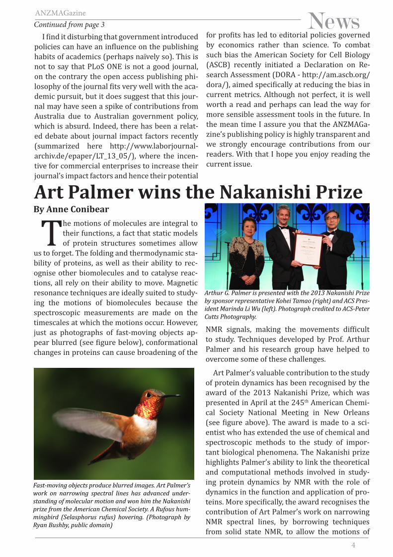
us to forget. The folding and thermodynamic sta-bility of proteins, as well as their ability to rec-ognise other biomolecules and to catalyse reac-tions, all rely on their ability to move. Magnetic resonance techniques are ideally suited to study-ing the motions of biomolecules because the spectroscopic measurements are made on the timescales at which the motions occur. However, just as photographs of fast-moving objects ap-pear blurred (see figure below), conformational changes in proteins can cause broadening of the
NMR signals, making the movements difficult to study. Techniques developed by Prof. Arthur Palmer and his research group have helped to overcome some of these challenges.
Art Palmer’s valuable contribution to the study of protein dynamics has been recognised by the award of the 2013 Nakanishi Prize, which was presented in April at the 245th American Chemi-cal Society National Meeting in New Orleans (see figure above). The award is made to a sci-entist who has extended the use of chemical and spectroscopic methods to the study of impor-tant biological phenomena. The Nakanishi prize highlights Palmer’s ability to link the theoretical and computational methods involved in study-ing protein dynamics by NMR with the role of dynamics in the function and application of pro-teins. More specifically, the award recognises the contribution of Art Palmer’s work on narrowing NMR spectral lines, by borrowing techniques from solid state NMR, to allow the motions of
Art Palmer wins the Nakanishi PrizeBy Anne Conibear
Fast-moving objects produce blurred images. Art Palmer’s work on narrowing spectral lines has advanced under-standing of molecular motion and won him the Nakanishi prize from the American Chemical Society. A Rufous hum-mingbird (Selasphorus rufus) hovering. (Photograph by Ryan Bushby, public domain)
Arthur G. Palmer is presented with the 2013 Nakanishi Prize by sponsor representative Kohei Tamao (right) and ACS Pres-ident Marinda Li Wu (left). Photograph credited to ACS-Peter Cutts Photography.
I find it disturbing that government introduced policies can have an influence on the publishing habits of academics (perhaps naïvely so). This is not to say that PLoS ONE is not a good journal, on the contrary the open access publishing phi-losophy of the journal fits very well with the aca-demic pursuit, but it does suggest that this jour-nal may have seen a spike of contributions from Australia due to Australian government policy, which is absurd. Indeed, there has been a relat-ed debate about journal impact factors recently (summarized here http://www.laborjournal-archiv.de/epaper/LT_13_05/), where the incen-tive for commercial enterprises to increase their journal’s impact factors and hence their potential
for profits has led to editorial policies governed by economics rather than science. To combat such bias the American Society for Cell Biology (ASCB) recently initiated a Declaration on Re-search Assessment (DORA - http://am.ascb.org/dora/), aimed specifically at reducing the bias in current metrics. Although not perfect, it is well worth a read and perhaps can lead the way for more sensible assessment tools in the future. In the mean time I assure you that the ANZMAGa-zine’s publishing policy is highly transparent and we strongly encourage contributions from our readers. With that I hope you enjoy reading the current issue.
Continued from page 3
5
ANZMAGazine
biomolecules to be studied more easily.
Colleagues and students of Koji Nakanishi, who has been a colleague of Art Palmer’s at Co-lumbia University for 21 years, established the Nakanishi Prize in 1995. Art considers it a spe-cial honour to receive the Nakanishi Prize as Koji’s influence on his career precedes their time together at Columbia and the award ‘closes a cir-cle in (his) mind’. In fact, the first symposium that Art attended as an undergraduate at Haverford College was presented by Koji Nakanishi and still stands out in his memory.
Art Palmer is the Robert Wood Johnson Jr. Pro-fessor of Biochemistry and Molecular Biophysics in the Department of Biochemistry and Molecu-lar Biophysics. His work on developing tools to measure spin relaxation rates of proteins and nucleic acids provides methods for investigating enzyme catalysis, protein folding and protein-nucleic acid interactions. For example, the Palm-er laboratory uses statistical mechanical meth-ods to analyse the free energy changes that occur when enzymes bind to their substrates. In one study, the energy cost involved when the yeast transcription factor GCN4 binds to DNA was de-termined and compared with results obtained by molecular dynamics simulations. Another exam-ple in which theoretical models have been inte-grated with biological applications is the study on the temperature dependence of the stability and activity of proteins. The study revealed that small differences in the sequences of ribonu-clease H from mesophilic (Escherichia coli) and thermophilic (Thermus thermophiles) bacteria (see figure on right) lead to differences in ther-mal stability, helping to explain why otherwise homologous enzymes are able to function at very different temperatures.
During his doctoral studies at the University of North Carolina, Art first became interested in time-dependent phenomena and started reading some papers on NMR given to him by a member of his doctoral committee. His career in NMR was launched during his postdoctoral work in the laboratory of Dr. Peter Wright at the Scripps Research Institute where he started to use NMR spectroscopy to study protein dynamics. Now as a professor, Art says that the interactions he has with his graduate students and postdocs are one
of the aspects he most enjoys about his work, and he credits most of the advances in the field to their ideas and insights. He particularly enjoys discussions with his postdocs and students about their data in which they prove themselves right and him wrong! Art has taught NMR spectrosco-py widely, including in Australia at the course or-ganised by Gottfried Otting, and comments that ‘working through problems with students and wrestling with their questions have taught me as much as I have taught them, maybe more’.
When asked about the new challenges in the NMR field, Art responded that the challenges in a technique-driven field like NMR are two-fold: ‘to improve the efficiency and robustness of the experiments so that a broader range of scien-tists can employ sophisticated methods in their own work and to improve the range of biologi-cal systems accessible to the methods, so that more difficult and complex questions in biology and biophysics can be answered. Many research groups have made tremendous progress in both areas over the past decade and I am excited for the next!’
The award of the Nakanishi prize is a fitting honour for someone who has contributed signifi-cantly to the NMR community in research, teach-ing, publications, and software and ANZMAG would like to congratulate Art Palmer on this prestigious award.
Cartoon representation of ribonuclease H (RNaseH, PDB code 2RN2) from Escherichia coli. The temperature depend-ence of protein dynamics allows homologous enzymes such as RNase H to have different temperature profiles in differ-ent organisms. Figure drawn using Pymol.
6
ANZMAGazine
News
My main research field is Electron Par-amagnetic Resonance (EPR) spec-troscopy, a technique that probes
the interaction of unpaired electrons with their surroundings. I became interested in EPR when I took my first postdoctoral position in 1999 at the ETH in Zürich, with Prof. Schweiger. In 2007 I moved to Oxford University, UK to help estab-lish and manage the Centre of Advanced Electron Spin Resonance (CAESR), and recently (2013) I started an ACR Future Fellowship (FF) at the Centre for Advanced Imaging (CAI), the Univer-
sity of Queensland.
One of my core research interests is methodol-ogy development and the application of pulse di-polar spectroscopy (PDS) in the biosciences. PDS refers to EPR techniques to measure the distance between two paramagnetic centres in the unique range ca 15-100 Å. The two main sequences of PDS are Double Electron-Electron Resonance (DEER), or synonymously pulsed electron dou-ble resonance (PELDOR), and double-quantum EPR (DQ-EPR). The paramagnetic centres uti-lized by PDS could be intrinsic to the biomole-cule, or added via site-directed spin labelling (e.g. for proteins the nitroxide MTSL). The technique is applicable to a wide range of samples, can be carried out in powders and frozen solutions, and is robust to sample preparation. PDS studies, for example, have been carried out with soluble and membrane proteins and their oligomers, DNA and RNA. A large part of my FF resources have been utilized on equipment upgrades to the ex-isting CAI spectrometers, in particular special-ized resonators for X-band (∼9.5 GHz) PDS stud-ies, and the procurement of a high-power TWT amplifier at Q-band (∼33 GHz), which provides a state-of-the-art spectrometer to carry out exper-iments with the highest sensitivity and resolu-tion. A very comprehensive overview of the field can be found in a Structure and Bonding volume ‘Spin-labels and intrinsic paramagnetic centres in the Biosciences: Structural information from distance measurements’ which contains 7 chap-ters, is edited by myself and Dr Timmel from Ox-ford University, and is due out this year.
I am also interested in the function of paramag-netic centres in metalloenzymes, having worked on the FeS clusters in Mitochondrial Complex I, P450’s, NiFe-Hydrogenase, and the nickel cen-tre at the active site of methyl-coenzyme M re-ductase. The paramagnetic centres in these en-zymes are probed by measuring the couplings to nearby nuclear spins e.g. 1H, 14N, 13C, 31P. The
DEER spectroscopy to determine the protein-protein com-plex between a P450 and its electron transfer partner using the MTSL spin label and a FeS cluster. Work in collaboration with Dr Wong and Dr Timmel, Oxford University, UK.
ANZM
AG w
elco
mes
new
EPR
exp
ert
By Jeff Harmer
Pulse and CW EPR in Biology, Materials Science, and Chemistry
7
ANZMAGazine
News
At the ANZMAG conference in Torquay in 2011, an education committee was cre-ated. Its inception was driven by discus-
sion on how to effectively teach magnetic reso-nance to those new to the field. Historically each group has had their own way of teaching their new staff and students and this is repeated as often as new staff arrive. It seemed like it would be more efficient to arrange for an ANZMAG run course that would benefit the entire community and ease the constant retraining by the various group leaders.
Indeed in the past there has been an ANZMAG led NMR course in conjunction with each AN-ZMAG conference, most recently run by amongst others Prof. Gottfried Otting. It seemed natural to continue this tradition but Gottfried soon sug-gested that perhaps if we were to record the lec-tures and make these freely available online, then the students could learn in their own time and this would be a good resource not only in Aus-tralia but more generally to the NMR community.
Given that we were tasked with recording such a lecture series which would last the test of time, it seemed appropriate to try to find the best NMR educator we could think of. A few names were floated but the on top the list was Dr James Keel-er from the University of Cambridge. Initially we
doubted that he would find time to fly half way across the world to stand in front of a camera and teach our students, but with some kind words from Gottfried and with ANZMAG generously of-fering to pick up the expenses, we received very encouraging news that he would be interested to give a series of lectures based on his popular “Understanding NMR Spectroscopy” book.
The next task was to find an appropriate place to run the course. We were somewhat con-
measurements provide structural and electronic information, for example identification of ligands and their position relative to a metal centre, and electronic information from hyperfine couplings which provide a map of the centres spin density.
At CAI we have equipment to carry out multi-fre-quency continuous wave (CW) EPR and pulse ex-periments, such as electron nuclear double res-onance (ENDOR), electron spin-echo envelope spectroscopy (ESEEM), and hyperfine sublevel correlation spectroscopy (HYSCORE).
I am very happy to provide more information or to discuss a potential collaboration upon re-quest.
Dr Jeffrey HarmerCentre for Advanced Imaging The University of QueenslandSt Lucia, Brisbane, QLD, 4072Email: [email protected]: (+61) 7 334 60351
HYSCORE characterization of the unique [4Fe-3S] cluster in the O2-tolerant [NiFe]-hydrogenase Hyd-1 from E. coli. JACS, 2012, 134 (37), 15581-15594.
Report from the “Keeler course”By Mehdi Mobli
8
ANZMAGazine
Newsstrained by Dr Keeler’s schedule, and needed to find somewhere appropriate to hold the course at the beginning of September. Given the time of the year it seemed that Queensland would have the best climate for the course. Myself and Dr Horst Schirra embarked on a hunt for a suitable venue, but as the course would run during Aus-tralian term time, it was difficult to find available student accommodation and lecture rooms at the main UQ campus in St Lucia, where we had held a similar course in 2008. Instead we ended up looking at the different research stations at UQ, and after dismissing the Heron Island research station due to excessive transport costs, we set-tled on the Moreton Bay Research Station on North Stradbroke Island.
The station manager Kevin Townsend was very excited that his new media room would be the venue of a series of publicly available lectures and we soon had booked nearly the entire re-search station for Dr Keeler’s course. The course was advertised and with help from Dr John Ge-hman we had soon set up a registration site and registered ~40 participants. The course also at-tracted a number of sponsors including Bruker, Agilent, Wiley, UQ and QNN, and we also had ex-cellent support from senior ANZMAG members.
Soon we found ourselves on the island with microphones and video recorders and a room full of students and tutors. It was clear that eve-ryone involved felt that it was a very important event, in particular we were impressed by Dr
Keeler bringing 5 identical changes of clothes to ensure continuity in the video production. With a couple of minor technical issues and interrup-tions by local noisy wildlife we had amassed 15 lectures and nearly as many hours of recorded material. The editing step took myself and Dr Ann Kwan nearly 6 months to finish, and after diligent proof watching work by ANZMAG volunteers, the lectures were finalised and finally advertised on the 20th of June 2013. The videos have since been viewed ~6,000 times with 10,000 minutes watched from 49 countries, with more people from USA and UK watching the videos than from Australia. It is clear that the videos have had a great and immediate impact in the 3 months they have been available, and this is a testament to the effectiveness of Dr Keelers book, which the series is based on as well as his passion for teaching which clearly shines through in these lectures. ANZMAG is indebted to Dr Keeler for coming to Australia and providing these lectures and on be-half of the society, Prof. Caroline Rae presented James with an award at the end of the lecture se-ries (see picture on previous page).
The “Keeler course” has set an excellent prec-edence, and soon we will record a lecture by Prof. Ad Bax on RDCs at the upcoming APNMR meet-ing in Brisbane, and we hope to continue to regu-larly add educational materials to the ANZMAG YouTube channel (http://www.youtube.com/user/ANZMAG/videos).
To the NMR cave…change is afootBy Oliver Jones
Affectionately referred to by its users as the NMR cave (visit and you’ll see why), change is afoot at Bio21’s NMR
facility. For a start it will soon (probably by the time you read this) not be known as Bio21 at all and will instead be Melbourne Magnetic Resonance. It will also be part of the ARIN - the Australian Research Infrastructure Network (http://arin.org.au/). Funded by the Victorian Government, ARIN is a new initiative to make all public research infrastructure (not just NMR)
easier to access and manage. The system will in-corporate online booking, billing and reporting capabilities and looks set to really make a differ-ence to analytical research in Victoria and the long-term plan is to invite other states to join. But enough about that, what about the facility itself?
Bio21 was established in 2002 and official-ly opened in 2005. A word of warning though, whether you call it Bio21 or Melbourne Magnet-
9
ANZMAGazine
ic Resonance the facility is not a place to go to if you like magnetic resonance and you get jealous easily. The facility is one of the best examples of its type in the southern hemisphere. Around AU$20 million worth of top end NMR spectrom-eters are spread out all over several hundred square feet of space and the area is a hive of ac-tivity. Dominating the lab is a massive 800 MHz high-field NMR spectrometer equipped with cyroprobe technology (see figure below) and running a close 2nd is a 700 MHz machine with a refrigerated auto-sampler. As part of ARIN, Mel-bourne Magnetic Resonance will also have ac-cess to additional instrumentation at Melbourne University and Monash Universities. Seven out of eight LIEF grants applied for to equip the fa-cility in recent years have been successful, so it will likely stay ahead of the pack in terms of new equipment in the future.
This aggregation of high- end instrumenta-tion in a single facility serves as a magnet (par-don the pun) for attracting academic research and industry collaborations. The Institute’s pri-mary uses of NMR are investigating the struc-tures of potential drug and pesticide targets, proteins pharmaceuticals, and studies of me-
tabolite biomarkers of disease, as well as the in-teractions of proteins with their specific ligands (including candidate drugs). This is not the limit though and the analytical capability of the facil-ity is limited only by the imagination of its us-ers. Said users are as diverse a list as one could think of and include scientists from all over Aus-tralia (and further afield) as well as industry, the CSIRO and DSTO. Hundreds, perhaps thousands of samples, are run here every day. As facility manager Dr. David Keizer tells me “basically, if you can think of a use for magnetic resonance then we can make it happen”.
If you want to take advantage of this facil-ity then your first stop should be to contact Dr Hamish Grant, Dr Shenggen Yao, or Dr Keiser. You will be trained up to use the machines and then will be able to run your own samples, with results being e-mailed back to you as soon as they are ready. For now you can find out more at http://www.bio21.unimelb.edu.au/platform-technologies/nmr-facility but in future the ARIN website will be the place to go. This is an excit-ing time to be involved in magnetic resonance in Victoria, make sure you are part of it.
View of the NMR cave from Bio21 reception
The beast - 800 MHz NMR
10
ANZMAGazine
Article
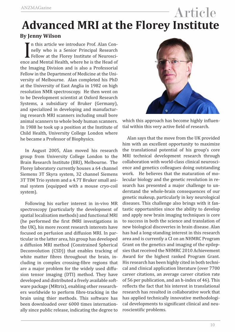
In this article we introduce Prof. Alan Con-nelly who is a Senior Principal Research Fellow at the Florey Institute of Neurosci-
ence and Mental Health, where he is the Head of the Imaging Division and is also a Professorial Fellow in the Department of Medicine at the Uni-versity of Melbourne. Alan completed his PhD at the University of East Anglia in 1982 on high resolution NMR spectroscopy. He then went on to be Development scientist at Oxford Research Systems, a subsidiary of Bruker (Germany), and specialized in developing and manufactur-ing research MRI scanners including small bore animal scanners to whole body human scanners. In 1988 he took up a position at the Institute of Child Health, University College London where he became a Professor of Biophysics.
In August 2005, Alan moved his research group from University College London to the Brain Research Institute (BRI), Melbourne. The Florey laboratory currently houses a 64 channel Siemens 3T Skyra system, 32 channel Siemens 3T TIM Trio system and a 4.7T Bruker small ani-mal system (equipped with a mouse cryo-coil system).
Following his earlier interest in in-vivo MR spectroscopy (particularly the development of spatial localisation methods) and functional MRI (he performed the first fMRI investigations in the UK), his more recent research interests have focused on perfusion and diffusion MRI. In par-ticular in the latter area, his group has developed a diffusion MRI method (Constrained Spherical Deconvolution (CSD)) that enables tracking of white matter fibres throughout the brain, in-cluding in complex crossing-fibre regions that are a major problem for the widely used diffu-sion tensor imaging (DTI) method. They have developed and distributed a freely available soft-ware package (MRtrix), enabling other research-ers worldwide to perform fibre-tracking in the brain using thier methods. This software has been downloaded over 6000 times internation-ally since public release, indicating the degree to
which this approach has become highly influen-tial within this very active field of research.
Alan says that the move from the UK provided him with an excellent opportunity to maximize the translational potential of his group’s core MRI technical development research through collaboration with world-class clinical neurosci-ence and genetics colleagues doing outstanding work. He believes that the maturation of mo-lecular biology and the genetic revolution in re-search has presented a major challenge to un-derstand the whole-brain consequences of our genetic makeup, particularly in key neurological diseases. This challenge also brings with it fan-tastic opportunities since the ability to develop and apply new brain imaging techniques is core to success in both the science and translation of new biological discoveries in brain disease. Alan has had a long-standing interest in this research area and is currently a CI on an NHMRC Program Grant on the genetics and imaging of the epilep-sies that received the NHMRC 2010 Achievement Award for the highest ranked Program Grant. His research has been highly cited in both techni-cal and clinical application literature (over 7700 career citations, an average career citation rate of 56 per publication, and an h-index of 46). This reflects the fact that his interest in translational research has resulted in collaborative work that has applied technically innovative methodologi-cal developments to significant clinical and neu-roscientific problems.
By Jenny WilsonAdvanced MRI at the Florey Institute
11
ANZMAGazine
ArticleMR at the NationalImaging FacilityBy Graham Galloway
The National Imaging Facility was estab-lished in 2006 as part of the Characteri-sation Capability within the National
Collaborative Research Infrastructure Strategy (NCRIS). From a relatively small investment of AU$7M by the federal government, it has grown to a distributed capability across 10 universities and institutes, partnering with ANSTO, to deliver over AU$200M of infrastructure to the research community. This has required cash support from State Governments and the institutions them-selves. It has also depended on the generosity of institutions making existing equipment available to the wider community. All access is charged at marginal cost recovery or less.
But NIF is more than the equipment. This is a technology that is in high demand by extremely good scientists who have no training in imag-ing. As Prof. Simon Ringer and I travelled around the country, preparing the development of the NCRIS Investment Plan, there was a consistent emphasis, from fellow scientists, on the need for expert support. And so, in the initial plan, and in developments since, NIF has ensured that every Flagship Instrument has a Facility Fellow, who will help users choose the best modality, design the experiments, analyse and interpret the data. This ensures the optimum use of the capabilities, but more importantly, it facilitates the best pos-sible data.
12
ANZMAGazine
Biomedical imaging is a major driver of imag-ing technology, both as a tool to understand basic physiology and biochemistry, in animal models as well as humans, and a tool to support clini-cal diagnosis and patient management. And so it is central to translational research. Transla-tion is a multi-faceted and often misused word. But, in whatever way it is used, imaging has a role. In bench to bedside, imaging can connect molecular process with clinical symptoms. From animal models to clinical trials, imaging is a key biomarker, in identifying disease, excluding con-founding co-morbidities, as well as following the efficacy of treatment. Imaging is an important tool in identifying targets for novel therapy, and in designing protocols for delivery of that ther-apy. As we move into the world of theranostics, nanomaterials incorporate functional groups to target the disease process, along with imaging probes to follow their progress through the body, so that they can deliver a therapeutic payload di-rectly to the underlying source of the pathology.
But NIF has projects way beyond biomedi-cal applications. Demand comes from a highly diverse range of fields, studying materials from
reinforced concrete to coral reefs, from under-standing frost resistance in grapes to following an avocado from tree to shopping bag to under-stand when it is bruised, and develop strategies to ensure you get a better quality fruit on your dinner plate. There are projects in ecology, look-ing at the evolution of our distinct fauna, and even projects to extract more information from fossils.
Whilst not only MR based, there is a very strong MR component to NIF, and many of the leaders have been active in MR and ANZMAG for many years. Contrast in MR is based on molecu-lar properties, and so understanding of the basics of NMR is critical to releasing the power of the technique. But while many of us remain passion-ate about MR, we recognise that it can contribute to the advances made in other imaging modali-ties, even transforming what in the past we con-sidered as competition. One of the strengths of NIF is that it combines the power of the different technologies to obtain complementary informa-tion. With the recent appearance of simultaneous MRI with PET, this has entered a new dimension, in which the exquisite anatomical and structural
PET measures metabolic activity, whilst MRI shows volume of fat. In Wildtype, we see lower glucose uptake and more fat. In genetically modified mice with high Brown Fat, we see higher glucose activity and less fat. Prof. G.Muscat, Dr P Lay (Institute for Molecular Bioscience), and Dr G.Cowin, Dr K.Mardon (NIF-UQ)
13
ANZMAGazine
Education
Saturation Transfer Difference (STD) NMR Spectroscopy
By Thomas Haselhorst
STD NMR spectroscopy[1] has become a very popular tool in academia and phar-maceutical industry alike to study pro-
tein-ligand complexes. Since Mayer and Meyer’s study was published in 1999 in Angew. Chem. Int. Ed.[1b] the number of published papers for the topic STD NMR has shown exponential growth[2]. In this short article I will briefly de-scribe the history of STD NMR and highlight the advantages and disadvantages of this method.
Although the phenomenon of saturation transfer itself is not novel and this technique has been known for many years it made a grand renaissance when Mayer and Meyer showed how to utilise this technique to identify ligands that bind to protein targets from a compound library [1b]. The difference spectra were obtained by subtract-ing a spectrum in which the protein is saturated from one acquired without protein saturation, resulting in a differ-ence spectrum, in which only the sig-nals of the binding ligand remain. The use of difference spectroscopy allows discriminatation between a binding and non-binding ligand and therefore has enormous potential as a screening tool. Furthermore, STD NMR spectros-copy can provide information beyond a simple screening approach as signals of protons and groups that are close to the protein surface show higher in-tensity and those that are more distant from the protein or are solvent exposed
show low or no STD NMR signal intensity[1a]. The degree of saturation a proton receives can be directly translated into the binding epitope of a ligand. This structural information becomes invaluable for the medicinal chemist in struc-ture-based drug design as it guides the chemical synthesis of lead structure optimisation.
An enormous advantage of STD NMR spectros-copy is that only a small amount of the protein is required and no expensive protein labelling is necessary. Saturation of the protein is normally achieved by selectively irradiating the protein resonances with a train of Gaussian soft pulses, however E-BURP pulses have been shown to increase sensitivity. The magnetisation is then
detail afforded by MRI is combined with the high molecular sensitivity of PET. The UQ node of NIF installed the world’s first commercial pre-clini-cal MR-PET, capable of simultaneous acquisition. One of the exciting areas of research is to use X-nuclei MR, imaging and spectroscopy, togeth-er with PET imaging. Imaging has come a long
way, but there is still so much more to learn as we delve into the untapped potential of this ever expanding access to contrast mechanisms that probe molecular structure in living systems.
14
ANZMAGazine
transferred rapidly through the entire protein mediated through spin diffusion. In case a small ligand or drug binds to the saturated protein, the saturation can then be transferred to this ligand. In addition, experiments can be quickly acquired, combined with virtually any 2D ex-periment (e.g. TOCSY and HSQC) and results can be obtained within less than an hour.
In a typical STD NMR experiment a ligand to protein ratio of about 100:1 is used and satu-ration of protein resonances (on-resonance) can be obtained by irradiating regions of the 1H NMR spectrum, typically in the aliphatic re-gion, between -1 and -2 ppm, a spectral region with no ligand protons but containing sufficient signals of the target protein. The off-resonance spectrum is acquired with a saturation frequen-cy where no protein or ligand signals are detect-able, usually at 30 ppm. For larger targets like cells and virus particles it is recommended to set the off-resonance frequency to 300 ppm due to the inherently large line width of high molec-ular weight macromolecules and the likelihood of resonance occurring at an Off-resonance fre-quency of 30 ppm. Subtraction of the on-reso-nance spectrum from the off-resonance one, re-sults in the final saturation transfer difference (STD) NMR spectrum showing only signals from ligands that have binding affinity.
The STD NMR effect can be quantified by group epitope mapping (GEM)[1a], which is only possible when the dissociation rate constant (koff) is greater than the T2 relaxation rate of the bound ligand. The STD amplification factor[1a] is obtained by multiplying the relative STD NMR effect (ASTD) of a given proton (ISTD/I0) at a given ligand concentration [L]T with the molar ratio of ligand in excess relative to the protein [L]T/[P].
The differences in ASTD for different protons can be quantitatively expressed by analysing the relative STD NMR effects at a given satura-tion time, mapping of moieties or residues of the ligand crucial for interaction. The difference
in the relative STD response (ISTD / I0 or ASTD) for each proton should reflect the proximity of that ligand proton to the protein surface.
The enormous potential of STD NMR spec-troscopy lies in the fact that there is no size lim-itation of the macromolecule. In fact, STD NMR has been extended to interrogate the interac-tion of ligands or substrates with immobilised proteins[3], Golgi enriched fractions[4], whole intact virions[5], virus like particles (VLPs)[6] and even fungi spores[7]. The large molecular weight of bulky particles makes them particu-larly attractive for STD NMR spectroscopy stud-ies because their inherently large line width en-ables saturation of the particle without affecting the ligand signals. Additionally, the larger cor-relation time of bulky particles results in effi-cient spin diffusion and consequently stronger saturation transfer. The introduction of double STD NMR (STDD) has been used successfully to eliminate unwanted binding and background noise [8].
Despite all the above mentioned advantages and practicality of STD NMR spectroscopy, it surely has its limitations. Firstly, this method is not suitable for detecting high-affinity ligands that undergo slow chemical exchange and typi-cally reside longer within the protein-bind-ing site. Similarly, if binding is very weak, the probability of the ligand being in the receptor site becomes very low resulting in weak or no STD NMR signals. Usually binding ligands with KD values in the 10-2 – 10-8 M range[1c] can be detected well, using this method. The introduc-tion of ‘spy’ molecules or low affinity binder in competition with high-affinity ligands is an al-ternative and has been shown to be useful but would require additional experiments and NMR time. Direct excitation of ligand signals can also be problematic if ligand resonances are close to the saturation frequency. In this case additional control STD NMR spectra are also required. Se-lective water flip-back and suppression might impose problems and can affect neighbouring ligand signals, especially in sugars. One must also pay close attention, when interpreting saturation in terms of distance, because signal reduction is sometimes distorted by inhomoge-neous saturation of the receptor[9]. Differences
15
ANZMAGazine
in the longitudinal relaxation times of individ-ual ligand protons have also been shown to be problematic[10]. Furthermore, spin diffusion within the ligand easily spoils the exact distance information between the receptor and ligand and complete relaxation matrix analysis may be required to interpret the data correctly[9]. The application of STD NMR in a recent fragment-based drug design (FBDD) study revealed that a large number of hits identified using STD NMR spectroscopy showed disappointingly only very little overlap with results from Surface Plasmon Resonance (SPR) [11]. A more recent paper by the Scanlon group reports that only ~50% of STD NMR hits identified in the primary screen are subsequently confirmed by a second screen-ing method [12]. A number of factors might be responsible that lead to false positive STD NMR effects in library screening experiments. For ex-ample, the naturally higher concentration of a compound mixture can potentially cause aggre-gation and therefore increased rapid relaxation rates and large NOEs that consequently produce false positive STD NMR effects. Therefore, the choice of the correct protein and ligand con-centration, library design, and the acquisition of appropriate control experiments are crucial factors that need to be optimised.
In summary it can be said that STD NMR is a practical method for screening and lead optimi-sation, but it is clear when STD NMR is applied in isolation, much like any other technique, it cannot be fully effective. However, if results are analysed with care and in combination with a ‘smart’ choice of control experiments and struc-tural analysis, this method has its merits, and I am sure, will continue to be used in the future in academia and pharmaceutical industry alike.
References:
[1] a) M. Mayer, B. Meyer, Journal of the American Chemical Society 2001, 123, 6108-6117; b) M. Mayer, B. Meyer, Angew Chem Int Ed Engl 1999, 38, 1784-1788; c) B. Meyer, T. Peters, Angew Chem Int Ed Engl 2003, 42, 864-890.
[2] Web of Knowledge, http://webofknowledge.com.[3] T. Haselhorst, A. K. Münster-Kühnel, M. Oschlies, J.
Tiralongo, R. Gerardy-Schahn, M. von Itzstein, Biochemical and biophysical research communications 2007, 359, 866-
870.[4] A. Maggioni, M. von Itzstein, J. Tiralongo, T. Hasel-
horst, Chembiochem : a European journal of chemical biol-ogy 2008, 9, 2784-2786.
[5] A. J. Benie, R. Moser, E. Bauml, D. Blaas, T. Peters, Journal of the American Chemical Society 2003, 125, 14-15.
[6] T. Haselhorst, J. M. Garcia, T. Islam, J. C. Lai, F. J. Rose, J. M. Nicholls, J. S. Peiris, M. von Itzstein, Angew Chem Int Ed Engl 2008, 47, 1910-1912.
[7] A. Maggioni, J. Meier, F. Routier, T. Haselhorst, J. Tiralongo, Chembiochem : a European journal of chemical biology 2011, 12, 2421-2425.
[8] B. Claasen, M. Axmann, R. Meinecke, B. Meyer, Jour-nal of the American Chemical Society 2005, 127, 916-919.
[9] V. Jayalakshmi, N. R. Krishna, J Magn Reson 2002, 155, 106-118.
[10] J. Yan, A. D. Kline, H. Mo, M. J. Shapiro, E. R. Zartler, J Magn Reson 2003, 163, 270-276.
[11] J. Wielens, S. J. Headey, D. I. Rhodes, R. J. Mulder, O. Dolezal, J. J. Deadman, J. Newman, D. K. Chalmers, M. W. Parker, T. S. Peat, M. J. Scanlon, Journal of biomolecular screening 2013, 18, 147-159.
[12] B. C. Doak, C. J. Morton, J. S. Simpson, M. J. Scanlon, Aust J Chem 2013, in press.
16
ANZMAGazine
Education
Quadrupolar nuclei outnumber spin-half nuclei by around three to one, and yet
they are far less frequently studied by solid-state NMR. Their defining feature is an asym-metric distribution of nuclear electric charge, which means that they interact not only with a magnetic field but also with an electric field gradient. This additional quadrupolar interac-tion can be very large and can have major con-sequences on the NMR spectrum, even altering the way the nucleus responds to RF pulses. As a result, slightly more advanced methods are of-ten required to observe these nuclei and inter-pret their spectra.
All quadrupolar nuclei have a spin number S > 1/2, and for now we will concentrate on half-integer quadrupoles and save integer spin nu-clei for the next issue. Some commonly studied examples of half-integer quadrupolar nuclei include 7Li (S = 3/2), 23Na (3/2), 27Al (5/2) and 87Rb (3/2). While rubidium is perhaps a less commonly encountered element than the for-mer three, it is particularly useful for testing out new solid-state NMR pulse sequences since it has a fairly high receptivity and is close in fre-quency to 13C.
A spin-3/2 nucleus has three transitions, while a spin-5/2 nucleus has five. Nuclei with
S = 7/2 and 9/2 also exist, though are less com-mon. In all cases, the central transition (CT, +1/2 ↔ −1/2) is unique, since it is affected by the quadrupolar interaction to a much smaller extent than the others (the so-called satellite transitions - ST). The CT therefore shows a much narrower line width, a correspondingly higher signal intensity, and is much easier to excite and observe than the satellites while still containing all of the information we are inter-ested in. The STs are broadened over extremely wide frequency ranges (usually several MHz or more) and are rarely observed. In most solid-state NMR experiments carried out on half-inte-ger quadrupoles, we only bother to observe the CT and can often ignore the satellites altogether.
In the previous article we learned that magic angle spinning (MAS) can be used to average anisotropic interactions to zero, enabling the acquisition of narrow, isotropic spectral peaks from solid powder samples. Unfortunately, this only works for spin-half nuclei, and MAS alone isn’t enough to average away the quadru-polar interaction. No matter how fast we spin
The Quadrupolar Quandary: Central Transitions
By Luke O’Dell
The Solid State
Around 75% of NMR-active isotopes are quadrupolar, and these can some-times be seen as inaccessible or intimidating by those unfamiliar with them. In the first of a two-part article, Luke O’Dell attempts to demystify these nuclei, briefly outlining what sets them apart from spin-half isotopes and discussing a few key techniques that can be used to study them in the solid state.
HLiNa
K NiRb Mo
GeCuCo
HeB C N O F NeAl Si P S Cl Ar
Ca Sc Ti Cr Fe Zn Ga As Se Br KrSr Y Zr Nb Tc Ru Rh Ag Cd In Sn Sb Te I Xe
Cs Ba La Hf Ta W Re Os Ir Pt Au Hg Tl Pb Bi Po AtPd
Rn
MgBe
V Mn
Elements highlighted in red feature a quadrupolar nucleus
−1/2
−3/2
+1/2
+3/2
fνL
CT
ST
STST
ST
CT
The central transition (CT) is much narrowerthan the satellite transitions (STs)
17
ANZMAGazine
the sample, the central transition signal from a quadrupolar nucleus will retain a characteristic second-order quadrupolar line shape. On one hand this can be useful since the shape of this powder pattern provides additional informa-tion on the local structural environment and can also be sensitive to dynamics. However, since the patterns from different sites will usually overlap, the spectral resolution suffers, making spectra hard to interpret.
Higher magnetic fields can be used to reduce the widths of CT powder patterns, but are not always available. Fortunately, several ingenious techniques exist that can allow high-resolution, isotropic peaks to be obtained from half-integer quadrupoles. Double Rotation (DOR) is a brute force way of fully averaging out the quadrupo-lar interaction by spinning the sample around two rotation axes simultaneously. Dynamic Angle Spinning (DAS) is based on the same principles but in this case the spinning angle is rapidly flipped during the experiment. These methods, however, are technically challenging and require specialised probes. A simpler ap-proach is to manipulate the nuclear magneti-sation itself rather than the sample as a whole. Multiple Quantum MAS (and its cousin Satellite Transition MAS) are pulse sequences that can be applied using standard MAS hardware. In these two-dimensional experiments, the inher-ent symmetry in the quadrupolar interactions experienced by the various transitions can be exploited to refocus the anisotropic broadening, resulting in high resolution isotropic spectra in the indirect dimension while retaining the ani-sotropic information in the direct one. The MQ-MAS experiment was introduced by Frydman & Harwood in 1995 and quickly became a stand-ard technique for quadrupolar nuclei (their ini-tial paper now has over 1000 citations).
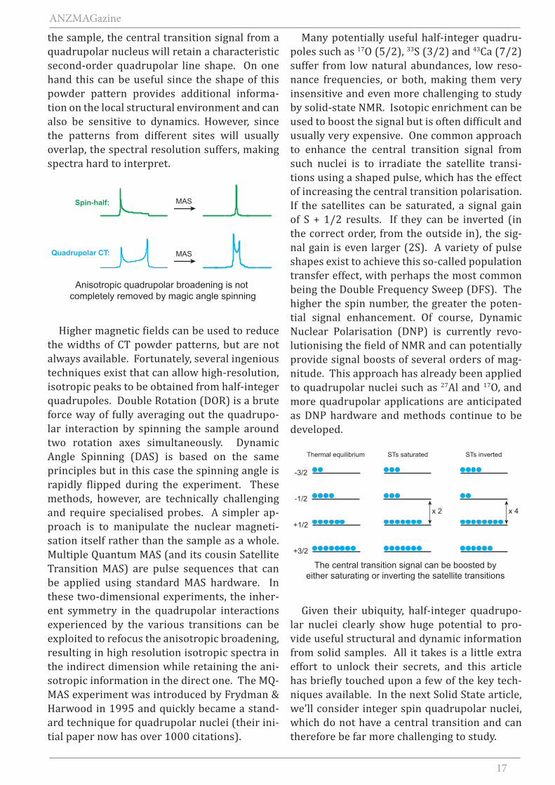
Many potentially useful half-integer quadru-poles such as 17O (5/2), 33S (3/2) and 43Ca (7/2) suffer from low natural abundances, low reso-nance frequencies, or both, making them very insensitive and even more challenging to study by solid-state NMR. Isotopic enrichment can be used to boost the signal but is often difficult and usually very expensive. One common approach to enhance the central transition signal from such nuclei is to irradiate the satellite transi-tions using a shaped pulse, which has the effect of increasing the central transition polarisation. If the satellites can be saturated, a signal gain of S + 1/2 results. If they can be inverted (in the correct order, from the outside in), the sig-nal gain is even larger (2S). A variety of pulse shapes exist to achieve this so-called population transfer effect, with perhaps the most common being the Double Frequency Sweep (DFS). The higher the spin number, the greater the poten-tial signal enhancement. Of course, Dynamic Nuclear Polarisation (DNP) is currently revo-lutionising the field of NMR and can potentially provide signal boosts of several orders of mag-nitude. This approach has already been applied to quadrupolar nuclei such as 27Al and 17O, and more quadrupolar applications are anticipated as DNP hardware and methods continue to be developed.
Given their ubiquity, half-integer quadrupo-lar nuclei clearly show huge potential to pro-vide useful structural and dynamic information from solid samples. All it takes is a little extra effort to unlock their secrets, and this article has briefly touched upon a few of the key tech-niques available. In the next Solid State article, we’ll consider integer spin quadrupolar nuclei, which do not have a central transition and can therefore be far more challenging to study.
MASSpin-half:
Quadrupolar CT:
Anisotropic quadrupolar broadening is not completely removed by magic angle spinning
MAS
x 2 x 4
-3/2
-1/2
+1/2
+3/2
STs saturatedThermal equilibrium
The central transition signal can be boosted byeither saturating or inverting the satellite transitions
STs inverted
18
ANZMAGazine
Industry
Cryoprobes have now been around for approaching 15 years but it took some time before the first Australian
labs committed to purchasing one. As well as waiting to see that the technology really worked (4-5 x sensitivity improvement with minimal compromise in other performance parameters), the expense of purchasing, siting & maintenance was not a trivial matter. The siting (3 phase power, provision of chilled water or locating an outdoor cooling unit) and the 10,000 hr service are costly. However, such cryoprobes have become an integral component of protein/large molecule NMR systems (typically 600 MHz and above).
To date in Australia the penetration of Cryoprobes into chemistry/small molecule labs has been minimal, even though they are available at 400 and 500 MHz. Whilst the costs mentioned above are no doubt a factor, this may also have been in part due to the absence of a more flexible multinuclear version.
With the introduction of the Bruker Cryoprobe Prodigy, these issues have been addressed to some extent. The Prodigy is not based on
cooling with Helium gas (which requires a complex and expensive Helium gas cooling and control system) but on cooling with liquid Nitrogen which just requires a similar probe, an LN2 dewar and a much simpler and less costly pumping/control unit. No special infrastructure is required - the system can be easily installed. Liquid Nitrogen refill interval is 10 days.
Liquid nitrogen cooled CryoprobesBy Peter Barron,
19
ANZMAGazine
Automated cool-down and warm-up take just a couple of hours. Maintenance is every 2 years and just involves exchange of vacuum pumps. Furthermore the standard version for 400-600 MHz is a BBO (Broadband Observe) and hence optimized for X/13C sensitivity. There is also a TCI (inverse triple HCN) version now for 500-700 MHz. Sensitivity improvement is less than for a Helium Cryoprobe as the coil is only cooled to 77 K and is ca x 2.5 that of an equivalent room temperature probe.
We have just installed such a 400 MHz Prodigy in our Alexandria facility and are very happy to demonstrate its performance. So if you would
like to see a Prodigy in action, don’t hesitate to contact us, and arrange to visit with samples to run. We expect to have the hardware on display at the APNMR/ANZMAG meeting. It is also possible that we can install the Prodigy on loan in labs with a suitable 400 MHz spectrometer.