Page 1
Aortic arch anomaliesCoarctation of the AortaInterrupted Aortic Arch
Echocardiography
V.Tomek, J. Marek, J. Škovránek, J. Gilík
Kardiocentrum, University Hospital Motol, Prague, Czech Republic
No disclosures
Page 2
Coarctation of the Aorta• occurs in 5 – 7% of CHD/ 7th form
• refers to narrowing of the AO isthmus
• circumferential shelf, more prominent along theposterior wall of AO isthmus (juxtaductal)
• wide anatomic spectrum, the length of COA variesfrom discrete to long- segment
• AOA is often elongated/hypoplastic
• >50% complex: + BAO, VSD, AS, MS, SV, TGA, DORV…
Page 3
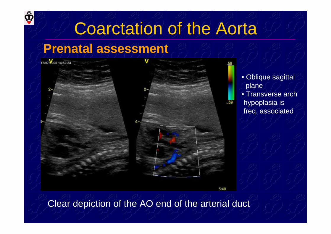
Coarctation of the AortaPrenatal assessment
Asymmetry of ventricular size/ LV afterload, outputLSVC/CS – powerful indicator Pasquini, Heart. 2005
Page 4
Coarctation of the AortaPrenatal assessment
• Oblique sagittalplane
• Transverse arch hypoplasia isfreq. associated
Clear depiction of the AO end of the arterial duct
Page 5
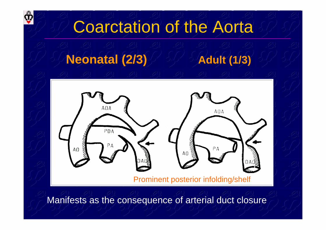
Coarctation of the Aorta
Neonatal (2/3) Adult (1/3)
Prominent posterior infolding/shelf
Manifests as the consequence of arterial duct closure
Page 6
Coarctation of the AortaPulsed Doppler flow pattern in abdom. AO
Critical COA
Closed PDA Opened PDA
Low flow velocityMinimal phasic variationIndicating body perfusion
Slightly decreased or normal amplitudeDiastolic component is reversed ornormal in case of PH
1. - abdominal situs determination
Page 7
Coarctation of the AortaPulsed Doppler flow pattern in abdom. AO
COA + restrictive PDA,preserved LV function
Low syst.wave amplitude
COA – 5 years old
Presence of collaterals
Antegrade diast.flow (= prox.obstr.)
Page 8
Coarctation of the AortaThe function and morpholgy of LV
Subnormal volume, enlarged RV, TR, PHmitral and aortic size
Page 9
Coarctation of the AortaThe function and morpholgy of LV
Normal/ enlarged volume, no PH, LV hypertrophy
Page 10
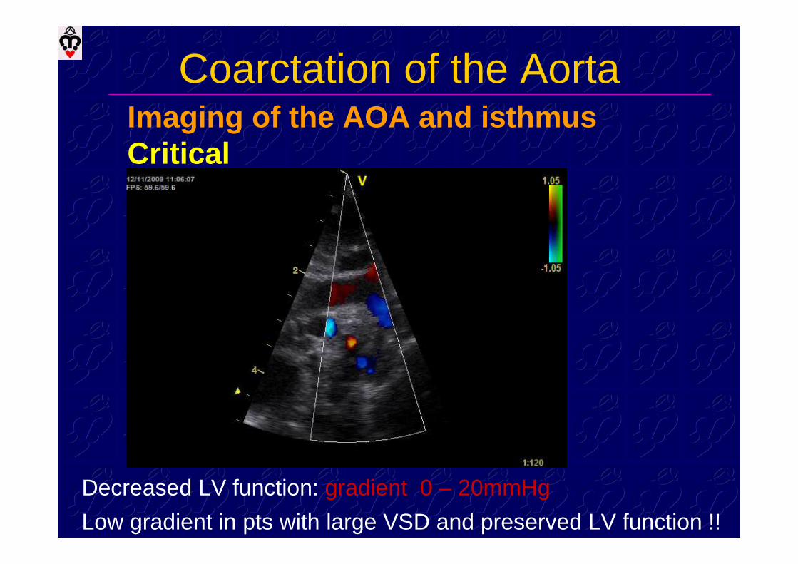
Coarctation of the AortaImaging of the AOA and isthmus
Severe form, normal/ decrease LV function
• Suprasternalnotch view
• Oblique sagittalplane
Page 11
Coarctation of the AortaImaging of the AOA and isthmusCW Doppler
velocity – preserved syst. function, restrictive/no PDA
• „Serrated“ pattern, rapid accel.,earlysystolic peak, gradual diastolicdecelerationcontinuousantegrade flowthroughout diastole
Page 12
Coarctation of the AortaImaging of the AOA and isthmusCritical
Decreased LV function: gradient 0 – 20mmHg
Low gradient in pts with large VSD and preserved LV function !!
Page 13
Coarctation of the AortaImaging of the AOA and isthmusMild or developing COA
Mild isthmic narrowing, usually no typical „ridge“
Page 14
Coarctation of the Aorta„False“ coarctation
Page 15
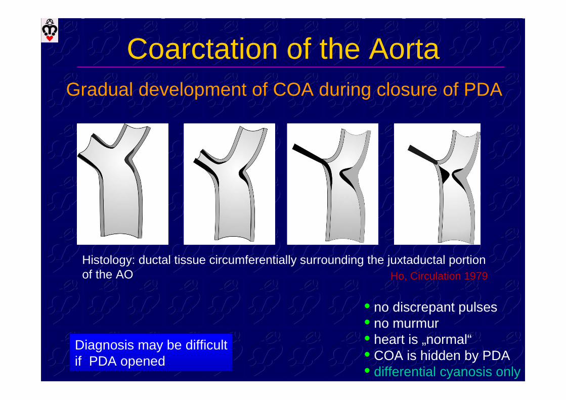
Diagnosis may be difficultif PDA opened
• no discrepant pulses• no murmur• heart is „normal“• COA is hidden by PDA• differential cyanosis only
Coarctation of the AortaGradual development of COA during closure of PDA
Histology: ductal tissue circumferentially surrounding the juxtaductal portionof the AO Ho, Circulation 1979
Page 16
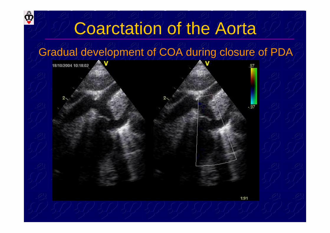
Coarctation of the AortaGradual development of COA during closure of PDA
Page 17
Coarctation of the AortaGradual development of COA during closure of PDA
Page 18
Coarctation of the AortaGradual development of COA during closure of PDA
Page 19
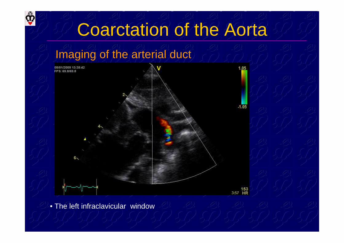
Coarctation of the AortaImaging of the arterial duct
• The left infraclavicular window
Page 20
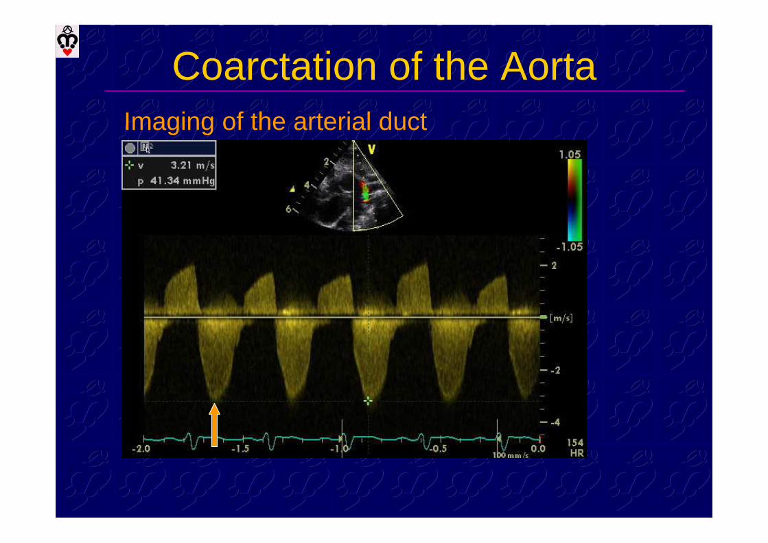
Coarctation of the AortaImaging of the arterial duct
Page 21
Coarctation of the AortaImaging of the arterial duct
• The left infraclavicular window
Page 22
Coarctation of the AortaHypoplastic aortic arch
Hypoplastic = diameter Z-score is less than –2.0
• The diameter of thenarrowest segment• 2D image measurement• the Z-score is calculated
Page 23
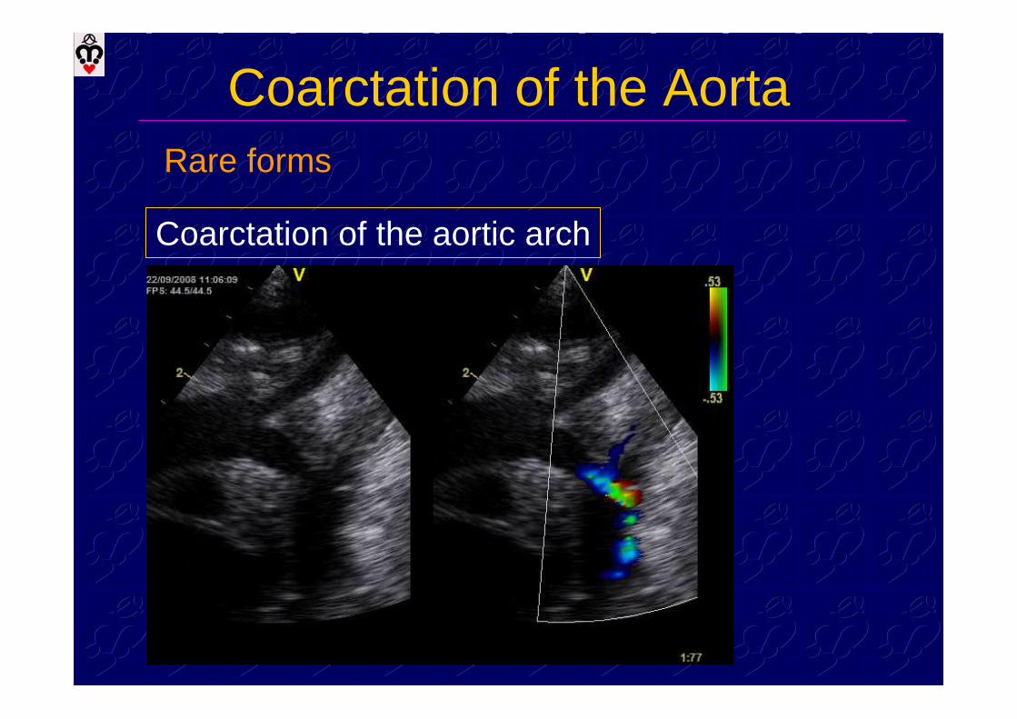
Coarctation of the AortaRare forms
Coarctation of the aortic arch
Page 24
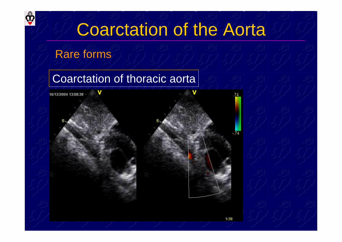
Coarctation of the Aorta
Coarctation of thoracic aorta
Rare forms
Page 25
Coarctation of the Aorta
Coarctation of thoracic aorta
Rare forms
Page 26
Coarctation of the AortaRare forms
Coarctation of abdominal aorta
Page 27
Interrupted aortic arch
• definition: discontinuity between two adjacementsegment of the aortic arch
• 0.38% of all and 1.3 % of critical CHD
Page 28
Embryonic arch diagramInterrupted aortic arch
Page 29
A1 B1 C1
B2 B1RAA
Interrupted aortic arch
Classificationaccording to the site
Page 30
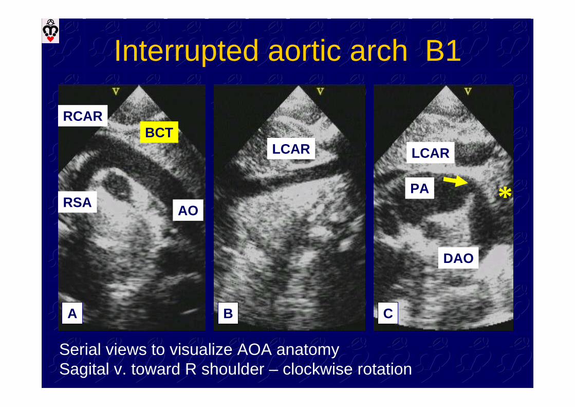
AORSA
RCAR
LCAR
PA
DAO
LCAR
BCT
*
A B C
Interrupted aortic arch B1
Serial views to visualize AOA anatomySagital v. toward R shoulder – clockwise rotation
Page 31
Interrupted aortic arch B2
Page 32
Interrupted aortic arch B2
Page 33
Interrupted aortic arch (A)x aortic arch atresia
Anatomic continuity through a fibrous strand/ lumen completely obstructed
Page 34
PA
LCAR
LSA
DAO
AOA
*
Interrupted aortic arch A1
Page 35
Interrupted aortic arch (A)x aortic arch atresia
Page 36
Interrupted aortic archVSD + LVOT obstruction
LVOT area < 0.7-0.8cm²/m² develop subAO obstructionLVOT/DAO ratio < or = 1.0 recomm. subAO resection
Minich 1992, Geva 1993
Ge, J Am Soc Echo.1997