Biochemical Pharmacology, Vol. 51, pp. 771-778, 1996. Copyright 0 1996 Elsevier Science Inc. ELSEVIER ISSN 0006-2952/96/$15.00 + 0.00 SSDI 0006-2952(95)02394-l AFLMediated Regulation of Human NAD( P)H:Quinone Oxidoreductase, (NQO,) Gene Expression Tao Xie and And K. Jaiswal” DEPARTMENT OF PHARMACOLOGY, Fox CHASE CANCER CENTER, PHILADELPHIA, PA 19111, U.S.A. ABSTRACT. NAD(P)H:quinone oxidoreductase, (NQO,) IS a flavoprotein that catalyzes two-electron re- duction and detoxification of quinones. We have shown previously that twenty-four base pairs of the human Antioxidant Response Element (hARE) mediate basal and xenobiotic-induced expression of the NQO, gene [Li and Jaiswal, .I Biol Chem 267: 15097-15104, 19921. In the present report, we have characterized a second &-element, Al’-2, at nucleotide position -157 of the human NQO, gene promoter that regulates basal and CAMP-induced transcription of the NQO, gene. The NQO, gene AP-2-mediated expression of the chloram- phenicol acetyl transferase (CAT) gene and the binding of nuclear proteins to the AP-2 element were observed in HeLa (Al’-2 positive) cells but not in human hepatoblastoma Hep-G2 (AP-2 deficient) cells, indicating the involvement of transcription factor AP-2 in the regulation of NQO, gene expression. Affinity purification of nuclear protein that binds to the NQO, gene AP-2 DNA element and western analysis revealed that AP-2 indeed binds to the NQO, gene AP-2 element and regulates its expression in HeLa cells. The involvement of AP-2 in the regulation of NQO, gene expression was confirmed by the observation that cDNA-derived AP-2 protein in Hep-G2 cells increased in the NQO, gene AP-2 but not mutant AP-2 mediated expression of CAT gene in Hep-G2 cells. BIOCHEM PHARMACOL 51;6:771-778, 1996. KEY WORDS. quinone oxidoreductase; AP-2; transcription; CAMP induction NQO,? (or DT diaphorase) catalyzes the two-electron reduc- tion of quinones and protects the cells from the adverse effects of semiquinones that are generated due to one-electron reduc- tion of quinones catalyzed by cytochrome I’450 reductase [l, 21. The semiquinones are known to enter in the redox cycling to produce oxygen free radicals that cause oxidative stress leading to neoplasia [3, 41. The expression of the NQO, gene is increased in tumor tissues as compared with normal tissues of similar origin and is induced in response to a variety of xenobiotics including polycyclic aromatic hydrocarbons, pla- nar aromatic compounds, antioxidants, oxidants, and tumor promoters [5-121. Deletion mapping and transfection studies have shown that twenty-four base pairs of the DNA segment present in the promoter region of the human NQO, gene designated as hARE mediate high basal and induced transcrip- tion of the NQO, gene [lo, 131. In addition to the hARE, the nucleotide sequence analysis of the human NQO, gene pro- moter indicates the presence of a perfect AI’-2 binding con- * Corresponding author: Dr. Anil K. Jaiswal, Department of Pharmacology, Fox Chase Cancer Center, 7701 Burholme Ave., Philadelphia, PA 19111. Tel. (215) 728-5282; FAX (215) 728-4333. Received 25 July 1995; accepted 12 October 1995. t Abbreuiations: NQO,, NAD(P)H:quinone oxidoreductase,, also known as quinone reductase (QR), quinone:(acceptor) oxidoreductase (QAO), and DT diaphorase (EC 1.6.99.2); hMTI1, h “man metallothionein II; hARE, &man Antioxidant Response Element; AP-2 element, binding site for AI’-2 cram- acting protein; and CAMP, cyclic AMP. sensus sequence [8]. The AP-2 binding site was characterized previously in the SV40 genome and in the promoter region of the hMTI1 gene [14, 151. AP-2 protein is known to bind to the AP-2 element and to regulate the target genes in response to CAMP, 12-0-tetradecanoylphorbol-13-acetate (TPA), and retinoic acid [14, 161. In the present report, we investigated the role of the AP-2 binding site and transcription factor AI’-2 in the regulation of NQO, gene expression. The deletion mutagenesis in the NQO, gene promoter and transfection in mammalian cells indicated that the AP-2 binding site is required for basal and CAMP-induced expression of the NQO, gene in human cer- vical carcinoma (HeLa) cells. The AP-2-mediated basal ex- pression and the CAMP response was absent in AP-2-deficient human hepatoblastoma (Hep-G2) cells, indicating the possi- ble involvement of transcription factor AP-2 in the regulation of expression and CAMP induction of the NQO, gene. The role of AP-2 in the AP-2-mediated regulation of NQO, gene expression was also supported by the observation that the AP-2 binding site from NQO, gene generated a slow-moving complex with nuclear proteins from HeLa cells and bacterially expressed cDNA-derived recombinant AP-2 protein but not with nuclear extract from AP-2-deficient Hep-G2 cells. The NQO, gene AP-2 binding site was used to affinity purify the interacting nuclear protein from HeLa cell nuclear extract to positively determine the identity of nuclear protein that binds to NQO, gene AP-2 DNA element. This nuclear protein ran
Transcript
Biochemical Pharmacology, Vol. 51, pp. 771-778, 1996. Copyright 0 1996 Elsevier Science Inc.
Tao Xie and And K. Jaiswal” DEPARTMENT OF PHARMACOLOGY, Fox CHASE CANCER CENTER, PHILADELPHIA, PA 19111, U.S.A.
ABSTRACT. NAD(P)H:quinone oxidoreductase, (NQO,) IS a flavoprotein that catalyzes two-electron re-
duction and detoxification of quinones. We have shown previously that twenty-four base pairs of the human
Antioxidant Response Element (hARE) mediate basal and xenobiotic-induced expression of the NQO, gene [Li
and Jaiswal, .I Biol Chem 267: 15097-15104, 19921. In the present report, we have characterized a second
&-element, Al’-2, at nucleotide position -157 of the human NQO, gene promoter that regulates basal and
CAMP-induced transcription of the NQO, gene. The NQO, gene AP-2-mediated expression of the chloram-
phenicol acetyl transferase (CAT) gene and the binding of nuclear proteins to the AP-2 element were observed
in HeLa (Al’-2 positive) cells but not in human hepatoblastoma Hep-G2 (AP-2 deficient) cells, indicating the
involvement of transcription factor AP-2 in the regulation of NQO, gene expression. Affinity purification of
nuclear protein that binds to the NQO, gene AP-2 DNA element and western analysis revealed that AP-2
indeed binds to the NQO, gene AP-2 element and regulates its expression in HeLa cells. The involvement of
AP-2 in the regulation of NQO, gene expression was confirmed by the observation that cDNA-derived AP-2
protein in Hep-G2 cells increased in the NQO, gene AP-2 but not mutant AP-2 mediated expression of CAT gene in Hep-G2 cells. BIOCHEM PHARMACOL 51;6:771-778, 1996.
NQO,? (or DT diaphorase) catalyzes the two-electron reduc-
tion of quinones and protects the cells from the adverse effects
of semiquinones that are generated due to one-electron reduc- tion of quinones catalyzed by cytochrome I’450 reductase [l, 21. The semiquinones are known to enter in the redox cycling to produce oxygen free radicals that cause oxidative stress leading to neoplasia [3, 41. The expression of the NQO, gene is increased in tumor tissues as compared with normal tissues of similar origin and is induced in response to a variety of xenobiotics including polycyclic aromatic hydrocarbons, pla- nar aromatic compounds, antioxidants, oxidants, and tumor promoters [5-121. Deletion mapping and transfection studies have shown that twenty-four base pairs of the DNA segment present in the promoter region of the human NQO, gene designated as hARE mediate high basal and induced transcrip- tion of the NQO, gene [lo, 131. In addition to the hARE, the nucleotide sequence analysis of the human NQO, gene pro- moter indicates the presence of a perfect AI’-2 binding con-
* Corresponding author: Dr. Anil K. Jaiswal, Department of Pharmacology, Fox Chase Cancer Center, 7701 Burholme Ave., Philadelphia, PA 19111. Tel. (215) 728-5282; FAX (215) 728-4333.
Received 25 July 1995; accepted 12 October 1995. t Abbreuiations: NQO,, NAD(P)H:quinone oxidoreductase,, also known as
quinone reductase (QR), quinone:(acceptor) oxidoreductase (QAO), and DT diaphorase (EC 1.6.99.2); hMTI1, h “man metallothionein II; hARE, &man Antioxidant Response Element; AP-2 element, binding site for AI’-2 cram- acting protein; and CAMP, cyclic AMP.
sensus sequence [8]. The AP-2 binding site was characterized previously in the SV40 genome and in the promoter region of the hMTI1 gene [14, 151. AP-2 protein is known to bind to the AP-2 element and to regulate the target genes in response to CAMP, 12-0-tetradecanoylphorbol-13-acetate (TPA), and retinoic acid [14, 161.
In the present report, we investigated the role of the AP-2 binding site and transcription factor AI’-2 in the regulation of NQO, gene expression. The deletion mutagenesis in the NQO, gene promoter and transfection in mammalian cells indicated that the AP-2 binding site is required for basal and CAMP-induced expression of the NQO, gene in human cer- vical carcinoma (HeLa) cells. The AP-2-mediated basal ex- pression and the CAMP response was absent in AP-2-deficient human hepatoblastoma (Hep-G2) cells, indicating the possi- ble involvement of transcription factor AP-2 in the regulation of expression and CAMP induction of the NQO, gene. The role of AP-2 in the AP-2-mediated regulation of NQO, gene expression was also supported by the observation that the AP-2 binding site from NQO, gene generated a slow-moving complex with nuclear proteins from HeLa cells and bacterially expressed cDNA-derived recombinant AP-2 protein but not with nuclear extract from AP-2-deficient Hep-G2 cells. The NQO, gene AP-2 binding site was used to affinity purify the interacting nuclear protein from HeLa cell nuclear extract to positively determine the identity of nuclear protein that binds to NQO, gene AP-2 DNA element. This nuclear protein ran
772 T. Xie and A. K. Jaiswal
as 50 kDa (approximately) protein on SDS-PAGE and cross- reacted with antibody against human transcription factor AP- 2. These results clearly established that AP-2 protein binds with the Al’-2 element and regulates the expression and CAMP induction of the NQO, gene. Further experiments us- ing cDNA-derived human Al’-2 protein in Hep-G2 cells pos- itively regulated the NQO, gene Al’-2-mediated expression of CAT gene, thus confirming the earlier conclusion that AI’-2 indeed regulates the expression of the human NQO, gene. The possible role of AI’-2 protein in the regulation of NQO, gene expression during differentiation and development is dis- cussed.
MATERIALS AND METHODS Chemicals and Reagents
Bacterially (Escherichia coli) expressed recombinant Al’-2 pro- tein was obtained from Promega (Catalogue No. E3050), Mad- ison, WI. This product has now been replaced by a new prod- uct that includes AP-2 as a single protein and not as a recom- binant protein with a molecular weight much higher than that of the AP-2 protein alone. HeLa and Hep-G2 cells were ob- tained from the American Type Culture Collection (ATCC), Bethesda, MD. The modifiers of CAMP levels (forskolin and Ro 20-1724) and other chemicals were purchased from the Sigma Chemical Co., St. Louis, MO.
Construction of Plasmids
The construction of plasmid pNQOrCAT1.55 has been de- scribed previously [8]. This plasmid contained 1.55 kb of 5’ flanking region and 110 bp of the first exon attached to the chloramphenicol acetyl transferase gene. For construc- tion of plasmids pNQO,CATO. 176 and pNQOiCATO.147, we first made a plasmid pNQOrCAT0.130. The plasmid pNQOtCAT0.130 contained 130 bp of the 5’ flanking region and 110 bp of the first exon attached to the CAT gene. This plasmid was created from the original plasmid pNQOrCAT1.85 by digestion with Pst I. The Pst I enzyme generated a deletion of 1720 bp from the 5’ end of the NQO, gene promoter. The remaining plasmid was self- ligated to make pNQOiCAT0.130. To construct the plasmid pNQOrCAT0.176, we amplified the region between -0.176 and -0.130 of the human NQO, gene promoter with Hind III and Pst I ends by polymerase chain reaction. This fragment of the DNA was subcloned in the plasmid pNQO,CAT0.130 at the Hind III-Pst I site to generate plasmid pNQO,CAT0.176. Similarly, the two strands of the oligonucleotides were synthe- sized corresponding to the region between -0.147 and -0.130 in the human NQO, gene promoter with Hind III and Pst I ends, annealed, kinased, and subcloned in the plasmid pNQOrCAT0.130 at the Hind III-Pst I site to make the re- combinant plasmid pNQOrCAT0.147.
Both strands of the NQO, gene containing the AP-2 bind- ing site (region between -162 and -143) were synthesized, annealed, kinased, and ligated to Bum HI linkers and cloned at the Burn HI site of the pBLCAT2 [17] to generate pNQ0, -2X
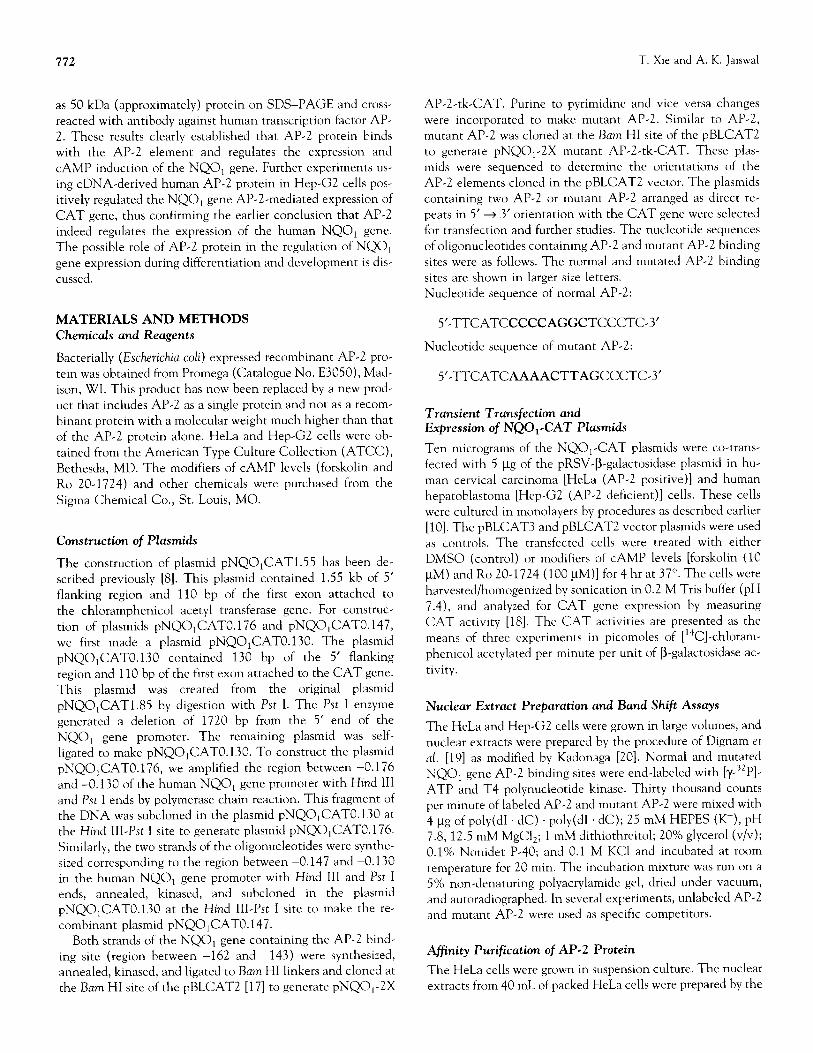
AP-2-tk-CAT. Purine to pyrimidine and vice versa changes were incorporated to make mutant AP-2. Similar to AP-2, mutant AP-2 was cloned at the Bum HI site of the pBLCAT2 to generate pNQO,-2X mutant AP-2-tk-CAT. These plas- mids were sequenced to determine the orientations of the AP-2 elements cloned in the pBLCAT2 vector. The plasmids containing two AP-2 or mutant AP-2 arranged as direct re- peats in 5’ + 3’ orientation with the CAT gene were selected for transfection and further studies. The nucleotide sequences of oligonucleotides containing AP-2 and mutant AP-2 binding sites were as follows. The normal and mutated AP-2 binding sites are shown in larger size letters. Nucleotide sequence of normal AP-2:
5’-TTCATCCCCCAGGCTCCCTC-3’
Nucleotide sequence of mutant AP-2:
5’-TTCATCAAAACTTAGCCCTC3
Transient Transfection and Expression of NQO,-CAT Plasmids
Ten micrograms of the NQO,-CAT plasmids were co-trans- fected with 5 l.tg of the pRSV-B-galactosidase plasmid in hu- man cervical carcinoma [HeLa (AP-2 positive)] and human hepatoblastoma [Hep-G2 (AP-2 deficient)] cells. These cells were cultured in monolayers by procedures as described earlier [lo]. The pBLCAT3 and pBLCAT2 vector plasmids were used as controls. The transfected cells were treated with either DMSO (control) or modifiers of CAMP levels [forskolin (10 PM) and Ro 20-1724 (100 l_tM)] for 4 hr at 37”. The cells were harvested/homogenized by sonication in 0.2 M Tris buffer (pH 7.4), and analyzed for CAT gene expression by measuring CAT activity [18]. The CAT activities are presented as the means of three experiments in picomoles of [14C]-chloram- phenicol acetylated per minute per unit of B-galactosidase ac- tivity.
Nuclear Extract Preparation and Band Shift Assays
The HeLa and Hep-G2 cells were grown in large volumes, and nuclear extracts were prepared by the procedure of Dignam et al. [19] as modified by Kadonaga [20]. Normal and mutated NQO, gene AP-2 binding sites were end-labeled with [Y-~~P]- ATP and T4 polynucleotide kinase. Thirty thousand counts per minute of labeled AP-2 and mutant AP-2 were mixed with 4 l_tg of poly(d1 . dC) . poly(d1 . dC); 25 mM HEPES (K+), pH 7.8; 12.5 mM MgCl,; 1 mM dithiothreitol; 20% glycerol (v/v); 0.1% Nonidet P-40; and 0.1 M KC1 and incubated at room temperature for 20 min. The incubation mixture was run on a 5% non-denaturing polyacrylamide gel, dried under vacuum, and autoradiographed. In several experiments, unlabeled AP-2 and mutant AP-2 were used as specific competitors.
Affinity Purification of AI’-2 Protein
The HeLa cells were grown in suspension culture. The nuclear extracts from 40 mL of packed HeLa cells were prepared by the
Al’-2 Mediated NQO, Gene Expression 773
procedure of Dignam et al. [19] as modified by Kadonaga [20]. The final protein concentration was 7.3 pg/pL of binding buffer. The proteins that bound to the NQO, gene AP-2 bind- ing site were affinity purified exactly by the method as de- scribed [20]. Two hundred and twenty micrograms of each strand of the normal NQO, gene AP-2 binding site (sequence of oligonucleotide as shwon above) were annealed, kinased, and self-ligated to obtain DNA fragments of 200-300 bp in length containing multiple AP-2 binding sites. This DNA was incubated with cyanogen bromide-activated Sepharose CL-2B to generate the AP-2 affinity matrix. The remaining reactive groups in cyanogen bromide-activated Sepharose CL-2B were blocked by its treatment with ethanolamine, and the affinity matrix was washed with 100 mL each of 0.01 M potassium phosphate buffer (pH 8.0), 1 M potassium phosphate buffer (pH 8.0), 1 M KCl, and water. The CL-2B-AP-2 affinity ma- trix was packed in a column and equilibrated with binding buffer [25 mM HEPES (K+), pH 7.8; 12.5 mM MgCl,; 1 mM dithiothreitol; 20% glycerol (v/v); 0.1% Nonidet P-40; 0.1 M KC1 and 2.2 mg/mL of poly(dI . dC) . poly(dI . dC)]. HeLa cell nuclear extract, as prepared above, was passed through this affinity column for binding and retention of specific nuclear proteins that bind to the AP-2 binding site of the NQO, gene. The bound proteins were eluted with a step gradient of 0.2 to 0.6 M KC1 in the binding buffer. All the fractions were ana- lyzed for protein content, SDS-PAGE, and staining with Coomassie blue and band shift assays with the NQO, gene AP-2 binding site. Various fractions containing AP-2 binding
CCCCAGGCT
-1550 + 4r
-467 -157 +’ pNQq CAT1.55
activity as determined by band shift analysis were pooled and reloaded on a second NQO, gene AP-2 affinity column. The bound proteins were eluted as earlier. This procedure was re- peated four times before purified AP-2 protein was obtained. To determine if the protein thus purified was AP-2, we ran an SDS-PAGE of denatured purified proteins from the fourth column, western blotted, and probed with antibodies directed against a peptide located at the C-terminus of human AP-2 (located after the DNA binding domain). AP-2 antibody was a gift from Dr. T. Williams (Yale University, New Haven, CT). The purified protein was also used in band shift assays to determine its specificity for NQO, gene AP-2 and its compe- tition with hMTI1 gene AP-2.
Nuclear Run-On Transcription
Nuclear run-on assays were performed using nuclei isolated from forskolin (10 PM) and Ro 20-1724 (100 PM) treated HeLa cells by a procedure described previously [21]. Human /3-actin was used as the control.
Co-transfection of Transcription Factor AI’-2
Expression and NQO, Gene Al’-2 Reporter Plasmids
The AP-2 expression plasmid SP(RSV)AP2 was obtained from Dr. T. Williams (Yale University). The construction of SP(RSV)AP2 plasmid has been described and was used suc- cessfully to express AP-2 protein in Hep-G2 cells to determine
2 Q
-176 h+ pNQq CAT0.176
-147-J+ pNQq CATO. 147
pBLCAT3 (Vector alone)
HeLa Hep-G2
Control 1239 + 31 51 f 06 Forskolin 2302 f 42 61 f 03
RoZO-1724 3597 f 69 51 f09
Control 51 f09 5 f 01 Forskolin 101 f 11 5 + 02 RoZO-1724 153+16 6 f02
Control 28 + 01 6 + 01 Forskolin 29 + 06 6 + 01 RoZO-1724 33 f02 8 f 02
Control 4fOl 5 f 01 Forskolin 5 +01 6 f 02 RoZO-1724 4 + 01 3 201
FIG. 1. Deletion mutagenesis in the NQO, gene promoter and transient transfection in mam- malian cells. The recombinant plasmids ~NQ0~cAT1.55, 0.176, and 0.147 containing dif- ferent lengths of the NQO, gene promoter and the vector control pBLCAT3 were transiently transfected in human cervical carcinoma (HeLa) and human hepatoblastoma (Hep-G2) cells. The RSV-P-galactosidase plasmid was co-transfected in each case to normalize the transfec- tion efficiency. The transfected cells were treated with DMSO (control), forskolin (10 PM), and Ro 20.1724 (100 PM) for 4 hr at 37”. The cells were harvested, homogenized by soni- cation, and analyzed for CAT gene expression by measuring CAT activity. The CAT activ- ities are represented as means f SD of three independent transfection experiments and expressed as picomoles of [‘4C]-chloramphenico1 acetylated per minute per unit of PMgalac- tosidase activitv.
774 T. Xie and A. K. Jaiswal
Iqtk-CAT
(pNQq -2XAP-2-tk-CAT)
Control 45f3 21f4
Forskolin 87 f 4 23 f 3 Ro20-1724 99 f 6 19+2
Mltk-CAT Control
Forskolin (pNQq -2X Mutant AP-Z-tk-CAT) Ro20-1724
pBLCAT2 (Vector alone)
Control Forskolin
Ro20-1724
HeLa Hep-GZ
10 + 1 5*0 12 * 1 7fl 11 f2 4fl
11 f02 4 f 01 10fOl 3 f 01 09 02 + 5 + 02
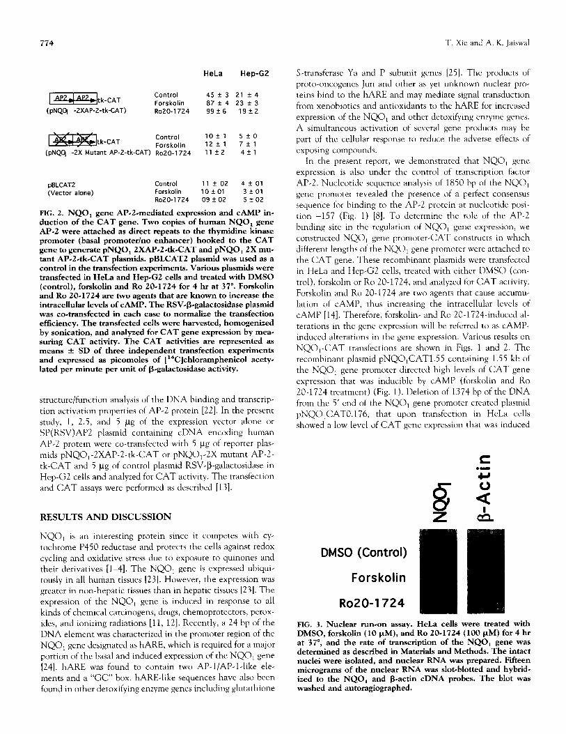
FIG. 2. NQO, gene Al’-2-mediated expression and CAMP in- duction of the CAT gene. Two copies of human NQO, gene Al’-2 were attached as direct repeats to the thymidine kinase promoter (basal promoter/no enhancer) hooked to the CAT gene to generate pNQO,2XAP-2.tk.CAT and pNQO,2X mu- tant AP.2-tk-CAT plasmids. pBLCAT2 plasmid was used as a control in the transfection experiments. Various plasmids were transfected in HeLa and Hep-G2 cells and treated with DMSO (control), forskolin and Ro 20.1724 for 4 hr at 37”. Forskolin and Ro 20-1724 are two agents that are known to increase the intracellular levels of CAMP. The RSV+galactosidase plasmid was co-transfected in each case to normalize the transfection efficiency. The transfected cells were harvested, homogenized by sonication, and analyzed for CAT gene expression by mea- suring CAT activity. The CAT activities are represented as means f SD of three independent transfection experiments and expressed as picomoles of [ 14C]chloramphenicol acety- lated per minute per unit of R.galactosidase activity.
structure/function analysis of the DNA binding and transcrip- tion activation properties of AP-2 protein [22]. In the present study, 1, 2.5, and 5 /_tg of the expression vector alone or SP(RSV)AP2 plasmid containing cDNA encoding human AP-2 protein were co-transfected with 5 /.tg of reporter plas- mids pNQO,-2XAP-2-tk-CAT or pNQO,-2X mutant AP-2- tk-CAT and 5 pg of control plasmid RSV-P-galactosidase in Hep-G2 cells and analyzed for CAT activity. The transfection and CAT assays were performed as described [13].
RESULTS AND DISCUSSION
NQO, is an interesting protein since it competes with cy- tochrome P450 reductase and protects the cells against redox cycling and oxidative stress due to exposure to quinones and their derivatives [14]. The NQO, gene is expressed ubiqui- tously in all human tissues [23]. However, the expression was greater in non-hepatic tissues than in hepatic tissues [23]. The expression of the NQO, gene is induced in response to all kinds of chemical carcinogens, drugs, chemoprotectors, perox- ides, and ionizing radiations [ll, 121. Recently, a 24 bp of the DNA element was characterized in the promoter region of the NQOr gene designated as hARE, which is required for a major portion of the basal and induced expression of the NQO, gene [24]. hARE was found to contain two AP-l/Al’-l-like ele- ments and a “GC” box. hARE-like sequences have also been found in other detoxifying enzyme genes including glutathione
S-transferase Ya and P subunit genes [25]. The products of proto-oncogenes Jun and other as yet unknown nuclear pro- teins bind to the hARE and may mediate signal transduction from xenobiotics and antioxidants to the hARE for increased expression of the NQO, and other detoxifying enzyme genes. A simultaneous activation of several gene products may be part of the cellular response to reduce the adverse effects of exposing compounds.
In the present report, we demonstrated that NQO, gene expression is also under the control of transcription factor Al’-2. Nucleotide sequence analysis of 1850 bp of the NQO, gene promoter revealed the presence of a perfect consensus sequence for binding to the Al’-2 protein at nucleotide posi- tion -157 (Fig. 1) [8]. To determine the role of the AP-2 binding site in the regulation of NQO, gene expression, we constructed NQO, gene promoter-CAT constructs in which different lengths of the NQO, gene promoter were attached to the CAT gene. These recombinant plasmids were transfected in HeLa and Hep-G2 cells, treated with either DMSO (con- trol), forskolin or Ro 20-1724, and analyzed for CAT activity. Forskolin and Ro 20-1724 are two agents that cause accumu- lation of CAMP, thus increasing the intracellular levels of CAMP [14]. Therefore, forskolin- and Ro 20.17240induced al- terations in the gene expression will be referred to as CAMP- induced alterations in the gene expression. Various results on NQO,-CAT transfections are shown in Figs. 1 and 2. The recombinant plasmid pNQOiCAT1.55 containing 1.55 kb of the NQO, gene promoter directed high levels of CAT gene expression that was inducible by CAMP (forskolin and Ro 20-1724 treatment) (Fig. 1). Deletion of 1374 bp of the DNA from the 5’ end of the NQO, gene promoter created plasmid pNQO,CAT0.176, that upon transfection in HeLa cells showed a low level of CAT gene expression that was induced
c
FIG. 3. Nuclear run-on assay. HeLa cells were treated with DMSO, forskolin (10 PM), and Ro 20-1724 (100 ~.LM) for 4 hr at 37”, and the rate of transcription of the NQO, gene was determined as described in Materials and Methods. The intact nuclei were isolated, and nuclear RNA was prepared. Fifteen micrograms of the nuclear RNA was slot-blotted and hybrid. ized to the NQO, and R-a&n cDNA probes. The blot was washed and autoragiographed.
AP-2 Mediated NQO, Gene Expression
2 8 c 2 a +
2 a
N
2 AP2 + AP2 extract
AP2 with reco AP2
AP2 complex with cellular AP2 protein
complex
Imbinant- protein
m+ + + ++++++++
E2 AP2 + HeLa Nu. Ext. AP2 + HepG2 Nu. Ext. aa
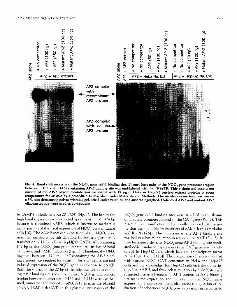
FIG. 4. Band shift assays with the NQO, gene Al’-2 binding site. Twenty base pairs of the NQO, gene promoter (region between - 162 and - 143) containing AP.2 binding site was end-labeled with [y3*P]ATP. Thirty thousand counts per minute of the AP.2 oligonucleotide was incubated with 15 pg of HeL.a or Hep-G2 nuclear extract proteins at room temperature for 20 min by a procedure as described under Materials and Methods. The incubation mixture was run on a 5% non~denaturing polyacrylamide gel, dried under vacuum, and autoradiographed. Unlabeled AP-2 and mutant AP-2 oligonucleotide were used as competitors.
by CAMP (forskolin and Ro 20-1724) (Fig. 1). The loss in the high basal expression was expected upon deletion of 1374 bp because it contained hARE, which is known to mediate a major portion of the basal expression of NQO, gene in tumor cells [lo]. The CAMP-induced expression of the NQO, gene remained unaffected by this deletion. In similar experiments, transfection of HeLa cells with pNQOtCAT0.147 containing 147 bp of the NQO, gene promoter resulted in loss of basal expression and CAMP induction (Fig. 1). Therefore, the DNA fragment between -176 and -147 containing the AP-2 bind- ing element was required for a part of the basal expression and induced expression of the NQO, gene in response to CAMP. Both the strands of the 20 bp of the oligonucleotide contain- ing AP-2 binding site and of the human NQO, gene promoter (region between nucleotides -0.162 and -0.153) were synthe- sized, annealed, and cloned in pBLCAT2 to generate plasmid pNQO,-ZXAP-2-tk-CAT. In this plasmid, two copies of the
NQO, gene AP-2 binding sites were attached to the thymi- dine kinase promoter hooked to the CAT gene (Fig. 2). This plasmid upon transfection in HeLa cells produced CAT activ- ity that was inducible by modifiers of CAMP levels (forskolin and Ro 20-1724). The mutations in the AP-2 binding site resulted in a loss of induction in response to CAMP (Fig. 2). It may be noteworthy that NQO, gene AP-2 binding site-medi- ated CAMP-induced expression of the CAT gene was not ob- served in Hep-G2 cells which lack the transcription factor AP-2 (Figs. 1 and 2) [14]. The comparison of results obtained with various NQO,-CAT constructs in HeLa and Hep-G2 cells and the knowledge that Hep-G2 cells lack the transcrip- tion factor AP-2, and thus lack stimulation by CAMP, strongly suggested the involvement of AP-2 protein in AP-2 binding site-mediated expression and induction of the NQO, gene expression. These experiments also raised the question of in- duction of endogenous NQO, gene expression in response to
776 T. Xie and A. K. Jaiswal
A. Affinity Purification of AP-2 B. Western analysis of
$ affinity purified AP-2
ii r”
g E 2
e E 3
$5 8 2 8
8 s
a 5 ‘i
z
z ; r” z .z
,o
AP-2 protein in the NQO, gene Al’-2 element-mediated ex- pression of human NQO, gene (Figs. 4 and 5). A bacterially expressed and purified recombinant AI’-2 protein (Promega) bound with high affinity to the NQO, gene AI’-2 binding site (Fig. 4). This binding was competed specifically by the unla- beled AP-2 binding site but not by the mutated AI’-2 binding site (Fig. 4). This clearly indicated that AI’-2 transcription factor indeed recognized and bound to the NQO, gene Al’-2 binding site. In addition, AP-2 binding activity was also oh- served in band shift assays with NQO, gene AP-2 binding site and HeLa cell nuclear extract (Fig. 4). The NQO, gene AP-2 nuclear complex observed with HeLa cell nuclear extract was much faster moving than that with bacterially expressed AP-2 protein (Fig. 4). This was expected because bacterially ex- pressed AP-2 protein is a recombinant protein that is much larger in size than normal AP-2 protein in HeLa cells. The NQO, gene AP-2 binding complex in HeLa cells was com-
--
ad- fAP-2
C. Band shift with purified AP-2
2 d AP2 + HeLa Nu. Ext.
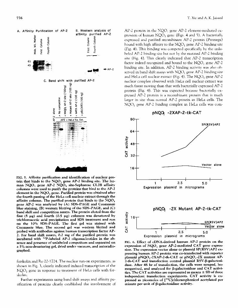
FIG. 5. Affinity purification and identification of nuclear pro- tein that binds to the NQO, gene Al?-2 binding site. The hue man NQO, gene AP.2 NQO, site-Sepharose CLZB afftnity columns were used to purify the proteins that bind to the AI’-2 element in the NQO, gene. Purified protein was obtained after the fourth passing of the HeLa cell nuclear extract through the affinity column. The purified protein that binds to the NQO, gene AP.2 was analyzed by (A) SDS-PAGE and Coomassie blue staining, (B) western blotting of the SDS-PAGE, and (C) band shift and competition assays. The protein eluted from the first (5 pg) and fourth (0.5 pg) columns was denatured by trichloroacetic acid precipitation and SDS treatment and run on the 10% SDS-PAGE. The first gel was stained with Coomassie blue. The second gel was western blotted and probed with antibodies against human transcription factor AP- 2. For band shift assays, 0.2 mg of the purified protein was incubated with 32PJabeled AP-2 oligonucleotides in the ab# sence and presence of unlabeled competitors and separated on a 5% non-denaturing gel, dried under vacuum, and autoradio- graphed.
forskolin and Ro 20-1724. The nuclear run-on experiments, as shown in Fig. 3, clearly indicated induced transcription of the NQO, gene in response to treatment of HeLa cells with for- skolin.
Further experiments using band shift assays and affinity pu- rification of proteins clearly established the involvement of
pNQCj -2XAP-2-tk-CAT
SP(RSV)APZ /
64 t /
x I /
t
16 Vector alone
I I I
1 2.5 5.0 Expression plasmid in micrograms
pNQq -2X Mutant AP-2-tk-CAT x ‘5 16-
2 SPCRSV)APZ
z I I Veqtor alone
I U 1 2.5 5.0
Expression plasmid in micrograms
FIG. 6. Effect of cDNA-derived human AP.2 protein on the expression of NQO, gene AP-2-mediated CAT gene expres- sion. The expression vector alone or plasmid SP(RSV)APZ exe pressing human AP-2 protein was co-transfected with reporter plasmid pNQO,.ZXAP-2.tkCAT or pNQO,-2X mutant AP- 2.&CAT and transfection control plasmid RSV+galactosi- dase. After 48 hr of transfection, the cells were scraped, ho* mogenized, and analyzed for Begalactosidase and CAT activi- ties. The CAT activities are represented as means f SD of three independent transfection experiments. CAT activity is ex- pressed as picomoles of [ “C]chloramphenicol acetylated per minute per unit of B-galactosidase activity.
AP-2 Mediated NQO, Gene Expression
peted specifically by unlabeled AI’-2 oligonucleotide but not by mutant AP-2 oligonucleotide. As compared with HeLa cells, no binding activity was detected in Hep-G2 cells, which are deficient in AP-2 protein (Fig. 4). Therefore, various band shift experiments, as shown in Fig. 4, clearly established that AI?-2 protein from HeLa cells binds to the Al’-2 binding site and mediates NQO, gene expression and CAMP induction. The binding of AI’-2 transcription factor to the NQOl gene AP-2 element was further confirmed by affinity purification of HeLa cell nuclear proteins that bind to the NQOl gene AP-2 binding site (Fig. 5). These nuclear proteins were purified by passing the HeLa cell nuclear extract on four NQOl gene AP-2 affinity columns in a sequence. The fourth column yielded purified protein that bound to the NQOl gene AI’-2 binding site with high affinity. SDS-PAGE analysis of the purified protein revealed the size of the protein to be approx- imately 50 kDa (Fig. 5). The purified protein cross-reacted with antibodies against human AI’-2 protein (Fig. 5). The binding of the purified protein with NQO, gene AI’-2 element was competed by unlabeled AI’-2 from NQO, (self) and hMTI1 gene but not by mutated AI’-2 (Fig. 5). These results confirmed the identity of the purified protein as AI’-2.
The role of AI’-2 in the regulation of NQO, gene expres- sion was also evident by the observation that cDNA-derived transcription factor AP-2 in Hep-G2 cells positively regulated the NQO, gene AP-2 but not mutant AI’-2-mediated expres- sion of CAT gene (Fig. 6). The increase in the AP-2-mediated expression was dependent on the concentration of plasmid SP(RSV)AP2 expressing cDNA encoded human AP-2 pro- tein (Fig. 6). The above results combined with earlier obser- vations confirmed that transcription factor AP-2 indeed reg- ulates the expression of the NQO, gene.
AP-2 is known to bind DNA as a homodimer [26]. The expression of AP-2 is reported to be regulated during differ- entiation of a teratocarcinoma cell line, which indicated that AP-2 may be involved in the control of developmentally reg- ulated gene expression [27]. In mouse embryos, a novel pattern of the expression of AP-2 was reported [27]. A majority of the expression of AP-2 was in neural crest cells and their major derivatives, including cranial and spinal sensory ganglia and facial mesenchyme. In addition, AP-2 was also found ex- pressed in ectoderm and a longitudinal column of the spinal cord and hindbrain that is contacted by neural crest-derived sensory ganglia. AP-2 also was expressed in limb bud mesen- thyme and in meso-metanephric regions. The expression pat- tern of the AP-2 has suggested a role for AP-2 in regulating the transcription of genes involved in the morphogenesis of the peripheral nervous system, face, limbs, skin, and nephric tis- sues [27]. In the present report, we have shown conclusively that NQO, gene expression is regulated by transcription factor AP-2. This raises important questions regarding NQO, gene expression and regulation by AP-2 in neural crest cells and their role during differentiation and development.
This work was supported by NIH Grant GM 47466. We thank our
colleaffues fog helpful discussions.
777
References
1.
2.
3.
4.
5.
6.
7.
8.
9.
10.
11.
12.
13.
14.
15.
16.
17.
18.
19.
Talalay P, Mechanisms of induction of enzymes that protect
against chemical carcinogenesis. Adv Enzyme Regul28: 149-159,
1989. Riley RJ and Workman P, DT-diaphorase and cancer chemo-
therapy. Biochem Pharmacol 43: 1657-1669, 1992.
Monks TJ, Hanzlik RP, Cohen GM, Ross D and Graham DG,
Quinone chemistry and toxicity. Toxicol Appl PhannacoI 112:
2-16, 1992.
Belinsky M and Jaiswal AK, NAD(P)H:quinone oxidoreductase,
(DT diaphorase) expression in normal and tumor tissues. Cancer
Metastasis Rev 12: 103-117, 1993.
Robertson JA, Chen H-C and Nebert DW, NAD(P)H:menadi-
one oxidoreductase: Novel purification of enzyme, cDNA and
complete amino acid sequence and gene regulation. .I Biol Chem
261: 15794-15799, 1986.
Williams JB, Lu AYH, Cameron RG and Pickett CB, Rat liver
Bayney RM, Morton MR, Favreau LV and Pickett CB, Rat liver
NAD(P)H:quinone reductase: Regulation of quinone reductase
gene expression by planar aromatic compounds and determina-
tion of the exon structure of the quinone reductase structural
gene. .I Biol Chem 264: 21793-21797, 1989.
Jaiswal AK, Human NAD(P)H:quinone oxidoreductase (NQO,)
gene structure and induction by dioxin. Biochemistry 30: 10647-
10653, 1991.
Cresteil T and Jaiswal AK, High levels of expression of the
NAD(P)H:quinone oxidoreductase (NQO,) gene in tumor cells
compared to normal cells of the same origin. Biochem Pharmacol
42: 1021-1027, 1991.
Li Y and Jaiswal AK, Regulation of human NAD(P)H:quinone
oxidoreductase gene: Role of APl binding site contained within
human antioxidant response element. J Biol Chem 267: 15097-
15104, 1992.
Prestera T, Holtzclaw MD, Zhang Y and Talalay P, Chemical
and molecular regulation of enzymes that detoxify carcinogens.
Proc Nat1 Acad Sci USA 90: 2965-2969, 1993.
Li Y and Jaiswal AK, Human antioxidant-response-element-me-
diated regulation of type 1 NAD(P)H:quinone oxidoreductase
gene expression: Effect of sulfhydryl modifying agents. Eur .J Bio-
them 226: 31-39, 1994.
Xie T, Belensky M, Xu Y and Jaiswal AK, ARE- and TRE-
mediated regulation of gene expression: Response to xenobiotics and antioxidants. ./ Biol Chem 270: 6894-6900, 1995.
Imagawa M, Chiu R and Karin M, Transcription factor AP-2
mediates induction by two different signal-transduction path-
ways: Protein kinase C and CAMP. Cell 51: 251-260, 1987.
Williams T, Admon A, Luscher B and Tjian R, Cloning and
expression of AP-2, a cell-type specific transcription factor that
activates inducible enhancer elements. Genes Dev 2: 1557-
1569, 1988.
Luscher B, Mitchell PJ, Williams T and Tjian R, Regulation of
transcription factor AP-2 by morphogen retinoic acid and by
second messengers. Genes Deo 3: 1507-1517, 1989.
Luckow B and Schtitz G, CAT constructions with multiple
unique restriction sites for the functional analysis of eukaryotic promoters and regulatory elements. Nucleic Acids Res 15: 5490,
1987.
Davis LG, Dibner MD and Battey JF, Measurement of CAT activity. Basic Methods in Molecular Biology, pp. 298-300.
Elsevier, Amsterdam, 1983.
Dignam JD, Lebovitz RM and Roeder RG, Accurate transcrip-
tion initiation by RNA polymerase II in a soluble extract from isolated mammalian nuclei. Nucleic Acids Res 11: 1475-1489,
1983.
778 T. Xie and A. K. Jaiswal
20. Kadonaga JT, Affinity purification of sequence-specific DNA binding proteins. Methods En~ymol 208: 1 l-24, 1991.
21. McKnight GS and Palmiter RD, Transcriptional regulation of the ovalbumin and conalbumin genes by steroid hormones in chick oviduct. J Biol Chem 254: 9050-9058, 1979.
22. Williams T and Tjian R, Analysis of the DNA-binding and ac- tivation properties of the human transcription factor AP-2. Genes Dee 5: 670-682, 1991.
25. Rushmore TH and Pickett CB, Glutathione-S-transferases, struc- ture, regulation and therapeutic implications. J Biol Chem 268: 11475-l 1478, 1993.
26. Williams T and Tjian R, Characterization of a dimerization motif in AP-2 and its function in heterologous DNA-binding protein. Science 25: 1067-1072, 1991.
23. Jaiswal AK, Human NAD(P)H:quinone oxidoreductase,: Gene 27. Mitchell PJ, Timmons PM, Hebert JM, Rigby PWJ and Tjian R, structure, activity, and tissue specific expression. J Biol Chem Transcription factor AP-2 is expressed in neural crest cell lin- 269: 14502-14508, 1994. eages during mouse embryogenesis. Genes Deu 5: 105-l 19, 1991.