Acta Protozool. (2012) 51: 291–304 http://www.eko.uj.edu.pl/ap doi:10.4467/16890027AP.12.023.0783 ACTA PROTOZOOLOGICA Ultrastructure of Diplophrys parva, a New Small Freshwater Species, and a Revised Analysis of Labyrinthulea (Heterokonta) O. Roger ANDERSON 1 and Thomas CAVALIER-SMITH 2 1 Biology and Paleo Environment, Lamont-Doherty Earth Observatory of Columbia University, Palisades, New York, U.S.A.; 2 Department of Zoology, University of Oxford, South Parks Road, Oxford, UK Abstract. We describe Diplophrys parva n. sp., a freshwater heterotroph, using fine structural and sequence evidence. Cells are small (L = 6.5 ± 0.08 μm, W = 5.5 ± 0.06 μm; mean ± SE) enclosed by an envelope/theca of overlapping scales, slightly oval to elongated-oval with rounded ends, (1.0 × 0.5–0.7 μm), one to several intracellular refractive granules (~ 1.0–2.0 μm), smaller hyaline peripheral vacuoles, a nucleus with central nucleolus, tubulo-cristate mitochondria, and a prominent Golgi apparatus with multiple stacked saccules (≥ 10). It is smaller than pub- lished sizes of Diplophrys archeri (~ 10–20 μm), modestly less than Diplophrys marina (~ 5–9 μm), and differs in scale size and morphology from D. marina. No cysts were observed. We transfer D. marina to a new genus Amphifila as it falls within a molecular phylogenetic clade extremely distant from that including D. parva. Based on morphological and molecular phylogenetic evidence, Labyrinthulea are revised to include six new families, including Diplophryidae for Diplophrys and Amphifilidae containing Amphifila. The other new families have dis- tinctive morphology: Oblongichytriidae and Aplanochytriidae are distinct clades on the rDNA tree, but Sorodiplophryidae and Althorniidae lack sequence data. Aplanochytriidae is in Labyrinthulida; the rest are in Thraustochytrida; Labyrinthomyxa is excluded. Key words: Diplophryidae, fine structure, molecular genetics, Labyrinthomyxa, labyrinthulean taxonomy. Address for correspondence: O. R. Anderson, Biology and Paleo En- vironment, Lamont-Doherty Earth Observatory, Palisades, New York 10964; Tel.: 845-365-8452; E-mail: [email protected]INTRODUCTION Barker (1868), in a brief report to the Dublin Micro- scopical Club (1867), originally described Diplophrys archeri as an ‘exceedingly minute, nearly orbicular or broadly elliptic’ freshwater rhizopod bearing at ‘two opposite points … a tuft of filiform pseudopodia’, and containing within the body ‘an oil-like refractive glob- ule of an orange or amber color’. That was the first de- scription of any Diplophrys, so D. archeri is the type species. Subsequent studies (e.g. Hertwig and Lesser 1874, Penard 1902) reported that this non-flagellate or- ganism, containing a prominent refractive granule and emergent polar filopodia, occurred as small mounds of cells on submerged aquatic plants. The cells appeared to be enclosed by a thin, hyaline test, and for some time Diplophrys, though apparently non-phagotrophic, was considered to be a rhizopod, now belonging either to the cercozoan family Amphitremidae of filose testate amoebae with bipolar tufts of branching filopodia (e.g. Calkins 1926) or the foraminiferan family Allogromi- idae, containing phagotrophic unicells enclosed by an
Ultrastructure of Diplophrys parva, a New Small Freshwater Species, and a Revised Analysis of Labyrinthulea (Heterokonta)
O. Roger ANDERSON1 and Thomas CAVALIER-SMITH2
1Biology and Paleo Environment, Lamont-Doherty Earth Observatory of Columbia University, Palisades, New York, U.S.A.; 2Department of Zoology, University of Oxford, South Parks Road, Oxford, UK
Abstract. We describe Diplophrys parva n. sp., a freshwater heterotroph, using fi ne structural and sequence evidence. Cells are small (L = 6.5 ± 0.08 μm, W = 5.5 ± 0.06 μm; mean ± SE) enclosed by an envelope/theca of overlapping scales, slightly oval to elongated-oval with rounded ends, (1.0 × 0.5–0.7 μm), one to several intracellular refractive granules (~ 1.0–2.0 μm), smaller hyaline peripheral vacuoles, a nucleus with central nucleolus, tubulo-cristate mitochondria, and a prominent Golgi apparatus with multiple stacked saccules (≥ 10). It is smaller than pub-lished sizes of Diplophrys archeri (~ 10–20 μm), modestly less than Diplophrys marina (~ 5–9 μm), and differs in scale size and morphology from D. marina. No cysts were observed. We transfer D. marina to a new genus Amphifi la as it falls within a molecular phylogenetic clade extremely distant from that including D. parva. Based on morphological and molecular phylogenetic evidence, Labyrinthulea are revised to include six new families, including Diplophryidae for Diplophrys and Amphifi lidae containing Amphifi la. The other new families have dis-tinctive morphology: Oblongichytriidae and Aplanochytriidae are distinct clades on the rDNA tree, but Sorodiplophryidae and Althorniidae lack sequence data. Aplanochytriidae is in Labyrinthulida; the rest are in Thraustochytrida; Labyrinthomyxa is excluded.
Key words: Diplophryidae, fi ne structure, molecular genetics, Labyrinthomyxa, labyrinthulean taxonomy.
Address for correspondence: O. R. Anderson, Biology and Paleo En-vironment, Lamont-Doherty Earth Observatory, Palisades, New York 10964; Tel.: 845-365-8452; E-mail: [email protected]
INTRODUCTION
Barker (1868), in a brief report to the Dublin Micro-scopical Club (1867), originally described Diplophrys archeri as an ‘exceedingly minute, nearly orbicular or broadly elliptic’ freshwater rhizopod bearing at ‘two opposite points … a tuft of fi liform pseudopodia’, and containing within the body ‘an oil-like refractive glob-ule of an orange or amber color’. That was the fi rst de-
scription of any Diplophrys, so D. archeri is the type species. Subsequent studies (e.g. Hertwig and Lesser 1874, Penard 1902) reported that this non-fl agellate or-ganism, containing a prominent refractive granule and emergent polar fi lopodia, occurred as small mounds of cells on submerged aquatic plants. The cells appeared to be enclosed by a thin, hyaline test, and for some time Diplophrys, though apparently non-phagotrophic, was considered to be a rhizopod, now belonging either to the cercozoan family Amphitremidae of fi lose testate amoebae with bipolar tufts of branching fi lopodia (e.g. Calkins 1926) or the foraminiferan family Allogromi-idae, containing phagotrophic unicells enclosed by an
O. R. Anderson and T. Cavalier-Smith292
organic test and having reticulose pseudopodia (e.g. Grassé 1953, p. 140). A second nominal species from the early literature, Diplophrys stercorea (Cienkowski 1876, Olive 1903), was later split off as a separate genus Sorodiplophrys because it makes multicellular fruiting bodies like a slime mold (Dykstra and Olive 1975), but its vegetative cells have the bipolar fi lopodial pheno-type that led Cienkowski to put it in Diplophrys. Dyk-stra and Porter (1984), however, considered that both Sorodiplophrys and Diplophrys might be distantly re-lated to labyrinthulids and thraustochytrids as all have tests of thin organic scales and naked thread-like ab-sorptive projections and none of them are phagotrophs, in marked contrast to fi lose amoebae.
More recently, Dykstra and Porter (1984) isolated a new species (Diplophrys marina) from marine vas-cular plants and described its transmission electron mi-croscopic fi ne structure and light microscopic morphol-ogy. They noted that its external envelope was not an organic membranous test characteristic of allogromids, but was composed of thin organic, overlapping scales, reinforcing their idea that Diplophrys and Sorodiploph-rys are related to Labyrinthulea similar to Labyrinthu-loides (Labyrinthuloides was later synonymized with Aplanochytrium: Leander and Porter 2000). Patterson et al. (2000), however, considered Diplophrys to have only one species, D. archeri, and as an amoeba of un-certain affi nities. Leander and Porter (2001) presented the fi rst molecular phylogenetic evidence, though with negligible bootstrap support, placing the non-fl agellate Diplophrys marina amongst the labyrinthulids and thraustochytrids, previously shown to be a very deep-branching part of the chromistan Heterokonta (= stra-menopiles) (Cavalier-Smith et al. 1994). Labyrinthulids and thraustochytrids mostly have fl agellate zoospores and together constitute the heterokont class Labyrinthu-lea and subphylum Sagenista (Olive 1975, Cavalier-Smith 1986), of the most deeply branching heterokont phylum Bigyra, which otherwise consists predominant-ly of diverse phagotrophic fl agellates (Cavalier-Smith 1997, Cavalier-Smith and Chao 2006). Labyrinthulea has two orders, Labyrinthulida (labyrinthulids) and Thraustochytrida (thraustochytrids), each with a single family (Cavalier-Smith and Chao 2006, Porter 1990). More comprehensive trees placed D. marina with very strong support fi rmly within Labyrinthulea (Cavalier--Smith and Chao 2006), but its position as sister to Laby-rinthula was only weakly supported. However, a sepa-rate 18S rDNA tree suggested that an undescribed fresh-
water Diplophrys sp. deposited in the American Type Culture Collection (ATCC 50360) is genetically ex-tremely distant from D. marina and does not group with it, but very weakly instead with an Aplanochytrium/Labyrinthuloides clade (Cavalier-Smith and Chao 2006); that cast doubt on whether strain ATCC 50360, for which there is no published morphology, really be-longs to the same genus as D. marina and suggested that Diplophrys-like organisms had much greater phy-logenetic depth than previously assumed.
To clarify more fully this putative deep diversity of Diplophrys-like species, we examined the light micro-scopic and fi ne structural morphology of strain ATCC 50360, isolated in 1992 from the intestinal tract of a goldfi sh (Carassius auratus) by S. A. Schaffer, and describe it as a new species, D. parva. We also carried out a more comprehensive phylogenetic analysis in-cluding 13 environmental 18S rDNA sequences related to D. marina or D. parva, and a comprehensive selec-tion of other Labyrinthulea, using improved methods for 327 heterokonts, which reveals at least 15 geneti-cally diverse Diplophrys-related species spread across two anciently diverged clades. Given their deep genetic divergence and ultrastructural differences, we transfer D. marina to a new genus Amphifi la and establish sep-arate new families: Diplophryidae for D. archeri and D. parva, and Amphifi lidae for Amphifi la marina. In the light of our 18S rDNA tree, we also conduct a broader taxonomic revision of Labyrinthulea at the family level to harmonize their classifi cation better with deep ge-netic divergences revealed on this and other recent trees (Tsui et al. 2009, Lara et al. 2011) and with marked morphological differences across the tree that are in-suffi ciently refl ected in current family demarcations. Altogether, we establish six new families within Laby-rinthulea, plus a seventh for Labyrinthomyxa (Laby-rinthomyxidae) which because of limited data (Dubosq 1921) cannot be included in Labyrinthulea: we place it incertae sedis within the chromist subkingdom Harosa.
MATERIALS AND METHODS
Light microscopyLight microscopic observations were made on live cells us-
ing the following equipment: (1) a Zeiss Axioskop compound mi-croscope equipped with an Optronics DEI-470 CCD camera, and (2) a Zeiss Axioplan compound microscope equipped with a Zeiss AxioCam digital camera. Images were captured electronically. Measurements were made from digital photographs.
Ultrastructure of Diplophrys 293
Electron microscopySamples were prepared for ultrathin sectioning and direct obser-
vation of the surface scales using negative staining. Cultures of ATCC 50360 isolate (designated as Diplophrys sp.), maintained in ATCC medium 802: Sonneborn’s Paramecium medium, were fi xed for transmission electron microscopy as previously published (Ander-son et al. 1997). The medium also contained Aerobacter aerogenes and mixed bacteria as prey. A suspension of cells placed in a 15 ml graduated conical centrifuge tube was mixed with an equal volume of TEM-grade glutaraldehyde (4% (w/v) in 0.2 M cacodylate buffer, pH 7.2), to yield a fi nal fi xative of 2% (w/v). After 20 min. at 3°C, the glutaraldehyde-fi xed cells were gently spun down to form a pellet, the supernatant was removed by aspiration, and 2 ml of cold osmium tetroxide solution (2% (w/v) in 0.2 M cacodylate buffer, pH 7.2) were added and the pellet thoroughly dispersed in the fi xative. After 1-h post-fi xation at 3°C, the cells were again pelleted and the supernatant removed. The cells were enrobed in 0.4% (w/v) solidifi ed agar. Small cubes (~ 1 mm) were cut from the agar block, washed in distilled water, dehydrated in a graded acetone/aqueous series, infi ltrated with and embedded in low viscosity epon (Energy Beam Sciences, Aga-wam, MA), and polymerized at 75°C for 12–18 h. Ultrathin sections were cut with a Porter-Blum MT-2 ultramicrotome (Sorvall, Nor-walk, CT) using a diamond knife, collected on uncoated copper grids, post-stained with Reynold’s lead citrate, and observed with a Philips TEM-201 transmission electron microscope (Einthoven, Nether-lands) operated at 60 kV accelerating voltage.
A portion of the glutaraldehyde-fi xed suspension of intact cells and shed scales was prepared for negative staining. Fixed cells were gently sedimented to form a pellet, the supernatant was aspirated away, and distilled water added to resuspend the pellet. Small ali-quots of suspended cells and free scales were deposited on carbon-coated formvar grids (200 mesh), excess liquid gently removed by placing a small segment of bibulous paper at the edge of the grid, and stained with 2% (w/v) ammonium molybdate adjusted to pH 6.8 with KOH solution. The air-dried grids were observed with the Philips TEM-201 transmission electron microscope (Einthoven, Netherlands).
Phylogenetic analysis Based on the 18S rDNA alignment of Gómez et al. (2011), we
added many more sequences manually (using MACGDE v. 2.4: Linton macgde.bio.cmich.edu), giving an alignment of 457 hetero-kont sequences, made preliminary distance trees and then selected for thorough phylogenetic analysis an additional 327 heterokont se-quences giving a balanced and broad sampling of all lineages, plus 27 broadly representing the closest outgroups Alveolata and Rhi-zaria (rich and representative sampling of relatively close outgroups is important for correct rooting within Heterokonta; many published trees use so few outgroup sequences that the root position may be erroneous). We analyzed 1,654 unambiguously aligned nucleotide positions (more than any previous study) by ML using the rapid bootstrap option (1,000 resamplings) of RAxML v. 7.0.3 (Stamata-kis 2006) with the GTRMIX model and evaluating the optimal tree by GTR + GAMMA with empirical base frequencies and 25 per site rate categories. To examine heterokont topology without out-groups, to see how taxon sampling affects the tree, we applied the same method to produce trees for the 327 Heterokonta alone (not shown) and for reduced data sets of 140, 188, 224, 268, 281, 300,
307, 327, 333, 342 and 354 sequences of Harosa (i.e. the chrom-ist subkingdom comprising Heterokonta, Alveolata and Rhizaria: Cavalier-Smith 2010a), which removed most Ochrophyta and vary-ing numbers of others by excluding more closely related sequences. To see which groupings were stable irrespective of method, we also ran neighbor joining (NJ) distance trees for these and other taxon samples using the F84 gamma model of Phylip v. 3.68 (Felsenstein http://evolution.genetics.washington.edu/phylip).
RESULTS
Light and electron microscopic morphology
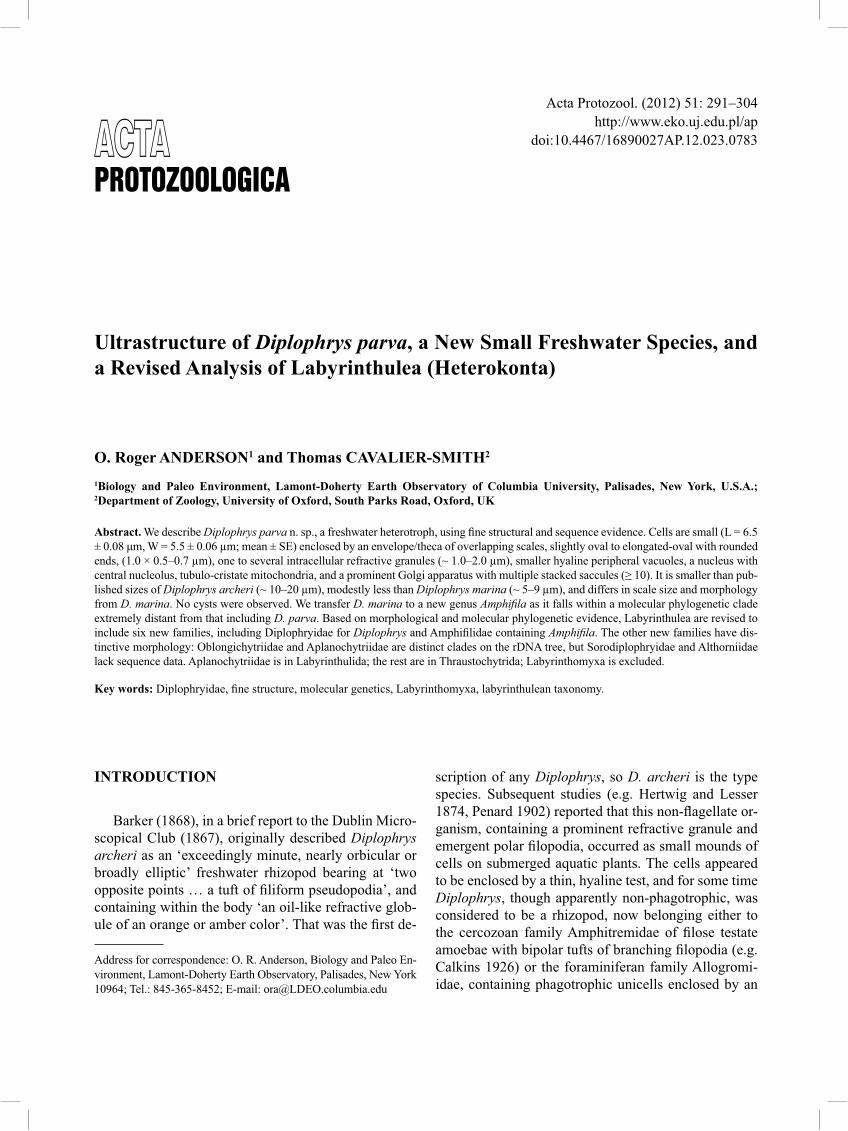
The light microscopic morphology of the ATCC isolate 50360, here described as Diplophrys parva n. sp., exhibits typical features of the genus Diplophrys (Fig. 1), including the ovoid to ellipsoidal cell shape (~ 5–7 μm), emergent tufts of branching pseudopodia at one or two protruding portions of the cell periphery, and one to several prominent internal refractive gran-ules (~ 1–2 μm) of unknown composition, but possi-
Fig. 1. Light micrographs of Diplophrys parva n. sp. Cells are ovate to rounded, containing one or more refractive granules (arrow). An empty cell envelope (theca: e) containing only a small fragment of cellular debris exhibits its thin composition. Bar: 5 μm.
O. R. Anderson and T. Cavalier-Smith294
bly lipid. Pseudopodia branch but do not anastomose. No evidence of cell aggregation, cysts, fruiting bodies, phagotrophy or cilia was seen, but the culture does con-tain bacteria. A small vacuole near the periphery of the cell, visible in light microscopic images particularly near the poles, appears to be a contractile vacuole.
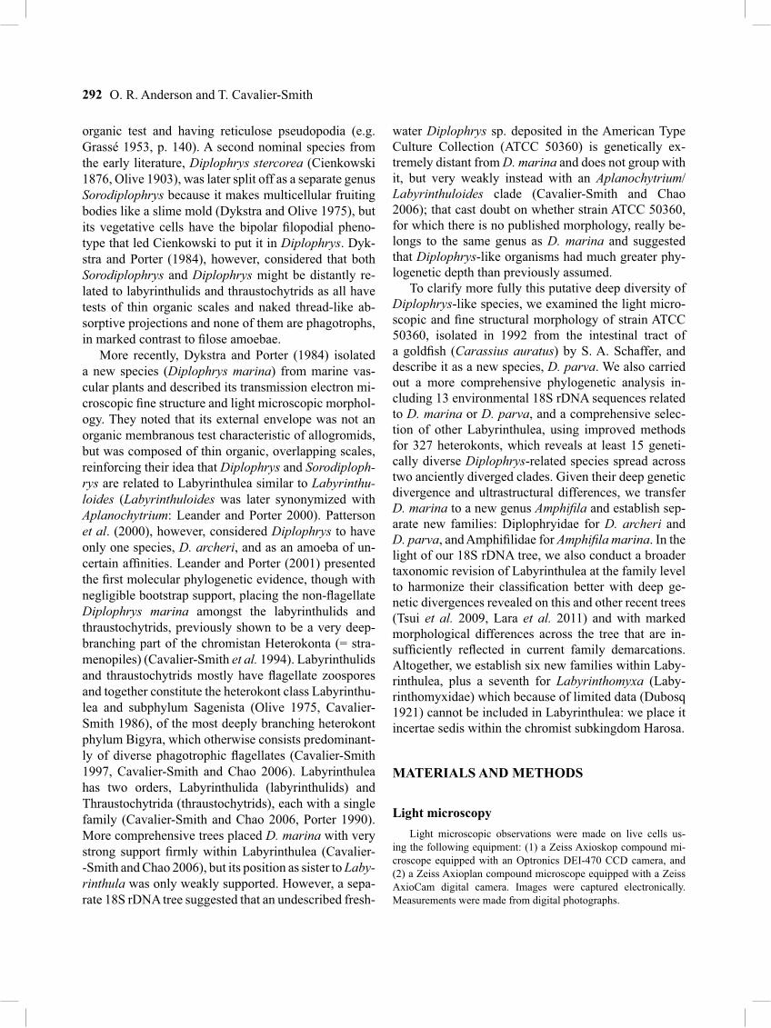
Images of ultrathin sections show fi ne structural details characteristic of the genus, including the sur-rounding theca (envelope) of overlapping scales and characteristic refractive bodies within the cell (Fig. 2). The overall features of a section through a cell (Fig. 2) show the loosely arranged organic scales (arrow) form-ing the theca. The prominent nucleus contains a some-what denser nucleolus (Fig. 3). Mitochondria with tubular cristae are scattered throughout the peripheral cytoplasm, and patches of convoluted smooth endoplas-mic reticulum are sometimes observed in the vicinity of the nucleus (Fig. 3). Pseudopodia emerge from the cell surface as electron dense conical projections, pos-sibly sagenetosomes (also known as bothrosomes) and become longer tubular extensions. At fi rst they may be contained within the surrounding envelope of scales, but eventually penetrate through the scales at one or a few places and emerge, becoming less electron dense. The peripheral cytoplasm also contains rounded vacuoles with less electron dense deposits of unknown compo-sition. The characteristic refractive granules appear to begin development as less-enlarged, irregularly shaped membrane-bound spaces (Fig. 2), often near electron-dense deposits that appear to be lipid. Eventually, they become much enlarged and more rounded (Fig. 4). The prominent Golgi apparatus contains multiple fl attened saccules (≥ 10) that are infl ated at the periphery where Golgi-derived vesicles are secreted (Fig. 4). The surface scales, when viewed with negative staining, are vari-able in shape; but typically are elongated and oval (Fig. 5), approximately 1 μm × 0.5 to 0.7 μm. Other scales are somewhat more broadly oval or become deformed,
appearing rounded or angular when dried on the grid in contiguity to one another (Fig. 5, inset). These scales are approximately the same size or somewhat smaller than the more elongated oval scales (Fig. 5).
Molecular phylogenetic evidence
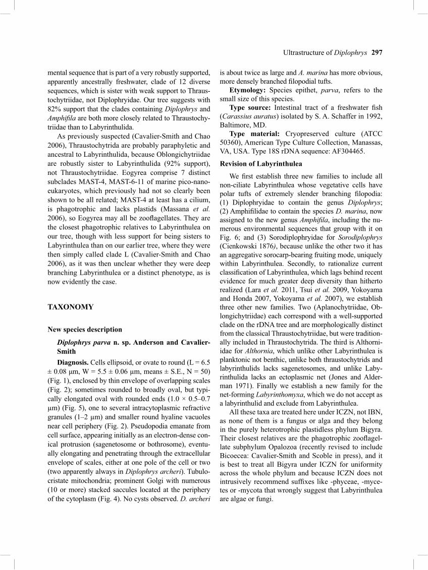
On a previously published distance tree, the Diploph-rys marina 18S rDNA sequence was weakly sister to Labyrinthula (Cavalier-Smith and Chao 2006); but that tree did not include the partial sequence of Diplophrys parva in case it distorted the topology. We have, there-fore, done a new analysis using maximum likelihood, which can place partial sequences more accurately, in-cluding it and a much larger number of labyrinthulean sequences, both cultured ones of known phenotype and environmental DNA sequences, in order to further clarify the position of D. parva. Figure 6 shows the branching order of the Labyrinthulea part only of the heterokont tree: the three main heterokont branches (Ochrophyta, Pseudofungi, Opalozoa) are collapsed as their internal branching order is irrelevant to this paper and would make the tree too large to fi t on the page. The internal branching order of the three collapsed branches on this same comprehensive tree is shown in a separate paper describing a novel opalozoan fl agellate (Cavalier-Smith and Scoble in press). Figure 6 gives strong support (98%) to the holophyly of Labyrinthulea including both D. par-va and A. marina, but shows that D. parva is extremely distant from both A. marina and Labyrinthulida. Dip-lophrys parva is sister with 100% bootstrap support to an environmental sequence from a peat bog as fi rst noted by Lara et al. (2011), and this clade is sister in turn to a much more deeply branching marine sequence of un-known phenotype (GU823043). The three together form a robust clade (labeled Diplophryidae) that is at least as deeply diverging from the clade containing A. marina as are Thraustochytriidae sensu stricto. By contrast A. ma-rina is sister with 84% support to a freshwater environ-
Figs 2–5. Fine structure of ultrathin sections of Diplophrys parva n. sp. 2 – Cell section showing the nucleus (n) with undulating to angular margin, mitochondria (m) with tubular cristae, segment of a “refractive granule” (rg), small peripheral vacuoles (v), emergent pseudopo-dium (ps) with characteristic electron dense cytoplasm, but typically becoming more granular and translucent after emerging through the scale coat, and profi le of the multi-lamellated scales (arrow) forming the envelope. Bar: 0.5 μm; 3 – nucleus (n), with a more electron dense nucleolus (nu), and nearby profi les of convoluted smooth endoplasmic reticulum (er), and a longitudinal section through an elongated mito-chondrion (m) with prominent tubular cristae. Bar: 0.5 μm; 4 – peripheral region of a cell showing the Golgi body (g) with multiple stacks of cisternae, mitochondria (m), and a prominent section through a membrane-enclosed “refractive granule” (rg). Bar: 0.2 μm; 5 – negatively stained preparations of scales (arrow) that typically appear elliptical (~ 1 × 0.5–0.7 μm) or occasionally much more irregular in shape (inset) from polygonal to broadly ovate (arrow, inset). Bars: 0.5 μm.
�
Ultrastructure of Diplophrys 295
O. R. Anderson and T. Cavalier-Smith296
Fig. 6. Maximum likelihood tree for 327 heterokonts emphasizing Labyrinthulea and Eogyrea of phylum Bigyra and the non-heterokont outgroups. Internal branches for the fi ve clades at the top of the tree are collapsed, but are being published separately (Cavalier-Smith and Scoble in press); the numbers to their right indicate how many sequences were included in each. Bootstrap supports for bipartitions are based on 1,000 resamplings using GTRMIX option of RAxML. Black bullets indicate 100% bootstrap support. The sequence attributed to ‘Labyrinthuloides haliotidis’ might be from a thraustochytrid contaminant rather than from Aplanochytrium (=Labyrinthuloides) haliotidis (Leander and Porter 2001).
Ultrastructure of Diplophrys 297
mental sequence that is part of a very robustly supported, apparently ancestrally freshwater, clade of 12 diverse sequences, which is sister with weak support to Thraus-tochytriidae, not Diplophryidae. Our tree suggests with 82% support that the clades containing Diplophrys and Amphifi la are both more closely related to Thraustochy-triidae than to Labyrinthulida.
As previously suspected (Cavalier-Smith and Chao 2006), Thraustochytrida are probably paraphyletic and ancestral to Labyrinthulida, because Oblongichytriidae are robustly sister to Labyrinthulida (92% support), not Thraustochytriidae. Eogyrea comprise 7 distinct subclades MAST-4, MAST-6-11 of marine pico-nano-eukaryotes, which previously had not so clearly been shown to be all related; MAST-4 at least has a cilium, is phagotrophic and lacks plastids (Massana et al. 2006), so Eogyrea may all be zoofl agellates. They are the closest phagotrophic relatives to Labyrinthulea on our tree, though with less support for being sisters to Labyrinthulea than on our earlier tree, where they were then simply called clade L (Cavalier-Smith and Chao 2006), as it was then unclear whether they were deep branching Labyrinthulea or a distinct phenotype, as is now evidently the case.
TAXONOMY
New species description
Diplophrys parva n. sp. Anderson and Cavalier-SmithDiagnosis. Cells ellipsoid, or ovate to round (L = 6.5
± 0.08 μm, W = 5.5 ± 0.06 μm, means ± S.E., N = 50) (Fig. 1), enclosed by thin envelope of overlapping scales (Fig. 2); sometimes rounded to broadly oval, but typi-cally elongated oval with rounded ends (1.0 × 0.5–0.7 μm) (Fig. 5), one to several intracytoplasmic refractive granules (1–2 μm) and smaller round hyaline vacuoles near cell periphery (Fig. 2). Pseudopodia emanate from cell surface, appearing initially as an electron-dense con-ical protrusion (sagenetosome or bothrosome), eventu-ally elongating and penetrating through the extracellular envelope of scales, either at one pole of the cell or two (two apparently always in Diplophrys archeri). Tubulo-cristate mitochondria; prominent Golgi with numerous (10 or more) stacked saccules located at the periphery of the cytoplasm (Fig. 4). No cysts observed. D. archeri
is about twice as large and A. marina has more obvious, more densely branched fi lopodial tufts.
Etymology: Species epithet, parva, refers to the small size of this species.
Type source: Intestinal tract of a freshwater fi sh (Carassius auratus) isolated by S. A. Schaffer in 1992, Baltimore, MD.
Type material: Cryopreserved culture (ATCC 50360), American Type Culture Collection, Manassas, VA, USA. Type 18S rDNA sequence: AF304465.
Revision of Labyrinthulea
We fi rst establish three new families to include all non-ciliate Labyrinthulea whose vegetative cells have polar tufts of extremely slender branching fi lopodia: (1) Diplophryidae to contain the genus Diplophrys; (2) Amphifi lidae to contain the species D. marina, now assigned to the new genus Amphifi la, including the nu-merous environmental sequences that group with it on Fig. 6; and (3) Sorodiplophryidae for Sorodiplophrys (Cienkowski 1876), because unlike the other two it has an aggregative sorocarp-bearing fruiting mode, uniquely within Labyrinthulea. Secondly, to rationalize current classifi cation of Labyrinthulea, which lags behind recent evidence for much greater deep diversity than hitherto realized (Lara et al. 2011, Tsui et al. 2009, Yokoyama and Honda 2007, Yokoyama et al. 2007), we establish three other new families. Two (Aplanochytriidae, Ob-longichytriidae) each correspond with a well-supported clade on the rDNA tree and are morphologically distinct from the classical Thraustochytriidae, but were tradition-ally included in Thraustochytrida. The third is Althorni-idae for Althornia, which unlike other Labyrinthulea is planktonic not benthic, unlike both thraustochytrids and labyrinthulids lacks sagenetosomes, and unlike Laby-rinthulida lacks an ectoplasmic net (Jones and Alder-man 1971). Finally we establish a new family for the net-forming Labyrinthomyxa, which we do not accept as a labyrinthulid and exclude from Labyrinthulea.
All these taxa are treated here under ICZN, not IBN, as none of them is a fungus or alga and they belong in the purely heterotrophic plastidless phylum Bigyra. Their closest relatives are the phagotrophic zoofl agel-late subphylum Opalozoa (recently revised to include Bicoecea: Cavalier-Smith and Scoble in press), and it is best to treat all Bigyra under ICZN for uniformity across the whole phylum and because ICZN does not intrusively recommend suffi xes like -phyceae, -myce-tes or -mycota that wrongly suggest that Labyrinthulea are algae or fungi.
O. R. Anderson and T. Cavalier-Smith298
Six new families of Labyrinthulea
1. Diplophryidae Cavalier-Smith fam. n. Diagnosis: Non-ciliated spherical, unicellular het-
erotrophic protists with scaly theca; without zoospores; with one or two polar tufts of sometimes branching but not anastomosing ectoplasmic threads stemming from a sagenetosome-like structure – if two, passing through the scaly theca on opposite sides of cell but with an offset; with a large (or few smaller), refractive golden yellow or amber lipid drop beside the nucleus. Glide slowly without obviously moving fi lopodia. Type genus Diplophrys Barker, 1868, p. 123.
Comment: As both have fi lopodia stemming from two pores Diplophrys was once grouped with Amphi-trema in Amphitremidae Poche, 1913, or in Amphis-tomina (Wailes 1915; Calkins 1926). We agree with Wailes (1915) that they are probably unrelated; Amphi-trema is not scaly but has an agglutinated test like the undoubtedly cercozoan fi lose amoeba Pseudodiffl ugia, so Amphitremidae (without Diplophrys) is now in-cluded in the order Tectofi losida of the cercozoan class Tectofi losea (Howe et al. 2011a). Amphistomidae is invalid, not being based on an included genus and pre-occupied by a family or subfamily including the fl uke genus Amphistoma Rudolphi, 1801 or Amphistomum Rudolphi, 1814. Diplophrys is not a foraminiferan, so cannot be kept in Allogromiidae, thus making a new family is essential as absence of zoospores and strong divergence on our tree prevent inclusion of Diplophrys in Thraustochytriidae.
2. Amphifi lidae Cavalier-Smith fam. n. Diagnosis: Non-ciliated spindle-shaped, thecate
unicellular heterotrophic protists; without defi nite sa-genetosome or zoospores; with two tufts of sometimes branching but not anastomosing fi lopodia emanating though two pores situated on opposite acute points of scaly theca or wall; with a large (or few smaller), refrac-tive lipid drop beside the nucleus. Glide with fi lopodia preceding cell. Type genus Amphifi la Cavalier-Smith gen. n. Diagnosis as for family. Type species Amphifi la marina Cavalier-Smith comb. n. Basionym Diplophrys marina Dykstra and Porter (1984, p. 627).
Etymology: Amphi Gk both; fi lum L. thread, as fi lo-podia extend from both ends of cell.
Comment: D. archeri Barker (1868), the type spe-cies, was a spherical freshwater organism. Although not then fi gured, the description and plates in Wailes (1915) and the photograph on p. 82 of Patterson (1992) cor-respond closely with Barker’s original description of
D. archeri and with a bloom of freshwater Diplophrys observed by TCS in South Africa. In marked contrast, A. marina was spindle-shaped with pointed ends, so we do not accept the opinion that they are the same spe-cies (Patterson 1989). Figure 6 shows that freshwater Diplophrys parva is exceedingly distantly related to the type strain of A. marina; each belongs to a separate genetically highly diverse clade. Patterson (1989) did not say whether the strain depicted in his Figs 19.17–19 was from freshwater or marine samples, but his light micrograph fi ts his identifi cation as D. archeri (differ-ent in shape from A. marina). His electron micrographs show a thinner wall than in A. marina and it is unclear if it is composed of scales; unlike in A. marina, the wall has no conical point where fi lopodia exit. His Fig. 17 shows many more fi lopodia in each tuft than in A. ma-rina (or D. parva) and no ectoplasmic swelling as in A. marina and Thraustochytriidae. As Amphifi la and Diplophrys are somewhat different morphologically and very distinct in sequence, naming spindle-shaped marine protists D. archeri (Vørs 1992; following Pat-terson 1989) was incorrect. Their marine habitat and spindle shape suggest they were A. marina; the cell la-beled ‘D. archeri after Vørs 1992’ in Patterson et al. (2000) also is probably A. marina and misleading as to the Diplophrys phenotype. The marine Arctic cells of Vørs (1993, Fig. 40F), more oval than D. archeri and less pointed than A. marina, are probably misidentifi ed as D. archeri and could be a third species. The Antarctic cells of Tong et al. (1997, Figs 5F, 6N) are probably also misidentifi ed as D. archeri, being broadly spindle-shaped with unusually short and unbranched fi lopodia (not stated whether the protists illustrated were marine or freshwater), probably a fourth species. These previ-ously overlooked subtle differences in morphology are consistent with our tree showing that there must be over a dozen undescribed species related to Diplophrys or Amphifi la and thus likely to have a broadly similar two-tufted phenotype; contrary to Patterson et al. (2000) there is not just one species – as in other formerly over-lumped taxa with relatively minor light microscopic variation (e.g. glissomonads: Howe et al. 2009, 2011b) there could be many.
3. Sorodiplophryidae Cavalier-Smith fam. n. Diagnosis: Coprophilic non-ciliated, unicellular
heterotrophic protists with fi lopodial gliding motility; without defi nite sagenetosome or zoospores; with pre-dominantly polar tufts of highly branched, sometimes anastomosing fi lopodia emanating at opposite points of cell wall composed of thin scales, often with lamellipo-
Ultrastructure of Diplophrys 299
dium at the base that may extend round the sides of the cell. On starvation, vegetative cells aggregate to form a stalked, golden yellow sorocarp containing numerous elliptical sorocytes, analogously to dictyostelid slime molds. Sorocytes with contractile vacuole and refrac-tive yellow bodies; vegetative cells ovoid to elliptical, with small colorless granules instead of yellow bodies. Glide at 30 μm/min. with fi lopodia at both ends, ante-rior ones shortening as they progress. Type genus Soro-diplophrys Cienkowski, 1876.
Comment: We agree with Dykstra and Olive (1975) that Sorodiplophrys cannot be included in Labyrinthuli-dae or Thraustochytridae, and with their conjecture that it may nonetheless be related to both. The more elon-gated nature of its cells than in Diplophrys, its tendency for fi lopodial anastomosis and marked mobility suggest that it is evolutionarily closer to Amphifi lidae than Dip-lophryidae. Therefore, we group Amphifi lidae and So-rodiplophryidae together as superfamily Amphifi loidea Cavalier-Smith fam. n.
Diagnosis: Vegetatively unicellular, non-ciliate typically elongated osmotrophic heterotrophs, with scaly walls and two opposite tufts of highly branching, sometimes anastomosing, fi lopodia; sagenetosome not obvious.
4. Aplanochytriidae Leander ex Cavalier-Smith fam. n.
Diagnosis: Marine saprophytic or parasitic hetero-trophic protists forming scaly walled sporangia that re-lease crawling non-fl agellate gliding cells and or bicili-ate zoospores; unlike Labyrinthulidae vegetative cells
not spindle-shaped, often typically spherical or nearly so and mobile upon an ectoplasmic net that does not completely enrobe them as it does in Labyrinthulidae; sagenetosome single unlike Labyrinthula.
Comment: Alderman et al. (1974) wrote that Aplanochytrium should be removed from Thraustochy-triidae as a new family; Leander and Porter named this Aplanochytriaceae but only in Leander’s 2001 PhD Thesis (Georgia University, USA), so the name was not validly published; it did not appear in their journal article (Leander and Porter 2001). Type genus Aplano-chytrium Bahnweg and Sparrow, 1972. The gliding mo-tility of vegetative cells (unlike Thraustochytriidae) and 100% BS support for Aplanochytrium being sisters to Labyrinthulidae justify placement within Labyrinthu-lida as a new family.
5. Oblongichytriidae Cavalier-Smith fam. n. Diagnosis: Thraustochytrids that have slender ob-
long zoospores, not squat oval ones as in Thraustochy-triidae; their 18S rRNA sequences do not group with those of Thraustochytriidae but near the base of the labyrinthulean clade. Type genus Oblongichytrium Yo-koyama and Honda (2007, p. 2002). Their non-group-ing with Thraustochytriidae sensu stricto (Table 1) is robustly on all our trees and the 3- and 4-gene trees of Tsui et al. (2009).
6. Althorniidae Cavalier-Smith fam. n. Diagnosis: Floating thraustochytrids with laterally
biciliate zoospores but no ectoplasmic net or sagene-tosomes. Type genus Althornia Jones and Alderman, 1971.
Class Labyrinthulea (Lister 1891) Olive ex Cavalier-Smith 1986
Order 1. Thraustochytrida Sparrow 1973Family 1. Thraustochytriidae Sparrow ex Cejp 1959 (Thraustochytrium, Ulkenia, Schizochytrium, Japonochytrium, Aurantiochytrium, Sicyoido-chytrium, Parietichytrium, Botryochytrium)Family 2. Oblongichytriidae Cavalier-Smith fam. n. (Oblongichytrium)Family 3. Althorniidae Cavalier-Smith fam. n. (Althornia)Family 4. Diplophryidae Cavalier-Smith fam. n. (Diplophrys)
Superfamily Amphifi loidea Cavalier-Smith superfam. n.Family 1. Amphifi lidae Cavalier-Smith fam. n. (Amphifi la)Family 2. Sorodiplophryidae Cavalier-Smith fam. n. (Sorodiplophrys)
Order 2. Labyrinthulida Dofl ein 1901Family 1. Labyrinthulidae Cienkowski 1867 (Labyrinthula)Family 2. Aplanochytriidae Leander ex Cavalier-Smith fam. n. (Aplanochytrium)
similar to labyrinthulids is Labyrinthomyxa (Dubosq 1921) with an anteriorly directed single cilium and a single amoeba phase; as its spindles do not move with-in the net, which in some respects is more like that of Leukarachnion (Grant et al. 2009), and there is no evi-dence for a laminate (or other) theca we exclude it from Labyrinthulea and establish a separate family (Laby-rinthomyxidae), here placed incertae sedis in Harosa as it is unclear whether it belongs in Heterokonta (possibly Leukarachnida; unlike Leukarachnion not known to be phagotrophic) or Cercozoa (possibly Endomyxa).
Labyrinthomyxidae Cavalier-Smith fam. n. Diagnosis: Filoplasmodial heterotrophs whose
spindle-shaped cells with bipolar projections form lin-ear, branching, anastomosing rows and parasitize sole-nocysts of the brown alga Laminaria; with uninucleate amoeba or uniciliate phases; cyst or theca unknown. Type genus Labyrinthomyxa (Dubosq 1921).
Another protist with similarities to labyrinthulids is Chlamydomyxa labyrinthuloides (Archer 1875), but its taxonomy is confused by probable later misidentifi ca-tions. Unless Archer confl ated multiple organisms, we support his interpretation of the original Chlamydo-myxa labyrinthuloides as probably a labyrinthulid (dis-tinct enough to merit its own family), unlike subsequent authors who questioned that or described other prob-ably unrelated ‘Chlamydomyxa’ species (Geddes 1882, Hieronymus 1898, Lankester 1896, Pascher 1930, Pearlmutter and Tumpano 1984, Penard 1904) – clonal cultures more similar to those of Archer are needed to check this. In particular we consider the non-reticulose, fi lose amoeboid heterokont alga identifi ed as Chlamy-domyxa labyrinthuloides by Wenderoth et al. (1999), whose 18S rDNA places it in Picophagea within the phylum Ochrophyta (Cavalier-Smith and Chao 2006), was misidentifi ed and is really a new species in an un-described genus – to be established elsewhere.
DISCUSSION
Our most striking conclusion is that Labyrinthulea includes two genetically extremely divergent clades of non-ciliated protists with two polar tufts of fi lopodia,
which are so similar in the light microscope that some have thought they were just one species (Patterson et al. 2000). Overall, based on available morphological evidence, we conclude that the ATCC 50360 strain is a new species in the genus Diplophrys, but Amphifi la is only remotely related.
Novelty of Diplophrys parva
The ATCC 50360 isolate that we name Diplophrys parva n. sp., differs substantially in size, shape, and/or fi ne structural features from published descrip-tions of D. archeri and A. marina. It is essentially the same length as A. marina, but only about half the size of D. archeri that typically has a much larger refrac-tive granule (often fi lling over half the diameter of the cell) and larger cell size (10 μm or larger) compared to D. parva (5–7 μm). However, the original description of D. archeri stated only that it was exceedingly minute and gave no size measurement or illustration. Archer, as described in Barker (1868), fi rst stated that its aver-age size was 1/2000 inch, i.e. 12.7 μm. As Archer was present at the meeting the previous year where D. ar-cheri was fi rst shown in public, described and its name published, we consider that this should be accepted as the average size of D. archeri. D. parva is about half the size of D. archeri, a suffi ciently large difference to make it unwise to treat them as one species. As many different species have probably been lumped under that name and most descriptions may relate to others, dif-ferent sizes given in some later studies should not be attributed to D. archeri. Despite their somewhat simi-lar size and general appearance, there is no possibility of confusing D. parva and A. marina. One is marine and the other freshwater; as they have ultrastructurally different scales and their rDNA sequences are radically different, two genera are merited.
Contrast between Diplophrys and Amphifi la marina
Conservation of the name Diplophrys for D. parva rather than A. marina merits discussion. Until an au-thentic culture of D. archeri is sequenced, we cannot be sure that retention of the generic name Diplophrys for the D. parva rather than the Amphifi la clade is correct, but a decision one way or the other had to be made. We picked D. parva for three reasons: fi rst because its more rounded, less pointed shape, is more like D. ar-cheri than is the spindle-shaped A. marina. The con-sistent phylogenetic contrast between the elongated Oblongichytrium and round Thraustochytriidae sensu stricto (Yokoyama and Honda 2007, and Fig. 6) shows
Ultrastructure of Diplophrys 301
that small differences in cell shape can have surpris-ingly deep phylogenetic signifi cance in Labryrinthulea. Second are the fi lopodia: in D. parva and archeri they are branched but non-anastomising, and both show only minimal cell motility if any – no locomotion was men-tioned in the original descriptions of D. archeri (Barker 1868). By contrast Amphifi la locomotes by active glid-ing and shows fi ne fi lopodial anastomoses, both char-acters shared with Sorodiplophrys, but not D. archeri. Thirdly, D. archeri and D. parva are both from fresh-water, whereas Amphifi la is marine, and conservatism of freshwater versus marine habitat is pronounced in many protists (Cavalier-Smith and Chao 2012), and also shows a non-random distribution across Labyrinthulea. One can argue that Labryrinthulea were probably an-cestrally marine. However, most lineages of the clade to which Amphifi la belongs are freshwater (or soil, eco-logically cognate), so that clade was probably freshwa-ter for most of its evolutionary history, and the ances-tor (or ancestors) must have made one relatively recent switch into the oceans, perhaps accompanying the sea grasses with which it is commensal. The sequence clos-est to D. parva comes from European peat bogs and D. archeri was from Irish moors, both consistent with the morphological evidence that D. parva and archeri are mutually closer than to Amphifi la.
Scale ultrastructure, often good phylogenetic in-dicators (Cavalier-Smith and Chao 2012, Howe et al. 2011a), strongly supports this; we found that D. parva has oval to elongated scales (~ 1 μm) decidedly differ-ent in size and shape from the round scales (~ 2 μm) of A. marina. A second ultrastructural difference is that D. parva has an obvious dense structure somewhat resembling a sagenetosome, whereas no evidence for a sagenetosome was seen in Amphifi la, in which respect also it resembles Sorodiplophrys (Dykstra and Olive 1975).
Increased diversity of Diplophrys-like protists
Only two previously described species were re-cently accepted as Diplophrys: Diplophrys archeri and Diplophrys marina (here moved to Amphifi la. Diploph-rys stercorea described by Cienkowski (1876) was re-assigned to a separate genus Sorodiplophrys (Dykstra and Olive 1975), with stercorea the type species. It is a sorocarp-producing protist, thus sharply distinct from Diplophrys and Amphifi la, despite having suffi ciently similar vegetative cells to A. marina (net-like fi lopodia) to make a relationship plausible. The sorocarp (stalk-borne fruiting body) is a product of cellular aggregation
as occurs among some slime molds, but as its vegeta-tive cell structure is dissimilar from slime molds, and cell aggregation is well known as a polyphyletic char-acter, it should not be placed in Mycetozoa. Though its ultrastructure remains unpublished, Dykstra and Olive (1975) stated that it lacks sagenetogens and has thin scales. Sorodiplophrys vegetative cells crawl using contractile non-granular fi lopodia, whose contractil-ity makes them perhaps more similar to those in the cercozoan superclass Ventrifi losa (Cavalier-Smith and Karpov 2012), comprising the fi lose amoeboid classes Imbricatea and Thecofi losea (Howe et al. 2011a), than to Diplophrys. As Imbricatea often also have scales, it is possible that Sorodiplophrys belongs in that class, which includes a variety of amoebae and fl agellates with similar contractile, non-granular branching fi lo-podia (Howe et al. 2011a, Cavalier-Smith and Chao 2012). Moreover, the testate Amphitremidae, with bipo-lar fi lopodia analogous to, but more robust than, those of Diplophrys, is currently assigned to Thecofi losea (Cavalier-Smith and Chao 2012). However, we have adopted the more conservative stance of retaining Soro-diplophrys within Labyrinthulea, for two reasons. First, Dykstra and Porter (1984) noted thin scales of Sorodip-lophrys resembling those of Labyrinthulea. If the scales had been more like any of the diverse siliceous scales of the scaly taxa now placed in Imbricatea, they would probably have mentioned that and even more strongly doubted its affi nity with Labyrinthulea. Thus Sorodip-lophrys is probably not an imbricate. Secondly, they stressed that Sorodiplophrys is osmotrophic and not phagotrophic, also making it unlikely that it is a scaly imbricate cercozoan amoeba (all have siliceous scales, not unmineralized organic ones like Labyrinthulea).
In contrast to Amphifi la and Sorodiplophrys, both currently recognized species of Diplophrys present di-agnostic features of the genus, i.e. ellipsoidal to ovoid cells, non-aggregating cells, enclosed by a thin enve-lope (shown to be imbricated scales by fi ne structure analysis) with pseudopodia emerging typically from two poles of the cell, forming a branching rhizopodial fan toward the periphery; there is at least one intracy-toplasmic refractive granule, presumed to be lipid. In D. archeri, the refractive granules (one or more) are typically very prominent, yellowish in color, and oc-cupy a large portion of the cell volume when viewed by light microscopy. Published images of D. archeri are typically in the range of 10–15 μm or somewhat larger (e.g. Barker 1868, Kudo 1977, p. 568). D. marina cells (3.7–5.9 × 5.1–8.5 μm) are ovoid with round Gol-
O. R. Anderson and T. Cavalier-Smith302
gi-derived scales (1.5–1.9 μm). The Diplophrys-like phenotype had only three named species (D. archeri, D. marina, and Sorodiplophrys stercorea) prior to this publication. Their placement now in three separate gen-era and families better refl ects their evolutionary diver-sity and should stimulate further research on this unique protist type – neither a rhizopod nor a fungus but a very distinctive, albeit neglected, osmotrophic phenotype. Many understudied protists are not in culture (e.g. D. ar-cheri), impeding molecular genetic analyses, but many more could probably be cultured with even a modest effort. Recent light micrographs of D. archeri with ac-curate diagnostic size and morphology for this species as described by Barker (1868) (e.g. http://starcentral.mbl.edu/microscope/portal.php?pagetitle=assetfactsheet&imageid=9704) show that D. archeri, and no doubt many genetically distinct look-alikes, can be isolated from the natural environment. Without a targeted study of Diplophrys, currently with only two verifi able spe-cies (archeri and parva), it is premature to judge wheth-er its taxonomic diversity is really limited to the three sequences that branch robustly together in Fig. 6, or is much more extensive. However, given the small size of D. parva, and its broad similarity to the genetically very distant Amphifi la, it is likely that many additional cryptic species will be discovered. The clade containing Amphifi la is currently more speciose. Possibly one of the two distinctly deep-branching soil lineages in that clade is related to the dung-dwelling Sorodiplophrys, as dung dwellers are most likely to have evolved from soil biota; if that could be confi rmed, it would make that quite speciose clade equivalent to the new superfam-ily Amphifi loidea. More intensive research on this mi-croscopically distinctive but remarkably conservative morphotype is warranted.
Large-scale evolution in Labyrinthulea
We can now conclude that there are not just two broad phenotypes in Labryinthulea, but three. The thraustochytrid-like condition appears to be ancestral; i.e. scaly thecate vegetative cells with a single aperture from which a sagenetosome emits slender branching, but not anastomosing, fi lopodia used not for locomotion but presumably to increase surface area for absorbing dissolved organic molecules; biciliate zoospores medi-ate dispersal. Secondly are the net-like Labyrinthulida, with cells stationary in the net (Aplanochytriidae) or self-propelling within it (Labyrinthulidae). The third major phenotype is the Diplophrys-like one with two
polar tufts of fi lopodia and no zoospores. Our trees show that Labyrinthulida and the Diplophrys-like phe-notype are both derived from thraustochrytrid-like an-cestors, but independently: both Diplophrys-like clades are unambiguously closer to Thraustochytriidae than to Labyrinthulida.
Our trees also raise the possibility that the Dip-lophrys-like phenotype evolved twice independently in Diplophryidae and Amphifi loidea. Such evolution involves only two things: loss of the zoospore, a very common evolutionary event in protists, and evolution of a second pore through the theca, which also is prob-ably not diffi cult; so we should not be concerned that Diplophryidae and the clade including Amphifi la are not sisters on our tree. But 18S rDNA clearly lacks the resolution to prove an independent origin of a second polar pore, though that would be consistent with the ultrastructural differences we found between D. parva and Amphifi la, but these also do not establish an inde-pendent origin, either. Probably sequences from many genes will be needed for a fi rmer conclusion. A better supported instance of convergent evolution is fi lopo-dial anastomosis, which seemingly created a net-like absorptive surface independently in Labyrinthulida and Amphifi loidea, also probably not diffi cult to evolve twice; net-like pseudopodia also involved independent-ly in Rhizaria (their ancestral state), Amoebozoa (e.g. leptomyxids) and elsewhere in Heterokonta in Chryso-monadea (e.g. Leukarachnion). However, they are all phagotrophs. We have used the word fi lopodium for the threadlike extensions of Labyrinthulea, but should stress that they are probably not homologous with fi lo-podia in rhizopods and have no phagotrophic function; they appear to be purely absorptive like the microvilli of the mammalian intestine and it is open to debate whether the term fi lopodium is somewhat misleading, especially in most thraustochrytrids where it lacks a lo-comotory function and is not in any sense a foot. This emphasizes that Labyrinthulea are protists sui generis that should not be slotted unthinkingly into convention-al textbook categories.
Acknowledgements. We thank Dr. Robert Molestina (American Type Culture Collection, Manassas, VA) for providing the light mi-crograph from the ATCC collection. We also appreciate information provided by Dr. Tom Nerad on the history of the cultures and some details of light microscopic observations made at ATCC of isolate 50360. This is Lamont-Doherty Earth Observatory Contribution No. 7610. TCS thanks NERC for past grant support and Josephine Scoble for help with old literature and agreeing to the inclusion of part of the tree stemming from our joint work on heterokonts.
Ultrastructure of Diplophrys 303
REFERENCES
Alderman D. J., Harrison J. L., Bremer G. B., Jones E. B. G. (1974) Taxonomic revisions in the marine bifl agellate fungi. The ultra-structural evidence. Mar. Biol. 25: 345–357
Anderson O. R., Rogerson A., Hannah F. (1997) Three new limax amoebae isolated from marine surface sediments: Vahlkampfi a caledonica n. sp., Saccamoeba marina n. sp., and Hartmannella vacuolata n. sp. J. Eukaryot. Microbiol. 44: 33–42
Archer W. (1869) On some freshwater Rhizopoda, new or little-known. Quart. J. Micr. Sci. 8: 101–124
Archer W. (1875) On Chlamydomyxa labyrinthuloides, nov. gen. et sp., a new freshwater sarcodic organism. Quart. J. Microscop. Sci. (N. S.) 15: 107–130
Barker J. (1868) No title. Quart. J. Microscop. Sci. (N. S.) 8: 123Calkins G. N. (1926) Biology of Protozoa. Lea and Fibiger, Phila-
delphiaCavalier-Smith T. (1986) The kingdom Chromista: origin and sys-
tematics. In: Progress in Phycological Research, Vol. 4, (Eds. F. E. Round, D. J. Chapman). Bristol, Biopress, 309–347
Cavalier-Smith T. (1997) Sagenista and Bigyra, two phyla of hetero-trophic heterokont chromists. Archiv Protistenkd. 148: 253–267
Cavalier-Smith T. (2010) Deep phylogeny, ancestral groups and the four ages of life. Phil. Trans. Roy. Soc. Lond. B 365: 111–132
Cavalier-Smith T., Chao E. E.-Y. (2006) Phylogeny and megasys-tematics of phagotrophic heterokonts (Kingdom Chromista). J. Mol. Evol. 62: 388–420
Cavalier-Smith T., Chao E. E. (2012) Oxnerella micra sp. n. (Ox-nerellidae fam. n.), a tiny naked centrohelid, and the diversity and evolution of Heliozoa. Protist 163: 574–601
Cavalier-Smith T., Karpov S. A. (2012) Paracercomonas kinetid ultrastructure, origins of the body plan of Cercomonadida, and cytoskeleton evolution in Cercozoa. Protist 163: 47–75
Cavalier-Smith T., Scoble J. M. (2012) Phylogeny of Heterokonta: Incisomonas marina, a uniciliate gliding opalozoan related to Solenicola (Nanomonadea), and evidence that Actinophryida evolved from raphidophytes. Eur. J. Protistol. (in press), http://dx.doi.org/10.1016/j.ejop.2012.09.002
Cavalier-Smith T., Allsopp M. T. E. P., Chao E. E.-Y. (1994) Thraus-tochytrids are chromists not fungi: 18S rRNA signatures of Het-erokonta. Phil. Trans. Roy. Soc. Lond. B 145: 209–220
Cienkowski L. (1876) Ueber enige Rhizopoden und verwandte Or-ganismen. Arch. Microskop. Anat. 12: 15–50
Dubosq O. (1921) Labyrinthomyxa sauvageaui n. g., n. sp., Pro-téomyxée parasite de Laminaria lejolisii Sauvageau. Les plas-modes de Labyrinthomyxa sauvageaui. C. R. Soc. Biol., Paris 84: 27–32
Dykstra M. J., Olive L. S. (1975) Sorodiplophrys: an unusual soro-carp-producing protist. Mycologia 67: 873–879
Dykstra M. J., Porter D. (1984) Diplophrys marina, a new scale-forming marine protist with Labyrinthulid affi nities. Mycologia 76: 626–632
Geddes P. (1882) Observations on the resting state of Chlamydo-myxa labyrinthuloides, Archer. Quart. J. Microsc. Sci. 22: 30–34
Gómez F., Moreira D., Benzerara K., López-García P. (2011) So-lenicola setigera is the fi rst characterized member of the abun-dant and cosmopolitan uncultured marine stramenopile group MAST-3. Environ. Microbiol. 13: 193–202
Grant J., Tekle Y., Anderson O. R., Patterson D. J., Katz L. A. (2009) Multigene evidence for the placement of a heterotrophic
amoeboid lineage Leukarachnion sp. among photosynthetic stramenopiles. Protist 160: 376–385
Grassé P.-P. (1953) Traité de Zoologie: Anatomie, Systématique, Biologie. Masson et Cie, Paris
Hertwig R., Lesser, E. (1874) Ueber Rhizopoden und denselben nahestehende Organismen. Arch. Mikroskop. Anat. (Suppl.) 10: 139–145
Hieronymus G. (1898) Zur Kenntnis von Chlamydomyxa Archer. Hedwigia 37: 1–40, plates 1–2
Howe A. T., Bass D., Vickerman K., Chao E. E., Cavalier-Smith T. (2009) Phylogeny, taxonomy, and astounding genetic diversity of Glissomonadida ord. nov., the dominant gliding zoofl agel-lates in soil (Protozoa, Cercozoa). Protist 160: 159–189
Howe A. T., Bass D., Scoble J. M., Lewis R., Vickerman K., Arndt H., Cavalier-Smith T. (2011a) Novel cultured protists identify deep-branching environmental DNA clades of Cercozoa, new genera Tremula, Micrometopion, Minimassisteria, Nudifi la, Peregrinia. Protist 162: 332–372
Howe A. T., Bass D., Chao E. E.-Y., Cavalier-Smith T. (2011b) New genera, species and improved phylogeny of Glissomonadida (Cercozoa). Protist 162: 710–722
Jones E. B. G., Alderman D. J. (1971) Althornia crouchii gen. et n. sp., a marine bifl agellate fungus. Nova Hedwigia 21: 381–400
Kudo R. R. (1977) Protozoology, 5th ed. Charles C. Thomas, Spring-fi eld, Illinois
Lankester E. R. (1896) Chlamydomyxa montana, n. sp., one of the Protozoa Gymnomyxa. Quart. J. Microsc. Sci. 39 n.s.: 233–244 + 232 plates (214, 215)
Lara E., Mitchell E. A., Moreira D., López García P. (2011) Highly diverse and seasonally dynamic protist community in a pristine peat bog. Protist 162: 14–32
Leander C. E., Porter D. (2000) Redefi ning the genus Aplanochy-trium (phylum Labyrinthulomycota). Mycotaxon 76: 439–444
Leander C. E., Porter D. (2001) The Labyrinthulomycota is com-prised of three distinct lineages. Mycologia 93: 459–464
Massana R., Terrado R., Forn I., Lovejoy C., Pedros-Alio C. (2006) Distribution and abundance of uncultured heterotrophic fl agel-lates in the world oceans. Environ. Microbiol. 8: 1515–1522
Olive E. W. (1903) Nuclear and cell division in Diplophrys ster-corea Cienk. Science 17: 260
Olive L. S. (1975) The Mycetozoans. Academic Press, New YorkPascher A. (1930) Ueber einen gruenen, assimilationsfaehigen plas-
modialen Organismus in den Blaettern von Sphagnum. Archiv Protistenkd. 72: 311–358
Patterson D. J. (1989) Stramenopiles, chromophytes from a protis-tan perspective. In: The Chromophyte Algae, (Eds. J. C. Green, B. S. Leadbeater, W. L. Diver). Clarendon Press, Oxford: 357–379
Patterson D. J. (1992) Free-living Freshwater Protozoa, a Colour Guide. Wolfe, London
Patterson D. J., Simpson A. G. B., Rogerson A. (2000) Amoebae of uncertain affi nities. In: An Illustrated Guide to the Protozoa, (Eds. J. J. Lee, G. F. Leedale, P. Bradbury), An Illustrated Guide to the Protozoa. Society of Protozoologists, Lawrence, Kansas: 804–827
Pearlmutter N., Timpano P. (1984) The biology of Chlamydomyxa montana: ultrastructure of the cyst. Protoplasma 122: 68–74
Penard E. (1902) Faune rhizopodique du bassin du Léman. Henry Kündig, Geneva, 714 pp.
Penard E. (1904) Étude sur la Chlamydomyxa montana. Arch. Pro-tistenkd. 4: 296–334, 19 Figs
O. R. Anderson and T. Cavalier-Smith304
Porter D. (1990) Phylum Labyrinthulomycota. In: Handbook of Protoctista, (Eds. L. Margulis, J. O. Corliss, M. Melkonian, D. Chapman). Jones and Bartlett, Boston: 388–398
Stamatakis A. (2006) RAxML-VI-HPC: maximum likelihood-based phylogenetic analyses with thousands of taxa and mixed models. Bioinformatics 22: 2688–2690
Tong S., Vørs N., Patterson D. J. (1997) Heterotrophic fl agellates, centrohelid heliozoa and fi lose amoebae from marine and fresh-water sites in the Antarctic. Polar Biol. 18: 91–106
Tsui C. K., Marshall W., Yokoyama R., Honda D., Lippmeier J. C., Craven K. D., Peterson P. D., Berbee M. L. (2009) Labyrinthu-lomycetes phylogeny and its implications for the evolutionary loss of chloroplasts and gain of ectoplasmic gliding. Mol. Phy-logenet. Evol. 50: 129–140
Vørs N. (1992) Heterotrophic amoebae, fl agellates and heliozoa from the Tvärminne area, Gulf of Finland, in 1988–1990. Oph-elia 36: 1–109
Vørs N. (1993) Heterotrophic amoebae, fl agellates, and heliozoa from Arctic marine waters (North West Territories, Canada and W. Greenland). Polar Biol. 13: 113–126
Wailes G. H. (1915) The British Freshwater Rhizopoda and Helio-zoa. Vol. III. Rhizopoda, Part III. Ray Society, London
Wenderoth K., Marquardt J., Fraunholz M., Van de Peer Y., Wastl J., Maier U.-G. (1999) The taxonomic position of Chlamydomyxa labyrinthuloides. Eur. J. Phycol. 34: 97–108
Yokoyama R., Honda D. (2007) Taxonomic rearrangement of the genus Schizochytrium sensu lato based on morphology, che-motaxonomic characteristics, and 18S rRNA gene phylogeny (Thraustochytriaceae, Labyrinthulomycetes), emendation for Schizochytrium and erection of Aurantiochytrium and Oblon-gichytrium gen. nov. Mycoscience 48: 199–211
Yokoyama R., Salleh B., Honda D. (2007) Taxonomic rearrange-ment of the genus Ulkenia sensu lato based on morphology, chemotaxonomical characteristics, and 18S rRNA gene phylog-eny (Thraustochytriaceae, Labyrinthulomycetes): emendation for Ulkenia and erection of Botryochytrium, Parietichytrium, and Sicyoidochytrium gen. nov. Mycoscience 48: 329–341
Received on 1st June, 2012; revised on 1st September, 2012; accept-ed on 9th September, 2012