Page 1
Copyright © Rebecca Rehder Wingerden
Investigation 7: Cell Division: Mitosis and Meiosis
AP Biology
Pearson Education, Inc., publishing as Person Benjamin Cummings College Board, AP Biology Curriculum Framework 2012-2013
Investigation 7: Cell DivisionLearning Objectives
• To describe the events in the cell cycle and how these events are controlled.
• To explain how DNA is transmitted to the next generation via mitosis.
• To explain how DNA is transmitted to the next generation via meiosis followed by fertilization.
• To understand how meiosis and crossing over leads to increased genetic diversity, which is necessary for evolution.
Copyright © 2012 Rebecca Rehder Wingerden
Background • One of the characteristics of living things is the ability
to replicate and pass on genetic information to the next generation.
• Cell division in individual bacteria and archaea usually occurs by binary fission.
• Mitochondria and chloroplast also replicate by binary fission which is evidence of the evolutionary relationship between these organelles and prokaryotes.
Investigation 7: Cell Division
Copyright © 2012 Rebecca Rehder Wingerden
Background • Cell division in eukaryotes is more complex. It requires
the cell to manage and complicated process of duplicating the nucleus, other organelles, and multiple chromosomes.
• The Cell Cycle is divided into: - interphase:
• G1 • S • G2
- mitosis and - cytokinesis
Investigation 7: Cell Division
Copyright © 2012 Rebecca Rehder Wingerden
Page 2
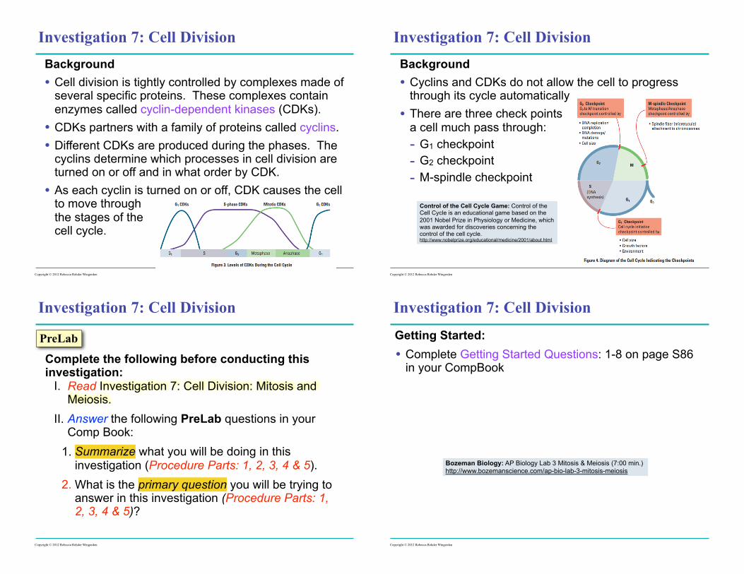
Background • Cell division is tightly controlled by complexes made of
several specific proteins. These complexes contain enzymes called cyclin-dependent kinases (CDKs).
• CDKs partners with a family of proteins called cyclins. • Different CDKs are produced during the phases. The
cyclins determine which processes in cell division are turned on or off and in what order by CDK.
• As each cyclin is turned on or off, CDK causes the cell to move through the stages of the cell cycle.
Investigation 7: Cell Division
Copyright © 2012 Rebecca Rehder Wingerden
Background • Cyclins and CDKs do not allow the cell to progress
through its cycle automatically • There are three check points
a cell much pass through: - G1 checkpoint - G2 checkpoint - M-spindle checkpoint
Investigation 7: Cell Division
Control of the Cell Cycle Game: Control of the Cell Cycle is an educational game based on the 2001 Nobel Prize in Physiology or Medicine, which was awarded for discoveries concerning the control of the cell cycle. http://www.nobelprize.org/educational/medicine/2001/about.html
Copyright © 2012 Rebecca Rehder Wingerden
Complete the following before conducting this investigation:
I. Read Investigation 7: Cell Division: Mitosis and Meiosis.
II. Answer the following PreLab questions in your Comp Book:
1. Summarize what you will be doing in this investigation (Procedure Parts: 1, 2, 3, 4 & 5).
2. What is the primary question you will be trying to answer in this investigation (Procedure Parts: 1, 2, 3, 4 & 5)?
Copyright © 2012 Rebecca Rehder Wingerden
PreLab
Investigation 7: Cell Division
Getting Started: • Complete Getting Started Questions: 1-8 on page S86
in your CompBook
Investigation 7: Cell Division
Copyright © 2012 Rebecca Rehder Wingerden
Bozeman Biology: AP Biology Lab 3 Mitosis & Meiosis (7:00 min.) http://www.bozemanscience.com/ap-bio-lab-3-mitosis-meiosis
Page 3
Procedure Part 1: Modeling Mitosis • Complete Activity #1: Mitosis
- Copy Analysis Sheet #1: Mitosis and use it to diagram each stage of mitosis (n = 1) as you simulate it in this exercise.
• Complete Activity #2: Mitosis with Two Pairs of Homologous Chromosome - Copy Analysis Sheet #2: Mitosis and use it to
diagram each stage of mitosis (n = 2) as you simulate it in this exercise.
• Post Activity Questions - answer the four question from Part 1: Modeling Mitosis at the top of page S87
Investigation 7: Cell Division
Copyright © 2012 Rebecca Rehder Wingerden
Analysis Sheet #1: Mitosis (n = 2)
Investigation 7: Cell Division
Copyright © 2012 Rebecca Rehder Wingerden
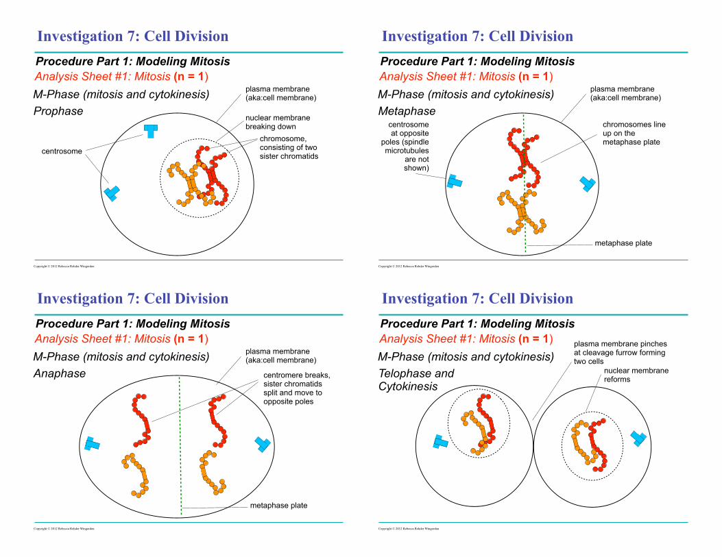
Procedure Part 1: Modeling Mitosis
Interphase (G1)
M-Phase (mitosis and cytokinesis)
Prophase
Metaphase
Anaphase
Telophase & Cytokinesis
Analysis Sheet #2: Mitosis (n = 3)Procedure Part 1: Modeling Mitosis
Interphase (G1)
Prophase
Metaphase
Anaphase
Telophase & Cytokinesis
M-Phase (mitosis and cytokinesis)
Analysis Sheet #1: Mitosis (n = 1)
Investigation 7: Cell Division
Copyright © 2012 Rebecca Rehder Wingerden
Procedure Part 1: Modeling Mitosis
Interphase (G1)
nuclear membrane
chromatin
plasma membrane (aka:cell membrane)
centrosomes nucleolus
Analysis Sheet #1: Mitosis (n = 1)
Investigation 7: Cell Division
Copyright © 2012 Rebecca Rehder Wingerden
Procedure Part 1: Modeling Mitosis
Interphase (G2)
nuclear membrane
chromatin
plasma membrane (aka:cell membrane)
centrosomes nucleolus
Page 4
Analysis Sheet #1: Mitosis (n = 1)
Investigation 7: Cell Division
Copyright © 2012 Rebecca Rehder Wingerden
Procedure Part 1: Modeling Mitosis
ProphaseM-Phase (mitosis and cytokinesis)
nuclear membrane breaking down
chromosome, consisting of two sister chromatids
plasma membrane (aka:cell membrane)
centrosome
Analysis Sheet #1: Mitosis (n = 1)
Investigation 7: Cell Division
Copyright © 2012 Rebecca Rehder Wingerden
Procedure Part 1: Modeling Mitosis
MetaphaseM-Phase (mitosis and cytokinesis)
chromosomes line up on the metaphase plate
plasma membrane (aka:cell membrane)
centrosome at opposite
poles (spindle microtubules
are not shown)
metaphase plate
Analysis Sheet #1: Mitosis (n = 1)
Investigation 7: Cell Division
Copyright © 2012 Rebecca Rehder Wingerden
Procedure Part 1: Modeling Mitosis
AnaphaseM-Phase (mitosis and cytokinesis)
centromere breaks, sister chromatids split and move to opposite poles
plasma membrane (aka:cell membrane)
metaphase plate
Analysis Sheet #1: Mitosis (n = 1)
Investigation 7: Cell Division
Copyright © 2012 Rebecca Rehder Wingerden
Procedure Part 1: Modeling Mitosis
Telophase and Cytokinesis
M-Phase (mitosis and cytokinesis)nuclear membrane reforms
plasma membrane pinches at cleavage furrow forming two cells
Page 5
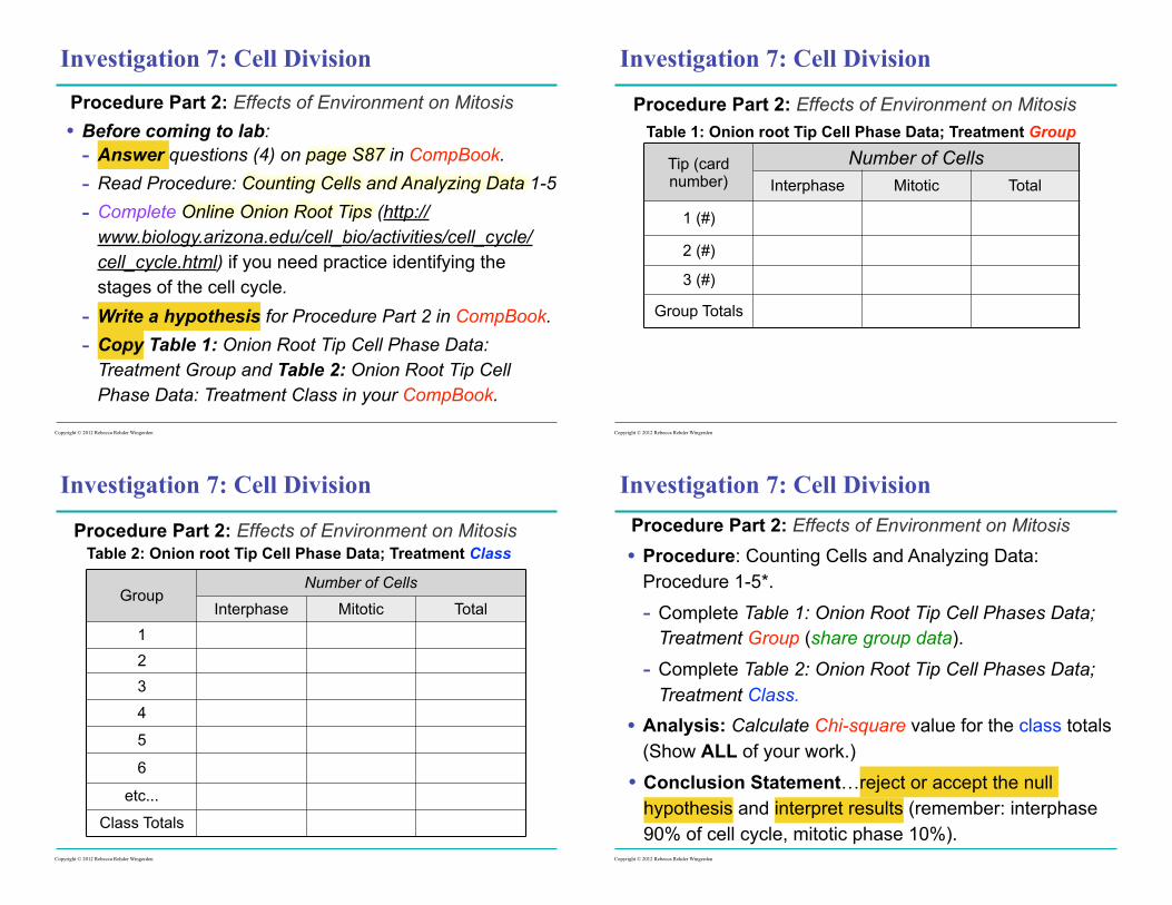
Procedure Part 2: Effects of Environment on Mitosis • Before coming to lab:
- Answer questions (4) on page S87 in CompBook. - Read Procedure: Counting Cells and Analyzing Data 1-5 - Complete Online Onion Root Tips (http://
www.biology.arizona.edu/cell_bio/activities/cell_cycle/cell_cycle.html) if you need practice identifying the stages of the cell cycle.
- Write a hypothesis for Procedure Part 2 in CompBook. - Copy Table 1: Onion Root Tip Cell Phase Data:
Treatment Group and Table 2: Onion Root Tip Cell Phase Data: Treatment Class in your CompBook.
Investigation 7: Cell Division
Copyright © 2012 Rebecca Rehder Wingerden
Procedure Part 2: Effects of Environment on Mitosis
Investigation 7: Cell Division
Copyright © 2012 Rebecca Rehder Wingerden
Tip (card number)
Number of CellsInterphase Mitotic Total
1 (#)
2 (#)
3 (#)
Group Totals
Table 1: Onion root Tip Cell Phase Data; Treatment Group
Procedure Part 2: Effects of Environment on Mitosis
Investigation 7: Cell Division
Copyright © 2012 Rebecca Rehder Wingerden
GroupNumber of Cells
Interphase Mitotic Total123
4
5
6
etc...
Class Totals
Table 2: Onion root Tip Cell Phase Data; Treatment Class
Procedure Part 2: Effects of Environment on Mitosis
• Procedure: Counting Cells and Analyzing Data: Procedure 1-5*.
- Complete Table 1: Onion Root Tip Cell Phases Data; Treatment Group (share group data).
- Complete Table 2: Onion Root Tip Cell Phases Data; Treatment Class.
• Analysis: Calculate Chi-square value for the class totals (Show ALL of your work.)
• Conclusion Statement…reject or accept the null hypothesis and interpret results (remember: interphase 90% of cell cycle, mitotic phase 10%).
Investigation 7: Cell Division
Copyright © 2012 Rebecca Rehder Wingerden
Page 6
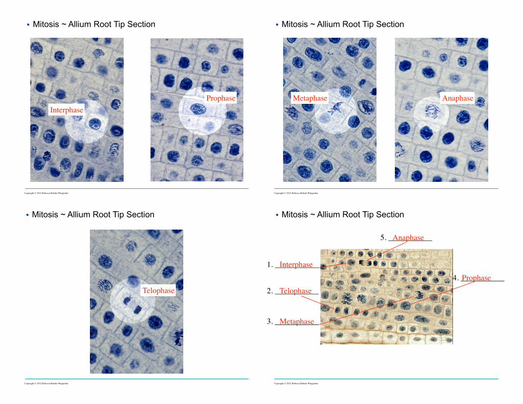
• Mitosis ~ Allium Root Tip Section
InterphaseProphase
Copyright © 2012 Rebecca Rehder Wingerden
Metaphase Anaphase
• Mitosis ~ Allium Root Tip Section
Copyright © 2012 Rebecca Rehder Wingerden
Telophase
• Mitosis ~ Allium Root Tip Section
Copyright © 2012 Rebecca Rehder Wingerden
1. ___________
2. ___________
3. ___________
4. ___________
5. ___________
Interphase
Telophase
Metaphase
Prophase
Anaphase
• Mitosis ~ Allium Root Tip Section
Copyright © 2012 Rebecca Rehder Wingerden
Page 7
Procedure Part 2: Effects of Environment on Mitosis
Investigation 7: Cell Division
Copyright © 2012 Rebecca Rehder Wingerden
GroupNumber of Cells
Interphase Mitotic Total123
4
5
6
etc...
Class Totals
Table 2: Onion root Tip Cell Phase Data; Treatment Class (5th)Procedure Part 2: Effects of Environment on Mitosis
Investigation 7: Cell Division
Copyright © 2012 Rebecca Rehder Wingerden
GroupNumber of Cells
Interphase Mitotic Total123
4
5
6
etc...
Class Totals
Table 2: Onion root Tip Cell Phase Data; Treatment Class (6th)
Procedure Part 2: Effects of Environment on Mitosis • Post Lab - Conclusion
• restate the purpose of this experiment. • restate your hypothesis for this experiment. • statement: do the results of your experiment support
or refute your hypothesis. • list and explain the data statistical test(s) that lead
you to your statement. • Therefore, conclusion (remember experimental
purpose).
Investigation 7: Cell Division
Copyright © 2012 Rebecca Rehder Wingerden
Procedure Part 3: Loss of Cell Cycle Control in Cancer • Before coming to lab:
- Complete Review from Part 1- answer the TWO questions in this section on page S90 in your CompBook
- Complete Prelab Questions for Part 3- answer the FIVE questions in this section on page S90 in your CompBook
Investigation 7: Cell Division
A HeLa cancer cell dividing
HeLa Cells: The History of the Hela Cell Line (9:00 min.)
Copyright © 2012 Rebecca Rehder Wingerden
Smithsonian.com The End of the Henrietta Lacks Saga?
Page 8
Procedure Part 3: Loss of Cell Cycle Control in Cancer • Lab Day: page S90-S91
- Form a hypothesis as to how the chromosomes of a cancer cell might appear in comparison to a normal cell and how those differences are related to the behavior of the cancer cell.
- Activity- Cancer and the Loss of Cell Cycle Control
Investigation 7: Cell Division
Copyright © 2012 Rebecca Rehder Wingerden
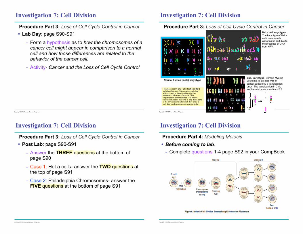
Procedure Part 3: Loss of Cell Cycle Control in Cancer
Investigation 7: Cell Division
Copyright © 2012 Rebecca Rehder Wingerden
Normal human (male) karyotype
HeLa cell karyotype- The karyotype of HeLa cells is extremely abnormal in part due to the presence of DNA from HPV.
CML karyotype- Chronic Myeloid Leukemia is just one type of cancer caused by a translocation error. The translocation in CML involves chromosomes 9 and 22.Fluorescence In Situ Hybridization (FISH)
technique know as “chromosome painting” which is used to detect and localize the presence or absence of specific DNA sequences on chromosomes. FISH uses fluorescent probes that bind to only those parts of the chromosome with which they show a high degree of sequence complementarity.
Procedure Part 3: Loss of Cell Cycle Control in Cancer • Post Lab: page S90-S91
- Answer the THREE questions at the bottom of page S90
- Case 1: HeLa cells- answer the TWO questions at the top of page S91
- Case 2: Philadelphia Chromosomes- answer the FIVE questions at the bottom of page S91
Investigation 7: Cell Division
Copyright © 2012 Rebecca Rehder Wingerden
Procedure Part 4: Modeling Meiosis • Before coming to lab:
- Complete questions 1-4 page S92 in your CompBook
Investigation 7: Cell Division
Copyright © 2012 Rebecca Rehder Wingerden
Page 9
Procedure 4: Modeling Meiosis • Lab day:
- Complete Model 1: Meiosis (2n) • Draw Parent Cell and Gametes (stamped)
- Complete Model 2: Meiosis (2n) Crossing Over • Draw Parent Cell and Gametes (stamped)
• Post Lab: questions 1-8 on page S93 in your CompBook
Investigation 7: Cell Division
Copyright © 2012 Rebecca Rehder Wingerden
Model #1: Meiosis (n = 2)
Investigation 7: Cell Division
Copyright © 2012 Rebecca Rehder Wingerden
Procedure Part 4: Modeling Meiosis
Interphase (G1) Parent Cell
Meiosis
Prophase I
Metaphase I
Metaphase II
Telophase II & Cytokinesis
Model #2: Meiosis (n = 2) Crossing OverProcedure Part 4: Modeling Meiosis
Meiosis
Prophase I
Metaphase I
Metaphase II
Telophase II & Cytokinesis
Interphase (G1) Parent Cell
Investigation 7: Cell Division
Parent Cell (2n) Prophase I (DNA replication
takes place in the S phase of Interphase)
Model 1: Meiosis with PopBeads (2n)
Copyright © 2012 Rebecca Rehder Wingerden
Investigation 7: Cell Division
Metaphase I
Model 1: Meiosis with PopBeads (2n)
Metaphase II
Telophase IICopyright © 2012 Rebecca Rehder Wingerden
Page 10
Investigation 7: Cell Division
Parent Cell (2n)
Model 2: Meiosis with PopBeads Crossing Over (2n)
Copyright © 2012 Rebecca Rehder Wingerden
Prophase I (DNA replication
takes place in the S phase of Interphase)
Investigation 7: Cell Division
Metaphase I
Model 2: Meiosis with PopBeads Crossing Over (2n)
Telophase II
Metaphase II
Copyright © 2012 Rebecca Rehder Wingerden
Procedure 5: Meiosis and Crossing Over in Sordaria - Sexual reproduction provides a mechanism to produce
genetic variation.
Investigation 7: Cell Division
- Sordaria fimicola, an ascomycete fungus is haploid for the bulk of its life cycle.
- The only diploid portion of the S. fimicola’s life cycle occurs when the nuclei of specialized hyphae come together.
Copyright © 2012 Rebecca Rehder Wingerden
Investigation 7: Cell Division
Sordaria becomes diploid only when two different strains of mycelia fuse allowing the two different haploid nuclei to form a diploid nucleus.
haploid (n) wild-type strain [+]
haploid mutant strain
[tn]
n
n
fertilization (2n) 2n
The diploid (2n) nucleus must then undergo meiosis to resume its haploid (n) state.
Meiosis, followed by mitosis, results in the formation of eight haploid ascospores contained within a sac called an ascus (plural, asci).
meiosis
n n n
n
mitosis
n n n n n
n n n
Copyright © 2012 Rebecca Rehder Wingerden
Page 11
Investigation 7: Cell Division
haploid (n) wild-type strain [+]
haploid mutant strain
[tn]
fertilization (2n)
n
n
2nmeiosis n
n
n
n
mitosis
n n n n n
n n n
If crossing over occurs, then the resulting ascospores contained within the ascus will be different.
Copyright © 2012 Rebecca Rehder Wingerden Copyright © 2012 Rebecca Rehder Wingerden
Procedure 5: Meiosis and Crossing Over in Sordaria - Identify crossing over in Sordaria
Investigation 7: Cell Division
When mycelia of two different strains come together and undergo meiosis, the asci that develop will contain four black and four tan ascospores.
The arrangement of the spores directly reflects whether or not crossing over has occurred. If crossing over occurs, then it will occur in the region between the gene for spore color and the centromere.
Procedure 5: Meiosis and Crossing Over in Sordaria • Before coming to lab:
- Read Background pages S94-S95 - Complete PreLab 3Q @ top of page S95 - Read Procedure 1-6 page S95 - Copy Table 3: Analysis of Result (p. S95)
Investigation 7: Cell Division
Copyright © 2012 Rebecca Rehder Wingerden
A Map Unit is an arbitrary unit of measure where one map unit corresponds to 1% crossover.
Procedure 5: Meiosis and Crossing Over in Sordaria
Investigation 7: Cell Division
Copyright © 2012 Rebecca Rehder Wingerden
Group Map Units12345678
TotalAverage
• Lab day: - Complete Procedure steps
1-6* on page S95 AND Table 3: Analysis of Result
- Calculate the Map Units for this gene AND the Class average Map Units for this gene
• Post Lab: - Complete Post Lab
Evaluating Results: questions 1-6 (p.S96)
Table 4: Map Unit Average