112
Muscle & Muscle Tissue
Muscle & Muscle Tissue
9 2
Muscle
Muscle functions:MovementMaintain postureStabilizes jointsGenerates heat
Functional characteristics:Excitability, contractility, extensibility, elasticity
9 3
Skeletal Muscle: The Organs
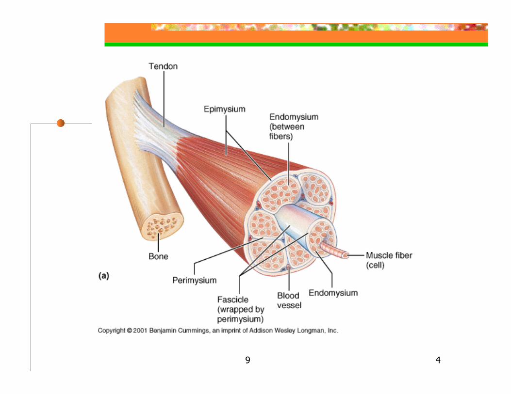
Skeletal muscle tissue, c. t., nerve fibers, blood vesselsConnective tissue wrappings (sheaths):
Epimysium – outer dense irreg. c. t., surrounds whole musclePerimysium – c. t. surrounds bundles of muscle fibers (fascicles)Endomysium – c. t. surrounds individual muscle fibers
9 4
9 5
Skeletal Muscle: The Organs
C. t. wrappings continuous with tendonsC. t. sheaths provide entry & exit routes for blood vessels & nervesAttachments:
Insertion – structure at more movable endOrigin – structure at less movable end
9 6
Skeletal Muscle: The Organs
Attachments cont.’d: Direct – epimysium directly to periosteum or perichondriumIndirect – more common
Tendon – ropelike interconnectionAponeurosis – sheetlike interconnection
9 7
Skeletal Muscle Microscopic Anatomy
Fiber size: Length - most 1-40 mm. long, but up to several cm.Diameter 10-100 µm
Fiber - Multinucleate syncytiumformed by fusion of myoblasts
Satellite cells – myoblasts that found in mature skeletal muscle
9 8
Skeletal Muscle Microscopic Anatomy
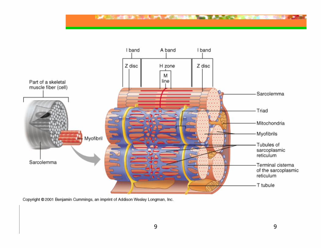
Skeletal Muscle Fiber:Sarcoplasm – cytoplasm
With glycosomes – stored glycogen inclusionsWith myoglobin – red pigment, stores oxygen
Sarcolemma – plasma membraneSarcoplasmic reticulum (SR) – E.R.
Terminal cisternae (lateral sacs) -perpendicular cross channels – store Ca++
attached to a protein, calsequestrin
9 9
9 10
Skeletal Muscle Microscopic Anatomy
Skeletal Muscle Fiber:Transverse tubules or t-tubules –tubular extensions of the sarcolemma
Runs between paired terminal cisternae forming a triad
Myofibrils – long cylindrical shaped, parallel long axis of muscle fiber, 2µm dia.
At intervals they are encircled by t-tubules100’s to 1000’s / muscle fiber, 80% cell volume
9 11
Skeletal Muscle Microscopic Anatomy
Myofibrils cont.’dStriations – a repeating series of dark A bands and light I bands
Responsible for cells striationsZ line (Z disc) – a darker midline interruption in the I band
Coin like-sheet composed of α-actinin (connectin)Sarcomere – area between two Z-lines, 2 µm longH zone – lighter region in middle of A bandM line – darker region bisecting H zone
Composed of myomesin (a protein) – helps hold adjacent thick filaments together

9 12
9 13
Skeletal MuscleMolecular Level
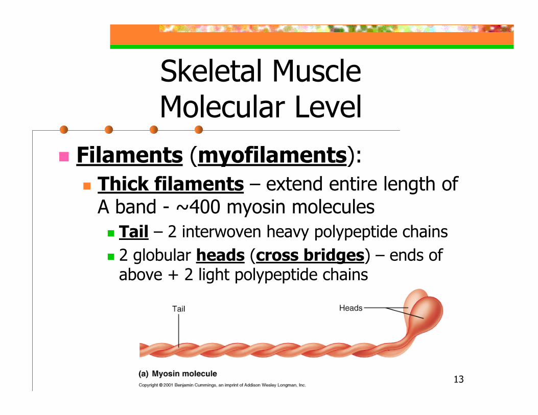
Filaments (myofilaments):Thick filaments – extend entire length of A band - ~400 myosin molecules
Tail – 2 interwoven heavy polypeptide chains2 globular heads (cross bridges) – ends of above + 2 light polypeptide chains
9 14
Skeletal MuscleMolecular Level
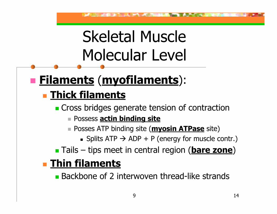
Filaments (myofilaments):Thick filaments
Cross bridges generate tension of contractionPossess actin binding sitePosses ATP binding site (myosin ATPase site)
Splits ATP ADP + P (energy for muscle contr.)
Tails – tips meet in central region (bare zone)
Thin filaments Backbone of 2 interwoven thread-like strands

9 15
9 16
Skeletal MuscleMolecular Level
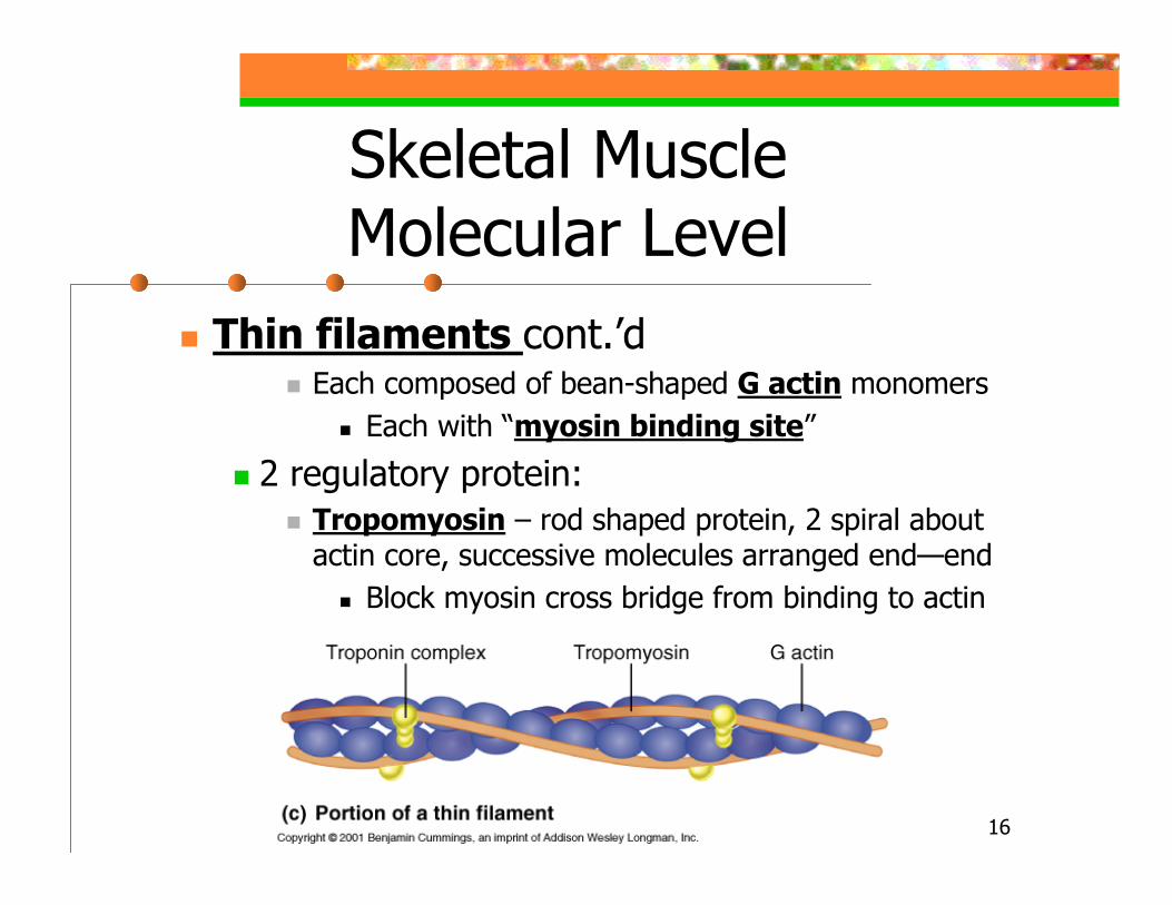
Thin filaments cont.’dEach composed of bean-shaped G actin monomers
Each with “myosin binding site”
2 regulatory protein:Tropomyosin – rod shaped protein, 2 spiral about actin core, successive molecules arranged end—end
Block myosin cross bridge from binding to actin
9 17
Skeletal MuscleMolecular Level
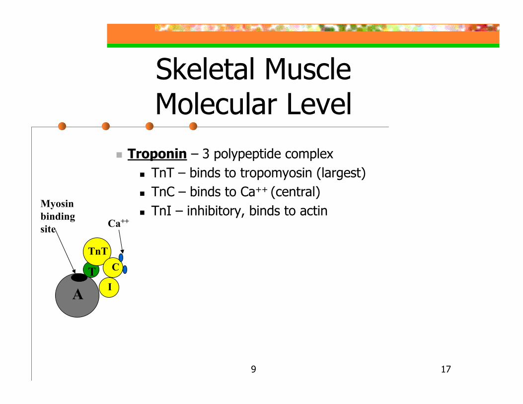
Troponin – 3 polypeptide complexTnT – binds to tropomyosin (largest)TnC – binds to Ca++ (central)TnI – inhibitory, binds to actin
AT
TnTC
I
Myosinbinding site Ca++
9 18
Skeletal Muscle Molecular Anatomy
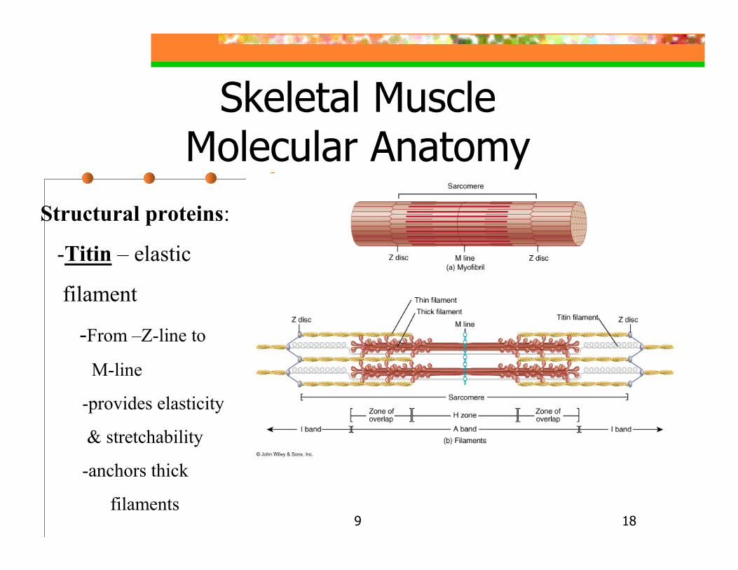
Structural proteins:
-Titin – elastic
filament
-From –Z-line to
M-line
-provides elasticity
& stretchability
-anchors thick
filaments
9 19
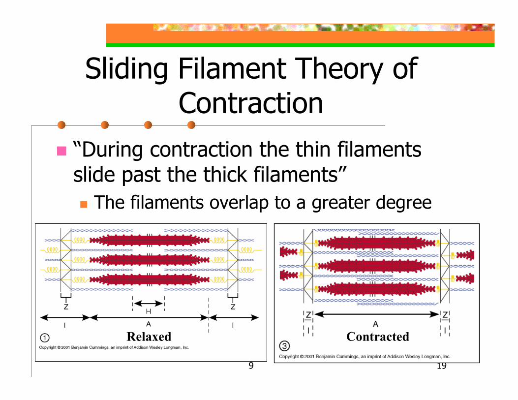
Sliding Filament Theory of Contraction
“During contraction the thin filaments slide past the thick filaments”
The filaments overlap to a greater degree
Relaxed Contracted
9 20
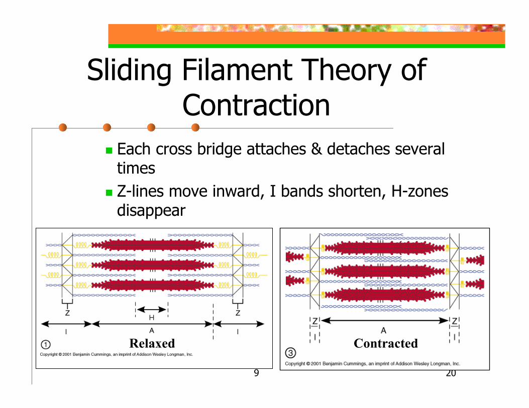
Sliding Filament Theory of Contraction
Each cross bridge attaches & detaches several timesZ-lines move inward, I bands shorten, H-zones disappear
Relaxed Contracted
9 21
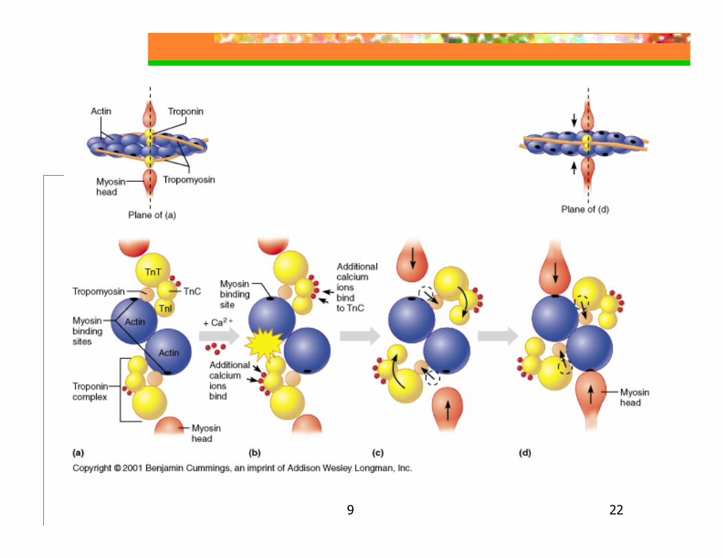
Role of Ca++ in Contraction
A) At low Ca++ levels tropomyosin blocks binding sites on actinB) At higher Ca++ levels additional Ca++
bind to troponin (2 additional)C) Ca++ activated troponin undergoes conformational change moves tropomyosin from actin’s binding site
9 22
9 23
Role of Ca++ in Contraction
D) Myosin cross bridge binds to actin’sbinding site permits contraction to begin
9 24
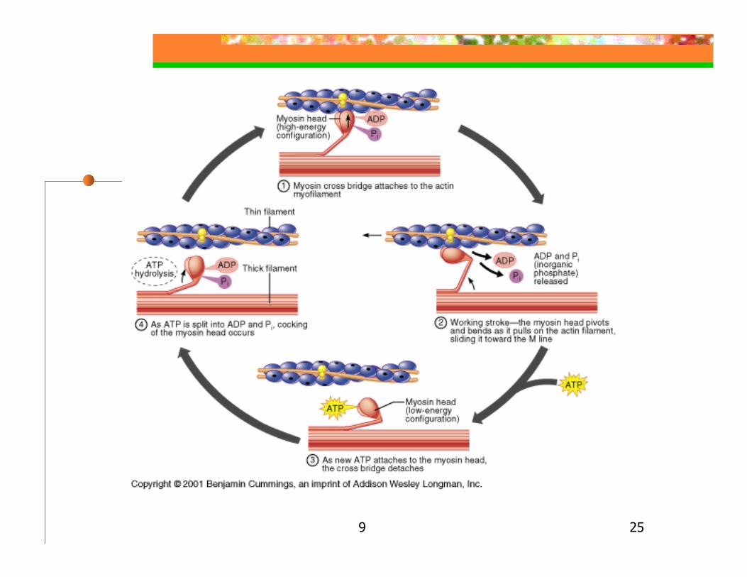
Events in the Sliding of Actin Filaments During Contraction1. High energy myosin cross bridge attaches to G-actin’s myosin binding site of thin filament
ADP & P attached to myosin + energy2. The myosin cross bridge swivels(pivots), energy used for swiveling
Attached thin filament movesADP & P are released
9 25
9 26
Events in the Sliding of Actin Filaments During Contraction3. New ATP molecule binds to myosin cross bridge cross bridge detaches from actin4. ATP hydrolysis occurs in presence of ATPase, that is ATP splits into ADP + P with some of its energy used to produce high energy myosin (“cocked position”)
9 27
Events in the Sliding of Actin Filaments During ContractionSingle working stroke of all cross bridges in a muscle shortening of 1%
But skeletal muscles contract between 30-35% of their resting lengthTherefore cross bridge cycling most occur many times during single contraction
Sliding continues while Ca++ are adequate & ATP is available
9 28
Ending Contraction
Ca++ pumped by active transport back into the SR
Troponin changes shapeActin active sites covered by
tropomyosinMuscle fiber relaxes
9 29
Rigor Mortis
Muscle stiffens usually 3-4 hours after deathPeak rigidity at about 12 hrs.-irreversible linkageDissipates over next 48-60 hrs.-protein breakdown
Dying cells unable to actively transport Ca++
into SR increase in Actin-Myosin cross bridge formationLack of ATP for detachment of actins from myosin cross bridges
9 30
The Neuromuscular Junction & The Nerve Stimulus
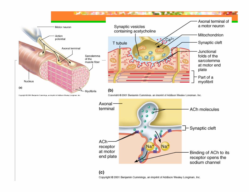
Neuromuscular junction (NMJ)–region where a motor neuron comes into close contact with a skeletal muscle fiber
Motor neuron – conducts nerve impulses to a muscle fiber or glandAxon terminal branches- short branches at end of axon ending in axon terminals
9 31
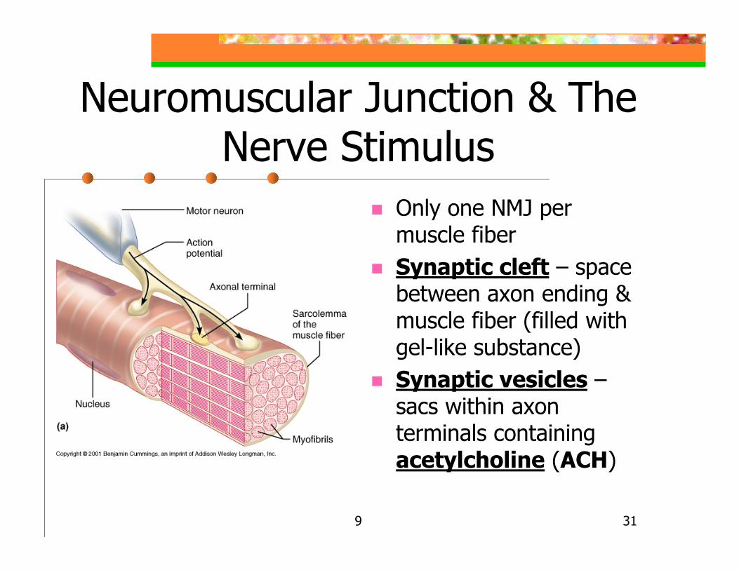
Neuromuscular Junction & The Nerve Stimulus
Only one NMJ per muscle fiberSynaptic cleft – space between axon ending & muscle fiber (filled with gel-like substance)Synaptic vesicles –sacs within axon terminals containing acetylcholine (ACH)
9 32
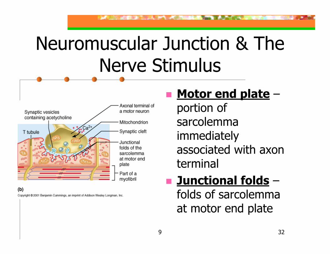
Neuromuscular Junction & The Nerve Stimulus
Motor end plate –portion of sarcolemma immediately associated with axon terminalJunctional folds –folds of sarcolemma at motor end plate
9 33
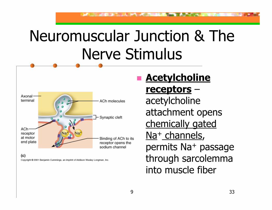
Neuromuscular Junction & The Nerve Stimulus
Acetylcholine receptors –acetylcholine attachment opens chemically gated Na+ channels, permits Na+ passage through sarcolemma into muscle fiber
9 34
Nerve Impulse To Muscle Fiber Action Potential
1. Action potential reaches axon terminal(AT) Voltage regulated Ca++
channels open Ca++ enter AT2. Causes exocytosis of synaptic vesicle ACH into synaptic cleft 3. ACH diffusion & attachment to ACH receptors of sarcolemma
9 35

9 36
Nerve Impulse To Muscle Fiber Action Potential
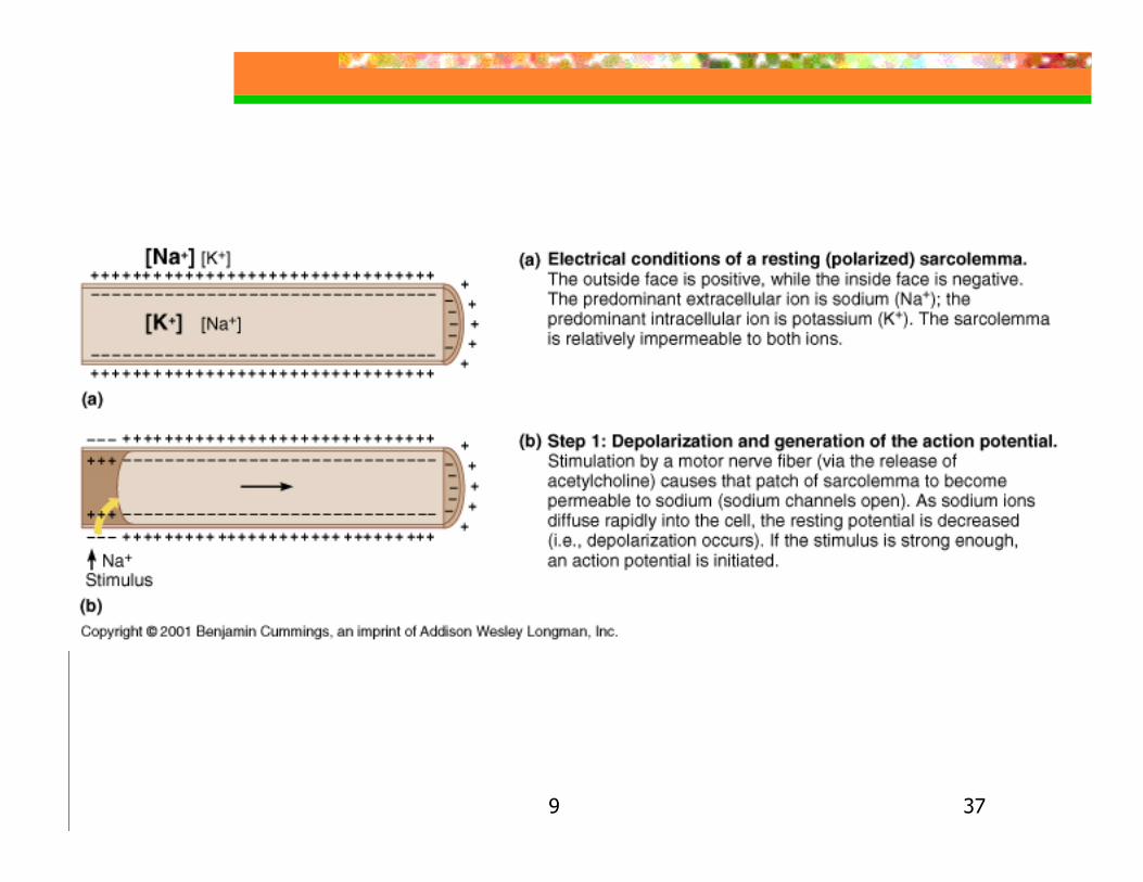
4. Chemically regulated Na+ channels of ACH receptors open Na+ enter through sarcolemma creating a less negative (more positive) region just beneath sarcolemma (depolarization)
If depolarization is strong enough, an action potential is generated that passes in all directions
9 37
9 38
Nerve Impulse To Muscle Fiber Action Potential
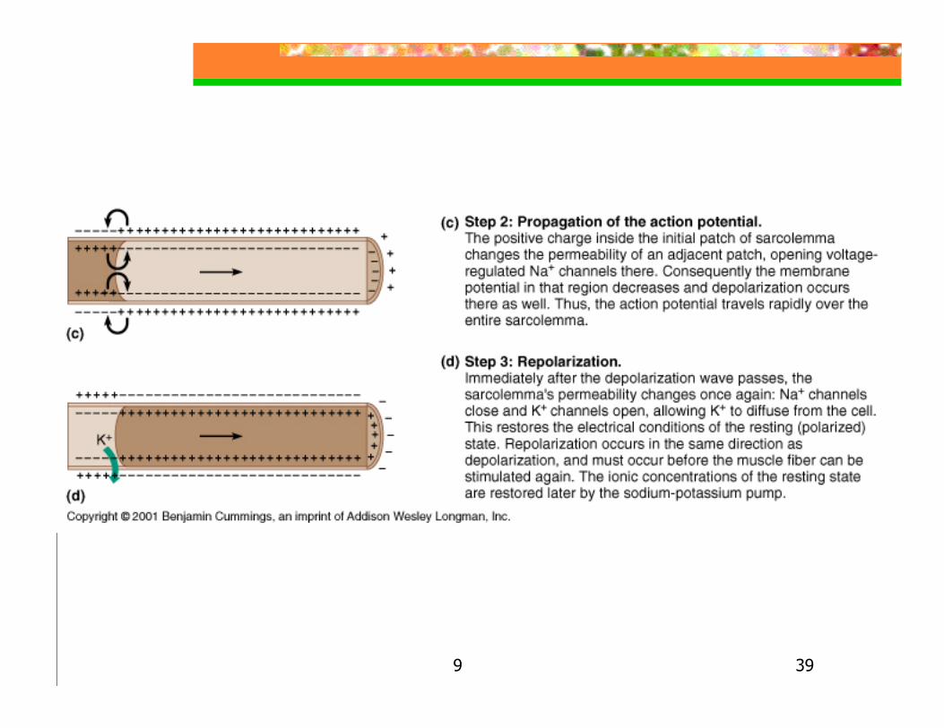
5. Action potential is self propagatingLocal depolarization wave spreads to adjacent areas of sarcolemma
Local voltage regulated Na+ channelsopen
Na+ diffuse in following their electrochemical gradient
9 39
9 40
Nerve Impulse To Muscle Fiber Action Potential
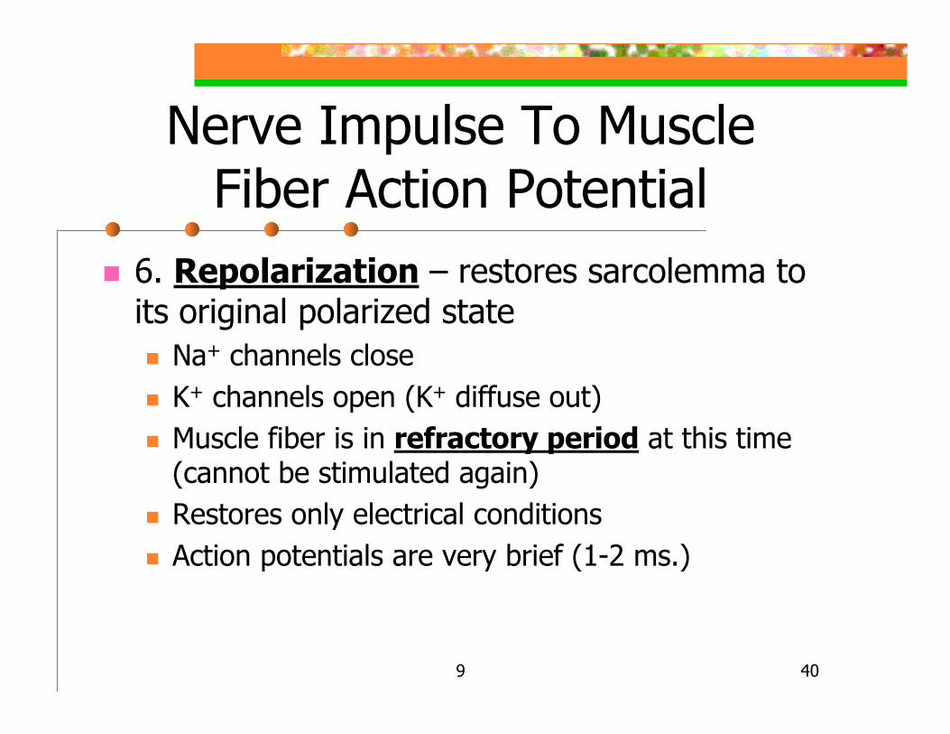
6. Repolarization – restores sarcolemma to its original polarized state
Na+ channels closeK+ channels open (K+ diffuse out)Muscle fiber is in refractory period at this time (cannot be stimulated again)Restores only electrical conditionsAction potentials are very brief (1-2 ms.)
9 41
Destruction of Acetylcholine
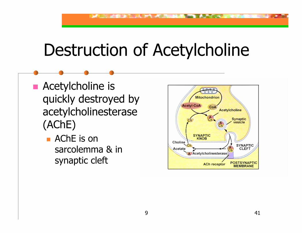
Acetylcholine is quickly destroyed by acetylcholinesterase (AChE)
AChE is on sarcolemma & in synaptic cleft
9 42
Myasthenia Gravis
Chronic autoimmune disease with progressive loss of functional ACH receptors
-Drooping of upper eyelids, difficulty swallowing and talking, and generalized muscle weakness
-1:10,000 people, female onset: between 20-40 yrs. of age, male onset: 50-60 yrs. of age
-AChE inhibitors for treatment: Pyridostigmine (Mestinon) or Neostigmine

9 43
Excitation-Contraction Coupling
Sequence of events by which transmission of action potential along sarcolemma leads to the sliding of myofilamentsSteps:
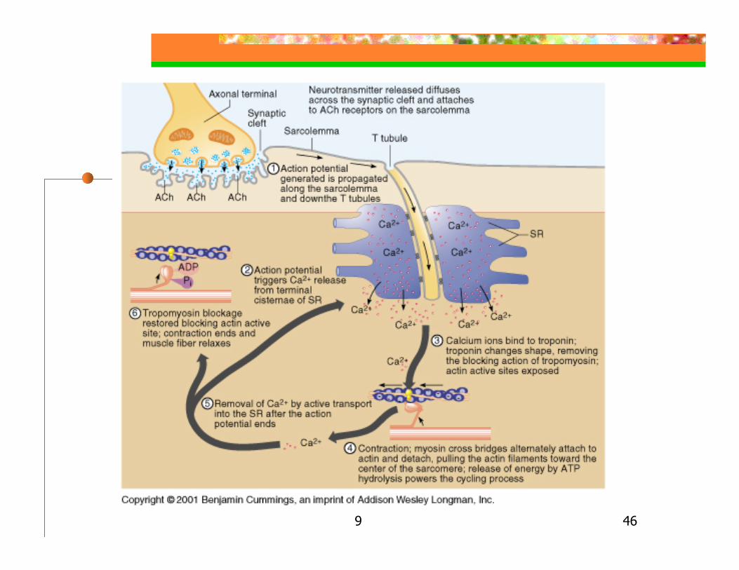
1. Action potential propagates along sarcolemma and down the T tubules

9 44

9 45
Excitation-Contraction Coupling
2. Transmission past terminal cisternae of SR causes release of Ca++ into sarcoplasm3. Ca++ bind to troponin
Troponin changes conformation Removes blocking action of tropomyosin Actin active site is exposed
4. Contraction – cross bridges alternately attach to actin & detach (at 10-5 M Intracellular Ca++ )
This pulls actin filaments toward center of sarcomereATP hydrolysis powers the cycling process
9 46

9 47
Excitation-Contraction Coupling
5. Removal of Ca++ by active transport into the SR after action potential6. Tropomyosin blockage restored
Contraction ends & muscle fibers relax
9 48
Muscle Mechanics: Terms & Rules
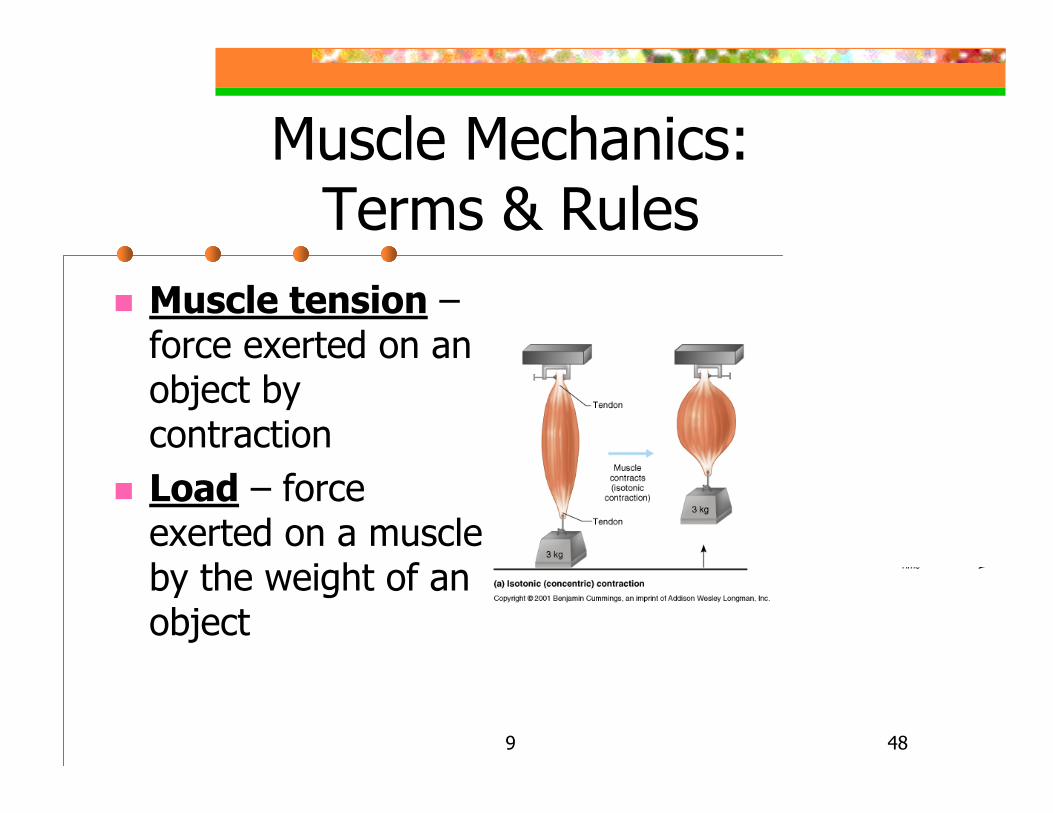
Muscle tension –force exerted on an object by contractionLoad – force exerted on a muscle by the weight of an object
9 49
Muscle Mechanics: Terms & Rules
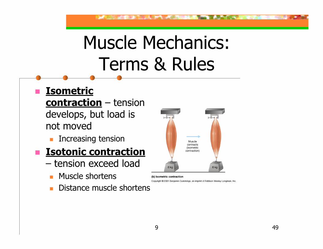
Isometric contraction – tension develops, but load is not moved
Increasing tension
Isotonic contraction– tension exceed load
Muscle shortensDistance muscle shortens
9 50
Muscle Mechanics:Terms & Rules
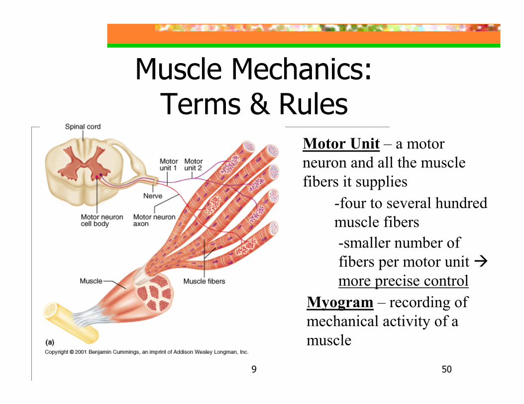
Motor Unit – a motor neuron and all the muscle fibers it supplies
-four to several hundred muscle fibers-smaller number of fibers per motor unit more precise control
Myogram – recording of mechanical activity of a muscle
9 51
Muscle Mechanics:Terms & Rules
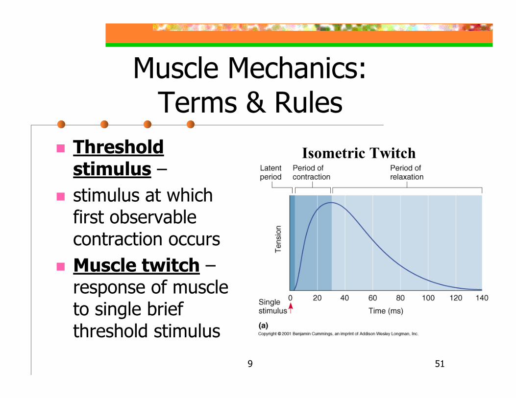
Threshold stimulus –stimulus at which first observable contraction occursMuscle twitch –response of muscle to single brief threshold stimulus
Isometric Twitch
9 52
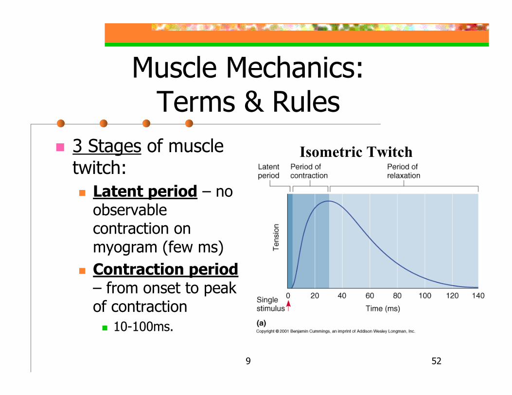
Muscle Mechanics:Terms & Rules
3 Stages of muscle twitch:
Latent period – no observable contraction on myogram (few ms)Contraction period– from onset to peak of contraction
10-100ms.
Isometric Twitch

9 53
Muscle Mechanics:Terms & Rules
3 Stages of muscle twitch:
Relaxation period –muscle tension decreases
10-100 msStarts with Ca++
reentry into SR
Isometric Twitch

9 54
Muscle Mechanics:Terms & Rules
Comparison between the lengths of contraction & relaxation periods in different skeletal muscles
9 55
Graded Muscle Responses
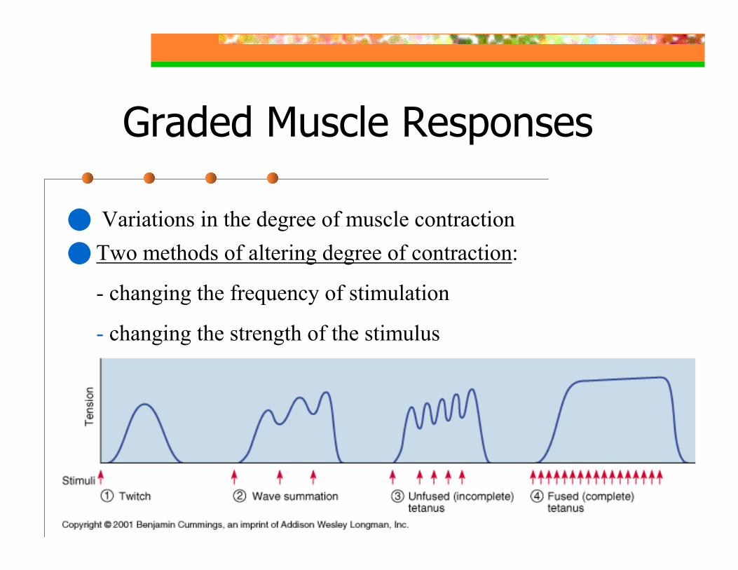
Variations in the degree of muscle contraction Two methods of altering degree of contraction:
- changing the frequency of stimulation
- changing the strength of the stimulus
9 56
Graded Muscle Responses –Frequency of Stimulation
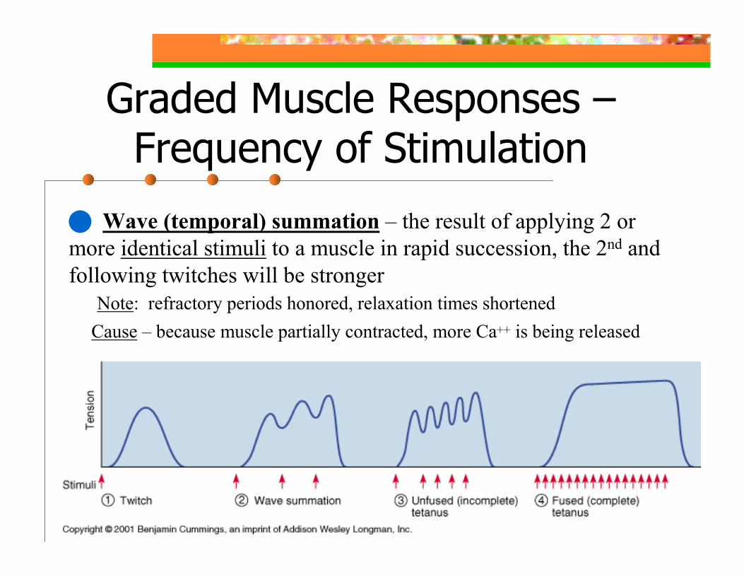
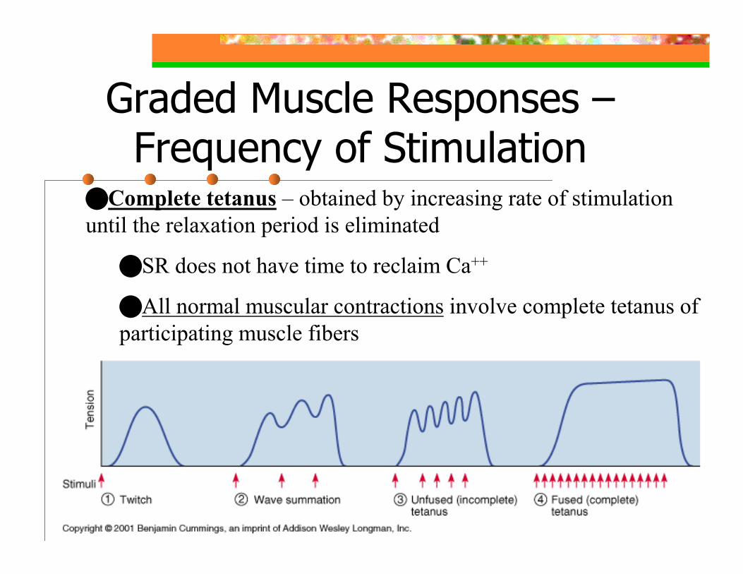
Wave (temporal) summation – the result of applying 2 or more identical stimuli to a muscle in rapid succession, the 2nd and following twitches will be stronger
Cause – because muscle partially contracted, more Ca++ is being releasedNote: refractory periods honored, relaxation times shortened
9 57
Graded Muscle Responses –Frequency of Stimulation
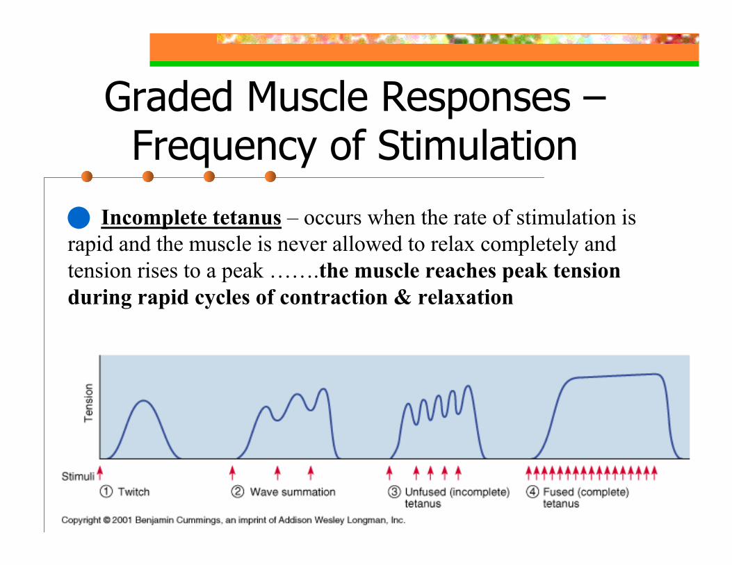
Incomplete tetanus – occurs when the rate of stimulation is rapid and the muscle is never allowed to relax completely and tension rises to a peak …….the muscle reaches peak tension during rapid cycles of contraction & relaxation
9 58
Graded Muscle Responses –Frequency of Stimulation
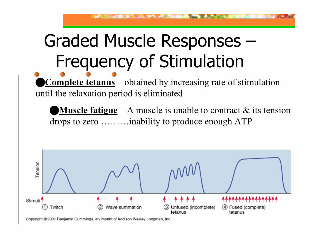
Complete tetanus – obtained by increasing rate of stimulation until the relaxation period is eliminated
SR does not have time to reclaim Ca++
All normal muscular contractions involve complete tetanus of participating muscle fibers
9 59
Graded Muscle Responses –Frequency of Stimulation
Complete tetanus – obtained by increasing rate of stimulation until the relaxation period is eliminated
Muscle fatigue – A muscle is unable to contract & its tension drops to zero ………inability to produce enough ATP
9 60
Graded Muscle Response –Stronger Stimulus
Multiple motor unit summation(recruitment) – caused by increasing strength of stimulus with the resultmore motor units contracting simultaneously
Maximal stimulus – the strongest stimulus that produces increased force of contraction
9 61
Graded Muscle Response –Treppe (Staircase Effect)
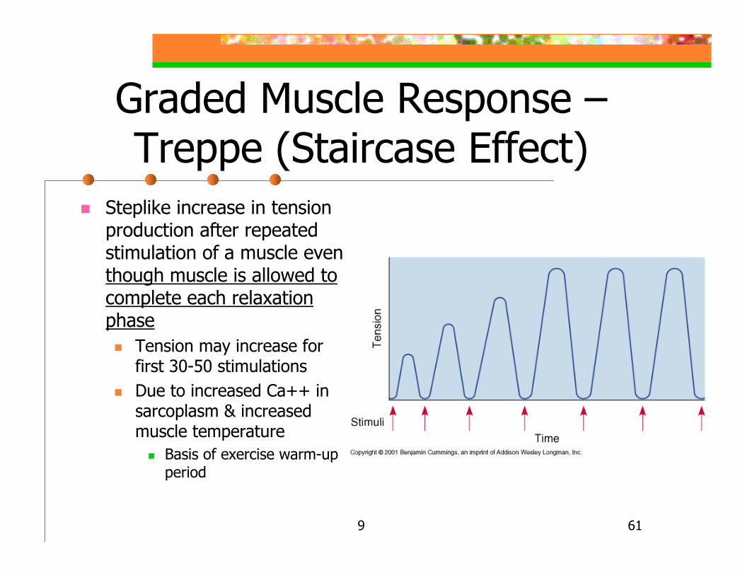
Steplike increase in tension production after repeated stimulation of a muscle even though muscle is allowed to complete each relaxationphase
Tension may increase for first 30-50 stimulationsDue to increased Ca++ in sarcoplasm & increased muscle temperature
Basis of exercise warm-up period

9 62
Muscle Tone
The slightly contracted state of normal relaxed muscles
Due to spinal reflexes that rotate the activation of groups of motor unitsDoes not produce active movementFunctions:
Keeps muscle ready to efficiently react to stimulusHelps stabilize joints and maintain posture
9 63
More On Isotonic & Isometric Contractions
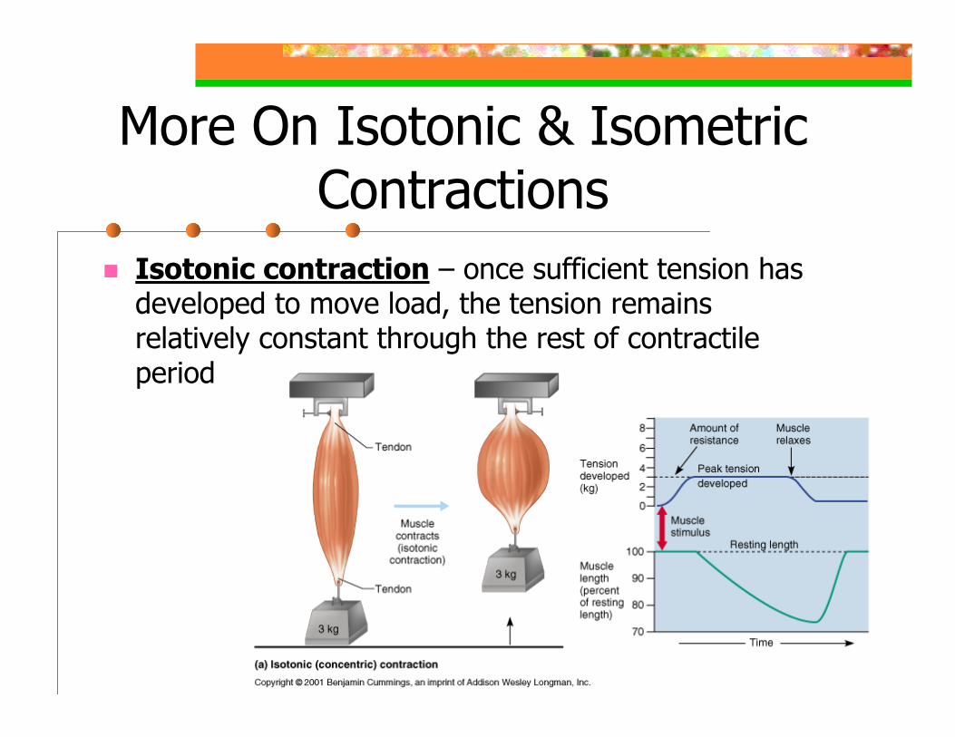
Isotonic contraction – once sufficient tension has developed to move load, the tension remains relatively constant through the rest of contractile period
9 64
More On Isotonic & Isometric Contractions
Isotonic contraction – 2 Types:Concentric – muscle shortens & does work
Picking up a weight, kicking a ballEccentric – muscle contracts as it lengthens
Calf muscle contraction when walking up long steep hillKnee flexion:
Quadriceps muscles are stretched & contract simultaneously
Counteracts force of gravity & controls descent of torso
50% more forceful than concentricMay cause delayed-onset muscle sorenessAll jumping & throwing activities involve both types
9 65
More On Isotonic & Isometric Contractions
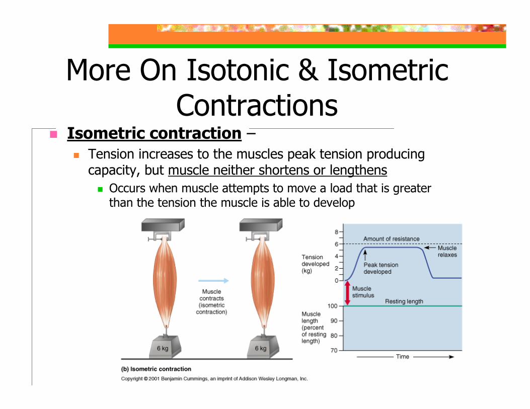
Isometric contraction –Tension increases to the muscles peak tension producing capacity, but muscle neither shortens or lengthens
Occurs when muscle attempts to move a load that is greater than the tension the muscle is able to develop
9 66
More On Isotonic & Isometric Contractions
Isometric contraction –Cross-bridges generating force, but are not moving the thin filamentsMaintaining upright posture, stabilizing jointsEx.: Knee flexion
Occurs when squat position is held for few seconds – both anterior & posterior thigh muscles contractAs one begins to rise to the upright position, until tension exceeds weight of upper body
9 67
Providing The ATP For Contraction
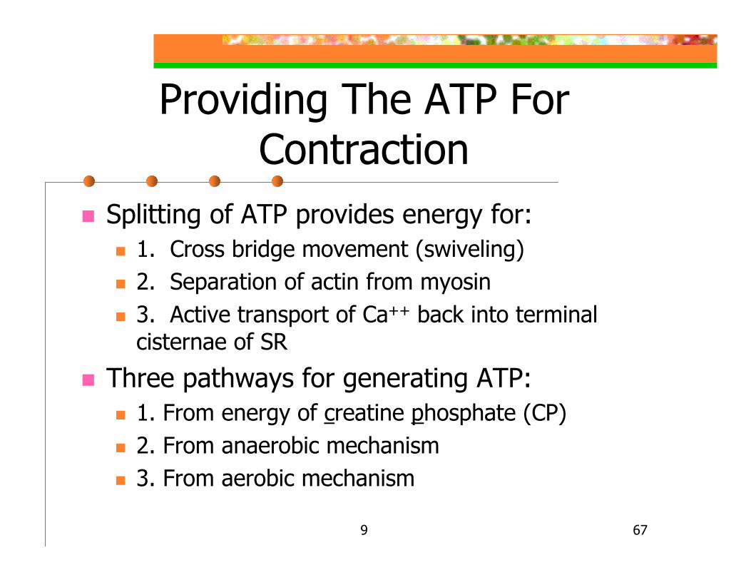
Splitting of ATP provides energy for:1. Cross bridge movement (swiveling)2. Separation of actin from myosin3. Active transport of Ca++ back into terminal cisternae of SR
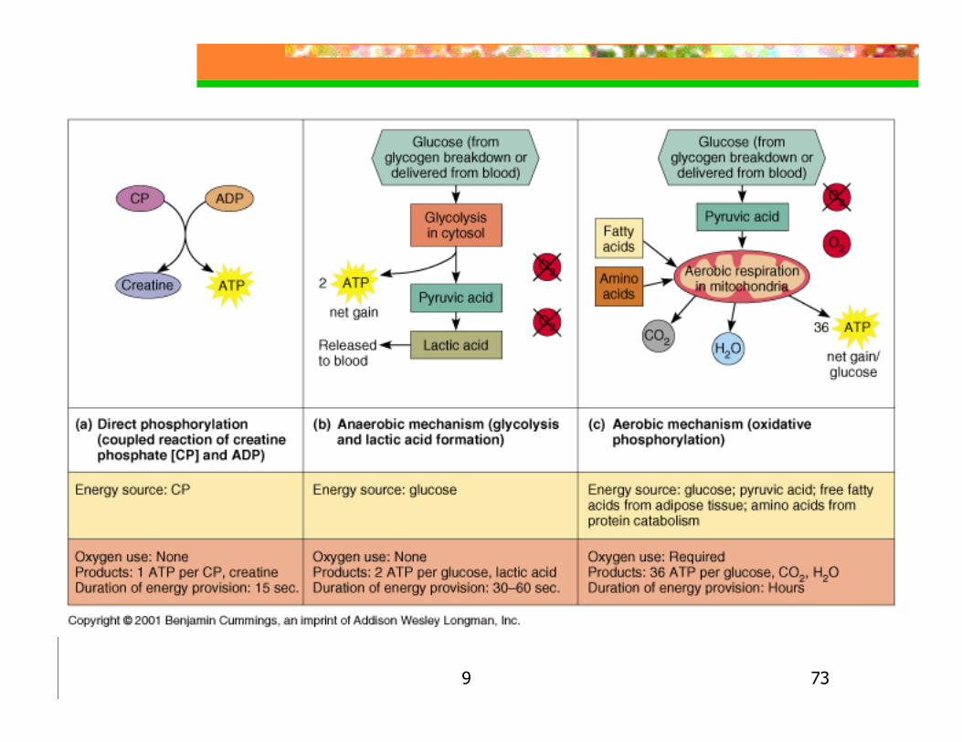
Three pathways for generating ATP:1. From energy of creatine phosphate (CP)2. From anaerobic mechanism3. From aerobic mechanism
9 68
1. From Energy Of Creatine Phosphate (CP)
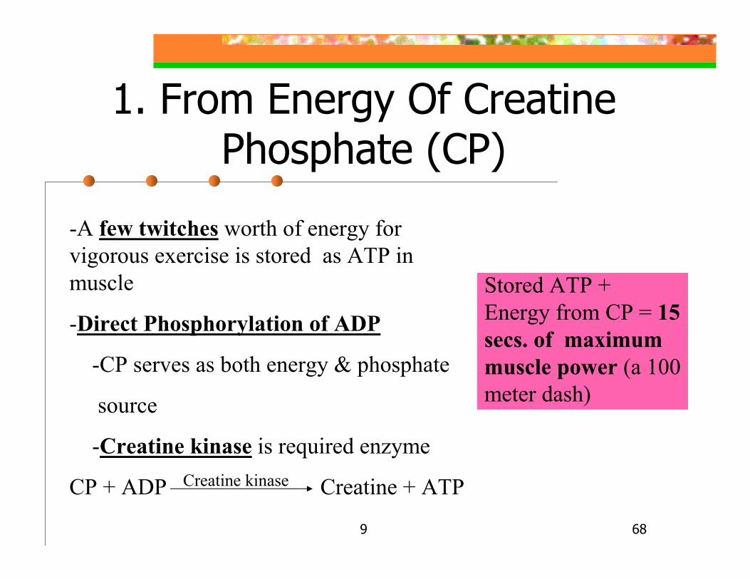
-A few twitches worth of energy for vigorous exercise is stored as ATP in muscle
-Direct Phosphorylation of ADP
-CP serves as both energy & phosphate
source
-Creatine kinase is required enzyme
CP + ADP Creatine + ATPCreatine kinase
Stored ATP + Energy from CP = 15 secs. of maximum muscle power (a 100 meter dash)

9 69
9 70
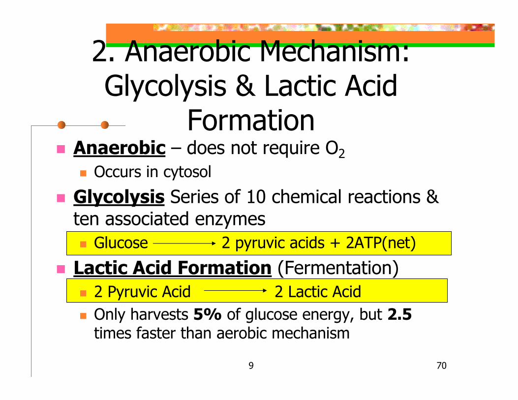
Anaerobic – does not require O2Occurs in cytosol
Glycolysis Series of 10 chemical reactions & ten associated enzymes
Glucose 2 pyruvic acids + 2ATP(net)
Lactic Acid Formation (Fermentation)2 Pyruvic Acid 2 Lactic AcidOnly harvests 5% of glucose energy, but 2.5times faster than aerobic mechanism
2. Anaerobic Mechanism: Glycolysis & Lactic Acid
Formation
9 71
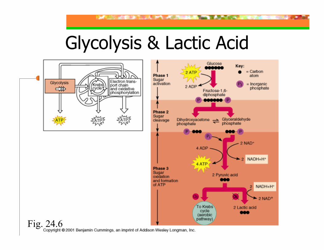
Glycolysis & Lactic Acid Formation
Fig. 24.6
9 72
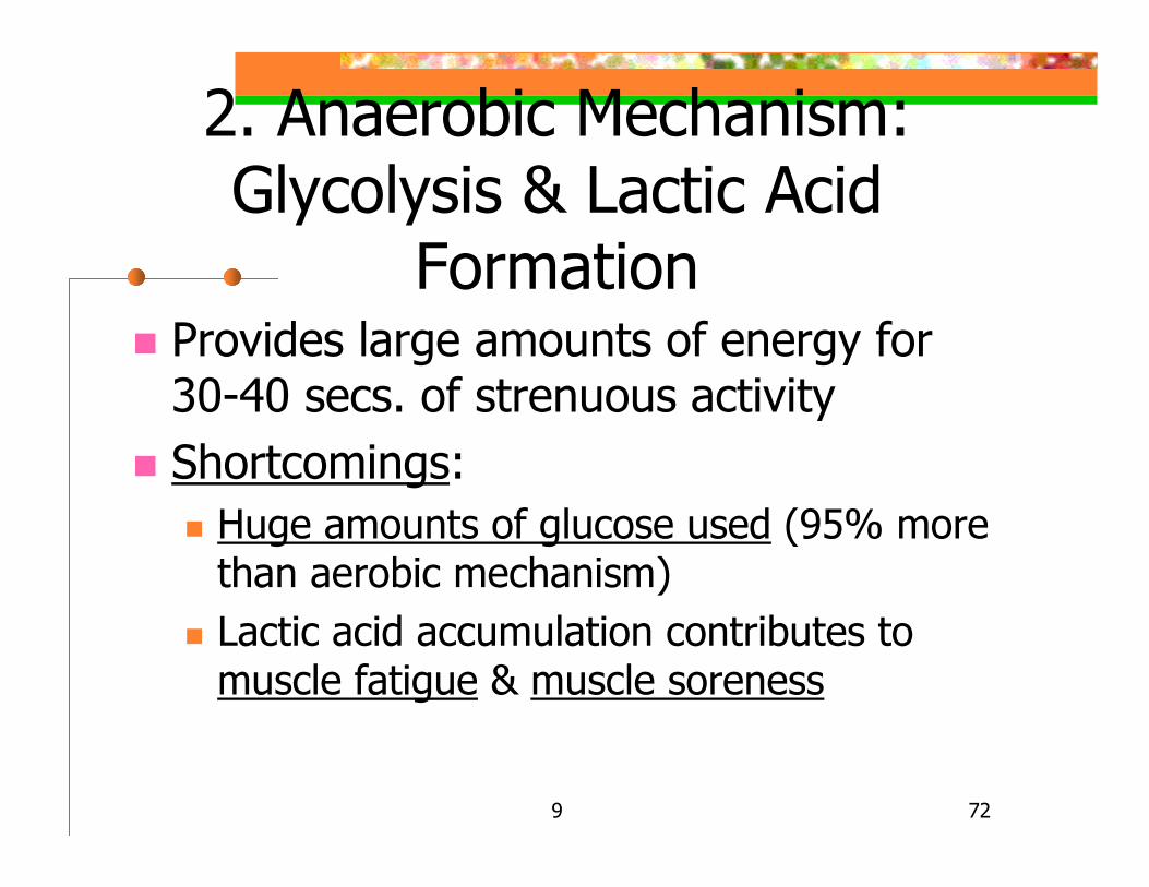
Provides large amounts of energy for 30-40 secs. of strenuous activityShortcomings:
Huge amounts of glucose used (95% more than aerobic mechanism)Lactic acid accumulation contributes to muscle fatigue & muscle soreness
2. Anaerobic Mechanism: Glycolysis & Lactic Acid
Formation
9 73
9 74
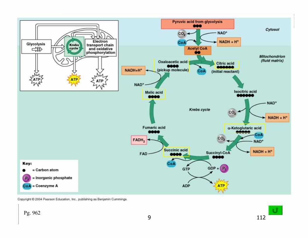
3. Aerobic Mechanism Aerobic Respiration
Requires O2
During rest & light to moderate exerciseOccurs in mitochondrionSummary reaction:Gycogen
Glucose + O2 + ADP CO2 + H2O + 36 ATP
9 75Fig.24.11
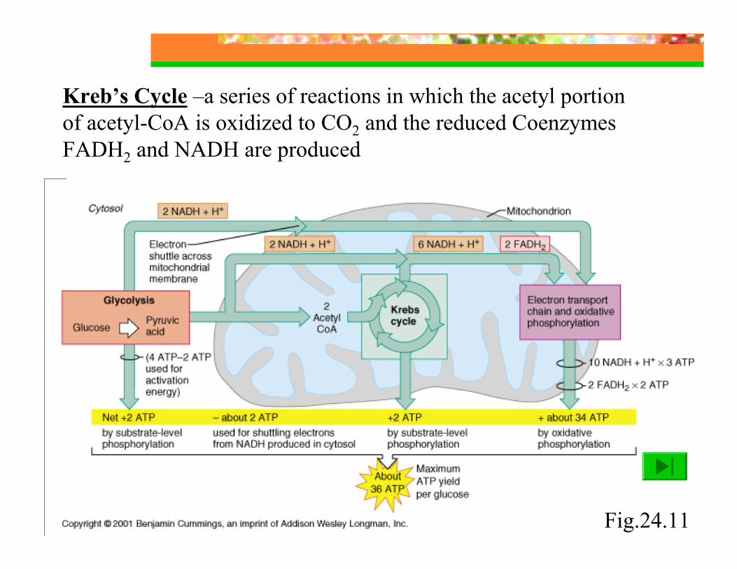
Kreb’s Cycle –a series of reactions in which the acetyl portion of acetyl-CoA is oxidized to CO2 and the reduced Coenzymes FADH2 and NADH are produced
9 76
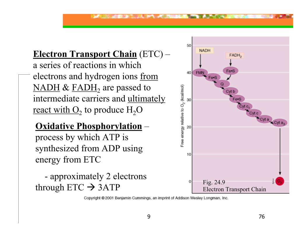
Electron Transport Chain (ETC) –a series of reactions in which electrons and hydrogen ions from NADH & FADH2 are passed to intermediate carriers and ultimately react with O2 to produce H2O
Electron Transport Chain
Oxidative Phosphorylation –process by which ATP is synthesized from ADP using energy from ETC
- approximately 2 electrons through ETC 3ATP Fig. 24.9

9 77
9 78
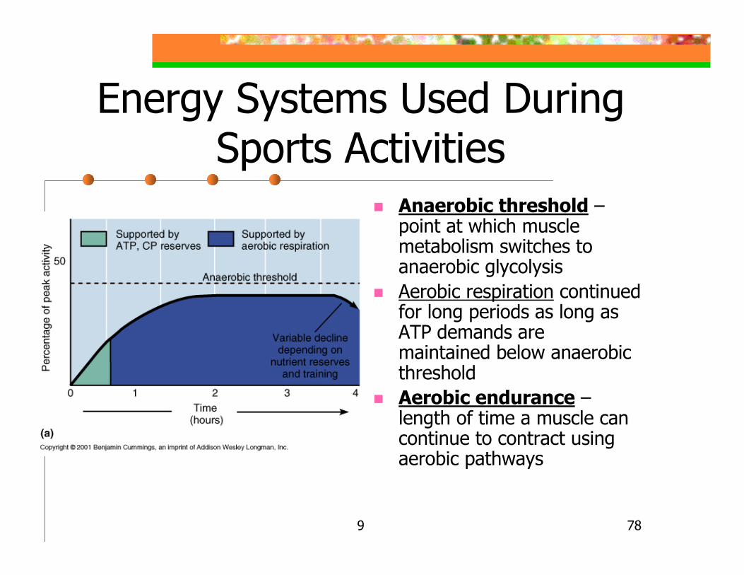
Energy Systems Used During Sports Activities
Anaerobic threshold –point at which muscle metabolism switches to anaerobic glycolysisAerobic respiration continued for long periods as long as ATP demands are maintained below anaerobic thresholdAerobic endurance –length of time a muscle can continue to contract using aerobic pathways
9 79
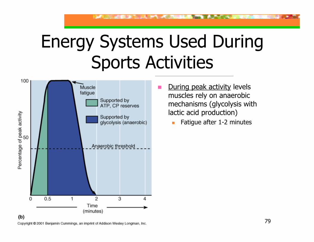
Energy Systems Used During Sports Activities
During peak activity levels muscles rely on anaerobic mechanisms (glycolysis with lactic acid production)
Fatigue after 1-2 minutes
9 80
Muscle Fatigue
Physiological inability to contractA relative deficit of ATP, not total absence
States of continuous contraction would occur with no ATP because myosin cross bridges could not separate from actin
Lactic acid accumulation (with low pH) & ionic imbalances contribute to fatigue
9 81
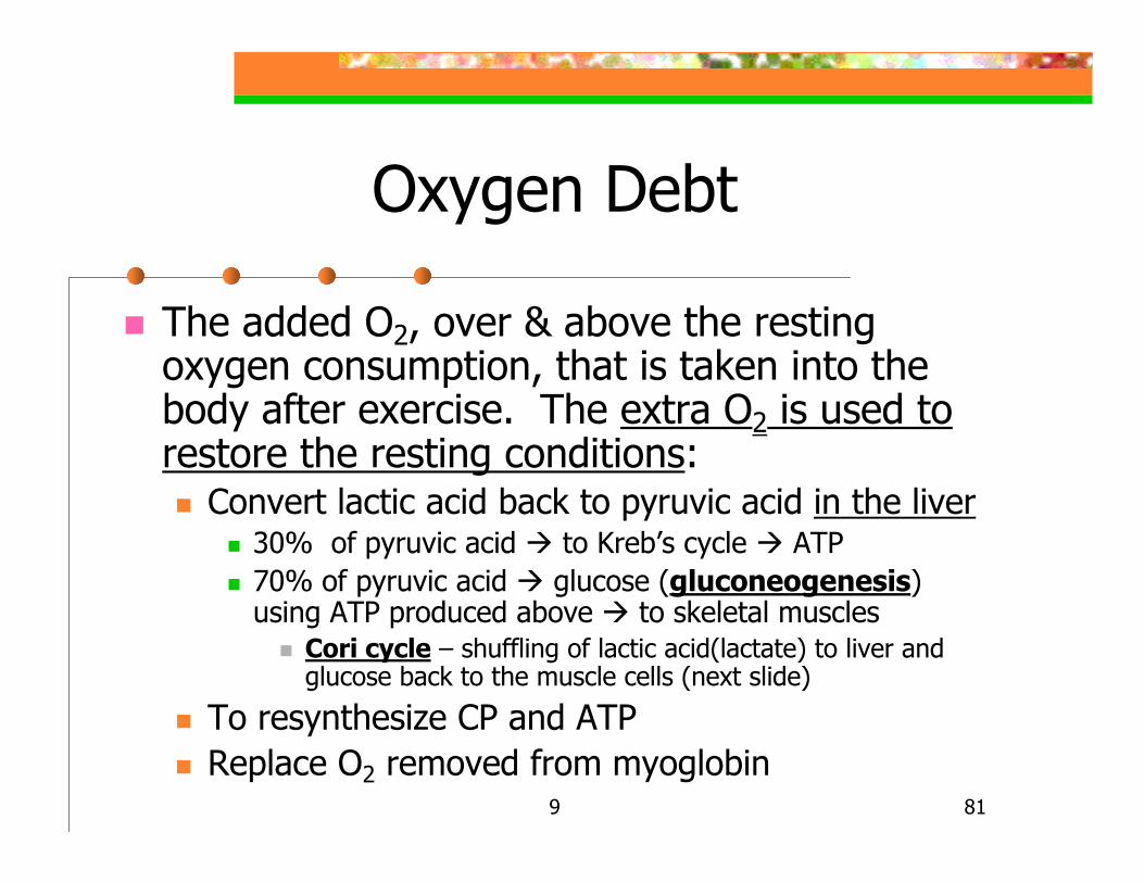
Oxygen Debt
The added O2, over & above the resting oxygen consumption, that is taken into the body after exercise. The extra O2 is used to restore the resting conditions:
Convert lactic acid back to pyruvic acid in the liver30% of pyruvic acid to Kreb’s cycle ATP70% of pyruvic acid glucose (gluconeogenesis) using ATP produced above to skeletal muscles
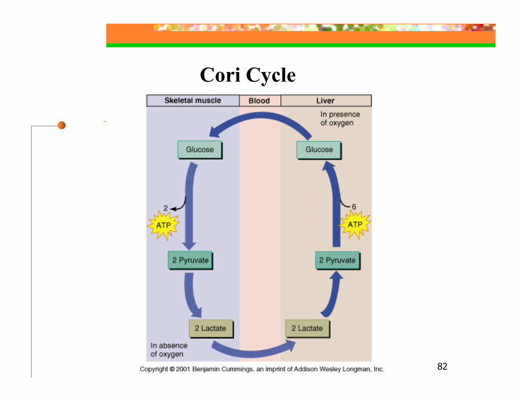
Cori cycle – shuffling of lactic acid(lactate) to liver and glucose back to the muscle cells (next slide)
To resynthesize CP and ATPReplace O2 removed from myoglobin
9 82
Cori Cycle
9 83
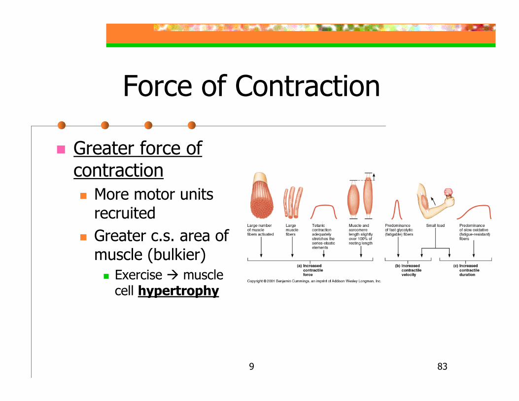
Force of Contraction
Greater force of contraction
More motor units recruitedGreater c.s. area of muscle (bulkier)
Exercise muscle cell hypertrophy
9 84
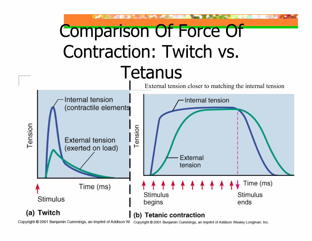
Force of Contraction
Greater force of contraction by increasing external tension to that of internal tension
Series elastic elements(SEE) – noncontractile elements of muscle
Connective tissue, tendon, etc.Develop external tension (tension transferred to the load)
Contractile elements – myofibrilsDevelop internal tension
Stretches the SEE, takes time to become tautTetanic contraction provides time to become taut ( take slack out of SEE)
9 85
Comparison Of Force Of Contraction: Twitch vs.
TetanusExternal tension closer to matching the internal tension
9 86
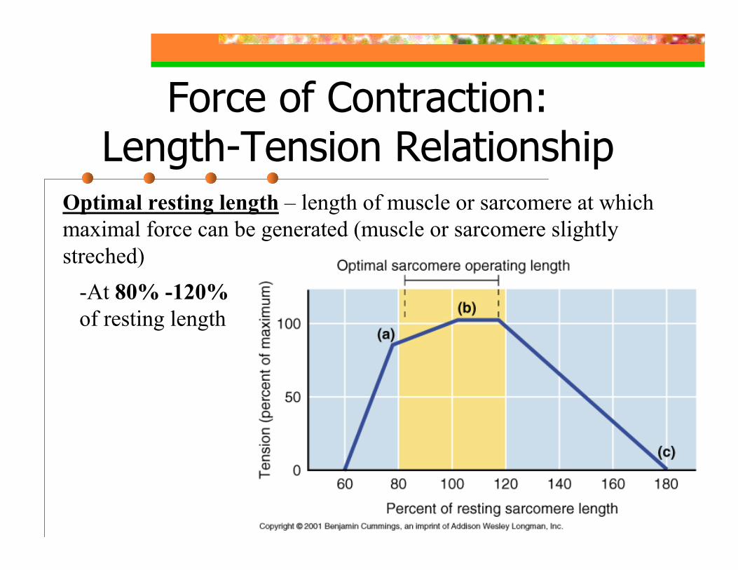
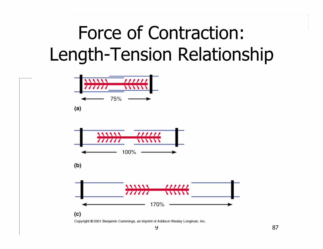
Force of Contraction:Length-Tension Relationship
Optimal resting length – length of muscle or sarcomere at which maximal force can be generated (muscle or sarcomere slightly streched)
-At 80% -120%of resting length
9 87
Force of Contraction:Length-Tension Relationship
9 88
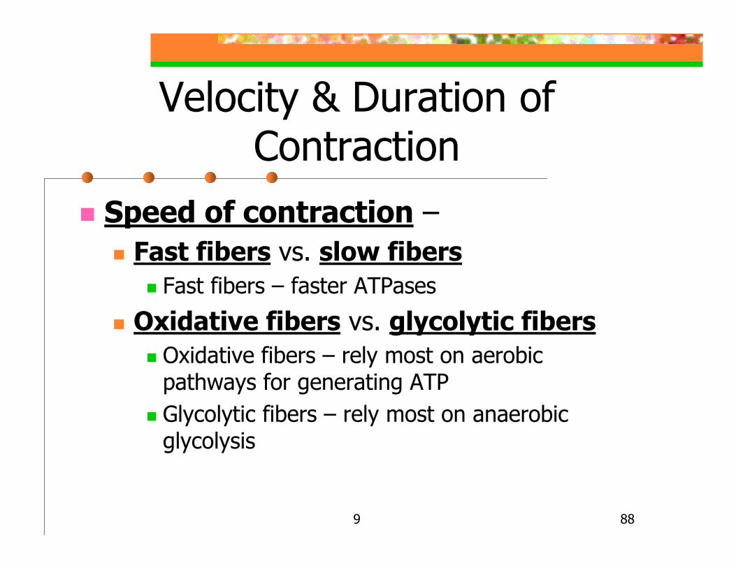
Velocity & Duration of Contraction
Speed of contraction –Fast fibers vs. slow fibers
Fast fibers – faster ATPases
Oxidative fibers vs. glycolytic fibersOxidative fibers – rely most on aerobic pathways for generating ATPGlycolytic fibers – rely most on anaerobic glycolysis
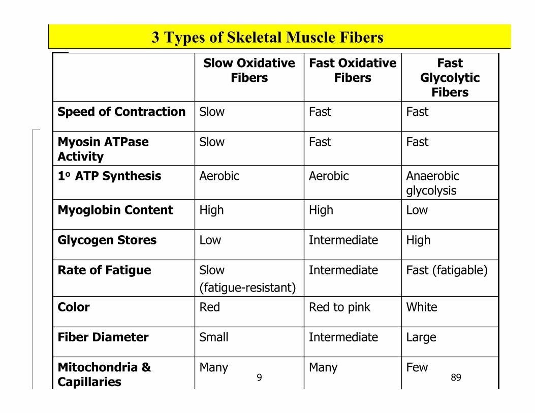
9 89FewManyManyMitochondria &
Capillaries
LargeIntermediate SmallFiber Diameter
WhiteRed to pinkRedColor
Fast (fatigable)IntermediateSlow (fatigue-resistant)
Rate of Fatigue
HighIntermediateLowGlycogen Stores
LowHighHighMyoglobin Content
Anaerobic glycolysis
AerobicAerobic1o ATP Synthesis
FastFastSlowMyosin ATPase Activity
FastFastSlowSpeed of Contraction
Fast Glycolytic
Fibers
Fast Oxidative Fibers
Slow Oxidative Fibers
3 Types of Skeletal Muscle Fibers
9 90
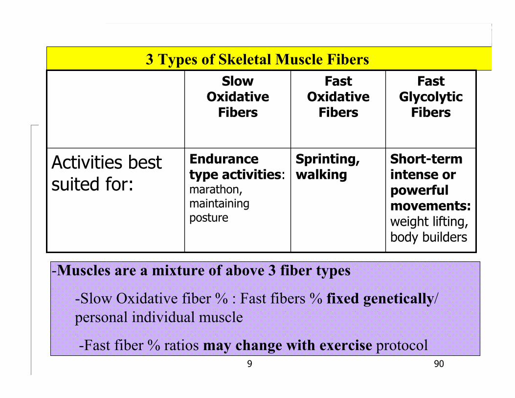
3 Types of Skeletal Muscle Fibers
Short-term intense or powerful movements:weight lifting, body builders
Sprinting, walking
Endurance type activities:marathon, maintaining posture
Activities best suited for:
Fast Glycolytic
Fibers
Fast Oxidative
Fibers
Slow Oxidative
Fibers
-Muscles are a mixture of above 3 fiber types
-Slow Oxidative fiber % : Fast fibers % fixed genetically/ personal individual muscle
-Fast fiber % ratios may change with exercise protocol
9 91
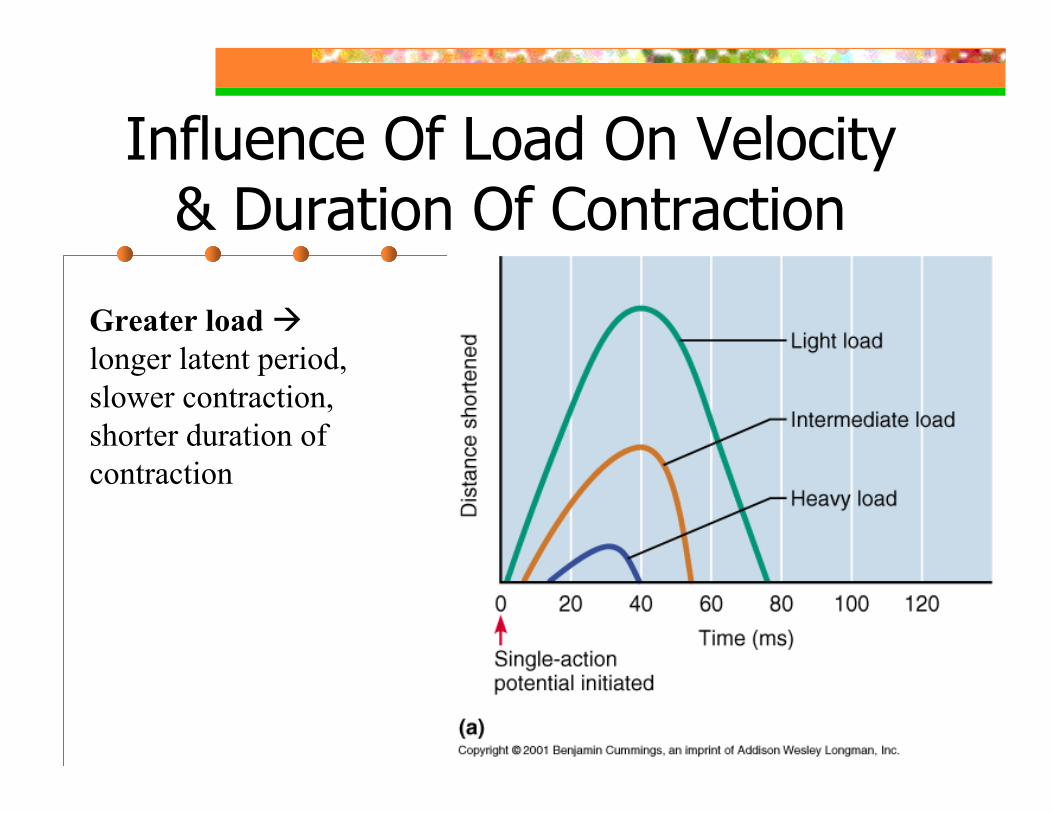
Influence Of Load On Velocity & Duration Of Contraction
Greater loadlonger latent period, slower contraction, shorter duration of contraction

9 92
Influence Of Load On Velocity Of Contraction
Greater load slower velocity of shortening
9 93
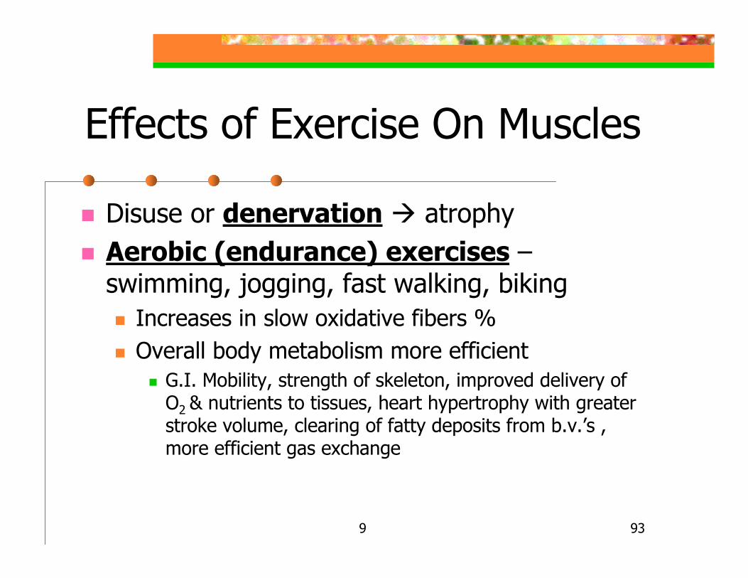
Effects of Exercise On Muscles
Disuse or denervation atrophyAerobic (endurance) exercises –swimming, jogging, fast walking, biking
Increases in slow oxidative fibers %Overall body metabolism more efficient
G.I. Mobility, strength of skeleton, improved delivery of O2 & nutrients to tissues, heart hypertrophy with greater stroke volume, clearing of fatty deposits from b.v.’s , more efficient gas exchange
9 94
Effects of Exercise On Muscles
Resistance exercises – weight lifting or isometric exercises
Increase in % of fast glycolytic fibers due to increase in size of individual muscle fibers and connective tissue
More mitochondria, more myofilaments, more myofibrils, more stored glycogenNeed to develop agonist & antagonist pairs
To prevent muscle-bound state & excessive loss of flexibility
Cross training – alternating aerobic & anaerobic exercises for optimal health
9 95
Smooth Muscle
Smooth muscle cells (fibers) Spindle shaped, centrally located nucleus, 2-10 µm x 30-200 µm long, no striations, no sarcomeres, no T-tubulesFibers secrete a small amount of endomysium thru which b.v. & nerves pass
Other c.t. sheaths absentFibers usually organized into sheets
Sheets in walls of many b.v.’s & of hollow organs
9 96
Smooth Muscle
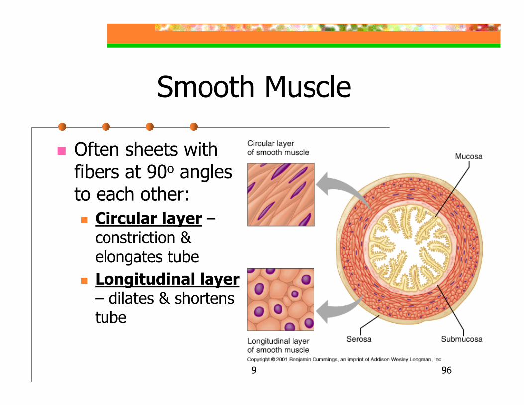
Often sheets with fibers at 90o angles to each other:
Circular layer –constriction & elongates tubeLongitudinal layer– dilates & shortens tube
9 97
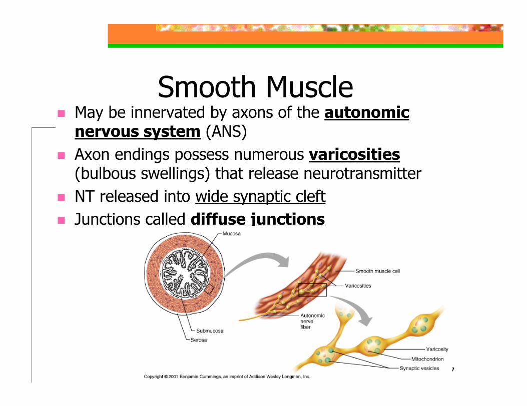
Smooth MuscleMay be innervated by axons of the autonomic nervous system (ANS)Axon endings possess numerous varicosities(bulbous swellings) that release neurotransmitterNT released into wide synaptic cleftJunctions called diffuse junctions
9 98
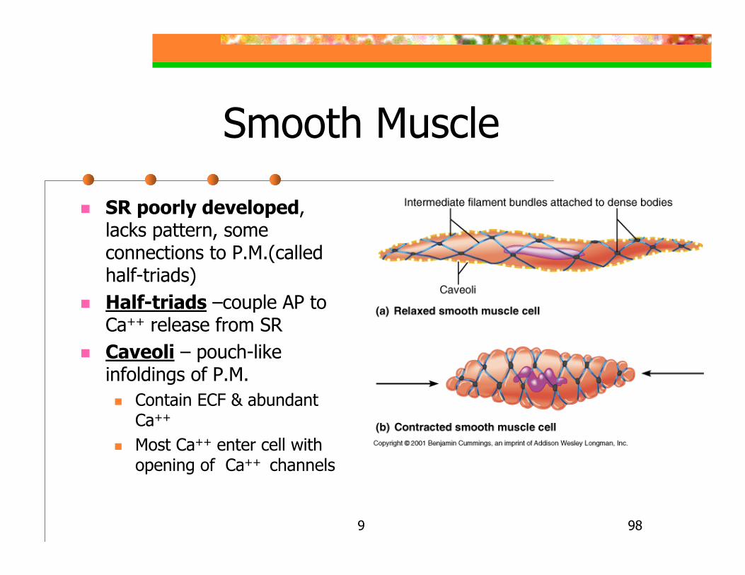
Smooth Muscle
SR poorly developed, lacks pattern, some connections to P.M.(called half-triads)Half-triads –couple AP to Ca++ release from SRCaveoli – pouch-like infoldings of P.M.
Contain ECF & abundant Ca++
Most Ca++ enter cell with opening of Ca++ channels
9 99
Smooth Muscle
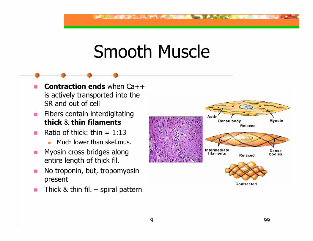
Contraction ends when Ca++ is actively transported into the SR and out of cellFibers contain interdigitating thick & thin filamentsRatio of thick: thin = 1:13
Much lower than skel.mus.
Myosin cross bridges along entire length of thick fil.No troponin, but, tropomyosin presentThick & thin fil. – spiral pattern
9 100
Smooth Muscle
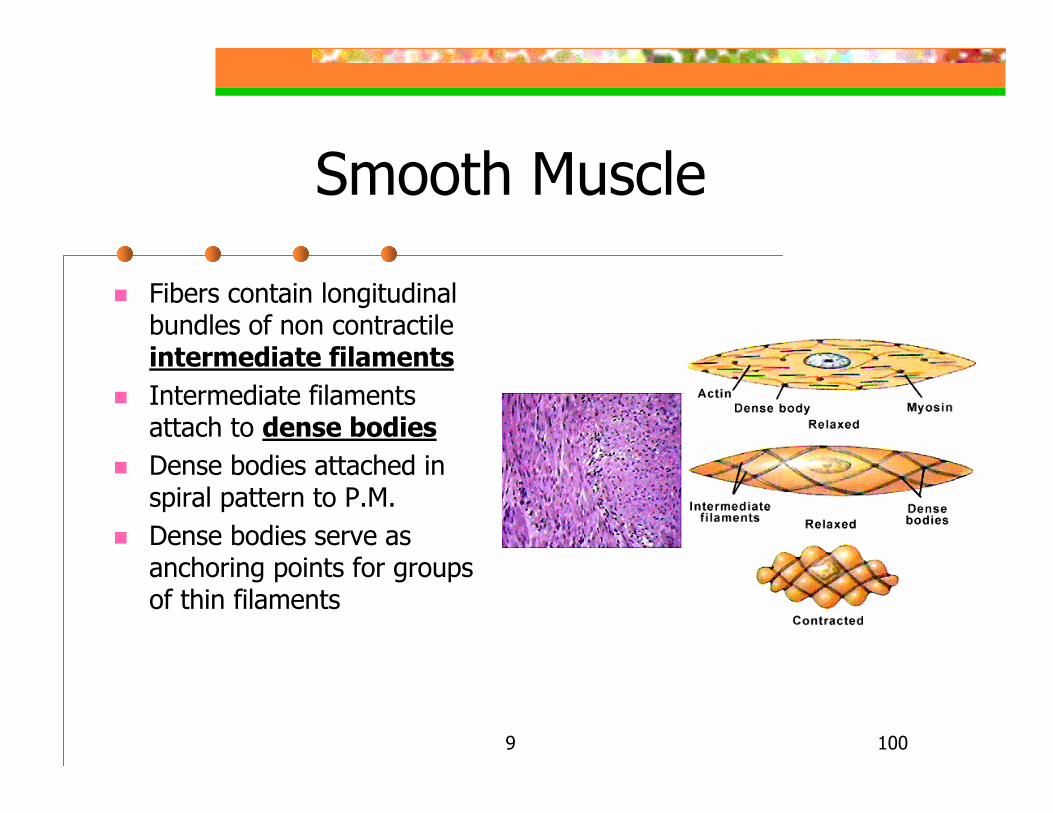
Fibers contain longitudinal bundles of non contractile intermediate filamentsIntermediate filaments attach to dense bodiesDense bodies attached in spiral pattern to P.M.Dense bodies serve as anchoring points for groups of thin filaments
9 101
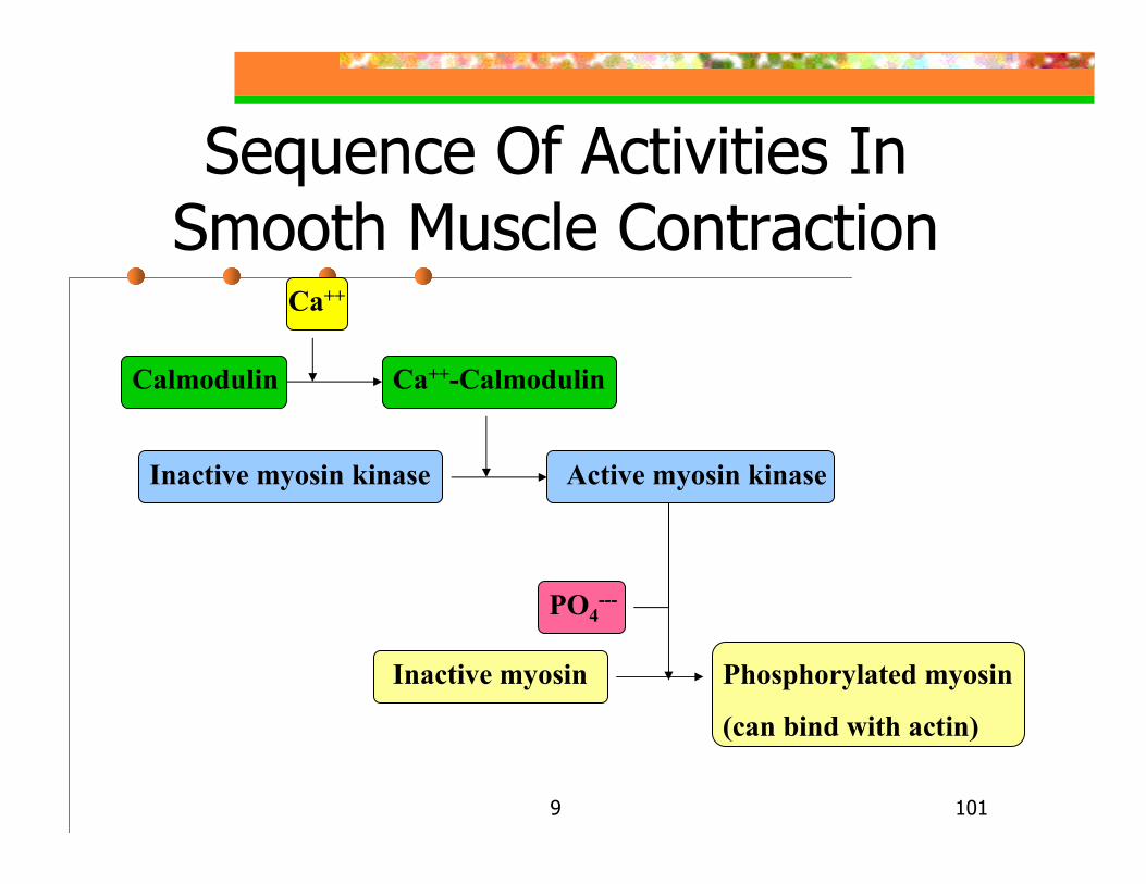
Sequence Of Activities In Smooth Muscle Contraction
Calmodulin Ca++-Calmodulin
Ca++
Inactive myosin kinase Active myosin kinase
PO4---
Inactive myosin Phosphorylated myosin
(can bind with actin)
9 102
Smooth Muscle Contraction: Characteristics
Slow – takes 30x longer to contract than skeletal muscleSustained – can maintain length contraction Resistant to fatigue – less than 1% energy cost of skeletal muscle
Sluggishness of active myosin kinaseMyofilaments latch together during long contractions – important for long term tone
ATP produced mainly by anaerobic processes(few mitochondria/fiber)
9 103
Smooth Muscle Contraction: Neural Regulation
Action potential(AP) may be generated by NT binding or a graded potential onlyDifferent NT’s may be involved (action dependent on receptor nature):
Ach receptors in bronchioles constrictionNorepinephrine (NE) receptors in bronchioles inhibition of contraction of smooth musclesNE receptors in walls of b.v. constriction
9 104
Smooth Muscle Contraction: Local Factors
Some lack nerve supply:Spontaneous depolarization may occurChemical stimulation may occur
Both nervous system & chemical stimulationChem. Stim. alters Ca++ entry into fiberChem. types: lack of O2, excess CO2, low pH, hormones (like gastrin)
Gastrin stimulates stomach contraction
9 105
Smooth Muscle Contraction: Length-Tension Changes
Stretches more than skeletal muscleGenerates more tension when comparably stretched or shortened than sk. mus.
Due to irregular overlapping arrangement of fibersSk. mus. Function at up to 30% longer than resting length & up to 30% shorterSm. mus. Function at up to100% longer than resting length and up to 50% shorter
Hollow organs tolerate great changes in volume without becoming flabby when empty
9 106
Smooth Muscle Contraction: Response To Stretch
Stretching provokes contractionIncreased tension last only briefly
Muscle quickly adapts to new length & relaxes, but maintains ability to contract
Called stress-relaxation responseImportant feature of stomach & urinary bladder…permits filling without expelling their contents
9 107
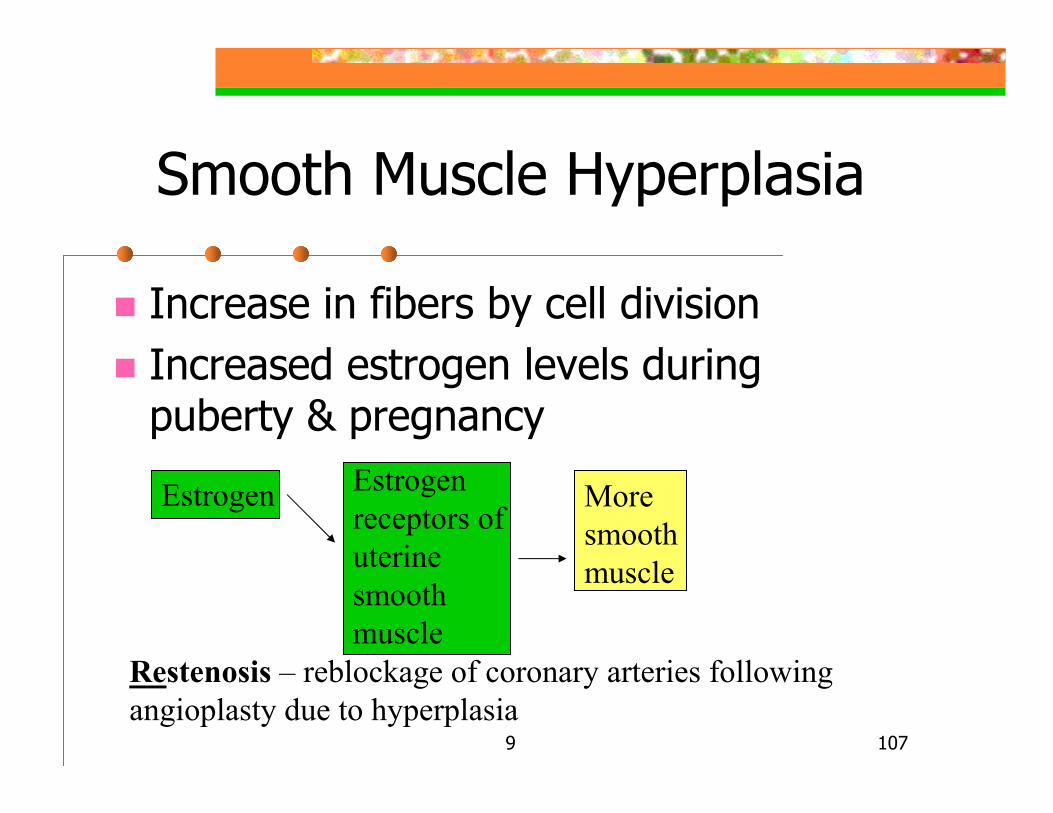
Smooth Muscle Hyperplasia
Increase in fibers by cell divisionIncreased estrogen levels during puberty & pregnancy
Estrogen Estrogen receptors of uterine smooth muscle
More smooth muscle
Restenosis – reblockage of coronary arteries following angioplasty due to hyperplasia
9 108
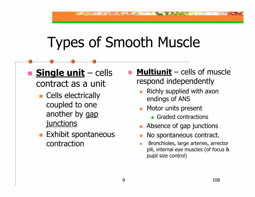
Types of Smooth Muscle
Single unit – cells contract as a unit
Cells electrically coupled to one another by gap junctionsExhibit spontaneous contraction
Multiunit – cells of muscle respond independently
Richly supplied with axon endings of ANSMotor units present
Graded contractions
Absence of gap junctionsNo spontaneous contract.Bronchioles, large arteries, arrector
pili, internal eye muscles (of focus & pupil size control)

9 109
Developmental Aspects Of Muscles
From mesodermSkeletal muscle fibers form by fusion of myoblasts
Unused myoblast become satellite cellsFunctional at 7 wks.ACH receptors on myoblasts aggregate in response to the release of growth factor called agrin released by invading nervesMuscular development parallels nervous system development, i.e. head-to toe & proximal-to distalGeneral male greater muscular development –testosterone related
9 110
Muscular Dystrophy
Group of inherited diseases with progressive muscular weakness, appearing generally in childhood
Muscles enlarge due to fat & c.t. depositsMuscle fibers degenerate & atrophyDuchene muscular dystrophy(DMD)
Inherited sex-linked recessive (mainly in males)1:3500 births, appear between 2-10 yrs. oldProgresses from legs upwardCytoplasm lacks dystrophin, a protein, that helps stabilize sarcolemma
9 111
Intermittent Claudication
Restricted blood delivery to legsAging & atherosclerosis (early stage of a arteriosclerosis with lipid in artery wall)
Excruciating pain in leg muscles while walkingPerson forced to stop & rest
9 112Pg. 962


