Page 1
CAMPBELL BIOLOGY IN FOCUS
© 2014 Pearson Education, Inc.
Urry • Cain • Wasserman • Minorsky • Jackson • Reece
Lecture Presentations by Kathleen Fitzpatrick and Nicole Tunbridge
13 The Molecular Basis of Inheritance
Page 2
© 2014 Pearson Education, Inc.
Overview: Life’s Operating Instructions
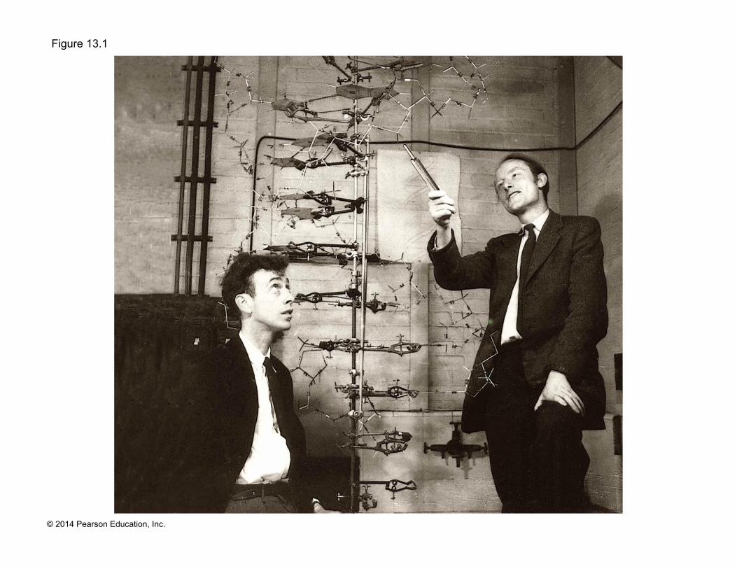
! In 1953, James Watson and Francis Crick introduced an elegant double-helical model for the structure of deoxyribonucleic acid, or DNA
! DNA, the substance of inheritance, is the most celebrated molecule of our time
! Hereditary information is encoded in DNA and reproduced in all cells of the body (DNA replication)
Page 3
© 2014 Pearson Education, Inc.
Figure 13.1
Page 4
© 2014 Pearson Education, Inc.
Concept 13.1: DNA is the genetic material
! Early in the 20th century, the identification of the molecules of inheritance loomed as a major challenge to biologists
Page 5
© 2014 Pearson Education, Inc.
The Search for the Genetic Material: Scientific Inquiry
! When T. H. Morgan’s group showed that genes are located on chromosomes, the two components of chromosomes—DNA and protein—became candidates for the genetic material
! The key factor in determining the genetic material was choosing appropriate experimental organisms
! The role of DNA in heredity was first discovered by studying bacteria and the viruses that infect them
Page 6
© 2014 Pearson Education, Inc.
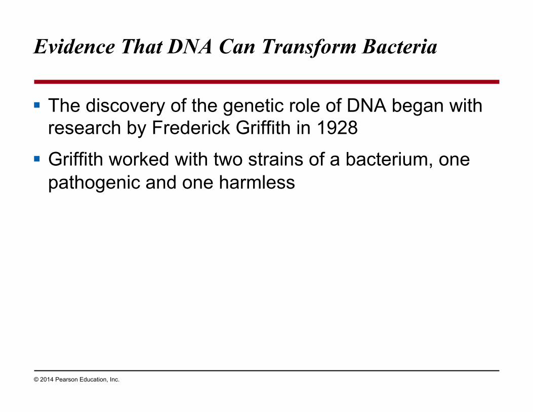
Evidence That DNA Can Transform Bacteria
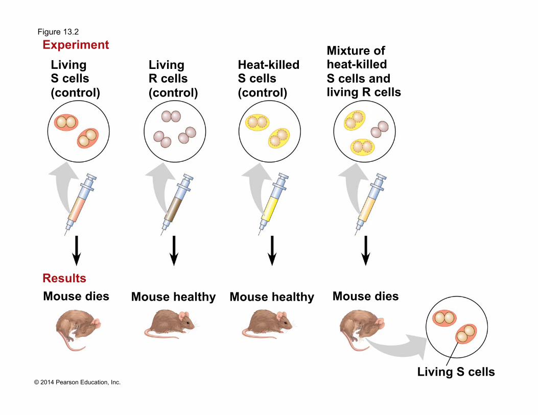
! The discovery of the genetic role of DNA began with research by Frederick Griffith in 1928
! Griffith worked with two strains of a bacterium, one pathogenic and one harmless
Page 7
© 2014 Pearson Education, Inc.
! When he mixed heat-killed remains of the pathogenic strain with living cells of the harmless strain, some living cells became pathogenic
! He called this phenomenon transformation, now defined as a change in genotype and phenotype due to assimilation of foreign DNA
Page 8
© 2014 Pearson Education, Inc.
Figure 13.2
Living S cells (control)
Mouse healthy Results
Experiment
Mouse healthy Mouse dies
Living S cells
Living R cells (control)
Heat-killed S cells (control)
Mixture of heat-killed S cells and living R cells
Mouse dies
Page 9
© 2014 Pearson Education, Inc.
! Later work by Oswald Avery and others identified the transforming substance as DNA
! Many biologists remained skeptical, mainly because little was known about DNA and they thought proteins were better candidates for the genetic material
Page 10
© 2014 Pearson Education, Inc.
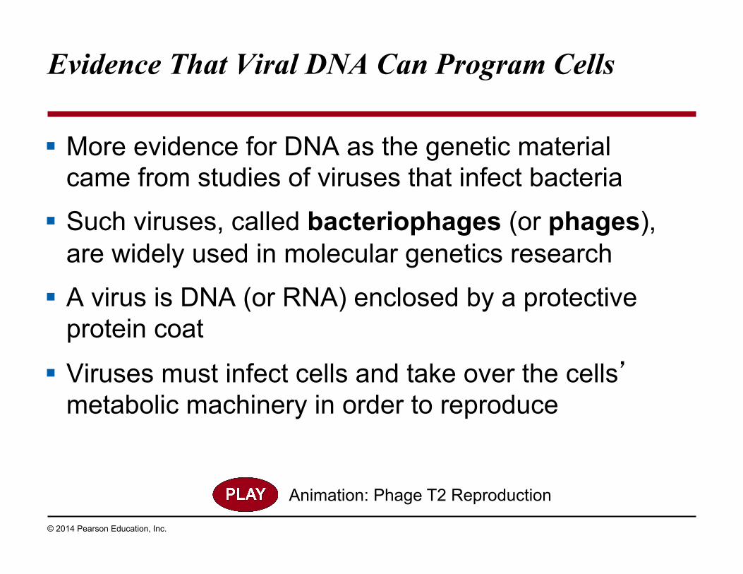
Evidence That Viral DNA Can Program Cells
! More evidence for DNA as the genetic material came from studies of viruses that infect bacteria
! Such viruses, called bacteriophages (or phages), are widely used in molecular genetics research
! A virus is DNA (or RNA) enclosed by a protective protein coat
! Viruses must infect cells and take over the cells’ metabolic machinery in order to reproduce
Animation: Phage T2 Reproduction
Page 11
© 2014 Pearson Education, Inc.
Figure 13.3
Phage head
Tail sheath
Tail fiber
DNA
Bacterial cell
100
nm
Page 12
© 2014 Pearson Education, Inc.
! In 1952, Alfred Hershey and Martha Chase showed that DNA is the genetic material of a phage known as T2
! To determine this, they designed an experiment showing that only the DNA of the T2 phage, and not the protein, enters an E. coli cell during infection
! They concluded that the injected DNA of the phage provides the genetic information
Animation: Hershey-Chase Experiment
Page 13
© 2014 Pearson Education, Inc.
Figure 13.4
Labeled phages infect cells.
Batch 1: Radioactive sulfur (35S) in phage protein Experiment
Agitation frees outside phage parts from cells.
Centrifuged cells form a pellet.
Radioactivity (phage protein) found in liquid
Batch 2: Radioactive phosphorus (32P) in phage DNA
Radioactivity (phage DNA) found in pellet
Radioactive protein
Radioactive DNA
Centrifuge
Centrifuge
Pellet
Pellet
1 2 3
4
4
Page 14
© 2014 Pearson Education, Inc.
Figure 13.4a
Labeled phages infect cells.
Batch 1: Radioactive sulfur (35S) in phage protein Experiment
Agitation frees outside phage parts from cells.
Centrifuged cells form a pellet.
Radioactivity (phage protein) found in liquid
Radioactive protein
Centrifuge
Pellet
1 2 3
4
Page 15
© 2014 Pearson Education, Inc.
Figure 13.4b
Batch 2: Radioactive phosphorus (32P) in phage DNA
Radioactivity (phage DNA) found in pellet
Radioactive DNA
Centrifuge Pellet
Labeled phages infect cells.
Agitation frees outside phage parts from cells.
Centrifuged cells form a pellet.
1 2 3
4
Experiment
Page 16
© 2014 Pearson Education, Inc.
Additional Evidence That DNA Is the Genetic Material
! It was known that DNA is a polymer of nucleotides, each consisting of a nitrogenous base, a sugar, and a phosphate group
! In 1950, Erwin Chargaff reported that DNA composition varies from one species to the next
! This evidence of diversity made DNA a more credible candidate for the genetic material
Animation: DNA and RNA Structure
Page 17
© 2014 Pearson Education, Inc.
Figure 13.5
Sugar– phosphate backbone
DNA nucleotide
Nitrogenous bases
3ʹ end
5ʹ end
Thymine (T)
Adenine (A)
Cytosine (C)
Guanine (G)
Page 18
© 2014 Pearson Education, Inc.
Figure 13.5a
Phosphate
DNA nucleotide Nitrogenous
base
3ʹ end
Sugar (deoxyribose)
Page 19
© 2014 Pearson Education, Inc.
! Two findings became known as Chargaff’s rules ! The base composition of DNA varies between
species
! In any species the number of A and T bases is equal and the number of G and C bases is equal
! The basis for these rules was not understood until the discovery of the double helix
Page 20
© 2014 Pearson Education, Inc.
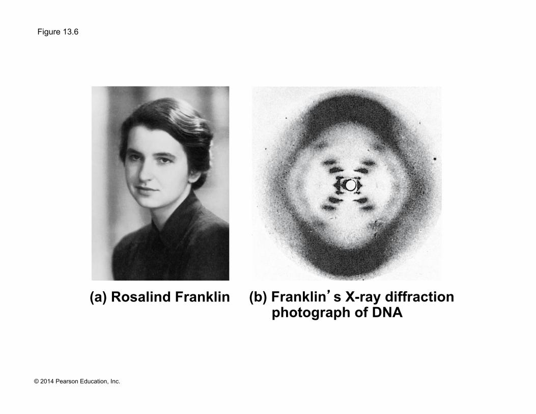
Building a Structural Model of DNA: Scientific Inquiry
! James Watson and Francis Crick were first to determine the structure of DNA
! Maurice Wilkins and Rosalind Franklin were using a technique called X-ray crystallography to study molecular structure
! Franklin produced a picture of the DNA molecule using this technique
Page 21
© 2014 Pearson Education, Inc.
Figure 13.6
(b) Franklin’s X-ray diffraction photograph of DNA
(a) Rosalind Franklin
Page 22
© 2014 Pearson Education, Inc.
Figure 13.6a
(a) Rosalind Franklin
Page 23
© 2014 Pearson Education, Inc.
Figure 13.6b
(b) Franklin’s X-ray diffraction photograph of DNA
Page 24
© 2014 Pearson Education, Inc.
! Franklin’s X-ray crystallographic images of DNA enabled Watson to deduce that DNA was helical
! The X-ray images also enabled Watson to deduce the width of the helix and the spacing of the nitrogenous bases
! The pattern in the photo suggested that the DNA molecule was made up of two strands, forming a double helix
Animation: DNA Double Helix
Video: DNA Surface Model
Page 25
© 2014 Pearson Education, Inc.
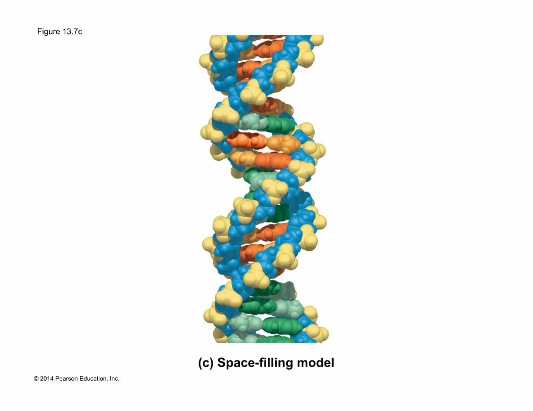
Figure 13.7
(c) Space-filling model
(a) Key features of DNA structure
(b) Partial chemical structure
3ʹ end
5ʹ end
3ʹ end
5ʹ end
Hydrogen bond
T A
C G
C G
3.4 nm
T A
T A
C G
C G
T
A
1 nm
0.34 nm
T A T
A
C G
C G
C
G
C G
T A
T A
C G C
G C G
Page 26
© 2014 Pearson Education, Inc.
Figure 13.7a
(a) Key features of DNA structure
3.4 nm
T A
C G
C
G
T
A
1 nm
0.34 nm
T A T
A
C G
C G
C
G
C
G
T A
T A
C G C
G C G
Page 27
© 2014 Pearson Education, Inc.
Figure 13.7b
(b) Partial chemical structure
3ʹ end
5ʹ end
3ʹ end
5ʹ end
Hydrogen bond
T A
C G
C G
T A
Page 28
© 2014 Pearson Education, Inc.
Figure 13.7c
(c) Space-filling model
Page 29
© 2014 Pearson Education, Inc.
! Watson and Crick built models of a double helix to conform to the X-ray measurements and the chemistry of DNA
! Franklin had concluded that there were two outer sugar-phosphate backbones, with the nitrogenous bases paired in the molecule’s interior
! Watson built a model in which the backbones were antiparallel (their subunits run in opposite directions)
Page 30
© 2014 Pearson Education, Inc.
! At first, Watson and Crick thought the bases paired like with like (A with A, and so on), but such pairings did not result in a uniform width
! Instead, pairing a purine with a pyrimidine resulted in a uniform width consistent with the X-ray data
Page 31
© 2014 Pearson Education, Inc.
Figure 13.UN02
Purine + purine: too wide
Pyrimidine + pyrimidine: too narrow
Purine + pyrimidine: width consistent with X-ray data
Page 32
© 2014 Pearson Education, Inc.
! Watson and Crick reasoned that the pairing was more specific, dictated by the base structures
! They determined that adenine (A) paired only with thymine (T), and guanine (G) paired only with cytosine (C)
! The Watson-Crick model explains Chargaff’s rules: in any organism the amount of A = T, and the amount of G = C
Page 33
© 2014 Pearson Education, Inc.
Figure 13.8
Sugar Sugar
Sugar
Sugar
Thymine (T) Adenine (A)
Cytosine (C) Guanine (G)
Page 34
© 2014 Pearson Education, Inc.
Concept 13.2: Many proteins work together in DNA replication and repair
! The relationship between structure and function is manifest in the double helix
! Watson and Crick noted that the specific base pairing suggested a possible copying mechanism for genetic material
Page 35
© 2014 Pearson Education, Inc.
Figure 13.9-1
(a) Parental molecule
T A
C G
C G
T A
T A
Page 36
© 2014 Pearson Education, Inc.
Figure 13.9-2
(a) Parental molecule
(b) Separation of parental strands into templates
T A
C G
C G
T A
T A T A
T A
C G
C G
T A
Page 37
© 2014 Pearson Education, Inc.
Figure 13.9-3
(a) Parental molecule
(b) Separation of parental strands into templates
(c) Formation of new strands complementary to template strands
T A
C G
C G
T A
T A T A
T A
C G
C G
T A
T A
C G
C G
T A
T A
T A
C G
C G
T A
T A
Page 38
© 2014 Pearson Education, Inc.
The Basic Principle: Base Pairing to a Template Strand
! Since the two strands of DNA are complementary, each strand acts as a template for building a new strand in replication
! In DNA replication, the parent molecule unwinds, and two new daughter strands are built based on base-pairing rules
Page 39
© 2014 Pearson Education, Inc.
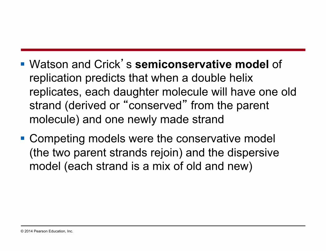
! Watson and Crick’s semiconservative model of replication predicts that when a double helix replicates, each daughter molecule will have one old strand (derived or “conserved” from the parent molecule) and one newly made strand
! Competing models were the conservative model (the two parent strands rejoin) and the dispersive model (each strand is a mix of old and new)
Page 40
© 2014 Pearson Education, Inc.
Figure 13.10
(a) Conservative model
(b) Semiconservative model
(c) Dispersive model
Parent cell First
replication Second
replication
Page 41
© 2014 Pearson Education, Inc.
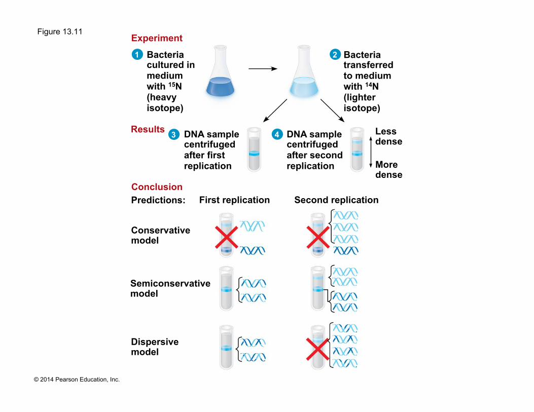
! Experiments by Matthew Meselson and Franklin Stahl supported the semiconservative model
Page 42
© 2014 Pearson Education, Inc.
Figure 13.11
Conservative model
Semiconservative model
Dispersive model
Predictions: First replication Second replication
DNA sample centrifuged after first replication
DNA sample centrifuged after second replication
Bacteria cultured in medium with 15N (heavy isotope)
Bacteria transferred to medium with 14N (lighter isotope)
Less dense
More dense
Experiment
Results
Conclusion
1
3
2
4
Page 43
© 2014 Pearson Education, Inc.
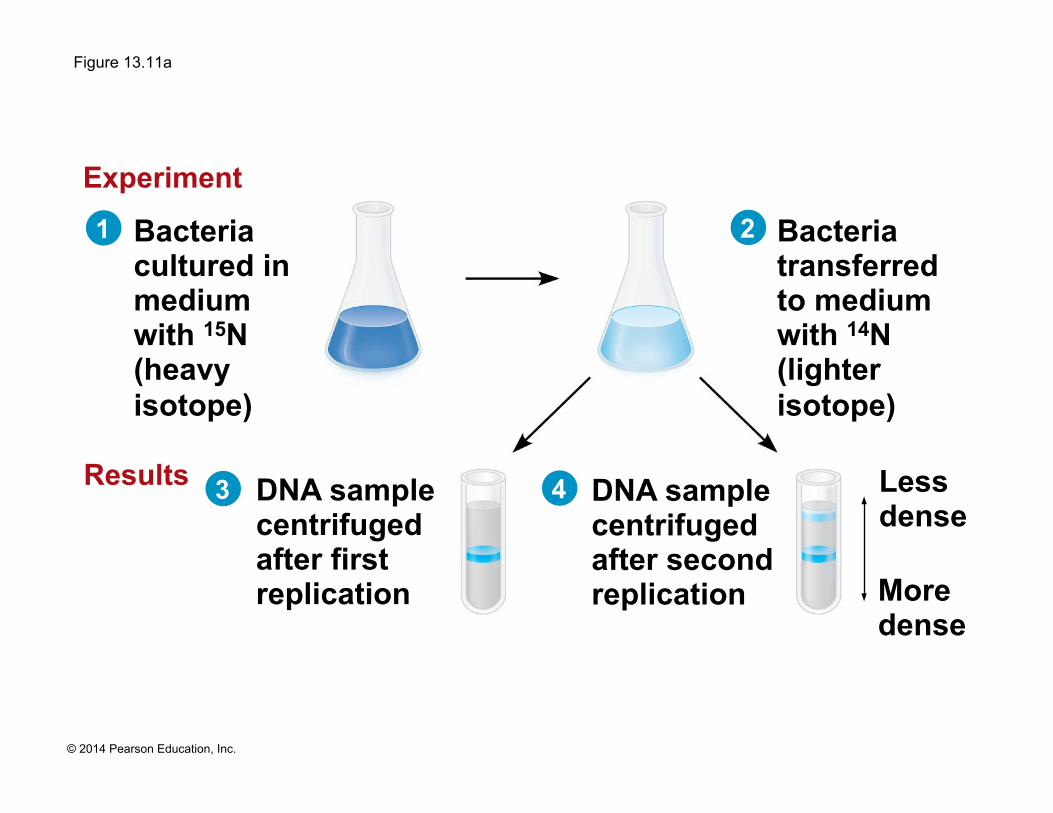
Figure 13.11a
DNA sample centrifuged after first replication
DNA sample centrifuged after second replication
Bacteria cultured in medium with 15N (heavy isotope)
Bacteria transferred to medium with 14N (lighter isotope)
Less dense
More dense
Experiment
Results
1
3 4
2
Page 44
© 2014 Pearson Education, Inc.
Figure 13.11b
Conservative model
Semiconservative model
Dispersive model
Predictions: First replication Second replication Conclusion
Page 45
© 2014 Pearson Education, Inc.
DNA Replication: A Closer Look
! The copying of DNA is remarkable in its speed and accuracy
! More than a dozen enzymes and other proteins participate in DNA replication
! Much more is known about how this “replication machine” works in bacteria than in eukaryotes
! Most of the process is similar between prokaryotes and eukaryotes
Page 46
© 2014 Pearson Education, Inc.
Getting Started
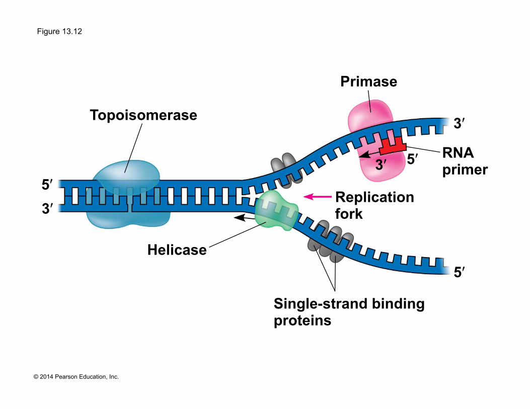
! Replication begins at particular sites called origins of replication, where the two DNA strands are separated, opening up a replication “bubble”
! At each end of a bubble is a replication fork, a Y-shaped region where the parental strands of DNA are being unwound
Animation: DNA Replication Overview
Animation: Origins of Replication
Page 47
© 2014 Pearson Education, Inc.
Figure 13.12
Single-strand binding proteins
Helicase
Topoisomerase
Primase
Replication fork
5ʹ
5ʹ 5ʹ 3ʹ
3ʹ
3ʹ
RNA primer
Page 48
© 2014 Pearson Education, Inc.
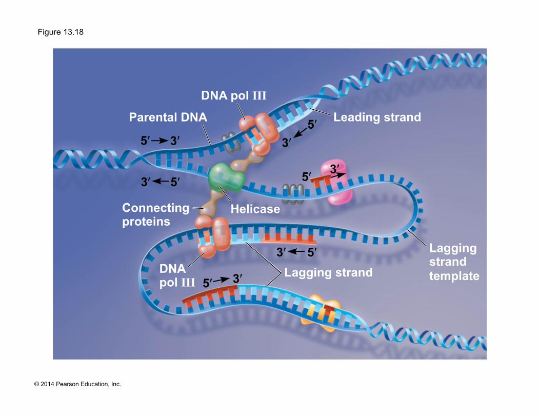
! Helicases are enzymes that untwist the double helix at the replication forks
! Single-strand binding proteins bind to and stabilize single-stranded DNA
! Topoisomerase relieves the strain caused by tight twisting ahead of the replication fork by breaking, swiveling, and rejoining DNA strands
Page 49
© 2014 Pearson Education, Inc.
Figure 13.13
Double- stranded DNA molecule
Two daughter DNA molecules
Replication bubble
Replication fork
Daughter (new) strand
Parental (template) strand Origin of
replication Double-stranded DNA molecule
Two daughter DNA molecules
Bubble Replication fork
Daughter (new) strand
Parental (template) strand
Origin of replication
(a) Origin of replication in an E. coli cell (b) Origins of replication in a eukaryotic cell
0.25
µm
0.5
µm
Page 50
© 2014 Pearson Education, Inc.
Figure 13.13a
Double- stranded DNA molecule
Two daughter DNA molecules
Replication bubble
Replication fork
Daughter (new) strand
Parental (template) strand Origin of
replication
(a) Origin of replication in an E. coli cell
0.5
µm
Page 51
© 2014 Pearson Education, Inc.
Figure 13.13aa
0.5
µm
Page 52
© 2014 Pearson Education, Inc.
! Multiple replication bubbles form and eventually fuse, speeding up the copying of DNA
Page 53
© 2014 Pearson Education, Inc.
Figure 13.13b
Double-stranded DNA molecule
Two daughter DNA molecules
Bubble Replication fork
Daughter (new) strand
Parental (template) strand
Origin of replication
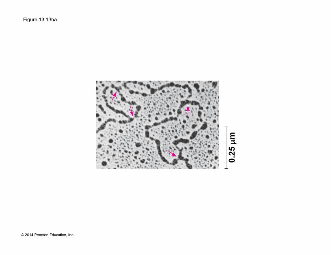
(b) Origins of replication in a eukaryotic cell
0.25
µm
Page 54
© 2014 Pearson Education, Inc.
Figure 13.13ba
0.25
µm
Page 55
© 2014 Pearson Education, Inc.
! DNA polymerases cannot initiate synthesis of a polynucleotide; they can only add nucleotides to an already existing chain base-paired with the template
! The initial nucleotide strand is a short RNA primer
Synthesizing a New DNA Strand
Page 56
© 2014 Pearson Education, Inc.
! The enzyme, primase, starts an RNA chain from a single RNA nucleotide and adds RNA nucleotides one at a time using the parental DNA as a template
! The primer is short (5–10 nucleotides long)
! The new DNA strand will start from the 3ʹ end of the RNA primer
Page 57
© 2014 Pearson Education, Inc.
! Enzymes called DNA polymerases catalyze the elongation of new DNA at a replication fork
! Most DNA polymerases require a primer and a DNA template strand
! The rate of elongation is about 500 nucleotides per second in bacteria and 50 per second in human cells
Page 58
© 2014 Pearson Education, Inc.
! Each nucleotide that is added to a growing DNA consists of a sugar attached to a base and three phosphate groups
! dATP is used to make DNA and is similar to the ATP of energy metabolism
! The difference is in the sugars: dATP has deoxyribose, while ATP has ribose
! As each monomer nucleotide joins the DNA strand, it loses two phosphate groups as a molecule of pyrophosphate
Page 59
© 2014 Pearson Education, Inc.
Figure 13.14
Pyro- phosphate
New strand
Phosphate
Nucleotide
5ʹ 3ʹ Template strand
Sugar Base
5ʹ
3ʹ
5ʹ
3ʹ
5ʹ 3ʹ
DNA poly-
merase T
A T
C G
A
C G
C
P
P i P
i 2
A T
C G
A
C G
C
Page 60
© 2014 Pearson Education, Inc.
Antiparallel Elongation
! The antiparallel structure of the double helix affects replication
! DNA polymerases add nucleotides only to the free 3ʹ end of a growing strand; therefore, a new DNA strand can elongate only in the 5ʹ to 3ʹ direction
Page 61
© 2014 Pearson Education, Inc.
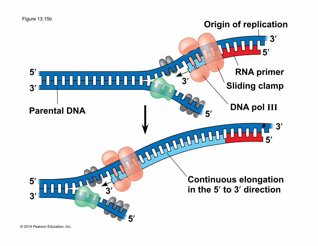
! Along one template strand of DNA, the DNA polymerase synthesizes a leading strand continuously, moving toward the replication fork
Animation: Leading Strand
Page 62
© 2014 Pearson Education, Inc.
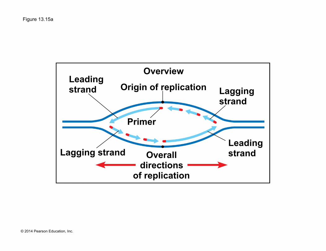
Figure 13.15
Parental DNA
5ʹ 3ʹ
5ʹ
3ʹ
5ʹ
3ʹ
Continuous elongation in the 5ʹ to 3ʹ direction
5ʹ 3ʹ
5ʹ
3ʹ
DNA pol III
RNA primer Sliding clamp
5ʹ 3ʹ
Origin of replication
Origin of replication
Lagging strand
Lagging strand
Overall directions
of replication
Leading strand
Leading strand
Overview
Primer
Page 63
© 2014 Pearson Education, Inc.
Figure 13.15a
Origin of replication
Lagging strand
Lagging strand
Overall directions
of replication
Leading strand
Leading strand
Overview
Primer
Page 64
© 2014 Pearson Education, Inc.
Figure 13.15b
Parental DNA
5ʹ 3ʹ
5ʹ
3ʹ
5ʹ
3ʹ
Continuous elongation in the 5ʹ to 3ʹ direction
5ʹ 3ʹ
5ʹ 3ʹ
DNA pol III
RNA primer Sliding clamp
5ʹ
3ʹ
Origin of replication
Page 65
© 2014 Pearson Education, Inc.
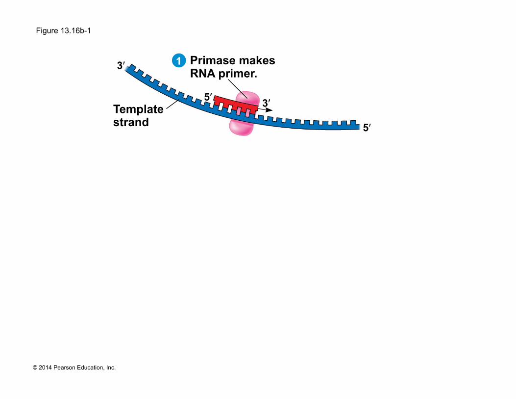
! To elongate the other new strand, called the lagging strand, DNA polymerase must work in the direction away from the replication fork
! The lagging strand is synthesized as a series of segments called Okazaki fragments
Page 66
© 2014 Pearson Education, Inc.
! After formation of Okazaki fragments, DNA polymerase I removes the RNA primers and replaces the nucleotides with DNA
! The remaining gaps are joined together by DNA ligase
Animation: Lagging Strand
Animation: DNA Replication Review
Page 67
© 2014 Pearson Education, Inc.
Figure 13.16
5ʹ 3ʹ
5ʹ
3ʹ
Origin of replication Lagging strand Lagging
strand
Overall directions of replication
Leading strand
Leading strand
Overview
Primase makes RNA primer.
RNA primer for fragment 1
Template strand
Okazaki fragment 1
DNA pol III makes Okazaki fragment 1.
DNA pol III detaches.
5ʹ 3ʹ
5ʹ
3ʹ
5ʹ
3ʹ 5ʹ
3ʹ
RNA primer for fragment 2
Okazaki fragment 2 DNA pol III
makes Okazaki fragment 2.
Overall direction of replication
DNA pol I replaces RNA with DNA.
DNA ligase forms bonds between DNA fragments.
5ʹ
3ʹ 5ʹ
3ʹ
5ʹ
3ʹ 5ʹ
3ʹ
5ʹ
3ʹ 5ʹ
3ʹ 1
2
3
4
5
6
Page 68
© 2014 Pearson Education, Inc.
Figure 13.16a
Origin of replication Lagging strand Lagging
strand
Overall directions of replication
Leading strand
Leading strand
Overview
Page 69
© 2014 Pearson Education, Inc.
Figure 13.16b-1
5ʹ 3ʹ
5ʹ
3ʹ Primase makes RNA primer.
Template strand
1
Page 70
© 2014 Pearson Education, Inc.
Figure 13.16b-2
5ʹ 3ʹ
5ʹ
3ʹ Primase makes RNA primer.
RNA primer for fragment 1
Template strand
DNA pol III makes Okazaki fragment 1.
5ʹ 3ʹ
5ʹ
3ʹ
1
2
Page 71
© 2014 Pearson Education, Inc.
Figure 13.16b-3
5ʹ 3ʹ
5ʹ
3ʹ Primase makes RNA primer.
RNA primer for fragment 1
Template strand
Okazaki fragment 1
DNA pol III makes Okazaki fragment 1.
DNA pol III detaches.
5ʹ 3ʹ
5ʹ
3ʹ
5ʹ
3ʹ 5ʹ
3ʹ
1
2
3
Page 72
© 2014 Pearson Education, Inc.
Figure 13.16c-1 RNA primer for fragment 2
Okazaki fragment 2 DNA pol III
makes Okazaki fragment 2.
5ʹ
3ʹ 5ʹ
3ʹ 4
Page 73
© 2014 Pearson Education, Inc.
Figure 13.16c-2 RNA primer for fragment 2
Okazaki fragment 2 DNA pol III
makes Okazaki fragment 2.
DNA pol I replaces RNA with DNA.
5ʹ
3ʹ 5ʹ
3ʹ
5ʹ
3ʹ 5ʹ
3ʹ 4
5
Page 74
© 2014 Pearson Education, Inc.
Figure 13.16c-3 RNA primer for fragment 2
Okazaki fragment 2 DNA pol III
makes Okazaki fragment 2.
Overall direction of replication
DNA pol I replaces RNA with DNA.
DNA ligase forms bonds between DNA fragments. 5ʹ
3ʹ 5ʹ
3ʹ
5ʹ
3ʹ 5ʹ
3ʹ
5ʹ
3ʹ 5ʹ
3ʹ 4
6
5
Page 75
© 2014 Pearson Education, Inc.
Figure 13.17
3ʹ 5ʹ
Origin of replication
Lagging strand
Lagging strand
Overall directions of replication
Leading strand
Leading strand
Overview
5ʹ 3ʹ
5ʹ
3ʹ
Leading strand
Lagging strand
DNA ligase DNA pol I DNA pol III
Primase
DNA pol III Primer
5ʹ 3ʹ
5ʹ
3ʹ
Lagging strand template
Parental DNA
Helicase
Single-strand binding proteins
Leading strand template
Page 76
© 2014 Pearson Education, Inc.
Figure 13.17a
Origin of replication
Lagging strand
Lagging strand
Overall directions of replication
Leading strand
Leading strand
Overview
Page 77
© 2014 Pearson Education, Inc.
Figure 13.17b
3ʹ 5ʹ
3ʹ
Leading strand
DNA pol III
Primase Primer
5ʹ
3ʹ
Lagging strand template
Parental DNA
Helicase
Single-strand binding proteins
Leading strand template
Page 78
© 2014 Pearson Education, Inc.
Figure 13.17c
5ʹ 5ʹ
3ʹ
5ʹ
3ʹ
Lagging strand
DNA ligase DNA pol I DNA pol III
Page 79
© 2014 Pearson Education, Inc.
The DNA Replication Complex
! The proteins that participate in DNA replication form a large complex, a “DNA replication machine”
! The DNA replication machine may be stationary during the replication process
! Recent studies support a model in which DNA polymerase molecules “reel in” parental DNA and “extrude” newly made daughter DNA molecules
Animation: DNA Replication
Page 80
© 2014 Pearson Education, Inc.
Figure 13.18
3ʹ 5ʹ
5ʹ
3ʹ 5ʹ
3ʹ
Lagging strand
DNA pol III
Leading strand
Lagging strand template
Parental DNA
Helicase Connecting proteins
DNA pol III
3ʹ 5ʹ
3ʹ 5ʹ
3ʹ 5ʹ
Page 81
© 2014 Pearson Education, Inc.
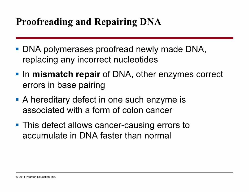
Proofreading and Repairing DNA
! DNA polymerases proofread newly made DNA, replacing any incorrect nucleotides
! In mismatch repair of DNA, other enzymes correct errors in base pairing
! A hereditary defect in one such enzyme is associated with a form of colon cancer
! This defect allows cancer-causing errors to accumulate in DNA faster than normal
Page 82
© 2014 Pearson Education, Inc.
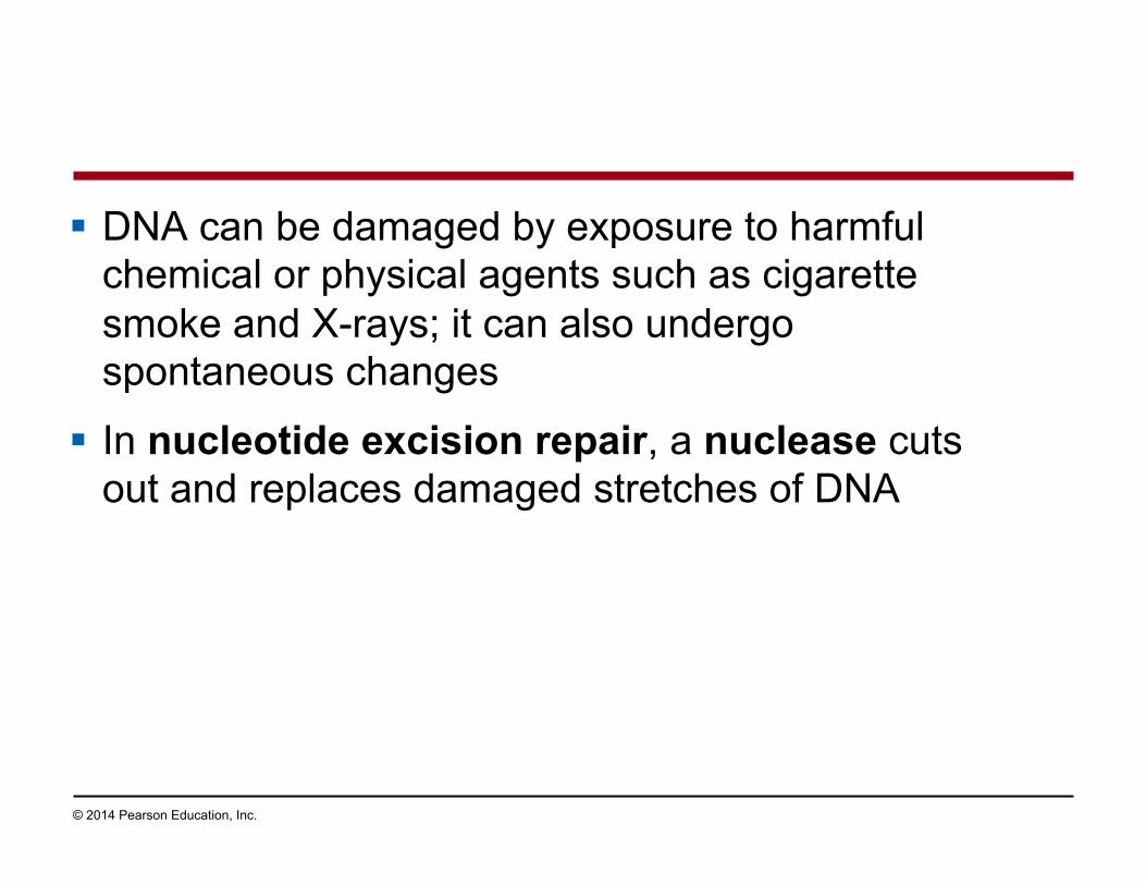
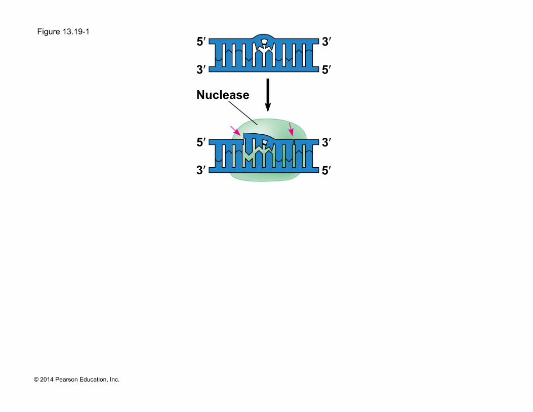
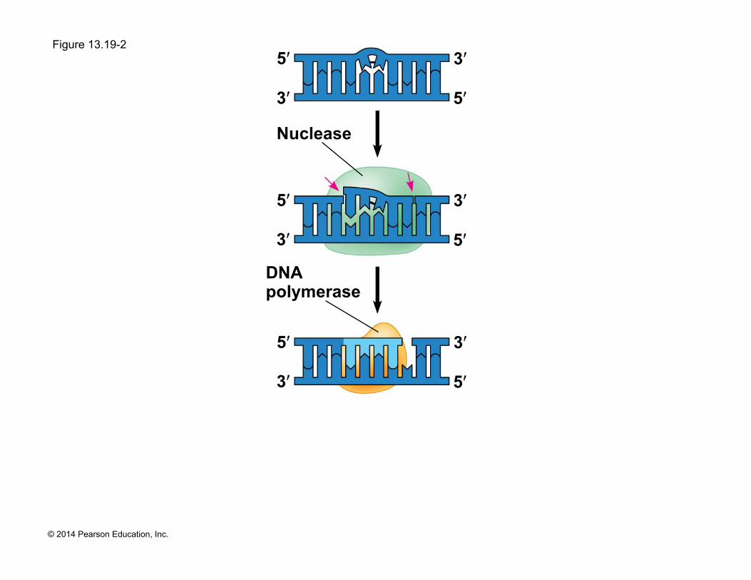
! DNA can be damaged by exposure to harmful chemical or physical agents such as cigarette smoke and X-rays; it can also undergo spontaneous changes
! In nucleotide excision repair, a nuclease cuts out and replaces damaged stretches of DNA
Page 83
© 2014 Pearson Education, Inc.
Figure 13.19-1
3ʹ
5ʹ
Nuclease
3ʹ
5ʹ
3ʹ
5ʹ 3ʹ
5ʹ
Page 84
© 2014 Pearson Education, Inc.
Figure 13.19-2
3ʹ
5ʹ
Nuclease
3ʹ
5ʹ
3ʹ
5ʹ
DNA polymerase
3ʹ
5ʹ
3ʹ
5ʹ 3ʹ
5ʹ
Page 85
© 2014 Pearson Education, Inc.
Figure 13.19-3
3ʹ
5ʹ
Nuclease
3ʹ
5ʹ
3ʹ
5ʹ
DNA polymerase
3ʹ
5ʹ
3ʹ
5ʹ 3ʹ
5ʹ
DNA ligase
3ʹ
5ʹ 3ʹ
5ʹ
Page 86
© 2014 Pearson Education, Inc.
Evolutionary Significance of Altered DNA Nucleotides
! Error rate after proofreading repair is low but not zero
! Sequence changes may become permanent and can be passed on to the next generation
! These changes (mutations) are the source of the genetic variation upon which natural selection operates
Page 87
© 2014 Pearson Education, Inc.
Replicating the Ends of DNA Molecules
! Limitations of DNA polymerase create problems for the linear DNA of eukaryotic chromosomes
! The usual replication machinery cannot complete the 5ʹ ends of daughter strands
! Repeated rounds of replication produce shorter DNA molecules with uneven ends
Animation: DNA Packing
Video: Nucleosome Model
Page 88
© 2014 Pearson Education, Inc.
Figure 13.20
1 µm
Page 89
© 2014 Pearson Education, Inc.
! Eukaryotic chromosomal DNA molecules have special nucleotide sequences at their ends called telomeres
! Telomeres do not prevent the shortening of DNA molecules, but they do postpone it
! It has been proposed that the shortening of telomeres is connected to aging
Page 90
© 2014 Pearson Education, Inc.
! If chromosomes of germ cells became shorter in every cell cycle, essential genes would eventually be missing from the gametes they produce
! An enzyme called telomerase catalyzes the lengthening of telomeres in germ cells
Page 91
© 2014 Pearson Education, Inc.
! Telomerase is not active in most human somatic cells
! However, it does show inappropriate activity in some cancer cells
! Telomerase is currently under study as a target for cancer therapies
Page 92
© 2014 Pearson Education, Inc.
Concept 13.3: A chromosome consists of a DNA molecule packed together with proteins
! The bacterial chromosome is a double-stranded, circular DNA molecule associated with a small amount of protein
! Eukaryotic chromosomes have linear DNA molecules associated with a large amount of protein
! In a bacterium, the DNA is “supercoiled” and found in a region of the cell called the nucleoid
Page 93
© 2014 Pearson Education, Inc.
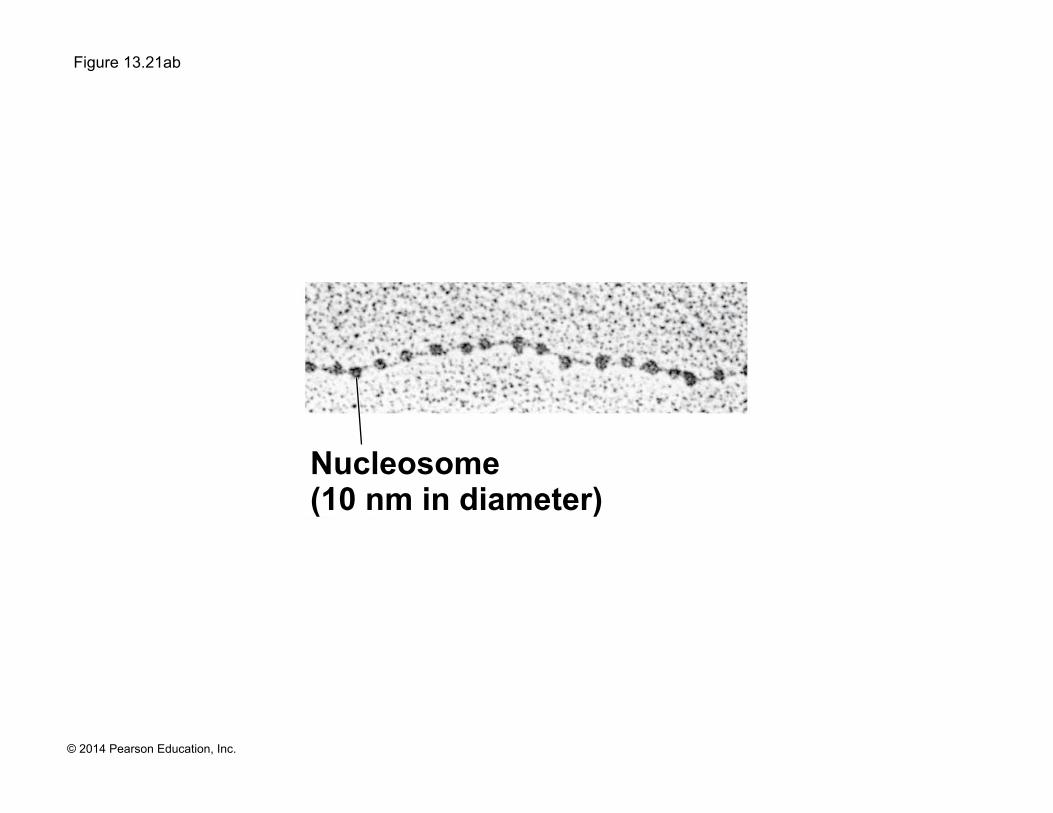
! Chromatin, a complex of DNA and protein, is found in the nucleus of eukaryotic cells
! Chromosomes fit into the nucleus through an elaborate, multilevel system of packing
! Chromatin undergoes striking changes in the degree of packing during the course of the cell cycle
Page 94
© 2014 Pearson Education, Inc.
Figure 13.21
Histone tail Histones H1
DNA double helix (2 nm in diameter)
Nucleosome (10 nm in diameter)
Loops
30-nm fiber
300-nm fiber
Replicated chromosome (1,400 nm)
Scaffold
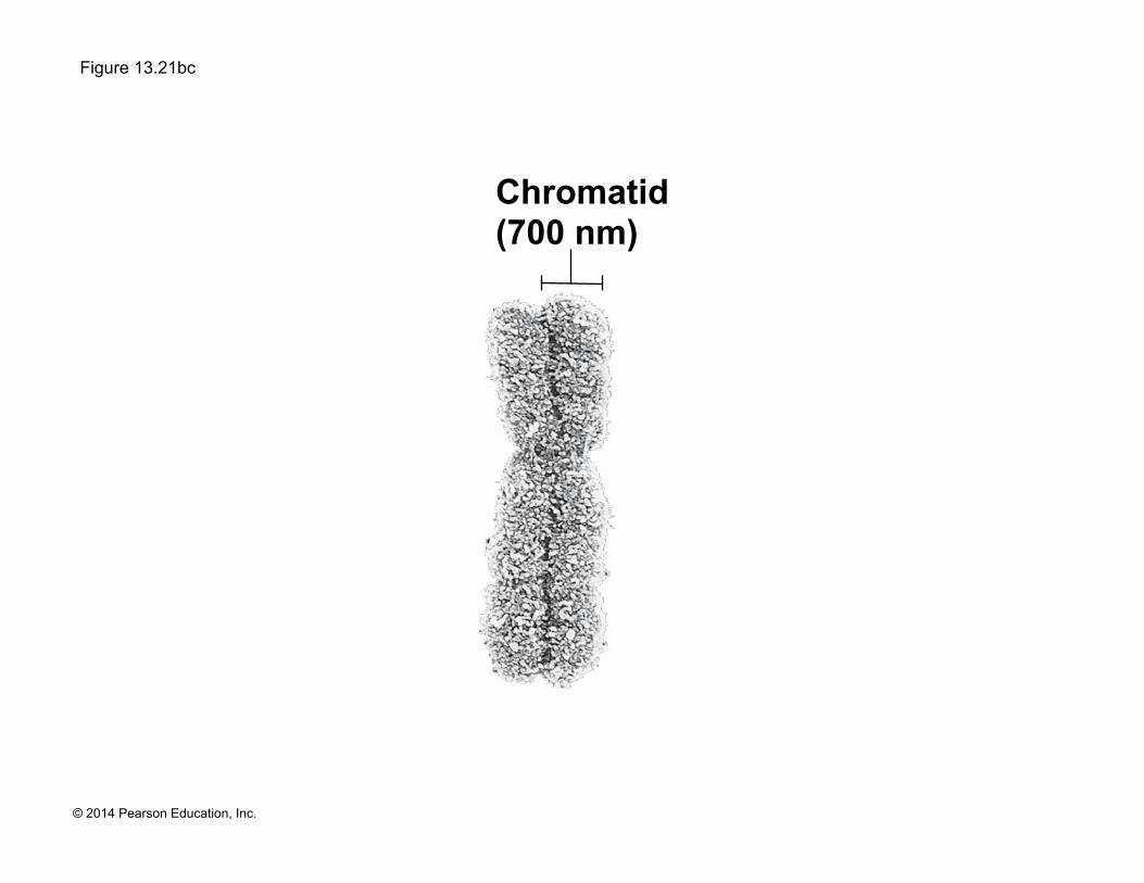
Chromatid (700 nm)
Page 95
© 2014 Pearson Education, Inc.
Figure 13.21a
Histone tail Histones
H1
DNA double helix (2 nm in diameter)
Nucleosome (10 nm in diameter)
Page 96
© 2014 Pearson Education, Inc.
Figure 13.21aa
DNA double helix (2 nm in diameter)
Page 97
© 2014 Pearson Education, Inc.
Figure 13.21ab
Nucleosome (10 nm in diameter)
Page 98
© 2014 Pearson Education, Inc.
Figure 13.21b
Loops
30-nm fiber
300-nm fiber
Replicated chromosome (1,400 nm)
Scaffold
Chromatid (700 nm)
Page 99
© 2014 Pearson Education, Inc.
Figure 13.21ba
30-nm fiber
Page 100
© 2014 Pearson Education, Inc.
Figure 13.21bb
Loops Scaffold
Page 101
© 2014 Pearson Education, Inc.
Figure 13.21bc
Chromatid (700 nm)
Page 102
© 2014 Pearson Education, Inc.
! At interphase, most of the chromatin is compacted into a 30-nm fiber, which is folded further in some areas by looping
! Even during interphase, centromeres and some other parts of chromosomes are highly condensed, similar to metaphase chromosomes
! This condensed chromatin is called heterochromatin; the more dispersed, less compacted chromatin is called euchromatin
Page 103
© 2014 Pearson Education, Inc.
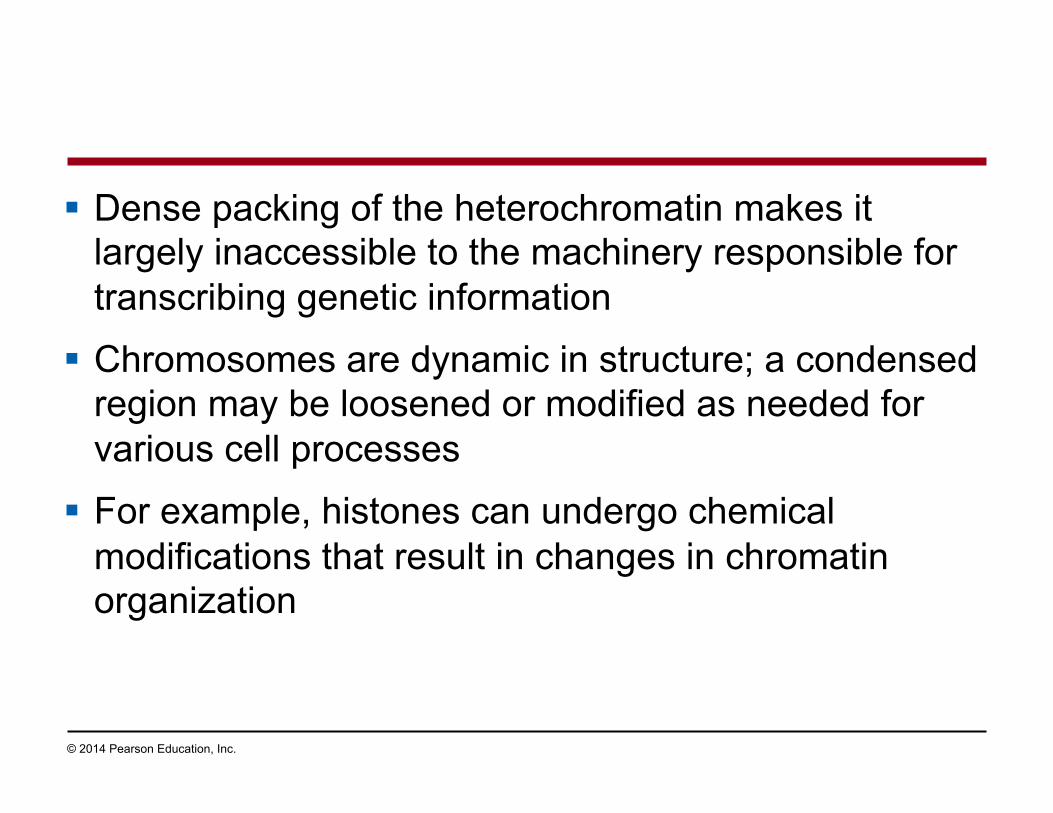
! Dense packing of the heterochromatin makes it largely inaccessible to the machinery responsible for transcribing genetic information
! Chromosomes are dynamic in structure; a condensed region may be loosened or modified as needed for various cell processes
! For example, histones can undergo chemical modifications that result in changes in chromatin organization
Page 104
© 2014 Pearson Education, Inc.
Concept 13.4: Understanding DNA structure and replication makes genetic engineering possible
! Complementary base pairing of DNA is the basis for nucleic acid hybridization, the base pairing of one strand of a nucleic acid to another, complementary sequence
! Nucleic acid hybridization forms the foundation of virtually every technique used in genetic engineering, the direct manipulation of genes for practical purposes
Page 105
© 2014 Pearson Education, Inc.
DNA Cloning: Making Multiple Copies of a Gene or Other DNA Segment
! To work directly with specific genes, scientists prepare well-defined segments of DNA in identical copies, a process called DNA cloning
! Most methods for cloning pieces of DNA in the laboratory share general features
Page 106
© 2014 Pearson Education, Inc.
! Many bacteria contain plasmids, small circular DNA molecules that replicate separately from the bacterial chromosome
! To clone pieces of DNA, researchers first obtain a plasmid and insert DNA from another source (“foreign DNA”) into it
! The resulting plasmid is called recombinant DNA
Animation: Restriction Enzymes
Page 107
© 2014 Pearson Education, Inc.
Figure 13.22
Copies of gene
Recombinant bacterium
Gene of interest
Gene used to alter bacteria for cleaning up toxic waste
Plasmid Bacterial chromosome
Gene for pest resistance inserted into plants
Protein dissolves blood clots in heart attack therapy
Recombinant DNA (plasmid)
Bacterium Gene inserted into plasmid
Plasmid put into bacterial cell
Cell containing gene of interest
DNA of chromosome (“foreign” DNA)
Gene of interest
Protein expressed from gene of interest
Human growth hormone treats stunted growth
Protein harvested
Host cell grown in culture to form a clone of cells containing the “cloned” gene of interest
Basic research and various applications
1
2
3
4
Page 108
© 2014 Pearson Education, Inc.
Figure 13.22a
Recombinant bacterium
Gene of interest
Plasmid Bacterial chromosome
Recombinant DNA (plasmid)
Bacterium Gene inserted into plasmid
Plasmid put into bacterial cell
Cell containing gene of interest
DNA of chromosome (“foreign” DNA)
Gene of interest
Protein expressed from gene of interest
Host cell grown in culture to form a clone of cells containing the “cloned” gene of interest
1
2
3
Page 109
© 2014 Pearson Education, Inc.
Figure 13.22b
Copies of gene
Gene of interest
Gene used to alter bacteria for cleaning up toxic waste
Gene for pest resistance inserted into plants
Protein dissolves blood clots in heart attack therapy
Protein expressed from gene of interest
Human growth hormone treats stunted growth
Protein harvested
Basic research and various applications
4
Page 110
© 2014 Pearson Education, Inc.
! The production of multiple copies of a single gene is called gene cloning
! Gene cloning is useful to make many copies of a gene and to produce a protein product
! The ability to amplify many copies of a gene is crucial for applications involving a single gene
Page 111
© 2014 Pearson Education, Inc.
Using Restriction Enzymes to Make Recombinant DNA
! Bacterial restriction enzymes cut DNA molecules at specific DNA sequences called restriction sites
! A restriction enzyme usually makes many cuts, yielding restriction fragments
Page 112
© 2014 Pearson Education, Inc.
Figure 13.23-1
Restriction enzyme cuts the sugar-phosphate backbones.
3ʹ
5ʹ Restriction site
DNA 3ʹ
5ʹ
Sticky end 3ʹ
5ʹ 3ʹ
5ʹ 3ʹ
5ʹ 3ʹ
5ʹ
G G C C A
T T A
A T T A
G G C C A
T T A
A T T
A
1
Page 113
© 2014 Pearson Education, Inc.
Figure 13.23-2
Restriction enzyme cuts the sugar-phosphate backbones.
3ʹ
5ʹ DNA
3ʹ
5ʹ
DNA fragment added from another molecule cut by same enzyme. Base pairing occurs.
Sticky end
One possible combination
3ʹ
5ʹ 3ʹ
5ʹ 3ʹ
5ʹ 3ʹ
5ʹ
3ʹ
5ʹ 3ʹ
5ʹ
3ʹ 5ʹ
3ʹ
5ʹ 3ʹ
5ʹ 3ʹ
5ʹ
3ʹ 5ʹ
3ʹ
5ʹ
G C A A T T
G G C C A
T T A
A T T A
G G C C A
T T A
A T T A
1
2
5ʹ
Restriction site
G G C C A
T T A
A T T A
G G C C A
T A
A T T
A T
Page 114
© 2014 Pearson Education, Inc.
Figure 13.23-3
Restriction enzyme cuts the sugar-phosphate backbones.
3ʹ
5ʹ DNA
3ʹ
5ʹ
DNA fragment added from another molecule cut by same enzyme. Base pairing occurs.
DNA ligase seals the strands.
Sticky end
One possible combination
Recombinant DNA molecule
3ʹ
5ʹ 3ʹ
5ʹ 3ʹ
5ʹ 3ʹ
5ʹ
3ʹ
5ʹ 3ʹ
5ʹ
3ʹ 5ʹ
3ʹ
5ʹ 3ʹ
5ʹ 3ʹ
5ʹ
3ʹ 5ʹ
3ʹ
5ʹ
3ʹ
5ʹ 3ʹ
5ʹ
G C A A T T
G G C C A
T T A
A T T A
G G C C A
T T A
A T T A
1
2
3
Restriction site
G G C C A
T T A
A T T A
G G C C A
T T A
A T T
A
Page 115
© 2014 Pearson Education, Inc.
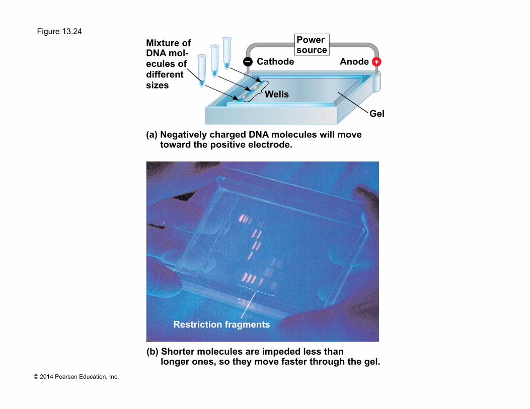
! To see the fragments produced by cutting DNA molecules with restriction enzymes, researchers use gel electrophoresis
! This technique separates a mixture of nucleic acid fragments based on length
Page 116
© 2014 Pearson Education, Inc.
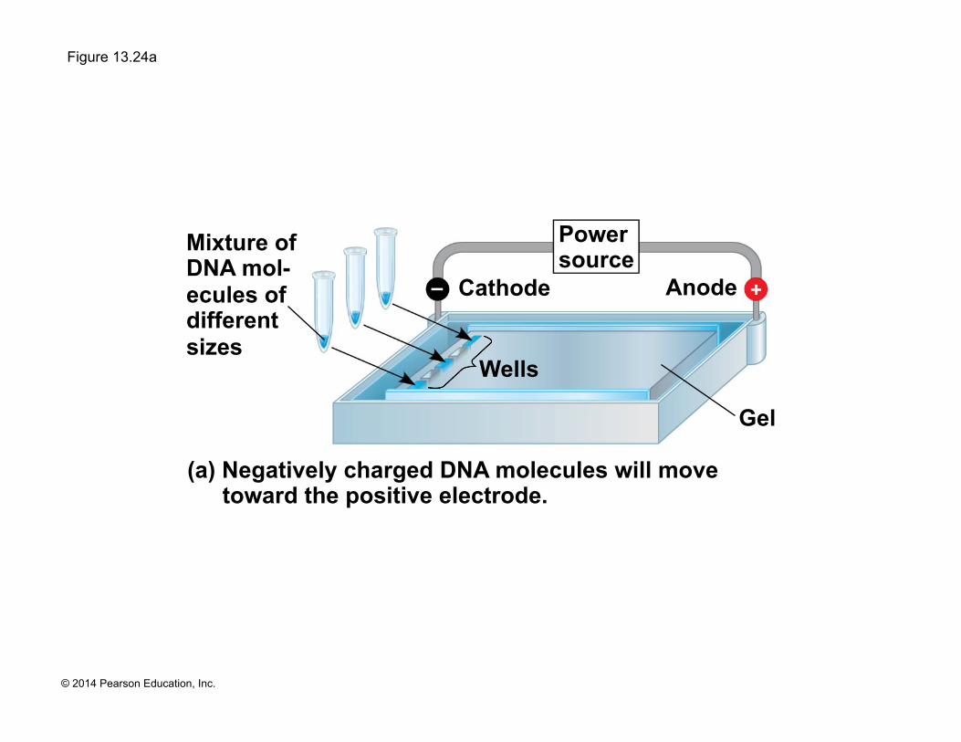
Figure 13.24 Mixture of DNA mol- ecules of different sizes
Cathode
Restriction fragments
Anode
Wells
Gel
Power source
(a) Negatively charged DNA molecules will move toward the positive electrode.
(b) Shorter molecules are impeded less than longer ones, so they move faster through the gel.
Page 117
© 2014 Pearson Education, Inc.
Figure 13.24a
Mixture of DNA mol- ecules of different sizes
Cathode Anode
Wells
Gel
Power source
(a) Negatively charged DNA molecules will move toward the positive electrode.
Page 118
© 2014 Pearson Education, Inc.
Figure 13.24b
Restriction fragments
(b) Shorter molecules are impeded less than longer ones, so they move faster through the gel.
Page 119
© 2014 Pearson Education, Inc.
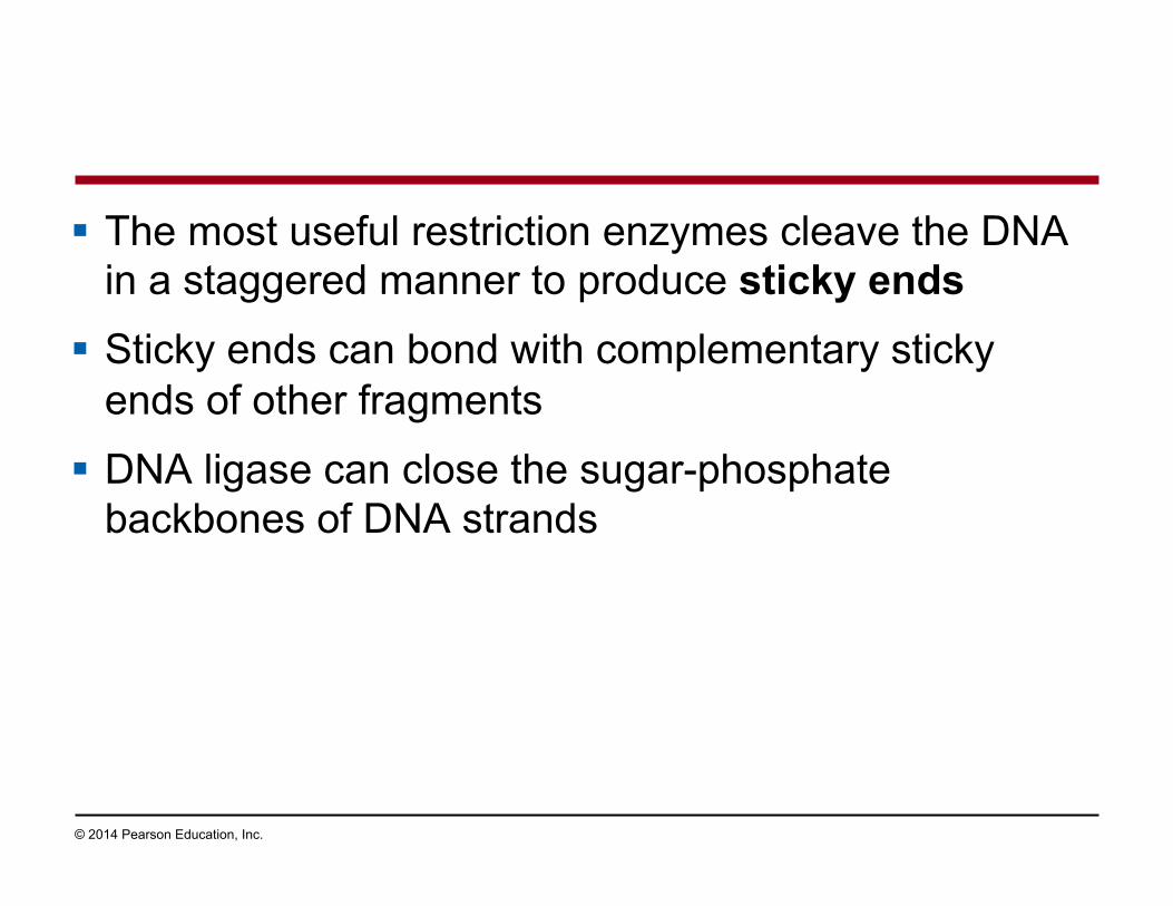
! The most useful restriction enzymes cleave the DNA in a staggered manner to produce sticky ends
! Sticky ends can bond with complementary sticky ends of other fragments
! DNA ligase can close the sugar-phosphate backbones of DNA strands
Page 120
© 2014 Pearson Education, Inc.
! In gene cloning, the original plasmid is called a cloning vector
! A cloning vector is a DNA molecule that can carry foreign DNA into a host cell and replicate there
Page 121
© 2014 Pearson Education, Inc.
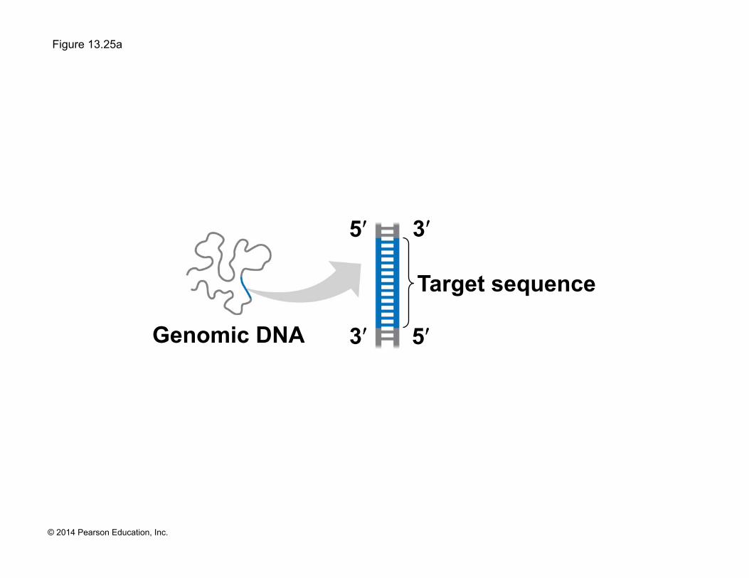
Amplifying DNA in Vitro: The Polymerase Chain Reaction (PCR) and Its Use in Cloning
! The polymerase chain reaction, PCR, can produce many copies of a specific target segment of DNA
! A three-step cycle brings about a chain reaction that produces an exponentially growing population of identical DNA molecules
! The key to PCR is an unusual, heat-stable DNA polymerase called Taq polymerase.
Page 122
© 2014 Pearson Education, Inc.
Figure 13.25
3ʹ
5ʹ
Cycle 1 yields 2 molecules
Genomic DNA
Denaturation
Target sequence
3ʹ
5ʹ
3ʹ
5ʹ
3ʹ
5ʹ
Primers
New nucleotides
Annealing
Extension
Cycle 2 yields 4 molecules
Cycle 3 yields 8 molecules;
2 molecules (in white boxes)
match target sequence
Technique
1
2
3
Page 123
© 2014 Pearson Education, Inc.
Figure 13.25a
3ʹ
5ʹ
Genomic DNA
Target sequence
3ʹ
5ʹ
Page 124
© 2014 Pearson Education, Inc.
Figure 13.25b-1
Cycle 1 yields 2
molecules
Denaturation
3ʹ
5ʹ 3ʹ
5ʹ
1
Page 125
© 2014 Pearson Education, Inc.
Figure 13.25b-2
Cycle 1 yields 2
molecules
Denaturation
3ʹ
5ʹ 3ʹ
5ʹ
Primers
Annealing
1
2
Page 126
© 2014 Pearson Education, Inc.
Figure 13.25b-3
Cycle 1 yields 2
molecules
Denaturation
3ʹ
5ʹ 3ʹ
5ʹ
Primers
New nucleotides
Annealing
Extension
1
2
3
Page 127
© 2014 Pearson Education, Inc.
Figure 13.25c
Cycle 2 yields 4 molecules
Cycle 3 yields 8 molecules;
2 molecules (in white boxes)
match target sequence
Results After 30 more cycles, over 1 billion (109) molecules match the target sequence.
Page 128
© 2014 Pearson Education, Inc.
! PCR amplification alone cannot substitute for gene cloning in cells
! Instead, PCR is used to provide the specific DNA fragment to be cloned
! PCR primers are synthesized to include a restriction site that matches the site in the cloning vector
! The fragment and vector are cut and ligated together
Page 129
© 2014 Pearson Education, Inc.
Figure 13.26
Cloning vector (bacterial plasmid)
DNA fragment obtained by PCR (cut by same restriction enzyme used on cloning vector)
Mix and ligate
Recombinant DNA plasmid
Page 130
© 2014 Pearson Education, Inc.
DNA Sequencing
! Once a gene is cloned, complementary base pairing can be exploited to determine the gene’s complete nucleotide sequence
! This process is called DNA sequencing
Page 131
© 2014 Pearson Education, Inc.
! “Next-generation” sequencing techniques, developed in the last ten years, are rapid and inexpensive
! They sequence by synthesizing the complementary strand of a single, immobilized template strand
! A chemical trick enables electronic monitors to identify which nucleotide is being added at each step.
Page 132
© 2014 Pearson Education, Inc.
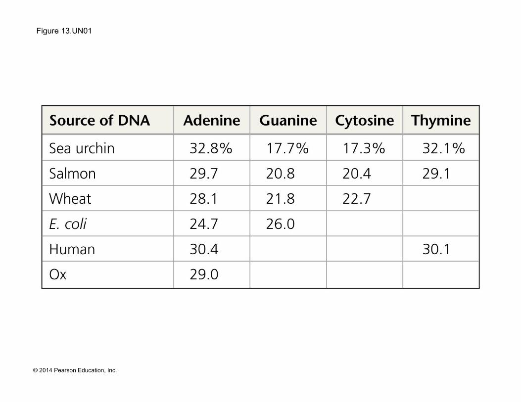
Figure 13.UN01
Page 133
© 2014 Pearson Education, Inc.
Figure 13.UN03
Sugar-phosphate backbone
Nitrogenous bases
Hydrogen bond
T A
C G
C
G
T
T
T A
A
A
C
G C
G
Page 134
© 2014 Pearson Education, Inc.
Figure 13.UN04
3ʹ 5ʹ
Origin of replication
3ʹ 5ʹ
Lagging strand synthesized in short Okazaki fragments, later joined by DNA ligase
DNA pol I replaces the RNA primer with DNA nucleotides
Primase synthesizes a short RNA primer
DNA pol III synthesizes leading strand continuously
DNA pol III starts DNA synthesis at 3ʹ end of primer continues in 5ʹ → 3ʹ direction 3ʹ
5ʹ
5ʹ
Parental DNA
Helicase
Page 135
© 2014 Pearson Education, Inc.
Figure 13.UN05
3ʹ
5ʹ Sticky end
G G C C A
T T A
A T T
A 3ʹ
5ʹ 3ʹ
5ʹ 3ʹ
5ʹ
Page 136
© 2014 Pearson Education, Inc.
Figure 13.UN06