122
Aperio ImageScope User’s Guide MAN-0001, Revision Q | 5 December 2017 For research use only. Not for use in diagnostic procedures.
Aperio ImageScopeUser’s Guide
MAN-0001, Revision Q | 5 December 2017
For research use only. Not for use in diagnostic procedures.
Aperio ImageScope User’s Guide, Revision Q © Leica Biosystems Imaging, Inc. 20172
Aperio ImageScope User’s GuideThis document applies to Aperio eSlide Manager Release 12.4 and later.
Copyright NoticeÌÌ Copyright © 2006 – 2017 Leica Biosystems Imaging, Inc. All Rights Reserved. LEICA and the Leica logo are registered trademarks of Leica
Microsystems IR GmbH. Aperio is a trademark of the Leica Biosystems group of companies in the USA and optionally in other countries. Other logos, product, and/or company names might be trademarks of their respective owners.
Customer ResourcesÌÌ For the latest information on Leica Biosystems Aperio ePathology products and services, please visit www.LeicaBiosystems.com/Aperio.
DisclaimersÌÌ Use normal care in maintaining and using Aperio ePathology servers. Interrupting network connections or turning off the servers while they are
processing data (such as when they are analyzing eSlides or generating an audit report) can result in data loss.
ÌÌ This manual is not a substitute for the detailed operator training provided by Leica Biosystems Imaging or for other advanced instruction. Leica Biosystems Imaging Field Representatives should be contacted immediately for assistance in the event of any instrument malfunction. Installation of hardware should only be performed by a certified Leica Biosystems Imaging Service Engineer.
ÌÌ ImageServer is intended for use with eSlides created by scanning glass slides with the scanner. Educators will use Aperio ePathology software to view and modify eSlides in Composite WebSlide (CWS) format.
PatentsÌÌ Aperio ePathology products are protected by U.S. Patents: 6,711,283; 6,917,696; 7,035,478; 7,116,440; 7,257,268; 7,428,324; 7,457,446; 7,463,761;
7,502,519; 7,518,652; 7,602,524; 7,646,496; 7,738,688 and licensed under one or more of the following U.S. Patents: 6,101,265; 6,272,235; 6,522,774; 6,775,402; 6,396,941; 6,674,881; 6,226,392; 6,404,906; 6,674,884; and 6,466,690.
Contact Information – Leica Biosystems Imaging, Inc.
Headquarters Customer Support General Information
Leica Biosystems Imaging, Inc. 1360 Park Center DriveVista, CA 92081USA
Tel: +1 (866) 478-4111 (toll free)Direct International Tel: +1 (760) 539-1100
US/Canada Tel: +1 (844) 534-2262 (toll free)Direct International Tel: +1 (760) 539-1150
US/Canada/Worldwide Email:[email protected]
US/Canada Tel: +1 (866) 478-4111 (toll free)Direct International Tel: +1 (760) 539-1100
Email: [email protected]
European Union Authorized Representative
CEpartner4UEsdoornlaan 133951 DB MaarnThe Netherlands

Aperio ImageScope User’s Guide, Revision Q © Leica Biosystems Imaging, Inc. 2017 3
Customer Service ContactsPlease contact the office for your country for technical assistance.
Australia:96 Ricketts RoadMount Waverly, VIC 3149AUSTRALIATel: 1800 625 286 (toll free) Between 8:30 AM-5 PM, Monday-Friday, AESTEmail: [email protected]
Austria:Leica Biosystems Nussloch GmbHTechnical Assistance CenterHeidelberger Strasse 17Nussloch 69226GERMANYTel: 0080052700527 (toll free)In-country Tel: +43 1 486 80 50 50Email: [email protected]
België/Belgique:Tel: 0080052700527 (toll free)In-country Tel: +32 2 790 98 50Email: [email protected]
Canada:Tel: +1 844 534 2262 (toll free)Direct International Tel: +1 760 539 1150Email: [email protected]
China:17F, SML Center No. 610 Xu Jia Hui Road, Huangpu DistrictShanghai, PRC PC:200025CHINATel: +86 4008208932Fax: +86 21 6384 1389Email: [email protected] Care email: [email protected]
Danmark:Tel: 0080052700527 (toll free)In-country Tel: +45 44 54 01 01Email: [email protected]
Deutschland:Leica Biosystems Nussloch GmbHTechnical Assistance CenterHeidelberger Strasse 17Nussloch 69226GERMANYTel: 0080052700527 (toll free)In-country Tel: +49 6441 29 4555Email: [email protected]
Eire:Tel: 0080052700527 (toll free)In-country Tel: +44 1908 577 650Email: [email protected]
España:Tel: 0080052700527 (toll free)In-country Tel: +34 902 119 094Email: [email protected]
France:Tel: 0080052700527 (toll free)In-country Tel: +33 811 000 664Email: [email protected]
Italia:Tel: 0080052700527 (toll free)In-country Tel: +39 0257 486 509Email: [email protected]
Japan:1-29-9 Takadannobaba, Sinjuku-kuTokyo 169-0075JAPAN
Nederland:Tel: 0080052700527 (toll free)In-country Tel: +31 70 413 21 00Email: [email protected]

Aperio ImageScope User’s Guide, Revision Q © Leica Biosystems Imaging, Inc. 20174
New Zealand:96 Ricketts RoadMount Waverly, VIC 3149AUSTRALIATel: 0800 400 589 (toll free) Between 8:30 AM-5 PM, Monday-Friday, AESTEmail: [email protected]
Portugal:Tel: 0080052700527 (toll free)In-country Tel: +35 1 21 388 9112Email: [email protected]
The Russian FederationBioLine LLCPinsky lane 3 letter ASaint Petersburg 197101THE RUSSIAN FEDERATIONTel: 8-800-555-49-40 (toll free)In-country Tel: +7 812 320 49 49Email: [email protected]
Sweden:Tel: 0080052700527 (toll free)In-country Tel: +46 8 625 45 45Email: [email protected]
Switzerland:Tel: 0080052700527 (toll free)In-country Tel: +41 71 726 3434Email: [email protected]
United Kingdom:Tel: 0080052700527 (toll free)In-country Tel: +44 1908 577 650Email: [email protected]
USA:Tel: +1 844 534 2262 (toll free)Direct International Tel: +1 760 539 1150Email: [email protected]
Aperio ImageScope User’s Guide, Revision Q © Leica Biosystems Imaging, Inc. 2017 5
1 Introduction .................................................................................................................... 10Intended Use .................................................................................................................................10Aperio ImageScope Features .............................................................................................................10Types of Files You Can View .............................................................................................................. 11Cybersecurity ................................................................................................................................ 11For More Information ......................................................................................................................12
2 Installing Aperio ImageScope ....................................................................................... 13Before You Start .............................................................................................................................13
Installation Requirements .............................................................................................................13Monitor and System Requirements .................................................................................................13Security Alerts ...........................................................................................................................13
Installation ...................................................................................................................................13Modifying or Removing the Aperio ImageScope Software ........................................................................14Starting Aperio ImageScope .............................................................................................................14
3 Opening an eSlide .......................................................................................................... 15About User Permissions ...................................................................................................................15Opening eSlide Images from Aperio eSlide Manager ..............................................................................16
Opening an Aperio eSlide Manager eSlide ........................................................................................16Opening an Aperio eSlide Manager eSlide from Aperio ImageScope .......................................................16
Opening an eSlide on Your Workstation or LAN .....................................................................................17Local Image Support ....................................................................................................................17Opening a Recently Viewed Local eSlide ..........................................................................................17
Opening and Viewing Multiple eSlides ................................................................................................18Managing eSlide Windows ...........................................................................................................19Keep Open Option .......................................................................................................................19
Viewing eSlide Information ...............................................................................................................20Status Bar .................................................................................................................................20
Saving and Opening an Image View ....................................................................................................21Closing eSlides ..............................................................................................................................21
4 Viewing an eSlide ........................................................................................................... 22
Contents
Aperio ImageScope User’s Guide, Revision Q © Leica Biosystems Imaging, Inc. 20176
Contents
Aperio ImageScope Viewing Window .................................................................................................22Aperio ImageScope Toolbar Quick Reference .....................................................................................23
Synchronizing Navigation of Multiple eSlides .......................................................................................25Smart Synchronization .................................................................................................................25
Moving Around the eSlide Image .......................................................................................................27Using the Magnifier Window ............................................................................................................28Changing Viewing Magnification ........................................................................................................28Viewing with Color Management .......................................................................................................29Viewing Scalebar, Axes, and Grid .......................................................................................................29Viewing Z-Stack eSlide Images ..........................................................................................................31
Viewing and Navigating a Z-Stack Image .........................................................................................31Viewing eSlides with IQ ...................................................................................................................32
IQ Features ................................................................................................................................32IQ Quick Reference ......................................................................................................................33
5 Rotating Images and Slide Labels ................................................................................ 34Rotating an Image ..........................................................................................................................34
Rotating an eSlide Label ...............................................................................................................34
6 Making Image Adjustments ........................................................................................... 35Making Image Adjustments ..............................................................................................................35
Saving and Loading Color Settings ..................................................................................................37For More Information ......................................................................................................................37
7 Working with Fluorescence eSlides ............................................................................. 38Applying a Temporary False Color .......................................................................................................38Adjusting Fluorescence Fused Images .................................................................................................39
Using the Fusion Adjustment Window .............................................................................................39Viewing All Channel Images ..........................................................................................................40
Seeing Channel Information .......................................................................................................41Cycling Channel Displays ..............................................................................................................41Adjusting Color ..........................................................................................................................42Adjusting Brightness, Contrast, and Gamma ......................................................................................42
Intensity Histogram .................................................................................................................43Adjusting Registration .................................................................................................................44
Fusing Fluorescence Images ..............................................................................................................45Notes on Creating an AFI ..............................................................................................................47
8 Image Resolution ........................................................................................................... 48Setting or Changing Image Resolution .................................................................................................48Computing the Resolution from the Image ............................................................................................49

Aperio ImageScope User’s Guide, Revision Q © Leica Biosystems Imaging, Inc. 2017 7
Contents
9 Working with Annotations ............................................................................................. 51Using the Annotation Tools ...............................................................................................................51
Measuring the Distance Between Two Line Annotations ......................................................................53Annotating Z-Stack Images ...........................................................................................................54Drawing Fixed Size Annotations .....................................................................................................54Drawing Annotations with a Fixed Aspect Ratio .................................................................................54Moving Annotations ....................................................................................................................55Editing Free-Form Annotations Created with the Pen or Negative Pen .....................................................55
Completing a Free-Form Shape ...................................................................................................56Editing a Free-Form Shape .........................................................................................................56Fixing a Problem Area ..............................................................................................................57
10 Using the Annotations Window .................................................................................... 58Annotations Summary View Window – Quick eIHC Analysis .....................................................................58
Slide-Specific Processing ..............................................................................................................58Using the Annotations Summary View Window .................................................................................59Enabling and Disabling Pre-Processing .............................................................................................61Incremental Processing ................................................................................................................61Other Options ............................................................................................................................61
The Annotations – Detailed View Window ...........................................................................................62Annotations Window Tools ........................................................................................................62
Annotation Length and Area Display ...............................................................................................63Adding Text to an Annotation ........................................................................................................63Moving Annotations ....................................................................................................................64Exporting and Importing Annotation Layers .......................................................................................64Using Attributes .........................................................................................................................66
Adding and Deleting Attributes ..................................................................................................66
11 Linking Annotations and eSlides ................................................................................. 67Working with the Link Manager .........................................................................................................67Creating a Link ...............................................................................................................................68Viewing Links ................................................................................................................................68Deleting Links ................................................................................................................................69
12 Tracking .......................................................................................................................... 70Turning on the Tracker .....................................................................................................................70Viewing a Track .............................................................................................................................71Playing a Track ...............................................................................................................................72Appending to a Track .......................................................................................................................72
13 Saving eSlides and Regions ......................................................................................... 73

Aperio ImageScope User’s Guide, Revision Q © Leica Biosystems Imaging, Inc. 20178
Contents
Copy and Paste an eSlide Image ........................................................................................................73Taking a Snapshot ..........................................................................................................................73Emailing a Snapshot .......................................................................................................................74
Receiving a Snapshot Email .......................................................................................................74Exporting Images ............................................................................................................................75Extracting a Region .........................................................................................................................76
Note on Fluorescent Images ..........................................................................................................78Extracting a Region of Interest from a Multi-Channel FL SCN eSlide .......................................................78Saving an Image of a Specific Size ..................................................................................................79
Extracting an Image of a Predefined Size or Aspect Ratio ..................................................................79Managing Viewing Applications .....................................................................................................80
Compatibility Notes .................................................................................................................80Defining a Viewing Application ...................................................................................................80Using the Viewing Application ...................................................................................................80Deleting Viewing Applications ...................................................................................................81
14 eSlide Conferencing ...................................................................................................... 82About eSlide Conferencing ...............................................................................................................82
Concepts ..................................................................................................................................82Starting an eSlide Conference ...........................................................................................................83
Connecting to an eSlide Conferencing Server ....................................................................................83Opening an Image to Share ...........................................................................................................84Joining a Conference ...................................................................................................................85
Viewing Slides in Conference ............................................................................................................86Changing the Conference Leader ........................................................................................................86
Leader Initiates Change in Leadership .............................................................................................86Follower Initiates Change in Leadership ...........................................................................................87Conference Creator Re-asserts Leadership .......................................................................................87
15 TelePath Live .................................................................................................................. 88Scanner Compatibility Notes .............................................................................................................88Before You Use TelePath Live ............................................................................................................88
Calibration ................................................................................................................................88Setting the ImageServerURL ..........................................................................................................88
What Is a Z-Stack? .........................................................................................................................89Connecting to an Aperio Scanner .......................................................................................................90Preparing a Slide for TelePath Live .....................................................................................................92Viewing Live Video from the Scanner ..................................................................................................92Capturing Z-stacks ..........................................................................................................................93Viewing Z-stacks ............................................................................................................................96

Aperio ImageScope User’s Guide, Revision Q © Leica Biosystems Imaging, Inc. 2017 9
Contents
16 Utilities and Diagnostics ............................................................................................... 97Logging ........................................................................................................................................97Cache Display ................................................................................................................................97Running Multiple Aperio ImageScope Sessions .....................................................................................98Tuning Parameters/Statistics ............................................................................................................99
Maximum Cache Size ..................................................................................................................99Statistics ..................................................................................................................................99
17 ImageScope Options ................................................................................................... 101General Options ...........................................................................................................................101
Magnification ..........................................................................................................................101Default Gamma Files .................................................................................................................102
Loading a Default Gamma Table File for the Main Image ................................................................102Loading a Default Gamma Table File for Z-stack Images .................................................................102
Analysis .....................................................................................................................................103Navigation Options .......................................................................................................................103
Synchronization Option ..............................................................................................................103Panning Options .......................................................................................................................104
Annotation Options .......................................................................................................................104Annotation Color Options ............................................................................................................104Fixed Size Annotations ...............................................................................................................104Automatically Saving Annotations ................................................................................................105
Tracking Options ..........................................................................................................................105Performance Options .....................................................................................................................106HTTP Proxy Option ........................................................................................................................107Report Image Options ....................................................................................................................107Color Management Options ............................................................................................................108Email Settings .............................................................................................................................109For More Information ....................................................................................................................109
A Keyboard Shortcuts ......................................................................................................110Aperio ImageScope Keyboard Shortcuts ............................................................................................ 110
B Aperio Integrated Color Management .........................................................................113ICC Profiles ................................................................................................................................. 113
Scanner ICC Profile .................................................................................................................... 113Display Monitor ICC Profile ......................................................................................................... 113
How Aperio ImageScope Uses Color Management ............................................................................... 114
Index ....................................................................................................................................115
Symbols.............................................................................................................................. 121

Aperio ImageScope User’s Guide, Revision Q © Leica Biosystems Imaging, Inc. 201710
1 Introduction
This chapter introduces you to Aperio ImageScope and indicates where to find information on specific features.
Intended UseFor research use only. Not for use in diagnostic procedures.
Aperio ImageScope FeaturesAperio ImageScope enables you to:
Ì` View eSlides from any workstation on the network, eliminating the delay of physically transporting glass slides.
Ì` Share and discuss eSlides in real-time in multiple remote locations by using eSlide conferencing.
Ì` View multiple eSlides concurrently.
Ì` Apply image adjustments for contrast, brightness, and gamma.
Ì` Analyze entire eSlides or selected regions using algorithms.
Ì` View, annotate, and analyze scanned z-stack images.
Ì` Interface directly to a Aperio scanner through a network connection to view slides “live” and in different focal planes.
Ì` Rotate eSlide images and labels.
Ì` Use Aperio Integrated Color Management to view eSlides to ensure the eSlides are displayed in accurate color.
Ì` Use the Image Quality (IQ) feature to optimize viewing of an eSlide based on its stain.
Ì` Add and manage various types of eSlide image annotations.
Ì` Interface to the Aperio ePathology ImageServer and Aperio eSlide Manager.
Ì` Instantly pan and zoom to any region of the slide.
Ì` Extract a region or selected regions of an eSlide to a file in a choice of formats.

Aperio ImageScope User’s Guide, Revision Q © Leica Biosystems Imaging, Inc. 2017 11
Chapter 1: Introduction
Types of Files You Can ViewYou can use Aperio ImageScope to view:
Ì` ScanScope Virtual Slides – .SVS files are created when the Aperio scanner scans glass microscope slides.
Ì` JPEG files – Both .JPG and .JP2 files.
Ì` TIFF and TIF files.
Ì` Aperio fluorescent images (Aperio Fused Image, .afi)
Ì` CWS files – Composite WebSlides1.
Ì` Hamamatsu NanoZoomer files – NDPI, NDPIS, and .VMS files.
Ì` 3D Histech MRXS / MRXS FL files – Note that MRXS and MRXS FL images are composite images that consist of a group of .DAT files.
Ì` ScanScope image set, .sis file – The Aperio ImageScope image view is what you see when one or more eSlides are viewed in the Aperio ImageScope window. Aperio ImageScope enables you to save the image view as a ScanScope image set so that you can open all the slides at once in the future.
Ì` SCN files – Leica Biosystems SCN brightfield and fluorescent images. Aperio ImageScope supports multiple region SCN image files.
Ì` Vectra QPTIFF – .QPTIFF files are created when the Vectra scanner scans glass microscope slides. They can be Brightfield or Fluorescence images.
CAUTION: The third-party image types listed above are supported for viewing only. Aperio Image Analysis algorithms are not supported for use with third-party fluorescence and brightfield image types. For details about Aperio Image Analysis, see the Aperio Image Analysis User’s Guide.
CybersecurityBe aware that workstations are susceptible to malware, viruses, data corruption, and privacy breaches. Work with your IT administrators to protect your workstation by following your institution’s password and security policies. For Aperio recommendations on protecting your workstations and servers, see the document Aperio Cybersecurity and Network Recommendations.
1 A Composite WebSlide, also known as a CWS slide, is a proprietary format created by Bacus Laboratories, Inc (“Bacus”). WebSlide® is a registered trademark of Bacus Laboratories Inc.
Aperio ImageScope User’s Guide, Revision Q © Leica Biosystems Imaging, Inc. 201712
Chapter 1: Introduction
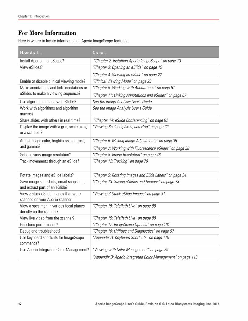
For More InformationHere is where to locate information on Aperio ImageScope features.
How do I... Go to...
Install Aperio ImageScope? “Chapter 2: Installing Aperio ImageScope” on page 13View eSlides? “Chapter 3: Opening an eSlide” on page 15
“Chapter 4: Viewing an eSlide” on page 22Enable or disable clinical viewing mode? “Clinical Viewing Mode” on page 23 Make annotations and link annotations or eSlides to make a viewing sequence?
“Chapter 9: Working with Annotations” on page 51
“Chapter 11: Linking Annotations and eSlides” on page 67Use algorithms to analyze eSlides? See the Image Analysis User’s GuideWork with algorithms and algorithm macros?
See the Image Analysis User’s Guide
Share slides with others in real time? “Chapter 14: eSlide Conferencing” on page 82Display the image with a grid, scale axes, or a scalebar?
“Viewing Scalebar, Axes, and Grid” on page 29
Adjust image color, brightness, contrast, and gamma?
“Chapter 6: Making Image Adjustments” on page 35
“Chapter 7: Working with Fluorescence eSlides” on page 38 Set and view image resolution? “Chapter 8: Image Resolution” on page 48 Track movements through an eSlide? “Chapter 12: Tracking” on page 70
Rotate images and eSlide labels? “Chapter 5: Rotating Images and Slide Labels” on page 34 Save image snapshots, email snapshots, and extract part of an eSlide?
“Chapter 13: Saving eSlides and Regions” on page 73
View z-stack eSlide images that were scanned on your Aperio scanner
“Viewing Z-Stack eSlide Images” on page 31
View a specimen in various focal planes directly on the scanner?
“Chapter 15: TelePath Live” on page 88
View live video from the scanner? “Chapter 15: TelePath Live” on page 88Fine-tune performance? “Chapter 17: ImageScope Options” on page 101Debug and troubleshoot? “Chapter 16: Utilities and Diagnostics” on page 97Use keyboard shortcuts for ImageScope commands?
“Appendix A: Keyboard Shortcuts” on page 110
Use Aperio Integrated Color Management? ”Viewing with Color Management” on page 29
“Appendix B: Aperio Integrated Color Management” on page 113

Aperio ImageScope User’s Guide, Revision Q © Leica Biosystems Imaging, Inc. 2017 13
2 Installing Aperio ImageScope
This chapter contains information on installing the client software for the Aperio ImageScope eSlide viewer.
Before You StartReview the information in this section prior to installing Aperio ImageScope.
Installation Requirements
To successfully install Aperio ImageScope, you must first log into Windows as a user with administrative privileges.
Monitor and System Requirements
Because eSlides are, by design, high resolution and information rich, for best results you should use a high-quality LCD monitor to view them. Make sure the monitor is at the proper viewing height and in a room with appropriate lighting.
Before installing Aperio ImageScope, make sure your workstation and monitor meet the minimum requirements discussed in the Aperio ePathology System Requirements.
Security Alerts
If during installation you see messages from Microsoft or third-party firewall, VPN, or virus software telling you that the installation has been blocked, you should consult your network administrator for help resolving these issues before continuing.
InstallationTo install Aperio ImageScope, follow these steps:
1. Ensure you are logged into Windows as a user with administrative privileges.
2. Double-click My Computer or open Windows Explorer and navigate to the Aperio ImageScope installer file. (This file may have been downloaded from the www.LeicaBiosystems.com/ePathology web site, may have been provided on CD, or may reside on your network; contact your network administrator for help if you have trouble finding it.)
If you are installing Aperio ImageScope on your DSR (Digital Slide Repository), use DSRInstall; if installing Aperio ImageScope on a user’s workstation, use ClientInstall.
3. Double-click the .exe file to start the installation wizard.
4. Follow the instructions on your screen to accept the terms of the license agreement and install Aperio ImageScope.

Aperio ImageScope User’s Guide, Revision Q © Leica Biosystems Imaging, Inc. 201714
Chapter 2: Installing Aperio ImageScope
Modifying or Removing the Aperio ImageScope SoftwareAt any time after Aperio ImageScope is installed, you can run the installer again to modify, repair, or remove the Aperio ImageScope software. If Aperio ImageScope is already installed, select from the following options on the installer window:
Ì` Modify to change the Aperio ImageScope installation by adding or deleting components.
Ì` Repair to reinstall all the components previously installed. This is the option to use if you are upgrading a previous installation to new software.
Ì` Remove to uninstall the Aperio ImageScope software.
Starting Aperio ImageScopeTo start Aperio ImageScope, click Start on the Windows taskbar, point to All Programs > ScanScope, and select ImageScope.
Aperio ImageScope User’s Guide, Revision Q © Leica Biosystems Imaging, Inc. 2017 15
3 Opening an eSlide
This chapter contains information on opening and viewing eSlides in Aperio ImageScope.
Connection speeds may affect Aperio ImageScope performance when viewing remote images. For best viewing, we recommend a connection speed of 100 mbps or greater.
Use Aperio ImageScope to view:
Ì` Local eSlides – images that reside on your workstation or your local network and are accessible using Microsoft file sharing (for example, by using Windows Explorer). Some features are not available when viewing local eSlides. See “Local Image Support” on page 17 for further details.
Ì` Remote eSlides – images that you open directly on an Aperio ePathology ImageServer or that you open using Aperio eSlide Manager.
Because Aperio ePathology eSlides are by design high resolution and information rich, for best results you should use a high quality monitor to view them. For details about monitor requirements, see the Aperio ePathology System Requirements.
About User PermissionsAperio ImageScope makes use of Aperio eSlide Manager security to enforce user permissions when viewing images.
The Aperio eSlide Manager administrator uses data groups and user roles to define what data you can see and what you can do when you see it. Data groups organize data such as eSlides into different groups that can be seen and used by different users. User roles define the commands you can use and the elements of an Aperio eSlide Manager page you can see.
What this means for Aperio ImageScope users is that when you open a remote image Aperio ImageScope may request that you log in so Aperio eSlide Manager can determine if you have the correct permissions to view the images you want to access. Type the same user name and password you use to log into Aperio eSlide Manager.
This also means that you may be restricted in what you can do with an eSlide. If, for example, you have read-only access to the data group that contains the eSlide you are viewing, you can use the Aperio ImageScope drawing tools to draw annotations but you won’t be able to save them. If you have questions about your user permissions, contact your Aperio eSlide Manager administrator for assistance.
Some of the features of the Aperio eSlide Manager security system you should know about are:
Ì` To keep user information secure, user credentials are encrypted and are never passed in clear text between the components of the Aperio eSlide Manager system.

Aperio ImageScope User’s Guide, Revision Q © Leica Biosystems Imaging, Inc. 201716
Chapter 3: Opening an eSlide
Ì` User credentials can time out. If enough time elapses after you log in, you may be asked to log in again.
Ì` Data groups and user permissions are defined in Aperio eSlide Manager by the administrator.
Depending on how Aperio eSlide Manager is configured, you may be able to log in as Guest to see public images that do not require user authentication.
Opening eSlide Images from Aperio eSlide ManagerYou can open a remote eSlide from Aperio eSlide Manager or you can open it from Aperio ImageScope. (For more details about using Aperio eSlide Manager, see the Aperio eSlide Manager Operator’s Guide.)
Opening an Aperio eSlide Manager eSlide
Aperio eSlide Manager has a Default Image Viewer setting. If Aperio ImageScope is set as the Default Image Viewer, then you do not need to press the “I” key in the instructions below.
To open an eSlide image from Aperio eSlide Manager, follow these steps:
1. In Aperio eSlide Manager, use the List commands or search feature to find the eSlide you want to view.
2. Press the I key and click the thumbnail image of the eSlide.
The eSlide opens in Aperio ImageScope.
Opening an Aperio eSlide Manager eSlide from Aperio ImageScope
To open an eSlide image from Aperio ImageScope, follow these steps:
1. Go to the Aperio ImageScope File menu and select Access Remote Server to connect to Aperio eSlide Manager.
2. Enter the name of the server where Aperio eSlide Manager resides, and set the Port value to 82.
3. Click Connect.
4. When prompted, enter your Aperio eSlide Manager user name and password.

Aperio ImageScope User’s Guide, Revision Q © Leica Biosystems Imaging, Inc. 2017 17
Chapter 3: Opening an eSlide
5. When the list of eSlide appears, select either the List or Thumbnail view from the drop-down list at the upper right.
6. Click the ImageScope link below the image.
Opening an eSlide on Your Workstation or LANTo open an eSlide that resides on your workstation or local area network:
1. Start Aperio ImageScope by clicking Start, pointing to All Programs > ScanScope, and then selecting ImageScope.
2. Go to the File menu and select Open Image (or click on the Aperio ImageScope toolbar).
3. On the Open Image window, navigate to the location that contains the image you want to view.
4. Click the name of the eSlide you want to open and click Open.
You may need to change the file type in the Open Image window to see the type of image you want to view. For example, to view a CWS image, click the file type drop-down list and select Composite WebSlides (*/SlideScan.ini).
Local Image Support
If you open a local image instead of an image in Aperio eSlide Manager, Smart sync, Tracking, and IQ are not supported for that image.
Opening a Recently Viewed Local eSlide
Aperio ImageScope displays a list of the last few eSlides that were viewed on the File menu. To open one of these images, go to the File menu and click one of the eSlides listed at the bottom of the menu.

Aperio ImageScope User’s Guide, Revision Q © Leica Biosystems Imaging, Inc. 201718
Chapter 3: Opening an eSlide
Opening and Viewing Multiple eSlidesYou can open multiple eSlides within Aperio ImageScope. To open multiple eSlides from Aperio eSlide Manager:
1. In Aperio eSlide Manager, use the List commands or search feature to find the eSlide you want to view.
2. Select the check boxes next to the eSlides you want to view.
3. Press the I key and click View Images in Aperio ImageScope:
You can view all the slides at once or view them separately by selecting Tile Vertical, Tile Horizontal, or
Aperio ImageScope User’s Guide, Revision Q © Leica Biosystems Imaging, Inc. 2017 19
Chapter 3: Opening an eSlide
Cascade from the Window menu.
You can move between the opened images by clicking on an image in the filmstrip, which appears in the left pane of the Aperio ImageScope window. If the Aperio ImageScope filmstrip is not visible, go to the View menu and select Filmstrip.
See “Chapter 4: Viewing an eSlide” on page 22 for more information on viewing images and using the Aperio ImageScope viewing tools.
Managing eSlide Windows
To maximize, minimize/restore, or close the individual eSlide windows within the Aperio ImageScope main window, click the slide icon on the image menu bar and select an action to perform.
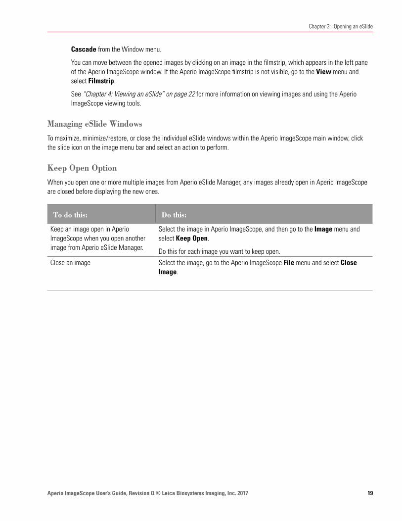
Keep Open Option
When you open one or more multiple images from Aperio eSlide Manager, any images already open in Aperio ImageScope are closed before displaying the new ones.
To do this: Do this:
Keep an image open in Aperio ImageScope when you open another image from Aperio eSlide Manager.
Select the image in Aperio ImageScope, and then go to the Image menu and select Keep Open.
Do this for each image you want to keep open.
Close an image Select the image, go to the Aperio ImageScope File menu and select Close Image.

Aperio ImageScope User’s Guide, Revision Q © Leica Biosystems Imaging, Inc. 201720
Chapter 3: Opening an eSlide
Viewing eSlide InformationTo view information, such as the image size, location, and compression ratio, about the active eSlide, go to the Image menu and select Information or click on the toolbar. The following information appears in the Image Information window.
Go to this tab: To view this information:
Information Provides detailed data regarding the eSlide, including the magnification, image ID, and description. The ICC profile is provided if one is being used. (See “Appendix B: Aperio Integrated Color Management” on page 113 for information on ICC profiles and color management.)
If this eSlide was scanned on an Aperio scanner, the time zone of the scan location and time of the scan appear. The Information tab is always shown. The other tabs only appear if those elements are associated with the eSlide. For example, if there is no label image for this eSlide, you do not see the Label Image tab.
For a z-stack eSlide image, information appears for each layer. The Depth is the layer separation value that is set during scanning, which is measured in microns (µm).
For an Aperio Fused Image (AFI), the Information window contains information on the separate channel images that comprise the AFI image.
Thumbnail A thumbnail image of this eSlide (the area of the glass slide that was scanned).
Label image The eSlide label.
Macro image A macro image of the entire slide.
Status Bar
Information about the active eSlide appears in the status bar at the bottom of the Aperio ImageScope window.
The sample status bar above shows the following information:
Ì` 73091 x 62821 = 12.8GB, file = 575MB – The entire eSlide is 73,091 by 62,821 pixels in size. The eSlide’s raw data is 12.8 gigabytes in size and the compressed size of the eSlide file is 575 megabytes.
Ì` 0, -12950 : 73091 x 62821 – The first two numbers indicate the pixel position of the top, left corner of the display. The second numbers indicate which part of the image is being viewed.
Ì` 1815, 37033 – The current pixel position of your cursor.
Ì` Prefetching/progressive rendering – Indicates which performance options are in effect. For information on performance options, see “Performance Options” on page 106.
Ì` PAN – The current navigation or annotation tool is selected. In this case, panning is selected.
You can turn the status bar off and on by going to the View menu and selecting Status Bar.
Aperio ImageScope User’s Guide, Revision Q © Leica Biosystems Imaging, Inc. 2017 21
Chapter 3: Opening an eSlide
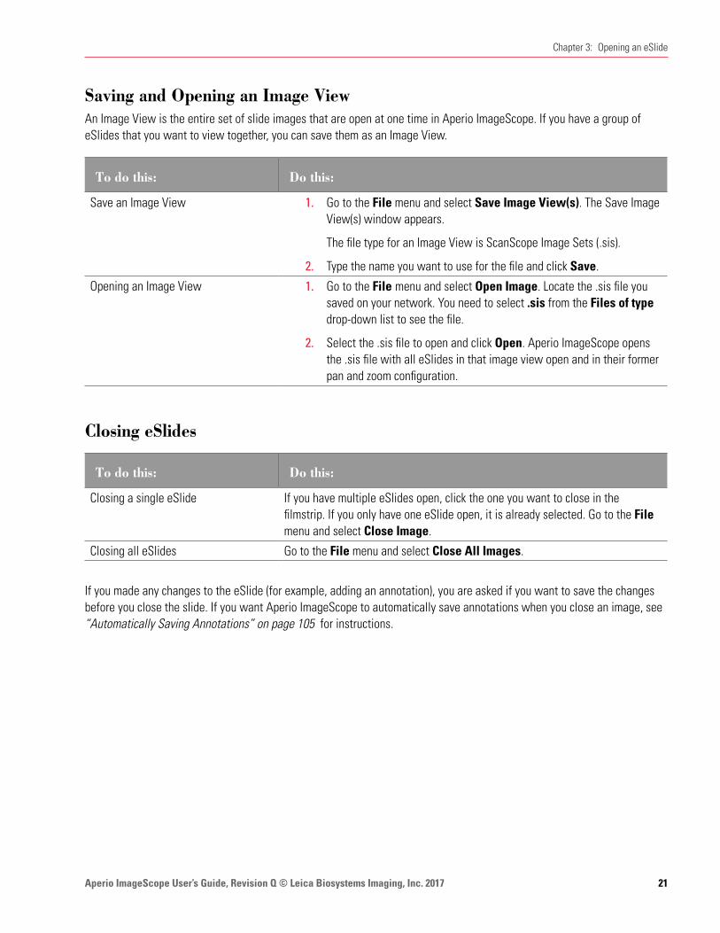
Saving and Opening an Image ViewAn Image View is the entire set of slide images that are open at one time in Aperio ImageScope. If you have a group of eSlides that you want to view together, you can save them as an Image View.
To do this: Do this:
Save an Image View 1. Go to the File menu and select Save Image View(s). The Save Image View(s) window appears.
The file type for an Image View is ScanScope Image Sets (.sis).
2. Type the name you want to use for the file and click Save.
Opening an Image View 1. Go to the File menu and select Open Image. Locate the .sis file you saved on your network. You need to select .sis from the Files of type drop-down list to see the file.
2. Select the .sis file to open and click Open. Aperio ImageScope opens the .sis file with all eSlides in that image view open and in their former pan and zoom configuration.
Closing eSlides
To do this: Do this:
Closing a single eSlide If you have multiple eSlides open, click the one you want to close in the filmstrip. If you only have one eSlide open, it is already selected. Go to the File menu and select Close Image.
Closing all eSlides Go to the File menu and select Close All Images.
If you made any changes to the eSlide (for example, adding an annotation), you are asked if you want to save the changes before you close the slide. If you want Aperio ImageScope to automatically save annotations when you close an image, see “Automatically Saving Annotations” on page 105 for instructions.

Aperio ImageScope User’s Guide, Revision Q © Leica Biosystems Imaging, Inc. 201722
This chapter provides a tour of the Aperio ImageScope main window and describes how to use the navigation and magnification tools.
Aperio ImageScope Viewing Window
Toolbar
eSlide label
Zoom slider
Filmstrip Thumbnail
ImageScope main window pane
MagnifierStatus bar
4 Viewing an eSlide

Aperio ImageScope User’s Guide, Revision Q © Leica Biosystems Imaging, Inc. 2017 23
Chapter 4: Viewing an eSlide
The main elements of the viewing window include:
Ì` Toolbar – You can perform many tasks from the toolbar. See the next section for a quick reference list of the Aperio ImageScope toolbar icons.
Ì` Zoom slider – You can magnify or shrink the current view. See “Zoom Slider” on page 28 for details.
Ì` Focus slider (not shown) – Appears with z-stack eSlide images only. Used to view different focal areas (z-stack layers) on a z-stack image. See “Viewing Z-Stack eSlide Images” on page 31 for details.
Ì` Filmstrip – Open eSlides appear in the filmstrip. Click a slide in the filmstrip to view it in the main window.
Ì` Label window – If the eSlide has an image of the slide label, it appears in the slide label window.
Ì` Thumbnail window – The thumbnail is a navigation tool that shows the complete eSlide. See “The Thumbnail Window” on page 27.
Ì` Magnifier window – Enables you to magnify a portion of the eSlide. See “Using the Magnifier Window” on page 28.
You can hide or show these tools from the View menu.
Clinical Viewing ModeClinical Viewing mode provides a simple toolbar that contains only the tools used in a clinical environment.
To do this: Do this:
Use the clinical toolbar Go to the View menu and select View Clinical Toolbar.
To provide quick and easy eSlide analysis, only the Summary View of the Annotations window is available when using clinical viewing mode.
Return to the full toolbar Go to the View menu and select View Standard Toolbar.
Aperio ImageScope Toolbar Quick Reference
Here is a quick list of the toolbar buttons.
*These icons are shown in clinical viewing mode.
Tool ActionGo to the Open Image window where you can browse for a local eSlide to open for viewing.
Close the eSlide that is currently open.
Export images of a specified area on the eSlide. You can export the raw eSlide image, the eSlide with annotations, and the mark-up image. See “Exporting Images” on page 75.
Aperio ImageScope User’s Guide, Revision Q © Leica Biosystems Imaging, Inc. 201724
Chapter 4: Viewing an eSlide
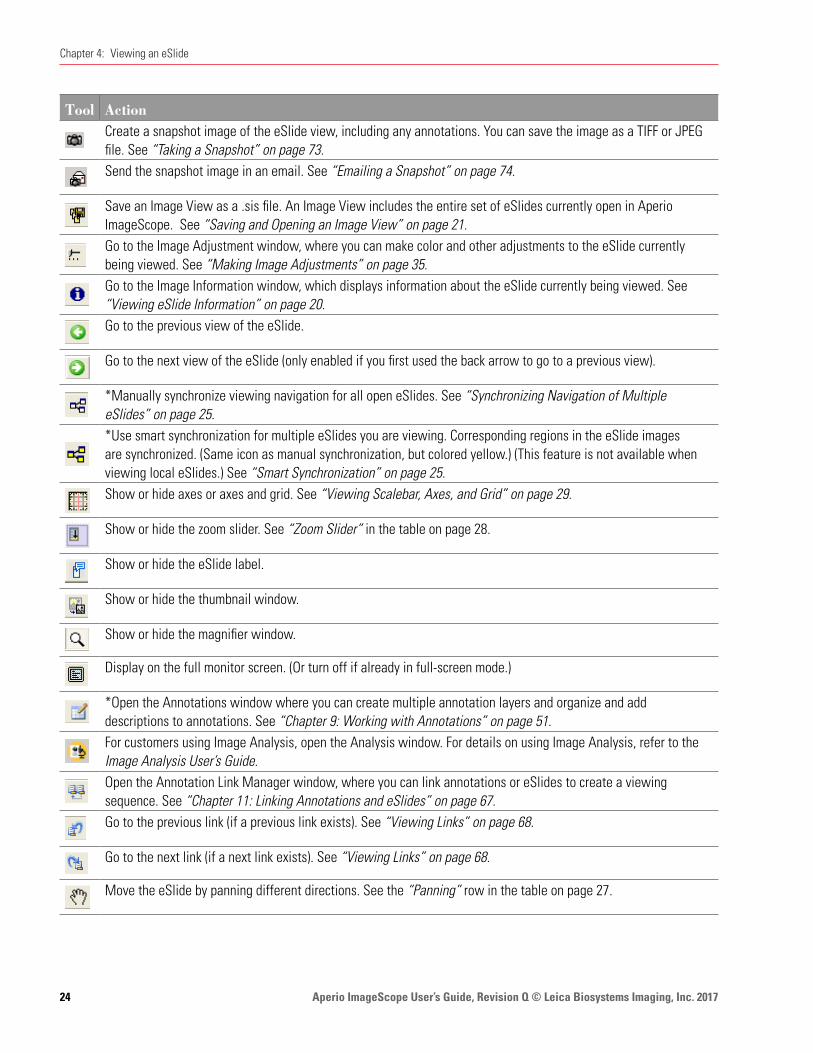
Tool ActionCreate a snapshot image of the eSlide view, including any annotations. You can save the image as a TIFF or JPEG file. See “Taking a Snapshot” on page 73.Send the snapshot image in an email. See “Emailing a Snapshot” on page 74.
Save an Image View as a .sis file. An Image View includes the entire set of eSlides currently open in Aperio ImageScope. See “Saving and Opening an Image View” on page 21.Go to the Image Adjustment window, where you can make color and other adjustments to the eSlide currently being viewed. See “Making Image Adjustments” on page 35.Go to the Image Information window, which displays information about the eSlide currently being viewed. See “Viewing eSlide Information” on page 20.Go to the previous view of the eSlide.
Go to the next view of the eSlide (only enabled if you first used the back arrow to go to a previous view).
*Manually synchronize viewing navigation for all open eSlides. See “Synchronizing Navigation of Multiple eSlides” on page 25.*Use smart synchronization for multiple eSlides you are viewing. Corresponding regions in the eSlide images are synchronized. (Same icon as manual synchronization, but colored yellow.) (This feature is not available when viewing local eSlides.) See “Smart Synchronization” on page 25.Show or hide axes or axes and grid. See “Viewing Scalebar, Axes, and Grid” on page 29.
Show or hide the zoom slider. See “Zoom Slider” in the table on page 28.
Show or hide the eSlide label.
Show or hide the thumbnail window.
Show or hide the magnifier window.
Display on the full monitor screen. (Or turn off if already in full-screen mode.)
*Open the Annotations window where you can create multiple annotation layers and organize and add descriptions to annotations. See “Chapter 9: Working with Annotations” on page 51.For customers using Image Analysis, open the Analysis window. For details on using Image Analysis, refer to the Image Analysis User’s Guide.Open the Annotation Link Manager window, where you can link annotations or eSlides to create a viewing sequence. See “Chapter 11: Linking Annotations and eSlides” on page 67.
Go to the previous link (if a previous link exists). See “Viewing Links” on page 68.
Go to the next link (if a next link exists). See “Viewing Links” on page 68.
Move the eSlide by panning different directions. See the “Panning” row in the table on page 27.

Aperio ImageScope User’s Guide, Revision Q © Leica Biosystems Imaging, Inc. 2017 25
Chapter 4: Viewing an eSlide
Tool ActionZoom the selected area of the eSlide. See the “Zoom Navigation” row on page 29.
*Extract a region of an eSlide. See “Exporting Images” on page 75.
*Draw a free-form annotation. See the “Pen” row in the table on page 51.
Draw a free-form annotation to be excluded from analysis. (This creates a negative annotation.) See the “Negative pen” row on page 52.*Measure an object on an eSlide. See the “Ruler” row on page 52.
*Draw a rectangular region (or a square if you hold down the Shift key while you draw).
Draw an elliptical annotation (or a circle if you hold down the Shift key while you draw).
*Draw an arrow pointing to an area of interest.
Mark the eSlide image with numeric counters. See the “Counter” row in the table on page 52.
*Select an image for a report. This feature is only useful if you have Aperio eSlide Manager Reporting option installed and the report template you are using uses images.Measure the distance (µm) between two free-form line annotations. See “Measuring the Distance Between Two Line Annotations” on page 53.Copy the selected annotation.
You can paste the annotation in any open eSlide. If you have run analysis on the annotation, only the annotation is copied (not the analysis results). Paste the copied annotation region in the active eSlide.
Turn Integrated Color Management on or off. Only useful if the image contains an embedded ICC profile. See “Viewing with Color Management” on page 29.*Turn Image Quality (IQ) mode on or off. See “Viewing eSlides with IQ” on page 32. (This feature is not available when viewing local eSlides.)*See help information for Aperio ImageScope.
Synchronizing Navigation of Multiple eSlidesIf you want all open eSlides to respond to the same navigation (for example, panning to the right) when you are viewing them side by side, go to the Aperio ImageScope toolbar and click .
Smart Synchronization
Smart synchronization is only available for remote images opened from Aperio eSlide Manager.
Smart synchronization is an extension of the manual synchronization feature discussed above. In addition to synchronizing navigation between the slides, corresponding regions in the eSlide images are also synchronized.

Aperio ImageScope User’s Guide, Revision Q © Leica Biosystems Imaging, Inc. 201726
Chapter 4: Viewing an eSlide
Click the icon on the Aperio ImageScope toolbar to use smart synchronization.
Smart synchronization compensates for rotation (non-flipped) but not for other factors such as stretched or missing tissue. In those cases, Aperio ImageScope tries to display the same tissue feature in all tiled images, but not necessarily in exactly the same location.
This feature is useful when the original microscope slides were prepared from the same tissue block but were stained differently, as shown in the example below. Using smart synchronization, the main features of the slide stay locked in step as you move around the slides.
You cannot use smart synchronization on images that were flipped vertically or horizontally. Also, you cannot flip an image while smart synchronization is turned on.
Aperio ImageScope User’s Guide, Revision Q © Leica Biosystems Imaging, Inc. 2017 27
Chapter 4: Viewing an eSlide
Moving Around the eSlide ImageThe table below describes different ways to move around an eSlide.
This feature: Works like this:
PanningWith the Panning tool selected , hold down the mouse button and drag the cursor across the eSlide.
Panning moves the slide the direction you are dragging. If you want to pan in reverse (“pathologist mode”), see“Panning Options” on page 104.
Autopanning Autopanning enables you to move at high speed over an eSlide.
With the cursor at the center of the main viewing area, click the scroll wheel on your mouse or right-click and select Autopan from the menu.
When the autopan icon appears , the eSlide starts moving toward your mouse pointer. The greater the distance between your mouse pointer and the
icon, the faster the scroll.
Scrolling Move your pointer toward any edge of the viewing window. When the pointer changes to an arrow: , click and hold the mouse button to scroll in that direction. To stop scrolling, release the mouse button.
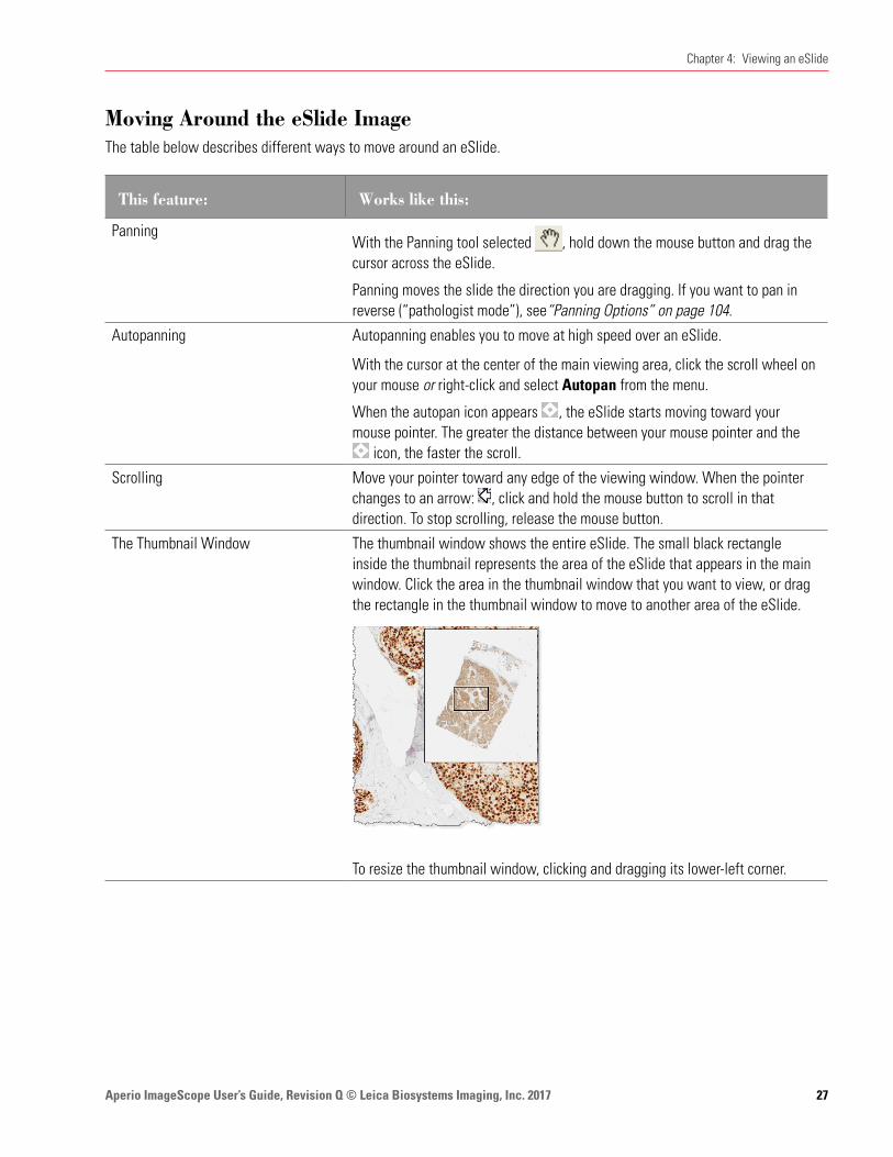
The Thumbnail Window The thumbnail window shows the entire eSlide. The small black rectangle inside the thumbnail represents the area of the eSlide that appears in the main window. Click the area in the thumbnail window that you want to view, or drag the rectangle in the thumbnail window to move to another area of the eSlide.
To resize the thumbnail window, clicking and dragging its lower-left corner.

Aperio ImageScope User’s Guide, Revision Q © Leica Biosystems Imaging, Inc. 201728
Chapter 4: Viewing an eSlide
This feature: Works like this:
Moving to a specific point on the eSlide
Go to the Image menu and select Go To. Using the current image size that appears as a reference, type an X Coordinate (horizontal) and a Y Coordinate (vertical) in pixels.
Ìy Click Go To: Center to position the point selected by those coordinates in the center of the current view.Ìy Click Go To: Corner to position the point selected by those coordinates in
the upper left corner of the current view.
Page panning Use the Shift+Arrow keys to move a page to the right or left, or up or down.
Using the Filmstrip To move between multiple eSlides, click an image from the filmstrip.
Using the Magnifier WindowUse the magnifier window to show a larger view of a particular portion of the eSlide. To use the Magnifier window:
Ì` Drag the magnifier window on the main window to the area you want to see in more detail.
Ì` Move your mouse pointer to the area that you want to display in the magnifier window.
Ì` Resize the magnifier window by dragging its lower right corner.
The default magnification is twice the resolution of the image in the main window. To change the resolution of the magnifier window, go to the Tools menu and select Options. For details, see “Magnification” on page 101.
Changing Viewing MagnificationYou can change the resolution of the entire main window image.
Use this feature: To do this:
Immediate Maximum Zoom Double-click the image to zoom to the maximum magnification. Double-click again to return to the previous magnification.
Zoom Slider Adjust the magnification of the image in the main window.
Ìy Click Fit to fit the entire eSlide within the main viewing area.Ìy Click a magnification level (2X, 4X, etc.).Ìy Drag the slider up or down to increase or decrease the magnification.Ìy Click the image in the main window and roll the scroll wheel.
To set the zoom slider magnification to percentages rather than X-magnification levels, go to the Tools menu and select Options.
Clear the Use “X” magnification rather than “%” check box and click OK.
Zoom keyboard shortcuts Press Ctrl+Minus key to zoom out, and Ctrl+Plus key to zoom in.

Aperio ImageScope User’s Guide, Revision Q © Leica Biosystems Imaging, Inc. 2017 29
Chapter 4: Viewing an eSlide
Use this feature: To do this:
Zoom Navigation To zoom into a particular area of the eSlide, click on the Aperio ImageScope toolbar. Click and drag in the main image window to draw a rectangle to outline the zoom area.
If you are using fixed size annotations, press the Ctrl key while you click on the area you want to zoom into. See“Fixed Size Annotations” on page 104 for more information.
Viewing with Color ManagementAperio Integrated Color Management controls the optical characteristics of your scanner and your display monitor to ensure the colors of the eSlides display accurately. For information on Aperio Integrated Color Management, see “Appendix B: Aperio Integrated Color Management” on page 113.
By default, Aperio ImageScope uses the scanner’s source ICC profile embedded in the eSlide and the target ICC profile for your monitor to make sure the image displays in accurate color. The ICC profile is embedded in the eSlide image during scanning.
You can turn Integrated Color Management on or off:
Ì` Click the icon on the Aperio ImageScope toolbar to turn color management on or off.
Ì` If an image has an embedded ICC profile and color management is turned on, the symbol displays at the bottom of the image. If color management is turned off, the symbol on the image looks like this: .
Viewing Scalebar, Axes, and GridYou can view a scalebar, scale axes, or a grid on an image. A scalebar shows the scale of an image and is often used on maps to allow you to estimate the distance between two points.
The units and spacing are adjusted to correspond to the resolution of the image and the current zoom level.
The zero point of the axes is in the center of the window; it is labeled with the current unit (for example, um for microns). If the resolution of the image is unknown, the units on the axes/grid are p (pixels), kp (kilopixels), or mp (megapixels). This is the case for photomicrographs and gross images before the resolution is set. The resolution on such images can be entered explicitly or by measuring a known item with a ruler (see “Chapter 8: Image Resolution” on page 48).
Aperio ImageScope User’s Guide, Revision Q © Leica Biosystems Imaging, Inc. 201730
Chapter 4: Viewing an eSlide
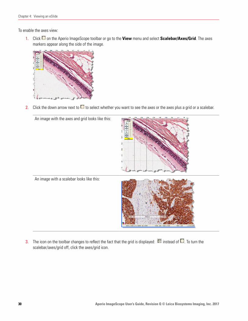
To enable the axes view:
1. Click on the Aperio ImageScope toolbar or go to the View menu and select Scalebar/Axes/Grid. The axes markers appear along the side of the image.
2. Click the down arrow next to to select whether you want to see the axes or the axes plus a grid or a scalebar.
An image with the axes and grid looks like this:
An image with a scalebar looks like this:
3. The icon on the toolbar changes to reflect the fact that the grid is displayed: instead of . To turn the scalebar/axes/grid off, click the axes/grid icon.

Aperio ImageScope User’s Guide, Revision Q © Leica Biosystems Imaging, Inc. 2017 31
Chapter 4: Viewing an eSlide
Viewing Z-Stack eSlide ImagesThe Aperio scanner can create multiple digital images of slide tissue scanned at different focal depths, creating a 3D image that you can visually navigate through much as a microscope user can navigate through different tissue focal depths by using the microscope objective fine and coarse adjustments. This ability to create a 3D image is called “z-stack scanning.”
Aperio ImageScope enables you to view and annotate specific layers of the z-stack image.
This section contains information about z-stack images that were scanned on an Aperio scanner. For information on z-stack images created from live video, see “Chapter 15: TelePath Live” on page 88.
Viewing and Navigating a Z-Stack Image
Aperio ImageScope automatically opens a z-stack image to the best focused layer, as determined by your Aperio scanner when the slide is scanned. The number of layers and the layer separation (depth, in microns (µm), between the layers) is set during scanning. For more information on scanning z-stack images, see the Console User’s Guide for your Aperio scanner.
Aperio ImageScope User’s Guide, Revision Q © Leica Biosystems Imaging, Inc. 201732
Chapter 4: Viewing an eSlide
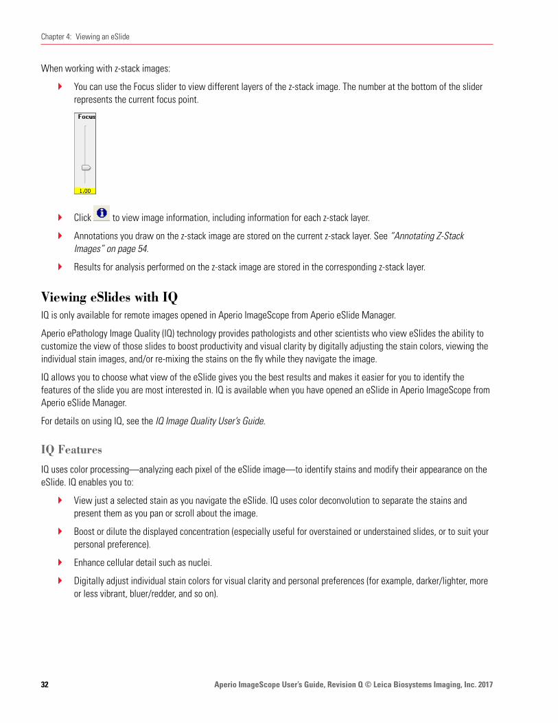
When working with z-stack images:
Ì` You can use the Focus slider to view different layers of the z-stack image. The number at the bottom of the slider represents the current focus point.
Ì` Click to view image information, including information for each z-stack layer.
Ì` Annotations you draw on the z-stack image are stored on the current z-stack layer. See “Annotating Z-Stack Images” on page 54.
Ì` Results for analysis performed on the z-stack image are stored in the corresponding z-stack layer.
Viewing eSlides with IQIQ is only available for remote images opened in Aperio ImageScope from Aperio eSlide Manager.
Aperio ePathology Image Quality (IQ) technology provides pathologists and other scientists who view eSlides the ability to customize the view of those slides to boost productivity and visual clarity by digitally adjusting the stain colors, viewing the individual stain images, and/or re-mixing the stains on the fly while they navigate the image.
IQ allows you to choose what view of the eSlide gives you the best results and makes it easier for you to identify the features of the slide you are most interested in. IQ is available when you have opened an eSlide in Aperio ImageScope from Aperio eSlide Manager.
For details on using IQ, see the IQ Image Quality User’s Guide.
IQ Features
IQ uses color processing—analyzing each pixel of the eSlide image—to identify stains and modify their appearance on the eSlide. IQ enables you to:
Ì` View just a selected stain as you navigate the eSlide. IQ uses color deconvolution to separate the stains and present them as you pan or scroll about the image.
Ì` Boost or dilute the displayed concentration (especially useful for overstained or understained slides, or to suit your personal preference).
Ì` Enhance cellular detail such as nuclei.
Ì` Digitally adjust individual stain colors for visual clarity and personal preferences (for example, darker/lighter, more or less vibrant, bluer/redder, and so on).
Aperio ImageScope User’s Guide, Revision Q © Leica Biosystems Imaging, Inc. 2017 33
Chapter 4: Viewing an eSlide
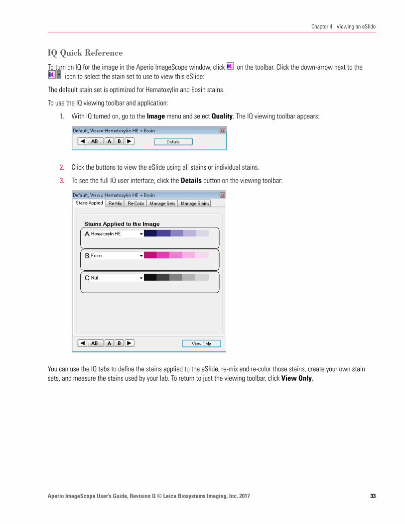
IQ Quick Reference
To turn on IQ for the image in the Aperio ImageScope window, click on the toolbar. Click the down-arrow next to the icon to select the stain set to use to view this eSlide:
The default stain set is optimized for Hematoxylin and Eosin stains.
To use the IQ viewing toolbar and application:
1. With IQ turned on, go to the Image menu and select Quality. The IQ viewing toolbar appears:
2. Click the buttons to view the eSlide using all stains or individual stains.
3. To see the full IQ user interface, click the Details button on the viewing toolbar:
You can use the IQ tabs to define the stains applied to the eSlide, re-mix and re-color those stains, create your own stain sets, and measure the stains used by your lab. To return to just the viewing toolbar, click View Only.

Aperio ImageScope User’s Guide, Revision Q © Leica Biosystems Imaging, Inc. 201734
Aperio ImageScope rotation tools allow you to rotate an image. You can also rotate an eSlide label image.
Rotating an ImageThe rotation setting is in effect only for the current viewing session and is not saved with the image. However, when you create a new image by using the Snapshot or Extract Region commands, the new image is saved in the current rotation. Saving an Image View also saves the current rotation settings so that opening the Image View displays the image with those rotation settings applied.
Image rotation is not enabled during a TelePath Live session.
To use image rotation:
1. Go to the Aperio ImageScope Image menu and select Rotate Image (Ctrl+E).
2. From the rotation toolbar, select the rotation setting you want to use:
Rotate zero degrees
Rotate 90 degrees right
Rotate 180 degrees
Rotate 90 degrees left
Flip vertically
Rotate 90 degrees right and flip vertically
Flip horizontally
Rotate 90 degrees left and flip vertically
Rotating an eSlide Label
Click the eSlide label to rotate it 90 degrees to the right. Aperio ImageScope saves the label rotation when you close the eSlide.
5 Rotating Images and Slide Labels
Aperio ImageScope User’s Guide, Revision Q © Leica Biosystems Imaging, Inc. 2017 35
You can modify the color settings of eSlides if particular colors do not show up well on your workstation monitor. This chapter discusses the different image adjustment settings.
For information on adjusting fluorescence images, see “Chapter 7: Working with Fluorescence eSlides” on page 38.
Image adjustments apply only to the current Aperio ImageScope session. Image adjustments do not modify your original eSlide, and they are not stored with the eSlide. You can save gamma settings to apply to the current eSlide or to apply to other eSlides later, and you can make a snapshot of the adjusted image if you want to save the adjusted eSlide image. (See“Chapter 13: Saving eSlides and Regions” on page 73 for information on making snapshots.)
Use the image adjustments to:
Ì` Adjust the brightness or contrast for all colors or for just the red, green, or blue channel.
Ì` Modify the color balance (for example, make reds less red and more cyan).
Ì` Adjust color curves for all colors or for just the red, green, or blue channel.
Ì` Save the color adjustments you have made in a gamma table file so they can be re-applied to the same or other eSlides that you view in the future.
Ì` Make image adjustments to the entire eSlide or to live video Z-stack images.
Making Image AdjustmentsFollow the instructions below to make color adjustments to your eSlide images using the brightness and contrast, color balance, color curves adjustment.
To make image adjustments, go to the Image menu and select Adjustments.
Here are some general tips for making image adjustments:
Ì` To modify the appearance of an entire eSlide, select Main Image. To modify just the current live video Z-stack images, select Z-stack Images. Note that the Z-stack Images option is for live video z-stack images created with TelePath Live.
Ì` Click and hold the Compare button to temporarily return the image to the original settings. Release the button to revert back to the changed settings.
Ì` Click the Reset button to return all colors to the original default settings.
6 Making Image Adjustments
Aperio ImageScope User’s Guide, Revision Q © Leica Biosystems Imaging, Inc. 201736
Chapter 6: Making Image Adjustments
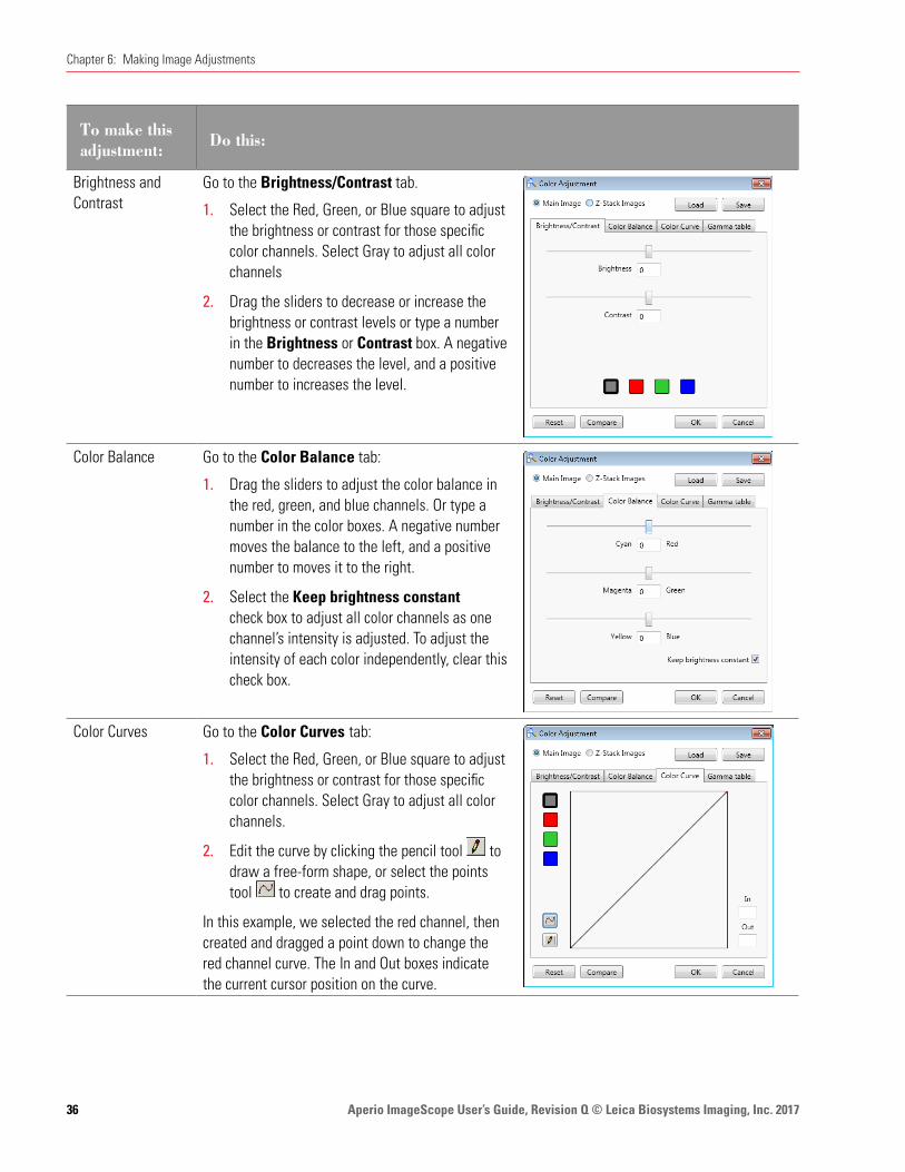
To make this adjustment: Do this:
Brightness and Contrast
Go to the Brightness/Contrast tab.
1. Select the Red, Green, or Blue square to adjust the brightness or contrast for those specific color channels. Select Gray to adjust all color channels
2. Drag the sliders to decrease or increase the brightness or contrast levels or type a number in the Brightness or Contrast box. A negative number to decreases the level, and a positive number to increases the level.
Color Balance Go to the Color Balance tab:
1. Drag the sliders to adjust the color balance in the red, green, and blue channels. Or type a number in the color boxes. A negative number moves the balance to the left, and a positive number to moves it to the right.
2. Select the Keep brightness constant check box to adjust all color channels as one channel’s intensity is adjusted. To adjust the intensity of each color independently, clear this check box.
Color Curves Go to the Color Curves tab:
1. Select the Red, Green, or Blue square to adjust the brightness or contrast for those specific color channels. Select Gray to adjust all color channels.
2. Edit the curve by clicking the pencil tool to draw a free-form shape, or select the points tool to create and drag points.
In this example, we selected the red channel, then created and dragged a point down to change the red channel curve. The In and Out boxes indicate the current cursor position on the curve.
Aperio ImageScope User’s Guide, Revision Q © Leica Biosystems Imaging, Inc. 2017 37
Chapter 6: Making Image Adjustments
Saving and Loading Color Settings
You can save the image adjustments, and load them to apply to other eSlide images.
To save the color adjustment settings: 1. Click the Save button that appears in the upper right corner of the Color Adjustments window.
2. Navigate to the directory where you want to save the gamma table file, type a file name, and click Save.
3. Click OK to exit the Image Adjustments window.
To load color adjustment settings 1. Click Load.
2. Navigate to the saved gamma table file, and click Open.
3. Click OK to exit the Image Adjustments window.
For More InformationÌ` For information on z-stack images created with TelePath Live, see “Chapter 15: TelePath Live” on page 88.
Ì` For information on loading color settings to be used every time Aperio ImageScope opens, see “General Options” on page 101.

Aperio ImageScope User’s Guide, Revision Q © Leica Biosystems Imaging, Inc. 201738
This chapter discusses how to view and adjust fluorescence eSlide images.
Images from the Aperio FL are grayscale images that are pseudo-colored during the scanning process.
Aperio ImageScope offers a full range of fluorescence features:
Ì` Temporarily apply a false color to a fluorescence image (this is not needed for fluorescence eSlides created by the Aperio FL)
Ì` For a fused image:
Ìy Change the display color for each channel image
Ìy Adjust brightness, contrast, and gamma (viewing the results on the image and on a histogram display)
Ìy Adjust registration between channels
Ì` Fuse multiple fluorescence channel images into a fused image (automatically done for images acquired with the Aperio FL)
Applying a Temporary False ColorIf you are using a grayscale fluorescence image and want to display it in color:
1. Open the image in Aperio ImageScope.
2. Go to the Image menu and select False Color. The False Color window displays:
3. Select a color by clicking a color box or using the color slider.
4. Select the Enable check box to see the image in the color you have selected. To view the image without the false color, clear the Enable check box.
Applying a false color in this way does not permanently change the display color for the image—this adjustment applies only to the current viewing session.
7 Working with Fluorescence eSlides
Aperio ImageScope User’s Guide, Revision Q © Leica Biosystems Imaging, Inc. 2017 39
Chapter 7: Working with Fluorescence eSlides
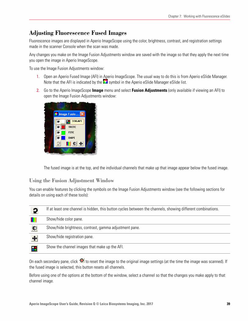
Adjusting Fluorescence Fused ImagesFluorescence images are displayed in Aperio ImageScope using the color, brightness, contrast, and registration settings made in the scanner Console when the scan was made.
Any changes you make on the Image Fusion Adjustments window are saved with the image so that they apply the next time you open the image in Aperio ImageScope.
To use the Image Fusion Adjustments window:
1. Open an Aperio Fused Image (AFI) in Aperio ImageScope. The usual way to do this is from Aperio eSlide Manager. Note that the AFI is indicated by the symbol in the Aperio eSlide Manager eSlide list.
2. Go to the Aperio ImageScope Image menu and select Fusion Adjustments (only available if viewing an AFI) to open the Image Fusion Adjustments window:
The fused image is at the top, and the individual channels that make up that image appear below the fused image.
Using the Fusion Adjustment Window
You can enable features by clicking the symbols on the Image Fusion Adjustments window (see the following sections for details on using each of these tools):
If at least one channel is hidden, this button cycles between the channels, showing different combinations.
Show/hide color pane.
Show/hide brightness, contrast, gamma adjustment pane.
Show/hide registration pane.
Show the channel images that make up the AFI.
On each secondary pane, click to reset the image to the original image settings (at the time the image was scanned). If the fused image is selected, this button resets all channels.
Before using one of the options at the bottom of the window, select a channel so that the changes you make apply to that channel image.

Aperio ImageScope User’s Guide, Revision Q © Leica Biosystems Imaging, Inc. 201740
Chapter 7: Working with Fluorescence eSlides
After opening a tool pane, click to close the pane to exit back to the main Image Fusion Adjustments window.
Viewing All Channel Images
Aperio eSlide Manager displays only the AFI and not the channel images that comprise the AFI for fluorescence images that were created by scanning a glass slide on a Aperio FL.
If you need to view the channel images that comprise an AFI, you can do so by using Aperio ImageScope.
1. Open the AFI by clicking its thumbnail on the Aperio eSlide Manager page. You see the AFI in the main viewing window along with the Image Fusion Adjustment window:
2. To view all of the channel images that make up the AFI, click the channel tile icon on the Image Fusion Adjustment window.
3. You can make channel adjustments, as described below.

Aperio ImageScope User’s Guide, Revision Q © Leica Biosystems Imaging, Inc. 2017 41
Chapter 7: Working with Fluorescence eSlides
To make this adjustment: Do this:
Hide a channel To hide a channel, click the eye symbol: next to it. (To add it back to the display, click again.)
Hide a channel color Click the color box next to a channel to turn off the false color for that channel (that is, to display it in grayscale). To turn the false color on, click the color box again.
Hide all color To remove all color, click the color box next to the AFI.
Seeing Channel Information
Point to a color box to see information about that channel.
Cycling Channel Displays
To cycle the display among the channels:
1. Hide a channel by clicking the eye symbol: next to it.
2. Click the button to cycle among different combinations of channels.
In this example, the FITC channel is hidden (this also turns off the AFI fused image, because all channels must be on to see the fused image):
Clicking the button cycles the display to a new combination of channels (TRITC and FTIC).
Clicking the button again cycles the display to another combination of channels. To turn off cycling, manually turn on all channels by clicking the
next to hidden channels.
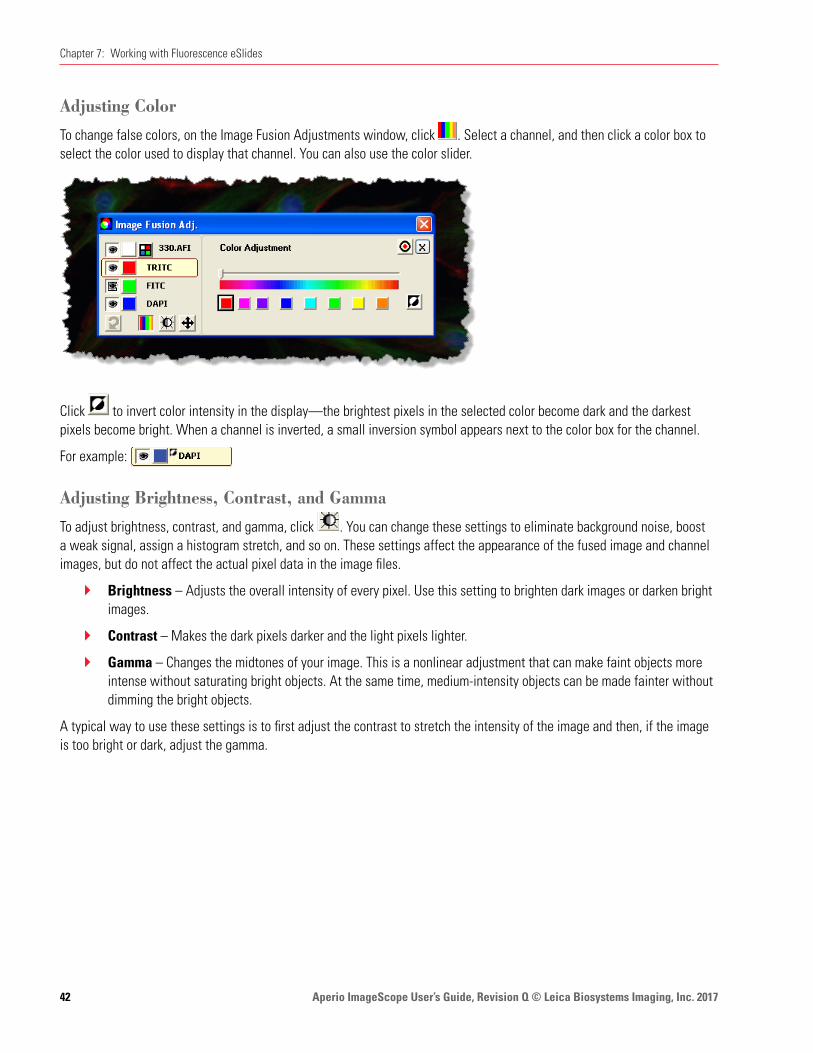
Aperio ImageScope User’s Guide, Revision Q © Leica Biosystems Imaging, Inc. 201742
Chapter 7: Working with Fluorescence eSlides
Adjusting Color
To change false colors, on the Image Fusion Adjustments window, click . Select a channel, and then click a color box to select the color used to display that channel. You can also use the color slider.
Click to invert color intensity in the display—the brightest pixels in the selected color become dark and the darkest pixels become bright. When a channel is inverted, a small inversion symbol appears next to the color box for the channel.
For example:
Adjusting Brightness, Contrast, and Gamma
To adjust brightness, contrast, and gamma, click . You can change these settings to eliminate background noise, boost a weak signal, assign a histogram stretch, and so on. These settings affect the appearance of the fused image and channel images, but do not affect the actual pixel data in the image files.
Ì` Brightness – Adjusts the overall intensity of every pixel. Use this setting to brighten dark images or darken bright images.
Ì` Contrast – Makes the dark pixels darker and the light pixels lighter.
Ì` Gamma – Changes the midtones of your image. This is a nonlinear adjustment that can make faint objects more intense without saturating bright objects. At the same time, medium-intensity objects can be made fainter without dimming the bright objects.
A typical way to use these settings is to first adjust the contrast to stretch the intensity of the image and then, if the image is too bright or dark, adjust the gamma.
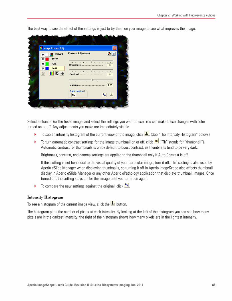
Aperio ImageScope User’s Guide, Revision Q © Leica Biosystems Imaging, Inc. 2017 43
Chapter 7: Working with Fluorescence eSlides
The best way to see the effect of the settings is just to try them on your image to see what improves the image.
Select a channel (or the fused image) and select the settings you want to use. You can make these changes with color turned on or off. Any adjustments you make are immediately visible.
Ì` To see an intensity histogram of the current view of the image, click . (See “The Intensity Histogram” below.)
Ì` To turn automatic contrast settings for the image thumbnail on or off, click (“Th” stands for “thumbnail”). Automatic contrast for thumbnails is on by default to boost contrast, as thumbnails tend to be very dark.
Brightness, contrast, and gamma settings are applied to the thumbnail only if Auto Contrast is off.
If this setting is not beneficial to the visual quality of your particular image, turn it off. This setting is also used by Aperio eSlide Manager when displaying thumbnails, so turning it off in Aperio ImageScope also affects thumbnail display in Aperio eSlide Manager or any other Aperio ePathology application that displays thumbnail images. Once turned off, the setting stays off for this image until you turn it on again.
Ì` To compare the new settings against the original, click .
Intensity Histogram
To see a histogram of the current image view, click the button.
The histogram plots the number of pixels at each intensity. By looking at the left of the histogram you can see how many pixels are in the darkest intensity; the right of the histogram shows how many pixels are in the lightest intensity.
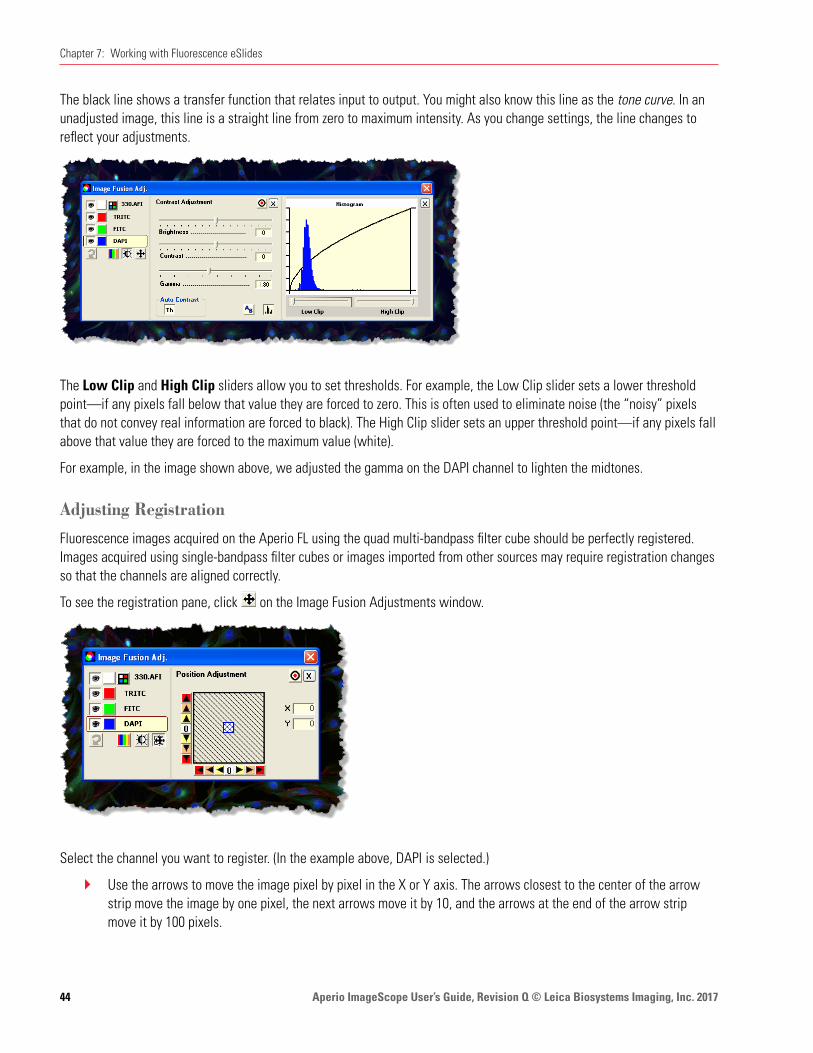
Aperio ImageScope User’s Guide, Revision Q © Leica Biosystems Imaging, Inc. 201744
Chapter 7: Working with Fluorescence eSlides
The black line shows a transfer function that relates input to output. You might also know this line as the tone curve. In an unadjusted image, this line is a straight line from zero to maximum intensity. As you change settings, the line changes to reflect your adjustments.
The Low Clip and High Clip sliders allow you to set thresholds. For example, the Low Clip slider sets a lower threshold point—if any pixels fall below that value they are forced to zero. This is often used to eliminate noise (the “noisy” pixels that do not convey real information are forced to black). The High Clip slider sets an upper threshold point—if any pixels fall above that value they are forced to the maximum value (white).
For example, in the image shown above, we adjusted the gamma on the DAPI channel to lighten the midtones.
Adjusting Registration
Fluorescence images acquired on the Aperio FL using the quad multi-bandpass filter cube should be perfectly registered. Images acquired using single-bandpass filter cubes or images imported from other sources may require registration changes so that the channels are aligned correctly.
To see the registration pane, click on the Image Fusion Adjustments window.
Select the channel you want to register. (In the example above, DAPI is selected.)
Ì` Use the arrows to move the image pixel by pixel in the X or Y axis. The arrows closest to the center of the arrow strip move the image by one pixel, the next arrows move it by 10, and the arrows at the end of the arrow strip move it by 100 pixels.

Aperio ImageScope User’s Guide, Revision Q © Leica Biosystems Imaging, Inc. 2017 45
Chapter 7: Working with Fluorescence eSlides
Ì` Click the 0 at the center of the arrow strip to move the image back to its original position on that axis.
Ì` The colored box in the middle of the display shows the position of the image relative to its original position. The box starts in the center and moves as you use the arrows. The color of the border of the box tells you which channel you are working with. (In the example above, the blue border is the same color used for the channel DAPI, so we know we are working with that channel.)
Ì` The movement of the image in the X/Y axes is shown in the X and Y boxes.
Fusing Fluorescence ImagesThe scanner creates a multi-layer “fused” image made up of the individual channel images. However, you can also create your own fused image or create one from individual channel images you received from another source.
Our example shows opening images in Aperio eSlide Manager.
1. Log into Aperio eSlide Manager and select All eSlides (As List) from the eSlides menu.
2. Select the check boxes next to the channel images you want to use to create a fused image and click View Images.
Aperio ImageScope opens each of the selected images:
In this example, we have three images, one for each type of fluorochrome used to stain the slide: DAPI, FITC, and TRITC.
If you want Aperio ImageScope to keep each of these images open after it creates the fused image, click the first image to select it, go to the Image menu, and select Keep Open (or type Ctrl+K). Repeat for the other images.
3. On the Aperio ImageScope menu bar, go to the Image menu and select Fuse Images.
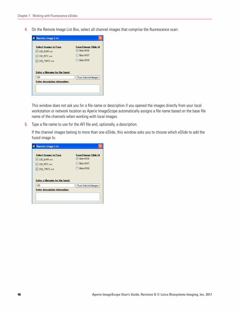
Aperio ImageScope User’s Guide, Revision Q © Leica Biosystems Imaging, Inc. 201746
Chapter 7: Working with Fluorescence eSlides
4. On the Remote Image List Box, select all channel images that comprise the fluorescence scan:
This window does not ask you for a file name or description if you opened the images directly from your local workstation or network location as Aperio ImageScope automatically assigns a file name based on the base file name of the channels when working with local images.
5. Type a file name to use for the AFI file and, optionally, a description.
If the channel images belong to more than one eSlide, this window asks you to choose which eSlide to add the fused image to.
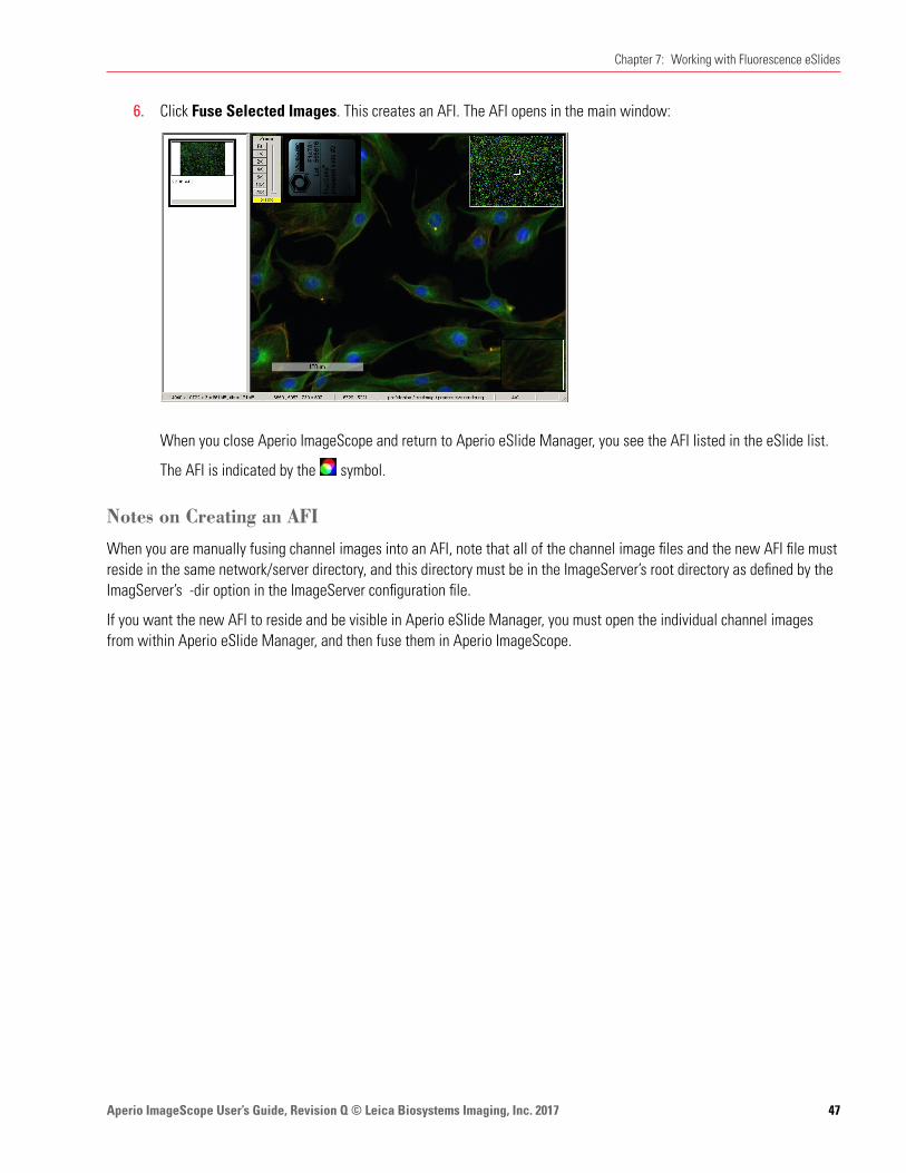
Aperio ImageScope User’s Guide, Revision Q © Leica Biosystems Imaging, Inc. 2017 47
Chapter 7: Working with Fluorescence eSlides
6. Click Fuse Selected Images. This creates an AFI. The AFI opens in the main window:
When you close Aperio ImageScope and return to Aperio eSlide Manager, you see the AFI listed in the eSlide list.
The AFI is indicated by the symbol.
Notes on Creating an AFI
When you are manually fusing channel images into an AFI, note that all of the channel image files and the new AFI file must reside in the same network/server directory, and this directory must be in the ImageServer’s root directory as defined by the ImagServer’s -dir option in the ImageServer configuration file.
If you want the new AFI to reside and be visible in Aperio eSlide Manager, you must open the individual channel images from within Aperio eSlide Manager, and then fuse them in Aperio ImageScope.

Aperio ImageScope User’s Guide, Revision Q © Leica Biosystems Imaging, Inc. 201748
8 Image Resolution
This chapter discusses how to see the resolution of an image and, if it is not known, how to set it.
For eSlides created by scanning microscope slides on an Aperio scanner, the resolution of the image is known and is stored in the image file. The resolution is used to display the magnification in the zoom slider, to compute ruler values, and to compute the length and area of annotation regions.
The resolution may not be known for other types of images you are working with.
To view and set an image’s resolution:
1. Go to the Image menu and select Resolution. The Image Resolution window displays:
If the resolution of the image is stored within the image file, Current Image Resolution and Apparent Magnification contains values—if the resolution is not known, these boxes will be empty.
Setting or Changing Image Resolution
When you change the resolution, you affect the way an image is viewed, but do not change the image file or information stored in it.
Because the resolution is used to display the magnification in the zoom slider, to compute ruler values, and to compute the length and width of annotations, these values change when you change the resolution.
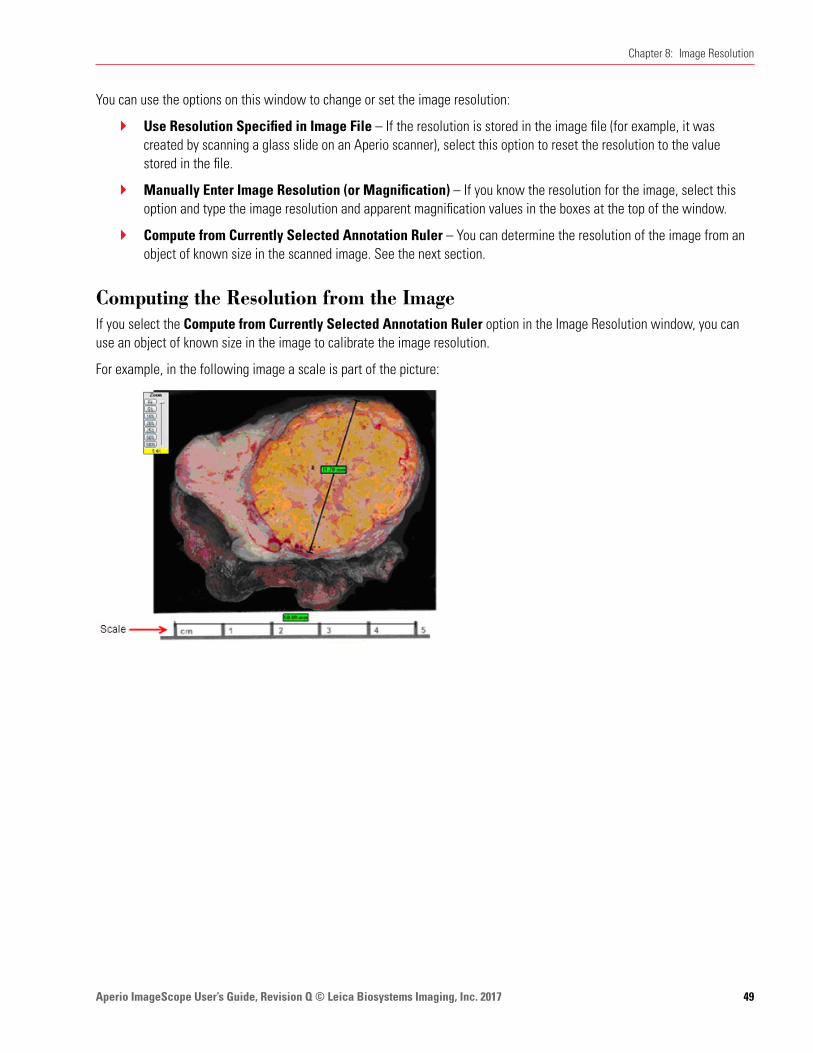
Aperio ImageScope User’s Guide, Revision Q © Leica Biosystems Imaging, Inc. 2017 49
Chapter 8: Image Resolution
You can use the options on this window to change or set the image resolution:
Ì` Use Resolution Specified in Image File – If the resolution is stored in the image file (for example, it was created by scanning a glass slide on an Aperio scanner), select this option to reset the resolution to the value stored in the file.
Ì` Manually Enter Image Resolution (or Magnification) – If you know the resolution for the image, select this option and type the image resolution and apparent magnification values in the boxes at the top of the window.
Ì` Compute from Currently Selected Annotation Ruler – You can determine the resolution of the image from an object of known size in the scanned image. See the next section.
Computing the Resolution from the ImageIf you select the Compute from Currently Selected Annotation Ruler option in the Image Resolution window, you can use an object of known size in the image to calibrate the image resolution.
For example, in the following image a scale is part of the picture:

Aperio ImageScope User’s Guide, Revision Q © Leica Biosystems Imaging, Inc. 201750
Chapter 8: Image Resolution
1. Draw an annotation ruler across the scale, which in this case shows a length of 5 centimeters.
2. Go to the Image menu and select Resolution. The Image Resolution window now has a place for you to enter the length of the ruler:
3. On the Image Resolution window, select Compute from Currently Selected Annotation Ruler and enter the length of the ruler in microns. In this case, enter a value of 50000 (5 cm = 50000 microns). If you want to delete the ruler after calibration, select the Delete Ruler after Resolution Computed check box.
Now all values measured by rulers will be accurately calibrated to this scale, such as the second ruler shown above which measures a length of 31mm across the specimen.
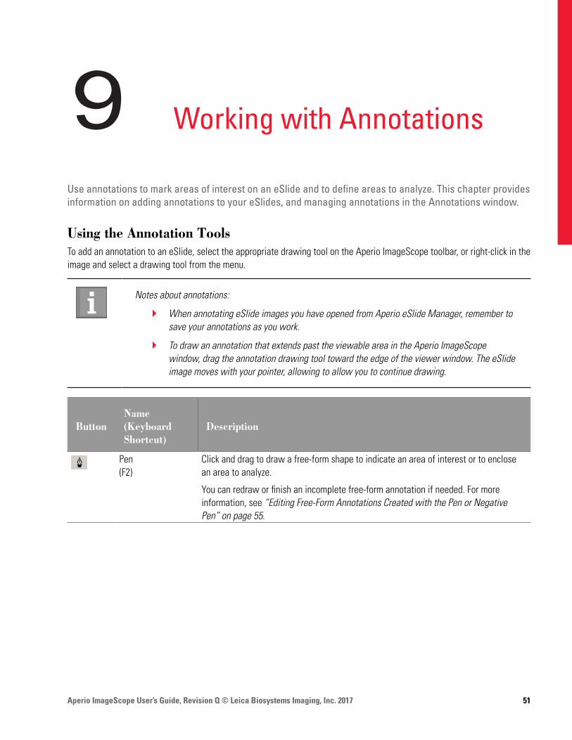
Aperio ImageScope User’s Guide, Revision Q © Leica Biosystems Imaging, Inc. 2017 51
9 Working with Annotations
Use annotations to mark areas of interest on an eSlide and to define areas to analyze. This chapter provides information on adding annotations to your eSlides, and managing annotations in the Annotations window.
Using the Annotation ToolsTo add an annotation to an eSlide, select the appropriate drawing tool on the Aperio ImageScope toolbar, or right-click in the image and select a drawing tool from the menu.
Notes about annotations:
Ì` When annotating eSlide images you have opened from Aperio eSlide Manager, remember to save your annotations as you work.
Ì` To draw an annotation that extends past the viewable area in the Aperio ImageScope window, drag the annotation drawing tool toward the edge of the viewer window. The eSlide image moves with your pointer, allowing to allow you to continue drawing.
ButtonName (Keyboard Shortcut)
Description
Pen(F2)
Click and drag to draw a free-form shape to indicate an area of interest or to enclose an area to analyze.
You can redraw or finish an incomplete free-form annotation if needed. For more information, see “Editing Free-Form Annotations Created with the Pen or Negative Pen” on page 55.
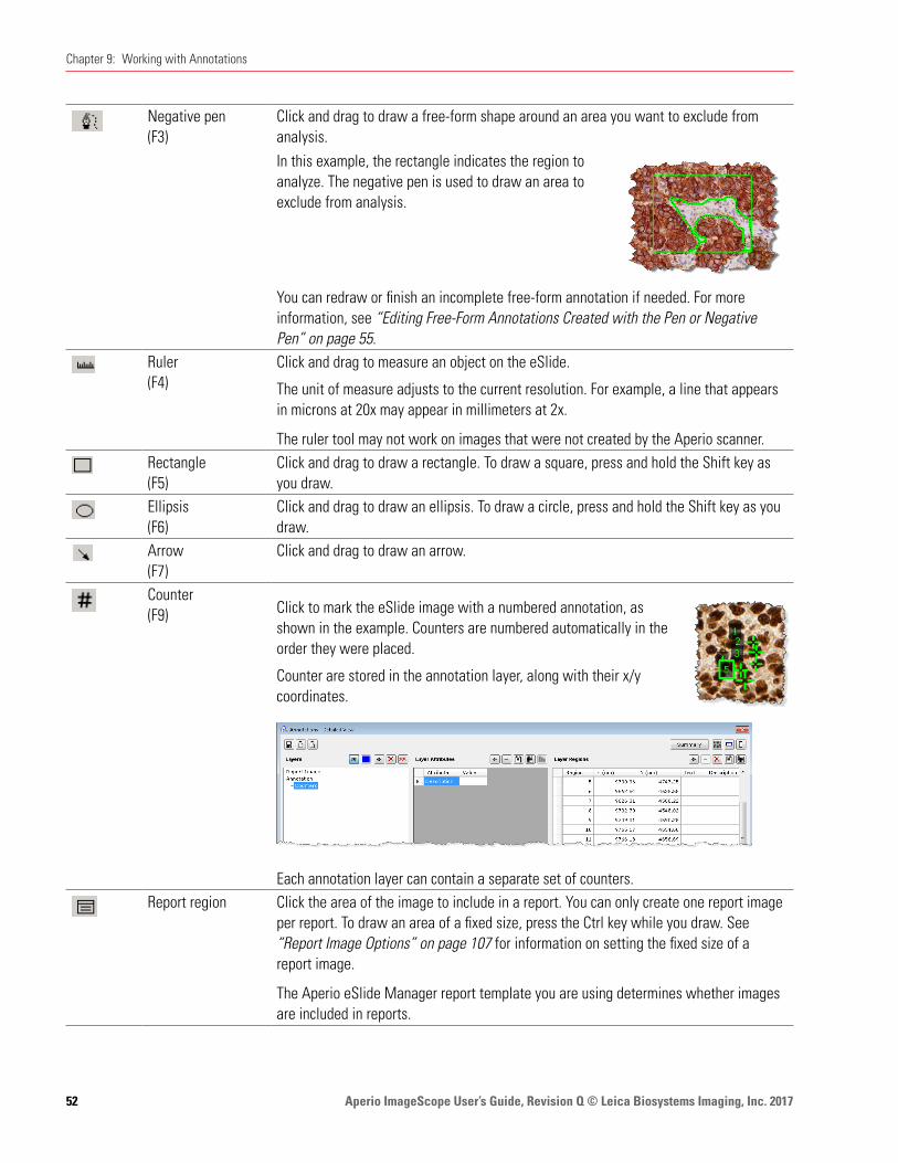
Aperio ImageScope User’s Guide, Revision Q © Leica Biosystems Imaging, Inc. 201752
Chapter 9: Working with Annotations
Negative pen(F3)
Click and drag to draw a free-form shape around an area you want to exclude from analysis. In this example, the rectangle indicates the region to analyze. The negative pen is used to draw an area to exclude from analysis.
You can redraw or finish an incomplete free-form annotation if needed. For more information, see “Editing Free-Form Annotations Created with the Pen or Negative Pen” on page 55.
Ruler(F4)
Click and drag to measure an object on the eSlide.
The unit of measure adjusts to the current resolution. For example, a line that appears in microns at 20x may appear in millimeters at 2x.
The ruler tool may not work on images that were not created by the Aperio scanner. Rectangle(F5)
Click and drag to draw a rectangle. To draw a square, press and hold the Shift key as you draw.
Ellipsis(F6)
Click and drag to draw an ellipsis. To draw a circle, press and hold the Shift key as you draw.
Arrow(F7)
Click and drag to draw an arrow.
Counter(F9)
Click to mark the eSlide image with a numbered annotation, as shown in the example. Counters are numbered automatically in the order they were placed.
Counter are stored in the annotation layer, along with their x/y coordinates.
Each annotation layer can contain a separate set of counters. Report region Click the area of the image to include in a report. You can only create one report image
per report. To draw an area of a fixed size, press the Ctrl key while you draw. See “Report Image Options” on page 107 for information on setting the fixed size of a report image.
The Aperio eSlide Manager report template you are using determines whether images are included in reports.
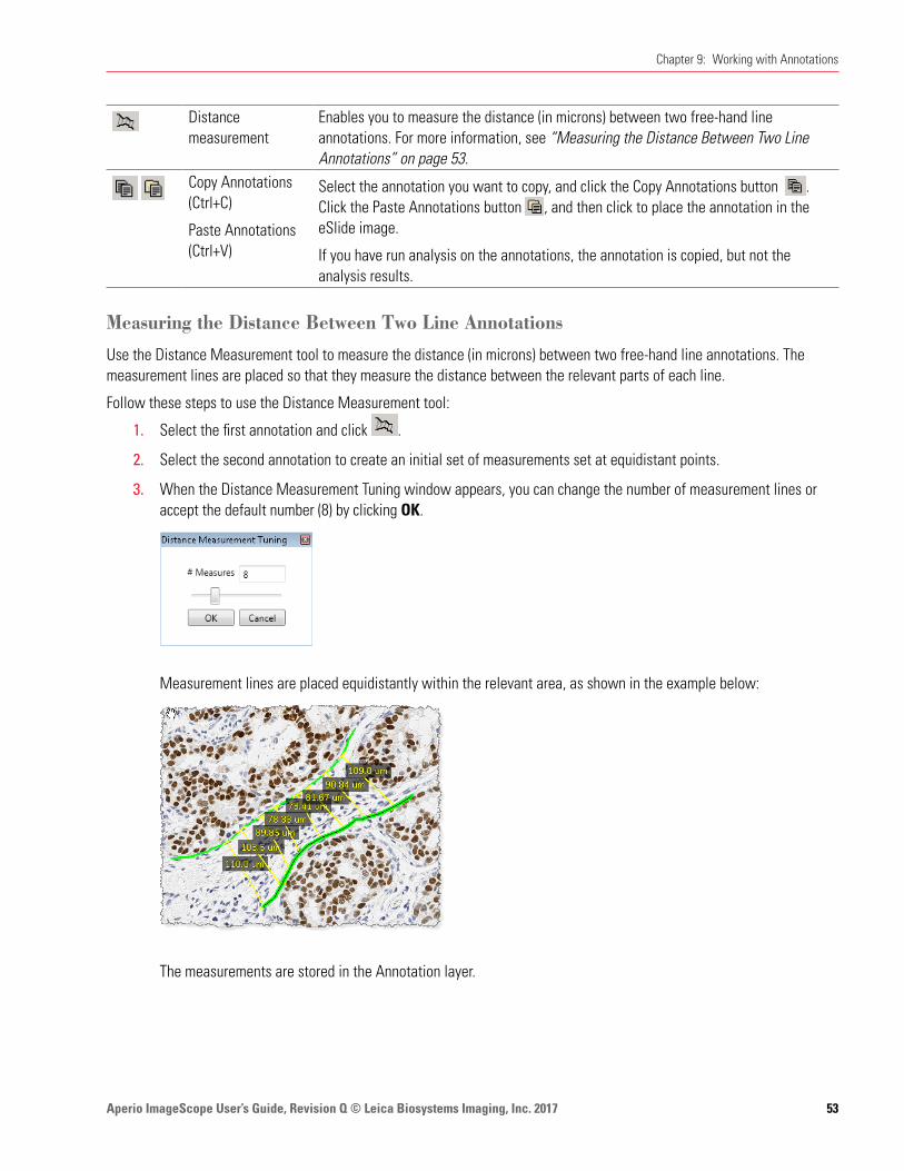
Aperio ImageScope User’s Guide, Revision Q © Leica Biosystems Imaging, Inc. 2017 53
Chapter 9: Working with Annotations
Distance measurement
Enables you to measure the distance (in microns) between two free-hand line annotations. For more information, see “Measuring the Distance Between Two Line Annotations” on page 53.
Copy Annotations (Ctrl+C)
Paste Annotations (Ctrl+V)
Select the annotation you want to copy, and click the Copy Annotations button . Click the Paste Annotations button , and then click to place the annotation in the eSlide image.
If you have run analysis on the annotations, the annotation is copied, but not the analysis results.
Measuring the Distance Between Two Line Annotations
Use the Distance Measurement tool to measure the distance (in microns) between two free-hand line annotations. The measurement lines are placed so that they measure the distance between the relevant parts of each line.
Follow these steps to use the Distance Measurement tool:
1. Select the first annotation and click .
2. Select the second annotation to create an initial set of measurements set at equidistant points.
3. When the Distance Measurement Tuning window appears, you can change the number of measurement lines or accept the default number (8) by clicking OK.
Measurement lines are placed equidistantly within the relevant area, as shown in the example below:
The measurements are stored in the Annotation layer.
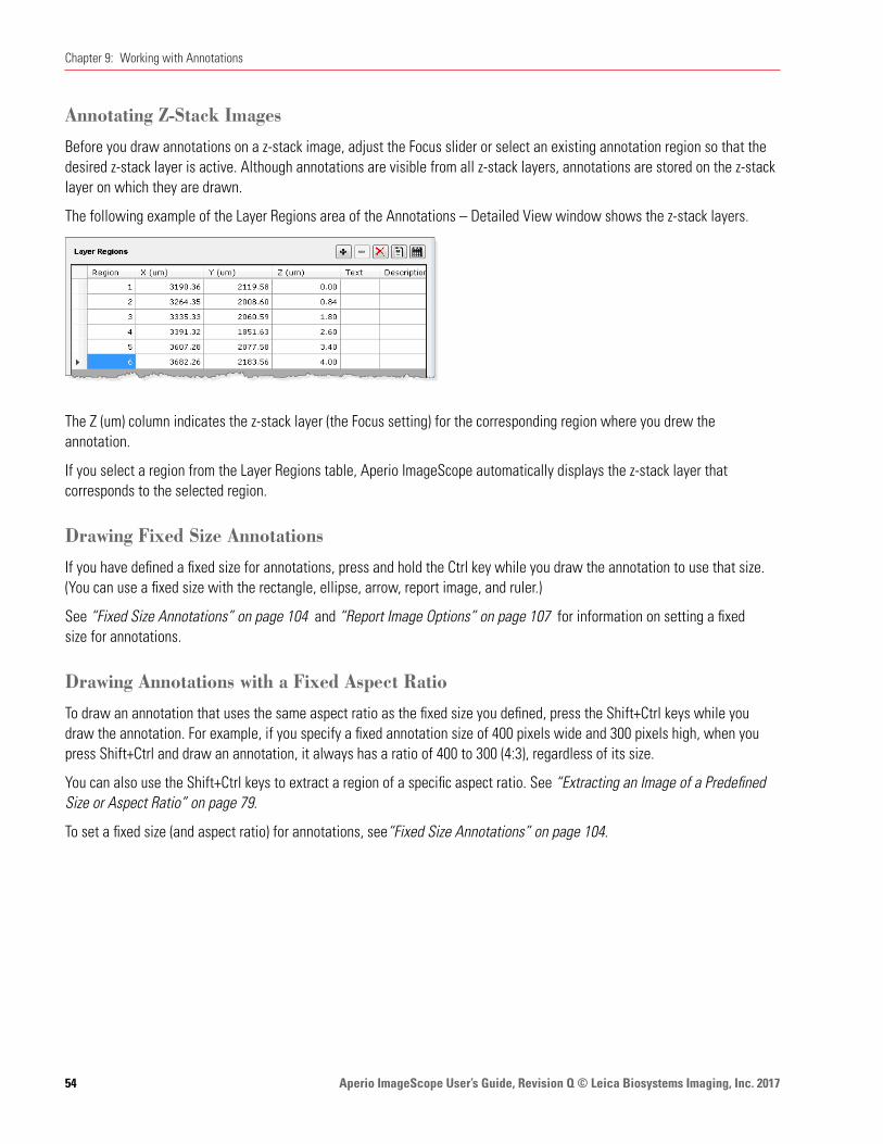
Aperio ImageScope User’s Guide, Revision Q © Leica Biosystems Imaging, Inc. 201754
Chapter 9: Working with Annotations
Annotating Z-Stack Images
Before you draw annotations on a z-stack image, adjust the Focus slider or select an existing annotation region so that the desired z-stack layer is active. Although annotations are visible from all z-stack layers, annotations are stored on the z-stack layer on which they are drawn.
The following example of the Layer Regions area of the Annotations – Detailed View window shows the z-stack layers.
The Z (um) column indicates the z-stack layer (the Focus setting) for the corresponding region where you drew the annotation.
If you select a region from the Layer Regions table, Aperio ImageScope automatically displays the z-stack layer that corresponds to the selected region.
Drawing Fixed Size Annotations
If you have defined a fixed size for annotations, press and hold the Ctrl key while you draw the annotation to use that size. (You can use a fixed size with the rectangle, ellipse, arrow, report image, and ruler.)
See “Fixed Size Annotations” on page 104 and “Report Image Options” on page 107 for information on setting a fixed size for annotations.
Drawing Annotations with a Fixed Aspect Ratio
To draw an annotation that uses the same aspect ratio as the fixed size you defined, press the Shift+Ctrl keys while you draw the annotation. For example, if you specify a fixed annotation size of 400 pixels wide and 300 pixels high, when you press Shift+Ctrl and draw an annotation, it always has a ratio of 400 to 300 (4:3), regardless of its size.
You can also use the Shift+Ctrl keys to extract a region of a specific aspect ratio. See “Extracting an Image of a Predefined Size or Aspect Ratio” on page 79.
To set a fixed size (and aspect ratio) for annotations, see“Fixed Size Annotations” on page 104.

Aperio ImageScope User’s Guide, Revision Q © Leica Biosystems Imaging, Inc. 2017 55
Chapter 9: Working with Annotations
Moving Annotations
To move one or more annotations on the eSlide image, do the following:
Move the selected annotation Press and hold the Ctrl key while you drag the annotation to a new location.
Move all annotations simultaneously Press and hold the Ctrl+Shift keys while dragging the selected annotation to a new location. All annotations move along with the one you are dragging. This is useful if you import annotations from a similar eSlide, and slight variations require some adjustments.
This feature moves all annotations in all layers except for Z-stacks and markup images.
Editing Free-Form Annotations Created with the Pen or Negative Pen
This feature enables you to edit the shape of free-form annotations that are created with the pen or negative pen tools. You can change the shape of an existing annotation, or finish an incomplete annotation.
You cannot edit an annotation if you have run analysis on it.
Tips for editing annotations:
Ìy Start and finish your edits in a problem-free area of the annotation. See “Fixing a Problem Area” on page 57 for an example.
Ìy Drag the pointer in a slow and smooth manner when redrawing annotations.
Ìy When you are redrawing an annotation, you can move back and forth over the same area until you release the left-mouse button.

Aperio ImageScope User’s Guide, Revision Q © Leica Biosystems Imaging, Inc. 201756
Chapter 9: Working with Annotations
Completing a Free-Form Shape
This example shows how to complete an annotation by connecting the two ends.
1. With the annotation selected, place the Pen tool at an open end of the shape.
2. Hold down the left mouse button and slowly drag the line until the two ends are connected.
3. When you are finished editing the annotation, release the mouse button.
Editing a Free-Form Shape
This example shows how to change the shape of an existing annotation.
1. With the annotation selected, position the Pen tool where you want to start the edit.
2. Hold down the left mouse button and slowly drag the line to change the shape, as shown below.
3. When you are finished editing the annotation, release the mouse button.
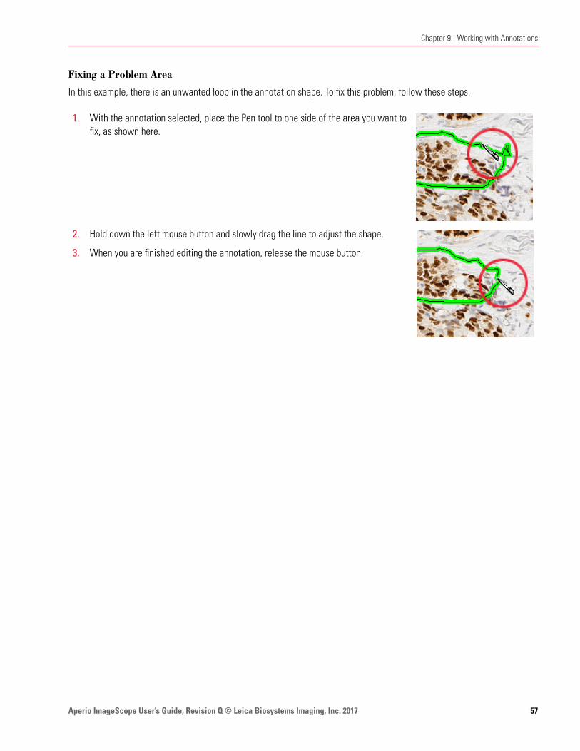
Aperio ImageScope User’s Guide, Revision Q © Leica Biosystems Imaging, Inc. 2017 57
Chapter 9: Working with Annotations
Fixing a Problem Area
In this example, there is an unwanted loop in the annotation shape. To fix this problem, follow these steps.
1. With the annotation selected, place the Pen tool to one side of the area you want to fix, as shown here.
2. Hold down the left mouse button and slowly drag the line to adjust the shape.
3. When you are finished editing the annotation, release the mouse button.
Aperio ImageScope User’s Guide, Revision Q © Leica Biosystems Imaging, Inc. 201758
10 Using the Annotations Window
Annotations are organized in the Annotations window in layers. Annotation layers also store algorithm analysis results. The Aperio ImageScope Annotations window contains the annotations associated with the image.
You can use the Annotations window in one of two modes:
Ì` Summary View – Designed for use with the eIHC product to provide one-step annotation and analysis for IHC eSlides. This view can also be used for quick analysis of any type.
Ì` Detailed View – Complete details on all annotations are available and you can add annotation attributes, text labels, and so on.
See the Aperio Image Analysis User’s Guide for information on using the Annotations Summary Window and on performing image analysis.
Annotations Summary View Window – Quick eIHC AnalysisThe detailed Annotations window provides a general solution for image analysis and handling annotations. However, a more streamlined version of the Annotations window is also available—the Annotations summary view. The Annotations summary view was specifically developed for analyzing IHC eSlides and makes the process quicker and simpler by fitting into a pathologist’s or researcher’s standard workflow.
Slide-Specific Processing
The key to the eIHC workflow is slide-specific processing, which defines how an eSlide is processed based on its stain and type of tissue (body site). Slide-specific processing can define what algorithm is used to analyze that type of slide, how analysis results are displayed and how to interpret those results (alternatively, manual scoring can be set up for the slide), and what comments are available for use by the pathologist or researcher viewing the slide. The slide-specific configuration for each stain/body site combination is defined by the Aperio eSlide Manager administrator. Once slide-specific processing is set up, viewing, annotating, and analyzing an eSlide becomes a quick process.
The summary view of the Annotations window is designed specifically for working with IHC eSlides to provide a quick way to mark tumor regions and analyze them in one simple step.
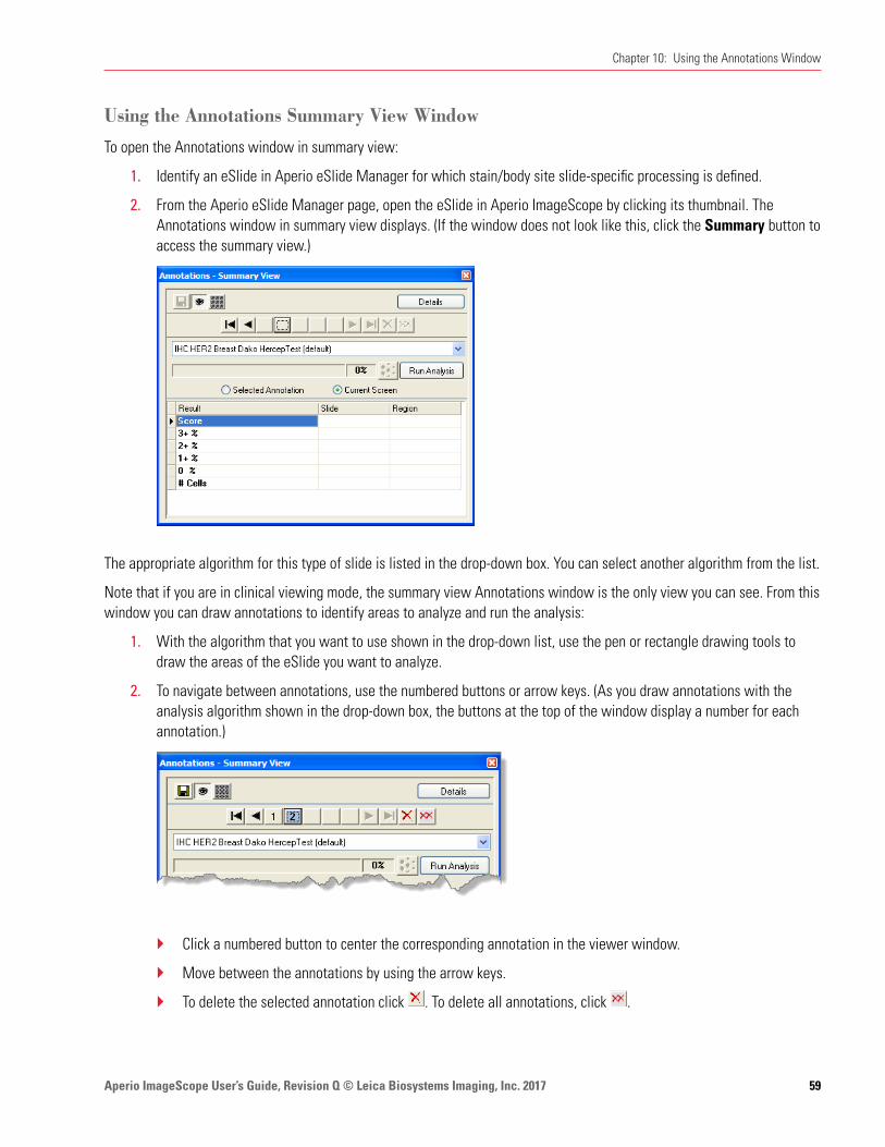
Aperio ImageScope User’s Guide, Revision Q © Leica Biosystems Imaging, Inc. 2017 59
Chapter 10: Using the Annotations Window
Using the Annotations Summary View Window
To open the Annotations window in summary view:
1. Identify an eSlide in Aperio eSlide Manager for which stain/body site slide-specific processing is defined.
2. From the Aperio eSlide Manager page, open the eSlide in Aperio ImageScope by clicking its thumbnail. The Annotations window in summary view displays. (If the window does not look like this, click the Summary button to access the summary view.)
The appropriate algorithm for this type of slide is listed in the drop-down box. You can select another algorithm from the list.
Note that if you are in clinical viewing mode, the summary view Annotations window is the only view you can see. From this window you can draw annotations to identify areas to analyze and run the analysis:
1. With the algorithm that you want to use shown in the drop-down list, use the pen or rectangle drawing tools to draw the areas of the eSlide you want to analyze.
2. To navigate between annotations, use the numbered buttons or arrow keys. (As you draw annotations with the analysis algorithm shown in the drop-down box, the buttons at the top of the window display a number for each annotation.)
Ì` Click a numbered button to center the corresponding annotation in the viewer window.
Ì` Move between the annotations by using the arrow keys.
Ì` To delete the selected annotation click . To delete all annotations, click .

Aperio ImageScope User’s Guide, Revision Q © Leica Biosystems Imaging, Inc. 201760
Chapter 10: Using the Annotations Window
Ì` To hide the selected annotation, click .
Ì` To save the annotations to Aperio eSlide Manager, click .
3. To analyze the current eSlide with the algorithm shown in the drop-down box:
a. Select the annotation drawn around the area you want to analyze and select Selected Annotation. (Or, if you want to analyze the entire area shown in the viewer window, select Current Screen.)
b. Click Run Analysis. As the analysis runs, you see progress information:
When the analysis is complete, you see the results:

Aperio ImageScope User’s Guide, Revision Q © Leica Biosystems Imaging, Inc. 2017 61
Chapter 10: Using the Annotations Window
Enabling and Disabling Pre-Processing
When using the Annotations window in summary view, you can temporarily turn off pre-processing region finding for algorithms that support that feature (for example, if you want to select possible tumor areas yourself to analyze rather than allowing the algorithm to do it for you).
To disable pre-processing, open the Annotations window in summary view and click the pre-processing button:
To turn pre-processing on again, click the button again. Note that this button is only enabled when you are using an algorithm that supports pre-processing.
Incremental Processing
The eIHC analysis applications are incremental algorithms, which means that as you add new regions and click Run Analysis on the Annotations window, only the new regions are analyzed, which can save a great deal of time. Any time you click Run Analysis again, all analysis results are updated.
Incremental processing is useful when a pathologist draws a single region and analyzes it, and after reviewing the analysis results wants to select additional regions and analyze them as well. The pathologist can add more annotation regions as needed or delete annotation regions and the analysis results will be updated accordingly.
If you delete a region, Aperio ImageScope automatically re-runs the analysis to update the summary analysis results that included that region. However, adding regions may make the analysis results incorrect until you re-run the analysis.
Other Options
Ì` To see a report image you have selected, select Report Image from the drop-down list.
Ì` To create an annotation that is not used for analysis (for example, a ruler or arrow), select Annotations from the drop-down list before drawing.
Ì` If the algorithm you are using supports result plots and they are enabled, the summary view window automatically opens all plots in separate windows when the analysis is done.

Aperio ImageScope User’s Guide, Revision Q © Leica Biosystems Imaging, Inc. 201762
Chapter 10: Using the Annotations Window
The Annotations – Detailed View WindowTo open the Annotations window go to the View menu and select Annotations.
The following Annotations window shows an eSlide with two annotations layers: “Dr. Bradley” and “Dr. Garcia.” (If your window does not look like this, click the Details button to return the Annotations window to the detailed view.)
The Annotations window in Detailed View contains the following sections:
Ì` Layers – lists all layers for this eSlide.
Ì` Layer Attributes – enables you to add and delete attributes for a layer. See “Using Attributes” on page 66 for more information.
Ì` Layer Regions – enables you to add and delete attributes for an annotation. You can use the Layer Regions to delete or move an annotation. This area also shows the length and area for the different annotation regions. If the resolution of the image is known, the length and area are displayed in microns. If not, the length and area are displayed in pixels.
Annotations Window Tools
Use the following tools to work with layers, layer attributes, and layer regions.
This tool: Is located here: And enables you to:Annotations window toolbar Arrange the Annotations window panes horizontally.
Annotations window toolbar Arrange the Annotations window panes vertically.
Annotations window toolbar Make Annotations window transparent.
Annotations window toolbar Save all annotations with the eSlide.
Annotations window toolbar Import annotations from a previously exported annotation file.
Annotations window toolbar Export the current annotations to an annotation file. Saved as XML with the same name as the eSlide file.
Layers pane Change the color of the annotations in the selected layer. (This box shows the current color for the layer annotations.)
You can also change the default color for each annotation layer. For more information, see “Annotation Color Options” on page 104.

Aperio ImageScope User’s Guide, Revision Q © Leica Biosystems Imaging, Inc. 2017 63
Chapter 10: Using the Annotations Window
This tool: Is located here: And enables you to:Layers pane Hide or show the annotations on the selected layer in the Aperio
ImageScope main window.Layer Attributes paneLayer Regions pane
Export the contents of the pane to a text file.
Layer Attributes Pane Display algorithm result plots.
Layer Attributes paneLayer Regions pane
Export the contents of the pane as an Excel spreadsheet.
Layers pane Layer Attributes pane Layer Regions pane
Add:New layer (if on the Layers pane)New layer attribute (if on the Layer Attributes pane)New annotation attribute (if on the Layer Regions pane)
Layer Attributes pane Layer Regions pane
Delete:Layer attribute (if on the Layer Attributes pane)Annotation attribute (if on the Layer Regions pane)
Layers pane Layer Regions pane
Delete:Selected layer (if on the Layers pane)Selected annotation (if on the Layer Regions pane)
Layers pane Delete all layers.
Annotation Length and Area Display
Aperio ImageScope measures and displays length and area for annotation regions. If the resolution of the image is known, length and area are displayed in microns; if not, they are displayed in pixels.
Adding Text to an Annotation
To add a text note that appears with an annotation.
1. In the Layers pane, select the layer that contains the annotation for which you want to add a note.
2. Click the annotation in the Layer Regions pane.

Aperio ImageScope User’s Guide, Revision Q © Leica Biosystems Imaging, Inc. 201764
Chapter 10: Using the Annotations Window
3. Type the note into the Text column.
The note appears next to the annotation, as shown below.
Moving Annotations
You can move a single annotation or all annotations.
To move: Do this:
One annotation Click the annotation to select it. Press and hold the Ctrl key, and drag the annotation to a new location.
All annotations You can move all annotations at the same time. For example, if you import annotations from a similar eSlide, slight variations in the new slide might require adjusting the position of the imported annotations.
Press and hold the Ctrl and Shift keys, and drag an annotation to a new location. All annotations move with the one you are dragging.
All annotations in all layers will be moved except for Z-stacks and markup images.
Exporting and Importing Annotation Layers
There are several ways to export and import information and annotations. You can also copy annotation layer information, and paste it into Word or Excel.
Algorithm analysis results are stored in an annotation layer so you may want to export that information into a text file to

Aperio ImageScope User’s Guide, Revision Q © Leica Biosystems Imaging, Inc. 2017 65
Chapter 10: Using the Annotations Window
include it in a report or to chart the information in a spreadsheet program.
You can also export annotations to be used on other eSlides. For example, if working with several very similar eSlides, you may find the same annotations apply to all of them.
To use this import or export feature: Do this:
Import or export annotations to the active eSlide
Ìy Export all annotations – on the Layers pane, click . Specify a name and location for the .xml file created.
Ìy Import an annotation file – on the Layers pane click . Navigate to the location of a previously exported annotations file.
Export text from Annotations Window Panes to a tab-delimited text file
This allows you to import the data into a spreadsheet program.
Ìy Export the text of the Layer Attributes pane – on the Layer Attributes pane, click . Specify the name and location of the text file to be created. Ìy Export the text of the Layer Regions pane – on the Regions Attributes
pane, click . Specify the name and location of the text file to be created.
Export text from Annotations Window panes to a Microsoft Excel Spreadsheet
Ìy Export text from the Layer Attributes pane – on the Layer Attributes pane, click . Specify the name and location of the new Excel (.xls) file.Ìy Export the text from the Layer Regions pane – on the Layer Regions
pane, click . Specify the name and location of the spreadsheet .xls file to be created.
Note: The numeric data is exported to Excel as text. Excel displays a note for each cell indicating that the numbers are in text format—you can click on the note to opt to transform the text to numeric format.
Copy and Paste Layer Attributes or Layer Region information
Select one or more cells from the Layer Attributes or Layer Region grids, and press Ctrl+C to copy the information to the Windows clipboard. Press Ctrl+V to paste the information into Word or Excel.
Note that the column headings are automatically copied over with the cell information.
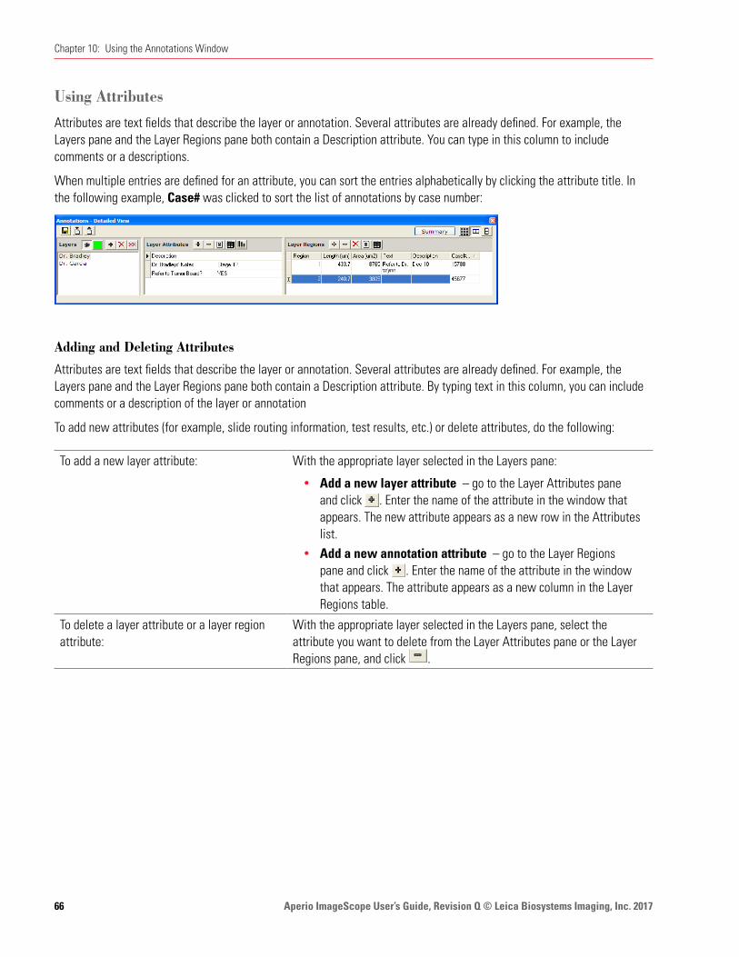
Aperio ImageScope User’s Guide, Revision Q © Leica Biosystems Imaging, Inc. 201766
Chapter 10: Using the Annotations Window
Using Attributes
Attributes are text fields that describe the layer or annotation. Several attributes are already defined. For example, the Layers pane and the Layer Regions pane both contain a Description attribute. You can type in this column to include comments or a descriptions.
When multiple entries are defined for an attribute, you can sort the entries alphabetically by clicking the attribute title. In the following example, Case# was clicked to sort the list of annotations by case number:
Adding and Deleting Attributes
Attributes are text fields that describe the layer or annotation. Several attributes are already defined. For example, the Layers pane and the Layer Regions pane both contain a Description attribute. By typing text in this column, you can include comments or a description of the layer or annotation
To add new attributes (for example, slide routing information, test results, etc.) or delete attributes, do the following:
To add a new layer attribute: With the appropriate layer selected in the Layers pane:
Ìy Add a new layer attribute – go to the Layer Attributes pane and click . Enter the name of the attribute in the window that appears. The new attribute appears as a new row in the Attributes list.Ìy Add a new annotation attribute – go to the Layer Regions
pane and click . Enter the name of the attribute in the window that appears. The attribute appears as a new column in the Layer Regions table.
To delete a layer attribute or a layer region attribute:
With the appropriate layer selected in the Layers pane, select the attribute you want to delete from the Layer Attributes pane or the Layer Regions pane, and click .
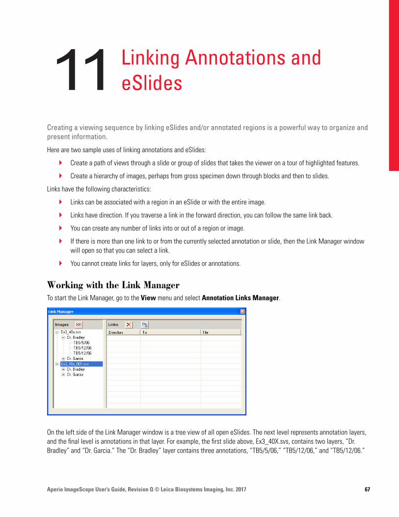
Aperio ImageScope User’s Guide, Revision Q © Leica Biosystems Imaging, Inc. 2017 67
11 Linking Annotations and eSlides
Creating a viewing sequence by linking eSlides and/or annotated regions is a powerful way to organize and present information.
Here are two sample uses of linking annotations and eSlides:
Ì` Create a path of views through a slide or group of slides that takes the viewer on a tour of highlighted features.
Ì` Create a hierarchy of images, perhaps from gross specimen down through blocks and then to slides.
Links have the following characteristics:
Ì` Links can be associated with a region in an eSlide or with the entire image.
Ì` Links have direction. If you traverse a link in the forward direction, you can follow the same link back.
Ì` You can create any number of links into or out of a region or image.
Ì` If there is more than one link to or from the currently selected annotation or slide, then the Link Manager window will open so that you can select a link.
Ì` You cannot create links for layers, only for eSlides or annotations.
Working with the Link ManagerTo start the Link Manager, go to the View menu and select Annotation Links Manager.
On the left side of the Link Manager window is a tree view of all open eSlides. The next level represents annotation layers, and the final level is annotations in that layer. For example, the first slide above, Ex3_40X.svs, contains two layers, “Dr. Bradley” and “Dr. Garcia.” The “Dr. Bradley” layer contains three annotations, “TB5/5/06,” “TB5/12/06,” and “TB5/12/06.”

Aperio ImageScope User’s Guide, Revision Q © Leica Biosystems Imaging, Inc. 201768
Chapter 11: Linking Annotations and eSlides
Click + to expand the lists, and click – to collapse them.
When you click a node in the tree, any links for that node appear in the list on the right.
Creating a LinkTo create a link:
1. In the left pane of the Link Manager window, drag a node and drop it onto another node.
For example, we dragged the first annotation on the Dr. Bradley layer onto the second annotation to create a link:
The symbol indicates that the link is a forward link. The symbol indicates the link is a backward link.
Viewing Links
The link navigation icons and commands are only enabled if a link exists. For example, if no previous link exists, you cannot use the Previous Annotation Link command.
You can follow links on the main Aperio ImageScope window or from within the Link Manager window. You do not need to open the Link Manager window to follow links.
If no annotation is selected, the viewing tools follow slide-level links; if there is more than one link to or from the selected annotation or slide, then the Link Manager window opens so that you can select a link.
Aperio ImageScope displays the target of the link in its main window. If the target is an eSlide, then it is fitted into the main window; if it is an annotation, then the annotation is centered and zoomed.

Aperio ImageScope User’s Guide, Revision Q © Leica Biosystems Imaging, Inc. 2017 69
Chapter 11: Linking Annotations and eSlides
To follow links in the main Aperio ImageScope window:
1. Open an eSlide that contains links.
2. To go to the next link, go to the View menu and select Next Annotation Link, or press Shift+F8, or click the icon on the Aperio ImageScope toolbar.
3. To go to a previous link, go to the View menu and select Previous Annotation Link, or press Shift+F7, or click the icon on the Aperio ImageScope toolbar.
To follow links in the Link Manager window: 1. Open an eSlide that contains links.
2. Go to the View menu and select Annotation Link Manager to open the Link Manager.
3. Click on the node for which links have been created and click in the right pane of the Link Manager window to follow the link.
Deleting Links
To delete a single link: In the Link Manager window, select a link in the right pane of the window and click .
To delete all links: In the Link Manager window, click .

Aperio ImageScope User’s Guide, Revision Q © Leica Biosystems Imaging, Inc. 201770
12 Tracking
The tracker tool enables you to record your movements through an eSlide and to save that record (known as a track) with the image as an annotation layer.
Tracking is only available for remote images opened in Aperio ImageScope from Aperio eSlide Manager.
Typical uses for the tracker tool are:
Ì` Histologists and pathologists might use this tool as reminder of which sections of the eSlide were visited.
Ì` The saved track might be useful for quality assurances purposes by providing permanent evidence of what sections of the eSlide were viewed.
Ì` The saved track could be used for educational purposes to provide students a tour of the eSlide.
Turning on the TrackerTo turn on the Tracker:
1. Go to the View menu and select Tracker. The tracker tool displays:
You can move this tool anywhere on your monitor display.
2. To start recording your movements, click the red record button :
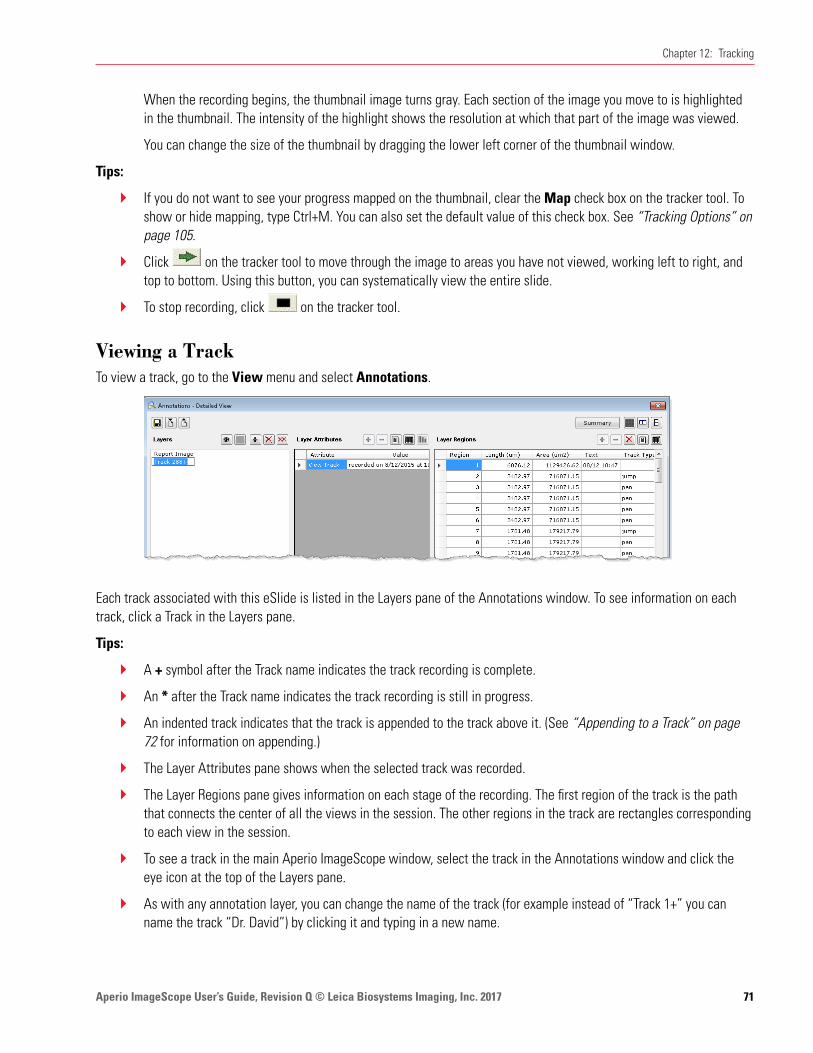
Aperio ImageScope User’s Guide, Revision Q © Leica Biosystems Imaging, Inc. 2017 71
Chapter 12: Tracking
When the recording begins, the thumbnail image turns gray. Each section of the image you move to is highlighted in the thumbnail. The intensity of the highlight shows the resolution at which that part of the image was viewed.
You can change the size of the thumbnail by dragging the lower left corner of the thumbnail window.
Tips:
Ì` If you do not want to see your progress mapped on the thumbnail, clear the Map check box on the tracker tool. To show or hide mapping, type Ctrl+M. You can also set the default value of this check box. See “Tracking Options” on page 105.
Ì` Click on the tracker tool to move through the image to areas you have not viewed, working left to right, and top to bottom. Using this button, you can systematically view the entire slide.
Ì` To stop recording, click on the tracker tool.
Viewing a TrackTo view a track, go to the View menu and select Annotations.
Each track associated with this eSlide is listed in the Layers pane of the Annotations window. To see information on each track, click a Track in the Layers pane.
Tips:
Ì` A + symbol after the Track name indicates the track recording is complete.
Ì` An * after the Track name indicates the track recording is still in progress.
Ì` An indented track indicates that the track is appended to the track above it. (See “Appending to a Track” on page 72 for information on appending.)
Ì` The Layer Attributes pane shows when the selected track was recorded.
Ì` The Layer Regions pane gives information on each stage of the recording. The first region of the track is the path that connects the center of all the views in the session. The other regions in the track are rectangles corresponding to each view in the session.
Ì` To see a track in the main Aperio ImageScope window, select the track in the Annotations window and click the eye icon at the top of the Layers pane.
Ì` As with any annotation layer, you can change the name of the track (for example instead of “Track 1+” you can name the track “Dr. David”) by clicking it and typing in a new name.

Aperio ImageScope User’s Guide, Revision Q © Leica Biosystems Imaging, Inc. 201772
Chapter 12: Tracking
Playing a TrackTo play the current track:
1. Click on the tracker tool.
To play a track made in the past:
1. Open an eSlide.
2. Go to the View menu and select Annotations to open the Annotations - Detailed View window. Select the track you want to play by clicking it in the Layers pane of the Annotations window.
3. Click on the tracker tool.
Tips:
While playing a track, use the tracker tool buttons to affect the playback:
Stop playing or recording the track.
Go to first view in the track
Go to the last view in the track
Go to the next view in the track
Go to the previous view in the track
As you play the track, the Annotations window updates to show your location in the Layer Regions pane.
Appending to a TrackYou can either start a new recording or append to a previous one. If a track already exists for this eSlide, when you click the record button, a message appears asking if you want to append the new track to the selected track.
To start a new recording, click No; to append to the previous track, click Yes.
If more than one track is created for the eSlide, to append to a specific track, open the Annotations window and select that track in the Layers pane before clicking the record button.
When a track is appended, the recording starts off as if all the views in the parent track have been viewed.
See “Viewing a Track” on page 71 for an example of an appended track.
Aperio ImageScope User’s Guide, Revision Q © Leica Biosystems Imaging, Inc. 2017 73
13 Saving eSlides and Regions
This chapter discusses several different ways to save images of eSlides.
WARNING: The images saved from Aperio ImageScope using the Copy, Snapshot, Export, and Extract features are for Research Use Only. Not for use in diagnostic procedures.
Copy and Paste an eSlide ImageYou can copy the viewable area of the eSlide image (and annotations) for use in reports or presentations.
1. Go to the Edit menu and select Copy.
2. Paste the image from the clipboard into an image or word editor, such as Microsoft Paint, Adobe Photoshop, or Microsoft Word.
Taking a SnapshotThe Snapshot tool enables you to take a picture of the current view of the eSlide, and save it as a JPEG or TIFF file for use in reports and presentations.
To take a snapshot, follow these steps.
1. Navigate to the area of the eSlide you want to capture so that it appears in the main Aperio ImageScope window.
2. Go to the File menu and select Save Snapshot or click on the toolbar.
3. In the Save Snapshot window, specify the location and name of the file, as well as the file type (JPEG or TIFF).
You can open and view the file from Windows Explorer or from within Aperio ImageScope.
If you make a snapshot of an image that has an embedded ICC profile, and Integrated Color Management is turned on in Aperio ImageScope, then Aperio ImageScope transforms the image using the monitor ICC profile. If Integrated Color Management is turned off, then Aperio ImageScope embeds the ICC profile from the scanner.
See “Viewing with Color Management” on page 29 for more information on color management.

Aperio ImageScope User’s Guide, Revision Q © Leica Biosystems Imaging, Inc. 201774
Chapter 13: Saving eSlides and Regions
Emailing a SnapshotYou can email the entire image displayed in the Aperio ImageScope window, or you can email snapshots of individual regions of the image.
To email a snapshot:
1. To draw attention to specific areas of the eSlide, use the rectangle drawing tool to mark those areas.
2. Go to the File menu and select Email Snapshot.
If you have not defined email settings for Aperio ImageScope, the SMTP Server tab of the Aperio ImageScope options appears. You must complete this information to continue. See “Email Settings” on page 109.
3. When the Mail Snapshot window appears, complete the email information by typing the recipient’s email address in the To field, and completing the Subject and Note fields.
4. In the Select Snapshot section, select from the following options:
Ì` Send current image view to send only the current viewable area of the image.
Ì` Send Regions in currently selected Annotation Layer to include regions in the selected annotation layer.
Ì` Delete Annotation Layer After Snapshot Mailed to delete the rectangle annotations after sending the email.
5. Click Mail.
Receiving a Snapshot Email
The Aperio ImageScope snapshot email includes the text you entered and JPEG files of the full Aperio ImageScope screen image and any annotations you selected for the email. The snapshot images are extracted at the same resolution used when the annotation was made.

Aperio ImageScope User’s Guide, Revision Q © Leica Biosystems Imaging, Inc. 2017 75
Chapter 13: Saving eSlides and Regions
Exporting ImagesUse this feature to export image files of a defined area of the eSlide image. You can export three versions of the same area, as shown below:
Original Image Annotated Image Markup Image
Images are exported as TIFF files, and are saved at the current viewing magnification.
To export images:
1. Click on the toolbar.
2. Click and drag a rectangle on the image to capture the area you want to export.
The Export Images window appears showing the area you specified, as shown below.
3. Select the check boxes next to the images you want to export:
Ì` Original Image – does not include annotations or markups.
Ì` Annotated Image – includes any annotations in the selected area.
Ì` Markup Image – includes any annotations and analysis markup areas.
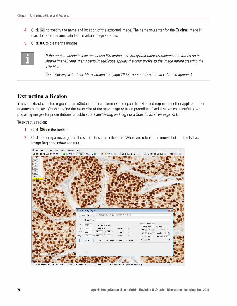
Aperio ImageScope User’s Guide, Revision Q © Leica Biosystems Imaging, Inc. 201776
Chapter 13: Saving eSlides and Regions
4. Click to specify the name and location of the exported image. The name you enter for the Original Image is used to name the annotated and markup image versions.
5. Click OK to create the images.
If the original image has an embedded ICC profile, and Integrated Color Management is turned on in Aperio ImageScope, then Aperio ImageScope applies the color profile to the image before creating the TIFF files.
See “Viewing with Color Management” on page 29 for more information on color management.
Extracting a RegionYou can extract selected regions of an eSlide in different formats and open the extracted region in another application for research purposes. You can define the exact size of the new image or use a predefined fixed size, which is useful when preparing images for presentations or publication (see“Saving an Image of a Specific Size” on page 79 ).
To extract a region:
1. Click on the toolbar.
2. Click and drag a rectangle on the screen to capture the area. When you release the mouse button, the Extract Image Region window appears.
Aperio ImageScope User’s Guide, Revision Q © Leica Biosystems Imaging, Inc. 2017 77
Chapter 13: Saving eSlides and Regions
3. Select from the following options:
Option DescriptionOutput File Location and name of the of the extracted file. If you do not use the Output File button, the new
file is created in the same location as the original eSlide, using the same name with a number appended.
Description Your title for the extracted region.Left Left pixel co-ordinate of the original image.Top Top pixel co-ordinate of the original image.Width Width of the extracted image in pixels.Height Height of the extracted image in pixels.Thumbnail Attach the thumbnail of the exported image.Label Attach the label image from the original scan if it exists.Macro Attach the macro image from the original if it exists.Tile Size Determines the organization of the data within the extracted image. For large SVS and TIFF files,
a tile size of 240 or 256 is optimal to enable fast access to the image.
To extract images for use with a third-party program that doesn’t support blocked TIFF files, you can set the tile size to zero to create a “stripped” image. Stripped TIFF files are supported by all software that processes TIFF files, but there is reduced performance for large images. JPEG files are always stripped (tile size is always zero).
Output/ Compression
The file formats and compression options available for the saved image:
Ìy SVS file format using JPEG2000 or JPEG compressionÌy JP2 file format using JPEG2000 compressionÌy TIF file format using LZW or no compressionÌy JPG file format using JPEG compressionÌy CWS file format using JPEG compression
Nx16 to 3x8 If you are using an Aperio fluorescent image (Aperio Fused Image, .afi file), this check box appears. Select this check box to create a single 3-channel, 8-bit RGB image file in the specified file format. See “Note on Fluorescent Images” below for more information.
ICC Profile If this image has an embedded ICC profile, you can select whether it is embedded, applied, ignored in the extracted image. For information on Aperio ePathology color management, see “Appendix B: Aperio Integrated Color Management” on page 113.
4. Choose the options you require and click Extract.
A progress bar shows the status of the extraction. When the extraction is complete, additional buttons are available for viewing the image.
5. Click the View button that appears at the bottom of the Extract Image Region window to open the extracted image in Aperio ImageScope. (For information on the Launch button, see “Compatibility Notes” on page 80.)
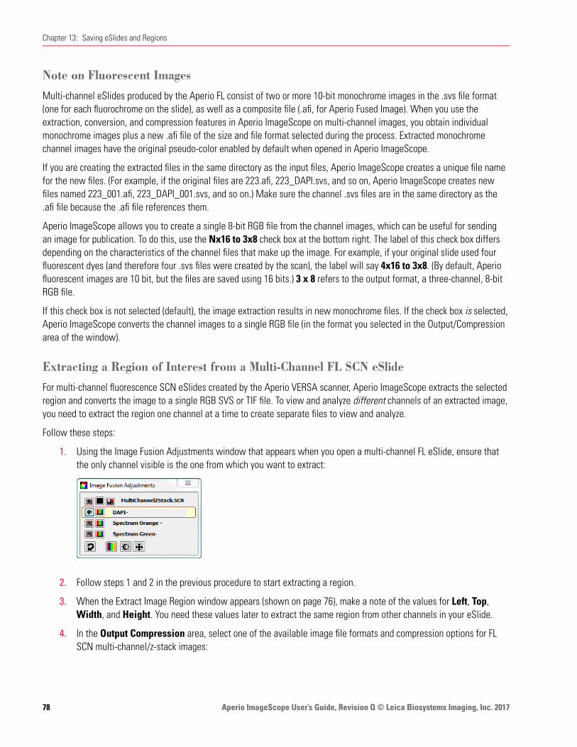
Aperio ImageScope User’s Guide, Revision Q © Leica Biosystems Imaging, Inc. 201778
Chapter 13: Saving eSlides and Regions
Note on Fluorescent Images
Multi-channel eSlides produced by the Aperio FL consist of two or more 10-bit monochrome images in the .svs file format (one for each fluorochrome on the slide), as well as a composite file (.afi, for Aperio Fused Image). When you use the extraction, conversion, and compression features in Aperio ImageScope on multi-channel images, you obtain individual monochrome images plus a new .afi file of the size and file format selected during the process. Extracted monochrome channel images have the original pseudo-color enabled by default when opened in Aperio ImageScope.
If you are creating the extracted files in the same directory as the input files, Aperio ImageScope creates a unique file name for the new files. (For example, if the original files are 223.afi, 223_DAPI.svs, and so on, Aperio ImageScope creates new files named 223_001.afi, 223_DAPI_001.svs, and so on.) Make sure the channel .svs files are in the same directory as the .afi file because the .afi file references them.
Aperio ImageScope allows you to create a single 8-bit RGB file from the channel images, which can be useful for sending an image for publication. To do this, use the Nx16 to 3x8 check box at the bottom right. The label of this check box differs depending on the characteristics of the channel files that make up the image. For example, if your original slide used four fluorescent dyes (and therefore four .svs files were created by the scan), the label will say 4x16 to 3x8. (By default, Aperio fluorescent images are 10 bit, but the files are saved using 16 bits.) 3 x 8 refers to the output format, a three-channel, 8-bit RGB file.
If this check box is not selected (default), the image extraction results in new monochrome files. If the check box is selected, Aperio ImageScope converts the channel images to a single RGB file (in the format you selected in the Output/Compression area of the window).
Extracting a Region of Interest from a Multi-Channel FL SCN eSlide
For multi-channel fluorescence SCN eSlides created by the Aperio VERSA scanner, Aperio ImageScope extracts the selected region and converts the image to a single RGB SVS or TIF file. To view and analyze different channels of an extracted image, you need to extract the region one channel at a time to create separate files to view and analyze.
Follow these steps:
1. Using the Image Fusion Adjustments window that appears when you open a multi-channel FL eSlide, ensure that the only channel visible is the one from which you want to extract:
2. Follow steps 1 and 2 in the previous procedure to start extracting a region.
3. When the Extract Image Region window appears (shown on page 76), make a note of the values for Left, Top, Width, and Height. You need these values later to extract the same region from other channels in your eSlide.
4. In the Output Compression area, select one of the available image file formats and compression options for FL SCN multi-channel/z-stack images:

Aperio ImageScope User’s Guide, Revision Q © Leica Biosystems Imaging, Inc. 2017 79
Chapter 13: Saving eSlides and Regions
Ì` SVS: JPEG2000 or JPG
Ì` TIF: LZW or NONE
5. Make any other necessary adjustments, and then click Extract.
6. Click Close to close the Extract Image Region dialog box.
7. To extract the same region from another channel on the same image:
a. Make the appropriate channel visible.
b. Click the Extract Region tool, and then click the eSlide to open the Extract Image Region dialog box to open the Extract Image Region dialog box.
c. Type in the values for Left, Top, Width, and Height that you recorded when extracting the region from the first channel.
d. Complete the process as described previously, repeating these steps for all channels in the eSlide.
Saving an Image of a Specific Size
To create an image of a specific size:
1. Follow steps 1 and 2 on page 76 to capture the area you want to extract.
2. When the Extract Image Region window displays, adjust the size of the saved file by changing the settings in the Width and Height boxes, located in the left area of the window.
Tips:
Ì` The first column of numbers after Width and Height is the number of pixels of the original extracted image. The second column is the output dimensions, initially set the same as the original values.
Ì` The percentages show the ratio between the original dimensions and the output dimensions. The check mark in the box causes both the width and height to be adjusted proportionally.
Ì` You can change the percentages to make an image smaller. If you are extracting a number of images, using the same percentage for them all ensures they all have the same resolution. You can adjust the percentage to greater than 100 to make the image larger.
Ì` Instead of changing the percentages, you can define a specific output dimension by typing the exact number of pixels you want for the height and width in the second width and height columns.
Extracting an Image of a Predefined Size or Aspect Ratio
If you have predefined a fixed size (see “Fixed Size Annotations” on page 104), to extract an image in the predefined size, hold down the Ctrl key while you click the extract tool on the toolbar.
To extract an image using the same aspect ratio of the predefined size (but not necessarily the same size), hold down the Shift and Ctrl keys while you use the extract tool to draw the region to extract.

Aperio ImageScope User’s Guide, Revision Q © Leica Biosystems Imaging, Inc. 201780
Chapter 13: Saving eSlides and Regions
Managing Viewing Applications
You can define applications other than Aperio ImageScope for viewing extracted regions. Doing so defines what viewing application Aperio ImageScope launches from the Extract Image Region window, but you can also open the extracted file outside of Aperio ImageScope using any compatible image viewing software (see the next section).
Compatibility Notes
Be aware that the viewing application you define may not be able to open an image of the type you’ve extracted. For example, Internet Explorer cannot open TIFF files, but can open JPEG files. If you click Launch and the application cannot open the extracted file, an error message appears from the application indicating it cannot open this type of file.
Also, the viewing application you define may not be able to handle a file as large as your extracted file. Some applications have a limit on the size of file they can open and work with. Some third-party programs cannot handle TIFF files, which use any form of compression. When in doubt, you can specify None for compression of TIFF files. However, if the region is large, using no compression results in a very large file.
Defining a Viewing Application
To define a viewing application:
1. Follow the instructions above to capture and extract a region of an eSlide.
2. Click the drop-down box next to the Launch button and select Define Application.
If no applications are defined, this list is empty except for this selection. If applications are defined, they display in alphabetical order.
3. When the Define Applications window appears, type a description of the application (for example, Photoshop) in the Application Name text box.
4. Click Browse to navigate to the location of the application executable file and select that file.
5. Click Add to add this application to the list of Aperio ImageScope viewing applications, and then click Done.
Using the Viewing Application
To view an extracted region with a viewing application (after one is defined):
1. Follow the steps above to extract a region.
2. Click the drop-down box next to the Launch button and select a viewing application.
3. Click the Launch button. (If no applications are defined, the Launch button is disabled.)
Aperio ImageScope User’s Guide, Revision Q © Leica Biosystems Imaging, Inc. 2017 81
Chapter 13: Saving eSlides and Regions
Deleting Viewing Applications
To delete applications previously defined:
1. Follow the steps above to extract a region.
2. Click the drop-down box next to the Launch button and select Define Application.
3. When the Define Applications window appears, click the Application Name drop-down box and select the application you want to delete from the list of Aperio ImageScope viewing applications. The Add button now changes to a Delete button.
4. Click Delete to delete the application from the list of Aperio ImageScope viewing applications, and then click Done. (This does not delete the application from your disk, but only from the list of Aperio ImageScope viewing applications.)
Aperio ImageScope User’s Guide, Revision Q © Leica Biosystems Imaging, Inc. 201782
14 eSlide Conferencing
eSlide conferencing makes it possible for several participants to view the same eSlide from multiple, remote locations. This section discusses how to use the eSlide conferencing feature of Aperio ImageScope.
About eSlide ConferencingAperio ImageScope eSlide conferencing not only allows multiple people to view the same eSlide at the same time, but also provides the following features:
Ì` Synchronized viewing so all participants see the same region of the eSlide at the same time
Ì` Real-time annotation sharing
Ì` Leader/follower roles and the ability to change those roles
All conference participants must have Aperio ImageScope installed on their workstations. (Download the latest free Aperio ImageScope software from www.LeicaBiosystems.com/ePathology.)
Concepts
Ì` The person hosting the conference is called the leader. This is usually the person who created the conference.
Ì` Participants who join the conference are called followers. Followers can see the eSlide the leader opens on the Aperio ImageScope viewer along with any annotations the leader makes.
Ì` Conferencing requires that all parties have access to a common DSC server and a common ImageServer or network share where images are stored.

Aperio ImageScope User’s Guide, Revision Q © Leica Biosystems Imaging, Inc. 2017 83
Chapter 14: eSlide Conferencing
Starting an eSlide Conference1. Log into Aperio eSlide Manager and open an eSlide.
2. Go to the View menu and click Digital Slide Conferencing. The following window displays:
Connecting to an eSlide Conferencing Server
1. On the Digital Slide Conferencing window, click DSC Server. The following window appears:
2. Type the following information:
Ìy Server Name – The example shows a DSC server running at dsc.scanscope.com. Type the name of the server that is running the DSC service on your network. Usually this is on your DSR, a dedicated server, or it may be on your workstation.
Ìy Port – Type the number of the port that has been configured for DSC. This is usually 80, but can be set to another port.
Ìy Email Address and Password are not required.

Aperio ImageScope User’s Guide, Revision Q © Leica Biosystems Imaging, Inc. 201784
Chapter 14: eSlide Conferencing
Ìy Screen Name – Type the name you want to use as the leader of the conference. You are identified by this name in the conference participant list.
3. Click Login. The status changes to Connected and the Create Conference button is enabled.
If you set up the DSC server on your workstation, verify your workstation is accessible to others. See your network administrator if necessary to ensure the correct ports are open, firewall permissions are set appropriately.
Creating a Conference1. Click the Create Conference button, and then enter the name of your conference:
2. Enter any name you wish and click OK.
You are notified that you are the conference leader. The baton next to your name indicates that you are the conference leader.
3. To display your mouse cursor in the participants’ view, select the Show Mouse check box.
The conference is now created.
Opening an Image to Share
The conference leader may open one or more eSlides to share in the conference. Because all participants must have access to the images too, the leader must open images on an ImageServer using the Access Remote Server command or directly from Aperio eSlide Manager. (You cannot share images in an eSlide conference if they are only on your workstation.)

Aperio ImageScope User’s Guide, Revision Q © Leica Biosystems Imaging, Inc. 2017 85
Chapter 14: eSlide Conferencing
The leader can open and move between multiple images during the conference using the Aperio ImageScope filmstrip.
Joining a Conference
Once a conference is created, participants may join.
1. Log into Aperio eSlide Manager and open any eSlide by clicking an eSlide thumbnail on the Aperio eSlide Manager page.
2. Follow the instructions in “Connecting to an eSlide Conferencing Server” on page 83 to connect to the DSC server. The following window displays listing any conferences running on that server:
Type the screen name you want to display to the other conference participants.
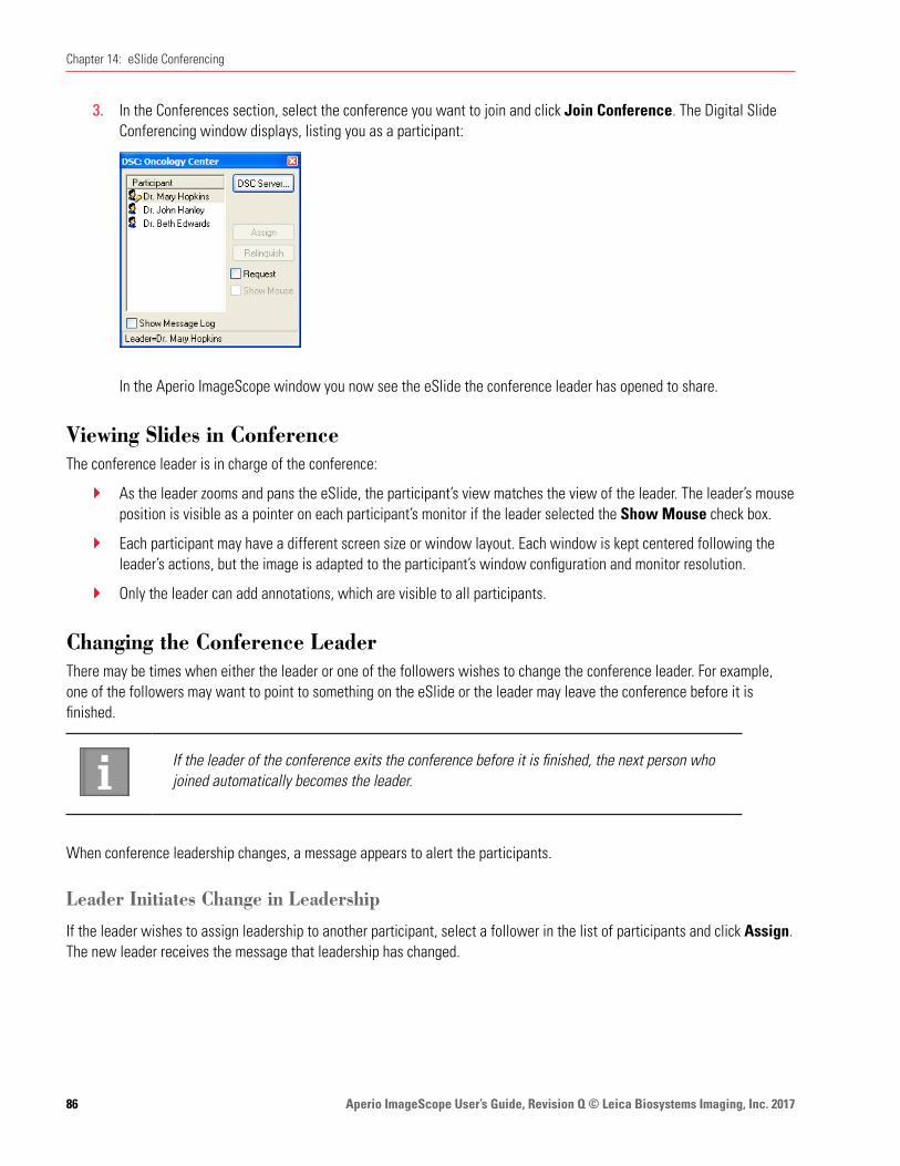
Aperio ImageScope User’s Guide, Revision Q © Leica Biosystems Imaging, Inc. 201786
Chapter 14: eSlide Conferencing
3. In the Conferences section, select the conference you want to join and click Join Conference. The Digital Slide Conferencing window displays, listing you as a participant:
In the Aperio ImageScope window you now see the eSlide the conference leader has opened to share.
Viewing Slides in ConferenceThe conference leader is in charge of the conference:
Ì` As the leader zooms and pans the eSlide, the participant’s view matches the view of the leader. The leader’s mouse position is visible as a pointer on each participant’s monitor if the leader selected the Show Mouse check box.
Ì` Each participant may have a different screen size or window layout. Each window is kept centered following the leader’s actions, but the image is adapted to the participant’s window configuration and monitor resolution.
Ì` Only the leader can add annotations, which are visible to all participants.
Changing the Conference LeaderThere may be times when either the leader or one of the followers wishes to change the conference leader. For example, one of the followers may want to point to something on the eSlide or the leader may leave the conference before it is finished.
If the leader of the conference exits the conference before it is finished, the next person who joined automatically becomes the leader.
When conference leadership changes, a message appears to alert the participants.
Leader Initiates Change in Leadership
If the leader wishes to assign leadership to another participant, select a follower in the list of participants and click Assign. The new leader receives the message that leadership has changed.
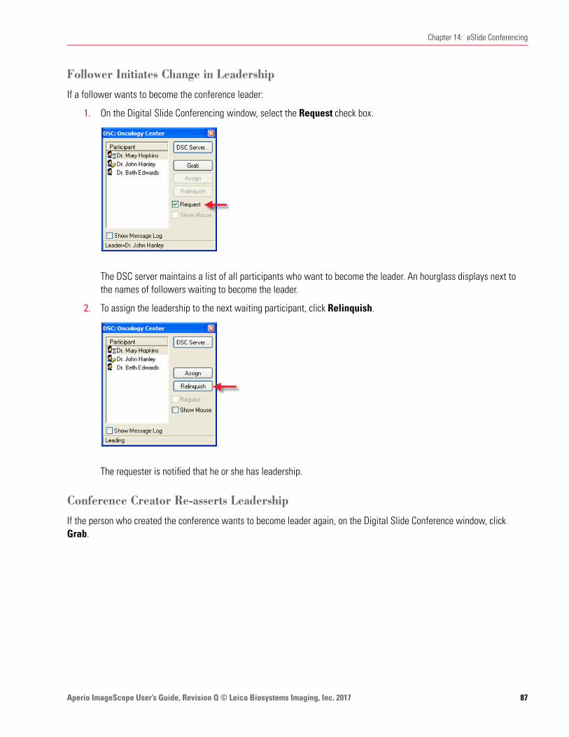
Aperio ImageScope User’s Guide, Revision Q © Leica Biosystems Imaging, Inc. 2017 87
Chapter 14: eSlide Conferencing
Follower Initiates Change in Leadership
If a follower wants to become the conference leader:
1. On the Digital Slide Conferencing window, select the Request check box.
The DSC server maintains a list of all participants who want to become the leader. An hourglass displays next to the names of followers waiting to become the leader.
2. To assign the leadership to the next waiting participant, click Relinquish.
The requester is notified that he or she has leadership.
Conference Creator Re-asserts Leadership
If the person who created the conference wants to become leader again, on the Digital Slide Conference window, click Grab.
Aperio ImageScope User’s Guide, Revision Q © Leica Biosystems Imaging, Inc. 201788
15 TelePath Live
TelePath Live, also known as Remote Revisit, provides a way to connect to your scanner remotely.
This chapter provides information and instructions on using TelePath Live to capture z-stack images from live video. For information on viewing z-stack eSlide images scanned on the Aperio scanner, see “Chapter 4: Viewing Z-Stack eSlide Images” on page 31.
By using TelePath Live to directly connect to your scanner, you can:
Ì` View a live video feed from the scanner from a remote location.
Ì` Capture Z-stacks and view specimens in multiple focal planes.
Ì` Perform a scan directly from Aperio ImageScope.
Scanner Compatibility NotesÌ` For scanners that contain an AutoLoader—a slide must be on the slide tray to use the Z-stack feature. You cannot
capture Z-stacks from slides in an AutoLoader.
Ì` Some older scanner models cannot use the TelePath Live feature; contact Leica Biosystems Imaging Technical Services if you have questions about whether your scanner can use TelePath Live.
Ì` Aperio CS scanners support TelePath Live from all five slide positions in the slide tray.
Before You Use TelePath LiveThere are some things you should do and know before you use TelePath Live.
Calibration
Your scanner is calibrated at the factory. However, if while using TelePath Live you find that the Live window does not line up with your cursor position in the Aperio ImageScope main window, you should calibrate it again.
For information on calibrating the scanner in preparation for using TelePath Live, please see the TelePath Live (Remote Revisit) Setup Application Note.
Setting the ImageServerURL
In order for the Console program View Slide button to display images in Aperio ImageScope that can be used for TelePath Live, the ImageServerURL parameter needs to be set up with your ImageServer name.

Aperio ImageScope User’s Guide, Revision Q © Leica Biosystems Imaging, Inc. 2017 89
Chapter 15: TelePath Live
To set up ImageServerURL:
1. Start the scanner Console program by clicking Start > All Programs > ScanScope > Console.
2. Open the Configure window. (See your Console User’s Guide for specific instructions.)
3. Click the ScanScope Properties tab.
4. In the Name field, type: ImageServerURL and click Get.
5. Verify that the name of your ImageServer displays in the Value field.
a. If the ImageServer name is correct, you are done.
b. If the ImageServer name is not correct or is blank, type the correct name of the ImageServer into the Value box and click Update.
6. Click Close to exit the Configure window. After you exit the Console, your change to ImageServerURL will take effect the next time you start the Console again.
Here is a little more information about why you need to set this parameter:
When ImageServerURL is set to a valid ImageServer machine name, Aperio ImageScope opens the eSlide by passing the imageID directly to ImageServer. ImageServer uses the database to locate the eSlide. You can tell that you are in the database mode if you see a number beginning with the @ symbol in the title bar of Aperio ImageScope.
You must use ImageServer in this database mode to save captured Z-stacks into the database. If ImageServerURL is not set, Z-stacks your capture in Aperio ImageScope will not be saved in the database when you open the eSlide using the View Slide button in the Console.
What Is a Z-Stack?A Z-stack is a three-dimensional image that allows you to view a specimen by moving up and down in the focal planes. This is especially useful for viewing “thick” specimens.
Z-stack images are cataloged in the ImageServer database, and linked to the original image as an annotation. One of the reasons for using the TelePath Live feature is to capture and view Z-stack images. See “Capturing Z-stacks” on page 93 for more information.

Aperio ImageScope User’s Guide, Revision Q © Leica Biosystems Imaging, Inc. 201790
Chapter 15: TelePath Live
Connecting to an Aperio Scanner1. Go to the Aperio ImageScope View menu and select TelePath Live. The following window displays:
2. Click Configure to define which scanner to connect to. The following window displays:
3. Type the name of the scanner and its port (usually 3333).
4. Define the ImageServer by typing the host name and port number. If you are using an Aperio CS or T3 scanner, this is the same name and port number as your scanner.
Note that the name of your ImageServer host is typically the same as the ImageServerURL setting (see ”Setting the ImageServerURL” on page 88) unless you access the ImageServer from outside a firewall, in which case it may be different—contact your network administrator for assistance.
5. Click OK.
6. On the TelePath Live window, click Connect.

Aperio ImageScope User’s Guide, Revision Q © Leica Biosystems Imaging, Inc. 2017 91
Chapter 15: TelePath Live
Troubleshooting Tips: If you are unable to connect to the scanner, contact your network administrator to verify the following conditions are true for your network:
y Port 3333 is open and directed to the scanner controller.
y Port 82 is open and directed to the ImageServer.
8. Log in using your Aperio eSlide Manager user name and password.
After a few seconds, a window appears showing slides that are loaded in the tray. You are now connected to a scanner.
The blue dot indicates the slide you are working on. You can click any displayed slide to select that slide.
If the window has no slides displayed, click the Refresh Snapshots button to load macro slide images.

Aperio ImageScope User’s Guide, Revision Q © Leica Biosystems Imaging, Inc. 201792
Chapter 15: TelePath Live
Preparing a Slide for TelePath LiveThere are two ways to prepare a slide for use with the TelePath Live window:
1. Scan a slide on the scanner and click the View Slide button in the Console. When Aperio ImageScope opens, you can begin placing Z-stack images on the eSlide from the Aperio ImageScope TelePath Live window.
2. Use the Revisit Quick Scan or Revisit Hi-Res Scan buttons on the Aperio ImageScope TelePath Live window. (See “Capturing Z-stacks” on page 93.)
Viewing Live Video from the ScannerConnect to the scanner and open the Aperio ImageScope TelePath Live window as discussed in “Connecting to an Aperio Scanner” on page 90.
After a scan is performed, click around on an eSlide in the TelePath Live window and see a live video feed from the scanner showing the tissue in the Live window.
When using TelePath Live, coarse and fine focus sliders provide fine-resolution focusing, allowing you to fine-tune the focus:
The right slider’s range is 10% of the entire focus range provided by the left slider, centered on the value of the left slider. For example, if the left slider is set at 50, the right slider’s range is 50 – 5% of the focus range to 50 + 5% of the focus range.
Use the slider on the left to get the image roughly into focus. Then use the slider on the right to fine-tune the focus.
Ì` Adjust the size of the Live window by using the Camera Mode drop-down list to select a window size:

Aperio ImageScope User’s Guide, Revision Q © Leica Biosystems Imaging, Inc. 2017 93
Chapter 15: TelePath Live
Ì` If you locate an area of interest, click the Capture Z-Stack button on the TelePath Live window to capture a Z-stack (3-dimensional) image of that area. (See the next section.)
Capturing Z-stacks1. Open the TelePath Live window and connect to a scanner as discussed in “Connecting to an Aperio Scanner” on
page 90.
The blue dot indicates the currently selected slide. Click on any other slide if you want to use it instead.

Aperio ImageScope User’s Guide, Revision Q © Leica Biosystems Imaging, Inc. 201794
Chapter 15: TelePath Live
2. Select your Z-stack options:
Ì` Depth – The total number of microns in the Z-stack.
Ì` Frame Count – The number of individual snapshots taken. The limit is 50.
Ì` Camera Mode – The width and height of the Z-stack snapshots. These are limited by the available modes of the area camera.
3. If you did not open Aperio ImageScope by clicking the View Slide button in the Console program, you may need to scan the slide:
Ì` Clicking the Revisit Quick Scan button on the TelePath Live window performs a high-resolution macro scan of the slide, generally used when time is short. The slide is scanned at 5x.
Ì` Clicking the Revisit Hi-Res Scan button on the TelePath Live window is the same as pressing the green button on the scanner. Only the currently selected slide is be scanned, and it is scanned at the currently selected magnification.
Whether the slide was scanned from the Console or from the TelePath Live window, when the slide is scanned its image displays in the Aperio ImageScope main window along with a Live window that shows the video feed from the scanner.
4. Click the image in the Aperio ImageScope main window. A yellow rectangle displays showing the area of the image displayed in the Live window. This also selects the area to perform a Z-stack capture. To move the selection rectangle, click another location in the main image.
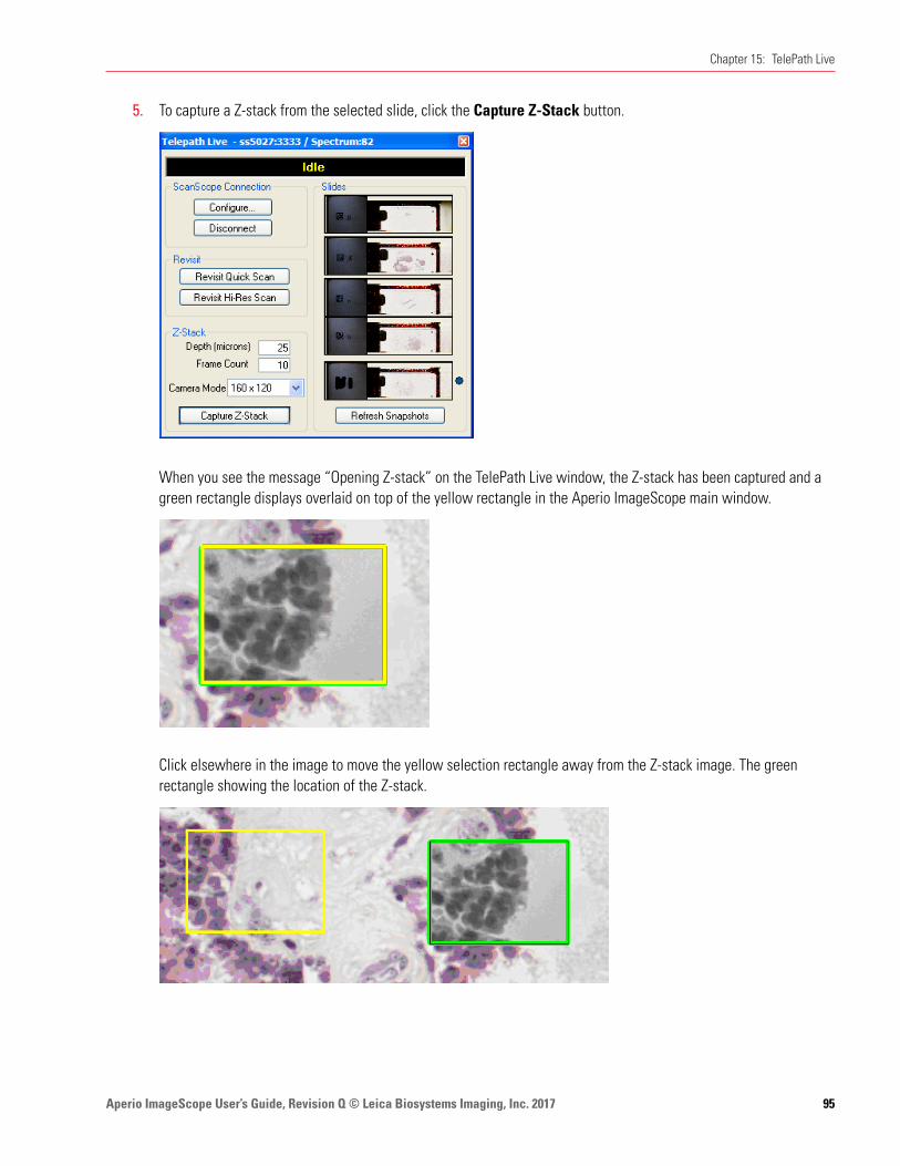
Aperio ImageScope User’s Guide, Revision Q © Leica Biosystems Imaging, Inc. 2017 95
Chapter 15: TelePath Live
5. To capture a Z-stack from the selected slide, click the Capture Z-Stack button.
When you see the message “Opening Z-stack” on the TelePath Live window, the Z-stack has been captured and a green rectangle displays overlaid on top of the yellow rectangle in the Aperio ImageScope main window.
Click elsewhere in the image to move the yellow selection rectangle away from the Z-stack image. The green rectangle showing the location of the Z-stack.
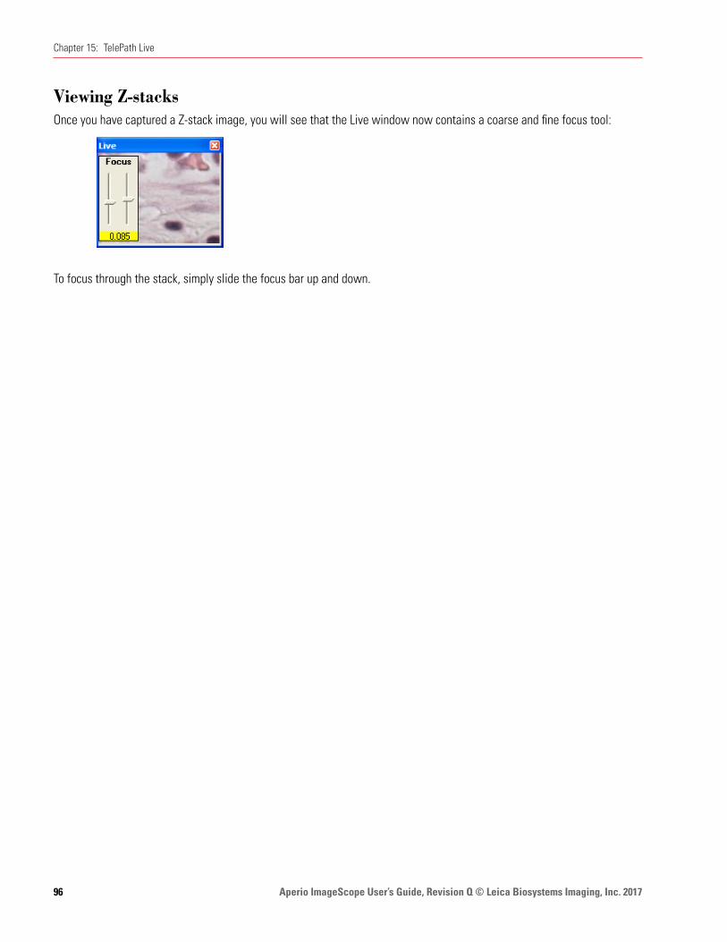
Aperio ImageScope User’s Guide, Revision Q © Leica Biosystems Imaging, Inc. 201796
Chapter 15: TelePath Live
Viewing Z-stacksOnce you have captured a Z-stack image, you will see that the Live window now contains a coarse and fine focus tool:
To focus through the stack, simply slide the focus bar up and down.
Aperio ImageScope User’s Guide, Revision Q © Leica Biosystems Imaging, Inc. 2017 97
16 Utilities and Diagnostics
The utilities discussed in this section are primarily used for troubleshooting. Leica Biosystems Imaging Service representatives may ask you to use these utilities to pinpoint an issue.
LoggingIf requested to do so by Leica Biosystems Imaging Services, enable logging by going to the Tools menu and selecting Logging. This creates a file named viewport.log where text messages on Aperio ImageScope actions are logged.
If you are having a problem, Technical Support typically asks you to:
1. Turn logging on.
2. Run until you encounter the problem again.
3. Then email Technical Support the viewport.log file.
You should then turn logging off (by again going to the Tools menu and selecting Logging) as logging affects system performance.
Cache DisplayBecause eSlides are typically much larger than can be loaded into memory all at once, Aperio ImageScope keeps portions of the eSlide in computer memory so it can display them quickly. It prefetches data for the eSlide from disk or from a remote server, determining which portions of the image to fetch by anticipating where the user is going to go next.
Enable the cache display by pressing the Shift+Ctrl+M keys. (Press Shift+Ctrl+M keys again to turn it off.)
The cache display shows which parts of the eSlide are loaded into Aperio ImageScope’s cache memory. The darker the area, the higher the resolution of the part of the image that is loaded.

Aperio ImageScope User’s Guide, Revision Q © Leica Biosystems Imaging, Inc. 201798
Chapter 16: Utilities and Diagnostics
Below is an image of the Aperio ImageScope screen showing the cache displays:
Running Multiple Aperio ImageScope SessionsAperio ImageScope normally allows only one instance of itself to run. (If Aperio ImageScope is open and you try to start it again, the first copy of Aperio ImageScope closes.) There may be special occasions when troubleshooting a problem or testing eSlide Conferencing, for example, when you want to run more than one copy of Aperio ImageScope at a time.
To run multiple copies of Aperio ImageScope:
1. Click Start and select Run.
2. Click Browse on the Browse window and navigate to the ImageScope.exe file on your workstation. (The default location is: C:\Program Files\ScanScope\ImageScope.)
3. Click Open.
The Run window now contains the path to the ImageScope.exe file.
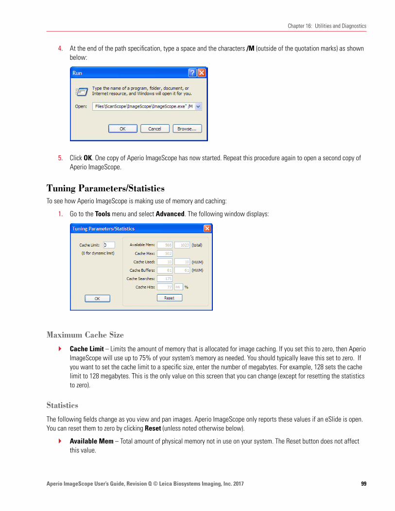
Aperio ImageScope User’s Guide, Revision Q © Leica Biosystems Imaging, Inc. 2017 99
Chapter 16: Utilities and Diagnostics
4. At the end of the path specification, type a space and the characters /M (outside of the quotation marks) as shown below:
5. Click OK. One copy of Aperio ImageScope has now started. Repeat this procedure again to open a second copy of Aperio ImageScope.
Tuning Parameters/StatisticsTo see how Aperio ImageScope is making use of memory and caching:
1. Go to the Tools menu and select Advanced. The following window displays:
Maximum Cache Size
Ì` Cache Limit – Limits the amount of memory that is allocated for image caching. If you set this to zero, then Aperio ImageScope will use up to 75% of your system’s memory as needed. You should typically leave this set to zero. If you want to set the cache limit to a specific size, enter the number of megabytes. For example, 128 sets the cache limit to 128 megabytes. This is the only value on this screen that you can change (except for resetting the statistics to zero).
Statistics
The following fields change as you view and pan images. Aperio ImageScope only reports these values if an eSlide is open. You can reset them to zero by clicking Reset (unless noted otherwise below).
Ì` Available Mem – Total amount of physical memory not in use on your system. The Reset button does not affect this value.
Aperio ImageScope User’s Guide, Revision Q © Leica Biosystems Imaging, Inc. 2017100
Chapter 16: Utilities and Diagnostics
Ì` Total Mem – Total amount of memory installed on your system. The Reset button does not affect this value.
Ì` Cache Max – Amount of cache memory used in megabytes.
Ì` Cache Used – Current amount of memory cache used in megabytes.
Ì` Cache Used HWM – High water mark for memory cache in megabytes.
Ì` Cache Buffers – Current number of buffers stored in cache.
Ì` Cache Buffers HWM – High water mark for buffers stored in cache.
Ì` Cache Searches – Number of cache searches.
Ì` Cache Hits – Ratio of cache hits to cache searches.
Ì` Cache Hits % – Ratio of cache hits to cache searches.
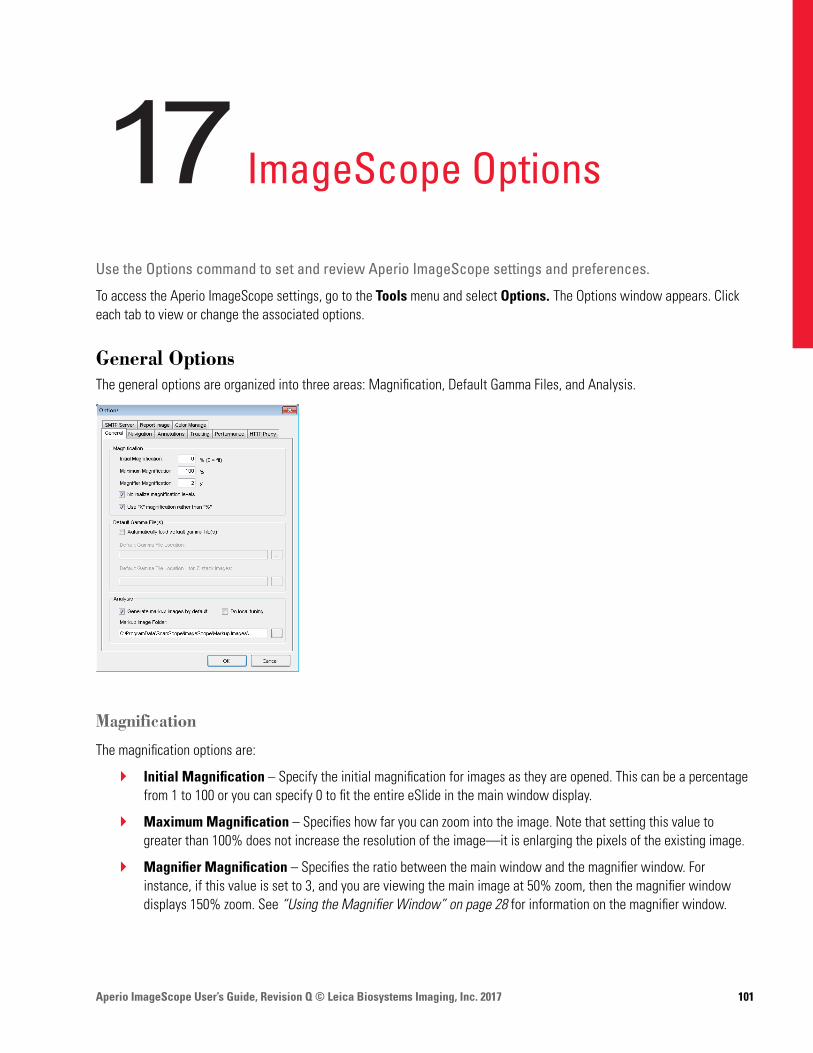
Aperio ImageScope User’s Guide, Revision Q © Leica Biosystems Imaging, Inc. 2017 101
17 ImageScope Options
Use the Options command to set and review Aperio ImageScope settings and preferences.
To access the Aperio ImageScope settings, go to the Tools menu and select Options. The Options window appears. Click each tab to view or change the associated options.
General OptionsThe general options are organized into three areas: Magnification, Default Gamma Files, and Analysis.
Magnification
The magnification options are:
Ì` Initial Magnification – Specify the initial magnification for images as they are opened. This can be a percentage from 1 to 100 or you can specify 0 to fit the entire eSlide in the main window display.
Ì` Maximum Magnification – Specifies how far you can zoom into the image. Note that setting this value to greater than 100% does not increase the resolution of the image—it is enlarging the pixels of the existing image.
Ì` Magnifier Magnification – Specifies the ratio between the main window and the magnifier window. For instance, if this value is set to 3, and you are viewing the main image at 50% zoom, then the magnifier window displays 150% zoom. See “Using the Magnifier Window” on page 28 for information on the magnifier window.
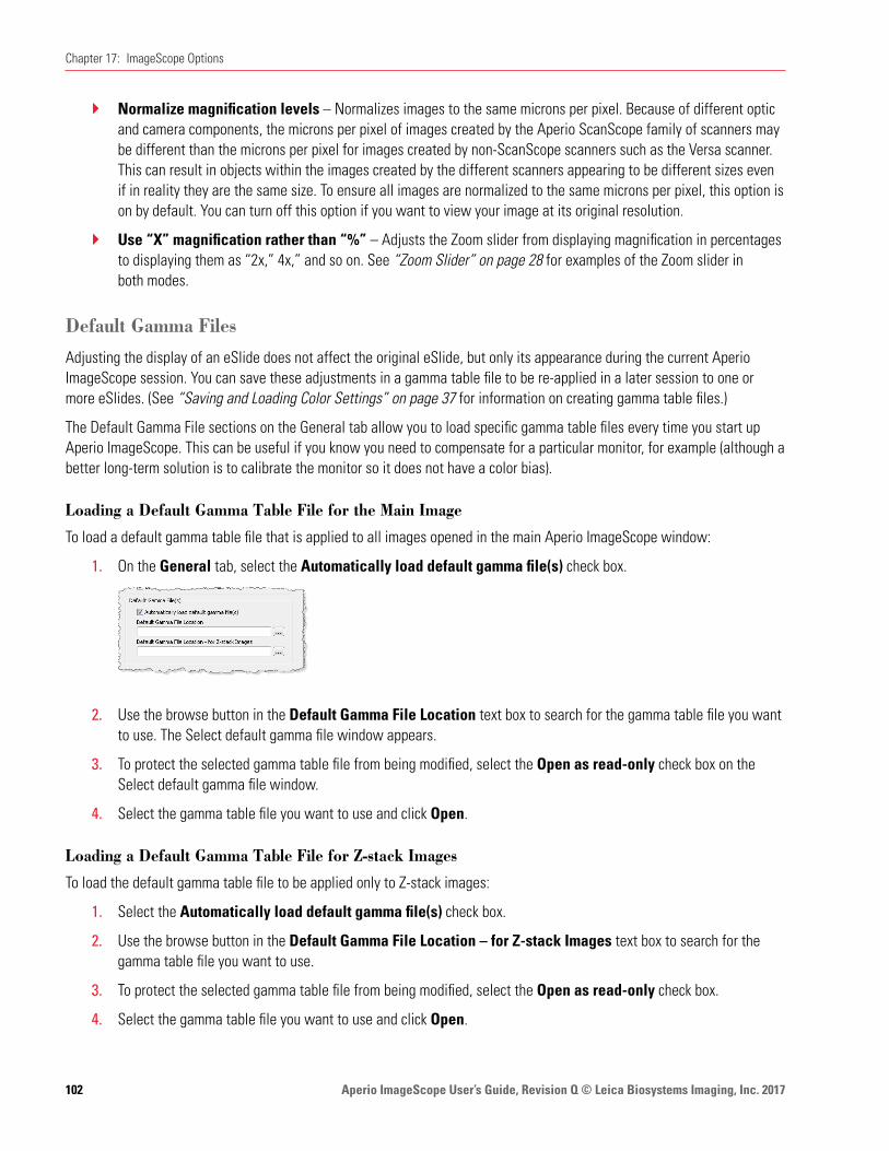
Aperio ImageScope User’s Guide, Revision Q © Leica Biosystems Imaging, Inc. 2017102
Chapter 17: ImageScope Options
Ì` Normalize magnification levels – Normalizes images to the same microns per pixel. Because of different optic and camera components, the microns per pixel of images created by the Aperio ScanScope family of scanners may be different than the microns per pixel for images created by non-ScanScope scanners such as the Versa scanner. This can result in objects within the images created by the different scanners appearing to be different sizes even if in reality they are the same size. To ensure all images are normalized to the same microns per pixel, this option is on by default. You can turn off this option if you want to view your image at its original resolution.
Ì` Use “X” magnification rather than “%” – Adjusts the Zoom slider from displaying magnification in percentages to displaying them as “2x,” 4x,” and so on. See “Zoom Slider” on page 28 for examples of the Zoom slider in both modes.
Default Gamma Files
Adjusting the display of an eSlide does not affect the original eSlide, but only its appearance during the current Aperio ImageScope session. You can save these adjustments in a gamma table file to be re-applied in a later session to one or more eSlides. (See “Saving and Loading Color Settings” on page 37 for information on creating gamma table files.)
The Default Gamma File sections on the General tab allow you to load specific gamma table files every time you start up Aperio ImageScope. This can be useful if you know you need to compensate for a particular monitor, for example (although a better long-term solution is to calibrate the monitor so it does not have a color bias).
Loading a Default Gamma Table File for the Main Image
To load a default gamma table file that is applied to all images opened in the main Aperio ImageScope window:
1. On the General tab, select the Automatically load default gamma file(s) check box.
2. Use the browse button in the Default Gamma File Location text box to search for the gamma table file you want to use. The Select default gamma file window appears.
3. To protect the selected gamma table file from being modified, select the Open as read-only check box on the Select default gamma file window.
4. Select the gamma table file you want to use and click Open.
Loading a Default Gamma Table File for Z-stack Images
To load the default gamma table file to be applied only to Z-stack images:
1. Select the Automatically load default gamma file(s) check box.
2. Use the browse button in the Default Gamma File Location – for Z-stack Images text box to search for the gamma table file you want to use.
3. To protect the selected gamma table file from being modified, select the Open as read-only check box.
4. Select the gamma table file you want to use and click Open.
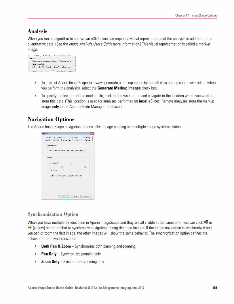
Aperio ImageScope User’s Guide, Revision Q © Leica Biosystems Imaging, Inc. 2017 103
Chapter 17: ImageScope Options
AnalysisWhen you run an algorithm to analyze an eSlide, you can request a visual representation of the analysis in addition to the quantitative data. (See the Image Analysis User’s Guide more information.) This visual representation is called a markup image.
Ì` To instruct Aperio ImageScope to always generate a markup image by default (this setting can be overridden when you perform the analysis), select the Generate Markup Images check box.
Ì` To specify the location of the markup file, click the browse button and navigate to the location where you want to store this data. (This location is used for analyses performed on local eSlides. Remote analyses store the markup image only in the Aperio eSlide Manager database.)
Navigation OptionsThe Aperio ImageScope navigation options affect image panning and multiple image synchronization.
Synchronization Option
When you have multiple eSlides open in Aperio ImageScope and they are all visible at the same time, you can click or (yellow) on the toolbar to synchronize navigation among the open images. If the image navigation is synchronized and
you pan or zoom the first image, the other images will show the same behavior. The synchronization option defines the behavior of that synchronization:
Ì` Both Pan & Zoom – Synchronizes both panning and zooming.
Ì` Pan Only – Synchronizes panning only.
Ì` Zoom Only – Synchronizes zooming only.

Aperio ImageScope User’s Guide, Revision Q © Leica Biosystems Imaging, Inc. 2017104
Chapter 17: ImageScope Options
Panning Options
The following panning options are available:
Ì` Scroll Speed – Specifies how fast the image scrolls when holding down the mouse button near the edge of an image.
Ì` Pan Speed – Specifies how fast the image pans when clicking and dragging the image.
Ì` Reverse panning direction (“Pathologist” mode) – Select this check box to pan in reverse. With this option enabled, as you click and drag to the left, the image moves to the right.
Annotation OptionsThe Annotations tab of the Options window allows you to select annotation options.
Annotation Color Options
Each annotation layer displays annotations in a different color. The Default Annotation Colors section allows you to select the color used for each layer.
1. Click the colored box above the annotation layer for which you want to change the color.
2. From the color selection window, select a color from the palette shown or click Define Custom Colors to define your own color.
3. Click OK.
If you use more than five annotation layers, the new layers re-use the colors defined for the first five layers. For example, layer 6 will use the color defined for layer 1 and layer 7 will use the color defined for layer 2.
Fixed Size Annotations
The Fixed Size Regions options on the Annotations tab set a fixed size for drawn annotations that is used when you press the Ctrl key while you draw. These settings are also used to extract a region of fixed size and to zoom to a region of fixed size.

Aperio ImageScope User’s Guide, Revision Q © Leica Biosystems Imaging, Inc. 2017 105
Chapter 17: ImageScope Options
Ì` Width and Height – these values define the size of rectangles or circles. For example, to always draw a square of 200 pixels by 200 pixels when you press the Ctrl key while using the Rectangle tool, type 200 into the Width and Height boxes.
Ì` Length – This value is used for the ruler and arrow annotations.
See “Report Image Options” on page 107 for information on setting the fixed size of a report image.
Automatically Saving Annotations
The Automatically Save Annotation Changes check box saves annotations and changes to annotations when you exit Aperio ImageScope without prompting for a confirmation.
If this option is enabled, but you have also selected Don’t Save Recorded Tracks on the Tracking options tab, tracks will not be saved when you exit even though other annotations are.
Tracking OptionsThe Tracking tab contains options that affect tracking. For example, you can enable tracking every time you open an eSlide in Aperio ImageScope. For information on tracking, see “Chapter 12: Tracking” on page 70.
The options on this tab include:
Ì` Automatically Start Tracking on Open – Always begin recording when you open an eSlide. If a track already exists for the eSlide, opening the slide starts appending a track to the original track.
Ì` Don’t Save Recorded Tracks – Disable saving the track with the eSlide.
Ì` Automatically Display Track Map – Enables track mapping by default. This also selects the Map check box on the Tracker tool.
Ì` Minimum Resolution – Specifies the minimum resolution of a view which is mapped. This defines the lowest resolution at which a region of the image is viewed.
Ì` Minimum Magnification – Specifies the minimum magnification of a view which is mapped. This defines the lowest magnification at which a region of the image is viewed.
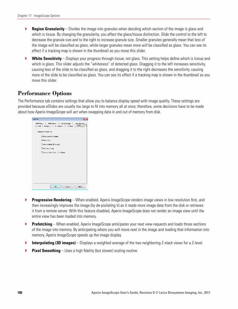
Aperio ImageScope User’s Guide, Revision Q © Leica Biosystems Imaging, Inc. 2017106
Chapter 17: ImageScope Options
Ì` Region Granularity – Divides the image into granules when deciding which section of the image is glass and which is tissue. By changing the granularity, you affect the glass/tissue distinction. Slide the control to the left to decrease the granule size and to the right to increase granule size. Smaller granules generally mean that less of the image will be classified as glass, while larger granules mean more will be classified as glass. You can see its effect if a tracking map is shown in the thumbnail as you move this slider.
Ì` White Sensitivity – Displays your progress through tissue, not glass. This setting helps define which is tissue and which is glass. This slider adjusts the “whiteness” of detected glass. Dragging it to the left increases sensitivity, causing less of the slide to be classified as glass, and dragging it to the right decreases the sensitivity, causing more of the slide to be classified as glass. You can see its effect if a tracking map is shown in the thumbnail as you move this slider.
Performance OptionsThe Performance tab contains settings that allow you to balance display speed with image quality. These settings are provided because eSlides are usually too large to fit into memory all at once; therefore, some decisions have to be made about how Aperio ImageScope will act when swapping data in and out of memory from disk.
Ì` Progressive Rendering – When enabled, Aperio ImageScope renders image views in low resolution first, and then increasingly improves the image (by de-pixilating it) as it reads more image data from the disk or retrieves it from a remote server. With this feature disabled, Aperio ImageScope does not render an image view until the entire view has been loaded into memory.
Ì` Prefetching – When enabled, Aperio ImageScope anticipates your next view requests and loads those sections of the image into memory. By anticipating where you will move next in the image and loading that information into memory, Aperio ImageScope speeds up the image display.
Ì` Interpolating (3D images) – Displays a weighted average of the two neighboring Z-stack views for a Z-level.
Ì` Pixel Smoothing – Uses a high fidelity (but slower) scaling routine.
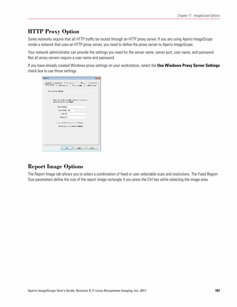
Aperio ImageScope User’s Guide, Revision Q © Leica Biosystems Imaging, Inc. 2017 107
Chapter 17: ImageScope Options
HTTP Proxy OptionSome networks require that all HTTP traffic be routed through an HTTP proxy server. If you are using Aperio ImageScope inside a network that uses an HTTP proxy server, you need to define the proxy server to Aperio ImageScope.
Your network administrator can provide the settings you need for the server name, server port, user name, and password. Not all proxy servers require a user name and password.
If you have already created Windows proxy settings on your workstation, select the Use Windows Proxy Server Settings check box to use those settings.
Report Image OptionsThe Report Image tab allows you to select a combination of fixed or user selectable sizes and resolutions. The Fixed Region Size parameters define the size of the report image rectangle if you press the Ctrl key while selecting the image area.

Aperio ImageScope User’s Guide, Revision Q © Leica Biosystems Imaging, Inc. 2017108
Chapter 17: ImageScope Options
Aperio eSlide Manager Reporting is an optional upgrade that create attractive reports with a few clicks of the mouse. The report image feature is used with this product if you are using report templates that contain images.
Color Management OptionsAperio Integrated Color Management considers the optical characteristics of your scanner and your display monitor to ensure eSlide color is displayed accurately. For information on color management and ICC profiles, see “Appendix B: Aperio Integrated Color Management” on page 113.
You can view the target monitor ICC profile or choose a new monitor:
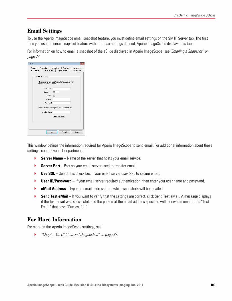
Aperio ImageScope User’s Guide, Revision Q © Leica Biosystems Imaging, Inc. 2017 109
Chapter 17: ImageScope Options
Email SettingsTo use the Aperio ImageScope email snapshot feature, you must define email settings on the SMTP Server tab. The first time you use the email snapshot feature without these settings defined, Aperio ImageScope displays this tab.
For information on how to email a snapshot of the eSlide displayed in Aperio ImageScope, see“Emailing a Snapshot” on page 74.
This window defines the information required for Aperio ImageScope to send email. For additional information about these settings, contact your IT department.
Ì` Server Name – Name of the server that hosts your email service.
Ì` Server Port – Port on your email server used to transfer email.
Ì` Use SSL – Select this check box if your email server uses SSL to secure email.
Ì` User ID/Password – If your email server requires authentication, then enter your user name and password.
Ì` eMail Address – Type the email address from which snapshots will be emailed
Ì` Send Test eMail – If you want to verify that the settings are correct, click Send Test eMail. A message displays if the test email was successful, and the person at the email address specified will receive an email titled “Test Email” that says “Successful!”
For More InformationFor more on the Aperio ImageScope settings, see:
Ì` “Chapter 16: Utilities and Diagnostics” on page 97.
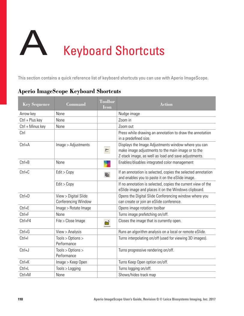
Aperio ImageScope User’s Guide, Revision Q © Leica Biosystems Imaging, Inc. 2017110
A Keyboard Shortcuts
This section contains a quick reference list of keyboard shortcuts you can use with Aperio ImageScope.
Aperio ImageScope Keyboard Shortcuts
Key Sequence Command Toolbar Icon Action
Arrow key None Nudge imageCtrl + Plus key None Zoom inCtrl + Minus key None Zoom out Ctrl Press while drawing an annotation to draw the annotation
in a predefined size.Ctrl+A Image > Adjustments Displays the Image Adjustments window where you can
make image adjustments to the main image or to the Z-stack image, as well as load and save adjustments.
Ctrl+B None Enables/disables integrated color management
Ctrl+C Edit > Copy If an annotation is selected, copies the selected annotation and enables you to paste it on the eSlide image.
Edit > Copy If no annotation is selected, copies the current view of the eSlide image and places it on the Windows clipboard.
Ctrl+D View > Digital Slide Conferencing Window
Opens the Digital Slide Conferencing window where you can create or join an eSlide conference.
Ctrl+E Image > Rotate Image Opens image rotation toolbarCtrl+F None Turns image prefetching on/off.Ctrl+F4 File > Close Image Closes the image that is currently open.
Ctrl+G View > Analysis Runs an algorithm analysis on a local or remote eSlide.Ctrl+I Tools > Options >
PerformanceTurns interpolating on/off (used for viewing 3D images).
Ctrl+J Tools > Options > Performance
Turns progressive rendering on/off.
Ctrl+K Image > Keep Open Turns Keep Open option on/off.Ctrl+L Tools > Logging Turns logging on/off.Ctrl+M None Shows/hides track map
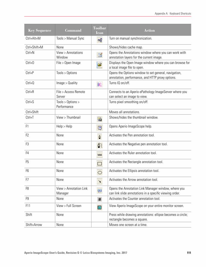
Aperio ImageScope User’s Guide, Revision Q © Leica Biosystems Imaging, Inc. 2017 111
Appendix A: Keyboard Shortcuts
Key Sequence Command Toolbar Icon Action
Ctrl+Alt+M Tools > Manual Sync Turn on manual synchronization.
Ctrl+Shift+M None Shows/hides cache map.Ctrl+N View > Annotations
WindowOpens the Annotations window where you can work with annotation layers for the current image.
Ctrl+O File > Open Image Displays the Open Image window where you can browse for a local image file to open.
Ctrl+P Tools > Options Opens the Options window to set general, navigation, annotation, performance, and HTTP proxy options.
Ctrl+Q Image > Quality Turns IQ on/off.
Ctrl+R File > Access Remote Server
Connects to an Aperio ePathology ImageServer where you can select an image to view.
Ctrl+S Tools > Options > Performance
Turns pixel smoothing on/off.
Ctrl+Shift Moves all annotations.Ctrl+T View > Thumbnail Shows/hides the thumbnail window.
F1 Help > Help Opens Aperio ImageScope help.
F2 None Activates the Pen annotation tool.
F3 None Activates the Negative pen annotation tool.
F4 None Activates the Ruler annotation tool.
F5 None Activates the Rectangle annotation tool.
F6 None Activates the Ellipsis annotation tool.
F7 None Activates the Arrow annotation tool.
F8 View > Annotation Link Manager
Opens the Annotation Link Manager window, where you can link slide annotations in a specific viewing order.
F9 None Activates the Counter annotation tool.
F11 View > Full Screen View Aperio ImageScope on your entire monitor screen.
Shift None Press while drawing annotations: ellipse becomes a circle; rectangle becomes a square.
Shift+Arrow None Moves one screen at a time.
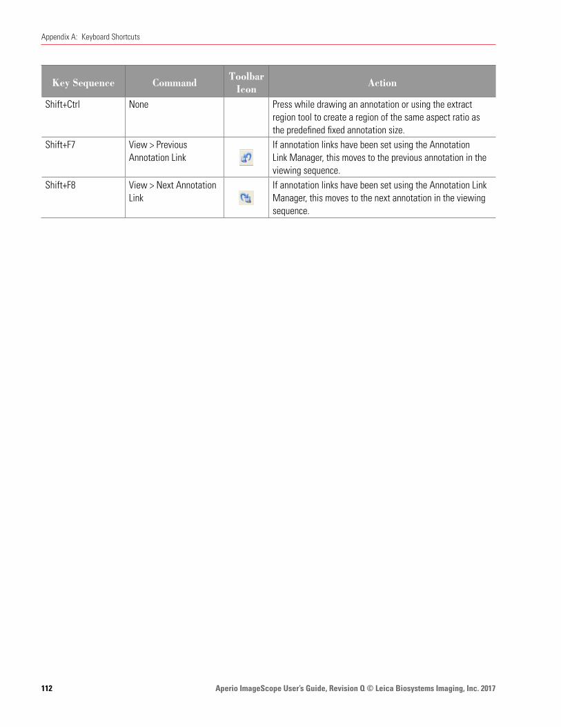
Aperio ImageScope User’s Guide, Revision Q © Leica Biosystems Imaging, Inc. 2017112
Appendix A: Keyboard Shortcuts
Key Sequence Command Toolbar Icon Action
Shift+Ctrl None Press while drawing an annotation or using the extract region tool to create a region of the same aspect ratio as the predefined fixed annotation size.
Shift+F7 View > Previous Annotation Link
If annotation links have been set using the Annotation Link Manager, this moves to the previous annotation in the viewing sequence.
Shift+F8 View > Next Annotation Link
If annotation links have been set using the Annotation Link Manager, this moves to the next annotation in the viewing sequence.
Aperio ImageScope User’s Guide, Revision Q © Leica Biosystems Imaging, Inc. 2017 113
B Aperio Integrated Color Management
Aperio Integrated Color Management considers the optical characteristics of your scanner and your display monitor to ensure the colors of the eSlides are displayed accurately.
A challenge for any provider of applications or devices that work with images is to ensure that the end result (in the case of Leica Biosystems Imaging, viewing an eSlide created by scanning a microscope slide) provides a true representation of the color of the original glass slide from which the eSlide was made.
Every image capturing device will transform the image color due to the particular characteristics of that device.
Fortunately, there is a way to take those characteristics into account to ensure that all along the path the image travels, from creation to display, color accuracy is maintained. As of Release 9, Leica Biosystems Imaging provides Integrated Color Management that works with the international-standard ICC color management specification to maintain image color accuracy.
ICC ProfilesThe ICC (International Color Consortium) is a body that maintains a specification defining a color management system that ensures the color of images moved among applications, devices, and operating systems is maintained accurately.
The ICC defines a format for ICC Profiles, which describes the color attributes of a particular device.
Scanner ICC Profile
In order to describe the behavior of image capture devices such as an Aperio scanner, the devices must be calibrated to a standard color space, which creates an ICC profile. At the Leica Biosystems Imaging facility, an ICC profile is created for your scanner. The scanner’s ICC profile is called the source profile. This profile is stored as a standalone file in \ScanScope\Profiles on the scanner workstation.
As you scan glass slides, the ICC profile for your scanner is also embedded in the resulting eSlide file.
The embedded profile tells ICC-aware applications, such as Aperio ImageScope and WebViewer, the color-transformation completed by the scanner. This allows the Aperio Integrated Color Management to transform the eSlide to its original color.
Display Monitor ICC Profile
The scanner’s ICC profile is not the only component of the integrated color management equation. The monitor you use to view the eSlide also has characteristics that affect color display. Your monitor may have shipped with drivers that contain an ICC profile for the monitor. Or, you can create a new monitor profile by using an external calibration tool. As the monitor is the final device in the image transformation chain, its profile is called the target profile.

Aperio ImageScope User’s Guide, Revision Q © Leica Biosystems Imaging, Inc. 2017114
Appendix B: Aperio Integrated Color Management
If a monitor ICC profile exists, the Aperio ICC-aware applications will use it with the scanner’s ICC profile to display the eSlide color accurately.
How Aperio ImageScope Uses Color ManagementAperio ImageScope is an ICC-aware application, which means that for any eSlide containing an embedded ICC profile it uses the embedded scanner ICC profile and your monitor ICC profile to ensure eSlides are displayed with accurate color.
For information on how Aperio ImageScope uses color management with various Aperio ImageScope features, see:
Ì` “Viewing with Color Management” on page 29
Ì` “Chapter 13: Saving eSlides and Regions” on page 73
Ì` “Color Management Options” on page 108
Aperio ImageScope User’s Guide, Revision Q © Leica Biosystems Imaging, Inc. 2017 115
Symbols3D images. See z-stack images, scanned
AAFI (Aperio Fused Image) 39Analysis button 24annotations
copy and paste 25, 53distance measurement 53drawing
editing free-form shapes 55ellipsis 52
fixed size 54grid, exporting 64layers
attributes 66exporting 64grid, exporting 65importing 64z-stack images 54
linking 67notes for working with 51panning while drawing 51redrawing freeform 55saving 51viewing length and area 62, 63z-stack images 54
Annotations windowdetailed view 58plots 61pre-processing 61summary view 58
Aperio Integrated Color Management 113arrow tool 52autopanning 27
axes and grid 29
Bbest focused layer, z-stack 31brightfield image adjustment. See image adjustments
Ccache
display 97settings 99
change annotation 55changing color defaults 104clinical viewing mode 23closing eSlides 21color adjustments. See image adjustmentscolor balance 36color management 108, 113
ICC profiles 113conferencing. See eSlide conferencingconnecting to scanner 90copy and paste annotations 25copy annotations 53counter annotation button 25Counter tool 52CWS files 11
Ddeleting regions 61diagnostics 97distance measurement button 25Distance Measurement Tuning window 53DSR 13
Index
Aperio ImageScope User’s Guide, Revision Q © Leica Biosystems Imaging, Inc. 2017116
Index
Eedit annotation 55ellipsis tool 52email settings 109email snapshot image 24, 74eSlide conferencing 82
changing leader 86creating 84joining 85viewing slides 86
eSlidesclosing 21information about 20opening local 17opening recent 17viewing multiple 18windows, managing 19
Excel spreadsheet, export annotation text 65export images, three versions 75export layer attributes text 65export regions attribute text 65export to Microsoft Excel 65extracted regions 76
fixed aspect ratio 79options 77predefined size 79saving specific size 79selecting 76viewing applications 80
extraction tool 75, 76
Ffalse color 38
applying temporary 38features 10file types supported 11filmstrip 19, 28fluorescence image 38
adjustments 42adjustments, tips 42AFI 39
adjusting 39brightness, adjusting 42
channelcycling display 41registering 44viewing information 41
coloradjusting 42inverting 42
contrast, adjusting 42creating RGB image 78extracting 78false color 38fused image 39fusing an image 45gamma, adjusting 42histogram
high clip 44intensity thresholds 44low clip 44tone curve 44transfer function 44
thumbnail, automatic contrast 43viewing channel images 40
Focus slider, z-stack images 32free-form shape annotation
draw 51edit 55
fused imageadjusting. See fluorescence image; See fluorescence
imagecreating 45
Hhistogram 43HTTP proxy option 107
IIHC analysis 58image adjustments 35
fluorescence images 39main image 35Z-stacks 35
image analysisresult plots 61
image information 20image resolution 48

Aperio ImageScope User’s Guide, Revision Q © Leica Biosystems Imaging, Inc. 2017 117
Index
Llabel rotation 34Layer Attributes pane
copy and paste 65export to Microsoft Excel 65export to text file 65
Layer Regions panecopy and paste 65export text file 65export to Microsoft Excel 65z-stack image layers 54
linking annotations 67Link Manager 67links
creating 68deleting 69viewing 68
local image support 17logging activities 97
Mmagnification
options 101magnification levels, normalize 102magnifier window 28
changing default resolution 28magnification settings 101resizing 28
main image gamma settings 102markup image 103measure distance annotation 53measure distance button 25microns per pixel, normalize 102Microsoft Excel, export annotation text 65multiple ImageScopes 98
Nnavigating
options 103z-stack images, scanned 31
NDPI files 11negative pen tool
editing annotations 55
computing 49setting/changing 49
image rotation 34ImageScope
disabling pre-processing 61features 10image view 11installing 13starting 14toolbar, quick reference 23window
filmstrip 19magnifier window 28quick reference 22status bar 20thumbnail window 27
ImageScope optionsanalysis 103annotation 104default gamma files 102general 101HTTP proxy 107integrated color management 108magnification 101navigation 103performance 106tracking 105
ImageServerURL 88image view 11, 21
saving 21Image view
opening 21installing 13integrated color management 29Intended Use 10interpolating 106IQ 32
JJPEG files 11
Kkeyboard shortcuts 110

Aperio ImageScope User’s Guide, Revision Q © Leica Biosystems Imaging, Inc. 2017118
Index
starting ImageScope 14status bar 20symbols 121synchronization options 103system requirements 13
Ttab-delimited file, export to 65TelePath Live
calibration 88connecting to scanner 90ImageServerURL 89live video 92prerequisites 88
thumbnail window 27TIFF files 11toolbar quick reference 23tracking 70
appending to a track 72options 105tips 71viewing track 71, 72
Uupdating analyses 61user permissions 15utilities 97
Vviewing
live video 92track 72with color management 29
VMS files 11
WWindows proxy server settings 107
XX-coordinate navigation 28
YY-coordinate navigation 28
using 52normalize scanned images 102
Oopening eSlides 17
Ppage panning 28panning
options 104panning while annotating 51Pen tool
editing annotations 55using 51
performance options 106prefetching images 97, 106pre-processing 61progressive rendering 106pseudo-color. See false color
RRectangle tool 52redraw annotation 55registering channels 44Report Region tool 52reverse panning 104rotating
images and labels 34Ruler tool 52
Ssaving
image view 21saving image to clipboard 73scalebar 29security 15slide-specific processing 58snapshot
creating 73save image 24send in email 24, 74
spreadsheet, export annotation text 65

Aperio ImageScope User’s Guide, Revision Q © Leica Biosystems Imaging, Inc. 2017 119
Index
Zzoom, immediate
29zoom navigation 29z-stack images
opening 20z-stack images, scanned
annotating 54best focused layer 31Focus slider 32viewing 31
Z-stacks, TelePath Livecalibrating 88capturing 93configuring 90gamma settings 102viewing 96

Aperio ImageScope User’s Guide, Revision Q © Leica Biosystems Imaging, Inc. 2017120
Index
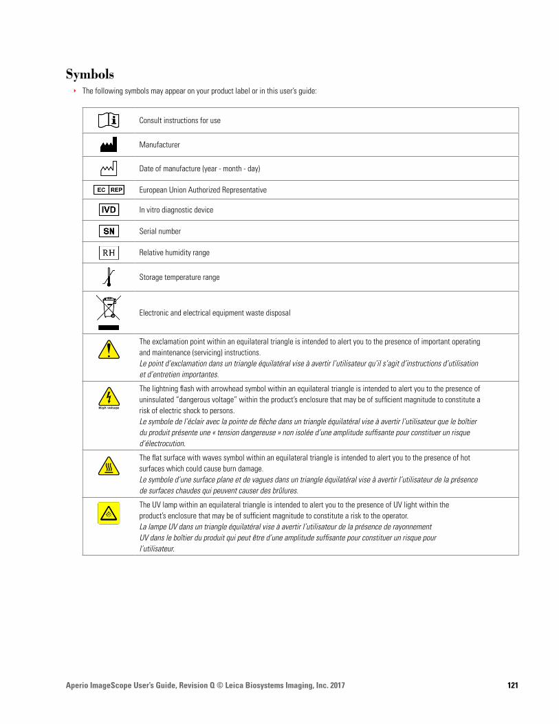
Aperio ImageScope User’s Guide, Revision Q © Leica Biosystems Imaging, Inc. 2017 121
SymbolsÌÌ The following symbols may appear on your product label or in this user’s guide:
Consult instructions for use
Manufacturer
Date of manufacture (year - month - day)
European Union Authorized Representative
In vitro diagnostic device
Serial number
Relative humidity range
Storage temperature range
Electronic and electrical equipment waste disposal
The exclamation point within an equilateral triangle is intended to alert you to the presence of important operatingand maintenance (servicing) instructions.Le point d’exclamation dans un triangle équilatéral vise à avertir l’utilisateur qu’il s’agit d’instructions d’utilisationet d’entretien importantes.
The lightning flash with arrowhead symbol within an equilateral triangle is intended to alert you to the presence ofuninsulated “dangerous voltage” within the product’s enclosure that may be of sufficient magnitude to constitute arisk of electric shock to persons.Le symbole de l’éclair avec la pointe de flèche dans un triangle équilatéral vise à avertir l’utilisateur que le boîtierdu produit présente une « tension dangereuse » non isolée d’une amplitude suffisante pour constituer un risqued’électrocution.
The flat surface with waves symbol within an equilateral triangle is intended to alert you to the presence of hotsurfaces which could cause burn damage.Le symbole d’une surface plane et de vagues dans un triangle équilatéral vise à avertir l’utilisateur de la présencede surfaces chaudes qui peuvent causer des brûlures.
The UV lamp within an equilateral triangle is intended to alert you to the presence of UV light within theproduct’s enclosure that may be of sufficient magnitude to constitute a risk to the operator.La lampe UV dans un triangle équilatéral vise à avertir l’utilisateur de la présence de rayonnementUV dans le boîtier du produit qui peut être d’une amplitude suffisante pour constituer un risque pourl’utilisateur.

www.LeicaBiosystems.com/Aperio


