57
Antepartum and Postpartum Hemorrhage . Maryam Al –Jaber nsultant, Family Medicine rch, 2015 Dr. Omnia Darweesh Resident, Family Medicin
| Date post: | 14-Aug-2015 |
| Category: |
Health & Medicine |
| Upload: | amir-mahmoud |
| View: | 125 times |
| Download: | 0 times |
Antepartum and Postpartum Hemorrhage
Dr. Maryam Al –JaberConsultant, Family Medicine
March, 2015
Dr. Omnia DarweeshResident, Family Medicine
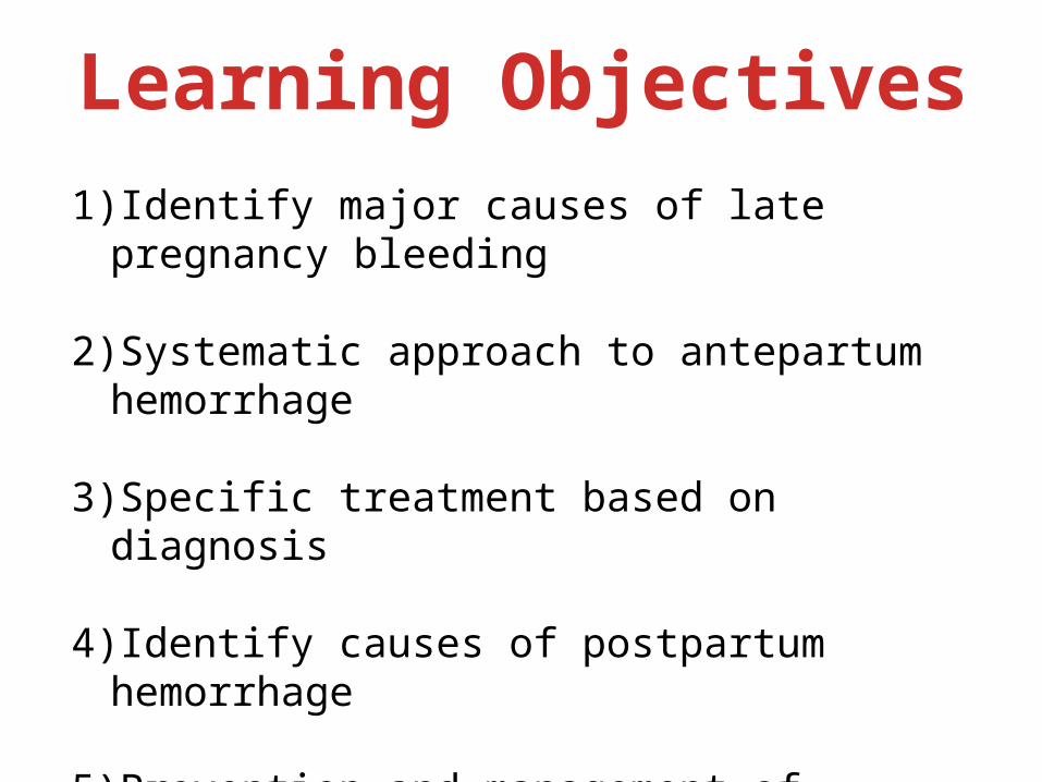
Learning Objectives1)Identify major causes of late pregnancy
bleeding
2)Systematic approach to antepartum hemorrhage
3)Specific treatment based on diagnosis
4)Identify causes of postpartum hemorrhage
5)Prevention and management of postpartum hemorrhage
Antepartum Hemorrhage
Antepartum hemorrhage (APH) is defined as bleeding from or in to the genital tract, occurring from 24+0 weeks of pregnancy and prior to the birth of the baby.
RCOG Guidelines
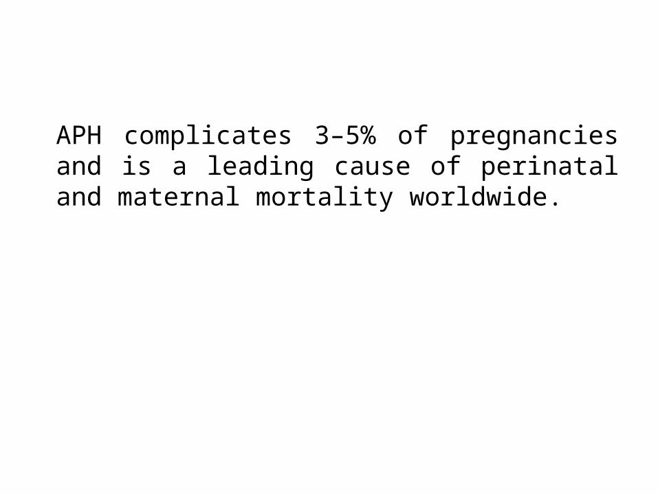
APH complicates 3–5% of pregnancies and is a leading cause of perinatal and maternal mortality worldwide.
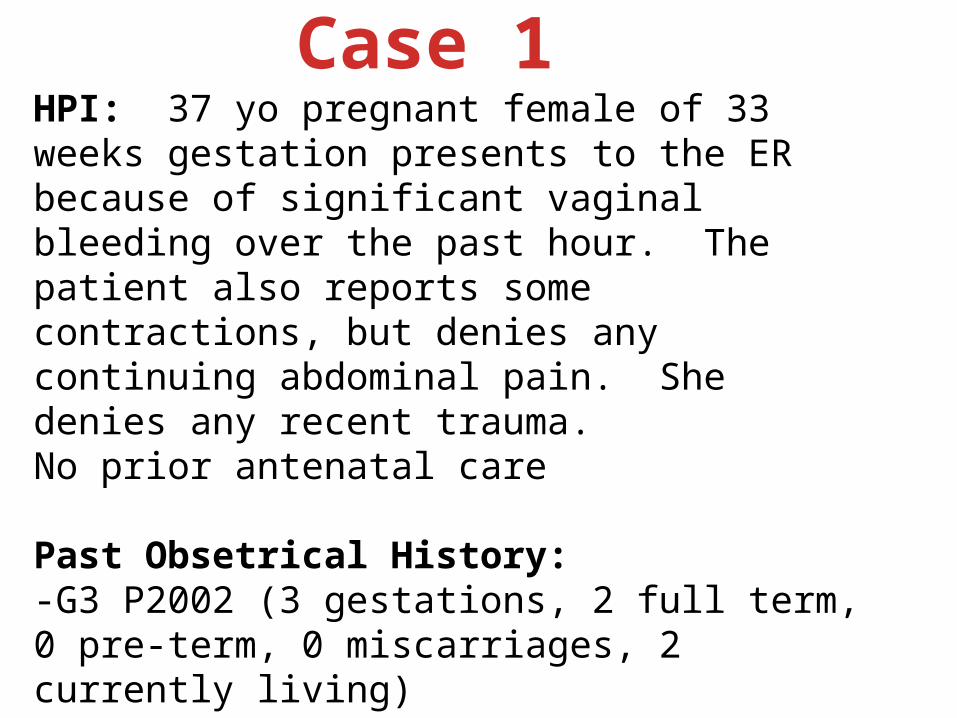
HPI: 37 yo pregnant female of 33 weeks gestation presents to the ER because of significant vaginal bleeding over the past hour. The patient also reports some contractions, but denies any continuing abdominal pain. She denies any recent trauma.No prior antenatal care
Past Obsetrical History:-G3 P2002 (3 gestations, 2 full term, 0 pre-term, 0 miscarriages, 2 currently living)-2 previous SVD’s (spontaneous vaginal delivery)-Last birth was 9 years ago by SVD, weighed 3800 grams-No previous obstetrical complications or morbidity
Case 1

No past medical or surgical historySocial History: Patient lives with her husband in the Santiago district of Cuzco. Denies any smoking, alcohol or drug use. No spousal abuse. Works as a housewife. Low economic status.Physical Exam:Vital Signs: Stable (BP – 110/70, P – 72)General Appearance: No apparent distress, appeared clinically stableSkin: Elastic, capillary reflex < 2 secondsUterine Height: 30 cmFetal Lie: LongitudinalContractions: PresentFetal Heart Tones: 144 x minute
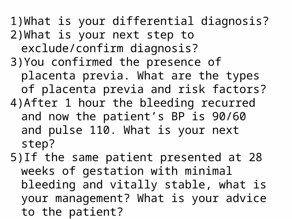
1)What is your differential diagnosis?2)What is your next step to exclude/confirm
diagnosis?3)You confirmed the presence of placenta
previa. What are the types of placenta previa and risk factors?
4)After 1 hour the bleeding recurred and now the patient’s BP is 90/60 and pulse 110. What is your next step?
5)If the same patient presented at 28 weeks of gestation with minimal bleeding and vitally stable, what is your management? What is your advice to the patient?
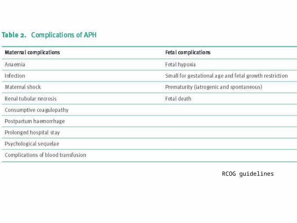
6)What are the complications of antepartum hemorrhage?
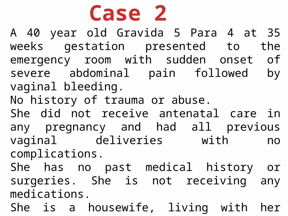
A 40 year old Gravida 5 Para 4 at 35 weeks gestation presented to the emergency room with sudden onset of severe abdominal pain followed by vaginal bleeding. No history of trauma or abuse.She did not receive antenatal care in any pregnancy and had all previous vaginal deliveries with no complications.She has no past medical history or surgeries. She is not receiving any medications.She is a housewife, living with her husband and children, she does not drink alcohol or use recreational drugs however she smokes ½ pack of cigarettes daily.
Case 2
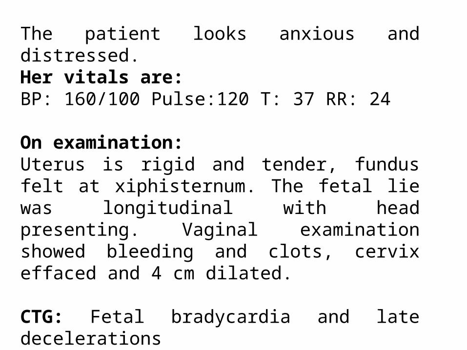
The patient looks anxious and distressed.Her vitals are:BP: 160/100 Pulse:120 T: 37 RR: 24
On examination:Uterus is rigid and tender, fundus felt at xiphisternum. The fetal lie was longitudinal with head presenting. Vaginal examination showed bleeding and clots, cervix effaced and 4 cm dilated.
CTG: Fetal bradycardia and late decelerations
Urine dipstick: +3 proteinuria

(1)What are the causes of antepartum hemorrhage?
(2)What is your diagnosis?(3)What are the risk factors? (4)What is your initial management?(5)What are the alarming signs in this
case?(6)If the same patient presented but
without bleeding what is your diagnosis?
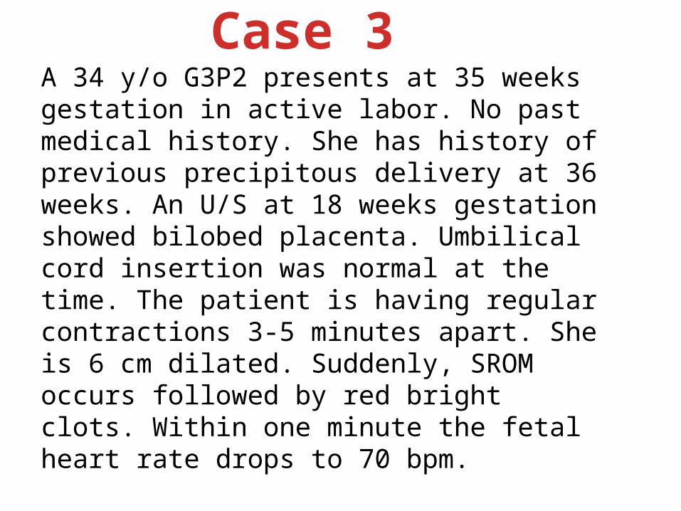
A 34 y/o G3P2 presents at 35 weeks gestation in active labor. No past medical history. She has history of previous precipitous delivery at 36 weeks. An U/S at 18 weeks gestation showed bilobed placenta. Umbilical cord insertion was normal at the time. The patient is having regular contractions 3-5 minutes apart. She is 6 cm dilated. Suddenly, SROM occurs followed by red bright clots. Within one minute the fetal heart rate drops to 70 bpm.
1)What is your most likely diagnosis?2)What are the risk factors for this
condition?3)What tests can you use to confirm
diagnosis?
Case 3
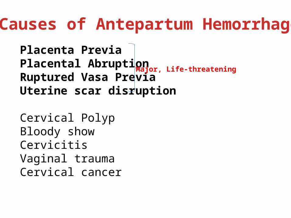
Placenta PreviaPlacental AbruptionRuptured Vasa PreviaUterine scar disruption
Cervical PolypBloody showCervicitisVaginal traumaCervical cancer
Causes of Antepartum Hemorrhage
Major, Life-threatening
RCOG guidelines
Management of Antepartum hemorrhage
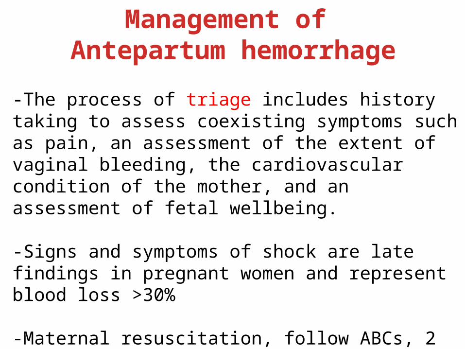
-The process of triage includes history taking to assess coexisting symptoms such as pain, an assessment of the extent of vaginal bleeding, the cardiovascular condition of the mother, and an assessment of fetal wellbeing.
-Signs and symptoms of shock are late findings in pregnant women and represent blood loss >30%
-Maternal resuscitation, follow ABCs, 2 wide bore cannulas, prompt fluid resuscitation and/or blood transfusion.
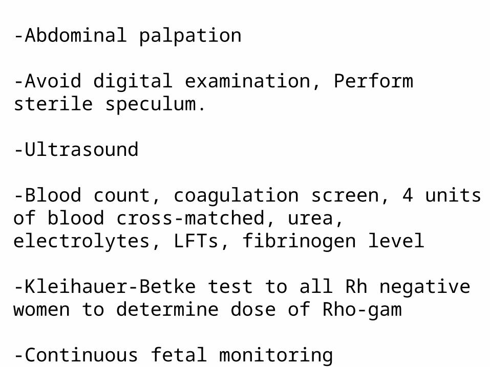
-Abdominal palpation
-Avoid digital examination, Perform sterile speculum.
-Ultrasound
-Blood count, coagulation screen, 4 units of blood cross-matched, urea, electrolytes, LFTs, fibrinogen level
-Kleihauer-Betke test to all Rh negative women to determine dose of Rho-gam
-Continuous fetal monitoring
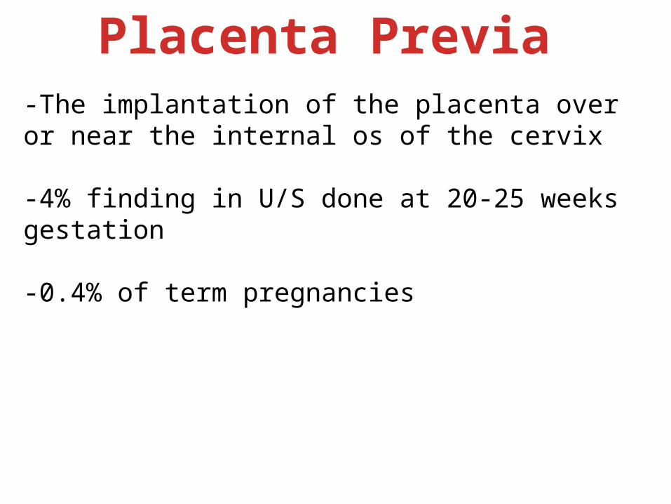
Placenta Previa-The implantation of the placenta over or near the internal os of the cervix
-4% finding in U/S done at 20-25 weeks gestation
-0.4% of term pregnancies
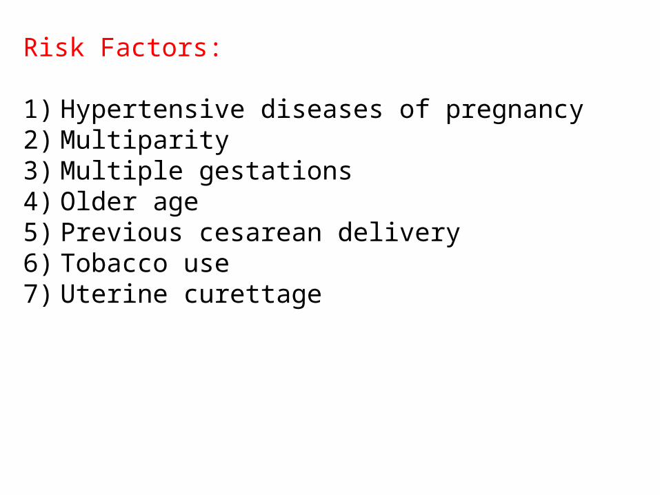
Risk Factors:
1) Hypertensive diseases of pregnancy2) Multiparity3) Multiple gestations4) Older age5) Previous cesarean delivery6) Tobacco use7) Uterine curettage
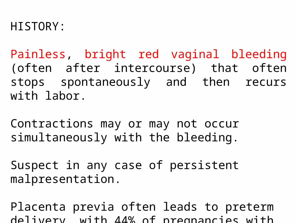
HISTORY:
Painless, bright red vaginal bleeding (often after intercourse) that often stops spontaneously and then recurs with labor.
Contractions may or may not occur simultaneously with the bleeding.
Suspect in any case of persistent malpresentation.
Placenta previa often leads to preterm delivery, with 44% of pregnancies with placenta previa delivered before 37 weeks.
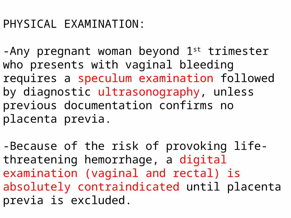
PHYSICAL EXAMINATION:
-Any pregnant woman beyond 1st trimester who presents with vaginal bleeding requires a speculum examination followed by diagnostic ultrasonography, unless previous documentation confirms no placenta previa.
-Because of the risk of provoking life-threatening hemorrhage, a digital examination (vaginal and rectal) is absolutely contraindicated until placenta previa is excluded.
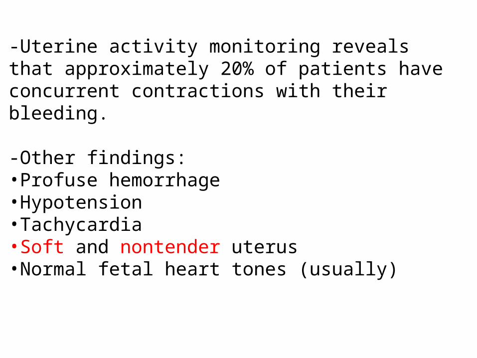
-Uterine activity monitoring reveals that approximately 20% of patients have concurrent contractions with their bleeding.
-Other findings:•Profuse hemorrhage•Hypotension•Tachycardia•Soft and nontender uterus•Normal fetal heart tones (usually)
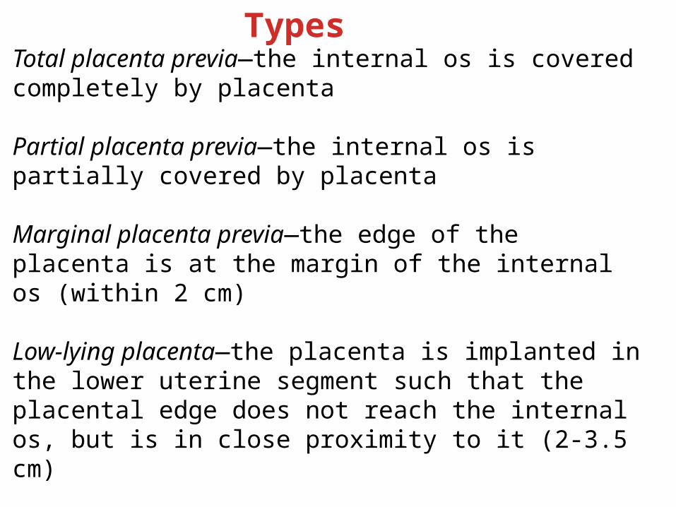
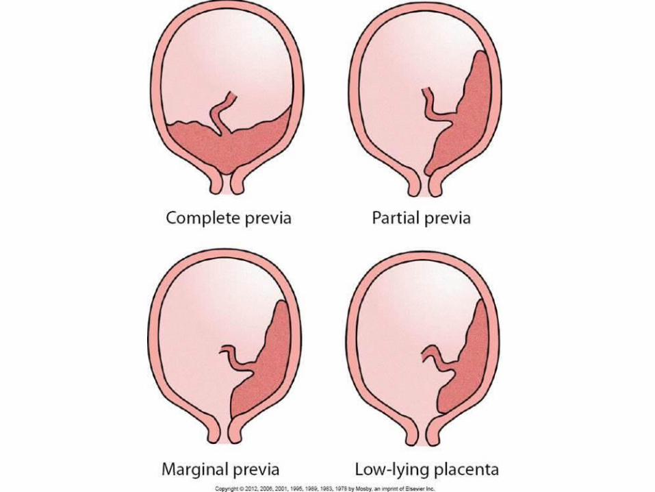
Total placenta previa—the internal os is covered completely by placenta
Partial placenta previa—the internal os is partially covered by placenta
Marginal placenta previa—the edge of the placenta is at the margin of the internal os (within 2 cm)
Low-lying placenta—the placenta is implanted in the lower uterine segment such that the placental edge does not reach the internal os, but is in close proximity to it (2-3.5 cm)
Vasa previa—the fetal vessels course through membranes and present at the cervical os
Types
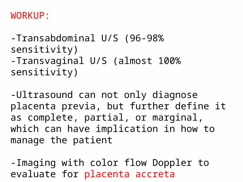
WORKUP:
-Transabdominal U/S (96-98% sensitivity)-Transvaginal U/S (almost 100% sensitivity)
-Ultrasound can not only diagnose placenta previa, but further define it as complete, partial, or marginal, which can have implication in how to manage the patient
-Imaging with color flow Doppler to evaluate for placenta accreta
-MRI for diagnosis of invasive placenta and organ involvement in placenta percreta
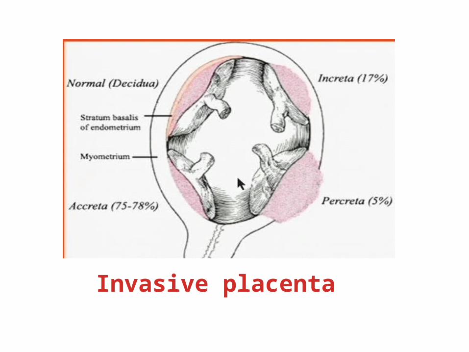
Invasive placenta

Laboratory Studies
-CBC--hCG levels-Rh compatibility test-FSP levels and fibrinogen - PT/aPTT-Blood type and cross; hold for at least 4 units-Apt test to determine fetal origin of blood (as in the case of vasa previa)-Wright stain applied to a slide smear of vaginal blood to look for nucleated red blood cells (RBCs), not adult blood-L/S ratio for fetal maturity-Kleihauer-Betke test (fetal-maternal transfusion)-Bedside clot test
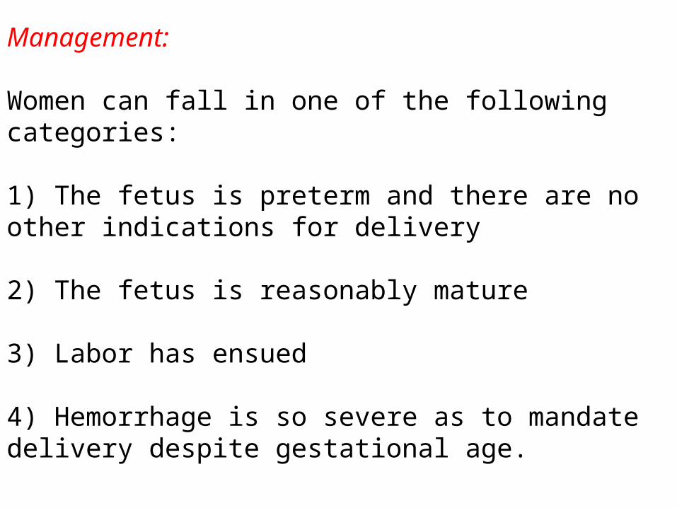
Management:
Women can fall in one of the following categories:
1) The fetus is preterm and there are no other indications for delivery
2) The fetus is reasonably mature
3) Labor has ensued
4) Hemorrhage is so severe as to mandate delivery despite gestational age.
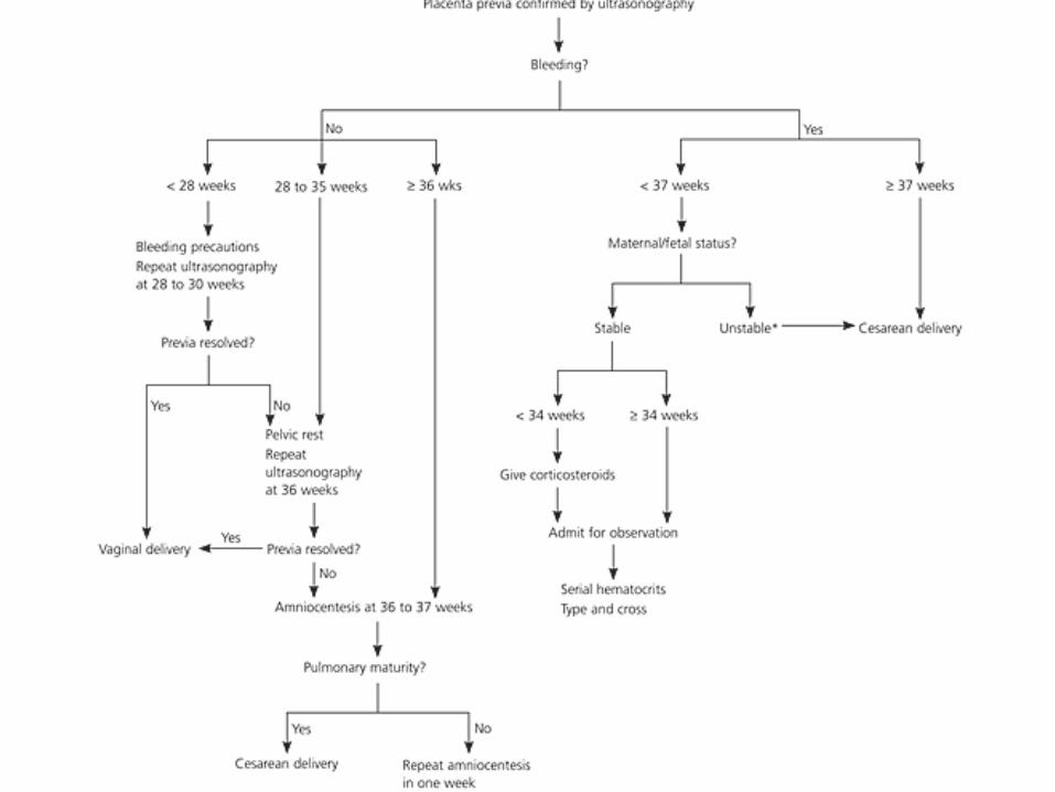
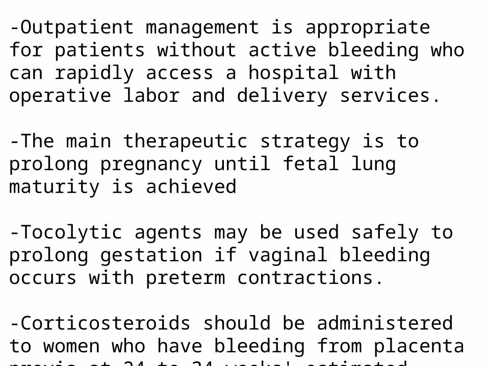
-Outpatient management is appropriate for patients without active bleeding who can rapidly access a hospital with operative labor and delivery services.
-The main therapeutic strategy is to prolong pregnancy until fetal lung maturity is achieved
-Tocolytic agents may be used safely to prolong gestation if vaginal bleeding occurs with preterm contractions.
-Corticosteroids should be administered to women who have bleeding from placenta previa at 24 to 34 weeks' estimated gestation.
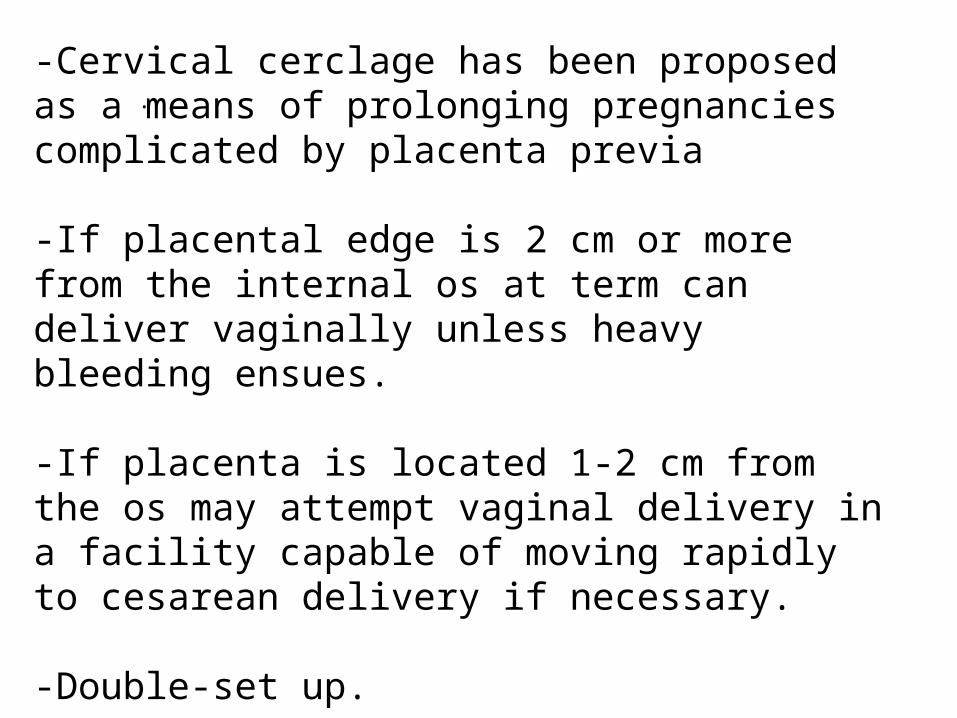
.-Cervical cerclage has been proposed as a means of prolonging pregnancies complicated by placenta previa
-If placental edge is 2 cm or more from the internal os at term can deliver vaginally unless heavy bleeding ensues.
-If placenta is located 1-2 cm from the os may attempt vaginal delivery in a facility capable of moving rapidly to cesarean delivery if necessary.
-Double-set up.
-Regional anesthesia is safer, less blood loss.

Patient Education:
-Women with asymptomatic previa in 2nd trimester can continue normal activities until follow-up U/S is performed at 28 weeks.
-Women with persistent previa in 3rd trimester should report any bleeding and abstain from intercourse and use of tampons and no digital examination.
-Counsel patients about the risk of recurrence. Instruct them to notify the obstetrician caring for their next pregnancy about their history of placenta previa.
-Encourage patients with known placenta previa to maintain intake of iron and folate as a safety margin in the event of bleeding.

Placental AbruptionRisk Factors:1) Hypertensive diseases of pregnancy2) Previous history of abruption3) Advanced maternal age/parity4) Trauma (abuse or accidents)5) Prolonged rupture of membranes6) Smoking7) Cocaine, Alcohol8) Over-distention e.g polyhydramnios9) Unexplained elevation of MSAFP10)Thrombophilias
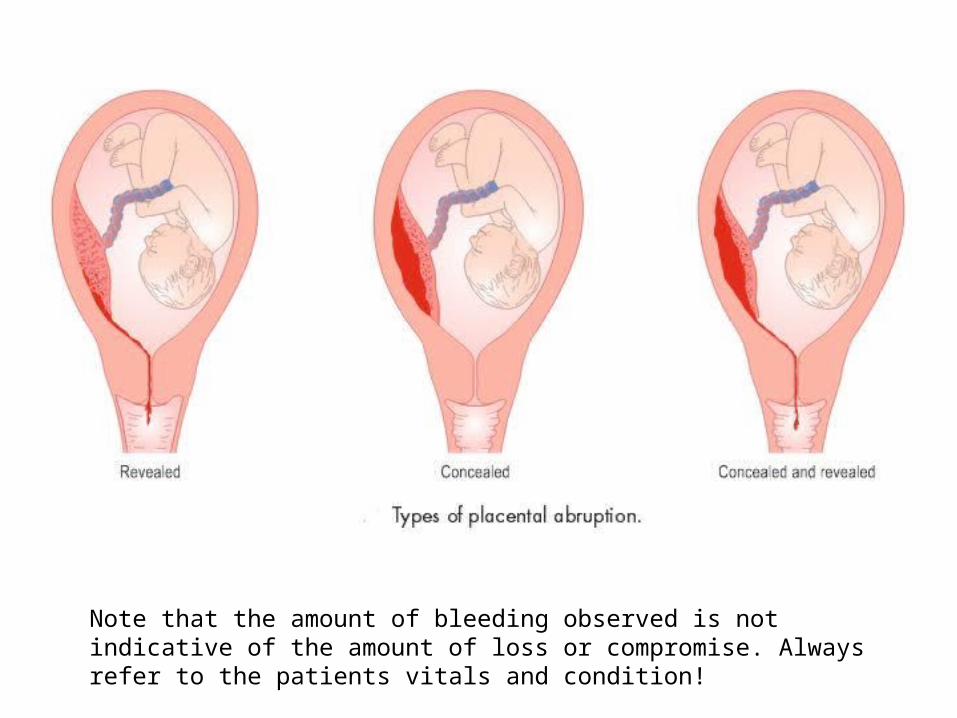
Note that the amount of bleeding observed is not indicative of the amount of loss or compromise. Always refer to the patients vitals and condition!
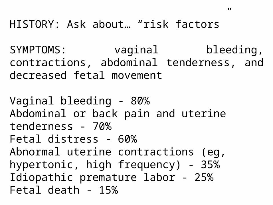
HISTORY: Ask about… “risk factors”
SYMPTOMS: vaginal bleeding, contractions, abdominal tenderness, and decreased fetal movement
Vaginal bleeding - 80%Abdominal or back pain and uterine tenderness - 70%Fetal distress - 60%Abnormal uterine contractions (eg, hypertonic, high frequency) - 35%Idiopathic premature labor - 25%Fetal death - 15%
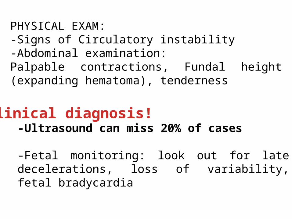
Clinical diagnosis!-Ultrasound can miss 20% of cases
-Fetal monitoring: look out for late decelerations, loss of variability, fetal bradycardia
PHYSICAL EXAM:-Signs of Circulatory instability-Abdominal examination:Palpable contractions, Fundal height (expanding hematoma), tenderness
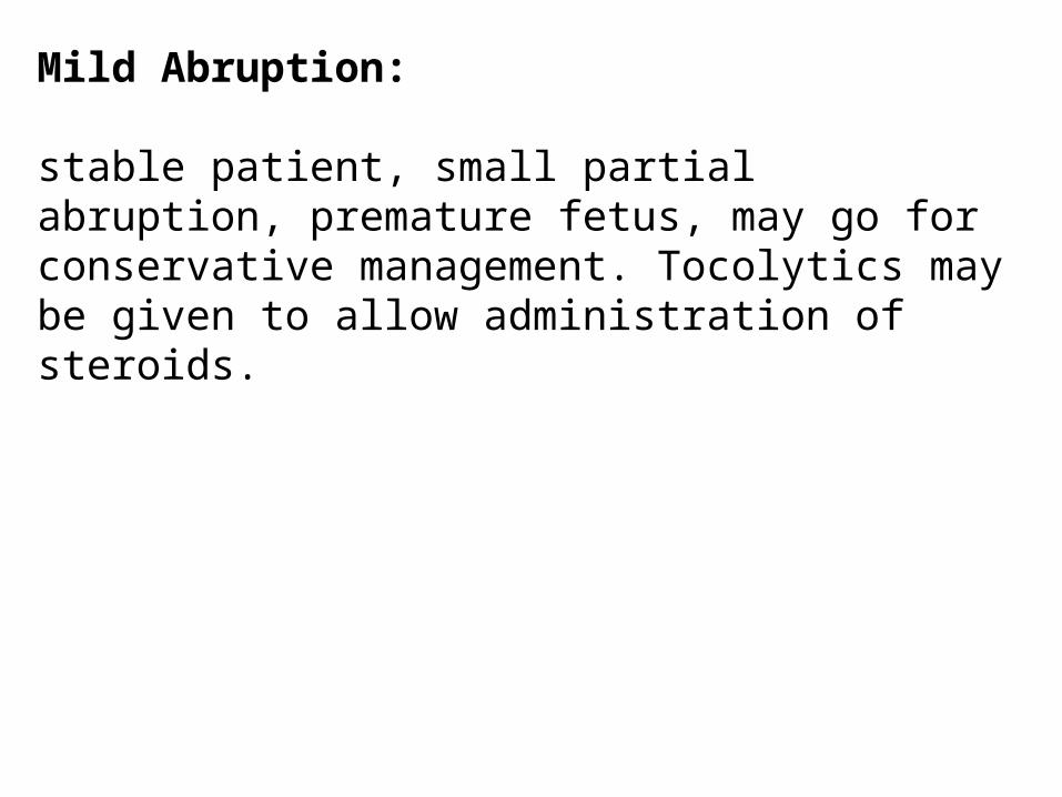
Mild Abruption:
stable patient, small partial abruption, premature fetus, may go for conservative management. Tocolytics may be given to allow administration of steroids.

Severe Abruption:
-Rapid stabilization of mother, ABCs, assessment of fetal well being.-Non-reassuring fetal heart tracing necessitates rapid cesarean delivery.-A decision-to-delivery interval 20 minutes or less improves neonatal outcomes.-If fetal demise is present, vaginal delivery is the goal.-Treatment of preeclampsia with magnesium slufate decreases risk of placental abruption.-1/3 of patients with abruption and fetal demise will develop coagulopathy
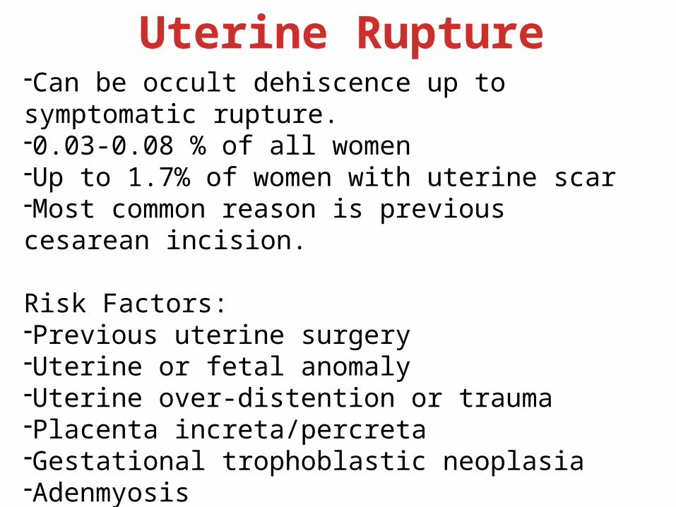
Uterine Rupture-Can be occult dehiscence up to symptomatic rupture.-0.03-0.08 % of all women-Up to 1.7% of women with uterine scar-Most common reason is previous cesarean incision.
Risk Factors:-Previous uterine surgery-Uterine or fetal anomaly-Uterine over-distention or trauma-Placenta increta/percreta-Gestational trophoblastic neoplasia-Adenmyosis-Excessive uterine stimulation
Clinical presentation-Classical presentation for significant rupture includes:
-Vaginal bleeding, Pain, Cessation of contractions, Absent fetal tones, Loss of station, easily palpable fetal parts through abdomen, profound maternal tachycardia and hypotension.
-Most cases present with abnormal fetal monitoring.
-13% of cases occur outside the hospital
Management-Asymptomatic scar disruption: Expectant
-Symptomatic rupture: Emergency C/S
Complications-Maternal:Hemorrhage, anemia, bladder rupture, hysterectomy, death
-Fetal:Respiratory distress, hypoxia, acidemia, death
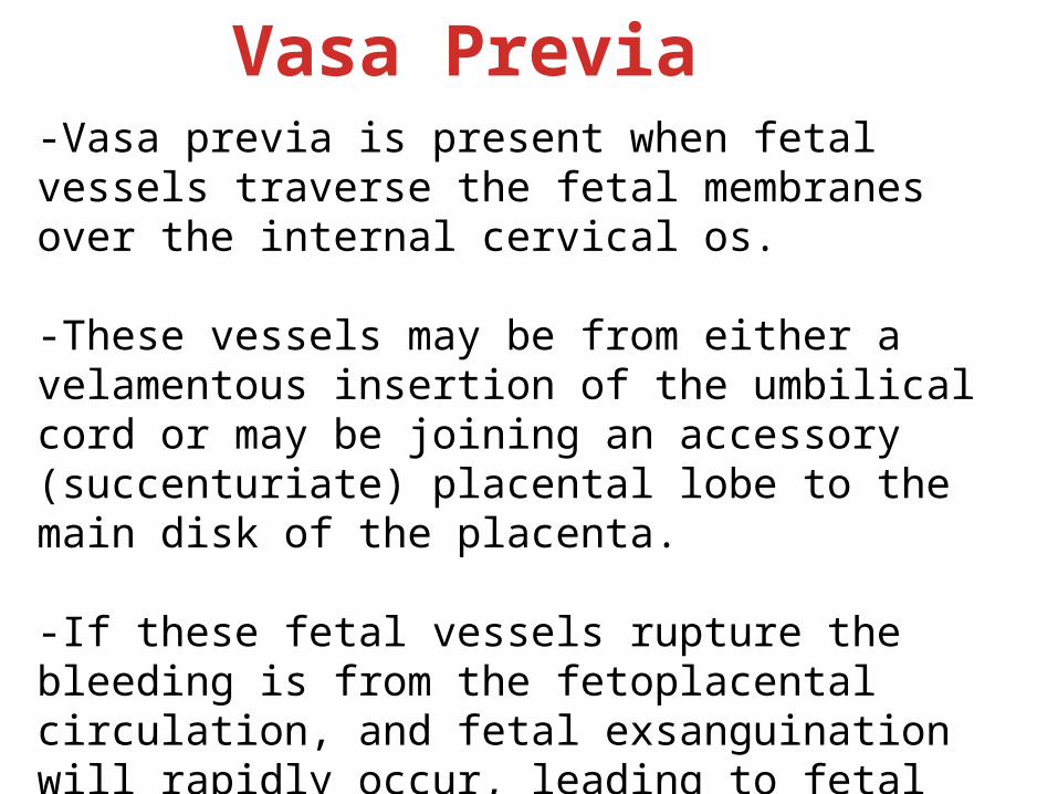
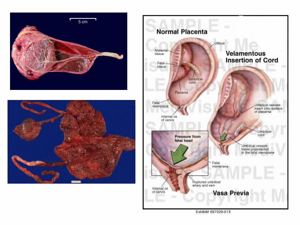
Vasa Previa-Vasa previa is present when fetal vessels traverse the fetal membranes over the internal cervical os.
-These vessels may be from either a velamentous insertion of the umbilical cord or may be joining an accessory (succenturiate) placental lobe to the main disk of the placenta.
-If these fetal vessels rupture the bleeding is from the fetoplacental circulation, and fetal exsanguination will rapidly occur, leading to fetal death.

RISK FACTORS:
1) Velamentous insertion of the cord2) Placenta previa3) IVF4) Bilobed and succenturiate-lobed placenta5) Multiple gestation
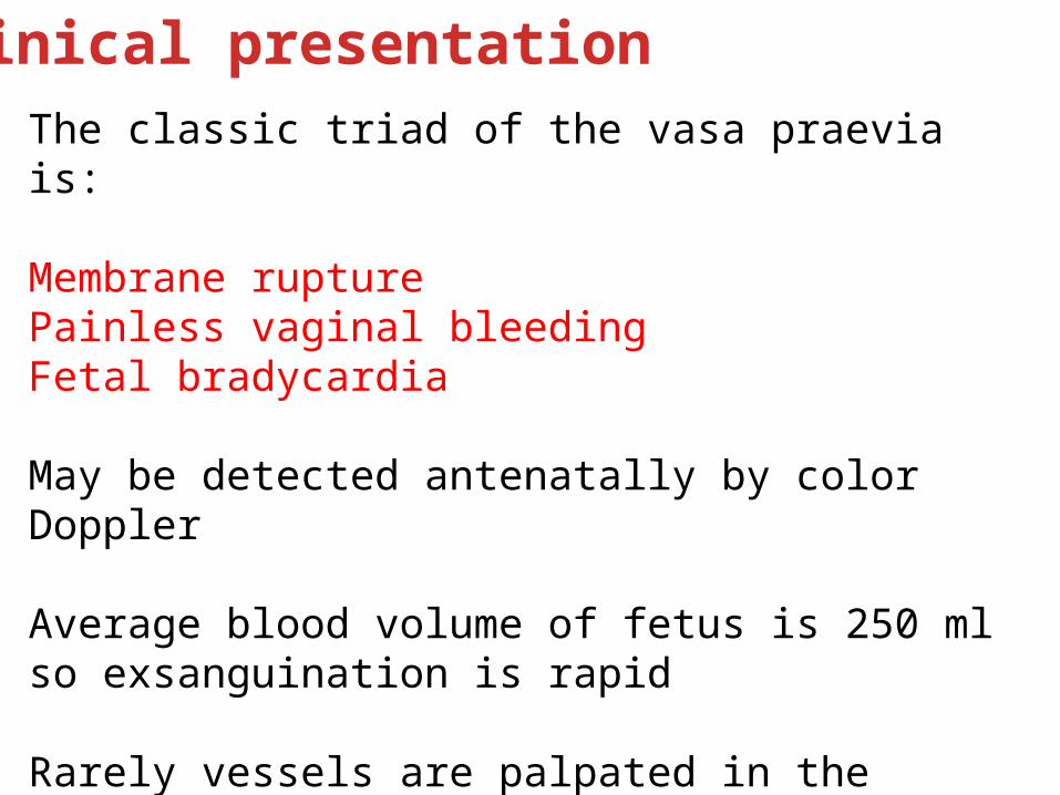
Clinical presentationThe classic triad of the vasa praevia is:
Membrane rupturePainless vaginal bleedingFetal bradycardia
May be detected antenatally by color Doppler
Average blood volume of fetus is 250 ml so exsanguination is rapid
Rarely vessels are palpated in the presenting membranes
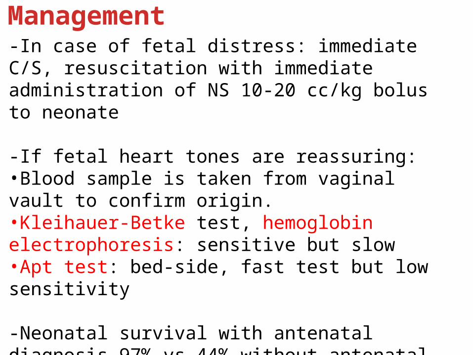
Management-In case of fetal distress: immediate C/S, resuscitation with immediate administration of NS 10-20 cc/kg bolus to neonate
-If fetal heart tones are reassuring:•Blood sample is taken from vaginal vault to confirm origin.•Kleihauer-Betke test, hemoglobin electrophoresis: sensitive but slow•Apt test: bed-side, fast test but low sensitivity
-Neonatal survival with antenatal diagnosis 97% vs 44% without antenatal diagnosis.
-Planned C/S at 35 weeks gestation after steroid administration for stable women detected antenatally
Postpartum Hemorrhage
PPH is defined as blood loss >500 mL following delivery.
Loss of >1000 mL is considered major PPH and is an emergent situation resulting in hemodynamic instability.
PPH is the most common maternal morbidity in developed countries and major cause of mortality worldwide.
Occurs in up to 18% of births.
Risk Factors:
-In most cases no identifiable risk factors-Prolonged third stage of labor-Preeclampsia-Cesarean section-Previous PPH-Multiple pregnancy-Fetal macrosomia-Episiotomy (more in mediolateral)
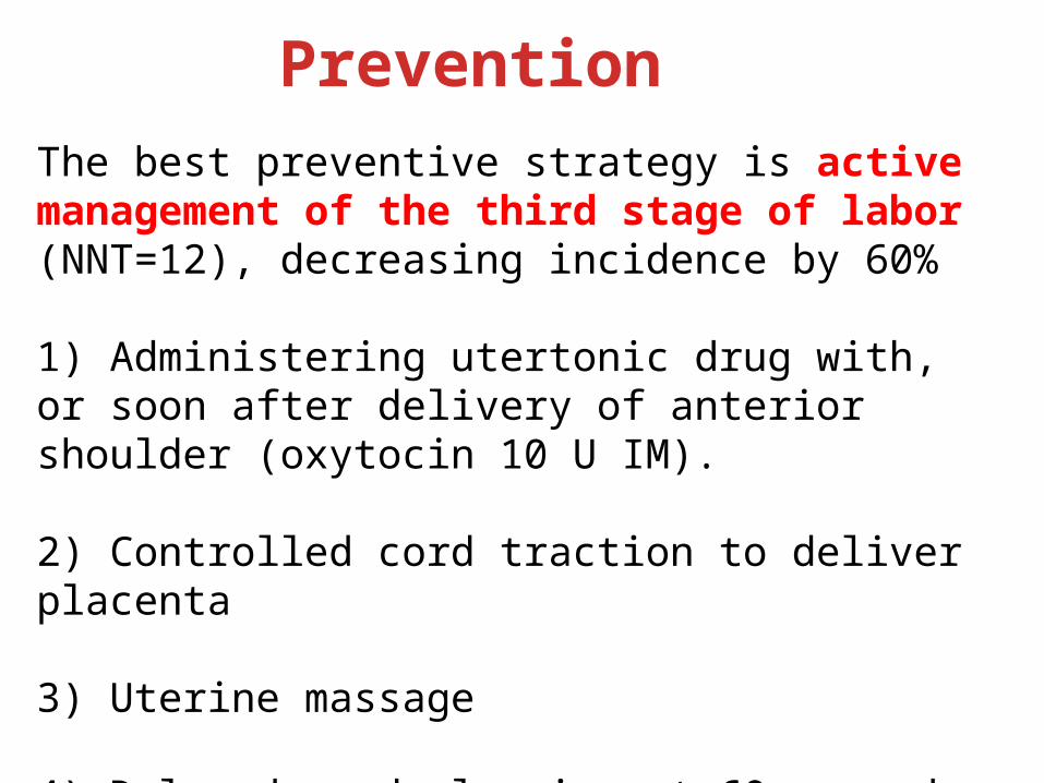
The best preventive strategy is active management of the third stage of labor (NNT=12), decreasing incidence by 60%
1) Administering utertonic drug with, or soon after delivery of anterior shoulder (oxytocin 10 U IM).
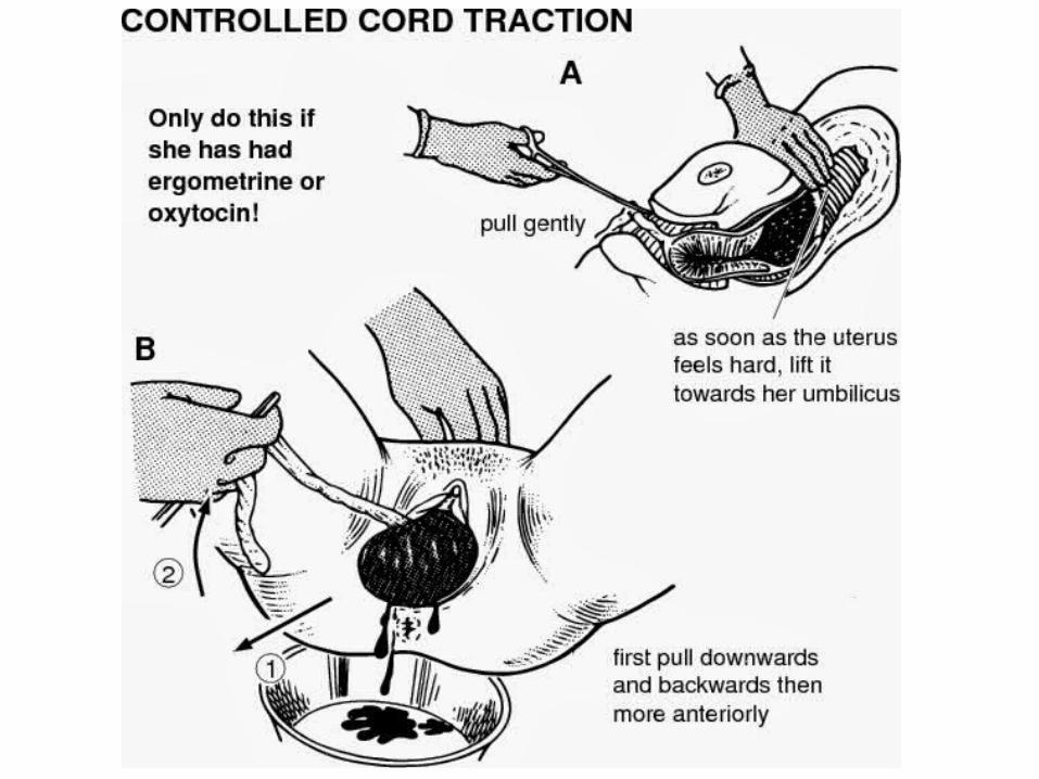
2) Controlled cord traction to deliver placenta
3) Uterine massage
4) Delayed cord clamping at 60 seconds.
Prevention

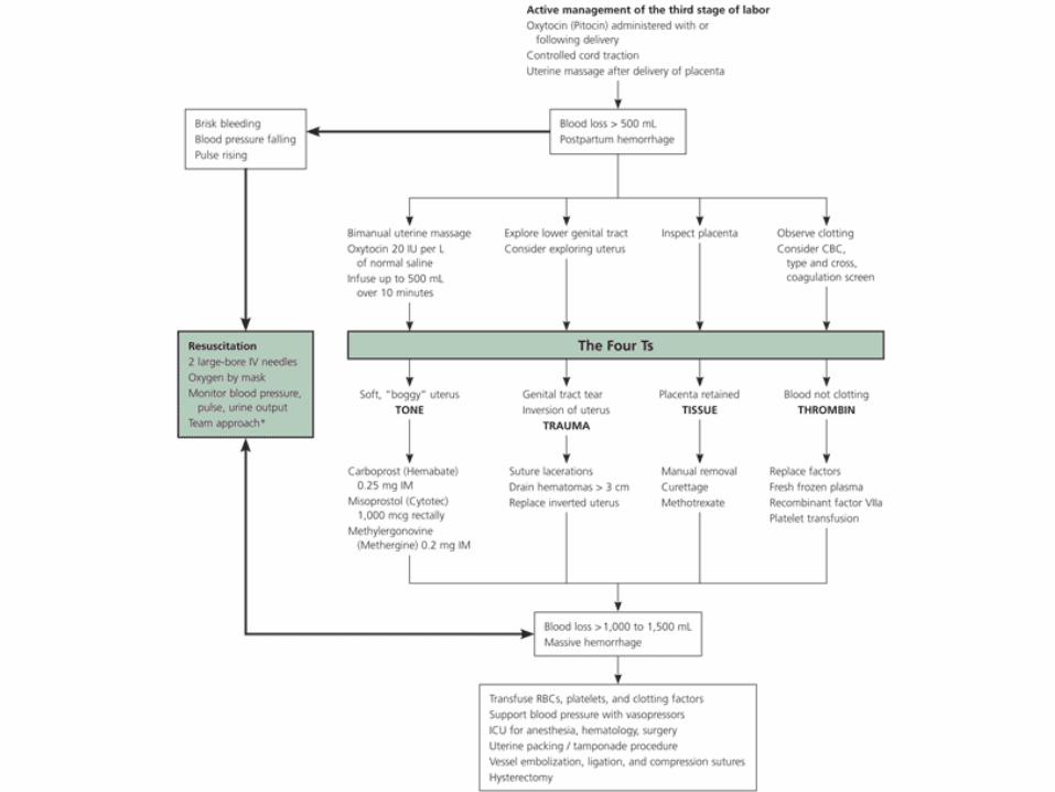
CausesThe 4 “Ts”
TONETRAUMA
TISSUETHROMBIN
70%
20% 1%
10%
Diagnosis of postpartum hemorrhage begins with recognition of excessive bleeding and methodic examination to determine its cause (Figure 1).
Diagnosis and Management
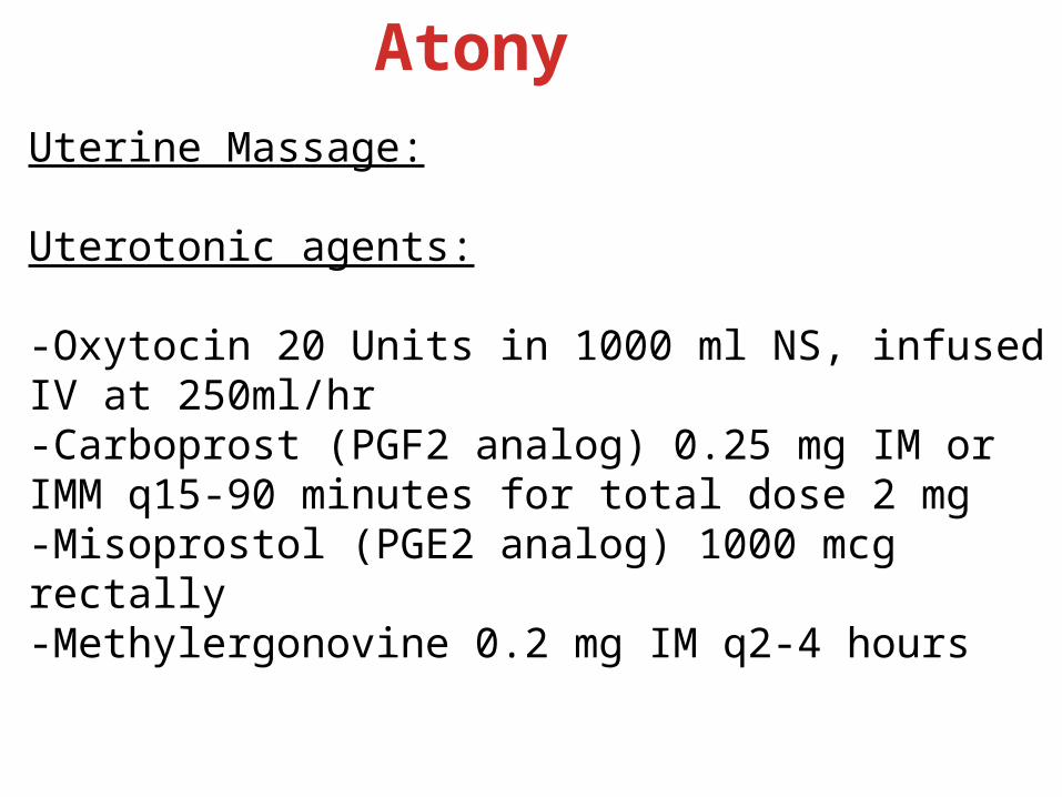
AtonyUterine Massage:
Uterotonic agents:
-Oxytocin 20 Units in 1000 ml NS, infused IV at 250ml/hr-Carboprost (PGF2 analog) 0.25 mg IM or IMM q15-90 minutes for total dose 2 mg-Misoprostol (PGE2 analog) 1000 mcg rectally-Methylergonovine 0.2 mg IM q2-4 hours

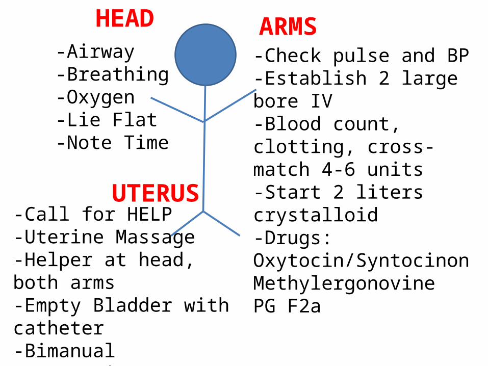
HEAD ARMS
UTERUS
-Airway-Breathing-Oxygen-Lie Flat-Note Time
-Check pulse and BP-Establish 2 large bore IV-Blood count, clotting, cross-match 4-6 units-Start 2 liters crystalloid-Drugs:Oxytocin/SyntocinonMethylergonovinePG F2a
-Call for HELP-Uterine Massage-Helper at head, both arms-Empty Bladder with catheter-Bimanual compression-Review other causes 4 Ts-Move to surgery