THE APICAL SYSTOLIC MURMUR IN MITRAL STENOSIS BY PATRICK MOUNSEY AND WALLACE BRIGDEN From the Cardiac Department of the London Hospital Received January 11, 1954 The purpose of this work was to determine how reliable a guide an apical systolic murmur can be to the finding at mitral valvotomy of incidental mitral regurgitation complicating dominant mitral stenosis. The history of the systolic murmur in mitral stenosis is a confused one, since general agreement was not reached about the timing of systolic and diastolic murmurs in mitral stenosis for nearly a century after Laennec (1819) first described the " bruit de souffiet " and " bruit de scie." Thus Ormerod (1864), Dickinson (1887), and Brockbank (1910) held that the characteristic murmur in mitral stenosis was in early systole and due to associated mitral regurgitation. On the other hand, Fauvel (1843), Gairdner (1861), and Fagge (1870) believed that the murmur was in late diastole and resulted from obstruction to the passage of blood through the mitral valve, as Laennec had originally suggested. With the advent of the electrocardiogram and phonocardiogram, the time of the murmur was fixed more accurately in the cardiac cycle. It became accepted that both diastolic and systolic murmurs were heard in mitral stenosis, the diastolic murmur being of chief importance as indicating stenosis, the systolic murmur, when present, being of secondary signifi- cance only, since it indicated merely a degree of incidental mitral regurgitation. With the intro- duction of mitral valvotomy and the consequent need for more detailed knowledge of the functional pathology of the mitral valve, interest has been re-awakened in the systolic murmur as one possible guide to the presence of regurgitation complicating mitral stenosis (Baker et al., 1952; Froment and Gravier, 1952; Abelmann et al., 1953; Sellors et al., 1953). METHOD Fifty patients, judged to have dominant mitral stenosis, were treated by mitral valvotomy, where the surgeon's finger estimated with special care the degree of incidental mitral regurgitation complicating dominant mitral stenosis.* The age of the patients varied between 17 and 49 years, the majority being in the 30 to 39 age group. Thirty-two patients were in sinus rhythm and 18 had auricular fibrillation. Clinical, radiological, and electrocardiographic evidence of left ventricular enlargement was absent in all patients in this series, with the exception of five, in whom the radiological evidence of slight enlarge- ment was thought to be due to aortic incompetence. In no case was there great enlargement of the left auricle. Right ventricular hypertrophy, as assessed by the electrocardiogram, was present in 32 of the 50 patients. Clinical examination was amplified by a phonocardiogram in every case and this was recorded on a four-channel phonocardiograph, as described by Leatham (1949); an electrocardiogram was used as a reference tracing. RESULTS Findings at Operation. The presence of mitral stenosis was confirmed at valvotomy in every patient, but the degree of stenosis varied (Fig. 1). In the majority the long diameter of the aperture * Mr. Vernon Thompson and Mr. Geoffrey Flavell performed the valvotomies. 255 on April 10, 2020 by guest. Protected by copyright. http://heart.bmj.com/ Br Heart J: first published as 10.1136/hrt.16.3.255 on 1 July 1954. Downloaded from
Transcript
THE APICAL SYSTOLIC MURMUR IN MITRAL STENOSISBY
PATRICK MOUNSEY AND WALLACE BRIGDEN
From the Cardiac Department of the London Hospital
Received January 11, 1954
The purpose of this work was to determine how reliable a guide an apical systolic murmur canbe to the finding at mitral valvotomy of incidental mitral regurgitation complicating dominantmitral stenosis.
The history of the systolic murmur in mitral stenosis is a confused one, since general agreementwas not reached about the timing of systolic and diastolic murmurs in mitral stenosis for nearly acentury after Laennec (1819) first described the " bruit de souffiet " and " bruit de scie." ThusOrmerod (1864), Dickinson (1887), and Brockbank (1910) held that the characteristic murmur inmitral stenosis was in early systole and due to associated mitral regurgitation. On the other hand,Fauvel (1843), Gairdner (1861), and Fagge (1870) believed that the murmur was in late diastoleand resulted from obstruction to the passage of blood through the mitral valve, as Laennec hadoriginally suggested. With the advent of the electrocardiogram and phonocardiogram, the timeof the murmur was fixed more accurately in the cardiac cycle. It became accepted that bothdiastolic and systolic murmurs were heard in mitral stenosis, the diastolic murmur being of chiefimportance as indicating stenosis, the systolic murmur, when present, being of secondary signifi-cance only, since it indicated merely a degree of incidental mitral regurgitation. With the intro-duction of mitral valvotomy and the consequent need for more detailed knowledge of the functionalpathology of the mitral valve, interest has been re-awakened in the systolic murmur as one possibleguide to the presence of regurgitation complicating mitral stenosis (Baker et al., 1952; Fromentand Gravier, 1952; Abelmann et al., 1953; Sellors et al., 1953).
METHODFifty patients, judged to have dominant mitral stenosis, were treated by mitral valvotomy,
where the surgeon's finger estimated with special care the degree of incidental mitral regurgitationcomplicating dominant mitral stenosis.*
The age of the patients varied between 17 and 49 years, the majority being in the 30 to 39 agegroup. Thirty-two patients were in sinus rhythm and 18 had auricular fibrillation. Clinical,radiological, and electrocardiographic evidence of left ventricular enlargement was absent in allpatients in this series, with the exception of five, in whom the radiological evidence of slight enlarge-ment was thought to be due to aortic incompetence. In no case was there great enlargement of theleft auricle. Right ventricular hypertrophy, as assessed by the electrocardiogram, was present in 32of the 50 patients.
Clinical examination was amplified by a phonocardiogram in every case and this was recordedon a four-channel phonocardiograph, as described by Leatham (1949); an electrocardiogram wasused as a reference tracing.
RESULTSFindings at Operation. The presence of mitral stenosis was confirmed at valvotomy in every
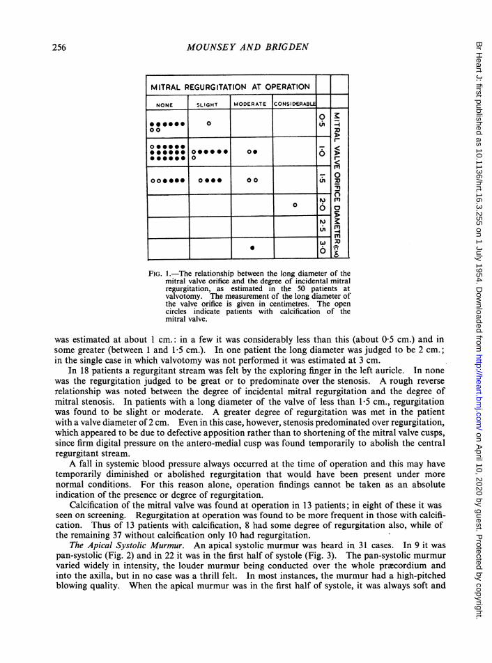
patient, but the degree of stenosis varied (Fig. 1). In the majority the long diameter of the aperture* Mr. Vernon Thompson and Mr. Geoffrey Flavell performed the valvotomies.
255
on April 10, 2020 by guest. P
rotected by copyright.http://heart.bm
j.com/
Br H
eart J: first published as 10.1136/hrt.16.3.255 on 1 July 1954. Dow
FIG.I.theyrlTinhip betwuementothe long diameter ofth
the valve orifice is given in centimnetres. The opencircles indicate patients with calcification of themitral valve.
was estimated at about I cm.: in a few it was considerably less than this (about 0 5 cm.) and insome greater (between I and 1-5 cm.). In one patient the long diameter was judged to be 2 cm.;in the single case in which valvotomy was not performed it was estimated at 3 cm.
In 18 patients a regurgitant stream was felt by the exploring finger in the left auricle. In nonewas the regurgitation judged to be great or to predominate over the stenosis. A rough reverserelationship was noted between the degree of incidental mitral regurgitation and the degree ofmitral stenosis. In patients with a long diameter of the valve of less than 1-5 cm., regurgitationwas found to be slight or moderate. A greater degree of regurgitation was met in the patientwith a valve diameter of 2 cm. Even in this case, however, stenosis predominated over regurgitation,which appeared to be due to defective apposition rather than to shortening of the mitral valve cusps,since firm digital pressure on the antero-medial cusp was found temporarily to abolish the centralregurgitant stream.A fall in systemic blood pressure always occurred at the time of operation and this may have
temporarily diminished or abolished regurgitation that would have been present under morenormal conditions. For this reason alone, operation findings cannot be taken as an absoluteindication of the presence or degree of regurgitation.
Calcification of the mitral valve was found at operation in 13 patients; in eight of these it wasseen on screening. Regurgitation at operation was found to be more frequent in those with calcifi-cation. Thus of 13 patients with calcification, 8 had some degree of regurgitation also, while ofthe remaining 37 without calcification only 10 had regurgitation.
The Apical Systolic Murmur. An apical systolic murmur was heard in 31 cases. In 9 it waspan-systolic (Fig. 2) and in 22 it was in the first half of systole (Fig. 3). The pan-systolic murmurvaried widely in intensity, the louder murmur being conducted over the whole prxcordium andinto the axilla, but in no case was a thrill felt. In most instances, the murmur had a high-pitchedblowing quality. When the apical murmur was in the first half of systole, it was always soft and
on April 10, 2020 by guest. P
rotected by copyright.http://heart.bm
j.com/
Br H
eart J: first published as 10.1136/hrt.16.3.255 on 1 July 1954. Dow
FIG. 2.-Apical pan-systolic murmur. MA=mitral area. HF=highfrequency filter. 1 and 2=first and second heart sounds. SM=systolicmurmur. MDM-mid-diastolicmurmur. PSM-presystolicmurmur.
QS... ..
4:t MDM PSM.. S.M :. .... ,5........ ................... .FIG. 3.-Apical systolic murmur, the bulk of which is in the first half of
systole. MA=mitral area. MF=medium frequency filter. 1 and2=first and second heart sounds. OS=opening snap. SM=systolicmurmur. MDM=mid-diastolicmurmur. PSM=presystolicmurmur.
might easily be missed on casual auscultation, being partly obscured by a loud first heart sound orpresystolic murmur, when present. No relationship was found between the presence or absenceof a presystolic murmur and the systolic murmur.The relationship between the presence and type of apical systolic murmur and the operation finding
of incidental mitral regurgitation complicating dominant mitral stenosis is shown in Fig. 4. A pan-systolic apical murmur was always associated with some degree of regurgitation at operation,whether the murmur was loud or soft. The loudness of the pan-systolic murmur, on the otherhand, although in general proportional to the degree of regurgitation found, was not always soand in one patient with a murmur of medium loudness tight mitral stenosis with only a small lateralregurgitant jet was found at operation.
-
257
on April 10, 2020 by guest. P
rotected by copyright.http://heart.bm
j.com/
Br H
eart J: first published as 10.1136/hrt.16.3.255 on 1 July 1954. Dow
FIG. 4.-The relationship in 50 patients between (a) thepresence, the type, and the loudness of an apicalsystolic murmur, and (b) the operation finding ofincidental mitral regurgitation.
The apical murmur in the first half of systole showed no direct relationship to the operationfinding of incidental mitral regurgitation, which was present, though slight, in 8 out of 22 patients..Absence of an apical systolic murmur was, with one exception, associated with absence of regurgita-tion at operation.
Other Systolic Murmurs in Mitral Stenosis. A systolic murmur, present during episodes of rightheart failure, was sometimes difficult to differentiate from the apical murmur of mitral regurgitation..It was met in four patients during congestive failure; it was loud, pan-systolic, best heard at the leftsternal edge in the fourth and fifth intercostal spaces, but well conducted to the apex also; and afterrecovery from failure it either diminished or disappeared completely. One patient had two episodesof right heart failure, while awaiting mitral valvotomy in hospital, and on each occasion the murmurcame with failure and went with recovery (Fig. 5). In another patient phasic variation in intensityof the murmur was noted with respiration, the murmur becoming louder during inspiration. Thiswas in contrast to apical systolic murmurs on which respiration had little effect. This murmur wasthought to be due to temporary tricuspid regurgitation and this is supported by recent hmmo-dynamic studies of tricuspid valve disease (Shillingford, 1953). Other signs of tricuspid incom-petence, such as a ventricular systolic wave in the jugular venous pulse and a pulsating liver, werenot present in our patients.A mid-systolic murmur, loudest in the aortic area but conducted to the apex, was heard in three
patients with slight aortic incompetence complicating the mitral stenosis. It was thought that thescaffed incompetent aortic valve cusps gave rise to the systolic murmur in the absence of aorticstenosis.
DISCUSSIONAlthough only the systolic murmur has so far been discussed in relation to the operation finding
of incidental mitral regurgitation, other signs of mitral regurgitation were carefully looked for inthis series, but yielded no additional information. No patient with left ventricular enlargement
258
on April 10, 2020 by guest. P
rotected by copyright.http://heart.bm
j.com/
Br H
eart J: first published as 10.1136/hrt.16.3.255 on 1 July 1954. Dow
FIG. 5.-Pan-systolic murmur, present during right heartfailure and disappearing after recovery. Both recordswere made at the left sternal edge (LSE) in the fourthintercostal space with a high frequency filter (HF). (A)recorded during right heart failure and (B) one weeklater after recovery from failure. The pan-systolicmurmur has been replaced by a small murmur confinedto the first half of systole.
was included in the series, with the exception of five, where slight enlargement was thought to bedue to aortic incompetence, and so this important sign of significant mitral regurgitation wasalways absent. It has been suggested that great enlargement of the left auricle is often associatedwith mitral regurgitation (Baker et al., 1952; and Abelmann et al., 1953). In our series, from whichpatients with gross left auricular enlargement were excluded, no relationship was noted betweenminor variations in the size of the left auricle and the presence of incidental mitral regurgitation.Bilateral systolic expansion of the left auricle seen in the anterior view, which Brigden and Leatham(1953) found to be the only reliable radiological sign of pure mitral regurgitation, was never seenin this series. Marked backward movement of the posterior border of the left auricle, which is nota reliable sign of mitral regurgitation, was found to be associated with the operation finding of puremitral stenosis as frequently as with stenosis complicated by regurgitation. Venner and Holling(1953), Wynn et al. (1952), and Ablemann et al. (1953) did not find the pulmonary capillary pressurecurve a reliable guide to the presence of incidental regurgitation complicating mitral stenosis and inthose of our patients on whom cardiac catheterization was performed the pulmonary capillarypressure curve did not give clear additional information about the presence of incidental mitralregurgitation.
It was thought that the loudness of the first heart sound and the finding of an opening snap mightbe of additional help in recognizing incidental mitral regurgitation complicating dominant mitralstenosis. Since in pure mitral stenosis the first heart sound is usually loud and the opening snapclear, and since in pure mitral regurgitation the first heart sound is not accentuated and the openingsnap absent (Brigden and Leatham, 1953), it might be expected that in mitral valve disease thesesounds would indicate whether stenosis or regurgitation predominated. Calcification of the mitralvalve, however, is also associated with a soft first heart sound and a soft or absent opening snap(Wynn, 1953; Mounsey, 1953). In this series the association of a soft first heart sound and asoft or absent opening snap was related to valve calcification and only indirectly related to the
259
on April 10, 2020 by guest. P
rotected by copyright.http://heart.bm
j.com/
Br H
eart J: first published as 10.1136/hrt.16.3.255 on 1 July 1954. Dow
operation finding of incidental mitral regurgitation, owing to the relatively higher incidence ofregurgitation in patients with valve calcification.
SUMMARY AND CONCLUSIONSIn a series of 50 patients with dominant mitral stenosis selected for mitral valvotomy, an apical
pan-systolic murmur was found to be the most reliable guide to the operation finding of incidentalmitral regurgitation. All patients with a pan-systolic apical murmur had mitral regurgitation,whether the murmur was loud or soft. The loudness of the murmur was not a reliable indicationof the varying degree of regurgitation.
An apical murmur in the first half of systole was not constantly related to the operation findingof regurgitation, which was never more than of slight degree in these patients. Absence of anapical systolic murmur was, with one exception, associated with absence of regurgitation atoperation.
A pan-systolic murmur, present in four patients during right heart failure and disappearingafter recovery, was sometimes difficult to differentiate from the apical murmur of mitral regurgita-tion. It was thought to be caused by temporary tricuspid regurgitation.
The association of a soft first heart sound and a soft or absent opening snap was related tocalcification of the mitral valve and only indirectly to incidental mitral regurgitation, owing to the-relatively higher incidence of regurgitation in patients with valve calcification.
We wish to thank Dr. William Evans for his helpful advice and criticism in the preparation of this paper, andMr. Vernon Thompson and Mr. Geoffrey Flavell for their co-operation. Thanks are also due to Mr. W. Dicks fortechnical assistance with the phonocardiograms.
REFERENCESAbelmann, W. H., Ellis, L. B., and Harken, D. E. (1953). Amer. J. Med., 15, 5.Baker, C., Brock, R. C., Campbell, M., and Wood, P. (1952). Brit. med. J., 1, 1043.Brigden, W., and Leatham, A. (1953). Brit. Heart J., 15, 55.Brockbank, E. M. (1910). Quart. J. Med., 3, 345.Dickinson, W. H. (1887). Lancet, 2, 650 and 695.Fagge, C. H., (1870-71). Guy's Hosp. Rep., 3rd ser., 16, 247.Fauvel, S. A. (1843). Arch gen. Med. Paris., ser. 4, 1 1.Froment, R., and Gravier, J. (1952). Rev. Lyon. Me'd., 1, 317.Gairdner, W. T. (1861). Edinb. med. J., 7, 438.Laennec, R. T. H. (1819). De l'Auscultation mediate ou Traite du Diagnostic des Maladies des Poumons et du Cwur,
Fonde principalement sur ce nouveau Moyen d'Exploration. Brosson et Chaude, Paris.Leatham, A. (1949). Post-grad. med. J., 25, 568.Mounsey, J. P. D. (1953). Brit. Heart J., 15, 135.Ormerod, E. L. (1864). Med. Times Gaz., 2, 153.Sellors, T. H., Bedford, D. E., and Somerville, W. (1953). Brit. med. J., 2, 1059.Shillingford, J. P. (1953). Personal communication.Venner, A., and Holling, H. E. (1953). Brit. Heart J., 15, 205.Wynn, A. (1953). Brit. Heart J., 15, 214.
, Mathews, M. B., McMillan, I. K. R., and Daley, R. (1952). Lancet, 2, 216.
260
on April 10, 2020 by guest. P
rotected by copyright.http://heart.bm
j.com/
Br H
eart J: first published as 10.1136/hrt.16.3.255 on 1 July 1954. Dow