Apo2L/TRAIL Inhibits Tumor Growth and Bone Destructionin a Murine Model of Multiple MyelomaAgatha Labrinidis,1Peter Diamond,2 Sally Martin,2 Shelley Hay,1Vasilios Liapis,1Irene Zinonos,1
NatalieA. Sims,3 GeraldJ. Atkins,1Cristina Vincent,1 Vladimir Ponomarev,4
DavidM. Findlay,1Andrew C.W. Zannettino,2 and Andreas Evdokiou1
Abstract Purpose:Multiple myeloma is an incurable disease, for which the development of new therapeu-tic approaches is required. Here, we report on the efficacy of recombinant solubleApo2L/tumornecrosis factor-related apoptosis-inducing ligand (TRAIL) to inhibit tumor progression and bonedestruction in a xenogeneic model of humanmultiple myeloma.Experimental Design: We established a mouse model of myeloma, in which Apo2L/TRAIL-sensitive RPMI-8226 or KMS-11 cells, tagged with a triple reporter gene construct(NES-HSV-TK/GFP/Luc), were transplanted directly into the tibial marrow cavity of nude mice.Tumor burden was monitored progressively by bioluminescence imaging and the developmentof myeloma-induced osteolysis was measured using high resolution in vivo micro-computedtomography.Results: Tumor burden increased progressively in the tibial marrow cavity of mice transplantedwith Apo2L/TRAIL-sensitive RPMI-8226 or KMS-11cells associated with extensive osteolysisdirectly in the area of cancer cell transplantation. Treatment of mice with recombinant solubleApo2L/TRAIL reduced myeloma burden in the bone marrow cavity and significantly protectedagainst myeloma-induced osteolysis.The protective effects of Apo2L/TRAIL treatment on boneweremediatedby the direct apoptotic actions of Apo2L/TRAIL onmyeloma cells within the bonemicroenvironment.Conclusions: This is the first in vivo study that investigates the efficacy of recombinant Apo2L/TRAIL on myeloma burden within the bone microenvironment and associated myeloma-inducedbone destruction. Our findings that recombinant solubleApo2L/TRAIL reduces myeloma burdenwithin the bonemicroenvironment and protects the bone frommyeloma-induced bone destructionargue against an inhibitory role of osteoprotegerin in Apo2L/TRAIL-induced apoptosis in vivo andhighlight theneed to clinically evaluateApo2L/TRAIL inpatientswithmultiplemyeloma.
Multiple myeloma is a B-cell malignancy characterized by thepresence of a monoclonal population of plasma cells, whichlocalize to sites in the bone marrow close to the endostealsurface (1). Multiple myeloma is unique among hematologic
malignancies in its capacity to cause massive osteoclast-mediated destruction throughout the axial and craniofacialskeleton. The focal osteolytic lesions result in a range ofdebilitating clinical symptoms including bone pain, pathologicfractures, spinal cord compression, hypercalcemia, and renalfailure (2). Although the precise mechanisms remain to bedetermined, numerous studies show that multiple myelomaplasma cells stimulate focal bone loss by promoting theaberrant recruitment and activation of osteoclast precursorsfrom the peripheral blood to the endosteal and trabecular bonesurfaces in close proximity to nests of multiple myelomaplasma cells and also inhibit osteoblastic bone formation [refs.3, 4 and reviewed by Heider et al. (5)]. Despite many recentadvances in clinical therapy, multiple myeloma remains anincurable disease, necessitating the development of new andsafe therapeutic approaches.
Apo2 ligand/tumor necrosis factor-related apoptosis-inducing ligand (TRAIL) represents a promising new candidatedrug for the treatment of multiple myeloma. Apo2L/TRAILinduces apoptosis in a variety of tumor cell types via interactionswith its death domain-containing receptors DR4 and DR5 toactivate caspases that carry out the cell death program. Threehomologous human decoy receptors for Apo2L/TRAIL have also
Cancer Therapy: Preclinical
Authors’Affiliations: 1Discipline of Orthopaedics and Trauma,The University ofAdelaide, Royal Adelaide Hospital, and Hanson Institute ; 2Myeloma andMesenchymal Research Laboratory, Bone and Cancer Laboratories, Division ofHaematology, Institute of Medical and Veterinary Science, and Hanson Institute,Adelaide, South Australia, Australia; 3St.Vincent’s Institute of Medical Research,Fitzroy, Victoria, Australia; and 4Department of Neurology, Memorial Sloan-Kettering Cancer Center, NewYork, NewYorkReceived 9/22/08; revised 10/29/08; accepted 11/25/08; published OnlineFirst3/10/09.Grant support: National Health and Medical Research Council of Australia,TheCancer Council of South Australia, and National Breast Cancer Foundation.The costs of publication of this article were defrayed in part by the payment of pagecharges.This article must therefore be hereby marked advertisement in accordancewith18 U.S.C. Section1734 solely to indicate this fact.Note: A.C.W. Zannettino and A. Evdokiou are considered equal senior authors.Requests for reprints:Andreas Evdokiou, Discipline of Orthopaedics andTrauma,The University of Adelaide, Royal Adelaide Hospital, and Hanson Institute, Level 4,Bice Building, NorthTerrace, Adelaide 5000, South Australia, Australia. Phone: 61-8-82223107; Fax: 61-8-82323065; E-mail: [email protected].
F2009 American Association for Cancer Research.doi:10.1158/1078-0432.CCR-08-2444
www.aacrjournals.orgClin Cancer Res 2009;15(6) March15, 2009 1998
been identified, including DcR1, DcR2, and osteoprotegerin(OPG; refs. 6, 7). Whereas DR4 and DR5 contain cytoplasmicdeath domains and on ligation induce apoptosis in Apo2L/TRAIL-sensitive cells, DcR1 and DcR2 lack functional deathdomains and cannot mediate apoptosis. OPG is a widelyexpressed soluble member of the tumor necrosis factor receptorfamily that is capable of binding to Apo2L/TRAIL, and althoughit has lower affinity for Apo2L/TRAIL at normal physiologictemperature, it can block Apo2L/TRAIL-induced apoptosisin vitro (8–11). Although other apoptosis-inducing membersof the tumor necrosis factor family, such as tumor necrosisfactor-a and Fas ligand, initially carried great promise asanticancer agents, their severe toxicity toward normal tissuesprecluded their clinical use. In contrast, Apo2L/TRAIL isselectively toxic to cancer cells and exhibits no toxicity tonormal cells (12, 13). For example, our previous studies haveshown that normal human bone cells, normal mammaryepithelial cells, and human fibroblasts are resistant to Apo2L/TRAIL-induced apoptosis in vitro despite constitutive expressionof Apo2L/TRAIL and its death receptors (14, 15). Although themechanisms underlying the differential sensitivity to Apo2L/TRAIL of different tumor types or between tumors of the sametype remain to be fully elucidated, multiple mechanisms appearto be involved. These include increased expression of the decoyreceptors for Apo2L/TRAIL (16–20) and the overexpression ofintracellular inhibitory proteins such as FLIP (16) or intracel-lular inhibitor of apoptosis molecules (21).
The antitumor activity of Apo2L/TRAIL has been welldemonstrated in several mouse xenograft models of humansoft tissue cancers, including colorectal (22), breast (12), lung(23), and glioma (24). Apo2L/TRAIL was active as a singleagent and exhibited synergistic activity with certain chemo-
therapeutic agents or radiotherapy, causing marked regressionor complete remission of tumors, with no evidence of toxicityto normal tissues and organs of the animals (25, 26). Phase Iaclinical trials of soluble, recombinant Apo2L/TRAIL in patientswith a variety of solid tumor types and non-Hodgkin’slymphoma have shown that Apo2L/TRAIL is safe and welltolerated (27). Preliminary results in six low-grade non-Hodgkin’s lymphomas, previously treated with rituximab,revealed that this particular combination with Apo2L/TRAILwas well tolerated and induced complete remission in twopatients and partial remission or stable disease in another threepatients (28). Further dose optimization is currently underway. In the context of multiple myeloma, previous studies haveshown that Apo2L/TRAIL induces apoptosis of myeloma cellsin vitro and reduces tumor burden in subcutaneous models ofmyeloma growth in vivo (8, 10, 29). However, the efficacy ofApo2L/TRAIL on myeloma burden in bone and myeloma-induced bone destruction has not been investigated. We haverecently shown that recombinant soluble Apo2L/TRAIL pre-vents breast cancer-induced bone destruction in a mousemodel by directly targeting cancer cells within the bonemicroenvironment and with no evidence of toxic side effectsto normal tissues and organs. This work highlighted thepotential for Apo2L/TRAIL therapy against bone-relatedmalignancies.
Previous studies have raised the possibility that the activityof soluble Apo2L/TRAIL may be abrogated in the bonemicroenvironment, where expression of the antagonisticdecoy receptor for Apo2L/TRAIL, OPG, is high and speculatedthat recombinant soluble Apo2L/TRAIL may have limited useas a therapeutic agent for the treatment of skeletal malignan-cies (8, 10, 11). To examine the antimyeloma activity ofApo2L/TRAIL within bone, we established a mouse model inwhich Apo2L/TRAIL-sensitive RPMI-8226 multiple myelomaor KMS-11 cells, tagged with a triple reporter gene construct(NES-HSV-TK/GFP/Luc; ref. 30), were transplanted directlyinto the tibial marrow cavity of nude mice. The intratibialinjection model results in the formation of focal osteolyticlesions, which are localized to a single bony site and producea consistent measurable outcome, enabling assessment of theefficacy of Apo2L/TRAIL treatment. An added advantage ofthis model is that the contralateral tibia in the same animalcan be used as a control. This allows evaluation of the effectsof treatment on myeloma burden and myeloma-inducedosteolysis as well as the effects of Apo2L/TRAIL treatment onnormal bone metabolism by comparing the non-tumor-bearing bones of the treated and vehicle-treated groups. Ourfindings that recombinant soluble Apo2L/TRAIL reducesmyeloma burden within the bone microenvironment andprotects the bone from myeloma-induced bone destructionargue against an inhibitory role of OPG in Apo2L/TRAIL-induced apoptosis in vivo and highlight the need to clinicallyevaluate recombinant soluble Apo2L/TRAIL in patients withmultiple myeloma.
Materials and Methods
Cells and reagents
The human multiple myeloma cell lines WL2, ARH-77, U266, RPMI-8226, KMS-11, LP-1, andNCI-929were obtained from the American TypeCulture Collection and cultured inDMEM, supplemented with glutamine
Translational Relevance
Multiple myeloma is an incurable disease, for which thedevelopment of new therapeutic approaches is required.Here, we report for the first time on the efficacy ofrecombinant soluble Apo2L/tumor necrosis factor-relatedapoptosis-inducing ligand (TRAIL) to inhibit tumorprogression and bone destruction in a xenogeneic modelof human multiple myeloma. Previous studies have raisedthe possibility that the activity of soluble Apo2L/TRAILmay be abrogated in the bone microenvironment, whereexpression of the antagonistic decoy receptor forApo2L/TRAIL, osteoprotegerin, is high, and speculated thatrecombinant soluble Apo2L/TRAIL may have limiteduse as a therapeutic agent for the treatment of skeletalmalignancies. Our findings that recombinant solubleApo2L/TRAIL reduces myeloma burden within the bonemicroenvironment and protects the bone from myeloma-induced bone destruction argue against an inhibitory roleof osteoprotegerin in Apo2L/TRAIL-induced apoptosisin vivo and highlight the need to clinically evaluateApo2L/TRAIL in patients with multiple myeloma. Givenits imminent use in the clinic, we strongly feel that theantimyeloma activity of the only available clinical grade ofrecombinant solubleApo2L/TRAIL should be reported.
Apo2L/TRAILReducesMyeloma Burden in Bone
www.aacrjournals.org Clin Cancer Res 2009;15(6) March15, 20091999
(2 mmol/L), penicillin (100 IU/mL), streptomycin (100 Ag/mL),gentamicin (160 Ag/mL), and 10% fetal bovine serum (Biosciences), ina humidified atmosphere containing 5% CO2. Recombinant solubleApo2L/TRAIL was a gift from Dr. Avi Ashkenazi (Genentech). Zoledronicacid was generously provided by Novartis Pharma. The tetrapeptidecaspase inhibitor zVAD-fmk was purchased from Calbiochem.
Measurement of cell viability
For determination of Apo2L/TRAIL effects on cell growth, 1 � 104
cells per well were seeded in 96-well microtiter plates. The followingday, fresh medium containing increasing concentrations of Apo2L/TRAIL (3-300 ng/mL) was added for 24 h. Cell viability was determinedusing the WST-1 cell proliferation reagent assay kit (Roche MolecularBiochemicals). The absorbance was measured at 420 to 480 nm usingan ELISA plate reader. Experiments comprised quadruplicate determi-nations and experiments were repeated at least three times. Results ofexperiments are given as the mean F SD.
Apoptosis analysis
4,6-Diamidine-2-phenylindole staining of nuclei. Cells were seededon plastic chamber slides and treated as indicated. After two washeswith PBS, cells were fixed in methanol for 5 min, washed again withPBS, and incubated with 0.8 mg/mL 4¶,6-diamidine-2-phenylindole(Roche Diagnostics) in PBS for 15 min at 37jC. After several washes inPBS, the coverslips were mounted on PBS/glycerol. 4¶,6-Diamidine-2-phenylindole staining was visualized by fluorescence microscopy.Measurement of DEVD-caspase activity. DEVD-caspase activity was
assayed by cleavage of zDEVD-AFC, a fluorogenic substrate based onthe peptide sequence at the caspase-3 cleavage site of poly(ADP-ribose)polymerase. Cells (5 � 105) grown in 24-well plates were treated asindicated, washed once with HBSS, and resuspended in 200 AL NP-40lysis buffer containing 5 mmol/L Tris-HCl, 5 mmol/L EDTA, and 0.5%NP-40 (pH 7.5). Cell lysate (20 AL) was added to each assay tubecontaining 8 Amol/L substrate in 1 mL protease buffer [50 mmol/LHEPES, 10% sucrose, 10 mmol/L DTT, 0.1% CHAPS (pH 7.4)]. After4 h at room temperature, fluorescence was quantified (Ex 400 andEm 505) in a Perkin-Elmer LS50 fluorescence spectrometer. Optimalamounts of added lysate and duration of assay were taken from linearportions of curves as determined in preliminary experiments.
Western blot analysis
Primary antibodies were purchased from Chemicon International(polyclonal antibody anti-bid), Pharmingen International (polyclonalantibody anti-caspase-8), Cell Signaling Technology (polyclonal anti-body anti-caspase-9), Medical and Biological Laboratories [monoclonalantibody (mAb) anti-caspase-10], Transduction Laboratories (mAbanti-caspase-3), and Roche Diagnostics [polyclonal antibody anti-poly(ADP-ribose) polymerase]. Anti-actin polyclonal antibody (SantaCruz Biotechnology) was used to normalize for protein concentration.Membranes were rinsed several times with PBS containing 0.1% Tween20 and incubated with 1:5,000 dilution of anti-mouse or anti-rabbitalkaline phosphatase-conjugated secondary antibodies (Amersham) for1 h. Visualization and quantification of protein bands were done usingthe Vistra ECF substrate reagent kit (Amersham) on a FluorImager(Molecular Dynamics).
Retroviral infection of RPMI-8226 and KMS-11 cells with the
triple reporter gene construct SFG-NES-TGL
TK/GFP/Luc-expressing RPMI-8226 and KMS-11 cell lines weregenerated using the retroviral expression vector SFG-NES-TGL (30).Viral particle-containing supernatant was generated and filtered toremove any cellular debris and used to infect RPMI-8226 and KMS-11cells as described previously (31). The retrovirally transduced cells weregrown as bulk cultures for 48 h and subsequently sorted for positivegreen fluorescent protein (GFP) expression using fluorescence-activatedcell sorting (Aria BD Biosciences). The cells were allowed to proliferateand the top 10% of GFP-expressing cells were sorted by fluorescence-
activated cell sorting to generate the sublines RPMI-8226-NES-TGL andKMS-11-TGL. To confirm luciferase expression, an in vitro biolumines-cence assay was done. Bioluminescence was measured using Lumino-scan Ascent and analyzed by Ascent software (ThermoFisher Scientific).
Animals
Female athymic nude mice at age 4 to 6 weeks (Institute of Medicaland Veterinary Science) were acclimatized to the animal housing facilityfor a minimum period of 1 week before the commencement ofexperimentation. All of the experimental procedures on animals werecarried out with strict adherence to the rules and guidelines for theethical use of animals in research and were approved by the AnimalEthics Committee of the Institute of Medical and Veterinary Science.
Intratibial implantation of multiple myeloma cells
Multiple myeloma RPMI-8226-NES-TGL or KMS-11-TGL cells wereinoculated intratibially as described previously (32, 33). Cells in log-phase growth were aspirated into a 25 AL Hamilton syringe (AlltechAssociates) coupled to a 27-gauge needle. The needle was insertedthrough the cortex of the anteria tuberosity of the left tibia. Once thebone cortex was traversed, the needle was inserted 3 to 5 mm down thediaphysis of the tibia, and 10 AL of the cell suspension (1 � 106 cellsper inoculum) were injected into the marrow space. The rightcontralateral tibia was injected with PBS alone as an internal control.For each of the two independent experiments in which RPMI-8226-TGLcells were injected intratibially, mice were randomized into two groupsof 10 mice per group. Two days after cancer cell transplantation, Apo2L/TRAIL was administered (intraperitoneally) at 30 mg/kg/dose once perday for 5 consecutive days followed by a once weekly dose untiltermination of the experiment. The control group was given PBSadministered intraperitoneally. Mice from experiments 1 and 2 werehumanely killed at 5 weeks after cancer cell implantation. In a parallel,experiment mice (n = 10) were injected subcutaneously with theantiresorptive bisphosphonate zoledronic acid (Novartis Pharma) at100 Ag/kg/dose once weekly for 5 weeks as a positive control forcomparison. Animals transplanted with KMS-11 cells were leftuntreated until tumors became well established and Apo2L/TRAILwas administered on day 21 (intraperitoneally) at 30 mg/kg/dose onceper day for 5 consecutive days followed by a once weekly dose untiltermination of the experiment on day 42.
In vivo bioluminescent imaging
Noninvasive, whole-body imaging to monitor luciferase-expressingRPMI-8226-NES-SFG-TGL or KMS-11-TGL cells in mice was doneweekly using the IVIS 100 Imaging System (Xenogen). About 30 minbefore analysis, mice were injected intraperitoneally with 100 ALD-luciferin solution at 250 mg/kg body weight (Xenogen) and thengas-anesthetized with isoflurane (Faulding Pharmaceuticals). Imageswere acquired for 0.5 to 10 s and the photon emission transmitted frommice was captured and quantitated in photons/s/cm2/sr using XenogenLiving image (Igor Pro version 2.5) software.
Micro-computed tomography analysis
In vivo live micro-computed tomography imaging. Computed to-mography (CT) images were obtained using a Skyscan-1076 in vivomicro-CT scanner (Skyscan) at fortnightly intervals while the animalswere anesthetized. The micro-CT scanner was operated at 80 kV,120 AA, rotation step 0. 5, 0.5 mm Al filter, scan resolution of 17.4 Am/pixel, and imaging time of 15 min. The cross-sections were reconstructedusing a cone-beam algorithm (software Cone_rec; Skyscan). Files werethen imported into CTAn software (Skyscan) for three-dimensionalanalysis and three-dimensional image generation. All images are viewedand edited using ANT visualization software.
Ex vivo micro-CT imaging. Limbs for micro-CT analysis weresurgically resected and scanned using the Skyscan-1072 high-resolutionmicro-CT scanner (Skyscan). The micro-CT scanner was operated at80 kV, 120 AA, rotation step 0.675, 0.5 mm Al filter, and scan resolution
Cancer Therapy: Preclinical
www.aacrjournals.orgClin Cancer Res 2009;15(6) March15, 2009 2000
of 5.2 Am/pixel. Using the two-dimensional images obtained from themicro-CT scan, the growth plate was identified and 750 sections wereselected starting from the growth plate/tibial interface and movingdown the tibia. For quantification, three-dimensional evaluation wasdone on all data sets acquired by selecting two separate regions ofinterest, one for total bone and another containing the trabecularspongiosa of the proximal tibia only, to determine three-dimensionalbone morphometric parameters (software CTAn; Skyscan).
Histology
Tibiae were fixed in 10% (v/v) buffered formalin (24 h at 4jC)followed by 2 weeks of decalcification in 0.5 mol/L EDTA/0.5%paraformaldehyde in PBS (pH 8.0) at 4jC. Complete decalcificationwas confirmed by radiography and tibiae were then paraffin embedded.Longitudinal sections (5 Am) were prepared and stained with H&E. Thesame sections were used for TUNEL staining using the ApopTag Plusin situ apoptosis detection kit (Chemicon International) as permanufacturer’s instructions. Analysis was done on an Olympus CX41microscope.
Isolation of tumors or explants from the bone marrow of tibia
Tibiae were cut below the growth plate and above the feet and a25-gauge needle was used to flush cells from the bone marrow cavitywith PBS. The ex vivo cells were sorted by fluorescence-activated cellsorting to isolate the highly GFP-expressing cells designated here asRPMI-8226-NES-TGL-ex vivo and seeded into 96-well plates beforetreatment with 100 ng/mL Apo2L/TRAIL to examine changes in drugsensitivity.
Generation of Apo2L/TRAIL-resistant RPMI-8226-NES-TGL cells
in vitro
RPMI-8226-NES-TGL cells were seeded in a T75 flask until 75%confluence. Cells were exposed to Apo2L/TRAIL at 25 ng/mL for thefirst 2 weeks, at which time the concentration of Apo2L/TRAIL wasincreased to 50 ng/mL for a further 2 weeks followed by treatment in100 ng/mL for a further 4 weeks. During the selection period, cell debriswas removed every 3 days and cells were incubated with fresh mediumcontaining Apo2L/TRAIL for 8 weeks. Thereafter, Apo2L/TRAIL-resistantcells, denoted RPMI-8226-NES-TGL-R, were cultured in Apo2L/TRAIL-free medium and were found to remain resistant.
Cell surface expression of DR4 and DR5
For each of the cell lines RPMI-8226-NES-TGL, RPMI-8226-NES-TGL-R, and RPMI-8226-NES-TGL-ex vivo , cells were collected, washedin 1% bovine serum albumin and 0.1% sodium azide in PBS, andresuspended in 50 AL blocking buffer (5% normal goat serum,1% bovine serum albumin in PBS + 0.1% sodium azide) on ice. mAbagainst TRAIL-R1/DR4 (IgG1, MAB347) was purchased from R&DSystems, whereas monoclonal antibodies against TRAIL-R2/DR5 (IgG1,M413), TRAIL-R3/DcR1 (IgG1, M430), and TRAIL-R4/DcR2 (IgG1,M444) were kindly supplied by Amgen. The mAb 1B5 was used as theIgG1 isotype control. Antibodies were added to cells at 20 Ag/mL andleft for 1 h on ice. Cells were washed and the secondary antibody wasadded (aIgG-PE; Southern Biotech) and left on ice for a further 45 min.Cells were washed and resuspended in fluorescence-activated cellsorting fix until analysis by flow cytometry.
Histomorphometric analysis
To determine the effects of Apo2L/TRAIL on normal bonemorphometry, fixed tibiae were embedded in methylmethacrylate(34, 35) and 5 Am sections were stained with toluidine blue (35).Histomorphometry was carried out in the secondary spongiosaof the proximal tibia (osteomeasure; Osteometrics) as describedpreviously (35).
Data analysis and statistics
Experiments were done in triplicate, and data are presented asmean F SE. All statistical analysis was done using SigmaStat forWindows version 3.0 (Systat Software) using the unpaired Student’s ttest. Measures of association between two variables were assessed usingthe Spearman rank correlation coefficient. Comparisons betweengroups were assessed using a one-way ANOVA test. In all cases, P <0.05 was considered statistically significant.
Results
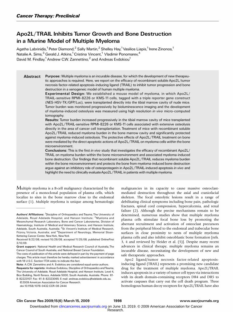
Effect of Apo2L/TRAIL on multiple myeloma cells in vitro. Apanel of seven well-established human multiple myeloma celllines were examined for their sensitivity to the cytotoxic effectsof Apo2L/TRAIL. As shown in Fig. 1A, only RPMI-8226 andKMS-11 exhibited a dose-dependent loss of cell viability aftertreatment for 24 h, reaching a maximum of 80% loss ofviability at the highest dose of 300 ng/mL Apo2L/TRAIL(Fig. 1A). The remaining cell lines were relatively resistant tothe effects of Apo2L/TRAIL. Treatment of RPMI-8226 cells withApo2L/TRAIL induced morphologic changes characteristic ofapoptosis, including chromatin condensation and DNA frag-mentation (Fig. 1B) and a dose-dependent activation ofcaspase-3, which reached maximum at 25 ng/mL and plateauedthereafter (Fig. 1C). The presence of a pan-caspase inhibitor,zVAD-fmk, completely reversed the reduction in cell viabilityinduced by Apo2L/TRAIL (Fig. 1C). Apoptosis induction wasassociated with processing and activation of the initiatorcaspase-8 and caspase-10, leading to cleavage pro-caspase-3,which was concomitant with cleavage of the apoptosis targetprotein poly(ADP-ribose) polymerase (Fig. 1D). Activation ofthe intrinsic apoptotic pathway was associated with caspase-8-mediated cleavage of the Bcl-2 protein family member Bid andactivation of caspase-9. Similar effects were also seen with KMS-11 cells (data not shown).Antitumor activity of Apo2L/TRAIL in a mouse model of
multiple myeloma. To evaluate the efficacy of recombinantsoluble Apo2L/TRAIL to inhibit tumor growth and bonedestruction, we established a xenogeneic tumor model ofhuman multiple myeloma, in which the Apo2L/TRAIL-sensitiveRPMI-8226 cells were transplanted directly into the marrowtibial cavity of athymic nude mice. For noninvasive biolumi-nescence imaging (BLI) of tumor growth, the parental cells wereretrovirally infected with a triple-fusion protein reporterconstruct encoding HSV-TK/GFP/Luc (30). After infection, themyeloma cells were enriched for high-level expression of GFPby two rounds of fluorescence-activated cell sorting, thusgenerating the subline RPMI-8226-NES-TGL. The selected cellsexhibited a 1,000-fold induction of luciferase activity whenanalyzed in vitro. When compared with the parental noninfect-ed myeloma cells, RPMI-8226-NES-TGL cells were equallyresponsive to Apo2L/TRAIL, with both lines showing highsensitivity to the apoptotic effects of the ligand and with similarkinetics of caspase-3 activity (Fig. 1E). RPMI-8226-NES-TGLcells were injected within the left tibiae of mice and the rightcontralateral tibiae were injected with PBS. Tumor burden wasassessed by weekly noninvasive BLI. Results from two separateanimal experiments showed an exponential increase of photonemission associated with an increase in tumor burden, whichwas clearly evident from day 7 onwards in the untreated groupof the first experiment and day 14 of the second experiment
Apo2L/TRAILReducesMyeloma Burden in Bone
www.aacrjournals.org Clin Cancer Res 2009;15(6) March15, 20092001
Fig.1. Apo2L/TRAIL-induced apoptosis inmultiple myeloma cell in vitro. A, comparisonof the effect of Apo2L/TRAIL in a panel of well-establishedhumanmultiplemyelomacell lines. Cells were left untreated or treated with increasing Apo2L/TRAIL concentrations of 3, 10, 30, 100, and100 ng/mL and cell viability was determined by MTTassay24 h thereafter. Representative experiment repeated at least three times. Points, mean; bars, SD. B, cells were seeded on chamber slides at 5 � 104 per chamber andtreatedwith Apo2L/TRAIL at100 ng/mL for 24 h. Cells were fixedwithmethanol and incubatedwith 4¶,6-diamidine-2-phenylindole beforewashing in PBS andmounting onPBS/glycerin. 4¶,6-Diamidine-2-phenylindole staining was visualized by fluorescence microscopy. C, left, RPMI-8226 cells were treated with increasing concentrations ofApo2L/TRAIL for 24 h. Cell lysates were used to determine caspase-3-like activity using the caspase-3 specific fluorogenic substrate, zDEVD-AFC, as described inMaterialsand Methods. Right, RPMI-8226 cells were treated for 24 h with100 ng/mL Apo2L/TRAIL alone or coincubated with the broad-specificity caspase inhibitor zVAD-fmk(50 Amol/L).To exclude possible toxic effects of the inhibitor, cells were also treatedwith the inhibitor alone. Cell viability was determined using theMTTassay and expressedas percentage of control. Quadruplicate results from a representative experiment repeated at least twice. Points, mean; bars, SD. D, RPMI-8226 cells were seeded at2� 106 perT25 flask and either left untreated or treated with Apo2L/TRAIL at a concentration of 100 ng/mL. Cells were then lysed and total cell lysates were analyzed byPAGE and transferred to polyvinylidene fluoride membranes for immunodetection.The caspase-8, caspase-9, caspase-10, and poly(ADP-ribose) polymerase antibodiesdetect both full-length and processed forms of the antigen, whereas caspase-3 and Bid antibodies detect only the full-length antigens. E, comparison of the effect of Apo2L/TRAIL on NES-HSV-TK/GFP/Luc retrovirally infected cells (RPMI-8226-NES-TGL) and their parental noninfected counterpart cells (RPMI-8226). Cells were left untreatedor treated for 24 h with increasing concentrations Apo2L/TRAIL. Cell viability (top) and caspase-3 activity (bottom) were determined as described above. Representativeexperiments repeated at least three times. Bars, SD.
Cancer Therapy: Preclinical
www.aacrjournals.orgClin Cancer Res 2009;15(6) March15, 2009 2002
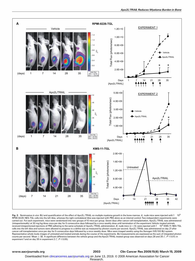
Fig. 2. Noninvasive in vivo BLI and quantification of the effect of Apo2L/TRAIL onmultiple myeloma growth in the bone marrow. A, nude mice were injected with1�106RPMI-8226-NES-TGL cells into the left tibia, whereas the right contralateral tibia was injected with PBS alone as an internal control. Two independent experiments werecarried out. For each experiment, mice were randomized into two groups of10 mice per group. Seven days after cancer cell transplantation, Apo2L/TRAIL was administered(intraperitoneally) at 30 mg/kg/dose once per day for 5 consecutive days followed by a once weekly dose until termination of the experiment.Vehicle-treated animalsreceived intraperitoneal injections of PBS adhering to the same schedule of Apo2L/TRAIL administration. B, nude mice (n = 6) were injected with1�106 KMS-11-NES-TGLcells into the left tibia and tumors were allowed to progress to a define size as measured by photon counts per second. Apo2L/TRAIL was administered on day 21aftercancer cell transplantation once per day for 5 consecutive days followed by a once weekly dose. Mice were imaged weekly using the Xenogen IVIS100 BLI system.Representative whole-body images of untreated and treated animals during the course of the experiments. BLImeasurements are expressed as the sum of integrated photoncounts per second. MeanF SE. A significant difference between the vehicle group and theApo2l/TRAIL-treated group was observed on days 28 and 35 (*, P < 0.01) inexperiment 1and on day 35 in experiment 2 (*, P < 0.05).
Apo2L/TRAILReducesMyeloma Burden in Bone
www.aacrjournals.org Clin Cancer Res 2009;15(6) March15, 20092003
(Fig. 2A). In contrast, animals treated intraperitoneally with30 mg/kg/dose of Apo2L/TRAIL for 5 consecutive days,followed by once weekly injection for 6 weeks, exhibited asignificant inhibition of tumor growth when compared withthe vehicle-treated groups (Fig. 2A). We next assessed thetumor-suppressive activity of Apo2L/TRAIL against the onlyother sensitive myeloma cell line KMS-11-TGL. However, inthis case, we allowed the tumors to establish and progress to adefined size with treatment initiated on day 21 after cancer celltransplantation. Treatment with Apo2L/TRAIL resulted in asignificant regression of the tumors within the first week oftreatment and bioluminescence signal was barely detectablethereafter (Fig. 2B). This indicates that this effect Apo2L/TRAILwas not cell line dependent but rather was a general
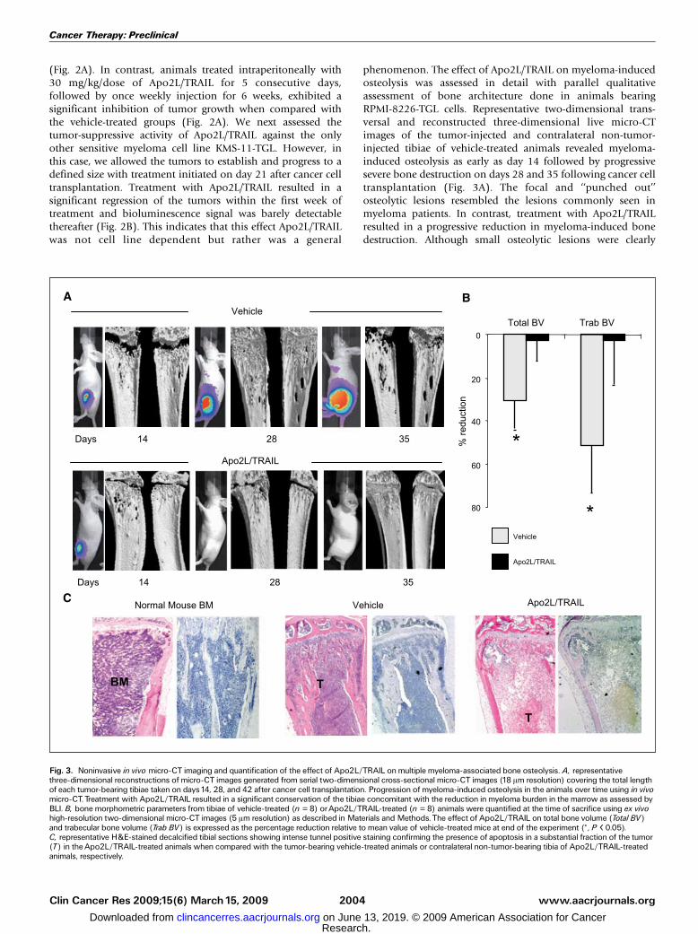
phenomenon. The effect of Apo2L/TRAIL on myeloma-inducedosteolysis was assessed in detail with parallel qualitativeassessment of bone architecture done in animals bearingRPMI-8226-TGL cells. Representative two-dimensional trans-versal and reconstructed three-dimensional live micro-CTimages of the tumor-injected and contralateral non-tumor-injected tibiae of vehicle-treated animals revealed myeloma-induced osteolysis as early as day 14 followed by progressivesevere bone destruction on days 28 and 35 following cancer celltransplantation (Fig. 3A). The focal and ‘‘punched out’’osteolytic lesions resembled the lesions commonly seen inmyeloma patients. In contrast, treatment with Apo2L/TRAILresulted in a progressive reduction in myeloma-induced bonedestruction. Although small osteolytic lesions were clearly
Fig. 3. Noninvasive in vivo micro-CT imaging and quantification of the effect of Apo2L/TRAIL onmultiple myeloma-associated bone osteolysis. A, representativethree-dimensional reconstructions of micro-CT images generated from serial two-dimensional cross-sectional micro-CT images (18 Am resolution) covering the total lengthof each tumor-bearing tibiae taken on days14, 28, and 42 after cancer cell transplantation. Progression of myeloma-induced osteolysis in the animals over time using in vivomicro-CT.Treatment with Apo2L/TRAIL resulted in a significant conservation of the tibiae concomitant with the reduction in myeloma burden in the marrow as assessed byBLI. B, bone morphometric parameters from tibiae of vehicle-treated (n = 8) orApo2L/TRAIL-treated (n = 8) animals were quantified at the time of sacrifice using ex vivohigh-resolution two-dimensional micro-CT images (5 Am resolution) as described in Materials and Methods.The effect of Apo2L/TRAIL on total bone volume (Total BV)and trabecular bone volume (Trab BV) is expressed as the percentage reduction relative to mean value of vehicle-treated mice at end of the experiment (*, P < 0.05).C, representative H&E-stained decalcified tibial sections showing intense tunnel positive staining confirming the presence of apoptosis in a substantial fraction of the tumor(T) in theApo2L/TRAIL-treated animals when compared with the tumor-bearing vehicle-treated animals or contralateral non-tumor-bearing tibia of Apo2L/TRAIL-treatedanimals, respectively.
Cancer Therapy: Preclinical
www.aacrjournals.orgClin Cancer Res 2009;15(6) March15, 2009 2004
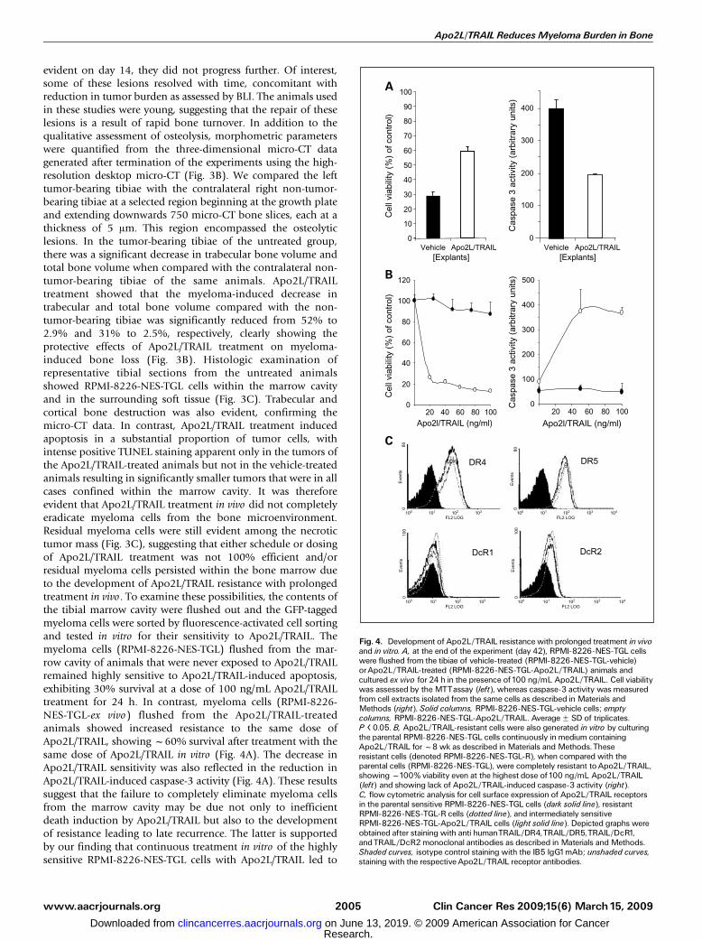
evident on day 14, they did not progress further. Of interest,some of these lesions resolved with time, concomitant withreduction in tumor burden as assessed by BLI. The animals usedin these studies were young, suggesting that the repair of theselesions is a result of rapid bone turnover. In addition to thequalitative assessment of osteolysis, morphometric parameterswere quantified from the three-dimensional micro-CT datagenerated after termination of the experiments using the high-resolution desktop micro-CT (Fig. 3B). We compared the lefttumor-bearing tibiae with the contralateral right non-tumor-bearing tibiae at a selected region beginning at the growth plateand extending downwards 750 micro-CT bone slices, each at athickness of 5 Am. This region encompassed the osteolyticlesions. In the tumor-bearing tibiae of the untreated group,there was a significant decrease in trabecular bone volume andtotal bone volume when compared with the contralateral non-tumor-bearing tibiae of the same animals. Apo2L/TRAILtreatment showed that the myeloma-induced decrease intrabecular and total bone volume compared with the non-tumor-bearing tibiae was significantly reduced from 52% to2.9% and 31% to 2.5%, respectively, clearly showing theprotective effects of Apo2L/TRAIL treatment on myeloma-induced bone loss (Fig. 3B). Histologic examination ofrepresentative tibial sections from the untreated animalsshowed RPMI-8226-NES-TGL cells within the marrow cavityand in the surrounding soft tissue (Fig. 3C). Trabecular andcortical bone destruction was also evident, confirming themicro-CT data. In contrast, Apo2L/TRAIL treatment inducedapoptosis in a substantial proportion of tumor cells, withintense positive TUNEL staining apparent only in the tumors ofthe Apo2L/TRAIL-treated animals but not in the vehicle-treatedanimals resulting in significantly smaller tumors that were in allcases confined within the marrow cavity. It was thereforeevident that Apo2L/TRAIL treatment in vivo did not completelyeradicate myeloma cells from the bone microenvironment.Residual myeloma cells were still evident among the necrotictumor mass (Fig. 3C), suggesting that either schedule or dosingof Apo2L/TRAIL treatment was not 100% efficient and/orresidual myeloma cells persisted within the bone marrow dueto the development of Apo2L/TRAIL resistance with prolongedtreatment in vivo. To examine these possibilities, the contents ofthe tibial marrow cavity were flushed out and the GFP-taggedmyeloma cells were sorted by fluorescence-activated cell sortingand tested in vitro for their sensitivity to Apo2L/TRAIL. Themyeloma cells (RPMI-8226-NES-TGL) flushed from the mar-row cavity of animals that were never exposed to Apo2L/TRAILremained highly sensitive to Apo2L/TRAIL-induced apoptosis,exhibiting 30% survival at a dose of 100 ng/mL Apo2L/TRAILtreatment for 24 h. In contrast, myeloma cells (RPMI-8226-NES-TGL-ex vivo) flushed from the Apo2L/TRAIL-treatedanimals showed increased resistance to the same dose ofApo2L/TRAIL, showing f60% survival after treatment with thesame dose of Apo2L/TRAIL in vitro (Fig. 4A). The decrease inApo2L/TRAIL sensitivity was also reflected in the reduction inApo2L/TRAIL-induced caspase-3 activity (Fig. 4A). These resultssuggest that the failure to completely eliminate myeloma cellsfrom the marrow cavity may be due not only to inefficientdeath induction by Apo2L/TRAIL but also to the developmentof resistance leading to late recurrence. The latter is supportedby our finding that continuous treatment in vitro of the highlysensitive RPMI-8226-NES-TGL cells with Apo2L/TRAIL led to
Fig. 4. Development of Apo2L/TRAIL resistance with prolonged treatment in vivoand in vitro. A, at the end of the experiment (day 42), RPMI-8226-NES-TGL cellswere flushed from the tibiae of vehicle-treated (RPMI-8226-NES-TGL-vehicle)orApo2L/TRAIL-treated (RPMI-8226-NES-TGL-Apo2L/TRAIL) animals andcultured ex vivo for 24 h in the presence of100 ng/mL Apo2L/TRAIL. Cell viabilitywas assessed by the MTTassay (left), whereas caspase-3 activity was measuredfrom cell extracts isolated from the same cells as described in Materials andMethods (right). Solid columns, RPMI-8226-NES-TGL-vehicle cells; emptycolumns, RPMI-8226-NES-TGL-Apo2L/TRAIL. AverageF SD of triplicates.P < 0.05. B, Apo2L/TRAIL-resistant cells were also generated in vitro by culturingthe parental RPMI-8226-NES-TGL cells continuously in medium containingApo2L/TRAIL forf8 wk as described in Materials and Methods.Theseresistant cells (denoted RPMI-8226-NES-TGL-R), when compared with theparental cells (RPMI-8226-NES-TGL), were completely resistant toApo2L/TRAIL,showingf100% viability even at the highest dose of100 ng/mL Apo2L/TRAIL(left) and showing lack of Apo2L/TRAIL-induced caspase-3 activity (right).C, flow cytometric analysis for cell surface expression of Apo2L/TRAIL receptorsin the parental sensitive RPMI-8226-NES-TGL cells (dark solid line), resistantRPMI-8226-NES-TGL-R cells (dotted line), and intermediately sensitiveRPMI-8226-NES-TGL-Apo2L/TRAIL cells (light solid line). Depicted graphs wereobtained after staining with anti humanTRAIL/DR4,TRAIL/DR5,TRAIL/DcR1,andTRAIL/DcR2 monoclonal antibodies as described in Materials and Methods.Shaded curves, isotype control staining with the IB5 IgG1mAb; unshaded curves,staining with the respectiveApo2L/TRAIL receptor antibodies.
Apo2L/TRAILReducesMyeloma Burden in Bone
www.aacrjournals.org Clin Cancer Res 2009;15(6) March15, 20092005
the development of resistant clones (RPMI-8226-NES-TGL-R)that were completely refractory to the apoptotic effects of theligand and showed no detectable Apo2L/TRAIL-inducedcaspase-3 activation when compared with the parental cells(Fig. 4B). To investigate the reason for the development ofApo2L/TRAIL resistance in vitro, we compared the expressionprofile of Apo2L/TRAIL receptors between sensitive andresistant cells using flow cytometric analysis to detect cellsurface expression of the receptors (Fig. 4C). The parental(sensitive) RPMI-8226-NES-TGL cells expressed high levels ofDR4 and DR5 on the cell surface but low levels of DcR1 andDcR2. Furthermore, this pattern of cell surface receptorexpression was not altered in either RPMI-8226-NES-TGL-R orRPMI-8226-NES-TGL-ex vivo cells, suggesting that in this casedeath receptor levels are not predictive of Apo2L/TRAILsensitivity (Fig. 4C).Effect of Apo2L/TRAIL on normal bone metabolism. Recent
work by Zauli et al. showed that injection of mice with solubleApo2L/TRAIL induced a significant increase in tibial trabecularthickness and total bone mass, suggesting that Apo2L/TRAILmay regulate normal bone metabolism via inhibition of hostosteoclast differentiation and bone resorption (36). To assessthe effect of Apo2L/TRAIL treatment on normal bone remodel-ing, we performed histomorphometry on the contralateral non-tumor-bearing tibiae from the Apo2L/TRAIL-treated animals
and compared these with the untreated group. The effect ofApo2L/TRAIL treatment on indices of bone resorption andformation, including osteoclast surface, osteoblast surface,osteoid volume, and osteoid surface, as well as bone structuralparameters, including trabecular bone volume, trabecularnumber, trabecular thickness, and trabecular spacing, was notsignificantly different compared with untreated animals(Table 1). In a different cohort of control animals, the effectof Apo2L/TRAIL treatment was assessed in parallel with thebisphosphonate zoledronic acid by three-dimensional micro-CT analysis. As anticipated, treatment with zoledronic acidinduced a profound and highly significant increase in allmorphometric parameters, including total bone volume andtrabecular volume, with respect to vehicle-treated animals(Table 2). These data were consistent with the qualitativeassessment of bone architecture as depicted from three-dimensional micro-CT scans (Fig. 5). In contrast, no significantchanges in any parameters were detected with Apo2L/TRAILtreatment, confirming the histomorphometric data.
Discussion
Previous studies have shown that Apo2L/TRAIL inducesapoptosis of myeloma cells in vitro and reduces tumor burdenin subcutaneous models of myeloma growth in vivo (10, 29).
Table 1. Bone histomorphometric analysis of vehicle-treated and Apo2L/TRAIL-treated animals
Parameters Vehicle control (n = 6) Apo2L/TRAIL (n = 6) P
Trabecular bone volume (%) 2.00 F 0.76 2.82 F 1.22 0.59Trabecular number (1/mm) 24.69 F 3.20 28.80 F 0.84 0.26Trabecular spacing (mm) 1,873 F 617 1,831 F 830 0.97Trabecular thickness (1/mm) 0.73 F 0.22 1.01 F 0.90 0.60Bone volume occupied by osteoid (%) 4.30 F 1.82 1.57 F 0.90 0.23Osteoid surface (%) 15.43 F 5.83 6.15 F 2.97 0.21Osteoblast surface (%) 15.16 F 5.90 6.63 F 3.11 0.25No. osteoblast/bone perimeter (#/mm) 9.30 F 3.47 3.94 F 1.80 0.22Osteoclast surface (%) 6.46 F 1.93 3.21 F 0.55 0.16
NOTE: Value in table expressed as mean F SE.
Table 2. Comparison of bone morphometric parameters of contralateral non-tumor-injected tibiae fromuntreated, Apo2L/TRAIL-treated, or zoledronic acid-treated animals
Parameters Vehicle control Apo2L/TRAIL ZOL
Mean F SE Mean F SE P Mean F SE P
Total bone volume (mm3) 3.16 F 0.10 2.94 F 0.08 0.13 5.02 F 0.22 <0.01Trabecular bone volume (mm3) 0.53 F 0.05 0.48 F 0.02 0.39 1.54 F 0.11 <0.01Bone surface/bone volume ratio (1/mm) 94.36 F 1.99 90.94 F 2.12 0.30 63.97 F 2.39 <0.01Bone surface (mm2) 49.15 F 3.95 43.65 F 0.90 0.20 95.98 F 5.38 <0.01Intersection surface (mm2) 3.97 F 0.29 3.60 F 0.24 0.35 7.51 F 0.29 <0.01Trabecular space (mm) 0.34 F 0.03 0.33 F 0.01 0.60 0.19 F 0.01 <0.01Trabecular number (1/mm) 4.43 F 0.20 4.30 F 0.13 0.52 9.60 F 0.44 0.03Trabecular thickness (1/mm) 0.05 F 0.00 0.05 F 0.00 0.69 0.05 F 0.00 <0.01Trabecular pattern factor (1/mm) -5.89 F 0.64 0.74 F 1.21 0.54 -88.80 F 5.67 <0.01Structure model index 1.60 F 0.06 1.65 F 0.06 0.58 -2.76 F 0.37 <0.01
NOTE: Total bone volume, trabecular bone volume, bone surface to bone volume ratio, bone surface, intersection surface, trabecular space,trabecular number, trabecular thickness, trabecular pattern factor, and structure model index were measured by three-dimensional analysis ofmicro-CT images of tibiae. Significance of results was determined using Student’s t test.
Cancer Therapy: Preclinical
www.aacrjournals.orgClin Cancer Res 2009;15(6) March15, 2009 2006
Despite this, the efficacy of Apo2L/TRAIL treatment onmyeloma burden in bone and myeloma-induced bone destruc-tion has not been reported. Our in vitro experiments confirmedthat only two of seven myeloma cell lines (RPMI-8226 andKMS-11) were highly sensitive to the apoptotic effects ofApo2L/TRAIL. We then evaluated the antimyeloma effects ofApo2L/TRAIL in a murine model of myeloma-induced osteol-ysis in which RPMI-8226 or KMS-11 cells were injected into theintratibial space of immunodeficient mice. A limitation inmeasuring tumor burden in bone is that it is not possible toaccurately assess the progression of tumor growth by palpationas in soft tissue tumors, because these tumors cannot be feltbefore they break through the cortical bone. To overcome thislimitation, we used noninvasive BLI, which enabled extremelysensitive tracking of myeloma burden in bone in real-timebefore and after treatment in live animals. In addition, themicro-CT imaging not only allowed a precise qualitativedescription of Apo2L/TRAIL treatment effects on bone archi-tecture but also provided quantitative bone morphometry.Using this methodology, we showed that Apo2L/TRAILtreatment reduced myeloma burden within the bone marrowand significantly protected the bone from myeloma-inducedosteolysis in two sensitive myeloma cell lines. Histologicassessment of Apo2L/TRAIL-treated animals revealed theinduction of apoptosis in a substantial fraction of the tumorwithin the marrow cavity, consistent with the decrease inbioluminescence. The bioavailability of Apo2L/TRAIL whenadministered via the peritoneal cavity, as in this study, isf30%and the peak plasma levels expected with 30 mg/kg intra-peritoneal dose given over 5 days is f7 Ag/mL.5 This is wellwithin the range previously shown to have efficacy in vivo (22).However, prolonged treatment with Apo2L/TRAIL failed tocompletely eradicate tumors from the bone microenvironment,suggesting that higher doses and/or more frequent drugadministration may be required to achieve complete eradica-
tion of tumors from the bone, because resistance appeared todevelop with prolonged treatment. Development of resistanceto Apo2L/TRAIL in the RPMI 8226 was not explained bydifferences in receptor expression between sensitive andresistant cells, indicating that other factors in the apoptoticpathways may be involved. These findings are consistent withour previously published studies, in which we have shown thatApo2L/TRAIL treatment reduced tumor burden and preventedbreast cancer-induced bone destruction in an animal model ofbreast cancer growth in bone. However, prolonged treatmentin vivo with Apo2L/TRAIL also resulted in the selection of breastcancer cells that were resistant to Apo2L/TRAIL, ultimatelyresulting in late recurrence (33). A limitation of the currentstudy is that only two of seven the multiple myeloma cell linestested were shown to be highly sensitive to Apo2L/TRAIL-induced apoptosis, suggesting that a combinatorial approachwill be required to improve the therapeutic potential or toreverse resistance in future Apo2L/TRAIL-based therapies formultiple myeloma.
The current literature presents many inconsistencies regard-ing the activity of different versions of recombinant solubleApo2L/TRAIL in modulating the differentiation and activity ofbone resorbing osteoclasts. Recent studies showed the ability ofthese Apo2L/TRAIL versions to inhibit both human and mouseosteoclastogenesis as well as influence the survival of matureosteoclasts (36–38). In addition, recent work by Zauli et al.showed that injection of mice with recombinant Apo2L/TRAILinduced a significant increase in tibial trabecular thickness andtotal bone mass, leading this group to suggest that, in additionto its effects on cancer cells, Apo2L/TRAIL may regulate normalbone metabolism via inhibition of host osteoclast differentia-tion and bone resorption; however, an increase in trabecularthickness is more likely to be due to enhanced osteoblastactivity (36). In contrast, other studies have suggested thatdifferent versions of recombinant Apo2/TRAIL might promoteosteoclastogenesis (8) and different molecular mechanisms toexplain these observations have been proposed, with theinterplay between Apo2L/TRAIL and the RANKL/RANK/OPG5 A. Ashkenazi, personal communication.
Fig. 5. Qualitative and quantitativeassessment of the effect of Apo2L/TRAILtreatment on normal bone. Representativethree-dimensional reconstructedcross-sectional and longitudinal micro-CTimages (5 Am resolution) of contralateralnormal non-tumor-bearing tibiae fromvehicle-treated and Apo2L/TRAIL-treatedmice.The antiresorptive bisphosphonatezoledronic acid (ZOL) was used as apositive control for comparison in aseparate cohort of animals (n = 10). Bonemorphometric parameters including totalbone volume and trabecular bone volumewere assessed using micro-CTsoftware asdescribed in Materials and Methods.Theextended evaluation of bone morphometricparameters is shown inTable 2. Dataindicate that treatment with zoledronicacid (100 mg/kg/dose once weekly for5 wk) resulted in significant changesin all bone morphometric parameters asexpected. In contrast, treatment withApo2L/TRAIL was without effect.
Apo2L/TRAILReducesMyeloma Burden in Bone
www.aacrjournals.org Clin Cancer Res 2009;15(6) March15, 20092007
system being central to these findings (8). In this study, ourdetailed histomorphometric and micro-CT analysis of thecontralateral non-tumor-bearing bones of Apo2L/TRAIL-treatedanimals showed no significant effect of Apo2L/TRAIL on anybone morphometric parameter. In contrast, treatment with theantiresorptive bisphosphonate zoledronic acid, which was usedfor comparison as a positive control, resulted in a significantincrease in bone mass as expected (39). Recently, we reportedon the effect of Apo2L/TRAIL on osteoclast differentiation andbone resorption using three independent in vitro models ofosteoclastogenesis (39). We showed that Apo2L/TRAIL did notblock RANKL-mediated osteoclast differentiation or boneresorption from human peripheral blood mononuclear cellsor from the murine monocytic cell line, RAW264.7. Further-more, Apo2L/TRAIL had no effect on bone resorption bymature osteoclasts isolated from human giant cell tumors ofbone. In addition, we found that Apo2L/TRAIL could notreverse the anti-osteoclastogenic effect of recombinant Fc-OPGor native OPG produced by cancer cells, a finding substantiatedby Zauli et al. (36). These results argue against a direct effect ofApo2L/TRAIL on osteoclast differentiation and activity and arein direct contrast to earlier studies. It is now well accepted thatvarious preparations of recombinant Apo2L/TRAIL may havedifferent biological activities, perhaps explaining the findings inosteoclasts (36–38), in addition to the toxicity reportedpreviously in human hepatocytes (40). The specificity andpurity of the soluble Apo2L/TRAIL protein used in the presentstudy has been verified in vitro (13), in animal models in vivo(22, 23), and in human clinical trials (27, 28).
Previous studies have raised the possibility that the activity ofsoluble Apo2L/TRAIL may be abrogated in the bone microen-vironment, where expression of the antagonistic decoy receptorfor Apo2L/TRAIL, OPG, is high and speculated that recombi-nant soluble Apo2L/TRAIL may have limited use as atherapeutic agent for the treatment of skeletal malignancies(8, 10, 11). Our previously published data on the tumor-suppressive activity of Apo2L/TRAIL against breast cancer cell
growth in bone (33) together with our findings here thatrecombinant soluble Apo2L/TRAIL reduces myeloma burdenwithin the bone microenvironment and protects the bone frommyeloma-induced bone destruction argue against an inhibitoryrole of OPG in Apo2L/TRAIL-induced apoptosis in vivo. Inaddition, we have shown that the protective effects ofrecombinant soluble Apo2L/TRAIL on myeloma burden inbone and myeloma-induced osteolysis are due to the directactions of the ligand on myeloma cells themselves within thebone microenvironment, resulting in the abrogation of the‘‘vicious cycle’’ of myeloma-induced bone destruction. Inconclusion, our study highlights the need to clinically evaluateApo2L/TRAIL alone and in combination with other antimye-loma agents to improve the outcome in patients with multiplemyeloma.
As an alternative to soluble Apo2L/TRAIL, antibodies specificfor both Apo2L/TRAIL proapoptotic receptors DR4 and DR5have been developed and have now entered early clinical trials(41–46). These activate Apo2L/TRAIL receptor-mediatedapoptotic pathways in a manner similar to the soluble ligandand may have enhanced therapeutic potential owing to theirprolonged half life in vivo . This, together with their non-OPGbinding properties, potentially makes monoclonal antibodieseffective therapeutic agent for the treatment of cancer in boneand their efficacy against multiple myeloma should also beinvestigated.
Disclosure of Potential Conflicts of Interest
No potential conflicts of interest were disclosed.
Acknowledgments
We thank Dr. Beiqing Pan for laboratory support, the University of Adelaidemicroscopy resources staff for assistance, and the Institute of Medical andVeterinary Science animal facility staff for the animal care assistance.
Cancer Therapy: Preclinical
www.aacrjournals.orgClin Cancer Res 2009;15(6) March15, 2009 2008
References1. Croucher PI, Apperley JF. Bone disease in multiplemyeloma. BrJHaematol 1998;103:902^10.
2. Kyle RA, Rajkumar SV. Multiple myeloma. Blood2008;111:2962^72.
3. Zannettino AC, Farrugia AN, Kortesidis A, et al.Elevated serum levels of stromal-derived factor-1aareassociated with increased osteoclast activity andosteolytic bone disease in multiple myeloma patients.Cancer Res 2005;65:1700^9.
4. Farrugia AN, Atkins GJ,To LB, et al. Receptor activa-tor of nuclear factor-nB ligand expression by humanmyeloma cells mediates osteoclast formation in vitroand correlates with bone destruction in vivo. CancerRes 2003;63:5438^45.
5. Heider U, Hofbauer LC, Zavrski I, Kaiser M, Jakob C,Sezer O. Novel aspects of osteoclast activation andosteoblast inhibition in myeloma bone disease. Bio-chem Biophys Res Commun 2005;338:687^93.
6. Bouralexis S, Findlay DM, Evdokiou A. Death to thebad guys: targeting cancer via Apo2L/TRAIL. Apo-ptosis 2005;10:35^51.
7. Ashkenazi A.Targeting death and decoy receptors ofthe tumour-necrosis factor superfamily. Nat RevCancer 2002;2:420^30.
8.Vitovski S, Phillips JS, Sayers J, Croucher PI. Investi-gating the interaction between osteoprotegerin andreceptor activator of NF-nB or tumor necrosis factor-related apoptosis-inducing ligand: evidence for a piv-
otal role for osteoprotegerin in regulating two distinctpathways. JBiol Chem 2007;282:31601^9.
9. Truneh A, Sharma S, Silverman C, et al. Temperaturesensitive differential affinity of TRAIL for its receptors:DR5 is the highest affinity receptor. J Biol Chem2000;275:23319^25.
10. Shipman CM, Croucher PI. Osteoprotegerin is a sol-uble decoy receptor for tumor necrosis factor-relatedapoptosis-inducing ligand/Apo2 ligand and can func-tion as a paracrine survival factor for human myelomacells. Cancer Res 2003;63:912^6.
11. EmeryJG, McDonnell P, Burke MB, et al. Osteopro-tegerin is a receptor for the cytotoxic ligand TRAIL.JBiol Chem1998;273:14363^7.
12. Walczak H, Miller RE, Ariail K, et al. Tumoricidalactivity of tumor necrosis factor-related apoptosis-inducing ligand in vivo. Nat Med1999;5:157^63.
13. Ashkenazi A, Pai RC, Fong S, et al. Safety and anti-tumor activity of recombinant soluble Apo2 ligand.JClin Invest 1999;104:155^62.
14. Atkins GJ, Bouralexis S, Evdokiou A, et al. Humanosteoblasts are resistant to Apo2L/TRAIL-mediatedapoptosis. Bone 2002;31:448^56.
15. Butler LM, Liapis V, Bouralexis S, et al. The his-tone deacetylase inhibitor, suberoylanilide hydroxa-mic acid, overcomes resistance of human breastcancer cells to Apo2L/TRAIL. Int J Cancer 2006;119:944^54.
16. GriffithTS, ChinWA, Jackson GC, Lynch DH, KubinMZ. Intracellular regulation ofTRAIL-induced apopto-sis in human melanoma cells. J Immunol 1998;161:2833^40.
17. Degli-Esposti MA, Smolak PJ, Walczak H, et al.Cloning and characterization of TRAIL-R3, a novelmember of the emergingTRAIL receptor family. J ExpMed1997;186:1165^70.
18. Keane MM, Ettenberg SA, Nau MM, Russell EK,Lipkowitz S. Chemotherapy augmentsTRAIL-inducedapoptosis in breast cell lines. Cancer Res 1999;59:734^41.
19. Zhang XD, FrancoAV, NguyenT, Gray CP, Hersey P.Differential localization and regulation of death anddecoy receptors forTNF-related apoptosis-inducing li-gand (TRAIL) in human melanoma cells. J Immunol2000;164:3961^70.
20. Zhang XD, NguyenT,ThomasWD, SandersJE,Her-sey P. Mechanisms of resistance of normal cells toTRAIL induced apoptosis vary between different celltypes. FEBSLett 2000;482:193^9.
21. Suliman A, Lam A, Datta R, Srivastava RK. Intracel-lular mechanisms of TRAIL: apoptosis through mito-chondrial-dependent and -independent pathways.Oncogene 2001;20:2122^33.
22. Kelley SK, Harris LA, Xie D, et al. Preclinical studiesto predict the disposition of Apo2L/tumor necrosisfactor-related apoptosis inducing ligand in humans:
www.aacrjournals.org Clin Cancer Res 2009;15(6) March15, 20092009
characterization of in vivo efficacy, pharmacokinetics,and safety. JPharmacol ExpTher 2001;299:31^8.
23. Jin H,Yang R, Fong S, et al. Apo2 ligand/tumor ne-crosis factor-related apoptosis-inducing ligand coop-erates with chemotherapy to inhibit orthotopic lungtumor growth and improve survival. Cancer Res2004;64:4900^5.
24. RothW, Isenmann S, Naumann U, et al. Locore-gional Apo2L/TRAIL eradicates intracranial humanmalignant glioma xenografts in athymicmice in the ab-sence of neurotoxicity. Biochem Biophys Res Com-mun1999;265:479^83.
25. Chinnaiyan AM, Prasad U, Shankar S, et al. Com-bined effect of tumor necrosis factor-related apopto-sis-inducing ligand and ionizing radiation in breastcancer therapy. Proc Natl Acad Sci U S A 2000;97:1754^9.
26. Gliniak B, LeT. Tumor necrosis factor-related apo-ptosis-inducing ligand’s antitumor activity in vivo isenhanced by the chemotherapeutic agent CPT-11.Cancer Res1999;59:6153^8.
27. PanY. Application of pharmacodynamic assays in aphase 1a trial of Apo2L/TRAIL in patients with ad-vanced tumours [abstract 3535]. J Clin Oncol(ASCO) 2007;25:3535.
28.Yee L. A phase1B safety and pharmacokinetic (PK)study of recombinant human Apo2L/TRAIL in combi-nation with rituximab in patients with low-gradenon-Hodgkin lymphoma [abstr 8078]. J Clin Oncol(ASCO) 2007;25:8078.
29. Mitsiades CS,Treon SP, Mitsiades N, et al. TRAIL/Apo2L ligand selectively induces apoptosis and over-comes drug resistance inmultiple myeloma: therapeu-tic applications. Blood 2001;98:795^804.
31. Zannettino AC, Rayner JR, Ashman LK, GondaTJ,Simmons PJ. A powerful new technique for isolatinggenes encoding cell surface antigens using retroviralexpression cloning. J Immunol 1996;156:611^20.
32. Berlin O, Samid D, Donthineni-Rao R, AkesonW,Amiel D,WoodsVL. Development of a novel sponta-neous metastasis model of human osteosarcomatransplanted orthotopically into bone of athymic mice.Cancer Res1993;53:4890^5.
33. Thai le M, Labrinidis A, Hay S, et al. Apo2L/tumornecrosis factor-related apoptosis-inducing ligand pre-vents breast cancer-induced bone destruction in amouse model. Cancer Res 2006;66:5363^70.
34. Sims NA, Brennan K, SpalivieroJ, Handelsman DJ,Seibel MJ. Perinatal testosterone surge is required fornormal adult bone size but not for normal boneremodeling. Am J Physiol Endocrinol Metab 2006;290:E456^62.
35. Sims NA, Clement-Lacroix P, Da Ponte F, et al.Bone homeostasis in growth hormone receptor-nullmice is restored by IGF-I but independent of Stat5.JClin Invest 2000;106:1095^103.
36. Zauli G, Rimondi E, Stea S, et al. TRAIL inhibitsosteoclastic differentiation by counteracting RANKL-dependent p27Kip1 accumulation in pre-osteoclastprecursors. JCell Physiol 2008;214:117^25.
37. Zauli G, Rimondi E, NicolinV, Melloni E, Celeghini C,Secchiero P. TNF-related apoptosis-inducing ligand(TRAIL) blocks osteoclastic differentiation inducedby RANKL plus M-CSF. Blood 2004;104:2044^50.
38. Roux S, Lambert-Comeau P, Saint-Pierre C, LepineM, Sawan B, Parent JL. Death receptors, Fas andTRAIL receptors, are involved in human osteoclastapoptosis. Biochem Biophys Res Commun 2005;333:42^50.
39. Labrinidis A, LiapisV,Thai le M, et al. Does Apo2L/TRAIL play any physiologic role in osteoclastogene-sis? Blood 2008;111:5411^2.
40. Lawrence D, Shahrokh Z, Marsters S, et al. Differ-ential hepatocyte toxicity of recombinant Apo2L/TRAIL versions. Nat Med 2001;7:383^5.
41. Adams C,Totpal K, Lawrence D, et al. Structural andfunctional analysis of the interaction between the ag-onistic and monoclonal antibody Apomab and theproapoptotic receptor DR5. Cell Death Differ 2008;15:751^6.
42. Pacey S, Plummer RE, Attard G, et al. Phase 1andpharmacokinetic study of HGS-ETR2, a humanmonoclonal antibody to TRAIL-R2, in patients withadvanced solid malignancies [abstract B114]. AACR-NCI-EORTC International Conference on MolecularTargets and Cancer Therapies; Philadelphia, PA;2005.
43.Camidge D, Herbst RS, Gordon M, et al. A phase 1safety and pharmacokinetic study of Apomab, ahuman DR5 agonistic antibody, in patients with ad-vanced cancer [abstract]. Proc ASCO 2007;25:3582.
44. Hotte SJ, Hirte HW, Chen EX, et al. HGS-ETR1, afully human monoclonal antibody to the tumor necro-sis factor-related apoptosis-inducing ligand death re-ceptor 1 (TRAIL-R1) in patients with advanced solidcancer: results of a phase1trial [abstract]. ProcASCO2005;23:3052.
45. Sikic BI,Wakelee HA, von MehrenM, et al. A phase1b study to assess the safety of lexatumumab, ahuman monoclonal antibody that activatesTRAIL-R2,in combination with gemcitabine, pemetrexed, doxo-rubicin or FOLFIRI [abstract]. Proc ASCO 2007;25:14006.
46. Tolcher AW, Mita M, Meropol NJ, et al. Phase 1pharmacokinetic and biologic correlative study ofmapatumumab, a fully human monoclonal antibodywith agonistic activity to tumour necrosis factor-related apoptosis-inducing ligand receptor-1. J ClinOncol 2007;25:1390^5.
2009;15:1998-2009. Clin Cancer Res Agatha Labrinidis, Peter Diamond, Sally Martin, et al. in a Murine Model of Multiple MyelomaApo2L/TRAIL Inhibits Tumor Growth and Bone Destruction