CANCER LETTERS ELSEVIER Apoptosis Cancer Letters 86 (1994) 151-157 in human tumor cells following treatment with ~120 antisense oligodeoxynucleotide ISIS 3466 Rose K. Busch, Laszlo Perlaky, Benign0 C. Valdez, Dale Henning, Harris Busch* Department of Pharmacology, Baylor College of Medicine, One Baylor Plaza. Houston. TX 77030, USA Received 22 July 1994; revision received 30 August 1994; accepted 31 August 1994 Abstract Previously, we reported that treatment of LOX cells in vitro with phosphorothioate oligonucleotide ISIS 3466 (an- tisense to the human nucleolar protein p120-FB2) produced a 70% cell kill and morphological changes including nucle- olar unravelling, chromatin condensation and fragmentation, and a reduction in mitotic figures consistent with apoptosis. This report shows that HeLa cells treated with ISIS 3466 also developed apoptosis: nucleosomal ladders were found when the DNA from the treated HeLa cells was extracted and run on agarose gels. The morphological changes consistent with apoptosis were found more frequently in the floating cells than in the attached cells. The percentages of floating cells and attached cells were indicators of the toxicity of the different oligonucleotides studied. Of these, oligonucleotide ISIS 3466 produced the highest percent of floating cells (78.4%). Treatment of HeLa cells with other oligonucleotides produced fewer floating cells, and the characteristic nucleosomal ladder was not found following DNA extraction. Keywords: Apoptosis; ~120; Phosphorothioate oligonucleotides; Antisense; Lipofectin@ 1. Introduction The human ~120 protein is a potentially useful target for antisense therapy [l-3] because of its abundance in cycling cancer cells. A number of ~120 antisense phosphorothioate oligodeoxy- nucleotides developed by ISIS Pharmaceuticals Abbreviations: DOTMA. N-l-1(2.3-dioleyloxy)propyl-N.N,N,N- trimethylammonium chloride; DOPE. dioleoyl phospha- tidylethanolamine; EDTA. ethylenediaminetetraacetic acid: Lipofectin@, 1:I DOTMA and DOPE in water; PBS. phosphate-buffered saline; SDS. sodium dodecyl sulfate. l Corresponding author. (Carlsbad, CA) were screened and tested for activ- ity in vitro and in vivo [4-61. Of those tested, oli- gonucleotide ISIS 3466 produced the most marked nucleolar and nuclear aberrations as revealed by DNA and methylene blue staining [7]. A number of these aberrations resulted from apoptosis. The ~120 protein was first identified by nucleo- lar immunofluorescence and by Western immuno- blots with a monoclonal antibody referred to as FB-2 [8,9]. The protein sequence was determined from the cDNA sequences [lo]; the genomic se- quences [ll], phosphorylation sites [12], and the epitope region [13] were then determined. 0304-3835/94/$07.00 0 1994 Elsevier Science Ireland Ltd. All rights reserved SSDI 0304-3835(94)03558-Z
Transcript
CANCER LETTERS
ELSEVIER
Apoptosis
Cancer Letters 86 (1994) 151-157
in human tumor cells following treatment with ~120 antisense oligodeoxynucleotide ISIS 3466
Rose K. Busch, Laszlo Perlaky, Benign0 C. Valdez, Dale Henning, Harris Busch*
Department of Pharmacology, Baylor College of Medicine, One Baylor Plaza. Houston. TX 77030, USA
Received 22 July 1994; revision received 30 August 1994; accepted 31 August 1994
Abstract
Previously, we reported that treatment of LOX cells in vitro with phosphorothioate oligonucleotide ISIS 3466 (an- tisense to the human nucleolar protein p120-FB2) produced a 70% cell kill and morphological changes including nucle- olar unravelling, chromatin condensation and fragmentation, and a reduction in mitotic figures consistent with apoptosis. This report shows that HeLa cells treated with ISIS 3466 also developed apoptosis: nucleosomal ladders were found when the DNA from the treated HeLa cells was extracted and run on agarose gels. The morphological changes consistent with apoptosis were found more frequently in the floating cells than in the attached cells. The percentages of floating cells and attached cells were indicators of the toxicity of the different oligonucleotides studied. Of these, oligonucleotide ISIS 3466 produced the highest percent of floating cells (78.4%). Treatment of HeLa cells
with other oligonucleotides produced fewer floating cells, and the characteristic nucleosomal ladder was not found following DNA extraction.
target for antisense therapy [l-3] because of its abundance in cycling cancer cells. A number of ~120 antisense phosphorothioate oligodeoxy- nucleotides developed by ISIS Pharmaceuticals
(Carlsbad, CA) were screened and tested for activ-
ity in vitro and in vivo [4-61. Of those tested, oli- gonucleotide ISIS 3466 produced the most marked nucleolar and nuclear aberrations as revealed by DNA and methylene blue staining [7]. A number of these aberrations resulted from apoptosis.
The ~120 protein was first identified by nucleo- lar immunofluorescence and by Western immuno-
blots with a monoclonal antibody referred to as FB-2 [8,9]. The protein sequence was determined from the cDNA sequences [lo]; the genomic se- quences [ll], phosphorylation sites [12], and the epitope region [13] were then determined.
0304-3835/94/$07.00 0 1994 Elsevier Science Ireland Ltd. All rights reserved
SSDI 0304-3835(94)03558-Z
152 R.K. Busch et al. /Cuncer Lett. 86 (1994) 151-157
Transforming properties due to excessive quan- tities of ~120 protein were demonstrated by trans-
fection of a ~120 cDNA plasmid into NIH3T3 cells [14]. The relevance of ~120 concentrations to human breast cancer was shown in a clinical study on human breast cancer in which increased ~120 protein correlated with poor prognosis [ 151.
Recently, multiple reports have emphasized the role of apoptosis in human cancer [ 16- 181. Apo- ptosis was induced by physical treatment such as heat and U.V. irradiation [19,20] and by drugs such as etoposide, tamoxifen, and daunorubicin [21- 231.
The present study reports the occurrence of apo- ptosis in HeLa cells in vitro following treatment with the antisense phosphorothioate oligonucleo- tide ISIS 3466.
2. Materials and methods
2.1. Treatment of tumor cells with oligonucleotides The HeLa 53 cells (ATCC.CCL2.2) in D-MEM
supplemented with 10% newborn calf serum, and penicillin/streptomycin, were seeded in 1 50-cm2 flasks at 4 x 10⁶/flask. After 24 h, the cells were treated with a complex [24] formed by 0.4 or 0.8 PM oligonucleotide plus 10 pg/ml Lipofectin@ reagent (GIBCO BRL) in serum-free Opti-MEM medium (GIBCO BRL). After 4 h, the treatment was terminated by aspirating the oligonucleotide- Lipofectin@ complex containing Opti-MEM and replacing it with fresh medium containing 10% serum. Growth was continued for a 20-h period.
2.2. Floating and attached cell samples The supernatant containing the floating cells
was removed, pelleted, washed and the cells were resuspended in PBS. The attached cells were released with a cell scraper, washed, pelleted and resuspended in PBS. The cells were counted and either pelleted for DNA extraction or attached to slides using a cytocentrifuge [25].
2.3. Hoechst stain for DNA The HeLa cells on slides were fixed in 2% form-
aldehyde and permeabilized with acetone [8]. The slides were washed twice for 15 min each in PBS and stained for DNA with 20 &ml Hoechst stain
33258 (Polysciences) in PBS. The samples were
analysed and photographed at a magnification of 375 with a Zeiss fluorescent microscope using a UV 2A/DM 400 filter block.
2.4. Extraction of DNA and preparation of agarose gels 1261
The combined floating and attached cells were suspended in TRIWEDTA buffer (pH 8) and lysed
at 37°C for 1 h in extraction buffer 10 mM TRIS-HCl/lOO mM EDTA (pH 8.0), 20 &ml pancreatic RNAase and 0.5% SDS. Proteinase K (20 mg/ml) was added and the lysed cells were
placed in a 50°C waterbath for 3 h. The DNA was extracted and purified with phenol/chloroform 2X and with phenol/chloroform/isoamyl alcohol 1X. The DNA was precipitated with l/l0 volume 3 M
sodium acetate (pH 5.2) plus 2 volumes of ethanol. A IO- 15-118 quantity of DNA/lane in gel
loading buffer, 0.25% bromophenol blue:40% sucrose (w/v) in water was electrophoresed into a
1.8% agarose mini-gel in 0.04 M TRIS-acetate/ 0.001 M EDTA (pH 8.0) buffer at 5 V/cm for ap- proximately 3 h. Ethidium bromide (0.5 pg/ml) was added either to the agarose gel or to the elec-
trophoresis buffer. The gels were photographed with transmitted ultraviolet light; Polaroid Type 667 lilm was used.
3. Results
Table 1 lists the phosphorothioate oligo- nucleotides used in this study. The two antisense
oligonucleotides tested were ISIS 3461 (-41 to -22, 5’UTR) and ISIS 3466 (2842 to 2861, 3 ‘UTR). The sense oligonucleotide ISIS 3790,
Table 1
Sequences of oligonucleotides (5’-3 ‘)
ISIS No. Sequence of oliqonucleotide Region of ~120 sequence
R.K. Busch et al. /Cancer Letr. 86 (1994) 151-157 153
A
100
0
6
I FLOATING CELL POPULATION 0 AnACHED CELL POPULATION
DOSE: 0.4 uM OLIGONUCLEOTDE T + 10 ug/ml LipofectinR r
T
L T
ISIS-3465
(antisense) ISIS-3790
(sense) ISIS-3461
(antisense) LipofectinR
(control)
T loo- I FLOATING CELL POPULATION
Y 0 ATTACHED CELL POPULATION
d ao- 0 -
a 6 60. k
k t
40-
8 k zo- a
0 ISIS-3790
(sense) 0.4 UM + Lipofecti+
1X-3790 (sense)
0.8 uM + Lipofecti+
Fig. 1. (A) Bar graph representing the ratio of the percent of
floating cells (filled bars) and the percent of attached cells (open
bars) relative to the total number of cells counted. HeLa cells were treated for 4 h with 0.4 pM oligonucleotides ISIS 3466,
ISIS 3790 or ISIS 3461 plus LipofectitP or Lipofectin@ alone.
The oligonucleotide-containing medium was removed, replaced with complete serum-containing medium, and cells were cul-
tured for an additional 20 h. The floating and attached cells
were then counted. (B) Bar graph representing the ratio of the
percent of floating cells (filled bars) and the percent of attached cells (open bars) relative to the total number of eells counted.
HeLa cells were treated for 4 h with 0.4 PM or 0.8 pM oligonu-
cleotide ISIS 3790 (sense) plus Lipofectin@. The oligonucleo-
tide containing medium was removed, replaced with complete
serum containing medium and cells were cultured for an ad-
ditional 20 h. The floating and attached cells were then
counted.
complementary to antisense ISIS 3466 was also tested. Previous reports from this laboratory have shown that the antisense oligonucleotide ISIS 3466 had the greatest antitumor effect [4].
Fig. 1 and Table 2 show the percentages of
floating and attached cells (relative to the total number of cells) after a 4-h treatment with the respective oligonucleotides. An inhibitory effect on growth caused the cells to detach from the sur- face of the culture flask. The oligonucleotide ISIS 3466 produced a floating population of 78.4% of the HeLa cells. Treatment with the Lipofectin@ alone or with the antisense oligonucleotide ISIS
3461 produced lesser effects; the floating popula- tion was only 1.3% and 7.3%, respectively. The lin- ding that the vast majority of cells, 98.7% and 92.7%, respectively, remained attached indicates
the minimal toxicity of the Lipofectin@ or of the antisense oligonucleotide ISIS 3461. In the
Fig. 2. DNA gel (1.8% agarose) stained with ethidium bromide
to show DNA extracted from HeLa cells as follows: lanes I and
5, markers; lane 2, DNA from cells treated with ISIS 3466 plus Lipofectin@; lane 3. DNA from cells treated with LipofectirP
alone; and lane 4. DNA from cells treated with ISIS 3790 plus
Lipofectin@. The arrowhead points to the area of the gel where
the DNA nucleosomal ladder is clearly visible.
154 R.K. Busch et al. /Cancer Lett. 86 (1994) 151-157
Table 2 The percentages of floating and attached cells after treatment
Treatment Percentage of total HeLa cells
Floating cells Attached cells
Lipofectin@ (10 pg/ml) 1.3 f 1.6 98.7 f 1.6
ISIS 3466 (0.4 pM) 78.4 f 7.5 21.6 zt 7.5
ISIS 3790 (0.4 pM) 25.4 + 2.1 74.6 f 2.1
ISIS 3790 (0.8 PM) 20.3 f 10.9 79.7 + 10.8
ISIS 3461(0.4 PM) 7.3 f 6.6 92.7 f 6.6
presence of the ISIS 3790 oligonucleotide, the sense sequence, 75% of the cells remained attach-
ed. When the sense oligonucleotide concentration was doubled to 0.8 PM, the number of detached cells did not increase significantly (Fig. IB, Table
2). Fig. 2, lane 2 (arrowhead) shows a nucleosomal
ladder that was obtained with DNA extracted from HeLa cells treated with 0.4 PM ISIS 3466 plus LipofectirP. The cells treated with
LipofectirP alone (lane 3) or LipofectirP plus 0.4 PM ISIS 3790 (lane 4) did not have DNA ladders.
Fig. 3. HeLa cells stained for DNA with Hoechst 33258. Cells were collected and cytocentrifuged as described in Mum-u/s und methods. (A) Cells treated with Lipofectin @ alone have normal nuclei with uniform DNA distribution (x 375). (Bl Cells treated with
antisense oligonucleotide ISIS 3461 have predominantly normal nuclei. The arrowhead points to one nucleus with irregularly con-
densed nuclear chromatin (x 375). (C) Cells treated with antisense oligonucleotide ISIS 3466. The arrowheads point to condensed
nuclear chromatin. (D) Additional cells treated with ISIS 3466. The arrowhead points to the condensed nuclear chromatin ( x 375).
(E) A field of ceils treated with the sense oligonucleotide ISIS 3790 showing some cells with condensed nuclear chromatin
(arrowheads. x 375).
R.K. Busch et al. /Cancer Lett. 86 (19941 151-157 155
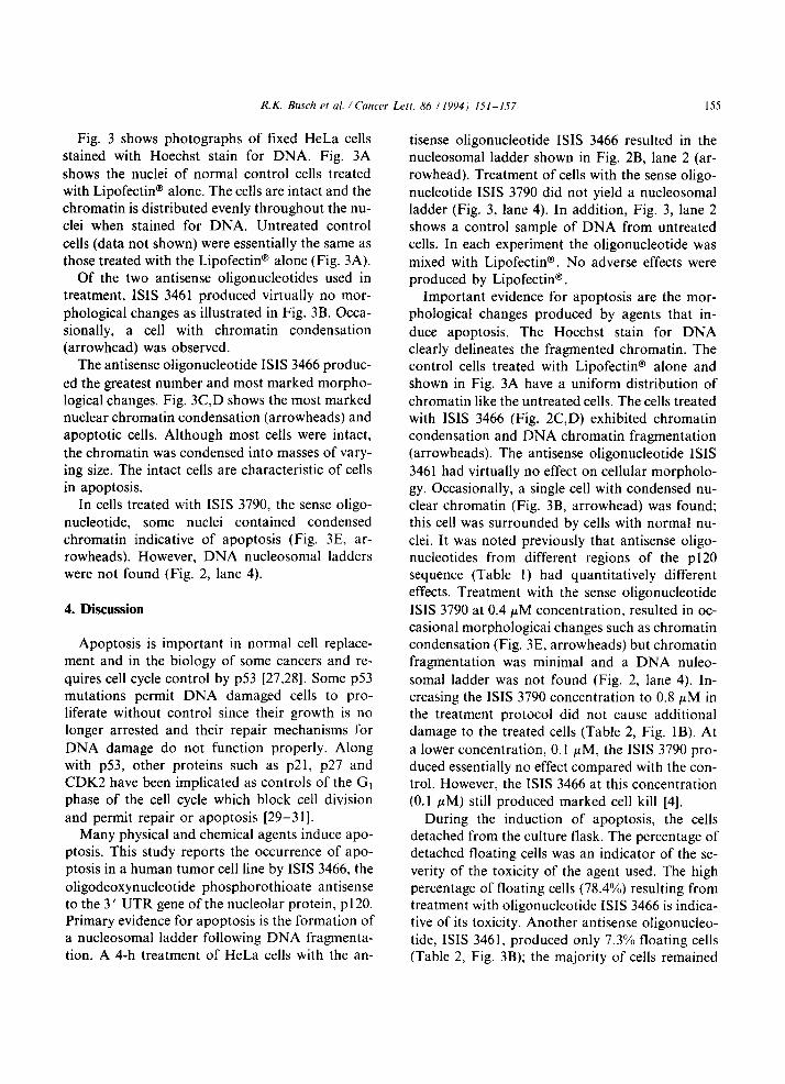
Fig. 3 shows photographs of fixed HeLa cells stained with Hoechst stain for DNA. Fig. 3A
shows the nuclei of normal control cells treated with Lipofectin@ alone. The cells are intact and the chromatin is distributed evenly throughout the nu- clei when stained for DNA. Untreated control
cells (data not shown) were essentially the same as those treated with the Lipofectin@ alone (Fig. 3A).
Of the two antisense oligonucleotides used in treatment, ISIS 3461 produced virtually no mor-
phological changes as illustrated in Fig. 3B. Occa- sionally, a cell with chromatin condensation (arrowhead) was observed.
The antisense oligonucleotide ISIS 3466 produc-
ed the greatest number and most marked morpho- logical changes. Fig. 3C,D shows the most marked nuclear chromatin condensation (arrowheads) and apoptotic cells. Although most cells were intact,
the chromatin was condensed into masses of vary- ing size. The intact cells are characteristic of cells in apoptosis.
In cells treated with ISIS 3790, the sense oligo- nucleotide, some nuclei contained condensed chromatin indicative of apoptosis (Fig. 3E, ar- rowheads). However, DNA nucleosomal ladders were not found (Fig. 2, lane 4).
4. Discussion
Apoptosis is important in normal cell replace- ment and in the biology of some cancers and re-
quires cell cycle control by ~53 [27,28]. Some ~53 mutations permit DNA damaged cells to pro- liferate without control since their growth is no longer arrested and their repair mechanisms for
DNA damage do not function properly. Along with ~53, other proteins such as ~21, p27 and CDK2 have been implicated as controls of the Gi phase of the cell cycle which block cell division and permit repair or apoptosis [29-311.
Many physical and chemical agents induce apo- ptosis. This study reports the occurrence of apo- ptosis in a human tumor cell line by ISIS 3466, the
oligodeoxynucleotide phosphorothioate antisense to the 3 ’ UTR gene of the nucleolar protein, ~120. Primary evidence for apoptosis is the formation of a nucleosomal ladder following DNA fragmenta- tion. A 4-h treatment of HeLa cells with the an-
tisense oligonucleotide ISIS 3466 resulted in the nucleosomal ladder shown in Fig. 2B, lane 2 (ar- rowhead). Treatment of cells with the sense oligo- nucleotide ISIS 3790 did not yield a nucleosomal ladder (Fig. 3, lane 4). In addition, Fig. 3, lane 2 shows a control sample of DNA from untreated cells. In each experiment the oligonucleotide was
mixed with Lipofectin@. No adverse effects were produced by Lipofectin@ .
Important evidence for apoptosis are the mor- phological changes produced by agents that in-
duce apoptosis. The Hoechst stain for DNA clearly delineates the fragmented chromatin. The control cells treated with Lipofectin@ alone and shown in Fig. 3A have a uniform distribution of chromatin like the untreated cells. The cells treated with ISIS 3466 (Fig. 2C,D) exhibited chromatin condensation and DNA chromatin fragmentation (arrowheads). The antisense oligonucleotide ISIS
3461 had virtually no effect on cellular morpholo- gy. Occasionally, a single cell with condensed nu- clear chromatin (Fig. 3B, arrowhead) was found; this cell was surrounded by cells with normal nu- clei. It was noted previously that antisense oligo- nucleotides from different regions of the ~120 sequence (Table 1) had quantitatively different effects. Treatment with the sense oligonucleotide
ISIS 3790 at 0.4 PM concentration, resulted in oc- casional morphologicai changes such as chromatin condensation (Fig. 3E, arrowheads) but chromatin fragmentation was minimal and a DNA nuleo-
somal ladder was not found (Fig. 2, lane 4). In- creasing the ISIS 3790 concentration to 0.8 PM in the treatment protocol did not cause additional damage to the treated cells (Table 2, Fig. 1B). At
a lower concentration, 0.1 PM, the ISIS 3790 pro- duced essentially no effect compared with the con- trol. However, the ISIS 3466 at this concentration (0.1 PM) still produced marked cell kill [4].
During the induction of apoptosis, the cells detached from the culture flask. The percentage of detached floating cells was an indicator of the se- verity of the toxicity of the agent used. The high percentage of floating cells (78.4%) resulting from treatment with oligonucleotide ISIS 3466 is indica- tive of its toxicity. Another antisense oligonucleo- tide, ISIS 3461, produced only 7.3% floating cells (Table 2, Fig. 3B); the majority of cells remained
156 R.K. Busch et al. /Cancer Lett. 86 (1994) 151-157
attached. Although treatment with the sense oligo- nucleotide ISIS 3790 resulted in some morphologi- cal change such as chromatin condensation (Fig. 3E) the number of floating cells remained between 20% and 25%. Even when the concentration of ISIS 3790 was doubled from 0.4 PM to 0.8 PM, no
additional changes were found in morphology or in the number of floating cells.
These experiments show that morphological changes alone do not necessarily correlate with
nucleosomal ladders. When 75% of the treated cells were detached and exhibited morphological changes associated with apoptosis, a DNA nucleosomal ladder was obtained.
Reports on antisense oligonucleotides as thera- peutic agents are increasing rapidly; analysis of the mechanisms of the toxic and apoptotic effects of antisense oligonucleotide ISIS 3466 in cultured human cancer cells may aid in the understanding of the effects of other antisense agents.
Acknowledgements
This work was supported by the DeBakey and Busch Funds.
References
111
121
[31
141
151
[61
Stein, C.A. and Cohen, J.S. (1988) Oligodeoxy-
nucleotides as inhibitors of gene expression: a review.
(1991) Prognostic significance of proliferation associated
nucleolar antigen ~120 in human breast carcinoma. Cancer Res.. 51. 1973-1978.
Green, D.R.. Bissonnette. R.P. and Cotter, T.G. (1994)
Apoptosis and cancer. Print. Pratt. Oncol.. 8, 1-14. Robertson, L.E., Huang. P.. Keating, M.J. and Plunkett.
W. (1994) Apoptosis in chronic lymphocytic leukemia.
Cancer Bull., 46. 130-135.
Fujiwara, T., Grimm, E.A., Mukhopadhyay, T., Cai.
D.W.. Owen-Schaub, L.B. and Roth, J.A. (1993) A
retroviral wild type ~53 expression vector penetrates
human lung cancer spheroids and inhibits growth by in- ducing apoptosis. Cancer Res.. 53, 4129-4133.
Takano. Y.S., Harmon. B.V. and Kerr. J.F.R. (1991) Apoptosis induced by mild hyperthermia in human and murine tumour cell lines: a study using electron micros-
copy and DNA gel electrophoresis. J. Pathol.. 163. 329-336.
Martin. S.J. and Cotter, T.G. (1991) lJltraviolet B irradi- ation of human leukemia HL-60 cells in vitro induces a
R.K. Busch et al. /Cancer Leii. 86 (19941 151-157 157
suicide cell response (apoptosis). Int. J. Radiat. Biol., 59, 9.16-9.19, 6.3-6.15, 6.19. Cold Spring Harbor Labora-
1001-1016. tory, Cold Spring Harbor.
[2l] Kaufman, H. (1989) Induction of endonucleolytic DNA
cleavage in human myelogenous leukemia cells by
etoposides, camptothecin and other cytotoxic anticancer
drugs: a cautionary note. Cancer Res., 49, 5870--5878. [22] Wren, B.C. (1993) Hormone therapy following breast
and uterine cancer. Baillieres Clin. Endocrinol. Metab.,
7. 225-242.
[23] Skladanowski, A. and Konopa, J. (1993) Adriamycin
and daunomycin induce programmed cell death (apopto-
sis) in tumour cells. Biochem. Pharmacol., 46, 375-382.
[24] Felgner, P.L. and Ringold, G.M. (1989) Cationic