Apoptosis of Peripheral Blood Leukocytes in Systemic Lupus Erythematosus: Studies on Serum Induction and Complement-Dependent Clearance Mechanisms Gullstrand, Birgitta 2010 Link to publication Citation for published version (APA): Gullstrand, B. (2010). Apoptosis of Peripheral Blood Leukocytes in Systemic Lupus Erythematosus: Studies on Serum Induction and Complement-Dependent Clearance Mechanisms. Department of Laboratory Medicine, Lund University. Total number of authors: 1 General rights Unless other specific re-use rights are stated the following general rights apply: Copyright and moral rights for the publications made accessible in the public portal are retained by the authors and/or other copyright owners and it is a condition of accessing publications that users recognise and abide by the legal requirements associated with these rights. • Users may download and print one copy of any publication from the public portal for the purpose of private study or research. • You may not further distribute the material or use it for any profit-making activity or commercial gain • You may freely distribute the URL identifying the publication in the public portal Read more about Creative commons licenses: https://creativecommons.org/licenses/ Take down policy If you believe that this document breaches copyright please contact us providing details, and we will remove access to the work immediately and investigate your claim. Download date: 20. Dec. 2021
Transcript
LUND UNIVERSITY
PO Box 117221 00 Lund+46 46-222 00 00
Apoptosis of Peripheral Blood Leukocytes in Systemic Lupus Erythematosus: Studieson Serum Induction and Complement-Dependent Clearance Mechanisms
Gullstrand, Birgitta
2010
Link to publication
Citation for published version (APA):Gullstrand, B. (2010). Apoptosis of Peripheral Blood Leukocytes in Systemic Lupus Erythematosus: Studies onSerum Induction and Complement-Dependent Clearance Mechanisms. Department of Laboratory Medicine,Lund University.
Total number of authors:1
General rightsUnless other specific re-use rights are stated the following general rights apply:Copyright and moral rights for the publications made accessible in the public portal are retained by the authorsand/or other copyright owners and it is a condition of accessing publications that users recognise and abide by thelegal requirements associated with these rights. • Users may download and print one copy of any publication from the public portal for the purpose of private studyor research. • You may not further distribute the material or use it for any profit-making activity or commercial gain • You may freely distribute the URL identifying the publication in the public portal
Read more about Creative commons licenses: https://creativecommons.org/licenses/Take down policyIf you believe that this document breaches copyright please contact us providing details, and we will removeaccess to the work immediately and investigate your claim.
From the Institute of Laboratory Medicine, Division of Microbiology, Immunology and
Glycobiology (MIG), Lund University.
Apoptosis of Peripheral Blood Leukocytes in Systemic Lupus Erythematosus:
Studies on Serum Induction and Complement-Dependent Clearance Mechanisms
Birgitta Gullstrand
Lund 2010
Akademisk avhandling Som med vederbörligt tillstånd av Medicinska Fakulteten vid Lunds Universitet för
avläggande av doktorsexamen i medicinsk vetenskap i ämnet laboratoriemedicin med inriktning experimentell klinisk immunologi kommer att offentligt försvaras i Segerfalksalen,
Wallenberg neurocenter BMC, onsdagen den 14 april 2010, klockan 09.00
Fakultetsopponent: Professor Cees van Kooten
Department of Nephrology, C3p, Leiden University Medical Center, Leiden, The Netherlands.
Apoptosis of Peripheral Blood Leukocytes in Systemic Lupus Erythematosus:
Studies on Serum Induction and Complement-Dependent Clearance Mechanisms
TABLE OF CONTENTS Table of contents 6 List of papers 7 Abbreviations 8 Summary 10 Introduction 11 The immune system 11 Leukocytes 12 Activation of T-and B-cells 13 Specific antibodies 15 Tolerance and autoimmunity 16 The complement system 17 The classical pathway 18 The alternative pathway 19 The lectin pathway 20 The terminal pathway 20 Regulation of complement activation 20 Complement deficiency 21 Programmed cell death 22 The external pathway 24 The internal pathway 25 Autophagy 29 The execution 29 Phagocytic clearance of dying cells 30 Systemic lupus erythematosus 31 Present investigation 35 Aims 35 Paper I and II 35 Results 36 Paper III 37 Results 38 Paper IV 39 Results 40
Discussion and future perspective 41 Conclusions 46
Populärvetenskaplig sammanfattning på svenska 47 Acknowledgment 52 References 55 Papers I-IV 63
- 7 -
LIST OF PAPERS
This thesis is based on the following papers, which will be referred to in the text by their
roman numerals (I-IV)
I. Induction of apoptosis in monocytes and lymphocytes by serum from patients with systemic lupus erythematosus - an additional mechanism to increased autoantigen load? Bengtsson AA, Sturfelt G, Gullstrand B, Truedsson L. Clin Exp Immunol. 2004 Mar;135(3):535-43.
II. SLE serum induces classical caspase-dependent apoptosis independent of death receptors. Bengtsson AA, Gullstrand B, Truedsson L, Sturfelt G. Clin Immunol. 2008 Jan;126(1):57-66.
III. Complement classical pathway components are all important in clearance of apoptotic and secondary necrotic cells. Gullstrand B, Mårtensson U, Sturfelt G, Bengtsson AA, Truedsson L. Clin Exp Immunol. 2009 May;156(2):303-11.
IV. Specificity of anti-histone antibodies determines complement-dependent phagocytosis of necrotic material by polymorphonuclear leukocytes in the presence of serum from patients with SLE. The LE cell phenomenon revisited. Birgitta Gullstrand, Helena Tydén, Andreas Jönsen, Christian Lood, Sören Jacobsen, Gunnar Sturfelt, Lennart Truedsson, and Anders A. Bengtsson. Manuscript.
- 8 -
ABBREVIATIONS ACR American College of Rheumatology AIE Apoptosis inducing effect AIF Apoptosis inducing factor Apaf 1 Apoptotic protease activating factor 1 AUC Area under curve divided by follow-up time C4BP C4b-binding protein CAD Caspase-activated deoxyribonuclease cFLIP Cellular Fas-associated death domain-like IL-1-converting enzyme inhibitory
protein CR Complement receptor CRP C-reactive protein DAMPs Damage associated molecular patterns DD Death domain DISC Death inducing signal complex ELISA Enzyme-linked immunosorbent assay ER Endoplasmic reticulum FADD Fas associated death domain FcR Fc receptor FITC Fluoroisothiocyanate IAPs Inhibitors of apoptosis ICAD Inhibitor of caspase-activated deoxyribonuclease IFN Interferon IL Interleukin LPS Lipopolysaccharide MASP Mannan-binding lectin-associated serine protease MBL Mannan-binding lectin MDM Monocytes to differentiate into macrophages MHC Major histocompability complex NC Necrotic material NHS Normal human serum NK Natural killer NODs Proteins carrying nucleotide-binding oligomerization domains PAC assay Phagocytosis of apoptotic cells assay PAMPs Pathogen-associated molecular patterns PARP Poly(ADP-ribose) polymerase PBMC Peripheral blood mononuclear cells PI Propidium iodide PMNs Polymorphonuclear leukocytes PNC assay Phagocytosis of necrotic cell material assay PRRs Pattern-recognition receptors PS Phosphatidylserine RA Rheumatoid arthritis ROS Reactive oxygen species SLE Systemic lupus erythematosus SLEDAI-2K SLE Disease Activity Index 2000 Smac/ Second mitochondria derived activator of caspase/Direct inhibitor of apoptosis DIABLO binding protein with low pI TGF-β Transforming growth factor beta
Systemic lupus erythematosus (SLE) is an autoimmune disease involving many organ
systems. The cause is not known, but a complex combination of environmental and genetic
factors seems to be involved. In SLE upregulation of type I interferons, a hyperactive B-cell
response, presence of autoantibodies against modified nuclear components, increased
complement consumption, increased apoptosis and decreased clearance of apoptotic cells are
seen. The purpose of this thesis was to investigate some of these mechanisms. The thesis is
based on four papers (I-IV).
(Papers I and II) We found that the apoptosis inducing effect was specific for sera from SLE
patients when comparing with sera from various control groups. However, the apoptosis
inducing effect was not related to SLE disease activity. Serum from SLE patients was
demonstrated to induce classical caspase-dependent apoptosis in monocytes and lymphocytes.
The apoptosis induction was not dependent on death receptors but involvement of the
mitochondrial pathway was indicated.
(Paper III) Phagocytosis of apoptotic cells by macrophages and C3 deposition on apoptotic
cells were investigated in the presence of sera lacking different complement proteins. We
found that complement-mediated opsonisation and phagocytosis of apoptotic cells,
particularly those undergoing secondary necrosis, are dependent mainly upon an intact
classical pathway. C1q was not more important than other classical pathway components,
suggesting a role in other pathogenetic processes than defect clearance of apoptotic cells.
(Paper IV) We evaluated the roles of serum complement and antibodies against histones in
relation to phagocytosis of necrotic cell material by polymorphonuclear neutrophil
granulocytes (PMNs). Phagocytosis of necrotic material by PMNs and high concentration of
antibodies against a broad spectrum of histones correlated with active SLE disease. The
specificities of these anti-histone antibodies appear to determine the complement-dependent
phagocytosis.
In conclusion, sera from SLE patients have the capacity to contribute to an increased load of
apoptotic cells. An efficient clearance of apoptotic and necrotic cell material is dependent on a
functional classical pathway, and autoantibodies against histones reflect the presence of
apoptotic or necrotic cells contributing to the autoimmune process in SLE.
- 11 -
INTRODUCTION
The immune system
The immune system is an integrated system of organs, tissues, proteins and cells that together
protect us against foreign substances and invading organisms. The first line of defence
consists of barriers such as the skin, mucous membranes, saliva, tears and the acid in the gut.
Besides the physical barriers, a number of different proteins, enzymes and cell types are
involved in the immune system, and these are able to differentiate between self and non-self.
The immune response is composed of two parts, the innate or nonspecific immune system and
the adaptive or specific immune system. The innate immune system is constitutively present
and ready to be mobilised upon infection, whereas the adaptive immune system requires some
time to react. These systems work together by various interactions and influence each other to
create an efficient immune defence [1, 2].
The innate immune response is a defence mechanism that recognises and reacts to pathogen-
associated molecular patterns (PAMPs), such as lipopolysaccharide (LPS), mannose-rich
glycan molecules exposed on microorganisms and DNA from bacteria or virus. However,
molecules released by stressed and injured human cells, such as heat shock proteins, high-
mobility group box 1 (HMGB1), S 100 proteins and DNA may also act as pro-inflammatory
mediators and these molecules are termed damage associated molecular patterns (DAMPs).
Pattern-recognition receptors (PRRs) recognise PAMPs and DAMPs and these receptors can
be cell bound such as Toll-like receptors (TLRs), intracellular, such as proteins carrying
nucleotide-binding oligomerization domains (NODs) and some TLRs or soluble such as the
complement proteins [1-5]. The cells involved in the innate immune response are phagocytic
cells such as neutrophils, monocytes and macrophages as well as the cytotoxic natural killer
(NK) cells. Upon activation, the cells release cytokines that act as signal molecules and
activate other cells involved in the immune response [2].
The function of the adaptive immune response is to destroy or inactivate foreign substances,
also called antigens. The adaptive immune system is composed of specialised cells, such as B-
cells and T-cells, which account for antibody and cell mediated immunity, respectively. These
cells become activated when a specific epitope of the antigen binds to the B-cell or T-cell
receptor. However, binding of antigen to the receptor is usually not sufficient to stimulate the
cells to proliferate and differentiate into an effector cell; a co-stimulatory signal provided by
- 12 -
another specialised cell is often required. Major histocompability complex (MHC) molecules
are proteins expressed on the cell surface and these molecules are recognised by T-cells. In
most cases, T-cells only bind to the antigen if it is presented in complex with MHC. MHC
class I molecules are expressed on the majority of nucleated cells and MHC class II are
expressed on antigen presenting cells such as macrophages, monocytes and B-cells. MHC
also acts as a self recognition molecule. The adaptive immune response also exhibits an
immunological memory [6].
Leukocytes
Polymorphonuclear leukocytes (PMNs) are the most common white blood cells in the
circulation. Neutrophils, the most abundant type of PMNs, possess a multi-lobed nucleus and
contain cytoplasmic granules. There are three types of granules, azurophilic (also called
primary), specific and small granules. The granules are generated during cell differentiation
and are used as storage for different substances. They contain cytotoxic substances, neutral
proteinases, acidic hydrolases and cytoplasmic membrane receptors. Their function is to
provide enzymes for hydrolytic substrate degradation and killing of bacteria, and also to
regulate various processes including inflammation [7]. Neutrophils rapidly engulf foreign
material that is covered with antibodies and complement fragments but they also clean up
damaged cells or cellular debris.
Mononuclear blood cells include cells such as monocytes, macrophages and lymphocytes.
The blood monocytes possess chemotactic, pinocytic and phagocytic abilities. During
migration into the tissue the monocytes undergo further differentiation to become
macrophages, and these take part in the initiation of T-cell activation by processing and
presenting antigens. Activated macrophages are central effectors and regulatory cells of the
immune response, and they achieve this by producing different substances such as cytokines
which modulate the function of other cells. Lymphocytes circulating in the bloodstream are
mostly in the resting state but the lymphocytes in the lymphoid tissues and organs can be
activated directly after antigen stimulation. There are different types of lymphocytes, T-cells,
B-cells and NK-cells. The T-cells are further divided into three types of cells, helper,
cytotoxic and regulatory. T-helper cells are needed for activation of B-cells and they
recognise antigen processed and presented by antigen presenting cells in complex with the
self molecule MHC class II. The cytotoxic T-cells target and destroy tumour cells and cells
infected with intracellular antigen such as virus, which are presented in complex with MHC
- 13 -
class I. Most nucleated cells express MHC class I, so any such cell that is infected with virus
or producing tumour antigens may present these antigens together with MHC class I and be
removed. The cytotoxic T-cells release perforin which forms channels in the cell membrane
of the target cell and causes death by osmotic lysis. The regulatory T-cells reduce the intensity
of the immune response by regulating transcription of different genes and by secreting
interleukin (IL)-10. B-cells become plasma cells upon activation which then secrete
antibodies or become memory cells. The NK-cells do not express a specific antigen binding
receptor. They have two types of receptors that either activate or inhibit activation. NK-cells
can bind antibody coated targets by immunoglobulin receptors (FcR), and they also bind to
cells missing the self marker MHC class I. Also the release of interferons (IFN) or cytokines
from virus-infected cells may activate these cells. NK-cells possess granules containing
perforin and granzymes, which can be released upon activation and induce cell death of the
target cell [6].
Activation of T- and B-cells
Antigen presenting cells, such as macrophages, phagocytose antigen, degrade it and expose
fragments of the antigen on the cell surface together with MHC class II molecules. The MHC-
antigen complex binds to a T-cell receptor specific to the presented antigen. The binding of
antigen to the T-cell receptor stimulates the expression of IL-2 receptors and secretion of IL-2
which binds to these receptors and stimulates the T-cell to proliferate. For activation of the T-
cell, a second co-stimulatory signal is needed. This may be mediated by the binding of the
signal protein B7 on the antigen presenting cell to the T-cell receptor protein CD28 [2]. Once
activated, the T-helper cells can be divided into subpopulations such as Th1, Th2 and Th17
cells depending on the cytokine signal received or become memory T-cells. The different
subsets of cells secrete different cytokines that regulate the immune response. Th1 cells
stimulate the cell-mediated response by secreting cytokines such as gamma IFN and tumour
necrosis factor (TNF) while Th2 cells stimulate B-cells to produce antibodies by secreting
cytokines such as IL-4, IL-5, IL-6 and IL-10 (Fig. 1) [6]. Th17 cells are a recently identified
subset of T-helper cells and these cells synthesise and secrete IL-17. IL-17 is involved in
inducing and mediating pro-inflammatory responses. However, little is known about the
function in humans but it is implicated to be an effector cell in autoimmune diseases [8].
Activation of B-cells occurs when the B-cell receptor recognises an antigen and binds to it.
Each B-cell has a unique receptor expressed on its surface and this receptor is a membrane
- 14 -
bound immunoglobulin. In most cases T-helper cells, activated with the same antigen as the
B-cell, are required for activation of the B-cell, a so-called T-cell dependent activation. The
interaction between the T- and B-cells occurs between the CD40 ligand present on the surface
of the activated T-helper cell and the CD40 protein present on the surface of the B-cell. In the
presence of different cytokines, such as IL-4, IL-5, IL-6 and IL10, the activated B-cell
differentiates into a plasma cell, producing antibodies of the same specificity as the B-cell
receptor that targets the antigen, or becomes a memory cell (Fig. 1) [6]. B-cells may also be
activated in a T-cell independent way. The antigen involved in this process is often
polysaccharides that are able to bind multiple B-cell receptors and activate the B-cell directly
to secrete IgM antibodies [6].
Figure 1. Activation of T-cell and T-cell dependent B-cell activation.
IL-1 IL-6
Antigen presenting cell
T-cell B7 CD28
T-cell receptor
MHC Class II
IL-2
Memory T-cell
Th1 Th2
T-cell receptor
B-cell receptor
IL-4 IL-5 IL-6 IL-10
CD40 ligand
CD40
IL-2 receptor
MHC Class II
IFN-γ TNF-α
Memory B-cell
Plasma cell
T-cell
- 15 -
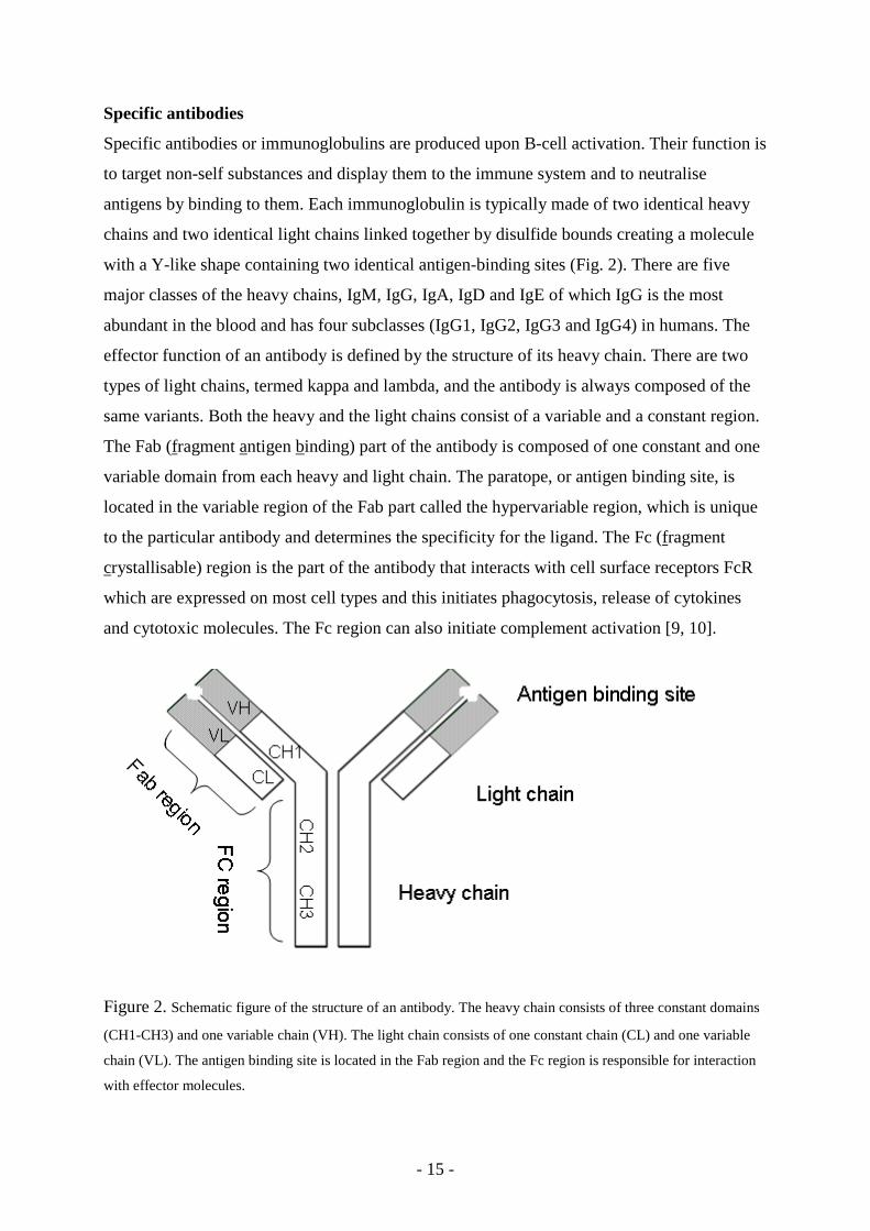
Specific antibodies
Specific antibodies or immunoglobulins are produced upon B-cell activation. Their function is
to target non-self substances and display them to the immune system and to neutralise
antigens by binding to them. Each immunoglobulin is typically made of two identical heavy
chains and two identical light chains linked together by disulfide bounds creating a molecule
with a Y-like shape containing two identical antigen-binding sites (Fig. 2). There are five
major classes of the heavy chains, IgM, IgG, IgA, IgD and IgE of which IgG is the most
abundant in the blood and has four subclasses (IgG1, IgG2, IgG3 and IgG4) in humans. The
effector function of an antibody is defined by the structure of its heavy chain. There are two
types of light chains, termed kappa and lambda, and the antibody is always composed of the
same variants. Both the heavy and the light chains consist of a variable and a constant region.
The Fab (fragment antigen binding) part of the antibody is composed of one constant and one
variable domain from each heavy and light chain. The paratope, or antigen binding site, is
located in the variable region of the Fab part called the hypervariable region, which is unique
to the particular antibody and determines the specificity for the ligand. The Fc (fragment
crystallisable) region is the part of the antibody that interacts with cell surface receptors FcR
which are expressed on most cell types and this initiates phagocytosis, release of cytokines
and cytotoxic molecules. The Fc region can also initiate complement activation [9, 10].
Figure 2. Schematic figure of the structure of an antibody. The heavy chain consists of three constant domains
(CH1-CH3) and one variable chain (VH). The light chain consists of one constant chain (CL) and one variable
chain (VL). The antigen binding site is located in the Fab region and the Fc region is responsible for interaction
with effector molecules.
- 16 -
Tolerance and autoimmunity
The immune defence is involved in detection and destruction of tumour cells and foreign
invaders causing infection. For the system to function properly it must be able to discriminate
between self and non-self molecules. Failure to do this may result in autoimmune diseases.
All immune and blood cells develop from multi-potent hematopoietic stem cells that originate
from the bone marrow and a highly diverse and random array of different specificities of T-
and B-cells are produced. These cells are capable of recognising an almost unlimited number
of antigens, including self-proteins. Immature T-cells undergo final maturation in the thymus
where they go through an important process which enables them to distinguish between self
and non-self. T-cells that recognise self-antigens are deleted by apoptosis or become
inactivated. The selection that occurs in the thymus is called central selection and the cells
undergo both positive and negative selection to produce T-cells that tolerate self-MHC
molecules but not self-peptides. In the positive selection, T-cells with receptors that bind with
neither too low nor too high affinity to surface MHC molecule on thymic epithelial cells are
selected, and the other cells die. This ensures that T-cells only recognise antigen in
association with MHC. The negative selection is mediated by macrophages and dendritic
cells, which present self-peptides bound to MHC and the cells that recognise self-peptides
bound to MHC undergo programmed cell death. The peripheral tolerance is developed after
the T-cell has matured and entered the periphery. The cell is regulated by regulatory T-cells
and the absence of co-stimulating signals. B-cell tolerance is not so tightly regulated. For B-
cell activation to occur, a T-cell with the same antigen molecule specificity as the one that
stimulates the particular B-cell is needed. This ensures the specificity of reactions to protein
antigen selected by the immune system. In the bone marrow the B-cells are tested for
interaction against self-antigens and the cells that recognise self-antigens are either processed
for change in receptor specificity or the cells undergo programmed cell death [11, 12].
Autoimmune diseases are conditions caused by the breakdown of immune tolerance resulting
in immune responses to self-antigens. Low levels of autoantibodies are found in blood from
healthy individuals without causing inflammation or damage. However, an autoimmune
disease occurs when a response against self-antigens involving T-cells, B-cells or
autoantibodies induces damage systemically or to a particular organ. Autoantibodies could
arise against novel epitopes expressed on modified self-proteins, cross-reacting antibodies if
non-self molecules closely resemble self-antigens. Autoantibodies could also arise from the
exposure of hidden self-molecules that would not be normally exposed to the immune system,
- 17 -
or as a result of hormonal component involvement, an imbalance of regulatory proteins or due
to a genetic predisposition combined with environmental factors [12]. Autoimmune diseases
can be organ specific and directly damage the organ target or systemic with involvement of
different self-molecules and cause disease through the formation of immune complex. Under
normal conditions immune complexes are rapidly cleared from the circulation by
phagocytosis or by transportation. If the clearance system fails, circulating immune complex
could be deposited in organs and cause inflammation or damage, such as glomerulonephritis,
vasculitis and arthritis [13].
The complement system
The complement system is an important part of the immune response, bridging innate and
adaptive immune mechanisms. Activities of the complement system include initiation of
inflammation, opsonisation of targets to promote phagocytosis, chemotaxis, lysis of cells and
clearance of immune complexes and apoptotic cells [14, 15]. This complex system consists of
more than 30 plasma and membrane proteins that interact and are activated through a cascade
reaction. There are three main pathways by which complement activation is initiated, the
classical pathway, the alternative pathway and the lectin pathway (Fig. 3). All pathways lead
to cleavage of C3 and activation of the common terminal pathway leading to assembly of the
membrane attack complex and eventually cell death by lysis [14, 16].
- 18 -
Figure 3. Activation of the classical, lectin and the alternative pathways initiate C3b opsonisation on target cells, lysis of cell by the membrane attack complex and the release of C3a and C5a, potent chemoattractants resulting in an inflammatory reaction, B (factor B), D (factor D), P (properdin).
The classical pathway
The classical pathway is activated by immune complexes containing IgM or IgG but other
molecules such as LPS, C-reactive protein (CRP), apoptotic cells and nucleic acids are also
able to activate this pathway (Fig. 4) [17]. The components involved in the classical pathway
are C1, C4 and C2. C1 is a Ca2+-dependent complex consisting of C1q, C1r2 and C1s2. When
the recognition molecule C1q binds to a target, a conformational change occurs, leading to
activation of C1r which, in turn, activates C1s. Activated C1s cleaves C4 into a small C4a
fragment and a larger C4b fragment. The small C4a fragment diffuses away while the larger
C4b fragment may attach covalently to the activator or other molecules in the vicinity. C2 is
also cleaved into two fragments by activated C1s; in the presence of Mg2+ the larger C2a
Classical Pathway Antibody/antigen, CRP
Lectin Pathway Mannose, N-acetyl-glucoseamine
Alternative Pathway C3b, microbes
C1q C1r2 C1s2
MBL/ficolins
MASPs
C3(H2O) C3b
C4b2a
C4b2a3b
C3
C3bBb
B, D and P
C4 and C2
P
C3bBb3b
C5b C5 C5b-C9 Membrane attack complex
C3b opsonisation
C3a inflammation
C5a inflammation
C3a inflammation
C4a and C2b
C3b opsonisation
C3
- 19 -
fragment can bind to C4b and form the classical pathway C3 convertase C4b2a. The C3
convertase cleaves C3 into two major fragments (C3a and C3b), and C3b molecules can bind
to the C3 convertase forming the C5 convertase C4b2a3b which cleaves C5 and activates the
terminal pathway (Fig. 3) [16].
Opsonisation of the target by C3b or C4b supports uptake and clearance of antigen, apoptotic
cells and immune complexes by phagocytic cells. The smaller fragments released during
complement activation, C3a and C5a are potent chemoattractants and their release results in
an inflammatory reaction and the recruitment of phagocytic cells to the damage site by
increasing permeability of the capillary beds [14].
Figure 4. Complement activation of the classical pathway.
The alternative pathway
The alternative pathway, described as an antibody-independent pathway, includes the
complement components C3, factor B, factor D and properdin. This pathway is initiated by
spontaneous hydrolysis of the internal thioester in C3, to form C3(H2O). Factor B binds to
C3(H2O) in the presence of Mg2+ and factor D then cleaves factor B into the fragments Bb and
Ba. The complex produced, C3(H2O)Bb, is also known as a fluid phase C3 convertase and
can cleave C3 into C3a and C3b. Various cell surfaces bind C3b which then can form a
complex with factor B and after cleavage by factor D generate the alternative pathway C3
convertase C3bBb, which is able to cleave C3 into C3a and C3b. In this way more C3b is
generated which can activate the alternative pathway creating an amplification loop. The C3b
formed by activation of the classical or lectin pathway also initiates activation of the
C1 molecule
Immune complexes
C4
C2
Classical pathway C3 convertase C4b2a
C4a
Cb2
- 20 -
alternative pathway [16]. Properdin binds and stabilises the alternative C3 convertase but has
recently been described to also act as an initiator of the alternative pathway as well [18-20].
Activation of C3 via a C2 bypass pathway initiated by C1 or Mannan-binding lectin (MBL)
but independent of C2 or MBL-associated serine protease (MASP)-2 has also been described
[21-24].
The lectin pathway
The lectin pathway is similar to the classical pathway and is initiated by the binding of MBL
or ficolins in complex with MASP molecules (MASP 1, 2 and 3), to sugar structures like N-
acetylglucosamine and mannose present on many microorganisms [25]. The binding is Ca2+ -
dependent and activates the MASP-2 which then can cleave C4 and C2, generating the C3
convertase C4b2a, the same C3 convertase as in the classical pathway.
The terminal pathway
The C5 convertases (C4b2a3b and C3bBbC3b) generated by the different pathways cleave C5
into its active form C5b and this remains bound to the C5 convertase. This cleavage initiates
the assembly of the membrane attack complex, with the binding of C6 and finally C7 to C5b,
forming a C5b67 complex which dissociates from the C5 convertase and binds to the
membrane surface. Thereafter C8 is incorporated, then C9 molecules bind to form the final
membrane attack complex which leads to the formation of pores into the cell causing osmotic
swelling and cell rupture [26]. The C5a fragment released after cleavage acts as a potent
anaphylatoxin and chemoattractant for phagocytotic cells [27].
Regulation of complement activation
Activation of the complement system needs to be carefully regulated to protect host cells and
tissue from damage and both fluid phase and membrane bound regulatory proteins are present
at high concentrations. The balance between activation and inhibition determines the
outcome. C1 esterase inhibitor (C1INH) controls activation of both the classical and the lectin
pathway by binding reversibly to C1 and MASP-2, but it can also bind to activated C1r and
C1s and inhibit their activity [28]. Factor I regulates complement activity by cleavage of C3b
and C4b using C4b-binding protein (C4BP) and factor H as soluble cofactors. C4BP also
regulates the classical pathway by dissociating the subunits of the C3 convertase and the
alternative pathway C3 convertase is regulated in the same way by factor H. Membrane bound
molecules acting as inhibitors of complement activation include membrane cofactor protein
- 21 -
(MCP, CD46) and complement receptor (CR) 1 (CD35), which regulates the C3 activation by
its function as a cofactor protein for factor I mediated cleavage of C3b. Decay accelerating
factor (DAF, CD55) prevents the assembly of C3 and C5 convertases and may also accelerate
the disassembly of preformed convertases. CR1, which binds to C1q, C3b, C4b and MBL on
opsonised targets, mediates transport of immune complexes by erythrocytes and promotes
phagocytosis [14]. The terminal pathway is regulated by clusterin and S-protein (vitronectin),
which binds to fluid phase C5b67 and inhibits the assembly of the membrane attack complex.
Protectin (CD59) prevents binding of C9 to the C5b678 complex [29]. To date, properdin is
the only positive regulator described for complement activation, by binding to and stabilising
the alternative pathway C3 convertase.
Complement receptors expressed on different cell types play a role in the regulation of
complement, and binding of complement to the receptor influences other cellular function
such as phagocytosis of immune complexes and apoptotic cells. CR2 (CD21) expressed on B-
cells binds to iC3b or C3d and binding of C3d opsonised antigen to CR2, when antigen is also
bound to B-cell receptor, results in B-cell activation and proliferation [30]. The complement
receptors CR3 (CD11b/CD18) and CR4 (Cd11c/CD18) belong to the family of integrins and
these receptors are also involved in the waste disposal of apoptotic cells by mediating
phagocytosis of iC3b opsonised targets [31]. There are also several receptors described for
C1q. Calreticulin in complex with CD91 binds to the collagen-like region of C1q and this
interaction is involved in clearance of apoptotic cells [32]. The gC1q receptor binds to the
globular region of C1q [33].
Complement deficiency
Deficiency of complement proteins can be inherited or acquired. Deficiencies are rare but
impaired complement function is associated with infections and autoimmity, especially
systemic lupus erythematosus (SLE). MBL-deficiency is the most common inherited defect
among the complement proteins resulting in increased susceptibility to infections especially in
early childhood [34]. Homozygous deficiency of the complement components in the classical
pathway (C1, C4 and C2) is associated with pyogenic infections, but also with risk of
development of SLE [35]. Alternative pathway deficiency states and deficiencies of
components in the terminal pathway are all associated with an increased risk of invasive
infections caused by predominantly Neisseria species. Acquired deficiencies may result from
- 22 -
decreased production or increased consumption of complement components caused by
activation, but autoantibodies against complement components can also contribute [35, 36].
Programmed cell death
Programmed cell death is a physiological process essential for the development of all
organisms and for maintenance of homeostasis by removing unwanted cells, but is also
involved in pathological conditions including immunological diseases and cancer [37, 38].
Programmed cell death is a complex, controlled, active process involving both biochemical
and morphological changes that are dependent on signals and activities within the dying cells.
During this process, the plasma membrane remains intact until the dying cell is phagocytosed
[39]. Apoptosis is a form of programmed cell death, but cell death can occur in a non-
apoptotic way and still be a physiological response. For the cell to be defined as apoptotic,
morphological features such as cell shrinkage, chromatin condensation, nuclear fragmentation
and membrane blebbing, with the maintenance of membrane integrity, should be fulfilled [40-
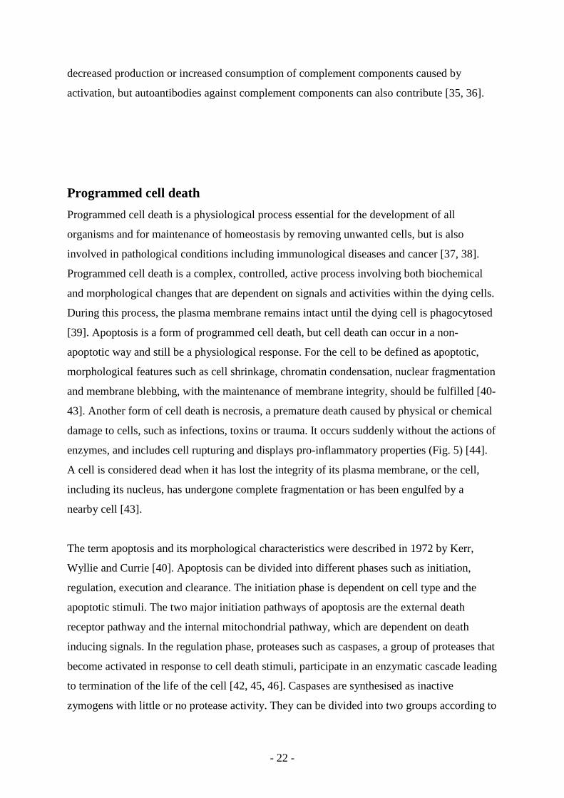
43]. Another form of cell death is necrosis, a premature death caused by physical or chemical
damage to cells, such as infections, toxins or trauma. It occurs suddenly without the actions of
enzymes, and includes cell rupturing and displays pro-inflammatory properties (Fig. 5) [44].
A cell is considered dead when it has lost the integrity of its plasma membrane, or the cell,
including its nucleus, has undergone complete fragmentation or has been engulfed by a
nearby cell [43].
The term apoptosis and its morphological characteristics were described in 1972 by Kerr,
Wyllie and Currie [40]. Apoptosis can be divided into different phases such as initiation,
regulation, execution and clearance. The initiation phase is dependent on cell type and the
apoptotic stimuli. The two major initiation pathways of apoptosis are the external death
receptor pathway and the internal mitochondrial pathway, which are dependent on death
inducing signals. In the regulation phase, proteases such as caspases, a group of proteases that
become activated in response to cell death stimuli, participate in an enzymatic cascade leading
to termination of the life of the cell [42, 45, 46]. Caspases are synthesised as inactive
zymogens with little or no protease activity. They can be divided into two groups according to
- 23 -
their active function; the initiator caspases 2, 8, 9, 10, and 12 and the effector caspases 3, 6
and 7. If the ‘point of no return’ is reached, the cell is executed and undergoes an organised
degradation. Apoptotic cells express phagocytic markers on their cell membrane and are
rapidly phagocytosed without induction of an inflammatory response [47-49]. This is in
contrast to necrosis which is a passive process characterised by cellular and nuclear swelling
and cell rupture leading to an inflammatory response.
The nucleus, endoplasmic reticulum (ER), Golgi apparatus and lysosome also play an
important role in the initiation and regulation of programmed cell death [50]. Once a cell gets
a death signal the most likely outcome is a programmed cell death, however, there are options
to arrest the process.
Figure 5. Apoptosis versus necrosis.
Viable cell
Apoptotic cell: •Single cell
•Energy dependent
•Cell shrinking •Membrane integrity maintained
•Activation of proteases
•Regulated
Necrotic cell: •Lots of cells
•Energy independent
•Cell swelling •Loss of membrane integrity
•Not regulated
•Leaking of cell components
No inflammation
Inflammation
- 24 -
The external pathway
The death receptors are transmembrane proteins belonging to the TNF receptor superfamily.
They contain an intracellular death domain (DD) which can activate the death cascade or
initiate a kinase pathway that turns on gene expression prolonging the life of the cell [51, 52].
The TNF receptor family contains several members that trigger apoptosis, the mechanisms of
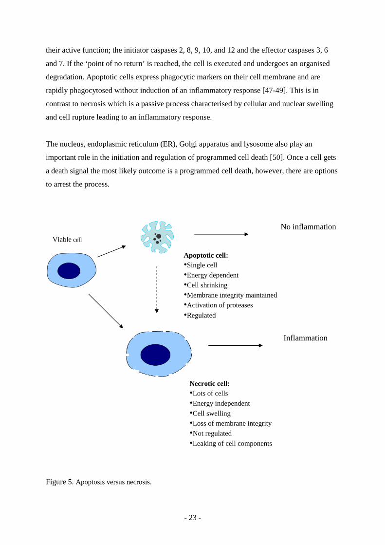
action of Fas and TNF receptor 1 (TNFR1) are the best elucidated. Binding of soluble or
membrane bound ligands to the extracellular domain of the death receptors causes receptor
trimerisation and aggregation of the cytoplasmic DDs. When apoptosis is trigged by Fas
ligand, Fas becomes activated and forms a complex with the adaptor protein FADD (Fas
associated death domain) [53, 54]. This recruits the apoptosis initiating protease procaspase 8
or 10 and the death-inducing signalling complex (DISC) is formed [55, 56]. This complex
triggers the intracellular signalling cascades that induce apoptosis. This step can be regulated
by cFLIP (cellular FADD-like interleukin-1-converting enzyme inhibitory protein), which is
an inactive homologue of caspase 8 and prolongs the survival of the cell [57]. By proteolytic
cleavage of procaspase 8 at a specific aspartic acid residue, a large and a small subunit are
released which associate to form heterodimers containing two active sites. Caspase 3, the
substrate for active caspase 8, is activated in two ways. The first mechanism involves direct
cleavage of procaspase 3 to yield activated caspase 3, which, in turn, stimulates other effector
caspases resulting in cleavage of structural and regulatory intracellular proteins and DNA
fragmentation, and finally cell death. The second mechanism is indirect activation where
caspase 8 cleaves the pro-apoptotic protein Bid which then can translocate to the
mitochondria where it triggers cytochrome c release, eventually leading to activation of
caspase 3 (Fig. 6).
TNF produced by activated T-cells and macrophages in the inflammatory response, is able to
activate the TNFR1 receptor. The TNFR1 receptor acts in a similar way as the Fas pathway
but it needs the adaptor molecule TRADD (TNFR-1-associated DD) [58] before the FADD
complex is formed, and procaspase 8 or 10 is recruited (Fig. 6).
- 25 -
Figure 6. Apoptosis induced by the death receptors Fas and TNF-α.
The interaction of TNF with its receptors can also inhibit apoptosis by activation of nuclear
factor κB (NF-κB), causing an up-regulation of the expression of several anti-apoptotic
proteins, which results in a prolonged cell survival [37].
TNF-related apoptosis inducing ligand (TRAIL) is able to activate five different receptors.
Two of the receptors, TRAIL-R1 and TRAIL-R2, contain an intracellular DD and they are
capable of inducing apoptosis but are mainly expressed on transformed cells. The other three
receptors (TRAIL-R3, TRAIL-R4 and TRAIL-R5) are decoy receptors with no or non-
functional DD and cannot induce apoptosis [59].
The internal pathway
The mitochondrial pathway is a central regulator of apoptosis and acts as a local sensor of
cellular stress, growth factor deprivation, irradiation and for the absence of survival signal.
Plasma membrane
DD
Fas
Fas ligand TNFα
DD TRADD FADD
Procaspase 8
Caspase 8
Procaspase 3 Caspase 3
Bid tBid
Mitochondrial pathway
TNFR1
Caspase 3
cFLIP
- 26 -
The mitochondrial pathway is triggered and regulated by proteins belonging to the Bcl-2
family, which possesses both anti-apoptotic and pro-apoptotic activity [60, 61]. Bcl-2 family
members control cell death by regulating the release of cytochrome c and other proteins from
the mitochondrial intermembrane space into the cytosol [62, 63]. The Bcl-2 family members
contain specific homologous regions, called Bcl-2 homology (BH) domains and all members
have one to four of these domains. They have been classified on the basis of their structural
similarities, resulting in four categories of BH domains (BH1 to BH4) and according to
functional criteria as either pro- or anti-apoptotic [64, 65]. Pro-apoptotic proteins contain
BH1, BH2 and BH3 type domains such as Bax and Bak. They act by perturbing the
mitochondria outer membrane allowing release of intermembrane space proteins and ions.
The proteins with only BH3 type domains, such as Bid, Bad and Bim are also pro-apoptotic;
they inhibit the activity of the anti-apoptotic proteins and promote activity of the BH1-BH3
pro-apoptotic proteins. The proteins with anti-apoptotic properties contain all four BH type
domains. Examples are Bcl-2 and Bcl-xL and they protect cells from death by stabilising the
mitochondrial outer membrane. The pro-apoptotic Bcl-2 family proteins are located in the
cytosol or associated with the cytoskeleton. After a death signal, the pro-apoptotic Bcl-2
family members undergo a conformational change that enables them to target and integrate
into membranes [66]. The anti-apoptotic Bcl-2 members are initially integral membrane
proteins found in the mitochondria, ER or nuclear membrane. They can inhibit the activation
of the pro-apoptotic Bcl-2 family members through dimerisation with them [67]. The pro-
apoptotic Bcl-2 family members are inserted into the outer mitochondrial membrane where
they form channels probably together with other proteins and mitochondria lipids. This
formation of specific pores in the outer membrane of the mitochondrion is reversible in
respect to mitochondrial function but it triggers other death signals by the release of proteins
and ions [63]. Another pathway results in the loss of mitochondrial membrane potential by
opening permeability transition pores (PT). There is a collapse in the electrochemical gradient
across the mitochondrial membrane which results in equilibration of ions between the matrix
and cytoplasm, osmotic swelling of the mitochondrial matrix, rupture of the mitochondrial
outer membrane and the release of proteins and ions. The PT pathway occurs mainly in
response to the release of Ca2+ from ER, and Bcl-2 members are also involved in the
regulation of Ca2+ release from the ER.
Upon release of cytochrome c from the mitochondrion [68] it forms a complex with apoptotic
protease activating factor 1 (Apaf-1) in combination with either ATP or dATP. Apaf-1
- 27 -
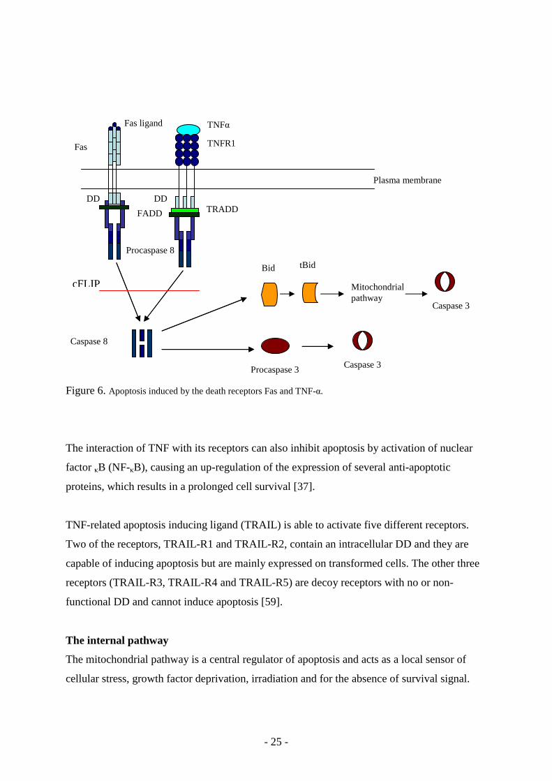
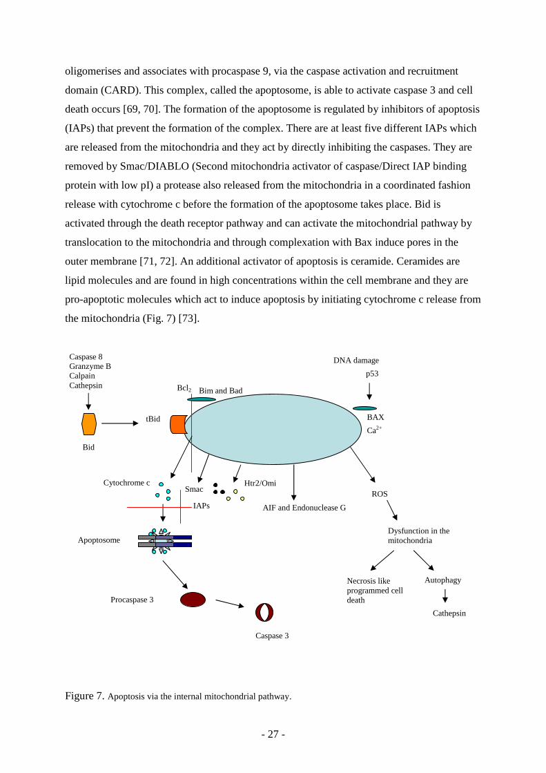
oligomerises and associates with procaspase 9, via the caspase activation and recruitment
domain (CARD). This complex, called the apoptosome, is able to activate caspase 3 and cell
death occurs [69, 70]. The formation of the apoptosome is regulated by inhibitors of apoptosis
(IAPs) that prevent the formation of the complex. There are at least five different IAPs which
are released from the mitochondria and they act by directly inhibiting the caspases. They are
removed by Smac/DIABLO (Second mitochondria activator of caspase/Direct IAP binding
protein with low pI) a protease also released from the mitochondria in a coordinated fashion
release with cytochrome c before the formation of the apoptosome takes place. Bid is
activated through the death receptor pathway and can activate the mitochondrial pathway by
translocation to the mitochondria and through complexation with Bax induce pores in the
outer membrane [71, 72]. An additional activator of apoptosis is ceramide. Ceramides are
lipid molecules and are found in high concentrations within the cell membrane and they are
pro-apoptotic molecules which act to induce apoptosis by initiating cytochrome c release from
the mitochondria (Fig. 7) [73].
Figure 7. Apoptosis via the internal mitochondrial pathway.
Bcl2
Cytochrome c
Apoptosome
Procaspase 3
Caspase 3
IAPs
Smac Htr2/Omi
AIF and Endonuclease G
ROS
Bim and Bad
tBid
Caspase 8 Granzyme B Calpain Cathepsin
Bid
DNA damage
p53
BAX
Ca2+
Dysfunction in the mitochondria
Autophagy
Cathepsin
Necrosis like programmed cell death
- 28 -
Programmed cell death can take many forms but not all fulfil the criteria of apopotosis.
Htr2/Omi, a serine protease released from the mitochondria, participates in both caspase
dependent and independent programmed cell death by virtue of its ability to function as an
inhibitor of IAPs and as a general protease. Apoptosis inducing factor (AIF) and
Endonuclease G, both released from the mitochondria, are able to translocate to the nucleus
and cause programmed cell death by a caspase independent DNA-fragmentation of chromatin,
resulting in high molecular weight DNA fragments [74]. Mitochondria outer membrane
permeabilisation can be induced by reactive oxygen species (ROS), Ca2+ release, and an
accumulation of misfolded proteins from the ER. Calpains are cysteine proteases that
naturally occur as inactive pro-enzymes. However, in the presence of high Ca2+ concentration
they become activated and participate in apoptosis initiated by glucocorticoids or irradiation
[75].
In normal cells, the p53 tumour suppressor protein is present at very low levels. The main
function of p53 is to regulate the cell cycle, but in response to cellular stress, such as DNA
damage, hypoxia and oncogene activation, the p53 protein is up-regulated and the cell
undergoes cell-cycle arrest. This allows for DNA to be repaired, or if that fails the cell dies by
apoptosis. The pro-apoptotic Bcl-2 family member protein, Bax, is up-regulated in response to
DNA damage and increased p53 levels. Furthermore, the anti-apoptotic Bcl-2 family
members are transcriptionally repressed by p53. Transcription of genes that increase
production of ROS, an activator of the mitochondrial apoptotic pathway, is induced by p53
and p53 may also up-regulate Fas, inducing Fas mediated programmed cell death.
Granzyme B and perforin (see page 13) is able to induce apoptosis in target cells by entering
the cell through a non-specific ion pore composed of perforin [76]. Granzyme B can also
initiate apoptosis by cleavage of Bid, caspase 3 and caspase 7.
Cathepsins are proteases involved in the digestion of apoptotic cells. Cathepsins are located in
the lysosome which is responsible for proteolysis of endocytosed and autophagocytosed
proteins at low pH. Destabilisation of the lysosomal membranes by oxidants, pore formation
through the Bcl-2 family members or shingosine, a lysosomotropic detergent, results in
release of cathepsins to the cytosol where they induce cell death through proteolytic effects in
the cytoplasm and the nucleus, or cleavage of Bid. Cathepsin is also able to activate caspases
by direct cleavage and shares many substrates with DNA repair enzyme poly(ADP-ribose)
- 29 -
polymerase (PARP), Bid and caspases [77-80].
Autophagy
In healthy cells, autophagy is a process where the cell degrades old and damaged organelles
within the cell, acting like a cell survival pathway but under certain circumstances it acts as an
alternative cell-death pathway [81]. During starvation or in hormone deprived cells, the
material can be recycled. The damaged organelles are engulfed by autophagosomes created by
donated membranes from the ER that surrounds the organelle. The autophagosomes fuse with
the lysosomes and form autophagic vacuoles. Lysosomes contain digestive enzymes and are
responsible for degrading old and damaged organelles within the cell. Autophagy can be
triggered by the same signals as apoptosis and is described as a backup system of apoptosis.
The execution
Caspase-activated deoxyribonuclease (CAD) is the nuclease that degrades the genomic DNA
between the nucleosomes into approximately 180 base pair fragments, which, when DNA is
analysed, appears as a DNA ladder and is a marker for apoptosis. The nuclease CAD exists as
an inactive complex (ICAD) with no DNase activity in living cells. However, ICAD becomes
activated upon caspase 3 mediated cleavage and can enter the nucleus and degrade the
chromosomal DNA [82]. The nuclear shrinkage and budding is caused by caspase mediated
cleavage of laminin, a network of protein filaments surrounding the nuclear periphery that
maintains the shape of the nucleus and mediates interactions between chromatin and the
nuclear membrane [83-87]. Cleavage of PAK2, a member of the p21-activated kinase family
mediates the active blebbing observed in apoptotic cells [88]. Caspases also cleave the
cytoskeleton proteins fodrin and gelsolin causing loss of cell shape [89]. The DNA repair
enzyme PARP is cleaved by caspases with the subsequent loss of its DNA repair activity [90].
DNA topoisomerase II, a nuclear enzyme essential for DNA replication and repair, could also
be inactivated by caspases leading to DNA damage.
- 30 -
Phagocytic clearance of dying cells
Phagocytosis of apoptotic cells is a very complex procedure, but under normal conditions it is
a fast and a non-inflammatory process preventing exposure of self-molecules. The uptake of
apoptotic cells actively suppresses the release of pro-inflammatory molecules and promotes
the release of anti-inflammatory molecules [91, 92]. The cell that is about to die sends out so-
called “find-me” signals such as lysophosphatidylcholine. For the recognition by the
phagocyte, the apoptotic cell displays so-called “eat-me” signals on its surface. These can be
pre-existing molecules, modified existing molecules, as well as the appearance of molecules
on the cell surface such as phosphatidylserine (PS), which is normally located on the inner
leaflet of the plasma membrane. At the same time the so-called “do not eat-me” signals are
down regulated, shed or internalised [48, 93, 94]. Many different recognition molecules on the
phagocyte orchestrate the clearance of apoptotic cells such as scavenger receptors (SR-A,
This study showed that apoptosis was induced in a time dependent manner in monocytes and
lymphocytes from healthy donors by supplementing the growth medium with 20% of serum
from SLE patients. The Annexin V binding to the cells preceded PI reactivity supporting the
idea that serum from SLE patients induces apoptosis and not primary necrosis. Morphological
studies with light and confocal microscopy showed that serum induced apoptosis on normal
cells incubated with serum from SLE patients displayed a classic apoptosis with shrunken
cells, nuclear condensation and formation of apoptotic bodies. This was confirmed by flow
cytometry by distribution of the cells in forward and side scatter properties (reflecting size
- 37 -
and structure, respectively) and by Annexin V binding. Caspase 8 and caspase 9 activity
measured over time mirrored each other with an increased activity already apparent after 4h.
The caspase activity could be reduced significantly (p<0.05) by the caspase inhibitors after 6h
incubation of cells with SLE serum, but no effect by the caspase inhibitors was seen after 16h
incubation. No decrease in the AIE of serum from SLE patients was seen when using
antibodies blocking the death receptor pathway, indicating that death receptor independent
pathways are more important than death receptor dependent ones.
Serum induced apoptosis was also seen when using Jurkat, U937 and FADD-deficient cell
lines, but with the cell line over-expressing Bcl-2, a significant decrease (p<0.05) of serum
induced apoptosis was seen, indicating the involvement of the mitochondrial pathway.
Depletion of IgG from serum and heat inactivation of serum did not have any effect on the
AIE, demonstrating that it was independent of IgG and complement activation.
The observed serum induced apoptosis was not seen when using sera from other autoimmune
diseases, infectious diseases or healthy individuals. Furthermore, no differences were seen
between sera from SLE patients with active or inactive disease.
There was no correlation between SLEDAI and the AIE on monocytes or lymphocytes using
serum from SLE patients. Patients with low C1q and/or low C4 levels had notably higher AIE
on both monocytes and lymphocytes. This was also seen for serum with low C3 levels, but in
this case the correlation was less pronounced. Levels of C5a in cell culture supernatants
correlated with the AIE on monocytes but much less with AIE on lymphocytes. The serum
concentration of anti-dsDNA antibody correlated weakly with AIE on monocytes and
between anti-cardiolipin antibody levels and lymphocyte apoptosis a correlation was also
found. Levels of TNF-α in cell culture supernatants, but not in the sera, correlated with the
AIE on both monocytes and lymphocytes.
Paper III
Inherited deficiencies in complement components of the classical pathway are associated with
a high risk for development of SLE. Therefore, the pathways involved in complement
activation by apoptotic cells were studied using serum from individuals deficient in the
- 38 -
complement proteins C1q, C2, C4, C3, properdin and MBL. Normal human serum and a pool
of sera from ten different healthy individuals were also used. Serum reagents lacking C1q and
factor D or factor D only were prepared as previously described [143]. The serum used for
C1q and factor D depletion was also deficient of MBL. Purified complement proteins C1q,
C2, C4, factor D and properdin were available in the laboratory and published methods for
purification had been used [144-148]. Recombinant MBL (rMBL) was kindly provided by
Professor J. C. Jensenius (Aarhus, Denmark).
For the generation of macrophages, peripheral blood monocytes were obtained from fresh
heparinised blood samples from healthy donors. The monocytes were cultured for 5 days in
RPMI 1640 media, containing 10% normal human serum, gentamycin and amphotericin
allowing the monocytes to differentiate into macrophages (MDM). Apoptotic cells were
obtained by treating Jurkat cells with staurosporine; yielding approximately 50% early
apoptotic cells and 50% late apoptotic cells, and then the cells were labelled with CFS-E. The
labelled apoptotic cells were allowed to interact with the MDMs in the presence of different
serum. The MDMs were collected and RPE-cy5 anti-CD14 was added. Engulfment of
labelled apoptotic Jurkat cells by the MDMs was analysed by flow cytometry.Cells positive
for both CD14 and CFS-E were considered as apoptotic cell-engulfing MDMs and the number
of those cells were calculated as a percentage of the total number of CD14-positive cells. For
measuring complement deposition on apoptotic cells, the cells were incubated with different
deficient sera or reagents. Complement activation was assessed by measuring C3 deposition
using antibodies against C3 fragment, with specificity for C3d, followed by incubation with
specific antibodies (anti-mouse) conjugated with phycoerythin before analysed by flow
cytometry.
Results
Sera from individuals deficient in classical pathway components (C1q, C2 and C4) all showed
a decreased phagocytosis of apoptotic cells. When adding back the missing complement
component, the phagocytosis increased to a level similar to that of normal human serum. The
lectin and the alternative pathway did not influence phagocytosis of apoptotic cells, since sera
lacking MBL, properdin or factor D showed equal levels of phagocytosis as normal human
serum. The C3 deficient serum showed decreased levels of phagocytosis of apoptotic cells but
increased after reconstitution with C3 albeit not to the same level as normal human serum.
- 39 -
Sera from healthy controls and sera with low MBL levels supported C3 deposition on
apoptotic cells over a wide range of serum concentrations (2.5-40%). The effect increased
with increasing serum concentration and was more pronounced on secondary apoptotic cells.
Serum deficient in the classical pathway only showed C3 deposition in high serum
concentrations, indicating involvement of the alternative pathway. Experiments with MBL-
deficient serum further depleted of C1q and factor D supported the predominance of the
classical pathway regarding C3 deposition on apoptotic cells.
Paper IV
Autoantibodies directed against autoantigens, such as nucleic acid and nucleic acid associated
proteins, are common in SLE, and apoptotic and necrotic cells are known to expose these
autoantigens. Phagocytosis of necrotic material (NC) and apoptotic cells by
polymorphonuclear leukocytes (PMNs) were studied in the presence of serum samples
collected consecutively over several years from 19 SLE patients (n=798). Routine laboratory
testing of complement proteins (C1q, C3 and C4) and anti-dsDNA antibodies had previously
been performed on serum from these SLE patients.
Sera from 64 SLE patients selected for time points at high and low disease activity estimated
by SLEDAI-2K [113] and sera from individuals deficient in the complement proteins C1q,
C2, C4 and properdin were also used. Purified complement proteins C1q, C2, C4, and
properdin were available in the laboratory and were used to restore deficient sera. These
proteins had previously been purified according to published methods [144-148]. As controls,
sera from 100 healthy blood donors were used.
Peripheral blood mononuclear cells (PBMC) were obtained from fresh heparinised blood
samples from healthy donors and PMNs were isolated by density gradient centrifugation
according to the manufacturer’s protocol. Engulfment of NC or apoptotic cells by the PMNs
in the presence of serum was analysed by flow cytometry and two assays for phagocytosis
were used. One of these assays measured phagocytosis of necrotic cell material (PNC assay)
and the other assay measured phagocytosis of apoptotic cells (PAC assay). The NC material
was generated by incubating PBMC for 10 min at 70ºC and apoptotic cells were generated by
treatment with staurosporine. Autoantibodies against histones were measured by ELISA. Two
- 40 -
different ELISAs were developed, one detecting antibodies against only histone 1, the linker
histone, and a second in which a mix of histones (1, 2, 3 and 4), of which 2, 3 and 4 belong to
the core histones, was used as antigen.
Results
Using flow cytometry, PMNs containing necrotic cells and apoptotic cells could be detected.
Increased levels of phagocytosis of NC and apoptotic cells were seen in the presence of serum
from SLE patients. Of the 19 SLE patients, 17 showed increased levels of phagocytosis of NC
at some time point and this was not seen in the healthy controls. The phagocytosis of NC was
dependent on antibodies and a functional complement system, shown by decreased levels in
the PNC assay after depletion of IgG and heat inactivation of sera.
To further establish the importance of the complement system for an efficient phagocytosis of
NC, different sera from complement deficient individuals were used, and anti-histone
antibodies were added to promote phagocytosis of NC. Sera with deficiencies within the
classical pathway (C1q, C2 or C4) all showed a clear decrease in phagocytosis of NC in the
presence of anti-histone antibodies when compared to normal human serum (NHS)
supplemented with anti-histone antibodies. After reconstitution of the missing complement
protein in the presence of anti-histone antibodies, the phagocytosis of NC equalled that of
anti-histone antibody-supplement NHS. The serum deficient of properdin showed an equal
capacity to promote phagocytosis of NC as the anti-histone antibody-supplemented NHS and
no change was seen after reconstitution with properdin.
Anti-histone antibody levels measured by ELISA showed a good correlation with the ability
to phagocytose NC. Classification of the anti-histone antibodies in the 19 SLE patients’ sera
by SDS-PAGE and Western blot showed that 10 sera contained autoantibodies against more
than one histone, 5 sera contained autoantibodies against only histone 1, and the remaining 4
patient sera did not have detectable levels of autoantibodies against any of the histones tested.
Among the patients who were positive in the PNC assay, with antibodies against more than
one histone, we found a trend of decreased concentrations of the complement proteins C1q,
C3 and C4 not seen in sera with only antibodies against histone 1. A similar trend was seen in
the correlation between antibodies against histones and the complement components C1q, C3
and C4.
- 41 -
Individual values of area under curve divided by follow-up time (AUC) in each of the 19
longitudinally followed patients were calculated and the patients were divided into groups
based on the presence and specificity of antibodies to histones. Sera containing antibodies
against more than one histone had increased activity in the PNC assay as compared to serum
containing antibodies against only histone 1 (p=0.01). Sera containing antibodies against
more than one histone were also higher in the PNC assay as compared to sera without
detectable anti-histone antibodies (p=0.008). No differences in AUC values were seen when
comparing phagocytosis of NC in serum containing antibodies against histone 1 with serum
negative in the PNC assay. A trend towards decreased concentrations of C3 and C4 was seen
in the group containing antibodies to more than one histone as compared to sera containing
antibodies to only histone 1 or without anti-histone antibodies, but this was not statistically
significant. Patients with antibodies to a broader spectrum of histones also had autoantibodies
against DNA. In the 64 patients where serum samples had been selected for time point of low
and high disease activity, a clear relation of increased phagocytosis of NC and high levels of
autoantibodies against histones was seen at time point of high disease activity.
Discussion and future perspectives
SLE is a complex disease and the cause is unknown, as in most other autoimmune diseases.
Multiple genetic factors interacting with environmental factors seem to play a role in
development of SLE and both the innate and the adaptive immune systems contribute to the
pathology seen in the disease. Apoptotic cells have been suggested to be a major source of
autoantigens in SLE since clustering and concentration of lupus autoantigens in the suface
blebs of apoptotic cells have been demonstrated [114]. SLE patients show an impaired
clearance of apoptotic cells, which may result in an increased exposure of autoantigens [149].
The formation of autoantibodies gives rise to circulating immune complexes causing damage
by deposition in many organ systems and activation of complement.
In Paper I we showed that the apoptosis inducing effect in sera from SLE patients was not
related to non-specific inflammatory events and it seems to be specific for SLE and still no
difference was seen between active and inactive disease. Others have shown accelerated Fas-
dependent apoptosis of monocytes and macrophages from SLE patients [124], and increased
apoptosis of CD34+ stem cells exposed to SLE serum has also been reported [128]. We have
- 42 -
further investigated the correlation between serum induced apoptosis and expression of Fas on
freshly prepared cells from SLE patients and no correlation was found. However, when using
serum from SLE patients, with the capacity to induce apoptosis, and autologous cells, the
serum also induced apoptosis on these cells (unpublished data). The presence of a pro-
apoptotic mechanism in serum from SLE patients could increase the load of autoantigens and
influence the efficiency of clearance of apoptotic cells. The apoptosis inducing effect was not
dependent on IgG and complement activation. This indicates that IgG containing immune
complexes are not directly involved and cell death is not caused by complement activation but
still split products of complement activation could be involved. The relationship seen between
the concentrations of the complement protein in the classical pathway and serum induced
apoptosis could not be seen when we extended the number of samples analysed (unpublished
data). It has been described that purified autoantibodies such as anti-dsDNA and anti-
cardiolipin antibodies do induce apoptosis in vitro [150, 151]. Even if only a weak correlation
was seen between the presence of these autoantibodies and serum induced apoptosis, and
removal of IgG by protein G absorption did not influence the serum induced apoptosis, further
studies regarding the presence of autoantibodies in serum are of interest. Ongoing studies with
a larger cohort regarding correlation between serum induced apoptosis and different diseases
manifestation are in progress.
In Paper II we showed that serum from SLE patients induces a classical caspase dependent
apoptosis, independent of death receptors but most likely involving the mitochondrial
pathway. Classical apoptosis is characterized by distinct morphological changes such as
compact chromatin condensation. Other criteria such as caspase activity and maintenance of
membrane integrity should also be fulfilled [43]. The serum induced apoptosis could be
reduced when the pan-caspase inhibitor Z-VAD fmk was used together with serum, indicating
a caspase dependent apoptosis. Activity of caspase 8 indicates involvement of the death
receptor pathway and activity of caspase 9 indicates involvement of the mitochondrial
pathway, but caspase 8 also has the possibility to activate caspase 9 via tBid-mediated
cytochrome c release. However, we saw both caspase 8 and caspase 9 activity occurring
simultaneously. These unexpected findings have also been described when anti-cancer drugs
are targeting the mitochondrial pathway where caspase 8 is activated in the absence of death
receptors [152]. In this situation caspase 8 is thought to function as an executioner caspase in
the mitochondrial pathway [153] and it has also been described that caspase 3 mediates
feedback activation on upstream caspases such as caspase 8 [154]. A reduction of the
- 43 -
apoptosis inducing effect was seen when using a Bcl-2 over-expressing cell line as compared
to corresponding vector control cells, indicating the involvement of the mitochondrial
pathway. No reduction of the serum induced apoptosis was seen when using a FADD-
deficient cell line or when blocking the death receptors Fas and TNF-α. This indicates that the
death receptor pathway is not involved in SLE serum induced apoptosis, but it cannot be ruled
out that a novel FADD-independent death receptor is involved. Furthermore, the balance
between pro- and anti-apoptotic stimuli may decide the destiny of the cell and the different
death pathways may co-exist. The factor in serum inducing apoptosis has not yet been
identified, current findings suggest involvement of a negatively charged protein, and further
investigations are ongoing.
In Paper III we investigated the role of complement in opsonisation of apoptotic cells.
Homozygous deficiency of the classical pathway components C1q, C4 or C2 is associated
with an increased susceptibility to SLE. Nearly all individuals with C1q deficiency develop
SLE, often at a young age, whereas C4-deficient individuals develop SLE less often, with
individuals deficient in C2 even less so [35]. We showed that the complement components of
the classical activation pathway are all important for an efficient phagocytosis of apoptotic
cells and the main contributor to opsonisation of C3 fragment on these cells. Thus, the strong
association between C1q-deficiency and SLE is only partly explained by the role of C1q in
the clearance of apoptotic cells. We studied the uptake of apoptotic cells by monocyte derived
macrophages in the presence of complement-deficient sera. The clearance of apoptotic cells
was shown to de dependent upon the classical pathway of complement activation, whereas
neither a functional lectin nor alternative pathway was necessary. These findings are in
accordance with most other reports within this research area [31, 155]. However, in our
experimental setup, we could not see any difference between serum from individuals deficient
in C1q, C4 or C2. This finding indicates that components of the classical pathway appear to
be equally important in the clearance of apoptotic cells, which contradicts the hypothesis of a
hierarchical role for classical pathway complements proteins with regard to their role in
clearance of apoptotic cells [156]. Elevated levels of serum IFN-α seen in SLE patients have
been proposed to have a significant role in the pathogenesis of the disease and our group
recently described a novel function of C1q in the regulation of immune complex induced
production of IFN-α. These findings suggest another role for C1q and contribute to the
explanation of why C1q-deficiency is such a strong risk factor for the development of SLE
[137].
- 44 -
The results obtained in this study using complement-deficient sera and sera depleted of C1q
and factor D showed that activation of complement by apoptotic cells resulted in deposition of
C3 fragments, which was dependent mainly upon the classical pathway. Deposition of C3
fragments through classical pathway activation was seen at low serum concentrations, and
was most pronounced on secondary necrotic cells. This is in agreement with results from
other investigators [96, 98]. The partly discrepant findings compared with previous studies
with regard to complement pathways in opsonisation and phagocytosis of apoptotic cells
could be due to the serum concentration used, but also to different cell types and cells at
different stages of apoptosis. In our experiments, human sera from complement-deficient
individuals were used, which could be another important difference. Nevertheless, our
experiments strongly indicate that activation of the classical pathway is beneficial for the
phagocytosis of cells undergoing apoptosis, especially of those also being secondarily
necrotic.
The major source of autoantigens in SLE, as mentioned above is thought to be apoptotic cells.
These autoantigens may be processed in a way that neo-epitopes will emerge to which the
immune system is not tolerant and autoantibodies are therefore formed against nuclear
antigens which are frequently seen in SLE patients. The presence of PMNs containing
phagocytosed nuclear material is also seen in SLE. This is described as the LE cell
phenomenon and was one of the first described laboratory findings associated with SLE
[116]. In Paper IV we evaluate the roles of complement and antibodies against histones in
relation to phagocytosis of necrotic cell material (NC) by PMNs. We found that
autoantibodies against histones were associated with high capacity of PMNs in phagocytosis
of NC and also that the classical complement pathway contributed to an efficient
phagocytosis. Serum deficient of C1q, C2 or C4 all showed decreased capacity to promote
phagocytosis of NC in the presence of antibodies against histones but no differences were
seen when using serum deficient of properdin as compared to normal serum with antibodies
histones added. In SLE patients a broad anti-histone, reactivity was associated with
complement consumption and clinical manifestations such as vasculitis and
glomerulonephritis. It has been suggested that histone 1 is the major autoantigen in SLE for
the generation of LE cells [118, 157]. However, our findings suggest that the LE cell
phenomenon is dependent on antibodies against histone 3 especially in combination with
antibodies against histone 1 or histone 2. The patients with antibodies to more than one
histone also had antibodies against dsDNA indicating a broad spectrum of autoantibody
- 45 -
specificities. This suggests that not exclusively autoantibodies against histone 1 but also
autoantibodies against other nuclear proteins, DNA, and perhaps autoantibodies against
nuclear ribonucleoproteins could be able to induce phagocytosis by PMNs. It has been
described that antibodies are able to penetrate into living cells and interact with their
respective antigen, which may affect intracellular functions as well as induction of apoptosis
[158]. This could lead to exposure of more autoantigens and generation of a broader spectrum
of autoantibodies if the apoptotic cells are not properly cleared from the system.
Possibly, the activation of the complement system seen in the patients with a broader
autoantibody profile reflects a higher density of bound antibodies making it more favourable
for C1 to bind resulting in complement activation. An Fc receptor-mediated phagocytosis of
antibody-coated nuclear material seems not to be sufficient; instead a cooperative binding of
complement fragments to receptors expressed on PMNs seems to be necessary for an efficient
phagocytosis. Further studies will be carried out regarding this issue.
Influence on phagocytosis of apoptotic cells by PMNs in the presence of autoantibodies with
different specificities will be studied. To further clarify the association between the
specificities of autoantibodies against histones and clinical findings, more patients will be
included in the study.
In this thesis, some of the mechanisms believed to be important in the pathogenesis of SLE
have been addressed. The ability of serum from SLE patients to induce apoptosis could
contribute to impaired clearance of apoptotic cells, an increased load of autoantigens, and the
formation of autoantibodies. Also low concentration of the complement classical pathway
components could cause impaired clearance of apoptotic cells and as a consequence, more
apoptotic cells may expose autoantigens. Autoantibodies against histones are important for
the uptake of the damaged cells in PMNs and may reflect the presence of apoptotic cells in the
circulation. The autoantibodies may form circulating immune complexes, and deposition of
these complexes in various organs may propagate an inflammatory response with complement
activation leading to complement consumption. The complement consumption which leads to
low concentrations of the components of the classical pathway may, in turn, contribute to
impaired clearance of apoptotic cells.
- 46 -
Conclusions
• Sera from SLE patients induce apoptosis in monocytes and lymphocytes from normal
healthy donors, as well as in the monocytoid cell line U937 and the T-cell line Jurkat.
• The apoptosis inducing effect is not related to non-specific inflammatory events and
seems to be specific for SLE but is not a marker for disease activity.
• The apoptosis inducing effect is not dependent on IgG complexes or complement
activation.
• Sera from SLE patients induce a classical caspase dependent apoptosis, independent of
death receptors but most likely involving the mitochondrial pathway.
• Deficiency of the factors C1q, C4 or C2, of the classical complement activation
pathway, resulted in an equally decreased ability to phagocytose apoptotic cells.
• Activation of the classical complement pathway seems to be the most important
contributor of opsonisation of C3 fragments on apoptotic cells and is required for an
efficient phagocytosis of apoptotic and necrotic cells.
• The strong association between C1q-deficiency and SLE is only partly explained by
the role of C1q in the clearance of apoptotic cells.
• Phagocytosis of necrotic material by PMNs is dependent on the presence of IgG and
on the classical complement activation pathway.
• Broad anti-histone antibody reactivity is strongly associated with increased
phagocytosis of necrotic material by neutrophils, disease activity and with
complement consumption.
- 47 -
POPULÄRVETENSKAPLIG SAMMANFATTNING PÅ
SVENSKA
Systemisk lupus erythematosus (SLE) är en kronisk autoimmun sjukdom, det vill säga en
sjukdom där immunförsvaret angriper kroppens egen vävnad och förorsakar inflammation.
SLE kan drabba många av kroppens organsystem så som hud, leder, njurar, hjärta, kärl,
lungor och det centrala nervsystemet. Sjukdomsorsaken är okänd men tros bero på en
samverkan mellan gener och miljöfaktorer. Immunsystemet är involverat i sjukdomsprocessen
på många sätt, bl. a. finns en ökad produktion av antikroppar mot många naturligt
förekommande strukturer i kroppens celler däribland mot DNA, så kallade autoantikroppar.
Granulocyter vilka aktivt tagit upp delar av cellkärnor, i regel i komplex med antikroppar
riktade mot cellkärnornas komponenter, bland annat mot histoner kan beskrivas som LE-
celler. Detta LE-cellsfenomen är associerat med SLE och redan 1948 beskrev M. Hargraves
en mikroskopbaserad metod för att detektera dessa celler.
Komplementsystemet består av ett 30-tal lösliga eller membranbundna proteiner, vilka utgör
en viktig del av immunsystemet. Vissa komplementfragment fungerar som opsoniner, d.v.s.
märker ut främmande ämnen (antigen), vilket underlättar för fagocyter (celler som äter upp)
att ta upp (fagocytera) dessa ämnen och oskadliggöra dem, vidare kan komplement också vid
aktivering göra hål på cellen så att den förstörs. Komplementsystemet kan aktiveras via tre
vägar, den klassiska vägen, den alternativa vägen och lektinvägen. Bristfällig funktion av
komplementsystemet är associerad med sjukdomen SLE, speciellt defekter av proteiner
involverade i den klassiska vägen. Vid brist av något av dessa proteiner ökar risken för att
utveckla SLE och en rangordning finns där ca 90 % av personer med brist av C1q utvecklar
SLE, 75 % vid brist av C4 och 10 % vid brist av C2. En ökad aktivering av
komplementsystemet kan också ses vid sjukdomen SLE framförallt vid sjukdomsskov.
Apoptos, en form av programmerad celldöd, är nödvändig för utvecklande och
upprätthållande av multicellulära organismer och som försvarsmekanism. Det är en aktiv
process som är beroende av signaler eller aktiviteter i den döende cellen och framkallar inte
något inflammatoriskt svar, i motsats till nekros som är en passiv celldöd orsakad av skada
vilken ger upphov till ett inflammatoriskt svar. Apoptos kan aktiveras externt genom
receptorsignal eller internt via mitokondrien som då frisätter pro-apoptotiska ämnen. Kaspaser
- 48 -
är en grupp enzymer som normalt finns i inaktiv form, men som aktiveras vid apoptos och då
kan bryta ner eller aktivera andra proteiner och cellen förbereds för död. Vid programmerad
celldöd förändras cellen, nya molekyler exponeras i cellmembranet, så att fagocyterande
celler känner igen och omedelbart kan ta upp den. Vid ett snabbt omhändertagande av de
apoptotiska cellerna undviks att de intracellulära komponenterna exponeras för
immunsystemet. Patienter med sjukdomen SLE har en försämrad förmåga att ta bort döende
celler, vilket leder till en ökad mängd av kvarvarande apoptotiska celler. Dessa apoptotiska
celler blir då tillgängliga för immunsystemet och de döende cellerna är troligtvis huvudkällan
för exponering av autoantigen som bidrar till bildandet av autoantikroppar.
Avsikten med detta projekt var att utvidga och beskriva det tidigare fyndet att serum från
patienter med sjukdomen SLE inducerar apoptos hos celler från friska givare. Vi ville även
studera komplementsystemets påverkan vid borttagandet av apoptotiska eller döda celler
genom att använda sera med komplementbrist och undersöka deponering av C3 på de döende
cellerna. Vidare var avsikten att undersöka relationen mellan autoantikroppar riktade mot
histon och förmågan att fagocytera döda celler.
Metoder: Flödescytometri är en teknik där man enkelt kan mäta och identifiera olika celler
genom att låta cellerna skickas en och en genom en laserstråle. Det reflekterande och
avböjande laserljuset registreras och omvandlas till elektriska signaler som ger ett mått på
cellens storlek och form. Cellerna kan även märkas med olika cellspecifika markörer till vilka
det är kopplat olika ämnen, fluorokromer som utsänder ljus vid olika våglängder och på så
sätt kan olika subpopulationer identifieras. Celler kan också undersökas med
konfokalmikroskop, här utnyttjas laserljus för att konstruera tredimensionella bilder.
Laserljuset flyttas över provet och endast det ljus som är i fokus skickas till detektor och
övrigt ljus filtreras bort, detta gör det möjligt att ta bilder i flera optiska plan. Förekomst av
antikroppar kan mätas med ELISA (enzyme-linked immunosorbent assay), principen för
denna metod är att klä en plastplatta med antigen. Därefter tillsätter man det prov man vill
undersöka och med hjälp av en enzymatisk reaktion kan man mäta inbindning av antikroppar
till den beklädda plattan.
Delarbete I: Induction of apoptosis in monocytes and lymphocytes by serum from patients with
systemic lupus erythematosus – an additional mechanism to increase autoantigen load? I
detta arbete studerade, bestyrkte och utökade vi observationen av att det i serum från SLE-
- 49 -
patienter finns en löslig faktor som har förmåga att inducera apoptos i celler från friska givare
och denna förmåga att förorsaka apoptos relaterades till kliniska och immunologiska data.
Annexin V (AV) binder till fosfatidylserin exponerat på cellytan på apoptotiska celler och
propidiumjodid (PI) binder till DNA. Levande, apoptotiska och nekrotiska celler kan
särskiljas genom att mäta AV- och PI-inbindning till celler med flödescytometri. Vi odlade
celler från friska givare i närvaro av serum från SLE-patienter och inbindning av AV och PI
mättes. Resultaten visar att serum från SLE-patienter förorsakar primärt apoptos och inte
nekros av dessa celler. För att undersöka om detta fenomen även fanns vid andra
sjukdomstillstånd undersökte vi sera från patienter med andra autoimmuna sjukdomar så som
reumatoid artrit, vaskulit och infektionssjukdomar samt friska kontroller. Förmåga att
inducera apoptos i celler från friska givare var unikt för sjukdomen SLE, men ingen skillnad
kunde ses mellan aktiv och inaktiv sjukdom. Således kunde vi visa att sera från SLE-patienter
har förmåga att inducera apoptos i celler och att denna förmåga verkar vara specifik för SLE-
sjukdomen. Denna egenskap skulle kunna bidra till en ökad exponering av autoantigen.
Delarbete II:SLE serum induces classical caspase-dependent apoptosis independent of death
receptors. I detta arbete gick vi vidare och försökte karakterisera på vilket sätt serum från
SLE-patienter initierar apoptos. Med konfokalmikroskopi kunde vi konstatera att de
apoptotiska cellerna hade ett typiskt klassisk kaspasberoende apoptotiskt utseende, det vill
säga att kromosomerna vilka innehåller DNA och protein hade sammanpressats och
apoptotiska cellkroppar hade börjat bildads. Andelen apoptotiska celler minskades vid
blockering av kaspasaktivitet. Däremot påverkades inte den apoptosinducerande effekten vid
blockering av dödsreceptorvägen. Bcl-2 är ett anti-apoptotiskt protein vilket hämmar apoptos
via mitokondrievägen. Vi undersökte om en cell-linje som överuttrycker Bcl-2 kunde hämma
apoptos förorsakad av sera från SLE-patienter. En signifikant minskning kunde ses vilket
indikerade att denna väg är inblandad. Således kunde vi konstatera att sera från SLE-patienter
inducerar en klassisk kaspasberoende apoptos oberoende av dödsreceptorer på friska celler.
Delarbete III: Complement classical pathway components are all important in clearance of
apoptotic cells. Här studerade vi fagocytos av apoptotiska celler i närvaro av
komplementdefekta sera. Genom att sätta upp en flödescytometerbaserad metod där
makrofager (fagocyterande celler) inkuberades med apoptotiska celler i närvaro av serum
kunde vi mäta antalet apoptotiska celler som fagocyterats av makrofagerna. Resultaten visade
att den fagocyterande förmågan försämrades lika mycket vid frånvaro av C1q, C2, C4 eller
- 50 -
C3 i serum, vilket indikerar att aktivering av alla komponenter i den klassiska vägen är av lika
stor betydelse för en effektiv fagocytos av apoptotiska celler. Detta innebär att den starka
kopplingen mellan brist på C1q och SLE inte enbart kan förklaras med den minskad förmåga
att fagocytera apoptotiska celler. Vi undersökte då deposition av C3-fragment på de
apoptotiska cellerna i närvaro av komplementdefekt serum. I dessa försök möjliggjordes
mätning av C3-deposition både på tidigt apoptotiska celler (AV-positiva/PI-negativa) och sent
apoptotiska celler (både AV- och PI-positiva). Resultaten visade inbindning av C3-fragment
på apoptotiska celler och denna inbindning var huvudsakligen beroende av aktivering via den
klassiska vägen. Inbindning av C3-fragment kunde ses på både tidigt och sent apoptotiska
celler men var mest uttalat på sent apoptotiska celler. En viss inbindning av C3 till
apoptotiska celler kunde detekteras i närvaro av komplementdefekta sera i hög koncentration,
vilket indikerar att C3-deposition via alternativa vägen mest fungerar som en förstärkning av
aktivering via den klassiska vägen.
Någon skillnad i brist på den klassiska vägens olika komponenter och utvecklandet av
sjukdomen SLE kunde inte ses i dessa försök utan här var alla komponenterna i den klassiska
vägen lika viktiga. Dessa experiment visade att komplementberoende opsonisering med C3-
fragment på döende celler är viktig för att få ett effektivt borttagande av dessa celler och
därmed en minskad exponering av autoantigen. Sålunda, den starka associationen mellan SLE
och brist av C1q kan inte enbart förklaras av den roll C1q har i borttagandet av apoptotiska
celler utan även andra förklaringar som exempelvis reglering av cytokiner via C1q skulle
kunna påverka denna association.
Delarbete IV: Specificity of anti-histone antibodies determines complement-dependent
phagocytosis of necrotic material by polymorphonuclear leukocytes in the presence of serum
from patients with SLE. The LE cell phenomenon revisited. Syftet med detta arbete var att
mäta förekomst av autoantikroppar riktade mot histoner i serum från patienter med sjukdomen
SLE och att undersöka om granulocyter fagocyterar necrotiskt material i närvaro av serum
från SLE-patienter eller serum från komplementdefekta individer. Två olika ELISA-metoder
utvecklades för att kunna mäta antikroppar mot histon 1 och antikroppar mot en mix av
histoner (1, 2, 3 och 4) och en flödescytometerbaserad metod användes för att mäta fagocytos
av nekrotiskt material. Dessa metoder utvärderades och jämfördes med varandra och med
kliniska data. Resultaten visade en god korrelation mellan de olika metoderna och att
förekomst av antikroppar mot histon framför allt antikroppar mot flera olika histoner främjar
- 51 -
fagocytos. Förutom närvaro av antikroppar behövdes ett fungerande komplement system, där
den klassiska vägen aktiveras, för att på ett effektivt sätt fagocytera det antikroppsklädda
nekrotiska materialet. Antikroppskoncentration visade ett starkt samband med
sjukdomsaktivitet. Således kunde vi här visa att ett brett spektrum av autoantikroppar mot
flera histoner främjar fagocytos av nekrotiskt material i närvaro av den klassiska vägen. Att
mäta antikroppar riktade mot histoner i serum från SLE-patienter skulle kunna vara ett sätt att
påvisa närvaron av döende celler, vilka kan bidra till ett ökat autoimmunsvar.
- 52 -
TACKORD Först och främst vill jag Tacka mina handledare, mina tre visa män, som har inspirerat och
uppmuntrat mig, gett goda råd och alltid funnits till hands. Utan er och övriga medarbetare i
vår lilla grupp hade denna bok aldrig blivit skriven.
Lennart Truedsson, du har inte bara varit min handledare utan också varit min chef i
oändligt många år. När jag tänker på denna tid är det stor tacksamhet jag känner. Du har som
chef och handledare visat mig stor medmänsklighet, omtanke, gett beröm, uppmuntrat till fritt
tänkande, delat med dig av dina gedigna kunskaper, och alltid funnits där när jag har behövt
dig. Du har en oerhörd förmåga att reda ut resultat som verkar obegripliga och komma med
kloka idéer och synpunkter och framför allt har du en otrolig förmåga att förstå vad jag
egentligen menar. Tack för att du har trott på mig och för allt du gett mig. Dessutom vill jag
passa på att ge ett Tack till Ann, det är alltid trevligt att vara i ditt sällskap.
Anders Bengtsson, var ska jag börja, med dig har man alltid roligt även om man inte alltid
riktigt vet var man är. Du har en mycket stor portion av engagemang och energi som du villigt
delar med dig av. Ditt stora kunnande och intresse i ämnet är mer än beundransvärt och din
omtanke och generositet är mycket stor. Att få ta del av din kunskap är inspirerande,
diskussionerna med dig är givande och uppmuntrande ord bjuder du också på. Tusen Tack,
för allt.
Gunnar Sturfelt , Tack för dina betydande synpunkter och för all den kunskap och
erfarenheter som jag har haft förmånen att få ta del av. Du är länken mellan patient och
provrör, vilket skapar en helhet och gör arbetet så mycket mer intressant och respektfyllt. Att
få ta del av din kompetens och erfarenhet har varit ett privilegium. Du är omtänksam och ditt
stöd och uppbackning har jag uppskattat mycket.
Eva Holmström, du har inte bara varit en mentor till mig på jobbet utan även i livet.
Innebörden av dina egenskaper som medmänsklig, god, kunnig och rolig är så mycket mer
och så mycket större hos dig än hos många andra. Tack Eva.
Gertrud Hellmer , i många år har vi delat på 4 kvadratmeter utan några som helst problem,
det säger nog det mesta. Du har tålmodigt orkat lyssna på allt och inget. Du är alltid villig att
skickligt ge en hjälpande hand både på labbet och i språkkunskap.
- 53 -
Christian Lood , denna lilla påg kommer att bli en stor man. Du går från klarhet till klarhet.
Din skicklighet, kunnande och ödmjukhet njuter jag av varje dag, lycka till Christian. Tack för
att vi får ha dig hos oss.
Lillemor Skattum , din kompetens kan vi luta oss mot och du ger framtiden ett ansikte, det är
alltid trevligt och givande att samarbeta med dig.
Till Malin vill jag säga mycket välkommen till vår lilla grupp.
Stort Tack vill jag även ge till, övriga och tidigare medarbetare Ulla Mårtensson, Göran
Jönsson, Malin Carlsson, Barbro Selander, Cecilia Klint och Gunilla Nordin-
Fredriksson.
Anders Sjöholm som allt förtidigt gick bort, fortfarande är saknaden stor.
Jag vill Tacka alla på cell immunologen för att ni alltid är villiga att ge en hjälpand hand,
Elisabeth, Lisette, Catarina. Speciellt vill jag Tacka Annica Andreasson för all hjälp jag
har fått med flödescytometri, för att du har outtröttligt svarat på alla mina frågor och för alla
år som en mycket trevlig medarbetare.
Stort Tack vill jag säga till alla på komplement avdelningen både nuvarande och tidigare
medarbetare, Mona, Christina, Pia, Anci, Karin, Malin och Eva N för all vänlighet och
hjälp med analyser. Dessutom vill jag Tacka alla andra på våning B för all välvilja och hjälp.
Tack Susanne för att du alltid ser möjligheter för samarbete och därmed underlättar det
dagliga arbetet. Jag vill Tacka alla på Sölvegatan 23 både tidigare och nuvarande medarbetare
för många år av ett trevligt arbetsklimat och mycket vänlighet och ett stort Tack vill jag ge till
alla studenter som passerat igenom här.
Jag vill också rikta ett stort Tack till Helena Tydén, Andreas Jönsen, Ola Nived, Ingrid
Johansson, Anita Nihlberg, och Maria Andersson på reumatologen för givande samarbete
och Lisa, Kerstin, Inger, Lena och Lena S för att ni alltid ger en ett varmt välkomnande.
Dessutom vill jag ge ett stort Tack till Maria och Peter på labbet för all hjälp med serum
prov och med mycket annat.
Jag vill Tacka alla medförfattare som bidragit med sina expertkunskaper och värdefulla
synpunkter.
Tack Catharina Svanborg för all uppmuntran du ger och möjligheten till vidareutbildning.
- 54 -
Jag vill Tacka alla andra både tidigare och nuvarande medarbetare på MIG för ett trevligt
arbetsklimat, sällskap i fikarummet och givande samarbete. Speciell vill jag Tacka Maj-Lis,
Anki, Barbro, Maria, Oskar, Petter, Pontus, Bryndis, Hans, Sonja, Emma, Micke och
Babu för all hjälp från reagens, dator bestyr till fina konfokal bilder.
Stort Tack till njurlab, Lena Gunnarsson, Thomas Hellmark och Mårten Segelmark för att
ni alltid är villiga att ge en hjälpande hand.
Jag vill också Tacka Ulrika, Liselott och Mats på Immunteknologi för all hjälp. Jag vill även
Tacka alla berörda medarbetare på Immunologen i Lund och på Active Biotech för givande
sammarbete.
Till Jakki, Paul och Brian , vill jag säga många Tack för en fantastisk vänskap. Jakki du är
inte bara en vän utan mer som en syster som alltid finns där för mig. Dessutom vill jag Tacka
dig för att du alltid ställer upp och hjälper mig med det engelska språket.
Ett jättestort Tack vill jag ge till ”the gang of Furulund” efter mer än 20 års vänskap uppstår
fortfarande ett fullständigt kaos av prat och skratt vid varje träff, Fam. Kron, Kristian, Fam.
Tobiasson-Håkansson och Fam. Jönsson Tack för att ni finns.
Vad vore livet utan hockey och hockey vänner, särskilt Tack till Fam. Olsson, Fam.
Svensson, Fam. Liljenfors, Fam. Clemensson och Fam. Westerberg för trogen och trevlig
vänskap och alla kloka match analyser. Förutom hockey finns ju golf och Fam. Mårtensson
vill jag Tacka för allt ni gjort för min Johan.
Tack alla ungdomar som ger oss så mycket glädje, David, Johanna, Dennis, Max, Johan S,
Eric, Jens S, Markus, Roger, Jenny, Johan C, Bengan, Scotte, Florim, Alex och Joel.
Speciellt vill jag tacka Jens, Julia och Jonna Kron för all hjälp med fest förberedelserna.
Sist men inte minst vill jag Tacka mina nära och kära. Monica och Jan, Tack för många år av
vänskap och all hjälp vi har fått av er.
Peter, du är min hjälte och min bästa vän, tålmodigt finns du vid min sida och fyller min
vardag med glädje och skratt, Tack för allt.
Joakim och Johan mitt allt, ni är livets ljus och ni är allt jag har önskat och mer där till.
- 55 -
REFERENCES 1. Medzhitov, R. and C.A. Janeway, Jr., Innate immunity: the virtues of a nonclonal
system of recognition. Cell, 1997. 91(3): p. 295-8. 2. Medzhitov, R. and C. Janeway, Jr., Innate immunity. N Engl J Med, 2000. 343(5): p.
338-44. 3. Janeway, C.A., Jr. and R. Medzhitov, Innate immune recognition. Annu Rev
Immunol, 2002. 20: p. 197-216. 4. Beutler, B., Innate immunity: an overview. Mol Immunol, 2004. 40(12): p. 845-59. 5. Kono, H. and K.L. Rock, How dying cells alert the immune system to danger. Nat Rev
Immunol, 2008. 8(4): p. 279-89. 6. Parkin, J. and B. Cohen, An overview of the immune system. Lancet, 2001. 357(9270):
p. 1777-89. 7. Nathan, C., Neutrophils and immunity: challenges and opportunities. Nat Rev
Immunol, 2006. 6(3): p. 173-82. 8. Yang, J., et al., Th17 and natural Treg cell population dynamics in systemic lupus
erythematosus. Arthritis Rheum, 2009. 60(5): p. 1472-83. 9. Burton, D.R. and J.M. Woof, Human antibody effector function. Adv Immunol, 1992.
51: p. 1-84. 10. Padlan, E.A., Anatomy of the antibody molecule. Mol Immunol, 1994. 31(3): p. 169-
217. 11. von Boehmer, H., Self recognition by the immune system. Eur J Biochem, 1990.
194(3): p. 693-8. 12. Lleo, A., et al., Definition of human autoimmunity - autoantibodies versus
autoimmune disease. Autoimmun Rev, 2009. 13. Elkon, K. and P. Casali, Nature and functions of autoantibodies. Nat Clin Pract
Rheumatol, 2008. 4(9): p. 491-8. 14. Walport, M.J., Complement. First of two parts. N Engl J Med, 2001. 344(14): p. 1058-
66. 15. Walport, M.J., Complement. Second of two parts. N Engl J Med, 2001. 344(15): p.
1140-4. 16. Friec, G.L. and C. Kemper, Complement: coming full circle. Arch Immunol Ther Exp
(Warsz), 2009. 17. Gewurz, H., et al., Nonimmune activation of the classical complement pathway.
Behring Inst Mitt, 1993(93): p. 138-47. 18. Spitzer, D., et al., Properdin can initiate complement activation by binding specific
target surfaces and providing a platform for de novo convertase assembly. J Immunol, 2007. 179(4): p. 2600-8.
19. Harboe, M. and T.E. Mollnes, The alternative complement pathway revisited. J Cell Mol Med, 2008. 12(4): p. 1074-84.
20. Kimura, Y., et al., Activator-specific requirement of properdin in the initiation and amplification of the alternative pathway complement. Blood, 2008. 111(2): p. 732-40.
21. May, J.E. and M.M. Frank, A new complement-mediated cytolytic mechanism--the C1-bypass activation pathway. Proc Natl Acad Sci U S A, 1973. 70(3): p. 649-52.
22. Knutzen Steuer, K.L., et al., Lysis of sensitized sheep erythrocytes in human sera deficient in the second component of complement. J Immunol, 1989. 143(7): p. 2256-61.
- 56 -
23. Selander, B., et al., Mannan-binding lectin activates C3 and the alternative complement pathway without involvement of C2. J Clin Invest, 2006. 116(5): p. 1425-34.
24. Daha, M.R., C. van Kooten, and A. Roos, Compliments from complement: A fourth pathway of complement activation? Nephrol Dial Transplant, 2006. 21(12): p. 3374-6.
25. Dommett, R.M., N. Klein, and M.W. Turner, Mannose-binding lectin in innate immunity: past, present and future. Tissue Antigens, 2006. 68(3): p. 193-209.
26. Muller-Eberhard, H.J., Molecular organization and function of the complement system. Annu Rev Biochem, 1988. 57: p. 321-47.
27. Nordahl, E.A., et al., Activation of the complement system generates antibacterial peptides. Proc Natl Acad Sci U S A, 2004. 101(48): p. 16879-84.
28. Davis, A.E., 3rd, Hereditary angioedema: a current state-of-the-art review, III: mechanisms of hereditary angioedema. Ann Allergy Asthma Immunol, 2008. 100(1 Suppl 2): p. S7-12.
29. Kim, D.D. and W.C. Song, Membrane complement regulatory proteins. Clin Immunol, 2006. 118(2-3): p. 127-36.
30. Fearon, D.T., The complement system and adaptive immunity. Semin Immunol, 1998. 10(5): p. 355-61.
31. Trouw, L.A., A.M. Blom, and P. Gasque, Role of complement and complement regulators in the removal of apoptotic cells. Mol Immunol, 2008. 45(5): p. 1199-207.
32. Lu, J.H., et al., The classical and regulatory functions of C1q in immunity and autoimmunity. Cell Mol Immunol, 2008. 5(1): p. 9-21.
33. Peerschke, E.I., K.B. Reid, and B. Ghebrehiwet, Identification of a novel 33-kDa C1q-binding site on human blood platelets. J Immunol, 1994. 152(12): p. 5896-901.
34. Thiel, S., P.D. Frederiksen, and J.C. Jensenius, Clinical manifestations of mannan-binding lectin deficiency. Mol Immunol, 2006. 43(1-2): p. 86-96.
35. Truedsson, L., A.A. Bengtsson, and G. Sturfelt, Complement deficiencies and systemic lupus erythematosus. Autoimmunity, 2007. 40(8): p. 560-6.
36. Sjöholm, A.G., et al., Complement deficiency and disease: an update. Mol Immunol, 2006. 43(1-2): p. 78-85.
37. Zimmermann, K.C., C. Bonzon, and D.R. Green, The machinery of programmed cell death. Pharmacol Ther, 2001. 92(1): p. 57-70.
38. Thompson, C.B., Apoptosis in the pathogenesis and treatment of disease. Science, 1995. 267(5203): p. 1456-62.
39. Martin, S.J. and D.R. Green, Protease activation during apoptosis: death by a thousand cuts? Cell, 1995. 82(3): p. 349-52.
40. Kerr, J.F., A.H. Wyllie, and A.R. Currie, Apoptosis: a basic biological phenomenon with wide-ranging implications in tissue kinetics. Br J Cancer, 1972. 26(4): p. 239-57.
41. Cohen, G.M., et al., Formation of large molecular weight fragments of DNA is a key committed step of apoptosis in thymocytes. J Immunol, 1994. 153(2): p. 507-16.
42. Cohen, G.M., Caspases: the executioners of apoptosis. Biochem J, 1997. 326 ( Pt 1): p. 1-16.
43. Kroemer, G., et al., Classification of cell death: recommendations of the Nomenclature Committee on Cell Death 2009. Cell Death Differ, 2009. 16(1): p. 3-11.
44. Zong, W.X. and C.B. Thompson, Necrotic death as a cell fate. Genes Dev, 2006. 20(1): p. 1-15.
45. Green, D. and G. Kroemer, The central executioners of apoptosis: caspases or mitochondria? Trends Cell Biol, 1998. 8(7): p. 267-71.
46. Thornberry, N.A. and Y. Lazebnik, Caspases: enemies within. Science, 1998. 281(5381): p. 1312-6.
- 57 -
47. Duvall, E., A.H. Wyllie, and R.G. Morris, Macrophage recognition of cells undergoing programmed cell death (apoptosis). Immunology, 1985. 56(2): p. 351-8.
48. Fadok, V.A., et al., Exposure of phosphatidylserine on the surface of apoptotic lymphocytes triggers specific recognition and removal by macrophages. J Immunol, 1992. 148(7): p. 2207-16.
49. Martin, S.J., et al., Early redistribution of plasma membrane phosphatidylserine is a general feature of apoptosis regardless of the initiating stimulus: inhibition by overexpression of Bcl-2 and Abl. J Exp Med, 1995. 182(5): p. 1545-56.
50. Jaattela, M. and J. Tschopp, Caspase-independent cell death in T lymphocytes. Nat Immunol, 2003. 4(5): p. 416-23.
51. Oehm, A., et al., Purification and molecular cloning of the APO-1 cell surface antigen, a member of the tumor necrosis factor/nerve growth factor receptor superfamily. Sequence identity with the Fas antigen. J Biol Chem, 1992. 267(15): p. 10709-15.
52. Cheng, J., et al., Protection from Fas-mediated apoptosis by a soluble form of the Fas molecule. Science, 1994. 263(5154): p. 1759-62.
53. Boldin, M.P., et al., Involvement of MACH, a novel MORT1/FADD-interacting protease, in Fas/APO-1- and TNF receptor-induced cell death. Cell, 1996. 85(6): p. 803-15.
54. Muzio, M., et al., FLICE, a novel FADD-homologous ICE/CED-3-like protease, is recruited to the CD95 (Fas/APO-1) death--inducing signaling complex. Cell, 1996. 85(6): p. 817-27.
55. Medema, J.P., et al., FLICE is activated by association with the CD95 death-inducing signaling complex (DISC). Embo J, 1997. 16(10): p. 2794-804.
56. Muzio, M., et al., An induced proximity model for caspase-8 activation. J Biol Chem, 1998. 273(5): p. 2926-30.
57. Irmler, M., et al., Inhibition of death receptor signals by cellular FLIP. Nature, 1997. 388(6638): p. 190-5.
58. Hsu, H., J. Xiong, and D.V. Goeddel, The TNF receptor 1-associated protein TRADD signals cell death and NF-kappa B activation. Cell, 1995. 81(4): p. 495-504.
59. Falschlehner, C., U. Schaefer, and H. Walczak, Following TRAIL's path in the immune system. Immunology, 2009. 127(2): p. 145-54.
60. Adams, J.M. and S. Cory, The Bcl-2 protein family: arbiters of cell survival. Science, 1998. 281(5381): p. 1322-6.
61. Gross, A., J.M. McDonnell, and S.J. Korsmeyer, BCL-2 family members and the mitochondria in apoptosis. Genes Dev, 1999. 13(15): p. 1899-911.
62. Yang, J., et al., Prevention of apoptosis by Bcl-2: release of cytochrome c from mitochondria blocked. Science, 1997. 275(5303): p. 1129-32.
63. Green, D.R. and J.C. Reed, Mitochondria and apoptosis. Science, 1998. 281(5381): p. 1309-12.
64. Cory, S. and J.M. Adams, Matters of life and death: programmed cell death at Cold Spring Harbor. Biochim Biophys Acta, 1998. 1377(2): p. R25-44.
65. Kelekar, A. and C.B. Thompson, Bcl-2-family proteins: the role of the BH3 domain in apoptosis. Trends Cell Biol, 1998. 8(8): p. 324-30.
66. Wolter, K.G., et al., Movement of Bax from the cytosol to mitochondria during apoptosis. J Cell Biol, 1997. 139(5): p. 1281-92.
67. Gross, A., et al., Enforced dimerization of BAX results in its translocation, mitochondrial dysfunction and apoptosis. Embo J, 1998. 17(14): p. 3878-85.
68. Liu, X., et al., Induction of apoptotic program in cell-free extracts: requirement for dATP and cytochrome c. Cell, 1996. 86(1): p. 147-57.
- 58 -
69. Li, P., et al., Cytochrome c and dATP-dependent formation of Apaf-1/caspase-9 complex initiates an apoptotic protease cascade. Cell, 1997. 91(4): p. 479-89.
70. Zou, H., et al., Apaf-1, a human protein homologous to C. elegans CED-4, participates in cytochrome c-dependent activation of caspase-3. Cell, 1997. 90(3): p. 405-13.
71. Luo, X., et al., Bid, a Bcl2 interacting protein, mediates cytochrome c release from mitochondria in response to activation of cell surface death receptors. Cell, 1998. 94(4): p. 481-90.
72. Li, H., et al., Cleavage of BID by caspase 8 mediates the mitochondrial damage in the Fas pathway of apoptosis. Cell, 1998. 94(4): p. 491-501.
73. Ghafourifar, P., et al., Ceramide induces cytochrome c release from isolated mitochondria. Importance of mitochondrial redox state. J Biol Chem, 1999. 274(10): p. 6080-4.
74. Susin, S.A., et al., Molecular characterization of mitochondrial apoptosis-inducing factor. Nature, 1999. 397(6718): p. 441-6.
75. Squier, M.K. and J.J. Cohen, Calpain, an upstream regulator of thymocyte apoptosis. J Immunol, 1997. 158(8): p. 3690-7.
76. Lowin, B., M.C. Peitsch, and J. Tschopp, Perforin and granzymes: crucial effector molecules in cytolytic T lymphocyte and natural killer cell-mediated cytotoxicity. Curr Top Microbiol Immunol, 1995. 198: p. 1-24.
77. Bursch, W., The autophagosomal-lysosomal compartment in programmed cell death. Cell Death Differ, 2001. 8(6): p. 569-81.
78. Leist, M. and M. Jaattela, Triggering of apoptosis by cathepsins. Cell Death Differ, 2001. 8(4): p. 324-6.
79. Stoka, V., et al., Lysosomal protease pathways to apoptosis. Cleavage of bid, not pro-caspases, is the most likely route. J Biol Chem, 2001. 276(5): p. 3149-57.
80. Boya, P., et al., Lysosomal membrane permeabilization induces cell death in a mitochondrion-dependent fashion. J Exp Med, 2003. 197(10): p. 1323-34.
81. Levine, B. and J. Yuan, Autophagy in cell death: an innocent convict? J Clin Invest, 2005. 115(10): p. 2679-88.
82. Enari, M., et al., A caspase-activated DNase that degrades DNA during apoptosis, and its inhibitor ICAD. Nature, 1998. 391(6662): p. 43-50.
83. Neamati, N., et al., Degradation of lamin B1 precedes oligonucleosomal DNA fragmentation in apoptotic thymocytes and isolated thymocyte nuclei. J Immunol, 1995. 154(8): p. 3788-95.
84. Lazebnik, Y.A., et al., Studies of the lamin proteinase reveal multiple parallel biochemical pathways during apoptotic execution. Proc Natl Acad Sci U S A, 1995. 92(20): p. 9042-6.
85. Orth, K., et al., The CED-3/ICE-like protease Mch2 is activated during apoptosis and cleaves the death substrate lamin A. J Biol Chem, 1996. 271(28): p. 16443-6.
86. Rao, L., D. Perez, and E. White, Lamin proteolysis facilitates nuclear events during apoptosis. J Cell Biol, 1996. 135(6 Pt 1): p. 1441-55.
87. Buendia, B., A. Santa-Maria, and J.C. Courvalin, Caspase-dependent proteolysis of integral and peripheral proteins of nuclear membranes and nuclear pore complex proteins during apoptosis. J Cell Sci, 1999. 112 ( Pt 11): p. 1743-53.
88. Rudel, T. and G.M. Bokoch, Membrane and morphological changes in apoptotic cells regulated by caspase-mediated activation of PAK2. Science, 1997. 276(5318): p. 1571-4.
89. Kothakota, S., et al., Caspase-3-generated fragment of gelsolin: effector of morphological change in apoptosis. Science, 1997. 278(5336): p. 294-8.
- 59 -
90. Lazebnik, Y.A., et al., Cleavage of poly(ADP-ribose) polymerase by a proteinase with properties like ICE. Nature, 1994. 371(6495): p. 346-7.
91. Voll, R.E., et al., Immunosuppressive effects of apoptotic cells. Nature, 1997. 390(6658): p. 350-1.
92. Fadok, V.A., et al., Macrophages that have ingested apoptotic cells in vitro inhibit proinflammatory cytokine production through autocrine/paracrine mechanisms involving TGF-beta, PGE2, and PAF. J Clin Invest, 1998. 101(4): p. 890-8.
93. Savill, J. and V. Fadok, Corpse clearance defines the meaning of cell death. Nature, 2000. 407(6805): p. 784-8.
94. Lauber, K., et al., Clearance of apoptotic cells: getting rid of the corpses. Mol Cell, 2004. 14(3): p. 277-87.
95. Roos, A., et al., Mini-review: A pivotal role for innate immunity in the clearance of apoptotic cells. Eur J Immunol, 2004. 34(4): p. 921-9.
96. Gaipl, U.S., et al., Complement binding is an early feature of necrotic and a rather late event during apoptotic cell death. Cell Death Differ, 2001. 8(4): p. 327-34.
97. Ogden, C.A., et al., C1q and mannose binding lectin engagement of cell surface calreticulin and CD91 initiates macropinocytosis and uptake of apoptotic cells. J Exp Med, 2001. 194(6): p. 781-95.
98. Nauta, A.J., et al., Direct binding of C1q to apoptotic cells and cell blebs induces complement activation. Eur J Immunol, 2002. 32(6): p. 1726-36.
99. Nauta, A.J., et al., Mannose-binding lectin engagement with late apoptotic and necrotic cells. Eur J Immunol, 2003. 33(10): p. 2853-63.
100. Trouw, L.A., et al., C4b-binding protein and factor H compensate for the loss of membrane-bound complement inhibitors to protect apoptotic cells against excessive complement attack. J Biol Chem, 2007. 282(39): p. 28540-8.
101. Xu, W., et al., Properdin binds to late apoptotic and necrotic cells independently of C3b and regulates alternative pathway complement activation. J Immunol, 2008. 180(11): p. 7613-21.
102. Gullstrand, B., et al., Complement classical pathway components are all important in clearance of apoptotic and secondary necrotic cells. Clin Exp Immunol, 2009. 156(2): p. 303-11.
103. Fraser, D.A., et al., C1q differentially modulates phagocytosis and cytokine responses during ingestion of apoptotic cells by human monocytes, macrophages, and dendritic cells. J Immunol, 2009. 183(10): p. 6175-85.
104. Kemper, C., et al., The complement protein properdin binds apoptotic T cells and promotes complement activation and phagocytosis. Proc Natl Acad Sci U S A, 2008. 105(26): p. 9023-8.
105. Paidassi, H., et al., C1q binds phosphatidylserine and likely acts as a multiligand-bridging molecule in apoptotic cell recognition. J Immunol, 2008. 180(4): p. 2329-38.
106. Elward, K., et al., CD46 plays a key role in tailoring innate immune recognition of apoptotic and necrotic cells. J Biol Chem, 2005. 280(43): p. 36342-54.
107. Stahl-Hallengren, C., et al., Incidence studies of systemic lupus erythematosus in Southern Sweden: increasing age, decreasing frequency of renal manifestations and good prognosis. J Rheumatol, 2000. 27(3): p. 685-91.
108. Kotzin, B.L., Systemic lupus erythematosus. Cell, 1996. 85(3): p. 303-6. 109. Herrmann, M., R.E. Voll, and J.R. Kalden, Etiopathogenesis of systemic lupus
erythematosus. Immunol Today, 2000. 21(9): p. 424-6. 110. Rhodes, B. and T.J. Vyse, The genetics of SLE: an update in the light of genome-wide
association studies. Rheumatology (Oxford), 2008. 47(11): p. 1603-11.
- 60 -
111. Tan, E.M., et al., The 1982 revised criteria for the classification of systemic lupus erythematosus. Arthritis Rheum, 1982. 25(11): p. 1271-7.
112. Hanly, J.G., Antiphospholipid syndrome: an overview. CMAJ, 2003. 168(13): p. 1675-82.
113. Gladman, D.D., D. Ibanez, and M.B. Urowitz, Systemic lupus erythematosus disease activity index 2000. J Rheumatol, 2002. 29(2): p. 288-91.
114. Casciola-Rosen, L.A., G. Anhalt, and A. Rosen, Autoantigens targeted in systemic lupus erythematosus are clustered in two populations of surface structures on apoptotic keratinocytes. J Exp Med, 1994. 179(4): p. 1317-30.
115. Muller, S., et al., Pathogenic anti-nucleosome antibodies. Lupus, 2008. 17(5): p. 431-6.
116. Hargraves, M.M., H. Richmond, and R. Morton, Presentation of two bone marrow elements; the tart cell and the L.E. cell. Mayo Clin Proc, 1948. 23(2): p. 25-8.
117. Schett, G., et al., The lupus erythematosus cell phenomenon: comparative analysis of antichromatin antibody specificity in lupus erythematosus cell-positive and -negative sera. Arthritis Rheum, 2000. 43(2): p. 420-8.
118. Schett, G., G. Steiner, and J.S. Smolen, Nuclear antigen histone H1 is primarily involved in lupus erythematosus cell formation. Arthritis Rheum, 1998. 41(8): p. 1446-55.
119. Stuart, L.M. and R.A. Ezekowitz, Phagocytosis and comparative innate immunity: learning on the fly. Nat Rev Immunol, 2008. 8(2): p. 131-41.
120. Fadok, V.A., et al., Regulation of macrophage cytokine production by phagocytosis of apoptotic and post-apoptotic cells. Biochem Soc Trans, 1998. 26(4): p. 653-6.
121. Perniok, A., et al., High levels of circulating early apoptic peripheral blood mononuclear cells in systemic lupus erythematosus. Lupus, 1998. 7(2): p. 113-8.
122. Munoz, L.E., et al., Apoptosis in the pathogenesis of systemic lupus erythematosus. Lupus, 2008. 17(5): p. 371-5.
123. Emlen, W., J. Niebur, and R. Kadera, Accelerated in vitro apoptosis of lymphocytes from patients with systemic lupus erythematosus. J Immunol, 1994. 152(7): p. 3685-92.
124. Shoshan, Y., et al., Accelerated Fas-mediated apoptosis of monocytes and maturing macrophages from patients with systemic lupus erythematosus: relevance to in vitro impairment of interaction with iC3b-opsonized apoptotic cells. J Immunol, 2001. 167(10): p. 5963-9.
125. Ren, Y., et al., Increased apoptotic neutrophils and macrophages and impaired macrophage phagocytic clearance of apoptotic neutrophils in systemic lupus erythematosus. Arthritis Rheum, 2003. 48(10): p. 2888-97.
126. Dhir, V., et al., Increased T-lymphocyte apoptosis in lupus correlates with disease activity and may be responsible for reduced T-cell frequency: a cross-sectional and longitudinal study. Lupus, 2009. 18(9): p. 785-91.
127. Klint, C., et al., Toxic effects of SLE serum on normal monocytes in vitro: cell death induced by apoptosis related to complement dysfunction. Lupus, 2000. 9(4): p. 278-87.
128. Tiefenthaler, M., et al., Apoptosis of CD34+ cells after incubation with sera of leukopenic patients with systemic lupus erythematosus. Lupus, 2003. 12(6): p. 471-8.
129. Bengtsson, A.A., et al., Induction of apoptosis in monocytes and lymphocytes by serum from patients with systemic lupus erythematosus - an additional mechanism to increased autoantigen load? Clin Exp Immunol, 2004. 135(3): p. 535-43.
130. Bengtsson, A.A., et al., SLE serum induces classical caspase-dependent apoptosis independent of death receptors. Clin Immunol, 2008. 126(1): p. 57-66.
- 61 -
131. Wu, D., et al., Apoptotic release of histones from nucleosomes. J Biol Chem, 2002. 277(14): p. 12001-8.
132. Courtney, P.A., et al., Lymphocyte apoptosis in systemic lupus erythematosus: relationships with Fas expression, serum soluble Fas and disease activity. Lupus, 1999. 8(7): p. 508-13.
133. Botto, M., et al., Homozygous C1q deficiency causes glomerulonephritis associated with multiple apoptotic bodies. Nat Genet, 1998. 19(1): p. 56-9.
134. Boes, M., et al., Accelerated development of IgG autoantibodies and autoimmune disease in the absence of secreted IgM. Proc Natl Acad Sci U S A, 2000. 97(3): p. 1184-9.
135. Gaipl, U.S., et al., Impaired clearance of dying cells in systemic lupus erythematosus. Autoimmun Rev, 2005. 4(4): p. 189-94.
136. Ronnblom, L. and G.V. Alm, Systemic lupus erythematosus and the type I interferon system. Arthritis Res Ther, 2003. 5(2): p. 68-75.
137. Lood, C., et al., C1q inhibits immune complex-induced interferon-alpha production in plasmacytoid dendritic cells: a novel link between C1q deficiency and systemic lupus erythematosus pathogenesis. Arthritis Rheum, 2009. 60(10): p. 3081-90.
138. Pickering, M.C., et al., Systemic lupus erythematosus, complement deficiency, and apoptosis. Adv Immunol, 2000. 76: p. 227-324.
139. Jonsson, G., et al., Hereditary C2 deficiency in Sweden: frequent occurrence of invasive infection, atherosclerosis, and rheumatic disease. Medicine (Baltimore), 2005. 84(1): p. 23-34.
140. Botto, M. and M.J. Walport, C1q, autoimmunity and apoptosis. Immunobiology, 2002. 205(4-5): p. 395-406.
141. Carroll, M.C., The lupus paradox. Nat Genet, 1998. 19(1): p. 3-4. 142. Svensson, B. and G. Sturfelt, Monocyte in vitro function in systemic lupus
erythematosus (SLE). II. Glass adherence and spreading in presence of SLE-sera. Scand J Rheumatol Suppl, 1980. 31: p. 43-52.
143. Praz, F., M.C. Barreira, and P. Lesavre, A one-step procedure for preparation of classical pathway (C1q) and alternative pathway (factor D) depleted human serum. J Immunol Methods, 1982. 50(2): p. 227-31.
144. Fredlund, H., et al., Serum bactericidal activity and induction of chemiluminescence of polymorphonuclear leukocytes: complement activation pathway requirements in defense against Neisseria meningitidis. Int Arch Allergy Immunol, 1993. 100(2): p. 135-43.
145. Klint, C., L. Truedsson, and G. Sturfelt, Binding to erythrocyte complement receptor type 1 of BSA/anti-BSA complexes opsonized by C4A3 or C4B1 in the presence of serum. Scand J Immunol, 1995. 42(4): p. 425-32.
146. Tenner, A.J., P.H. Lesavre, and N.R. Cooper, Purification and radiolabeling of human C1q. J Immunol, 1981. 127(2): p. 648-53.
147. Truedsson, L. and G. Sturfelt, Human factor D of the alternative pathway: purification and quantitation by enzyme amplified electroimmunoassay. J Immunol Methods, 1983. 63(2): p. 207-14.
148. Williams, S.C. and R.B. Sim, Dye-ligand affinity purification of human complement factor B and beta 2 glycoprotein I. J Immunol Methods, 1993. 157(1-2): p. 25-30.
149. Andrade, F., L. Casciola-Rosen, and A. Rosen, Apoptosis in systemic lupus erythematosus. Clinical implications. Rheum Dis Clin North Am, 2000. 26(2): p. 215-27, v.
- 62 -
150. Tsai, C.Y., et al., Effect of antibodies to double stranded DNA, purified from serum samples of patients with active systemic lupus erythematosus, on the glomerular mesangial cells. Ann Rheum Dis, 1992. 51(2): p. 162-7.
151. Tsai, C.Y., et al., Polyclonal anticardiolipin antibodies purified from sera of patients with active systemic lupus erythematosus induce apoptosis of the cultured glomerular mesangial cells. Scand J Rheumatol, 2000. 29(6): p. 370-9.
152. Wesselborg, S., et al., Anticancer drugs induce caspase-8/FLICE activation and apoptosis in the absence of CD95 receptor/ligand interaction. Blood, 1999. 93(9): p. 3053-63.
153. Engels, I.H., et al., Caspase-8/FLICE functions as an executioner caspase in anticancer drug-induced apoptosis. Oncogene, 2000. 19(40): p. 4563-73.
154. Yang, S., et al., Caspase-3 mediated feedback activation of apical caspases in doxorubicin and TNF-alpha induced apoptosis. Apoptosis, 2006. 11(11): p. 1987-97.
155. Mevorach, D., et al., Complement-dependent clearance of apoptotic cells by human macrophages. J Exp Med, 1998. 188(12): p. 2313-20.
156. Taylor, P.R., et al., A hierarchical role for classical pathway complement proteins in the clearance of apoptotic cells in vivo. J Exp Med, 2000. 192(3): p. 359-66.
157. Stummvoll, G.H., et al., Characterisation of cellular and humoral autoimmune responses to histone H1 and core histones in human systemic lupus erythaematosus. Ann Rheum Dis, 2009. 68(1): p. 110-6.
158. Ruiz-Arguelles, A., L. Rivadeneyra-Espinoza, and D. Alarcon-Segovia, Antibody penetration into living cells: pathogenic, preventive and immuno-therapeutic implications. Curr Pharm Des, 2003. 9(23): p. 1881-7.