+ All Categories
Home > Documents > Appendix A: American Academy of Periodontology …978-3-662-46623-0/1.pdf · 199 Fig. 1 Dr. Gary...

Appendix A: American Academy of Periodontology …978-3-662-46623-0/1.pdf · 199 Fig. 1 Dr. Gary...

Date post: 05-Sep-2018
Category:
Upload: builien
View: 214 times
Download: 0 times
Share this document with a friend
17
197 © Springer-Verlag Berlin Heidelberg 2016 P.A. Levi, Jr. et al., Non-Surgical Control of Periodontal Diseases: A Comprehensive Handbook, DOI 10.1007/978-3-662-46623-0 Appendix A: American Academy of Periodontology Classification of Periodontal Diseases In order to properly diagnose and classify peri- odontal disease, the American Academy of Periodontology current standard is printed below. Chronic and aggressive forms of periodontitis can be further classified on the basis of extent and severity. Extent of disease can be characterized as either localized or generalized disease. Localized = <30 % of sites involved Generalized = >30 % of sites involved Severity can be characterized on the basis of the amount of clinical attachment loss (CAL) as follows: Slight = 1–2 mm CAL Moderate = 3–4 mm CAL Severe = 5 mm CAL Figures reprinted with the permission of the American Academy of Periodontology: Armitage G, Development of a classification system for periodontal diseases and conditions. Ann Periodontol. 1999;4(1): 2–3. Fig. 1
Transcript

197© Springer-Verlag Berlin Heidelberg 2016 P.A. Levi, Jr. et al., Non-Surgical Control of Periodontal Diseases: A Comprehensive Handbook, DOI 10.1007/978-3-662-46623-0

Appendix A: American Academy of Periodontology Classifi cation of Periodontal Diseases

In order to properly diagnose and classify peri-odontal disease, the American Academy of Periodontology current standard is printed below.

Chronic and aggressive forms of periodontitis can be further classifi ed on the basis of extent and severity.

Extent of disease can be characterized as either localized or generalized disease.

Localized = <30 % of sites involved Generalized = >30 % of sites involved

Severity can be characterized on the basis of the amount of clinical attachment loss (CAL) as follows:

Slight = 1–2 mm CAL Moderate = 3–4 mm CAL Severe = ≥5 mm CAL

Figures reprinted with the permission of the American Academy of Periodontology: Armitage G, Development of a classifi cation system for periodontal diseases and conditions. Ann Periodontol. 1999;4(1): 2–3. Fig. 1

198

Appendix A: American Academy of Periodontology Classifi cation of Periodontal Diseases

199

Fig. 1 Dr. Gary Armitage Periodontal Disease Classifi cation

Appendix A: American Academy of Periodontology Classifi cation of Periodontal Diseases

201© Springer-Verlag Berlin Heidelberg 2016 P.A. Levi, Jr. et al., Non-Surgical Control of Periodontal Diseases: A Comprehensive Handbook, DOI 10.1007/978-3-662-46623-0

Cardiac Conditions Associated with the Highest Risk of Adverse Outcome from Endocarditis for Which Prophylaxis with Dental Procedures Is Recommended

• Prosthetic cardiac valve • Previous infective endocarditis (IE) • Congenital heart disease (CHD)

– Unrepaired cyanotic CHD – Palliative shunts and conduits – Completely repaired congenital heart

defects with prosthetic material or device, whether placed by surgery or catheter intervention, during the fi rst 6 months after the procedure

– Repaired CHD with residual defects at the site of a prosthetic patch or prosthetic device

• Cardiac transplantation recipients who develop cardiac valvulopathy

Management of Patients with Prosthetic Joints Undergoing Dental Procedures

For patients with prosthetic joint implants, pro-phylactic antibiotics are NOT recommended prior to dental procedures to prevent prosthetic joint infection. For patients with a history of complications associated with their joint replace-ment surgery who are undergoing dental proce-dures that include gingival manipulation or

manipulation of the periapical region, prophylac-tic antibiotics should be considered after consul-tation with the orthopedic surgeon.

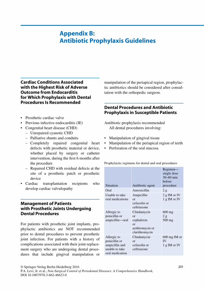
Dental Procedures and Antibiotic Prophylaxis in Susceptible Patients

Antibiotic prophylaxis recommended All dental procedures involving:

• Manipulation of gingival tissue • Manipulation of the periapical region of teeth • Perforation of the oral mucosa

Appendix B: Antibiotic Prophylaxis Guidelines

Prophylactic regimens for dental and oral procedures

Situation Antibiotic agent

Regimen—single dose 30–60 min before procedure

Oral Amoxicillin 2 g Unable to take oral medications

Ampicillin or cefazolin or ceftriaxone

2 g IM or IV 1 g IM or IV

Allergic to penicillin or ampicillin—oral

Clindamycin or cephalexin or azithromycin or clarithromycin

600 mg 2 g 500 mg

Allergic to penicillin or ampicillin and unable to take oral medication

Clindamycin or cefazolin or ceftriazone

600 mg IM or IV 1 g IM or IV

202

Reference

Prevention of infective endocarditis: Guidelines from the American Heart Association. A guide-line from the American Heart Association Rheumatic Fever, Endocarditis and Kawasaki Disease Committee, Council on Cardiovascular Disease in the Young, and the Council on Clinical Cardiology, Council on Cardiovascular Surgery and Anesthesia, and the Quality of Care and

Outcomes Research Interdisciplinary Working Group. JADA . 2008;139(1):3S–24S.

The use of prophylactic antibiotics prior to dental procedures in patients with prosthetic joints. Evidence-based clinical practice guide-lines for dental practitioners—a report of the American Dental Association Council on Scientifi c Affairs. Sollecito TP, Abt E, Lockhart PB, et al. JADA . 2015;146(1):11–6. e8.

Appendix B: Antibiotic Prophylaxis Guidelines

203© Springer-Verlag Berlin Heidelberg 2016 P.A. Levi, Jr. et al., Non-Surgical Control of Periodontal Diseases: A Comprehensive Handbook, DOI 10.1007/978-3-662-46623-0

Appendix C: Treating Tobacco Use: Pharmacological Therapy

Medication Cautions Side effects Dosage Use

Buproprion SR 150

Not for use if you: Are taking

monoamine oxidase inhibitor

Have had seizures in the past

Have or have had an eating disorder

Have had a stroke Have had a brain

tumor or brain surgery

Diffi culty sleeping Dry mouth Depressed mood Hostility Agitation

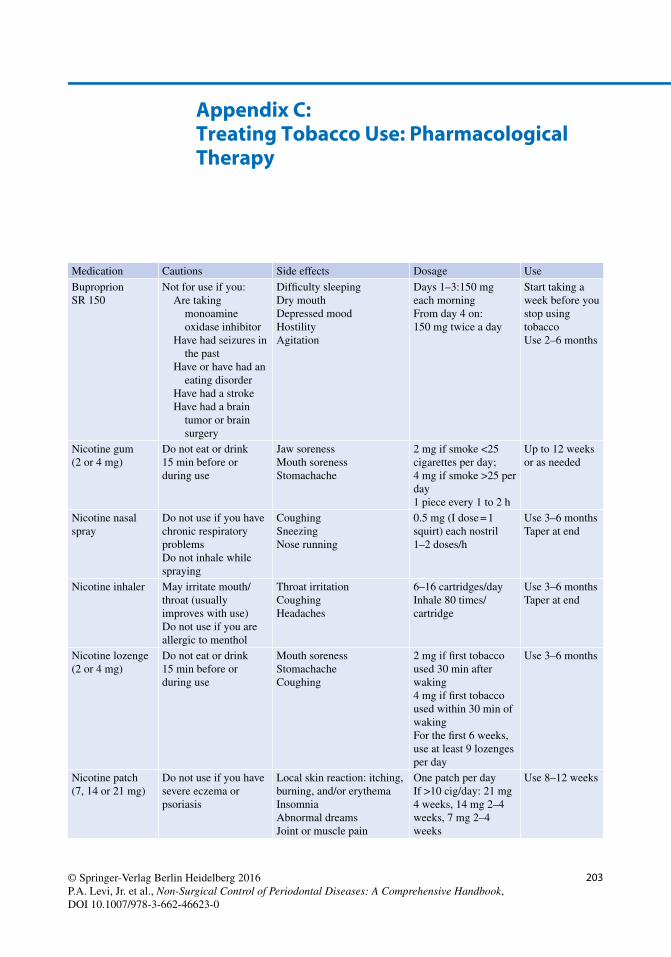
Days 1–3:150 mg each morning From day 4 on: 150 mg twice a day

Start taking a week before you stop using tobacco Use 2–6 months

Nicotine gum (2 or 4 mg)

Do not eat or drink 15 min before or during use

Jaw soreness Mouth soreness Stomachache

2 mg if smoke <25 cigarettes per day; 4 mg if smoke >25 per day 1 piece every 1 to 2 h

Up to 12 weeks or as needed

Nicotine nasal spray

Do not use if you have chronic respiratory problems Do not inhale while spraying

Coughing Sneezing Nose running

0.5 mg (I dose = 1 squirt) each nostril 1–2 doses/h

Use 3–6 months Taper at end

Nicotine inhaler May irritate mouth/throat (usually improves with use) Do not use if you are allergic to menthol

Throat irritation Coughing Headaches

6–16 cartridges/day Inhale 80 times/cartridge

Use 3–6 months Taper at end

Nicotine lozenge (2 or 4 mg)

Do not eat or drink 15 min before or during use

Mouth soreness Stomachache Coughing

2 mg if fi rst tobacco used 30 min after waking 4 mg if fi rst tobacco used within 30 min of waking For the fi rst 6 weeks, use at least 9 lozenges per day

Use 3–6 months

Nicotine patch (7, 14 or 21 mg)

Do not use if you have severe eczema or psoriasis

Local skin reaction: itching, burning, and/or erythema Insomnia Abnormal dreams Joint or muscle pain

One patch per day If >10 cig/day: 21 mg 4 weeks, 14 mg 2–4 weeks, 7 mg 2–4 weeks

Use 8–12 weeks

204

Medication Cautions Side effects Dosage Use

Varenicline (0.5 or 1 mg)

Use with caution in patients: With signifi cant

renal impairment With serious

psychiatric illness Undergoing dialysis Under 18

Nausea Insomnia Abnormal dreams Constipation/vomiting/gas May cause worsening of psychiatric illness Depression Agitation Suicidal thoughts (although rare)

Days 1–3: 0.5 mg every morning Days 4–7: 0.5 mg twice daily From day 8 on: 1 mg twice daily

Start 1 week before quit date; use 3–6 months

Appendix C: Treating Tobacco Use: Pharmacological Therapy

205© Springer-Verlag Berlin Heidelberg 2016 P.A. Levi, Jr. et al., Non-Surgical Control of Periodontal Diseases: A Comprehensive Handbook, DOI 10.1007/978-3-662-46623-0

Appendix D: Fagerstrom Test

207© Springer-Verlag Berlin Heidelberg 2016 P.A. Levi, Jr. et al., Non-Surgical Control of Periodontal Diseases: A Comprehensive Handbook, DOI 10.1007/978-3-662-46623-0

Now that your active treatment has been com-pleted by your doctor and hygienist, you are in periodontal health. To be able to maintain long- term benefi ts of healthy gum and bone tissues, you must continue to demonstrate effective plaque control habits at home as well as schedul-ing periodic appointments for professional care and supervision.

Your maintenance therapy appointments will be scheduled once every three months. During your visit, a doctor will do a thorough examina-tion to evaluate your progress. You will then see the dental hygienist who will help review and improve your plaque control techniques as well as remove accumulated bacterial plaque, calculus (tartar), and stain by scaling and polishing your teeth. Usually a combination of hand instruments

and power instruments are used during this pro-cess. There will be minimal discomfort during the procedure—the hygienist will endeavor to be gentle and will also apply topical anesthetic (numbing gel) to the gum tissues.

Every two to fi ve years, we will also check your progress (stability of bone height) with a complete mouth radiographic (x-ray) examination. A main-tenance therapy appointment card or e-mail reminder will be sent to you approximately one month before the previously scheduled appoint-ment time. Please contact our offi ce at least one week before your appointment to confi rm or adjust the exact time that is most convenient to you. A nominal fee will be charged for maintenance ther-apy services, and they can be discussed in greater detail beforehand with our offi ce manager.

Appendix E: Sample Patient Brochure Describing the Maintenance Therapy Program

209© Springer-Verlag Berlin Heidelberg 2016 P.A. Levi, Jr. et al., Non-Surgical Control of Periodontal Diseases: A Comprehensive Handbook, DOI 10.1007/978-3-662-46623-0

A Active/initial therapy . See Phase I therapy Acute ulcerative necrotizing gingivitis/periodontitis

(ANUG/NUP) , 4 Adjunctive therapy

DNA probe sampling , 171 laser therapy , 169–170 oral bacterial culturing , 170 periodontal endoscopes , 171 pharmacotherapeutics , 163–166 sensitivity tests , 170 smoking cessation , 166–169

Aggressive periodontitis , 13 Air abrasion , 125 AirFloss® , 66, 67 Alveolar bone , 3 Alveolar bone proper , 3 Amount of attached gingiva

periodontal charting , 90, 92 tissue evaluation , 81

Anatomy gingival and tooth anatomy

patient hygiene therapy , 9–10 periodontal diseases/deformation , 1–5 professional hygiene therapy , 5–8

tooth , 8 Area specifi c curettes , 117–121 .

See also Curettes Arestin , 164 Articaine (Septocaine®) , 152 Atridox , 164 Attached gingiva , 1, 2 . See also Amount of attached

gingiva inadequate , 29, 30 width of , 4, 6

Attachment apparatus , 4, 6 Automated periodontal probes , 113

B Bacterial species , 16 Bass technique , 51

Bleeding on probing (BOP) circumferential step probing technique , 40–41 gingival infl ammation , 40, 42 for motivation , 42–43 papillary bleeding index , 86–87 sulcular circumferential sweep method , 42 sulcus bleeding index , 87

Bulk fl uids , 16 Bundle bone , 3 Bupivacaine (Marcaine®) , 152 Butler Sunstar end-tuft brushes , 47, 48

C Calculus

composition , 43–44 detection , 45, 95–96, 141, 142 photomicrograph , 44 subgingival scaling and root planing , 44 types , 43

Cementum , 3 Children’s brushes , 47 Chisel scaler , 115, 116 Chlorhexidine gluconate solution , 67, 68 Chlorhexidine mouth rinse , 165 Circumferential step probing technique , 40–41 Climax community , 16 Closed gingival curettage . See also Curettes

contraindications , 143–144 description , 143

Coincidental curettage , 142–145 . See also Curettes Colgate Total® Advanced Pro-Shield™ , 68, 69 Combine push and pull stroke , 110 Connective tissue attachment , 2, 4 Cratered , 4 Crest Pro Health complete mouthwash , 68, 69 Curettes

area specifi c , 117–119 descripiton , 116 furcation , 120 sharpening , 125, 127–128 universal , 116–117, 125 vision , 119, 120

Index

210

D Defi nitive soft tissue curettage . See Closed gingival

curettage Dental assessment , 11, 12 Dental caries , 16 Dental implant design , 34 Dental implant-supported prostheses , 34, 35 Dental lasers , 169–170 Dental plaque , 1, 2, 16 Dental plaque/biofi lm removal

barriers/challenges , 26–27 bleeding on probing ( see Bleeding on probing (BOP)) calculus , 43–45 concordance , 23 defective restorative margins , 31 defective toothbrush , 33 dental disease control factors , 23 dental fl oss/tape , 57–59 dental implant design , 34 dental implant-supported prostheses , 34, 35 detection using disclosing agents

coaching brushing techniques , 39 digital intraoral camera usage , 40, 41 mouth mirror usage , 40 oral hygiene technique instruction , 39–40 stained plaque , 37–39 tablet and liquid forms , 37

disclosing solution , 25 exposed furcation , 26, 28 fl ossing technique , 59–60 gingival morphologic alterations , 29–30 goal , 21 improper plaque removal techniques , 32 ineffectual dental fl oss technique , 34 interproximal brushes , 60–61, 63 interproximal cleaning devices

AirFloss® , 66, 67 fl oss threaders , 62–63, 65 mouthwashes , 67–69 rubber tips , 62, 64–65 tongue cleaners , 64–65 toothpick-type devices , 63–64, 66 water irrigator/fl osser , 65–67

interproximal plaque removal , 57–59 malposed teeth , 30–31 manual toothbrushes

children’s brushes , 47 clinician’s determination , 46 designs and bristle confi gurations , 45, 46 end-tuft brushes , 47–48 and power brushes , 47 Right Kind® toothbrush , 46, 47 safety , 47 sulcus brushes , 48

maturation of dental biofi lm , 22 orthodontic appliances , 31 pocketing , 26, 27 power toothbrushes

advantage , 48 Oral-B® , 49, 50

Sonicare® , 48–49 Sunstar Butler , 49, 50 wear of , 49, 50

probing depth , 27–29 professional hygiene therapy , 26 provisional fi xed restorations , 31–32 stiff brush , 32–33 supra-and subgingival calculus , 29 supragingival calculus , 26, 27 tactile root sensitivity , 32 thoroughness and completeness , 25 tooth anatomic irregularities , 29 tooth brushing techniques , 49, 50

Bass technique , 51 with fi xed orthodontic appliances , 56–57 modifi ed Bass technique , 52 power brush , 56 stationary bristle technique , 52–55 Stillman’s technique , 55–56

tooth cleaning process , 24–25 Dentinal hypersensitivity

chemical agents , 160 etiology and treatment options , 160 physical agents , 160 treatment recommendations , 161

Diamond-coated fi le , 120 DNA probe sampling , 171

E E-cigarettes , 168 Electronic nicotine delivery systems (ENDS) , 168 ENDS . See Electronic nicotine delivery systems (ENDS) End-tuft brushes , 47–48 Epinephrine , 153 Epithelial attachment , 2, 4 Explorers

description , 113 functions , 114 No. 23 , 114 ODU 11–12 , 114 TU-17 , 114

Extraoral radiographs analysis and interpretation , 102–103 cephalometric projections , 101 cone-beam computed tomography , 101–102 panoramic radiograph , 100–102

F Facial pocket , 9, 10 Favorable prognosis , 18 File scaler , 116 Finger rest position, periodontal hand instrumentation

description , 107–108 extraoral fi nger rest , 108–109 intraoral fi nger rest , 108

Flat marginal gingival architecture , 4 Flossing technique , 59–62 Floss threaders , 62–63, 65

Index

211

FMX , 100 Fremitus , 93–94 Full-mouth series , 100 Furcation curettes , 122 . See also Curettes Furcations

attachment , 8 Glickman classifi cation system , 94 Hamp classifi cation system , 94–95 probing , 94

G Gel-Kam® , 161 Gingival anatomy

patient hygiene therapy , 9–10 periodontal diseases/deformation , 1–5 professional hygiene therapy , 5–8

Gingival biotypes , 5, 9 Gingival hyperplasia , 9 Gingival index , 98–99 Gingival tissue evaluation

amount of attached gingiva , 81 biotype , 81–82 color , 81 consistency , 81 gingival recession , 81 occlusal analysis , 82 shape , 81 size , 81 surface texture , 81

Gingivitis , 13, 14 Gingivitis/Periodontitis

periodontal instruments adjunctive hand instruments , 119–120 air/water syringe , 112 blade/working end , 111 curettes , 116–119 dental/mouth mirror , 111–112 explorers , 113–114 handles , 110 nomenclature , 110, 111 periodontal probe ( see Periodontal probes) polishing instruments , 123–125 power instruments , 120–123 scalers , 114–116 shank , 110–111 sharpening , 125–131

usage of periodontal instrumentation adaptation , 109 angulation , 109 fi nger rest position , 107–109 lateral pressure , 109 modifi ed pen grasp , 107 palm grasp , 107 pen grasp , 107 proper wrist position , 107, 108 strokes (activation of instrument) , 110 thumb and palm grasp , 107

Glickman classifi cation of furcation , 18, 94 Glide™ dental fl oss , 57

Gracey Curettes , 117–121 . See also Curettes GUM loop fl oss threader , 65 GUM plastic stiff fl oss threaders , 65 Gum stimulators . See Rubber tips

H Habitat , 16 Hamp classifi cation system , 94–95 Hand instrument sharpening , 128–129 Hoe scaler , 115 Hopeless prognosis , 18 Hygiene index , 180

I Implant design , 34 Implant maintenance

dental implant-supported prostheses , 181 home care, protocol for

dental fl oss , 186 with electric/power toothbrushes , 185 interproximal brush , 186, 187 with manual toothbrushes , 185 oral irrigator , 186 rubber tip , 186 super fl oss , 186

implant health evaluation healthy sign , 182 parameters , 181–182 peri-implantitis , 183 peri-implant mucositis , 182–183

instrumentation air polishing , 184, 185 hand instruments , 183–184 rubber cup with polishing paste , 185 titanium instruments , 184 ultrasonic scalers , 184

Improper scrubbing brushing technique , 32 Incomplete passive eruption , 9 Ineffectual dental fl oss technique , 34 Infl amed gingiva , 11 Injectable local anesthesia , 152, 153 Interproximal brushes

advantages , 60 disadvantages , 60–61, 63

Interproximal cleaning devices AirFloss® , 66, 67 fl oss threaders , 62–63, 65 mouthwashes , 67–69 rubber tips , 62, 64–65 tongue cleaners , 64–65 toothpick-type devices , 63–64, 66 water irrigator/fl osser , 65–67

Interproximal gingiva , 4 Intraoral examination , 11 Intraoral radiographs

analysis and interpretation , 102–103 complete mouth series , 100, 101 horizontal bitewing , 99

Index

212

Intraoral radiographs ( cont .) occlusal radiographs , 100 periapical radiograph , 100 vertical bitewing , 99–100

Intrasulcular technique, of power brushing , 57

L Laser curettage . See Sulcular debridement Laser pocket sterilization , 170 Laser root planing , 170 Laser therapy , 169–170 Lidocaine (Xylocaine®) , 152 Listerine® , 68 Local anesthesia, for scaling and root planing

anatomy , 151–152 epinephrine , 153 injectable , 152, 153 injection techniques , 154 mandible

buccal nerve block , 158, 159 inferior alveolar nerve block , 156–158 lingual nerve block , 158 mental nerve block , 158–159

maxilla anterior superior alveolar nerve block , 155 greater palatine nerve block , 156 nasopalatine nerve block , 156, 157 posterior superior alveolar nerve

block , 155–156 needle types , 153, 154 non-injectable topical anesthesia , 152, 153 periosteal injection , 154–155 types , 152, 154 use of , 151

M Maintenance therapy program

appointment/basic protocol , 180–181 attitude adjustment , 192 degree of attachment loss , 190–192 diagnostic goals , 187–188 evaluation guidelines , 179 gingival/periodontal pocket wall , 190–192 goals , 177–178 high-risk patients , 178 implant maintenance

dental implant-supported prostheses , 181 home care, protocol for , 185–187 implant health evaluation , 181–183 instrumentation , 183–185

importance , 192 objectives , 179 patient compliance , 193 periodontal therapy , 188–189 personal involvement , 189–190 prevention, of gingivitis/periodontitis , 178–179 psychological diffi culties , 189–190

Malposed teeth , 30–31 Manual toothbrushes

children’s brushes , 47 clinician’s determination , 46 designs and bristle confi gurations , 45, 46 end-tuft brushes , 47–48 and power brushes , 47 Right Kind® toothbrush , 46, 47 safety , 47 sulcus brushes , 48

Marginal gingiva , 1, 2 Marginal gingival architecture , 4 Marquis color-coded probe , 112 McGuire classifi cation system , 18 Mepivicaine (Carbocaine® ) , 152 Microbial biofi lm , 1 . See also Plaque Miller classifi cation, of recession , 90, 91 Miller index, for tooth mobility , 90, 93 Modifi ed Bass technique (MBT) , 52 Modifi ed plaque control record , 98 Motivational interviewing techniques , 169 Mouthwashes

chlorhexidine gluconate solution , 67, 68 Colgate Total® Advanced Pro-Shield™ , 68, 69 Crest Pro Health complete mouthwash , 68, 69 description , 67 Listerine® , 68

Mucogingival deformity , 87–90

N Nabers furcation probe , 112 Negative gingival architecture , 4 Nicotine replacement therapies (NRTs) , 168 No. 23 explorer , 114 Non-injectable topical anesthesia , 152, 153 Nonprescription-based oral rinses , 165 Non-pre-threaded fl oss holder , 59 Nonsurgical uses, of dental lasers , 170

O Occlusal analysis , 11–13 Occlusal trauma , 17 ODU 11–12 explorer , 114 Oral bacterial culturing test , 170 Oral-B Glide® , 57–59 Oral-B® power toothbrushes , 49, 50 Oral rinses , 165 Oraqix non-injectable anesthetic , 152, 153 Orthodontic tooth movement , 6

P Papillary bleeding index , 86–87 Patient examination and assessment

baseline vital signs , 77–78 daily oral hygiene habits , 79, 80 disinfected operatory , 71 extraoral examination , 79–80

Index

213

gingival tissue ( see Gingival tissue evaluation) initial patient interviews , 77 intraoral and extraoral photographs , 104 intraoral soft tissue examination , 80–81 medical/social history , 78, 79 operator visibility , 71 operatory setup , 71, 72 oral habits , 79 patient positioning , 72, 73 patient’s chief complaint , 77 patient’s expectations , 77 periodontal charting

amount of attached gingiva , 90, 92 bleeding on probing , 86–87 calculus detection , 95–96 fremitus , 93–94 furcation involvement , 94–95 gingival index , 98–99 mucogingival defects , 90, 93 plaque , 96–98 probing depth measurement , 82–86 recession measurement , 87–90 tooth mobility , 90, 93

periodontal documentation , 104 radiographs

angular bone loss , 103, 104 crestal lamina dura , 103 extraoral radiographs ( see Extraoral radiographs) furcation involvement , 104, 105 horizontal bone loss , 103, 104 intraoral radiographs ( see Intraoral radiographs) limitations , 103 during periodontal exam , 104 periodontal ligament space , 103–104

seated operator position , 72–75 seating the patient , 72, 73 semi-sitting position , 72 study casts , 104 supine position , 72 trendelenburg position , 72 upright position , 72 visibility

dental loupes , 76 dental magnifi cation , 75–76 head-mounted lighting , 74–76 magnifi cation lenses , 76–77 overhead lighting , 74, 76

Patient hygiene therapy, gingival and tooth anatomy , 9–10

PDI . See Periodontal disease index (PDI) Peridex , 165 Peri-implantitis , 16, 183 Peri-implant mucositis , 15–16, 182–183 Perioaid® plastic holder , 63, 66 PerioChip , 164 Periodontal charting

amount of attached gingiva , 90, 92 bleeding on probing

papillary bleeding index , 86–87 sulcus bleeding index , 87

calculus detection , 95–96 fremitus , 93–94 furcations

Glickman classifi cation system , 94 Hamp classifi cation system , 94–95 probing , 94

gingival index , 98–99 modifi ed plaque control record , 98 mucogingival defects , 90, 93 periodontal disease index , 98 plaque control record , 97 plaque index , 96–97 probing depth measurements

circumferential probing , 84–85 clinical attachment level , 82 interferences/obstruction , 83–84 interproximal probing , 85–86 pocket depth , 82 pressure , 84 probe insertion , 82, 83 probing, defi ned , 82 probing depth , 82 reading the probe , 86 sextants used , 82, 83 sulcus depth , 82

recession measurement consequences , 89–90 defi nition , 87 destructive periodontal disease , 88 hidden recession , 87 high frenal attachment , 89 histopathology , 89 infl ammation , 89 malpositioned teeth , 88 mechanical factors , 88 Miller classifi cation , 90, 91 mucogingival deformities , 88, 89 orthodontics , 88 restorative margins , 88 thin tissue biotype , 89 trauma , 88 vision recession , 87

tooth mobility causing factors , 93 Miller index , 90, 93

Periodontal disease index (PDI) , 98 Periodontal diseases/deformation

diagnosis of dental caries , 16 gingivitis , 13, 14 peri-implantitis , 16 peri-implant mucositis , 15–16 periodontitis , 14–15

etiology , 16–17 examination

integral parts of , 10–13 plaque removal techniques , 12–14

gingival anatomy , 1–5 prognosis , 17–19 risk factors for , 17

Index

214

Periodontal endoscopes , 171 Periodontal examination

integral parts of , 10–13 plaque removal techniques , 12–14

Periodontal health , 1 Periodontal ligament , 3 Periodontal probes

automated , 113 functions , 113 manual , 112 noninvasive applications , 113

Periodontal surgical services, indications for , 139–140 Periodontitis , 14–15 Perioscopy unit , 171 Periostat® , 164 Pharmacotherapeutics

local delivery antibiotic therapy , 164, 165 oral rinses , 165 subantimicrobial dose doxycycline , 164 systemic antibiotics

amoxicillin/metronidazole , 163–164 azithromycin , 164 ciprofl oxacin , 164 clindamycin , 164 doxycycline , 164 minocycline , 164 tetracyclines , 164

Phase I therapy , 133 facial histological appearance , 135 interdental histological appearance , 135, 136 purpose of , 134–135 reevaluation , 134, 136–139 six-step protocol , 138–139

Phase II therapy , 133, 139–140 Phase III therapy , 133, 140 Phase IV therapy , 133, 140 Phasing treatment plans, advantages of , 133–134 Pigtail explorer . See ODU 11–12 explorer Plaque

description , 1 identifi cation , 2

Plaque control record , 97 Plaque index , 96–97 Plaque removal techniques , 12–14 . See also Dental

plaque/biofi lm removal Plastic implant care instrument , 120 Polishing instruments

air powder polishing , 124–125 polishing pastes , 123, 124 rubber cup , 123, 124

Positive gingival architecture , 4 Powered sharpening techniques , 129–131 Power instruments

advantages , 122–123 disadvantages , 123 instrument tips , 122, 123 magnetostrictive ultrasonic instruments , 120, 121 piezoelectric instruments , 120–121 piezo tip angulation , 121, 122

sonic instruments , 120 ultrasonic instrument stroke , 121–122

Power toothbrushes advantage , 48 Oral-B® , 49, 50 Sonicare® , 48–49 Sunstar Butler , 49, 50 wear of , 49, 50

Pre-threaded fl oss holder , 59 Preventative dentistry, principles of , 178 Primary occlusal trauma , 17 Probing depth measurements

circumferential probing , 84–85 clinical attachment level , 82 interferences/obstruction , 83–84 interproximal probing , 85–86 pocket depth , 82 pressure , 84 probe insertion , 82, 83 probing, defi ned , 82 probing depth , 82 reading the probe , 86 sextants used , 82, 83 sulcus depth , 82

Professional hygiene therapy, gingival and tooth anatomy , 5–8

Pull stroke , 110 Push stroke , 110

Q Questionable prognosis , 18 Quorum sensing , 17

R Recession measurement

consequences , 89–90 defi nition , 87 destructive periodontal disease , 88 hidden recession , 87 high frenal attachment , 89 histopathology , 89 infl ammation , 89 malpositioned teeth , 88 mechanical factors , 88 Miller classifi cation , 90, 91 mucogingival deformities , 88, 89 orthodontics , 88 restorative margins , 88 thin tissue biotype , 89 trauma , 88 vision recession , 87

Right Kind® toothbrush , 46, 47 Root debridement , 6 Root planing procedure , 6, 8, 148–150

advantages , 140 basic clinical situations , 150–151 description , 141

Index

215

erbium lasers , 170 exploring subgingival topography , 142, 147 local anesthesia , 142 patient management protocol , 147–150 pertinent insurance codes in North America , 151

Rubber tips , 62, 64–65

S Salivary calculus . See Supragingival calculus Scalers

Chisel scaler , 115, 116 fi le scaler , 116 Hoe scaler , 115 sharpening techniques , 127–128 sickle scaler , 114–115

Scaling procedure , 8 advantages , 140 basic clinical situations , 150–151 calculus detection , 141, 142 description , 140 patient management protocol , 147–150 pertinent insurance codes in North America , 151 root surface assessment , 141, 145 shrinkage of infl amed papillae , 141, 146 supragingival vs. subgingival scaling , 141, 143

Scalloped gingiva , 4 Secondary occlusal trauma , 17 Second-hand smoke , 167 Sensitivity relief products , 161 Serumnal calculus . See Subgingival calculus Sharpening Cards® , 126 Sharpening instruments

advantages , 125 hand instrument sharpening , 128–129 lateral surfaces, of curettes and scalers , 125–126 objectives , 125 powered sharpening , 129–131 sharpening stones , 126–127 sharpness evaluation , 127

Shepherd’s hook explorer . See No. 23 explorer Sickle scaler , 114–115, 125 Single-rooted teeth , 5 Smoking cessation

counseling and referrals , 168 dental practitioner, role of , 167–168 E-cigarettes , 168 ENDS , 168 intervention steps , 168–169 motivational interviewing techniques , 169 pharmacological therapies , 168 stages of readiness , 169 tobacco

cancers , 167 chronic diseases , 166–167 dental implications , 167 for medicinal purposes , 166

Soft-Picks® , 64, 66 Sonicare® power toothbrushes , 48–49

Stationary bristle technique (SBT) , 52–55 Stillman’s technique (ST) , 55–56 Stimudents® , 63–64, 66 Subgingival calculus , 43–45 . See also Calculus Sulcabrush , 48 Sulcular circumferential sweep method , 42 Sulcular debridement , 170 Sulcus bleeding index , 87 Sunstar Butler power toothbrushes , 49, 50 Super Floss® , 57–58, 65 Supragingival calculus , 43, 45 . See also Calculus Supragingival plaque removal , 37

T Therapy , 197

closed gingival curettage contraindications , 143–144 description , 143

coincidental curettage , 142–143 dental prophylaxis , 140

basic clinical situations , 150–151 description , 144 patient management protocol , 144–147 pertinent insurance codes in

North America , 151 topical anesthetic , 148

dentinal hypersensitivity chemical agents , 160 etiology and treatment options , 160 physical agents , 160 treatment recommendations , 161

local anesthesia ( see Local anesthesia, for scaling and root planing)

phase I therapy ( see Phase I therapy) phase II therapy , 133, 139–140 phase III therapy , 133, 140 phase IV therapy , 133, 140 root planing procedure , 148–150

advantages , 140 basic clinical situations , 150–151 description , 141 exploring subgingival topography , 142, 147 local anesthesia , 142 patient management protocol , 147–150 pertinent insurance codes in

North America , 151 scaling procedure

advantages , 140 basic clinical situations , 150–151 calculus detection , 141, 142 description , 140 patient management protocol , 147–150 pertinent insurance codes in North America , 151 root surface assessment , 141, 145 shrinkage of infl amed papillae , 141, 146 supragingival vs. subgingival scaling , 141, 143

Tongue cleaners , 64–65 Tongue Sweeper® , 64–66

Index

216

Tooth anatomy , 8 patient hygiene therapy , 9–10 periodontal diseases/deformation , 1–5 professional hygiene therapy , 5–8

Tooth brushing techniques , 49, 50 Bass technique , 51 with fi xed orthodontic appliances , 56–57 modifi ed Bass technique , 52 power brush , 56 stationary bristle technique , 52–55 Stillman’s technique , 55–56

Tooth decay , 16 Tooth mobility

causing factors , 93 Miller index , 90, 93

Toothpick-type devices , 63–64, 66 Trauma from occlusion , 17 Traumatic occlusion , 17 TU-17 explorer , 114

U UNC 15 mm probe , 112 Unfavorable prognosis , 18 Universal curettes , 116–119 . See also Curettes

W Walking stroke , 110 Water irrigator/fl osser , 65–67 Williams probe , 112

Index


Recommended