115
CENTER FOR DRUG EVALUATION AND RESEARCH APPLICATION NUMBER: PHARMACOLOGY REVIEW(S) 21-560s000

CENTER FOR DRUG EVALUATION AND RESEARCH
APPLICATION NUMBER:
PHARMACOLOGY REVIEW(S)
21-560s000

1
DEPARTMENT OF HEALTH AND HUMAN SERVICES PUBLIC HEALTH SERVICE
FOOD AND DRUG ADMINISTRATION CENTER FOR DRUG EVALUATION AND RESEARCH
PHARMACOLOGY/TOXICOLOGY NDA REVIEW AND EVALUATION
Application number: 21-560
Applicant’s letter date: June 30, 2009
CDER stamp date: June 30, 2009
Product: Zortress® (everolimus, RAD001)
Indication: Prophylaxis of Organ Rejection in Renal
Transplantation
Applicant: Novartis Pharmaceuticals Corporation
Review Division: Division of Special Pathogen and Transplant
Products
Reviewer: William H. Taylor, PhD, DABT
Supervisor/Team Leader: William H. Taylor, PhD, DABT
Division Director: Renata Albrecht, MD
Project Manager: Jacquelyn E. Smith, M.A.
1 Executive Summary
1.1 Recommendations
No Pharmacology/Toxicology recommendations are currently indicated.
1.1.1 Approvability
The original Pharmacology/Toxicology reviewer for this application, Dr. Steven Kunder, indicated approval for this application in his 2003 review:
The New Drug Application 21-560 for Certican is approvable based on the preclinical pharmacology and toxicology submission. Toxicities demonstrated in preclinical studies may be monitored or be superseded by the benefit/risk ratio
2
determined by the clinical studies supporting kidney and heart transplant indications.
This reviewer agrees with Dr. Kunder’s assessment and conclusion.
1.1.3 Labeling
The purpose of this Pharmacology/Toxicology review is to present an updated label for everolimus (under the new trade name Zortress) for NDA 21-560. There are two principal differences between the Pharm/Tox portions of the label for Zortress, and the Pharm/Tox portions of the approved label for Afinitor®. The first difference is that the comparison of animal exposures to human exposures (for the transplant indication) is based on human AUC data from kidney transplant trials. The doses (and clinical exposures) for the oncology indication are considerably higher than those for the prophylaxis of kidney rejection. The consequence is that some animal toxicity seen only at higher doses in animals may be appropriate for inclusion in the oncology label, but not in the label for the transplant indication. Additionally, in the oncology Division, human doses are generally expressed in units of mg/m2, which are not appropriate, in this case, for this transplant indication. Secondly, the Afinitor label Pregnancy Category (Section 8.1) is a “D”, whereas the Zortress Pregnancy Category is a “C”. John K. Leighton, Ph.D., DABT, Associate Director for Oncology Pharmacology/Toxicology responded to an email request to clarify why the Afinitor label has a “D” Pregnancy Category. Dr. Leighton provided the following explanation in an email on November 17, 2009:
Because when ODAC [Oncology Drug Advisory Committee] reviewed this topic (pregnancy categories) they determined that the mechanism of action (investigational data) was likely relevant to humans, and thus merited D. We have discussed this numerous times with maternal health, and the clinicians in DDOP wanted to stick with what works (the D). There is positive evidence of human fetal risk based on adverse reaction data from investigational or marketing experience or studies in humans, but potential benefits may warrant use of the drug in pregnant women despite potential risks.
Specifically, there are no human data to support the “D” label. After discussing this issue with Abby Jacobs, Ph.D., Associate Director for Pharmacology/ Toxicology, Office of New Drugs, I selected the “C” Pregnancy Category for Zortress based on (1) the data from animal studies, (2) the lack of human pregnancy data supporting a “D” category, and (3) the “C” Pregnancy Category for the same-class drug, Rapamune® (sirolimus/ rapamycin).

3
The recommended Pharmacology/Toxicology sections for the Zortress label are as follows:
8 USE IN SPECIFIC POPULATIONS 8.1 Pregnancy Pregnancy Category C There are no adequate and well-controlled studies of Zortress in pregnant women. In rats and rabbits, everolimus crossed the placenta and was toxic to the conceptus. The potential risk for humans is unknown. Zortress should be given to pregnant women only if the potential benefit to the mother justifies the potential risk to the fetus. Women of childbearing potential should be advised to use effective contraception methods while they are receiving Zortress, and if
, up to 8 weeks after stopping treatment. Everolimus administered daily to pregnant rats by oral gavage at 0.1 mg/kg from before mating and through organogenesis resulted in increased preimplantation loss and early resorptions of fetal implants. AUCs in rats at this dose were approximately one-third those in humans administered the starting dose (0.75 mg b.i.d.). Everolimus administered daily by oral gavage at 0.8 mg/kg to pregnant rabbits during organogenesis resulted in increased late resorptions of fetal implants. At this dose, AUCs in rabbits were slightly less than AUCs in humans administered the starting clinical dose. 8.3 Nursing Mothers It is not known whether everolimus is excreted in human milk. Everolimus and/or its metabolites readily transferred into the milk of lactating rats at a concentration 3.5 times higher than in maternal serum. Because many drugs are excreted in human milk and because of the potential for serious adverse reactions in nursing infants from everolimus, women should avoid breast-feeding during treatment with everolimus. 13 NONCLINICAL TOXICOLOGY 13.1 Carcinogenesis, Mutagenesis, Impairment of Fertility Everolimus was not carcinogenic in mice or rats when administered daily by oral gavage for 2 years at doses of 0.9 mg/kg. In these studies, AUCs in mice were much higher (at least 20 times) than those in humans receiving 0.75 mg b.i.d., and AUCs in rats were in the same range as those in humans receiving 0.75 mg b.i.d. Everolimus was not mutagenic in the bacterial reverse mutation assay, the mouse lymphoma thymidine kinase assay, or the chromosome aberration assay usingV79 Chinese hamster cells, or in vivo following two daily doses of 500 mg/kg in the mouse micronucleus assay.
(b) (4)
4
In a 13-week male fertility oral gavage study in rats, testicular morphology was at 0.5 mg/kg and above, and sperm motility, sperm head count and plasma
testosterone concentrations were diminished at 5 mg/kg, which caused a decrease in male fertility. There was evidence of reversibility of these findings in animals examined after 13 weeks post-dosing. The 0.5 mg/kg dose in male rats resulted in AUCs in the range of clinical exposures, and the 5 mg/kg dose resulted in AUCs approximately 5 times the AUC in humans receiving 0.75 mg b.i.d. Everolimus did not affect female fertility in nonclinical studies, but everolimus crossed the placenta and was toxic to the conceptus. [see Pregnancy (8.1)]
2 Drug Information 2.1 Drug
2.1.2 Generic Name
Everolimus, RAD-001, RAD001
2.1.7 Pharmacologic class
Everolimus (RAD001) is a derivative of rapamycin (sirolimus) and is an mTOR (mammalian target of rapamycin) kinase inhibitor and an immunsuppressant.
2.2 Relevant IND/s, NDA/s, and DMF/s
IND 52,003, NDA (Afinitor)
2.3 Clinical Formulation
Zortress (everolimus) is available as 0.25 mg, 0.5 mg, 0.75 mg and 1 mg tablets.
2.3.1 Drug Formulation
Zortress Tablets include the following inactive ingredients: BHT, lactose monohydrate, hypromellose, magnesium stearate, crospovidone, and lactose anhydrous.
2.4 Proposed Clinical Population and Dosing Regimen
Adult patients at low-moderate immunologic risk receiving a kidney transplant
2.5 Regulatory Background
(b) (4)
(b) (4)
5
On December 19, 2002, Novartis Pharmaceuticals Corporation initially filed NDA 21-560 with the FDA for the prophylaxis of organ rejection in allogenic kidney and heart transplant patients. The Office of Drug Evaluation IV issued Approvable letters to Novartis for NDA 21-560 on October 20, 2003 and August 27, 2004. On June 30, 2009 Novartis filed amendments to NDA 21-560 which included new clinical studies to restart the agency’s review. In the June 30, 2009 submission, Novartis amended the product label indication to eliminate heart transplant recipients. During the current review cycle, the CDER Division of Medication Error Prevention and Analysis (DMEPA)
approved the trade name “Zortress.” Everolimus is approved in Europe as Certican for use as an immunosuppressant to prevent rejection of solid organ transplants. On March 30, 2009, FDA approved everolimus as Afinitor under NDA 22-334 for the treatment of patients with advanced renal cell carcinoma after failure of treatment with sunitinub or sorafenib.
3 Studies Submitted 3.1 Studies Reviewed
Steven C. Kunder, Ph.D. completed an initial Pharmacology/Toxicology NDA 21-560 review of everolimus (see DARRTS October 21, 2003). Shwu-Luan Lee, Ph.D. completed a Pharmacology/Toxicology review for NDA 22-334 in the Division of Oncology Products for the Afinitor product (DARRTS, March 12, 2009). Neither the applicant’s original study reports nor literature were examined for this review. The source materials for this review are the two previous Pharmacology/ Toxicology reviews in DARRTS for everolimus, the approved label for Afinitor, and the applicant’s June 30, 2009 proposed label for Zortress. William H. Taylor, PhD, DABT Pharmacology/Toxicology Reviewer
(b) (4)
ApplicationType/Number
SubmissionType/Number Submitter Name Product Name
-------------------- -------------------- -------------------- ------------------------------------------NDA-21560 ORIG-1 NOVARTIS
PHARMACEUTICALS CORP
CERTICAN (EVEROLIMUS)TABLETS
---------------------------------------------------------------------------------------------------------This is a representation of an electronic record that was signedelectronically and this page is the manifestation of the electronicsignature.---------------------------------------------------------------------------------------------------------/s/----------------------------------------------------
WILLIAM H Taylor12/18/2009
RENATA ALBRECHT12/22/2009

Note:
This will be the Standard CDER Coversheet

TABLE OF CONTENTS
EXECUTIVE SUMMARY............................................................................................................................................. 3
PHARMACOLOGY/TOXICOLOGY REVIEW......................................................................................................... 5
3.1 INTRODUCTION AND DRUG HISTORY ................................................................................................................. 5
3.2 PHARMACOLOGY....................................................................................................................................................... 8 3.2.1 Brief summary ........................................................................................................................................ 8 3.2.2 Primary pharmacodynamics ................................................................................................................... 8 3.2.3 Secondary pharmacodynamics ............................................................................................................... 8 3.2.4 Safety pharmacology .............................................................................................................................. 8 3.2.5 Pharmacodynamic drug interactions....................................................................................................... 9
3.3 PHARMACOKINETICS/TOXICOKINETICS........................................................................................................... 9 3.3.1 Brief summary ........................................................................................................................................ 9 3.3.3 Absorption .............................................................................................................................................. 9 3.3.4 Distribution........................................................................................................................................... 11 3.3.5 Metabolism........................................................................................................................................... 16 3.3.6 Excretion .............................................................................................................................................. 17 3.3.7 Pharmacokinetic drug interactions........................................................................................................ 18 3.3.10 Tables and figures to include comparative TK summary ..................................................................... 18
3.4 TOXICOLOGY............................................................................................................................................................. 21 3.4.1 Overall toxicology summary ................................................................................................................ 21 3.4.2 Single-dose toxicity .................................................................................................................................. 3.4.3 Repeat-dose toxicity ............................................................................................................................. 26 3.4.4. Genetic toxicology................................................................................................................................ 63 3.4.5. Carcinogenicity..................................................................................................................................... 68 3.4.6. Reproductive and developmental toxicology........................................................................................ 86 3.4.7 Local tolerance ......................................................................................................................................... 3.4.8 Special toxicology studies .................................................................................................................... 97
3.6 OVERALL CONCLUSIONS AND RECOMMENDATIONS................................................................................ 107
3.7. APPENDIX/ATTACHMENTS........................................................................................................................................
NDA 21-560 page 3
EXECUTIVE SUMMARY 1. Recommendations 1.1 Recommendation on Approvability The New Drug Application 21-560 for Certican is approvable based on the preclinical pharmacology and toxicology submission. Toxicities demonstrated in preclinical studies may be monitored or be superceded by the benefit/risk ratio determined by the clinical studies supporting kidney and heart transplant indications. 1.2 Recommendation for nonclinical studies It is recommended that possible impurities arising from the manufacturer or degradation of everolimus be qualified according to ICH guidelines. It is also recommended that the sponsor elucidate the mechanism of toxicity to the female reproductive organs, attempting to determine hormonal changes caused by everolimus. 1.3 Recommendations on labeling
(b) (4)
(b) (4)
(b) (4)

NDA 21-560 page 4
2. Summary of nonclinical findings 2.1 Brief overview of nonclinical findings Everolimus was studied in mice, rats, minipigs and cynomolgus monkeys to evaluate its toxicities and in rats and rabbits to determine its reproductive toxicity potential. Everolimus causes toxicities in animal studies both by its pharmacologic mechanism of action, immunosuppression, as well as by direct toxicity. Immunosuppression by everolimus resulted in atrophy of the lymphoid organs (thymus, spleen, lymph nodes) as well decreased circulating lymphocytes and total leukocytes. Toxicities affected by immunosuppression included myocardial degeneration/myocarditis in monkeys and rats at $1.5 mg/kg ($1.7-4.5x human exposure); this is likely related to viral infection emerging under immunosuppression. Other toxicities included reproductive organ toxicity in all species tested including testicular atrophy in monkeys at 0.3 mg/kg (0.9x human exposure) and uterine atrophy and reduced follicular development in mice and monkeys at doses of $1.5 mg/kg ( 4.5-18x human exposure); renal toxicity in rats with tubular degeneration at doses of $5.0 mg/kg (55x human exposure); pancreatic toxicity was seen in monkeys with islet cell degeneration at 5.0 mg/kg (4.5x human exposure) and vacuolation of the exocrine pancreas in minipigs at 5.0 mg/kg (23x human exposure); lung toxicity in mice at $1.5 mg/kg (15x human exposure) and rats at $0.5 mg/kg (0.1x human exposure); and toxicity to the eye as swelling and disruption of cortical fibers of the lens at a dose of 0.9 mg/kg in rats (0.4 x human exposure). In carcinogenicity studies, no significant tumor findings occurred in rats (0.4x human exposure) and one tumor, osteoma of the femur was significant in mice at 0.9 mg/kg (8.6x human exposure). Genotoxicity for RAD was not observed in the in vitro bacterial reversion (Ames), in vitro mammalian mouse lymphoma assay, in vitro chromosomal aberration test or in vivo mammalian micronucleus assay. Reproductive toxicity was seen in rat and rabbit studies. Male fertility was decreased in rats at 5.0 mg/kg (0.9x human exposure); sperm were misshapen at 0.5 mg/kg (0.12x human exposure) without effect on fertility. In rat embryo-fetal development studies, skeletal malformations increased at doses $0.3 mg/kg (0.09x human exposure). In rabbit embryo-fetal development studies, no fetal effects were seen until doses were maternally toxic (0.8 mg/kg, 0.14x human exposure). 2.2 Pharmacologic activity Everolimus is an orally active immunosuppressant drug acting as an inhibitor of intracellular proliferative signaling in activated T lymphocytes at FK binding protein 12 (FKB-12). The complex of RAD/FKB-12 is then believed to bind to and inhibit the kinase mTOR. This arrests activated T lymphocytes in the G1 phase of their cell cycle, preventing their proliferation in response to the response to the foreign antigens of transplanted tissues. Inhibition of mTOR blocks the postreceptor IL-2 signals which mediate T-call proliferation while calcineurin inhibitors (eg. Cyclosporin, Tacrolimus) block IL-2 synthesis and stop the cell cycle as it progresses from G0 to G1 phase.
Nonclinical safety issues relevant to clinical use -Renal toxicity is of prime importance, especially for renal transplantation. It is well characterized for calcineurin inhibitors. -Pancreatic toxicity, also well characterized for calcineurin inhibitors, potentially leading to post-transplantation diabetes mellitus. -Reproductive toxicity/male fertility, counterindicates Certican for pregnant women; however, organ transplantation is typically not conducted in pregnant women. Decreased male fertility may prevent males from impregnating partners after transplantation. -Eye toxicity seen in rats (disruption of fibers in lens) may cause vision problems. -Hypercholesterolemia and hypertriglyceridemia, seen in rats and monkeys, and typical of other immunosuppressant drugs used for organ transplantation, may be treated with current antihyperlipodemic therapies following transplantation.
NDA 21-560 page 5
PHARMACOLOGY/TOXICOLOGY REVIEW
3.1 INTRODUCTION AND DRUG HISTORY NDA number: 21-560 Review number: 001 Sequence number/date/type of submission: 001 Information to sponsor: Yes ( ) No (x ) Sponsor and/or agent: Novartis Pharmaceutical Corporation East Hanover, NJ Manufacturer for drug substance: Novartis Pharmaceutical Corporation Reviewer name: S.Kunder Division name: Special Pathogen and Immunologic Drug Products HFD #: 590 Review completion date: 10 Oct 2003 Drug: Trade name: Certican Generic name: everolimus Code name: RAD, SDZ RAD, RAD 666, RAD 001
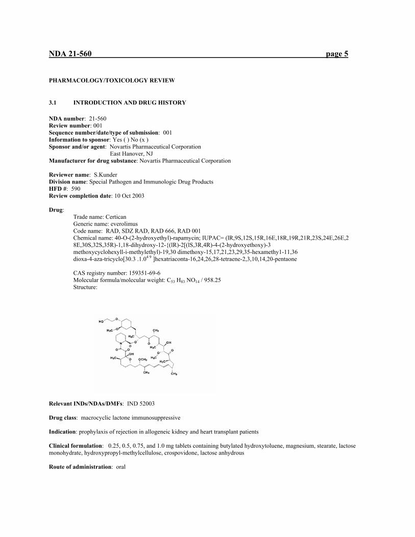
Chemical name: 40-O-(2-hydroxyethyl)-rapamycin; IUPAC= (lR,9S,12S,15R,16E,18R,19R,21R,23S,24E,26E,2 8E,30S,32S,35R)-1,18-dihydroxy-12-{(lR)-2[(lS,3R,4R)-4-(2-hydroxyethoxy)-3 methoxycyclohexyll-i-methylethyl)-19,30 dimethoxy-15,17,21,23,29,35-hexamethy1-11,36 dioxa-4-aza-tricyclo[30.3 .1.04 9 ]hexatriaconta-16,24,26,28-tetraene-2,3,10,14,20-pentaone
CAS registry number: 159351-69-6 Molecular formula/molecular weight: C53 H83 NO14 / 958.25 Structure:
Relevant INDs/NDAs/DMFs: IND 52003 Drug class: macrocyclic lactone immunosuppressive Indication: prophylaxis of rejection in allogeneic kidney and heart transplant patients Clinical formulation: 0.25, 0.5, 0.75, and 1.0 mg tablets containing butylated hydroxytoluene, magnesium, stearate, lactose monohydrate, hydroxypropyl-methylcellulose, crospovidone, lactose anhydrous Route of administration: oral

NDA 21-560 page 6
Proposed use: kidney and heart transplantation Disclaimer: Tabular and graphical information are constructed by the reviewer unless cited otherwise. In results where no sex designation is specified, the finding is applied to both sexes. For study results, increases and decreases are with respect to controls
INTRODUCTION Certican is an immunosuppressant macrolide, structurally related to rapamycin, under development for organ transplantation. Certican is derived from chemical modification of rapamycin, a product of Streptomyces hygroscopicus. Certican appears to share the same mechanism of action with rapamycin but its immunosuppressive biochemical mechanism of action is distinct from that of cyclosporin (CsA), tacrolimus (FK506), mycophenolate mofetil, or azathioprine. Certican inhibits T lymphocyte activation and proliferation that occurs in response to antigenic and cytokine (interleukin [IL]-2, IL-4, IL-7, and IL-15) stimulation by a mechanism different from that of immunosuppressants previous to rapamycin. Certican binds to the immunophilin, FK binding protein 12 (FKBP-12), producing an immunosuppressive complex. Unlike cyclosporin and tacrolimus, the rapamycin FKBP complex appears to have no effect on calcineurin activity. This complex binds to and inhibits the activation of a kinase called the mammalian target of rapamycin (mTOR). Inhibition of mTOR by rapamycin suppresses cytokine-driven T-cell proliferation, inhibiting the progression from the G1 to the S phase of the cell cycle. Certican prolongs allograft survival in animal models of transplantation, including rodents and primates, both for solid organ and for cellular allografts. Nephrotoxicity is a concern with many preceding immunosuppressants, particularly cyclosporin. Certican, possibly due to its differing mechanism of action, appears not to have the severity of nephrotoxicity seen with cyclosporin. Combinations of Certican with other immunosuppressants are problematic. Other toxicities and affects seen in other immunosuppressive drugs including elevation of glucose and hyperlipidemia, are of concern with Certican as well. Due to the similarity of chemical structure and toxicologic issues, the reader may wish to compare the Pharmacology/Toxicology review of NDA 21-083 for rapamycin. Studies reviewed within this submission: Pharmacology studies are summarized in pharmacology section Pharmacokinetics and excretion after single intravenous and peroral administration (0.9 mg/kg) of H3-labeled RAD001 to mice Pharmacokinetics in mice after intravenous bolus administration (0.9 mg/kg) with RAD001 Quantitative determination of rapamycin and SDZ RAD in blood samples after single and multiple administration in human and monkey In vitro distribution, plasma protein binding and stability of RAD001 in mouse plasma Stability in mouse, monkey and human plasma. Addendum to the study: In vitro distribution, plasma protein binding and stability of RAD001 in mouse plasma (above) In vitro blood distribution and plasma protein binding of RAD001 in rat plasma. Addendum to the study: In vitro distribution, plasma protein binding and stability of RAD001 in mouse plasma (above) Distribution and excretion of total radioactivity in rats after peroral administration of 1.5 mg/kg 14C-labelled SDZ RAD Whole-body autoradioluminography in albino and pigmented rats after po and iv doses of [3H] RAD001 Embryofetal transfer in pregnant rats on day 13 and day 17 of gestation after po administration of [3H] RAD001 Galactogenic transfer, kinetics and metabolism in milk and blood after single peroral administration (0.9 mg/kg) of 3H-labeled RAD001 to lactating rats Dose-dependent brain penetration in rats Disposition in rats after single and repeated once daily peroral administration (0.5 mg/kg/day) of 3H-labeled RDA001 for 21 consecutive days Intestinal absorption and presystemic metabolism of SDZ RAD Permeability study across Caco-2 cell monolayers Biotransformation in mice following a single oral and intravenous dose (0.9 mg/kg) of 3H-RAD Biotransformation in cynomolgus monkey following a single oral dose of 3H-RAD Inhibition of RAD 001 in vitro metabolism by ketoconazole, itraconazole and fluconazole An oral dose-escalating study in dogs A comparative 2-week oral (gavage) toxicity study in the rat with a micro-emulsion and a solid dispersion A 2-week oral (gavage) dose-range-finding study in minipigs 4-week oral (gavage) toxicity study in minipigs 28-day oral gavage toxicity study in juvenile cynomolgus monkeys with a 2-week reversibility period SDZ RAD: Toxicity study by oral gavage administration to cynomologus monkeys for 26 weeks followed by a four week reversibility period
NDA 21-560 page 7
52-week oral (gavage) toxicity study in the cynomolgus monkey Toxicity study by intravenous (bolus) administration to HanIbm Wistar rats for 2 weeks Two-week intravenous infusion toxicity study in minipigs 4-week toxicity study in cynomolgus monkeys by intravenous infusion followed by a 2-week reversibility period 4-week toxicity study in cynomolgus monkeys by intravenous infusion followed by a 2-week reversibility period RAD 001, rapamycin and Neoral: 2-week exploratory study in rats FTY720 in combination with RAD001: 4-week oral (gavage) toxicity in male monkeys with a 4-week recovery period RAD001 and methotrexate: 4-week oral combination toxicity study in rats Comparative toxicity study in HanIbm Wistar rats with batches differing in by-product content A comparative 2-week oral (gavage) toxicity study in the rat with two different batches 4-week oral toxicity study in rats (batch comparison) An oral 13-week investigative fertility study in male rats with 13 weeks recovery An oral fertility and embryo-fetal development study in female rats An oral embryo-fetal development study in rabbits An oral pre- and postnatal development study in rats SDZ RAD 666: mutagenicity test using Salmonella typhimurium (batch control) SDZ RAD 666: mutagenicity test using Salmonella typhimurium (batch control) SDZ RAD (solid dispersion): mutagenicity test using Salmonella typhimurium (batch control) RAD 001: mutagenicity test using Salmonella typhimurium (batch control) SDZ RAD (solid dispersion) Chromosomal aberration test with V79 Chinese hamster cells RAD001:Chromosomal aberration test with V79 Chinese hamster cells Oncogenicity study by oral gavage administration to CD-1 mice for 104 weeks Oncogenicity study by oral gavage administration to Hanibm rats for 104 weeks Comparative study of ophthalmic toxicity by oral gavage administration to CD rats and HanIbm rats for 4 weeks A 2-week oral (gavage) mechanistic toxicity study in rats An oral neonatal and juvenile developmental dose range-finding study in rats An oral neonatal and juvenile development study in rats with 13- and 26-week recovery periods Studies reviewed in IND 52003: Acute oral toxicity study in mice. Acute intravenous toxicity study in mice. Acute intravenous toxicity study in mice (amendment No. 1 to final report) Acute oral toxicity study in rats. Acute intravenous toxicity study in rats. Acute intravenous toxicity study in rats (amendment No. 1 to final report). Dose range finding study (intravenous infusion administration) in rats. A 2-week oral (gavage) dose-range-finding study in rats. Comparative intravenous infusion study in rats. Toxicity study by oral gavage administration to HanIbm Wistar rats for 4 weeks followed by a 2 week reversibility period. A repeat toxicity study by oral gavage administration to HanIbm Wistar rats for 4 weeks followed by a 2 week reversibility period. Dose escalating study (oral administration) in cynomolgus monkeys. 14-Day dose range finding study (oral route) in cynomolgus monkeys. Dose finding study in cynomolgus monkeys by intravenous infusion for 2 weeks. 14-Day dose range finding study (intravenous infusion administration) in cynomolgus monkeys. Supplement No. 1: Toxicokinetic report. Toxicity study by oral (gavage) administration to cynomolgus monkeys for 4 weeks followed by a 2 week reversibility period. An oral fertility dose-range-finding study in male rats An oral reproductive toxicity dose-range-finding study in female rats with toxicokinetics and placental transfer. An oral embryo-fetal development dose-range-finding study in rabbits with toxicokinetics and placental transfer. Mutagenicity test using Salmonella typhimurium Range-finding toxicity study by oral gavage administration to CD-1 mice for 13 weeks Toxicity study by oral gavage administration to Hanlbm Wistar rats for 4 weeks followed by a 2 week reversibility period Toxicity study by oral gavage administration to Hanlbm Wistar rats for 26 weeks followed by a 4 week reversibility period Combination of Sandimmun-Neoral and SDZ RAD 4-week oral (gavage) toxicity study in Cynomolgus monkeys
NDA 21-560 page 8
3.2 PHARMACOLOGY 3.2.1 Brief summary Everolimus had minimal effects in neurological, cardiovascular, pulmonary, renal, and gastrointestinal assays. The primary mechanism of action, immunosuppression, is described below. 3.2.2 Primary pharmacodynamics Mechanism of action: The main immunosuppressive action of RAD is an inhibition of an intracellular proliferative signal in activated T lymphocytes at FK Binding protein 12 (FKB-12). Growth-factor stimulated phosphorylation of p70 S6 kinase is then inhibited. This kinase is involved in the initiation of protein synthesis. Phosphorylation of p70 S6 kinase is believed to be controlled by FKB-12-rapamycin associated protein (mTOR). The complex of RAD/FKB-12 is then believed to bind to and inhibit mTOR, a regulatory protein controlling cell metabolism, growth and proliferation. This arrests activated T lymphocytes in the G1 phase of their cell cycle, preventing their proliferation in response to the foreign antigens of transplanted tissues. Clonal expansion of antigen specific T cells in response to foreign antigen and IL-2 stimulation is prevented. For further detail refer to the microbiology review. Drug activity related to proposed indication: As described above, RAD inhibits the T cell response to the antigens of allogeneic tissue involved in organ transplantation thereby preventing immunologic rejection. 3.2.3 Secondary pharmacodynamics The antiproliferative action of everolimus inhibits growth factor-stimulated proliferation of vascular smooth muscle cells. In a human umbilical vein, RAD inhibited proliferation stimulated by vascular endothelial growth factor. In the rat carotid artery balloon model, rats subjected to carotid artery angioplasty were treated with RAD and injury –induced neointima formation was inhibited. Similar results were seen in a pig model with a 14-day RAD treatment following coronary balloon injury. In rat aorta transplantation, everolimus inhibits neointimal formation due to damaged endothelial cells. The likely application for these antiproliferative studies is in the drug-coated stent. In rabbits, RAD-coated stents were examined at 28 days after implantation, demonstrating absence of restenosis. 3.2.4 Safety pharmacology
(b) (4)
NDA 21-560 page 9
Neurological effects: The Irwin primary observation test for effects on motor activity, locomotion, behavioral stimulation, behavioral depression, muscle tone, neurologic activity, autonomic activity, pupilar diameter, rectal temperature and lethargy was performed with oral doses of 2, 20 and 50 mg/kg. Motor activity was increased at all doses within 5 min of dosing. Rats receiving 20 mg/kg showed an increase in flight reaction. Pupil diameter was increased at 50 mg/kg for at least 23 h after dosing. Food consumption in the 50 mg/kg group also decreased. RAD appears to exert slight CNS effects at doses of $20 mg/kg. Cardiovascular effects: In an i.v. study in anesthetized pigs, doses of 0.01, 0.1, 1.0 and 10 mg/kg were used to determine the effects of RAD upon blood pressure, heart rate, blood flow, respiratory rate and electrocardiogram. No cardiovascular effects were seen at doses #10 mg/kg i.v. in pigs. In the in vitro assay on HERG currents recorded from stably transfected HEK293 cells, RAD did not inhibit HERG currents. Pulmonary effects: Pulmonary effects of RAD were examined in anesthetized, ventilated guinea pig model at doses of 0.3, 3 and 30 mg/kg i.v. to determine effects on airway resistance and dynamic compliance. No effects were seen at these doses. There was a reduction in airway reactivity to histamine at 3 and 30 mg/kg, indicating some antihistaminic activity. Renal effects: In saline-treated mice, at RAD doses of 15 and 50 mg/kg p.o., slight increases in total excretion of urinary potassium and chloride were seen. Gastrointestinal effects: The effect of RAD on intestinal transit time with charcoal was studied in mice at doses up to 50 mg/kg, p.o. No effect on gastrointestinal transit time was seen. Abuse liability: none Other: none 3.2.5 Pharmacodynamic drug interactions The major enzyme of human metabolism of RAD is believed to be CYP3A4. Using CYP3A4 inhibitors ketoconazole and itraconazole as well as a poor inhibitor (fluconazole), the inhibitors were shown to inhibit biotransformation of RAD while fluconazole did not. This supports the role of CYP3A4 in the metabolism of RAD.
3.3 PHARMACOKINETICS/TOXICOKINETICS 3.3.1 Brief summary
Preclinical pharmacokinetics were studied in mice, rats and cynomolgus monkeys following oral and intravenous administration. Oral absorption of single dose radiolabeled RAD was determined in the mouse (12%), monkey (18%), rat (39-43%) and humans (11%). Oral bioavailability was determined in the mouse (5%), rat (14-26%), and monkey (6%). Plasma protein binding was similar among the monkey (16% free), the rat (7.6% free), and humans (25% free), while in the mouse extensive protein binding occurred (0.1% free). Tissue distribution was determined by radiolabel studies in the rat in which tissue levels following i.v. dosing were highest in the liver and kidney after 5 minutes. Half-lives of label in most tissues were between 1.4 and 1.9 days, excepting brain (10 days), testes (13 days) and epididymides (5 days). Following oral dosing, the greatest amounts of label were found in the heart, liver, lung, kidney, spleen, thyroid and adrenal glands after 2 hours. The metabolism of RAD is also the major mechanism of elimination. Parent drug is the predominant form found in blood, with parent compound averaging in mice, rats and humans between 31 and 63% of total radioactivity, and 12% in monkeys. Five major metabolites are found in vivo in human, monkey, rat and mouse. These metabolites are typically the result of conjugation with fatty acids and hydrolytic and hydroxylated products. The related compound rapamycin may arise as a metabolite but was found in small amounts (~5%) of total AUC in clinical pharmacokinetic studies. Hydrolytic and hydroxylated metabolites were studied in vitro for immunosuppressive activity and were approximately 60- to 500 fold less active than the parent, RAD. The majority of radiolabeled RAD was eliminated in the feces in mouse (95-99%), rat (68-89%), monkey (66-75%), and humans (79%). Urinary excretion was consistently low (<7%). Bile duct cannulated rats demonstrated approximately 71% biliary excretion. See summary tables below. 3.3.3 Absorption Study title: Pharmacokinetics and excretion after single intravenous and peroral administration (0.9 mg/kg) of H3-labeled RAD001 to mice

NDA 21-560 page 10
Study no. DMPK(CH) R98-707 Study facility: Novartis Pharma AG Preclinical Safety/Drug Metabolism
ADE Section 4002 Basel Switzerland
Date of study: Nov, 1998-Jan 1999 GLP (no) Dose & formulation: [H3]- RAD001, specific activity=49.8 MBq/mg Animals: male CD-1 mice Protocol: Mice were administered H3-labeled RAD001 by intravenous route (2 mg) and oral gavage (1.0 mg). Blood was collected at 0.083, 0.5, 1, 3, 8, 24 and 72 h post dosing. Urine and feces were collected daily up to 72 h post dosing. Blood, fecal and urine levels were analyzed by liquid scintillation counting. RESULTS: The pharmacokinetic values for this study : Dose (mg/kg) route H3 tmax H3 Cmax H3 AUC (:mol.L) 0.9 i.v. 1 3.98 38.3 0.9 p.o. 0.5 0.51 4.39 CONCLSIONS With either i.v. or p.o. dosing, radioactivity was excreted rapidly and nearly completely within 48 h (>96%). Excretion was nearly all in feces with p.o. or i.v. administration (>95%). Study title: Pharmacokinetics in mice after intravenous bolus administration (0.9 mg/kg) with RAD001 Study no. DMPK(CH) R00-874 Study facility: Novartis Pharma AG Preclinical Safety/Drug Metabolism
ADE Section 4002 Basel Switzerland
Date of study: 25-28 Jan 2000 GLP (no) Dose & formulation: placebo for Sandimmune/saline Animals: mice, CD-1, males Protocol: Mice were administered a single i.v. injection (0.9 mg/kg) of RAD 001. Blood samples were collected at 0.083, 0.25, 0.5, 1, 2, 4, 6, 8, 24, 48 and 72 h after dosing. Blood samples were analyzed by LC-APCI-MS. RESULTS: AUC= 18.7 h.:/ml; t1/2 =9.83 h; clearance= 0.67 l/kg CONCLUSIONS: Blood levels of RAD in this i.v. study were maintained near injection levels for 8 h, then declined steadily from 8 to 72 h. Study title: Quantitative determination of rapamycin and SDZ RAD in blood samples after single and multiple administration in human and monkey Study no. DMPK(CH) 1997/287 Study facility: Novartis Pharma AG Preclinical Safety/Drug Metabolism
ADE Section 4002 Basel Switzerland Date of study: Aug 4-5 1997 GLP (no)
NDA 21-560 page 11
Dose & formulation: samples used in this study were obtained from patients receiving a single dose of RAD (25 mg ), multiple doses of 0.75 mg in stable renal transplant patients and cynomolgus monkeys receiving 4-week multiple dose 0.5 mg/kg/day treatment. Animals: see above Protocol: existing blood samples (patient and monkey) had rapamycin and RAD concentrations determined by HPLC RESULTS: Human blood samples: 25 mg single dose, rapamycin/RAD =3.0-5.4% 0.75 mg multi-dose, rapamycin/RAD=3.8-5.2% Cynomolgus monkey: 0.5 mg/kg/day, 4-weeks, rapamycin/RAD=7.8-10.7% CONCLUSIONS: Rapamycin was found in blood samples of patients and cynomolgus monkeys receiving RAD in amounts ranging from 3.0-5.2% in patients to 7.8-10.7% in monkeys. 3.3.4 Distribution Study title: In vitro distribution, plasma protein binding and stability of RAD001 in mouse plasma. Study no. DMPK (CH) R00-1253 Study facility: Novartis Pharma AG Preclinical Safety/Drug Metabolism
ADE Section 4002 Basel Switzerland Date of study: 15-26 May 2000 GLP: (no) Dose & formulation: H3-RAD001 batch 98902, 25.3 MBq/ml Animals: pooled blood from CD-1 mice Protocol: Plasma protein binding was determined using mouse blood erythrocytes by the erythrocyte partitioning method. Plasma dilutions from 0.1 to 60 % were used with H3-RAD001/RAD001 at a concentration range of 5-5000 ng/ml. RESULTS: blood distribution in plasma: plasma fraction = 98"4% from 5 to 5000 ng/ml Bound fraction in plasma= 99.9% at 10 ng/ml CONCLUSIONS: RAD was highly bound to mouse blood plasma proteins (99..9%). This is greater than the plasma binding seen in other species ranging from 75 to 84%. Study title: Stability in mouse, monkey and human plasma. Addendum to the study: In vitro distribution, plasma protein binding and stability of RAD001 in mouse plasma (above) Study no. DMPK(CH) R00-1253-01 Study facility: Novartis Pharma AG Preclinical Safety/Drug Metabolism
ADE Section 4002 Basel Switzerland Date of study: May to Sept 2000 GLP (no) Dose & formulation: H3-RAD001 batch RA 910-7, 100 ng/ml added to plasma Animals: pooled plasma from mouse (CD-1), cynomolgus monkey, healthy human volunteers Protocol: H3-RAD001 was added to mouse, cynomolgus monkey and human plasma to determine the stability of RAD. Incubation followed at 37BC. RAD levels in plasma determined by HPLC from samples collected after 0.5, 1, 2, 6 and 24 h following addition to plasma. Structures of degradation products were determined.

NDA 21-560 page 12
RESULTS: Plasma stability: The stability half-life of RAD was: 19.7 h in mouse plasma 1.9 h in cynomolgus monkey plasma 4.0 h in human plasma Degradants of RAD in this study consisted mainly of the ring-opened forms . For structures see charts below. CONCLUSIONS: RAD has a short half-life in monkey and human plasma at 37BC. RAD in mouse plasma has a somewhat longer half-life, almost 20 h. This information may be useful in design of in vitro assays involving RAD with plasma. Ring-opened structures were the primary degradation products in this assay. Study title: In vitro blood distribution and plasma protein binding of RAD001 in rat plasma. Addendum to the study: In vitro distribution, plasma protein binding and stability of RAD001 in mouse plasma (above) Study no. DMPK(CH) R00-1253-02 Study facility: Novartis Pharma AG Preclinical Safety/Drug Metabolism
ADE Section 4002 Basel Switzerland Date of study: 15-26 May 2000 GLP (no) Dose & formulation: H3-RAD001 batch RA 919-7, specific activity 87.7 MBq/mg, RAD, batch 98902, total concentrations 5, 50 , 100, 500, 1000 and 5000 ng/ml Animals: pooled blood and plasma from rats, HAN/WIST Protocol: Erythrocytes were suspended in plasma solutions, incubated with RAD and the resulting supernatant collected. RAD concentration was determined by scintillation counting. Protein binding of RAD added to rat plasma was determined by the erythrocyte partitioning method. A shortened (5 min) RAD incubation time (compared with previous assays) was used due to the findings of the stability study (above). RESULTS: The blood distribution of RAD, or fraction in plasma, was concentration dependent, ranging from 33.6% (5 ng/ml) to 85.5% (5000 ng/ml). The bound fraction in plasma was 92.4%. CONCLUSIONS: RAD bound to plasma proteins in a concentration-dependent manner as seen with rat, monkey and human plasma. The bound fraction in rat plasma was 92.4% Study title: Distribution and excretion of total radioactivity in rats after peroral administration of 1.5 mg/kg 14C-labelled SDZ RAD Study no. DMPK(CH) 1997/515 Study facility: Novartis Pharma AG Preclinical Safety/Drug Metabolism
ADE Section 4002 Basel Switzerland Date of study: 10 May 1998 GLP (no) Dose & formulation: 1.5 mg/kg 14C-labelled SDZ RAD, batch RSE 009-1, specific activity 4.27 MBq/mg Animals: rats, male HAN Wistar Protocol: Rats were orally administered 1.5 mg/kg 14C-labelled SDZ RAD. Rats were sacrificed at 2, 8, 24 and 144 h after dosing. Tissue specimens were processed and analyzed by scintillation counting. Other rats were placed in cages to collect urine and feces for determination of 14C-labelled-RAD elimination. RESULTS: The radiolabeled RAD was extensively distributed in the organs of treated rats after 2 h, mostly at concentrations greater than that of blood. Highest concentrations were in the gastrointestinal tract and liver. At 8 and 24 h, tissue concentrations were greatly reduced but similar in distribution pattern. The greatest tissue concentrations were in liver,
(b) (4)

NDA 21-560 page 13
mesenteric lymph node, thymus, spleen and gastrointestinal tract. The testis and brain remained at low concentrations. At 144 h, overall tissue concentrations decreased, with greatest concentrations located in kidney, liver, and gastrointestinal tract. A total of 0.5% of the original dose was estimated to remain in the carcass at 144 h. Excretion studies showed 89% of the dose eliminated in feces in 24 h. After 7 days, 94% elimination via feces had occurred, with only 1.4% via urine. CONCLUSIONS: Extensive distribution of radiolabeled RAD was demonstrated in the rat, with highest concentrations in liver, gastrointestinal tract and immunologic tissues. Rapid elimination was seen predominantly by the fecal route. Study title: Whole-body autoradioluminography in albino and pigmented rats after po and iv doses of [3H] RAD001 Study no. DMPK(CH) R98-194 Study facility: Novartis Pharma AG Preclinical Safety/Drug Metabolism
ADE Section 4002 Basel Switzerland Date of study: 19 Nov 1998 GLP (no) Dose & formulation: 1.5 mg/kg p.o, 1 mg/kg i.v., H3-RAD001 batch RA 919-6, specific activity 36.1 MBq/mg in 1:20 RAD placebo/0.9% saline Animals: rats, male HAN:WIST (albino) and Long Evans (pigmented) Protocol: Intravenously dosed rats were sacrificed at 5 min, and 168 h after injection and orally dosed rats were sacrificed at 2 and 168 h following dosing and immediately frozen. Sagital sections of the carcasses were exposed on tritium sensitive imaging plates for image development, then digitally scanned and processed and quantified. RESULTS: In both albino and pigmented rats tissue distribution was similar; highest concentrations were found in the gastrointestinal tract, liver and kidney. The melanin containing tissues of the pigmented rats did not retain radiolabeled RAD with greater affinity than non-melanin continuing tissues of the albino rats. CONCLUSIONS: After both oral and intravenous dosing to albino and pigmented rats, radiolabeled RAD distributed similarly among tissues and had no affinity for melanin in tissues of the pigmented rats. Study title: Embryofetal transfer in pregnant rats on day 13 and day 17 of gestation after po administration of [3H] RAD001 Study no. DMPK(CH) R98-732 Study facility: Novartis Pharma AG Preclinical Safety/Drug Metabolism
ADE Section 4002 Basel Switzerland Date of study: 2 Sept –12 Nov 1998 GLP (no) Dose & formulation: H3-RAD001 batch RA 919-6, specific activity 36.1 MBq/mg in 1:20 RAD placebo/0.9% saline Animals: rats, female HAN:WISTon day 13, 17 of gestation Protocol: Pregnant female rats were administered approximately 0.9 mg/kg [3H] RAD001 by oral gavage. Day 13 and 17 gestation rats were sacrificed at 0.5, 2, 6 and 14 h after dosing with delivery of fetuses and placentas. Also from day 17 rats amnion and amniotic fluid were collected. All day 13 rat tissues and fetuses were processed for liquid scintillation counting. Day 17 fetuses and tissues had radiolabel concentration determined by quantitative whole body radioluminography. RESULTS: Gestation day 13 rats showed highest label concentrations at 0.5 h post-dosing in liver, gastrointestinal tract and lung. At 2 h post-dosing, a similar distribution was seen. Peak tissue levels were seen 6 h post-dosing in the lung, pituitary gland, adrenal cortex spleen and thyroid gland. At 24h post dosing, tissue levels had decreased greatly and no individual tissues predominating. Fetal radioactivity was detected at all timepoints at levels lower than maternal blood. Gestation day 17 rats showed radioactive tissue peaks in the heart, lung, liver, spleen, gastrointestinal tract and placenta at 6 h post-dosing. Radioactivity was detected in the fetus at 6 h post –dose at levels similar to maternal blood. Placenta had detectable radioactivity at all timepoints

NDA 21-560 page 14
CONCLUSIONS: Fetal transfer of [3H] RAD001was seen in pregnant rats administered an oral dose (0.9 mg/kg) on days 13 and 17 of gestation. Study title: Galactogenic transfer, kinetics and metabolism in milk and blood after single peroral administration (0.9 mg/kg) of 3H-labeled RAD001 to lactating rats Study no. DMPK(CH) R98-708 Study facility: Novartis Pharma AG Preclinical Safety/Drug Metabolism
ADE Section 4002 Basel Switzerland Date of study: April, 1999- July, 2000 GLP (no) Dose & formulation: 0.9 mg/kg, H3-RAD001 batch RA 919-7, specific activity 0.15 MBq/mg Animals: rats, HAN:WIST, lactating day 9 parturition; pups/litter reduced to 5 Protocol: Female rats were administered H3-RAD001by oral gavage. Oxytocin was administered i.p. 15 min prior to milking. Milk was collected by vacuum milking device. Milk and blood samples were collected at 0.5, 2, 4, 8, 24, 48, 72 and 96 h postdosing. H3-RAD001 in the milk and blood samples was measured by liquid scintillation. Blood samples were also subjected to HPLC analysis for metabolites. RESULTS: The following pharmacokinetic parameters for H3-RAD001 were seen in this study: Dose (mg/kg) H3 tmax (h) H3 Cmax (nmol.l) H3 AUC (nmol.h/l)
0-24h/0-96 h 0.9 blood 0.5 21.3 114/191 0.9 milk 2.0 25.7 416/634 Metabolites found in blood and milk included fatty acid conjugation, hydrolytic and hydroxylated products typical of those seen in other metabolism studies. CONCLUSIONS: Metabolite patterns seen in lactating rats in this study appear to similar to those seen in monkeys and humans. H3-RAD001 was rapidly transferred from blood to milk. Radiolabel was concentrated in milk compared to blood, which may due to the lipid content of milk and the lipophilic nature of RAD. Study title: Dose-dependent brain penetration in rats Study no. DMPK(CH) R00-2214 Study facility: Novartis Pharma AG Preclinical Safety/Drug Metabolism
ADE Section 4002 Basel Switzerland Date of study: GLP (no) Dose & formulation: 0.1, 0.3, 1, 3, 10, 30 mg/kg, i.v.; H3-RAD001 batch RA 910-2, specific activity 58.2mCi/mg Animals: rats, male Wistar Protocol: Rats were administered H3-RAD001by i.v. infusion over 0.5 m. At 2 hours post, rats were sacrificed, blood samples drawn and brains dissected. In a second part of the study, rats received a bolus injection (0.17 min) of 1mg/kg H3-
NDA 21-560 page 15
RAD001. Rats were sacrificed at 0.08, 2, 8, 24 and 168 h after dosing. Blood samples were taken and brains dissected. Radioactivity in tissues was measured by liquid scintillation counting. RESULTS: Brain and blood levels of H3-RAD001 increased linearly up to 1 mg/kg, then (at 2 h post dose) in a nonlinear manner in both tissues. At 168 h concentrations were higher in brain as blood concentrations were barely detectable. CONCLUSIONS: Examination of parent drug and total radioactivity concentrations in the brain appears to indicate that parent and not metabolites enter the brain. The difference in concentration between brain and blood at 168h post dose may indicate brain as a depot for this lipophilic drug. Study title: Disposition in rats after single and repeated once daily peroral administration (0.5 mg/kg/day) of 3H-labeled RDA001 for 21 consecutive days Study no. DMPK(CH) R98-706 Study facility: Novartis Pharma AG Preclinical Safety/Drug Metabolism
ADE Section 4002 Basel Switzerland Date of study: 20 June 2001 GLP (no) Dose & formulation: 0.5 mg/kg, H3-RAD001 batch RA 919-7, specific activity 51.3 MBq/mg Animals: rats, males, HAN:WIST Protocol: Rats were administered H3-RAD001, 0.5 mg/kg/day by oral gavage in the following groups:
Group Sample collection No. of doses PK+ metabolism Day 1: 2, 4, 8, 24h post
dose, day 6, 13, 18, 20, 24 h post dose; day 21, 2, 4, 8, 24, 72, 120, 168 h post dosing
21
Excretion Urine, feces collected daily, to 168 h after last dose
21
Distribution Day 1, day 21 autoluminography of carcass
1, 21
Metabolite profiling Day 21, 24 h after dose 21 RESULTS: Excretion of H3-RAD001 was mostly through the fecal route; after day 8 fecal excretion was $90%. Excretion was nearly complete within 168 h of the last dose. Tissue distribution showed the highest concentrations in the nasal turbinates, esophagus, glandular stomach mucosa, and intestinal wall throughout the sampling period. Accumulation was seen in multiple versus single dose administration, with the thymus seminal vesicle and lachrymal gland showing higher concentrations later in the multiple dose samples. Hydroxylated, hydrolytic and fatty acid conjugate metabolites predominated in metabolites recovered from excretions, and tissues as see in other rats metabolism studies. Other complex, unresolved metabolites were seen in feces and urine. CONCLUSIONS: Rats dosed with 0.5 mg/kg H3-RAD001 showed predominantly fecal excretion as seen in other studies. Tissue distribution was concentrated in the digestive/gastrointestinal tract throughout the 21 day treatment period. Accumulation was seen in the multiple dose rats compared with the single dose rats. Metabolic patterns of fatty acid conjugates and hydroxylated/hydrolytic metabolites were seen both in tissues and excretions.

NDA 21-560 page 16
3.3.5 Metabolism Study title: Intestinal absorption and presystemic metabolism of SDZ RAD Study no. DMPK (CH) 1997/417 Study facility: Novartis Pharma AG BT/Drug Metabolism
4002 Basel Switzerland and Novartis Pharma AG
CFSS/Drug Metabolism 4002 Basel Switzerland
Date of study: 17 Dec 1997 GLP (no) Dose & formulation: 0.15, 1.5 mg/kg 3H-RAD, in placebo microemulsion: saline Animals: Wistar rats, fasted overnight, ligated mesenteric vein; human intestinal Caco-2 cell monolayers; rat intestinal single pass permeability model Protocol: 3H-RAD was injected into the ligated segment of the mesenteric vein. Total mesenteric blood was collected from the ligated rats after 5 min. Blood was analyzed for 3H-RAD by liquid scintillation counting and HPLC. Cells from the Caco-2 cell line were plated into diffusion chambers with 3H-RAD with either verapamil ( a gp-transport protein inhibitor) or cyclosporin A. Media was collected to measure 3H-RAD transport across the cells. In a rat intestinal single pass permeability model the jejunum was cannulated and 3H-RADpumped through the segment. Mucosa was scraped from this jejunum segment for liquid scintillation counting. RESULTS: The rats with a ligated segment of the mesenteric vein received either 0.15 mg/kg or 1.5 mg/kg doses. Rats receiving 0.15 mg/kg displayed approximately 47 % biotransformation while rats receiving 1.5 mg/kg had 29 % biotransformation. Caco-2 cell layers showed active pumping of RAD across them which was inhibited by the gr-pump inhibitor verapamil and slightly by cyclosporin A. CONCLUSIONS: These studies show that RAD may be readily absorbed through the intestine as seen in the Caco-2 cell transport. Increasing the dose of 3H-RAD appeared to slow transport in the intestinal wall, indicating a saturable mechanism. Study title: Permeability study across Caco-2 cell monolayers Study no. DMPK(CH) R99-2602 Study facility: Novartis Pharma AG Preclinical Safety/Drug Metabolism
ADE Section 4002 Basel Switzerland Date of study: 29 Nov –17 Dec 1999 GLP (no) Dose & formulation: , seco acid metabolite of RAD, batch LJ2/62613535 :m Animals: Human intestinal cell line Caco-2 on permeable filter support Protocol: was added to Caco-2 cells on a permeable filter support to measure bidirectional transport. The sponsor is unclear as to the method to determine transport. RESULTS: The sponsor claims that no transport was observed. CONCLUSIONS: If the claim of a lack of transport of across Caco-2 cells is valid, it would appear that this seco acid metabolite would not be absorbed through the intestinal wall into general circulation. Study title: Biotransformation in mice following a single oral and intravenous dose (0.9 mg/kg) of 3H-RAD Study no. DMPK(CH) R00-1806 Study facility: Novartis Pharma AG Preclinical Safety/DMPK/ADME
Basel Switzerland
(b) (4)
(b) (4)
(b) (4)
NDA 21-560 page 17
Date of study: Aug-Oct 2000 GLP (no) Dose & formulation: 0.9 mg/kg, 3H-RAD in Sandimmune placebo solution/0.9% sodium chloride Animals: mice, male CD-1 Protocol: Rats were administered 3H-RAD by either oral or i.v. route. Rats were housed in metabolic cages to collect urine and feces for 24 h following dosing. Blood was collected 0.083, 1, 3, 8, and 24 h post dose.. Concentrations of 3H-RAD in blood and excretion samples were determined by liquid scintillation counting. Blood samples were analyzed by HPLC to determine metabolite patterns. RESULTS: Oral administration resulted in a blood Cmax of 178pmol/ml at a tmax of 1 h. AUC for the oral dosing was 1693 pmol.h/ml. Intravenous dosing did not result in detectable blood levels of 3H-RAD. The major component from blood of the oral dosing study was parent RAD (63%). Fecal excretion accounted for 73.4% of elimination. Metabolites included hydrolysis/dehydration and hydroxylation forms. CONCLUSIONS: Fecal elimination was the major excretory route for orally administered 3H-RAD. Metabolism was through hydrolysis/dehydration and hydroxylation forms seen in human and monkey studies. Study title: Biotransformation in cynomolgus monkey following a single oral dose of 3H-RAD Study no. DMPK(CH) R98-1404 Study facility: Novartis Pharmaceuticals Corporation Absorption Distribution Metabolism &Excretion
East Hanover, NJ USA Date of study: Nov 1998-June 2000 GLP (no) Dose & formulation: 5 mg/kg, 3H-RAD, batch RA 919-6, specific activity 1.35 mCi/mg Animals: cynomolgus monkey, males Protocol: Cynomolgus monkeys were administered a single dose of 3H-RAD, 5 mg/kg, by orogastric tube. Blood was collected at 1, 2, 4, 7 and 24h post dose. Urine, feces, were collected for the intervals 0-24, 24-48, 48-72 and 72-96h postdose. Radioactivity of blood and excretions were measured by liquid scintillation counting. Blood was also analyzed by HPLPC for metabolite content. RESULTS: The pharmacokinetic parameters determined in this study, cmax= 776, pmol/ml, tmax=1.5 h, AUC=6251 pmol.h/ml are similar to those seen in other monkey studies. Excretion was primarily by feces (~93%). Metabolism was through hydrolysis/dehydration and hydroxylation forms. CONCLUSIONS: Following verification of 3H-RAD pharmacokinetics in cynomolgus monkeys, metabolites were analyzed in blood, feces and urine. Metabolism through ring-opening hydrolysis and hydroxylated forms, are similar to those found in human in vivo metabolism. 3.3.6 Excretion See studies above

NDA 21-560 page 18
3.3.7 Pharmacokinetic drug interactions Study title: Inhibition of RAD 001 in vitro metabolism by ketoconazole, itraconazole and fluconazole Study no. DMPK (CH) R99-2448 Study facility: Novartis Pharma AG Preclinical Safety/Drug Metabolism
ADE Section 4002 Basel Switzerland Date of study: 31 May 2000 GLP (no) Dose & formulation: RAD 001, batch RAD001-NXB; ketoconazole, itraconazole, fluconazole; each in DMSO Animals: human liver microsomes Protocol: Microsomal preparations were incubated with RAD 1 :l, 0.25 :m alone and with one of either ketoconazole, itraconazole or fluconazole at concentrations of 0.05 to 2 :m for 15 min. Analysis: Supernatants from incubations were measured for RAD by LC-MS analysis RESULTS: Ketoconazole inhibited biotransformation of RAD as is known in CYP3A4 systems. Itraconazole inhibited biotransformation of RAD. Fluconazole did not inhibit RAD biotransformation, an expected result as fluconazole is not a good inhibitor of CYP3A4. CONCLSIONS: CYP3A4 is believed to be the main enzyme of human metabolism of RAD. Using known inhibitors of this enzyme (ketoconazole, itraconazole) and a poor inhibitor (fluconazole), the inhibitors were shown to inhibit biotransformation of RAD. These results indicate that ketoconazole and itraconazole might not be recommended for use with RAD. No clinical drug-drug interaction studies were performed using ketoconazole or itraconazole with Certican. 3.3.10 Tables and figures to include comparative TK summary Absorption parameters of RAD in animals and human species Dose
Mg/kg route formulation Tmax
radioactivity h
Tmax RAD, h
Dose absorbed, %
Bioavailability %
Mouse 0.9 P.O. microemulsion 0.5 1 12 5 Rat 1.5
15.0 PO Microemulsion
Microemulsion 4.1"4.6 1.8"0.4
1.6"1.5 2.2"1.1
39 43
14 26
Monkey 5 P.O. microemulsion 2.0"0 1.2"0.8 18 6 human 0.039 P.O. Solid dispersion 1.7 1.5 11 na RAD blood concentration after oral and intravenous dosing in ADME studies Species Dose
Mg/kg Route and frequency Cmax
Ng/ml AUC Ng.h/ml
Mouse 0.9 0.9
P.O., single Iv, single
108 1940
1013 20283
Rat 1.5 15 1 10
P.O., single P.O., single Iv, single Iv, single
16.7 210 90.5 1347
2034 818 5169 43.8
NDA 21-560 page 19
0.5 0.5
P.O., single P.O., 21 days
3.39 3.75
79.4
Monkey 5 1
P.O., single Iv, single
102 415
1732 5471
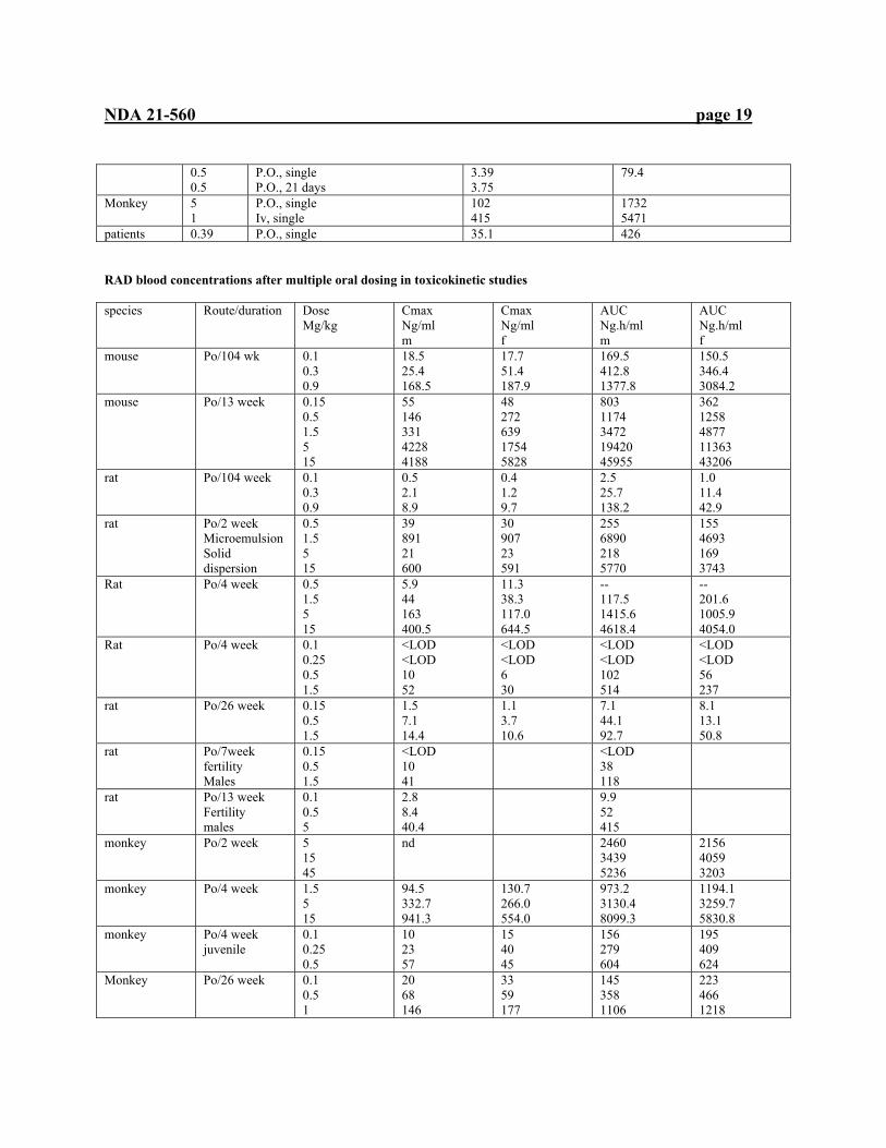
patients 0.39 P.O., single 35.1 426 RAD blood concentrations after multiple oral dosing in toxicokinetic studies species Route/duration Dose
Mg/kg Cmax Ng/ml m
Cmax Ng/ml f
AUC Ng.h/ml m
AUC Ng.h/ml f
mouse Po/104 wk 0.1 0.3 0.9
18.5 25.4 168.5
17.7 51.4 187.9
169.5 412.8 1377.8
150.5 346.4 3084.2
mouse Po/13 week 0.15 0.5 1.5 5 15
55 146 331 4228 4188
48 272 639 1754 5828
803 1174 3472 19420 45955
362 1258 4877 11363 43206
rat Po/104 week 0.1 0.3 0.9
0.5 2.1 8.9
0.4 1.2 9.7
2.5 25.7 138.2
1.0 11.4 42.9
rat Po/2 week Microemulsion Solid dispersion
0.5 1.5 5 15
39 891 21 600
30 907 23 591
255 6890 218 5770
155 4693 169 3743
Rat Po/4 week 0.5 1.5 5 15
5.9 44 163 400.5
11.3 38.3 117.0 644.5
-- 117.5 1415.6 4618.4
-- 201.6 1005.9 4054.0
Rat Po/4 week 0.1 0.25 0.5 1.5
<LOD <LOD 10 52
<LOD <LOD 6 30
<LOD <LOD 102 514
<LOD <LOD 56 237
rat Po/26 week 0.15 0.5 1.5
1.5 7.1 14.4
1.1 3.7 10.6
7.1 44.1 92.7
8.1 13.1 50.8
rat Po/7week fertility Males
0.15 0.5 1.5
<LOD 10 41
<LOD 38 118
rat Po/13 week Fertility males
0.1 0.5 5
2.8 8.4 40.4
9.9 52 415
monkey Po/2 week 5 15 45
nd 2460 3439 5236
2156 4059 3203
monkey Po/4 week 1.5 5 15
94.5 332.7 941.3
130.7 266.0 554.0
973.2 3130.4 8099.3
1194.1 3259.7 5830.8
monkey Po/4 week juvenile
0.1 0.25 0.5
10 23 57
15 40 45
156 279 604
195 409 624
Monkey
Po/26 week 0.1 0.5 1
20 68 146
33 59 177
145 358 1106
223 466 1218
NDA 21-560 page 20
5 537 383 4913 3322 monkey Po/52 week 0.1
0.5 0.9
8.5 24.1 84.9
10.1 20.5 47.8
98.0 275.6 941.3
59.6 176.2 471.8
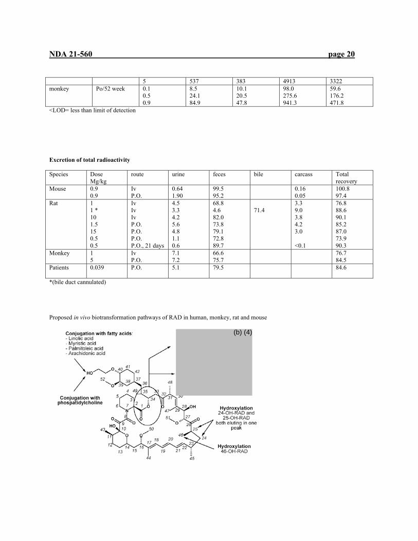
<LOD= less than limit of detection Excretion of total radioactivity Species Dose
Mg/kg route urine feces bile carcass Total
recovery Mouse 0.9
0.9 Iv P.O.
0.64 1.90
99.5 95.2
0.16 0.05
100.8 97.4
Rat 1 1 * 10 1.5 15 0.5 0.5
Iv Iv Iv P.O. P.O. P.O. P.O., 21 days
4.5 3.3 4.2 5.6 4.8 1.1 0.6
68.8 4.6 82.0 73.8 79.1 72.8 89.7
71.4
3.3 9.0 3.8 4.2 3.0 <0.1
76.8 88.6 90.1 85.2 87.0 73.9 90.3
Monkey 1 5
Iv P.O.
7.1 7.2
66.6 75.7
76.7 84.5
Patients 0.039 P.O. 5.1
79.5 84.6
*(bile duct cannulated) Proposed in vivo biotransformation pathways of RAD in human, monkey, rat and mouse
(b) (4)
NDA 21-560 page 21
3.4 TOXICOLOGY 3.4.1 Overall toxicology summary General toxicology: Toxicities observed in animal studies with RAD may be classified as related to pharmacologic effect i.e. immunosuppression and those which appear independently of immunosuppression, direct toxicity. Multiple dose toxicity studies provided the most informative portion of the submission. Multiple dose studies were conducted in mice for up to 13 weeks, rats up to 26 weeks and in cynomolgus monkeys up to 52 weeks. Additional toxicity findings may be gleaned from the 104-week mouse and rat carcinogenicity studies. Due to gastrointestinal intolerability in dogs, cynomolgus monkeys were selected by the sponsor to serve as the non-rodent species for toxicity studies. Toxicity studies with rapamycin also had the same toxicity with dogs. Body weight reduction was seen at the higher doses used for all species studied, at $5.0 mg/kg in mice, at $0.5 mg/kg in rats, at $1.5 mg/kg in minipigs and at $0.5 mg/kg in monkeys. Inflammatory gastrointestinal findings, possibly related to changes in gastrointestinal microflora under immunosuppression at these doses may be responsible for the effect through decreased absorption. Immunosuppression, the intended action of everolimus, was observed in all species tested, manifesting as thymic atrophy, splenic atrophy and lymph node atrophy associated with a decrease in circulating lymphoid and total white cells. In studies at the higher doses, other effects secondary to immunosuppression were observed. Effects to skin such as abrasions, ulceration, inflammation and scabs, were observed in monkeys (4-week study, $5.0 mg/kg; 52-week study, 0.3 mg/kg) and mice (13-weeks, $1.5 mg/kg). These effects were seen in previous studies with rapamycin and other immunosuppressive drugs in animal studies. The abrasion by the grating of cages causes sores on feet and ears which are likely entry sites for microorganisms to infect the immunosuppresed animals, despite animal husbandry efforts to maintain a relatively germ-free environment. Heart lesions exemplified by myocardial degeneration were exacerbated by higher doses of RAD in the rat ($ 1.5 mg/kg). In cynomolgus monkeys, myocardial degeneration was observed at $5.0 mg/kg in a 2-week study and at 1.5 and 5.0 mg/kg in a 26-week study. These findings were also seen in rapamycin studies. In the RAD studies, preexisting viral infections may have been exacerbated by immunosuppression. Endogenous viruses are well-characterized in cynomolgus monkey breeding colonies and may only emerge under immunosuppression. Other infections and/or inflammation related to immunosuppression included a 4-week study in minipigs which was compromised by a coccidial infection in the intestine, while the high dose group in the 52-week monkey study was terminated after 39 weeks due to gastrointestinal inflammation. Reproductive organs were subject to RAD toxicity in all species studied. Decreases in organ weight were accompanied by histopathology findings including testicular atrophy (monkey, 0.3 mg/kg) as well as depletion of germ cells and tubular vacuolation in testes, reduced sperm count in epididymides in males, and in females, uterine atrophy and reduced follicular development. These reproductive effects were seen at doses as low as 1.5 mg/kg in mice and monkeys. Male reproductive toxicity appears related to RAD –related reduction of testosterone as seen in the male rat 13-week fertility study, correlated with impaired fertility. A hormonal basis for the RAD-related toxicity to reproductive organs observed in females remains unclear without further studies. Toxicity to the lung was observed in both mouse and rat studies. Increased alveolar macrophages were observed in mice at doses $1.5 mg/kg and $0.5 mg/kg in the rat. Eosinophilic deposits in the lung were seen in a rat study comparing batches with different byproduct content. Toxicity to the eye was observed in a rats study in which swelling and disruption of cortical lens fibers at a dose of 5 mg/kg in a one-month study and at 0.9 mg/kg in the 104-wek carcinogenicity study. Renal toxicity was seen in several rat studies. In CD-1 mice, renal tubular degeneration was observed in a 13-week study at doses of $5.0 mg/kg. Studies in rats demonstrated greater incidence and severity of lipofuscin in the renal tubular epithelial cells after 26 weeks at $0.5 mg/kg and in the 104-week carcinogenicity study at doses $0.3 mg/kg. Pancreatic toxicity was observed in a 4-week minipigs and 26-week monkey study. The minipigs study showed vacuolation of the exocrine pancreas with necrosis at 15 mg/kg. The monkey study demonstrated pancreatic islet cell degeneration at 5.0 mg/kg and degranulation in the exocrine pancreas. The toxciologic basis of the pancreatic findings is unclear. It is a concern in human organ transplantation which is complicated by so-called post-transplant diabetes mellitus, also observed with immunosuppression by calcineurin inhibitors.
NDA 21-560 page 22
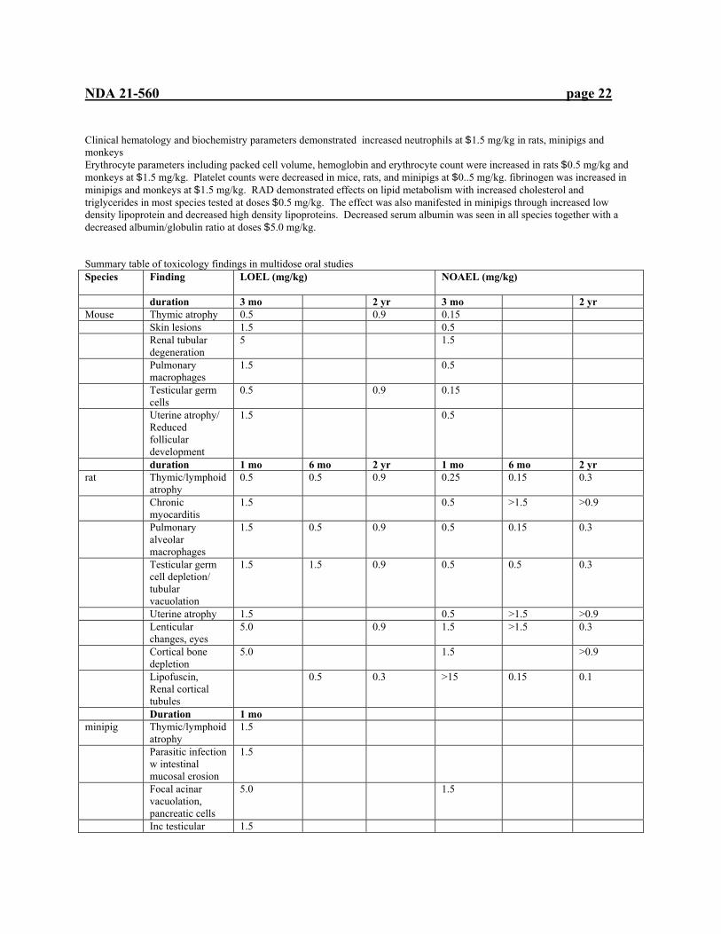
Clinical hematology and biochemistry parameters demonstrated increased neutrophils at $1.5 mg/kg in rats, minipigs and monkeys Erythrocyte parameters including packed cell volume, hemoglobin and erythrocyte count were increased in rats $0.5 mg/kg and monkeys at $1.5 mg/kg. Platelet counts were decreased in mice, rats, and minipigs at $0..5 mg/kg. fibrinogen was increased in minipigs and monkeys at $1.5 mg/kg. RAD demonstrated effects on lipid metabolism with increased cholesterol and triglycerides in most species tested at doses $0.5 mg/kg. The effect was also manifested in minipigs through increased low density lipoprotein and decreased high density lipoproteins. Decreased serum albumin was seen in all species together with a decreased albumin/globulin ratio at doses $5.0 mg/kg. Summary table of toxicology findings in multidose oral studies Species Finding LOEL (mg/kg)
NOAEL (mg/kg)
duration 3 mo 2 yr 3 mo 2 yr Mouse Thymic atrophy 0.5 0.9 0.15 Skin lesions 1.5 0.5 Renal tubular
degeneration 5 1.5
Pulmonary macrophages
1.5 0.5
Testicular germ cells
0.5 0.9 0.15
Uterine atrophy/ Reduced follicular development
1.5 0.5
duration 1 mo 6 mo 2 yr 1 mo 6 mo 2 yr rat Thymic/lymphoid
atrophy 0.5 0.5 0.9 0.25 0.15 0.3
Chronic myocarditis
1.5 0.5 >1.5 >0.9
Pulmonary alveolar macrophages
1.5 0.5 0.9 0.5 0.15 0.3
Testicular germ cell depletion/ tubular vacuolation
1.5 1.5 0.9 0.5 0.5 0.3
Uterine atrophy 1.5 0.5 >1.5 >0.9 Lenticular
changes, eyes 5.0 0.9 1.5 >1.5 0.3
Cortical bone depletion
5.0 1.5 >0.9
Lipofuscin, Renal cortical tubules
0.5 0.3 >15 0.15 0.1
Duration 1 mo minipig Thymic/lymphoid
atrophy 1.5
Parasitic infection w intestinal mucosal erosion
1.5
Focal acinar vacuolation, pancreatic cells
5.0 1.5
Inc testicular 1.5

NDA 21-560 page 23
tubular atrophy Uterine/vaginal
atrophy 15.0 5.0
Necrotic follicles, ovaries
5.0 1.5
Duration 1 mo 6 mo 1 yr 1 mo 6 mo 1 yr Monkey cynomolgus
Thymic atrophy 5.0 1.5 1.5 0.5 >0.9
Splenic lymphoid atrophy
1.5 0.5 0.9 <1.5 <0.5 0.3
Skin lesions 5.0 0.5 1.5 <0.5 >0.9 Intestinal
histiocytosis 5.0 0.5 1.5 <0.5 >0.9
GI tract inflammation
5.0 0.3 >15 1.5 0.1
Myocardial degeneration/ Necrosis
1.5 >15 0.5 >0.9
Pancreatic islet cell degeneration
5.0 >15 1.5 >0.9
Reduced follicular cell development, ovaries
1.5 >15 0.5 >0.9
Tubular atrophy, testes
0.3 >15 0.1
Comparative systemic exposure in oral studies species Dose Duration
Weeks AUC(0-24) Ng.h/ml
Multiple of human exposure for NOAEL
mouse 0.15 0.5 1.5 5 15
13
M 803 1174 3472 19420 45955
F 362 1258 4877 11363 43206
3.1 4.8
mouse 0.1 0.3 0.9
101 160 380 2231
8.6
rat 0.5 1.5 5 15
4 79 435 1468 6076
0.3
rat 0.15 0.5 1.5
26 8 29 72
0.03
rat 0.1 0.3 0.9
104 2 19 91
0.4
minipig 1.5 5 15
4 2670 6057 7856
Monkey 1.5 5 15
4 1086 3204 6978

NDA 21-560 page 24
Monkey 0.1 0.5 1.5
26 189 412 1162
1.6
monkey 0.1 0.3 0.9
52 (39)
79 226 706
0.86
NOAEL exposures in bold These may be compared to the human exposure for a 3 mg/day dose resulting in an AUC(0-24h) of 260 ng.h/ml in a 3-month steady state in clinical study B251 Genetic toxicology: The requirements of the ICH genotoxicity battery were fulfilled by the sponsor with both in vivo and in vitro assays. The following studies were performed: Study type species route dose Outcome In vitro-bacterial reversion (Ames)
S. typhimurium In vitro 8-5000 :g/plate, +/- S9 negative
In vitro mammalian Mouse lymphoma L5178Y cells
---- In vitro 7.5-90 :g/ml, +/- S9 15-120 :g/ml, +/- S9
Negative
V79 Chinese hamster cells
---- In vitro 15-81:g/ml,- S9 57-131:g/ml, + S9
negative
In vivo mammalian Micronucleus
mouse In vivo 50, 160, 500 mg/kg negative
Based on these negative findings, everolimus does not appear to have genotoxic activity. Carcinogenicity: In a 104-week carcinogenicity study in mice, statistical review showed no dose mortality trends for the male and female mice. No positive linear trends were found for male mice and one positive trend for female mice. Femur (including joint) osteoma, a rare tumor, was significant in the females (p=0.021) compared with the combined control group but within historical control incidence rates. Pairwise comparisons for this tumor were not significant for any of the dose groups compared to the combined control. None of the other observed neoplastic tumors were statistically significant by trend analysis or exact tests. Survival was approximately 55% in males at 104 weeks and 42% of females after 101 weeks. Survival among treated mice was highest in the high dose groups correlating with lower bodyweight gain. Food consumption was unaffected by treatment. Histopathology findings included treatment-related changes in the thymus, testes, and epididymides. High dose females had thymic involution. Leukocytic infiltration of the renal cortex was reduced in mid- and high dose females and submandibulary salivary gland in treated females, possibly related to immunosuppression. Reproductive effects were observed in the testes and epididymides of high dose males. The statistical review of the 104-week carcinogenicity study in rats showed no dose mortality trends for the male and female mice. No positive linear trends were found for male or female rats. None of the observed neoplastic tumors were statistically significant by trend analysis or exact tests. No dose mortality trend were found for the male rats. A significant negative trend across treatment groups for mortality was found for female rats. Survival was approximately 58% in males and 62% of females after 104 weeks. Survival among treated rats was highest in the mid and high dose groups correlating with lower bodyweight gain. Food consumption was slightly decreased in the high dose group; the rest of the groups were unaffected by treatment. Histopathology findings included treatment-related changes in the testes, epididymides, ovaries and uterus in the 0.9 mg/kg group. Immunosuppression-related changes included thymic atrophy, inflammatory changes in the Harderian glands, mesenteric lymph nodes, lachrymal glands, lungs, pancreas, skeletal muscle and submandibular gland. In the lung, increased incidence of alveolar macrophages was found, with eosinophilic deposition and pigment-laden macrophages. In the liver, age-related effects such as increased incidence of senile portal liver tract changes in males receiving 0.3 and 0.9 mg/kg appear treatment-related. Axonal degeneration of the sciatic nerve in females receiving 0.9 mg/kg also treatment-related. Lens changes included anterior suture line opacity and increased incidence of lenticular degeneration in males at 0.9 mg/kg. Age-related effects of the adrenal cortex, focal hypertrophy, hyperplasia and fatty vacuolation, were reduced in treatment groups
NDA 21-560 page 25
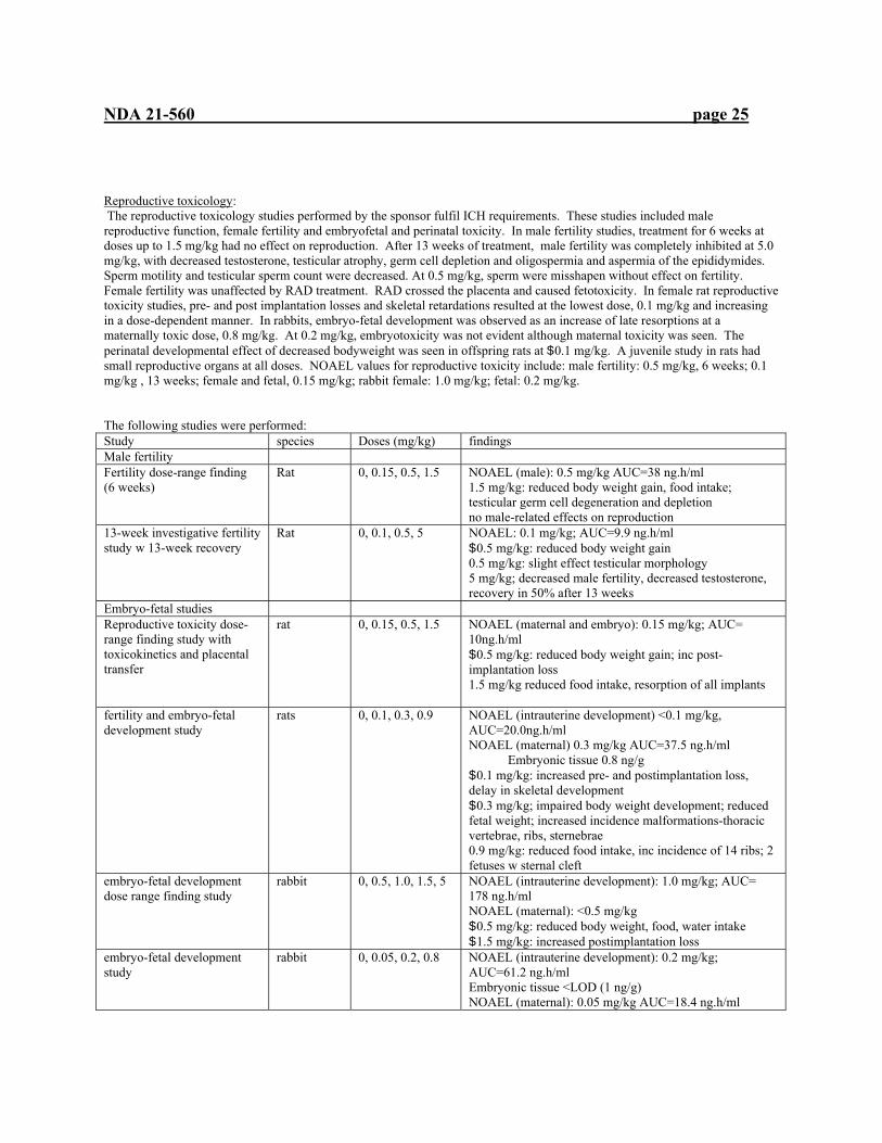
Reproductive toxicology: The reproductive toxicology studies performed by the sponsor fulfil ICH requirements. These studies included male reproductive function, female fertility and embryofetal and perinatal toxicity. In male fertility studies, treatment for 6 weeks at doses up to 1.5 mg/kg had no effect on reproduction. After 13 weeks of treatment, male fertility was completely inhibited at 5.0 mg/kg, with decreased testosterone, testicular atrophy, germ cell depletion and oligospermia and aspermia of the epididymides. Sperm motility and testicular sperm count were decreased. At 0.5 mg/kg, sperm were misshapen without effect on fertility. Female fertility was unaffected by RAD treatment. RAD crossed the placenta and caused fetotoxicity. In female rat reproductive toxicity studies, pre- and post implantation losses and skeletal retardations resulted at the lowest dose, 0.1 mg/kg and increasing in a dose-dependent manner. In rabbits, embryo-fetal development was observed as an increase of late resorptions at a maternally toxic dose, 0.8 mg/kg. At 0.2 mg/kg, embryotoxicity was not evident although maternal toxicity was seen. The perinatal developmental effect of decreased bodyweight was seen in offspring rats at $0.1 mg/kg. A juvenile study in rats had small reproductive organs at all doses. NOAEL values for reproductive toxicity include: male fertility: 0.5 mg/kg, 6 weeks; 0.1 mg/kg , 13 weeks; female and fetal, 0.15 mg/kg; rabbit female: 1.0 mg/kg; fetal: 0.2 mg/kg. The following studies were performed: Study species Doses (mg/kg) findings Male fertility Fertility dose-range finding (6 weeks)
Rat 0, 0.15, 0.5, 1.5 NOAEL (male): 0.5 mg/kg AUC=38 ng.h/ml 1.5 mg/kg: reduced body weight gain, food intake; testicular germ cell degeneration and depletion no male-related effects on reproduction
13-week investigative fertility study w 13-week recovery
Rat 0, 0.1, 0.5, 5 NOAEL: 0.1 mg/kg; AUC=9.9 ng.h/ml $0.5 mg/kg: reduced body weight gain 0.5 mg/kg: slight effect testicular morphology 5 mg/kg; decreased male fertility, decreased testosterone, recovery in 50% after 13 weeks
Embryo-fetal studies Reproductive toxicity dose-range finding study with toxicokinetics and placental transfer
rat 0, 0.15, 0.5, 1.5 NOAEL (maternal and embryo): 0.15 mg/kg; AUC= 10ng.h/ml $0.5 mg/kg: reduced body weight gain; inc post-implantation loss 1.5 mg/kg reduced food intake, resorption of all implants
fertility and embryo-fetal development study
rats 0, 0.1, 0.3, 0.9 NOAEL (intrauterine development) <0.1 mg/kg, AUC=20.0ng.h/ml NOAEL (maternal) 0.3 mg/kg AUC=37.5 ng.h/ml Embryonic tissue 0.8 ng/g $0.1 mg/kg: increased pre- and postimplantation loss, delay in skeletal development $0.3 mg/kg; impaired body weight development; reduced fetal weight; increased incidence malformations-thoracic vertebrae, ribs, sternebrae 0.9 mg/kg: reduced food intake, inc incidence of 14 ribs; 2 fetuses w sternal cleft
embryo-fetal development dose range finding study
rabbit 0, 0.5, 1.0, 1.5, 5 NOAEL (intrauterine development): 1.0 mg/kg; AUC= 178 ng.h/ml NOAEL (maternal): <0.5 mg/kg $0.5 mg/kg: reduced body weight, food, water intake $1.5 mg/kg: increased postimplantation loss
embryo-fetal development study
rabbit 0, 0.05, 0.2, 0.8 NOAEL (intrauterine development): 0.2 mg/kg; AUC=61.2 ng.h/ml Embryonic tissue <LOD (1 ng/g) NOAEL (maternal): 0.05 mg/kg AUC=18.4 ng.h/ml
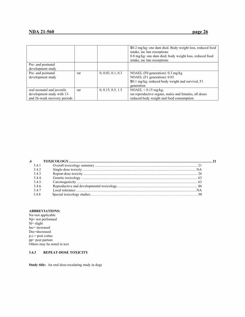
NDA 21-560 page 26
$0.2 mg/kg: one dam died. Body weight loss, reduced food intake, inc late resorptions 0.8 mg/kg: one dam died; body weight loss, reduced food intake, inc late resorptions
Pre- and postnatal development study
Pre- and postnatal development study
rat 0, 0.03, 0.1, 0.3 NOAEL (F0 generation): 0.3 mg/kg NOAEL (F1 generation): 0.03 $0.1 mg/kg: reduced body weight and survival, F1 generation
oral neonatal and juvenile development study with 13- and 26-week recovery periods
rat 0, 0.15, 0.5, 1.5 NOAEL < 0.15 mg/kg; sm reproductive organs, males and females, all doses reduced body weight and food consumption
.4 TOXICOLOGY............................................................................................................................................................. 21 3.4.1 Overall toxicology summary ................................................................................................................ 21 3.4.2 Single-dose toxicity .............................................................................................................................NA 3.4.3 Repeat-dose toxicity ............................................................................................................................. 26 3.4.4. Genetic toxicology ............................................................................................................................... 63 3.4.5. Carcinogenicity .................................................................................................................................... 63 3.4.6. Reproductive and developmental toxicology........................................................................................ 86 3.4.7 Local tolerance ....................................................................................................................................NA
3.4.8 Special toxicology studies………………………………………………………………………………98 ABBREVIATIONS: Na=not applicable Np= not performed Sl= slight Inc= increased Dec=decreased p.c.= post coitus pp= post partum Others may be noted in text 3.4.3 REPEAT-DOSE TOXICITY Study title: An oral dose-escalating study in dogs

NDA 21-560 page 27
Key study findings: Ulcerations are seen throughout the gastrointestinal tract, contributing to the diarrhea and dehydration also occurring. Study no: 41DED (BS-674)
Volume #, and page #: v.16, p1
Conducting laboratory and location: Novartis Pharma AG Basel, Switzerland Date of study initiation: not provided (approx. 26 Jan 1994) GLP compliance: no QA report: yes ( ) no ( x ) Drug, lot #, radiolabel, and % purity: batch BT7/695, na, np Formulation/vehicle: not provided Methods (unique aspects):
Dosing: Species/strain: dog (no breed provided) #/sex/group or time point (main study): 1 Satellite groups used for toxicokinetics or recovery: none Age: not provided Weight: not provided
Doses in administered units: 2 mg/kg (days 1-3), 6 mg/kg (days 4-6), 20 mg/kg (days 7-9)
Route, form, volume, and infusion rate: oral capsule
Observations and times: Clinical signs: daily Body weights: days 0, 3, 6, 9 Food consumption: days 1-7, 8-9 Ophthalmoscopy: not performed EKG: not performed Hematology: day 0, 24 h after last dose Clinical chemistry: day 0, 24 h after last dose Urinalysis: not performed Gross pathology: at necropsy Organs weighed: not performed Histopathology: at necropsy Toxicokinetics: not performed Other: not performed
Results: Mortality: all dogs survived until necropsy Clinical signs: tremor 18 mg/kg: sedation, diarrhea, ataxia, salivation, exsiccosis Body weights: body weight loss at 6, 18 mg/kg Food consumption: decreased Ophthalmoscopy: not performed Electrocardiography: not performed Hematology: increased partial prothrombin time, increased sedimentation rates;
Clinical chemistry: increased total bilirubin, alkaline phosphatase, cholesterol, triglycerides; decreased glucose Urinalysis: not performed Organ weights: not performed
Gross pathology: ulceration of oral and gastrointestinal tract Histopathology: multifocal ulceration of oral cavity, esophagus, stomach, and large intestine, with neutrophil infiltration,
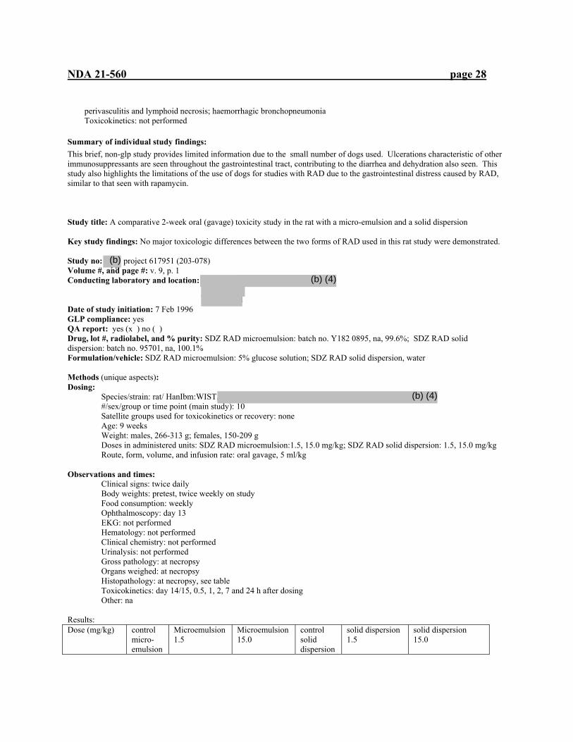
NDA 21-560 page 28
perivasculitis and lymphoid necrosis; haemorrhagic bronchopneumonia Toxicokinetics: not performed
Summary of individual study findings: This brief, non-glp study provides limited information due to the small number of dogs used. Ulcerations characteristic of other immunosuppressants are seen throughout the gastrointestinal tract, contributing to the diarrhea and dehydration also seen. This study also highlights the limitations of the use of dogs for studies with RAD due to the gastrointestinal distress caused by RAD, similar to that seen with rapamycin.
Study title: A comparative 2-week oral (gavage) toxicity study in the rat with a micro-emulsion and a solid dispersion Key study findings: No major toxicologic differences between the two forms of RAD used in this rat study were demonstrated. Study no: project 617951 (203-078) Volume #, and page #: v. 9, p. 1 Conducting laboratory and location: nd Date of study initiation: 7 Feb 1996 GLP compliance: yes QA report: yes (x ) no ( ) Drug, lot #, radiolabel, and % purity: SDZ RAD microemulsion: batch no. Y182 0895, na, 99.6%; SDZ RAD solid dispersion: batch no. 95701, na, 100.1% Formulation/vehicle: SDZ RAD microemulsion: 5% glucose solution; SDZ RAD solid dispersion, water Methods (unique aspects): Dosing:
Species/strain: rat/ HanIbm:WIST, #/sex/group or time point (main study): 10 Satellite groups used for toxicokinetics or recovery: none Age: 9 weeks Weight: males, 266-313 g; females, 150-209 g Doses in administered units: SDZ RAD microemulsion:1.5, 15.0 mg/kg; SDZ RAD solid dispersion: 1.5, 15.0 mg/kg Route, form, volume, and infusion rate: oral gavage, 5 ml/kg
Observations and times: Clinical signs: twice daily Body weights: pretest, twice weekly on study Food consumption: weekly Ophthalmoscopy: day 13 EKG: not performed Hematology: not performed Clinical chemistry: not performed Urinalysis: not performed Gross pathology: at necropsy Organs weighed: at necropsy Histopathology: at necropsy, see table Toxicokinetics: day 14/15, 0.5, 1, 2, 7 and 24 h after dosing Other: na Results: Dose (mg/kg) control
micro-emulsion
Microemulsion 1.5
Microemulsion 15.0
control solid dispersion
solid dispersion 1.5
solid dispersion 15.0
(b) (4)
(b) (4)
(b) (4)
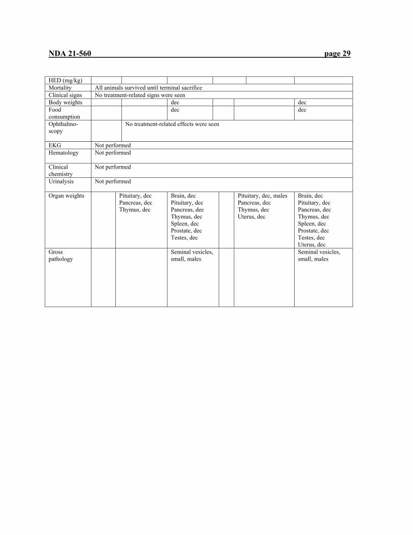
NDA 21-560 page 29
HED (mg/kg) Mortality All animals survived until terminal sacrifice Clinical signs No treatment-related signs were seen Body weights dec dec Food consumption
dec dec
Ophthalmo-scopy
No treatment-related effects were seen
EKG Not performed Hematology
Not performed
Clinical chemistry
Not performed
Urinalysis
Not performed
Organ weights
Pituitary, dec Pancreas, dec Thymus, dec
Brain, dec Pituitary, dec Pancreas, dec Thymus, dec Spleen, dec Prostate, dec Testes, dec
Pituitary, dec, males Pancreas, dec Thymus, dec Uterus, dec
Brain, dec Pituitary, dec Pancreas, dec Thymus, dec Spleen, dec Prostate, dec Testes, dec Uterus, dec
Gross pathology
Seminal vesicles, small, males
Seminal vesicles, small, males
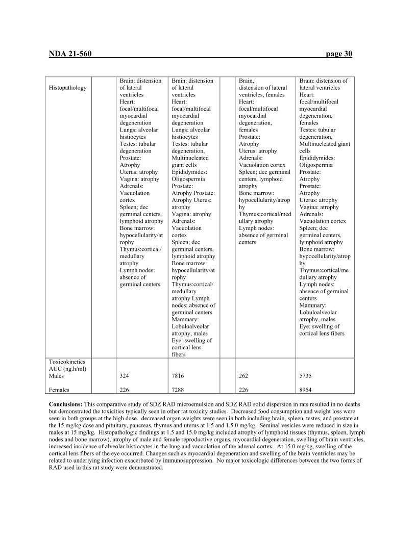
NDA 21-560 page 30
Histopathology
Brain: distension of lateral ventricles Heart: focal/multifocal myocardial degeneration Lungs: alveolar histiocytes Testes: tubular degeneration Prostate: Atrophy Uterus: atrophy Vagina: atrophy Adrenals: Vacuolation cortex Spleen; dec germinal centers, lymphoid atrophy Bone marrow: hypocellularity/atrophy Thymus:cortical/medullary atrophy Lymph nodes: absence of germinal centers
Brain: distension of lateral ventricles Heart: focal/multifocal myocardial degeneration Lungs: alveolar histiocytes Testes: tubular degeneration, Multinucleated giant cells Epididymides: Oligospermia Prostate: Atrophy Prostate: Atrophy Uterus: atrophy Vagina: atrophy Adrenals: Vacuolation cortex Spleen; dec germinal centers, lymphoid atrophy Bone marrow: hypocellularity/atrophy Thymus:cortical/medullary atrophy Lymph nodes: absence of germinal centers Mammary: Lobuloalveolar atrophy, males Eye: swelling of cortical lens fibers
Brain,: distension of lateral ventricles, females Heart: focal/multifocal myocardial degeneration, females Prostate: Atrophy Uterus: atrophy Adrenals: Vacuolation cortex Spleen; dec germinal centers, lymphoid atrophy Bone marrow: hypocellularity/atrophy Thymus:cortical/medullary atrophy Lymph nodes: absence of germinal centers
Brain: distension of lateral ventricles Heart: focal/multifocal myocardial degeneration, females Testes: tubular degeneration, Multinucleated giant cells Epididymides: Oligospermia Prostate: Atrophy Prostate: Atrophy Uterus: atrophy Vagina: atrophy Adrenals: Vacuolation cortex Spleen; dec germinal centers, lymphoid atrophy Bone marrow: hypocellularity/atrophy Thymus:cortical/medullary atrophy Lymph nodes: absence of germinal centers Mammary: Lobuloalveolar atrophy, males Eye: swelling of cortical lens fibers
Toxicokinetics AUC (ng.h/ml) Males Females
324 226
7816 7288
262 226
5735 8954
Conclusions: This comparative study of SDZ RAD microemulsion and SDZ RAD solid dispersion in rats resulted in no deaths but demonstrated the toxicities typically seen in other rat toxicity studies. Decreased food consumption and weight loss were seen in both groups at the high dose. decreased organ weights were seen in both including brain, spleen, testes, and prostate at the 15 mg/kg dose and pituitary, pancreas, thymus and uterus at 1.5 and 1.5.0 mg/kg. Seminal vesicles were reduced in size in males at 15 mg/kg. Histopathologic findings at 1.5 and 15.0 mg/kg included atrophy of lymphoid tissues (thymus, spleen, lymph nodes and bone marrow), atrophy of male and female reproductive organs, myocardial degeneration, swelling of brain ventricles, increased incidence of alveolar histiocytes in the lung and vacuolation of the adrenal cortex. At 15.0 mg/kg, swelling of the cortical lens fibers of the eye occurred. Changes such as myocardial degeneration and swelling of the brain ventricles may be related to underlying infection exacerbated by immunosuppression. No major toxicologic differences between the two forms of RAD used in this rat study were demonstrated.
NDA 21-560 page 31
Study title: A 2-week oral (gavage) dose-range-finding study in minipigs
Key study findings: Due to the small group sizes, it is difficult to draw definitive conclusions about the results of this study. Study no: 212DFP (BS-610) Volume #, and page #: v. 16, p. 17 Conducting laboratory and location: Novartis Pharma AG
Basel, Switzerland Date of study initiation: 3 Sept 1996 GLP compliance: yes QA report: yes ( ) no ( x )
Drug, lot #, radiolabel, and % purity: X035 0396, na, 86.8% Formulation/vehicle: 9.1% solid dispersion Methods (unique aspects):
Dosing: Species/strain: minipig, Gottingen SPF #/sex/group or time point (main study): 1 Satellite groups used for toxicokinetics or recovery: na Age: 4-5 mo Weight: 9.3-11.3 kg Doses in administered units: 0, 0.5, 1.5, 5 mg/kg Route, form, volume, and infusion rate: oral gavage, 5 ml/kg, na
Observations and times: Clinical signs: daily Body weights: weekly Food consumption: weekly Ophthalmoscopy: not performed EKG: days 10/11 Hematology: day 14 Clinical chemistry: day 14 Urinalysis: not performed Gross pathology: at necropsy Organs weighed: see histopathology table Histopathology: at necropsy Toxicokinetics: day 14 at 1, 3, 7 and 14 h after dosing Other: na
Results: Mortality: 1 female (5 mg/kg) sacrificed day 3-poor health, tremors, sedation, weight loss, increased sedimentation rate; necropsy: multiple granulomas with pneumonia Clinical signs: no treatment related effects seen Body weights: no treatment related effects seen Food consumption: no treatment related effects seen Ophthalmoscopy: not performed Electrocardiography: no treatment related effects seen Hematology: all treated males and females, decreased platelets, lymphocytes
Clinical chemistry: increased creatinine, females, 0.5, 1.5 mg/kg, males, 5 mg/kg;
Albumin, (-globulin decreased , males 1.5 mg/kg, females, 1.5, 5 mg/kg Urinalysis: not performed Organ weights: lower weights, pituitary, testes, treated males; spleen, adrenal, pituitary, ovaries, uterus, treated females Gross pathology: lung nodule, female, 1.5 mg/kg

NDA 21-560 page 32
Histopathology:
Finding 0.5 mg/kg 1.5 mg/kg 5 mg/kg thymic cortical lymphocytolysis
all all All
Lung: foreign body granuloma
1F 1M, 1F
Seminiferous tubules: vacuolated germ cells
1M 1M 1M
Leydig cell hyperplasia 1M 1M 1M
Toxicokinetics:
0.5 mg/kg 1.5 mg/kg 5 mg/kg AUC (0-24h) M: ng.h/ml F:
1454 1694
5731 6271
5007 *
Cmax (ng/ml) M: F:
88 91
473 418
424 *
* value not calculated due to single surviving female
Conclusion: One high dose female died on day 3; weight loss was noted prior to treatment and pneumonitis was seen at necropsy with foreign body granuloma. Immunosuppression may have exaggerated a preexisting condition. Otherwise, all minipigs survived until necropsy. Drug-induced effects included an increased creatinine, females, 0.5, 1.5 mg/kg, males, 5 mg/kg; albumin and (-globulin decreased, males 1.5 mg/kg, females, 1.5, 5 mg/kg; decreased weights, pituitary, testes, treated males; spleen, adrenal, pituitary, ovaries, uterus, treated females; thymic cortical lymphocytosis seen in all treated groups. Testicular effects were seen in all treated males including vacuolated cells in the seminiferous tubules and Leydig cell hyperplasia. The small numbers of minipigs in each group permits only generalization concerning the effects seen, especially immunosuppression and testicular effects.
Study title: 4-week oral (gavage) toxicity study in minipigs Key study findings: Immunosuppressive effects seen as effects on leukocytes and lymphatic organs (spleen, thymus, lymph nodes). Skin also had drug-related dermatitis, possibly due to immunosuppression. Pancreas and adrenal gland (cortex and medulla) were affected, as were male (testis) and female (ovaries and uterus) by toxicity. A NOEL was not determined in this study. Study no: 971033 Volume #, and page #: v. 16, p. 117 Conducting laboratory and location: Novartis Pharma AG Basel, Switzerland Date of study initiation: 29 July 1997 GLP compliance: yes QA report: yes ( x ) no ( )
Drug, lot #, radiolabel, and % purity: X011 0397, na, 94.1% Formulation/vehicle: 9.1% solid dispersion in water Methods (unique aspects):
Dosing: Species/strain: minipig, Gottingen SPF #/sex/group or time point (main study): 3
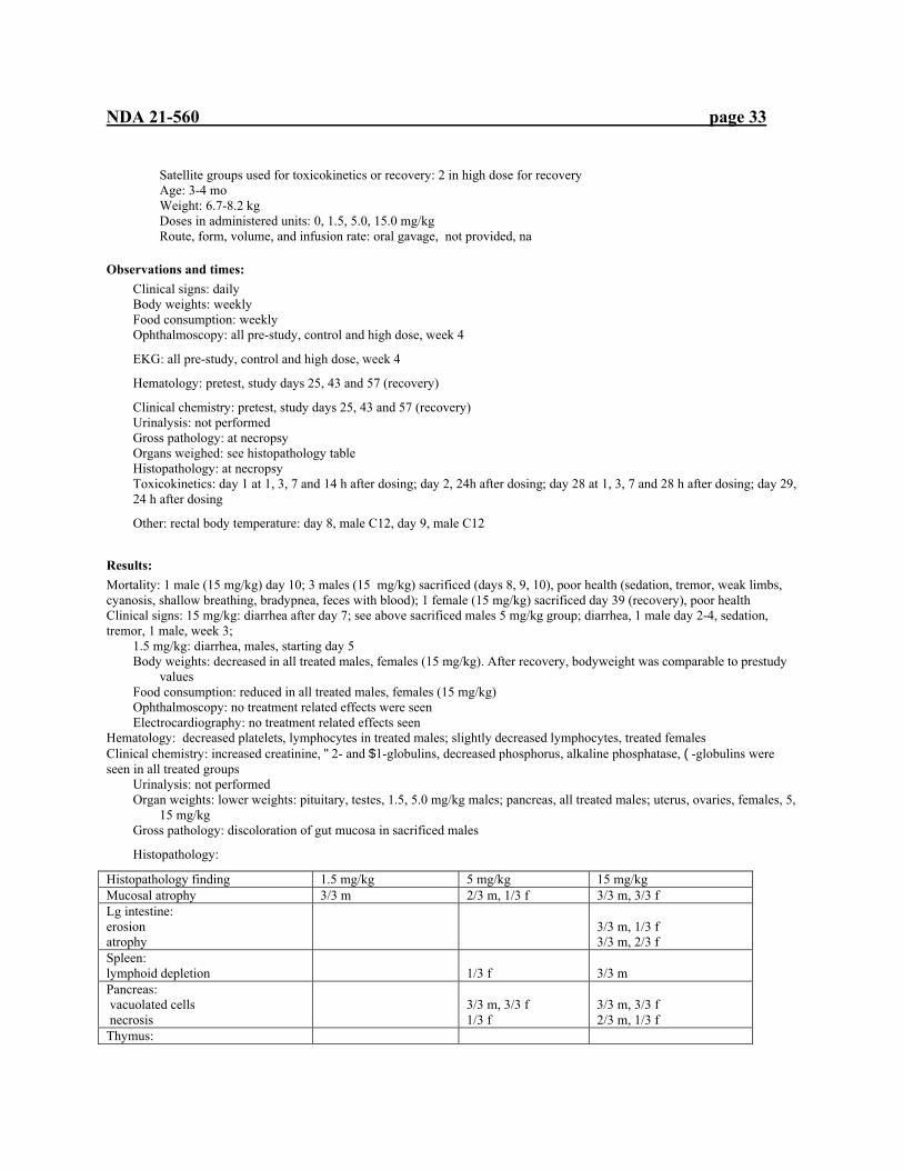
NDA 21-560 page 33
Satellite groups used for toxicokinetics or recovery: 2 in high dose for recovery Age: 3-4 mo Weight: 6.7-8.2 kg Doses in administered units: 0, 1.5, 5.0, 15.0 mg/kg Route, form, volume, and infusion rate: oral gavage, not provided, na
Observations and times: Clinical signs: daily Body weights: weekly Food consumption: weekly Ophthalmoscopy: all pre-study, control and high dose, week 4
EKG: all pre-study, control and high dose, week 4
Hematology: pretest, study days 25, 43 and 57 (recovery)
Clinical chemistry: pretest, study days 25, 43 and 57 (recovery) Urinalysis: not performed Gross pathology: at necropsy Organs weighed: see histopathology table Histopathology: at necropsy Toxicokinetics: day 1 at 1, 3, 7 and 14 h after dosing; day 2, 24h after dosing; day 28 at 1, 3, 7 and 28 h after dosing; day 29, 24 h after dosing
Other: rectal body temperature: day 8, male C12, day 9, male C12
Results: Mortality: 1 male (15 mg/kg) day 10; 3 males (15 mg/kg) sacrificed (days 8, 9, 10), poor health (sedation, tremor, weak limbs, cyanosis, shallow breathing, bradypnea, feces with blood); 1 female (15 mg/kg) sacrificed day 39 (recovery), poor health Clinical signs: 15 mg/kg: diarrhea after day 7; see above sacrificed males 5 mg/kg group; diarrhea, 1 male day 2-4, sedation, tremor, 1 male, week 3;
1.5 mg/kg: diarrhea, males, starting day 5 Body weights: decreased in all treated males, females (15 mg/kg). After recovery, bodyweight was comparable to prestudy
values Food consumption: reduced in all treated males, females (15 mg/kg) Ophthalmoscopy: no treatment related effects were seen Electrocardiography: no treatment related effects seen
Hematology: decreased platelets, lymphocytes in treated males; slightly decreased lymphocytes, treated females Clinical chemistry: increased creatinine, "2- and $1-globulins, decreased phosphorus, alkaline phosphatase, (-globulins were seen in all treated groups
Urinalysis: not performed Organ weights: lower weights: pituitary, testes, 1.5, 5.0 mg/kg males; pancreas, all treated males; uterus, ovaries, females, 5,
15 mg/kg Gross pathology: discoloration of gut mucosa in sacrificed males
Histopathology:
Histopathology finding 1.5 mg/kg 5 mg/kg 15 mg/kg Mucosal atrophy 3/3 m 2/3 m, 1/3 f 3/3 m, 3/3 f Lg intestine: erosion atrophy
3/3 m, 1/3 f 3/3 m, 2/3 f
Spleen: lymphoid depletion
1/3 f
3/3 m
Pancreas: vacuolated cells necrosis
3/3 m, 3/3 f 1/3 f
3/3 m, 3/3 f 2/3 m, 1/3 f
Thymus:
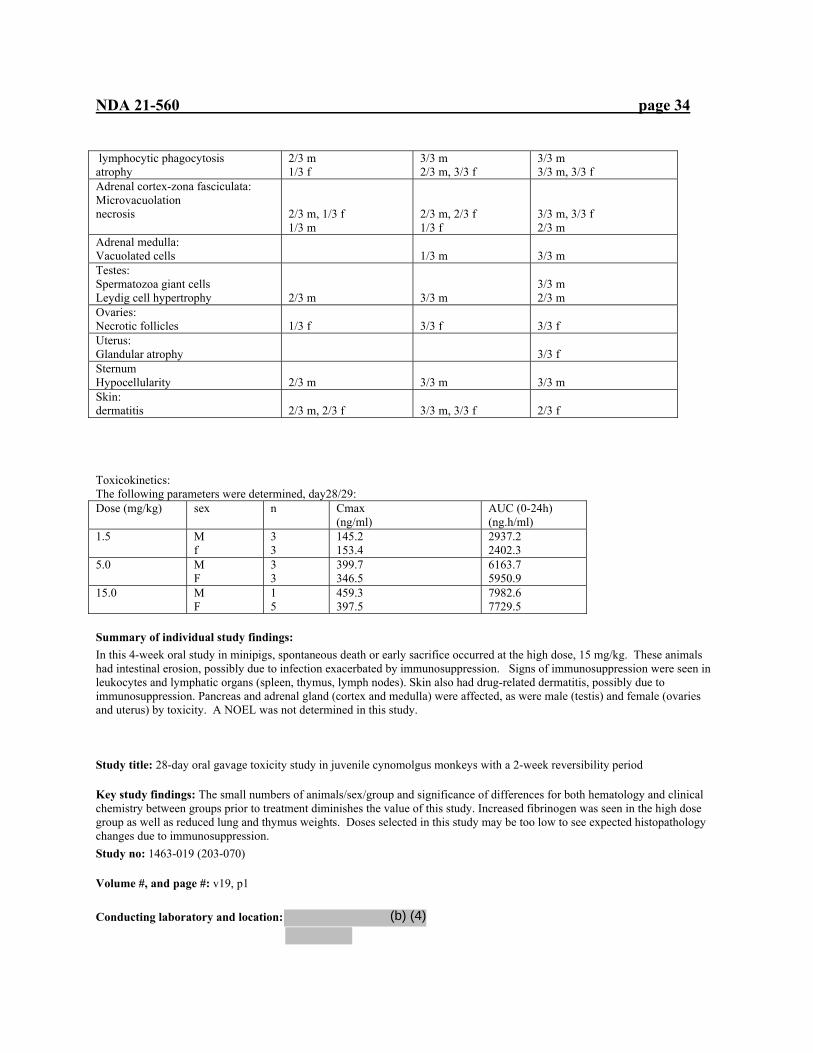
NDA 21-560 page 34
lymphocytic phagocytosis atrophy
2/3 m 1/3 f
3/3 m 2/3 m, 3/3 f
3/3 m 3/3 m, 3/3 f
Adrenal cortex-zona fasciculata: Microvacuolation necrosis
2/3 m, 1/3 f 1/3 m
2/3 m, 2/3 f 1/3 f
3/3 m, 3/3 f 2/3 m
Adrenal medulla: Vacuolated cells
1/3 m
3/3 m
Testes: Spermatozoa giant cells Leydig cell hypertrophy
2/3 m
3/3 m
3/3 m 2/3 m
Ovaries: Necrotic follicles
1/3 f
3/3 f
3/3 f
Uterus: Glandular atrophy
3/3 f
Sternum Hypocellularity
2/3 m
3/3 m
3/3 m
Skin: dermatitis
2/3 m, 2/3 f
3/3 m, 3/3 f
2/3 f
Toxicokinetics: The following parameters were determined, day28/29: Dose (mg/kg) sex n Cmax
(ng/ml) AUC (0-24h) (ng.h/ml)
1.5 M f
3 3
145.2 153.4
2937.2 2402.3
5.0 M F
3 3
399.7 346.5
6163.7 5950.9
15.0 M F
1 5
459.3 397.5
7982.6 7729.5
Summary of individual study findings: In this 4-week oral study in minipigs, spontaneous death or early sacrifice occurred at the high dose, 15 mg/kg. These animals had intestinal erosion, possibly due to infection exacerbated by immunosuppression. Signs of immunosuppression were seen in leukocytes and lymphatic organs (spleen, thymus, lymph nodes). Skin also had drug-related dermatitis, possibly due to immunosuppression. Pancreas and adrenal gland (cortex and medulla) were affected, as were male (testis) and female (ovaries and uterus) by toxicity. A NOEL was not determined in this study. Study title: 28-day oral gavage toxicity study in juvenile cynomolgus monkeys with a 2-week reversibility period
Key study findings: The small numbers of animals/sex/group and significance of differences for both hematology and clinical chemistry between groups prior to treatment diminishes the value of this study. Increased fibrinogen was seen in the high dose group as well as reduced lung and thymus weights. Doses selected in this study may be too low to see expected histopathology changes due to immunosuppression. Study no: 1463-019 (203-070)
Volume #, and page #: v19, p1
Conducting laboratory and location:
(b) (4)
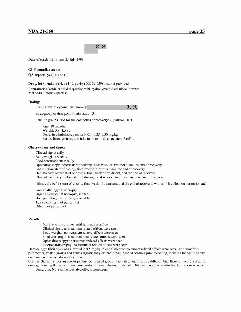
NDA 21-560 page 35
Date of study initiation: 25 July 1996
GLP compliance: yes QA report: yes ( x ) no ( )
Drug, lot #, radiolabel, and % purity: XO 35 0396, na, not provided Formulation/vehicle: solid dispersion with hydroxymethyl cellulose in water Methods (unique aspects):
Dosing: Species/strain: cynomolgus monkey,
#/sex/group or time point (main study): 3
Satellite groups used for toxicokinetics or recovery: 2 (control, HD)
Age: 10 months Weight: 0.8- 1.5 kg Doses in administered units: 0, 0.1, 0.25, 0.50 mg/kg Route, form, volume, and infusion rate: oral, dispersion, 5 ml/kg
Observations and times: Clinical signs: daily Body weights: weekly Food consumption: weekly Ophthalmoscopy: before start of dosing, final week of treatment, and the end of recovery EKG: before start of dosing, final week of treatment, and the end of recovery Hematology: before start of dosing, final week of treatment, and the end of recovery Clinical chemistry: before start of dosing, final week of treatment, and the end of recovery
Urinalysis: before start of dosing, final week of treatment, and the end of recovery; with a 16 h collection period for each
Gross pathology: at necropsy Organs weighed: at necropsy, see table Histopathology: at necropsy, see table Toxicokinetics: not performed Other: not performed
Results: Mortality: all survived until terminal sacrifice Clinical signs: no treatment related effects were seen Body weights: no treatment related effects were seen Food consumption: no treatment related effects were seen Ophthalmoscopy: no treatment related effects were seen Electrocardiography: no treatment related effects were seen
Hematology: fibrinogen was elevated in 0.5 mg/kg m and f; no other treatment related effects were seen. For numerous parameters, treated groups had values significantly different than those of controls prior to dosing, reducing the value of any comparative changes during treatment. Clinical chemistry: For numerous parameters, treated groups had values significantly different than those of controls prior to dosing, reducing the value of any comparative changes during treatment. Otherwise no treatment-related effects were seen.
Urinalysis: No treatment-related effects were seen.
(b) (4)
(b) (4)
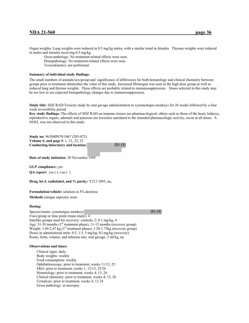
NDA 21-560 page 36
Organ weights: Lung weights were reduced in 0.5 mg/kg males, with a similar trend in females. Thymus weights were reduced in males and females receiving 0.5 mg/kg.
Gross pathology: No treatment-related effects were seen. Histopathology: No treatment-related effects were seen. Toxicokinetics: not performed.
Summary of individual study findings: The small numbers of animals/sex/group and significance of differences for both hematology and clinical chemistry between groups prior to treatment diminishes the value of this study. Increased fibrinogen was seen in the high dose group as well as reduced lung and thymus weights. These effects are probably related to immunosuppression. Doses selected in this study may be too low to see expected histopathology changes due to immunosuppression. Study title: SDZ RAD:Toxicity study by oral gavage administration to cynomologus monkeys for 26 weeks followed by a four week reversibility period Key study findings: The effects of SDZ RAD on immune tissues are pharmacological; others such as those of the heart, kidneys, reproductive organs, adrenals and pancreas are toxicities unrelated to the intended pharmacologic activity, occur at all doses. A NOEL was not observed in this study.
Study no: 96/SMP078/1067 (203-072) Volume #, and page #: v. 21, 22, 23 Conducting laboratory and location:
Date of study initiation: 28 November 1995
GLP compliance: yes QA report: yes ( x ) no ( )
Drug, lot #, radiolabel, and % purity: Y213 1095, na,
Formulation/vehicle: solution in 5% dextrose Methods (unique aspects): none
Dosing: Species/strain: cynomolgus monkeys, #/sex/group or time point (main study): 4 Satellite groups used for recovery: controls, 2; 0.1 mg/kg, 4 Age: 31-39 months (1st treatment phase), 11-15 months (recovery group) Weight: 1.69-2.47 kg (1st treatment phase), 1.28-1.75kg (recovery group) Doses in administered units: 0.5, 1.5, 5 mg/kg; 0.l mg/kg (recovery) Route, form, volume, and infusion rate: oral gavage, 5 ml/kg, na
Observations and times: Clinical signs: daily Body weights: weekly Food consumption: weekly Ophthalmoscopy: prior to treatment, weeks 11/12, 25 EKG: prior to treatment, weeks 1, 12/13, 25/26 Hematology: prior to treatment, weeks 4, 13, 26 Clinical chemistry: prior to treatment, weeks 4, 13, 26 Urinalysis: prior to treatment, weeks 4, 12 24 Gross pathology: at necropsy
(b) (4)
(b) (4)
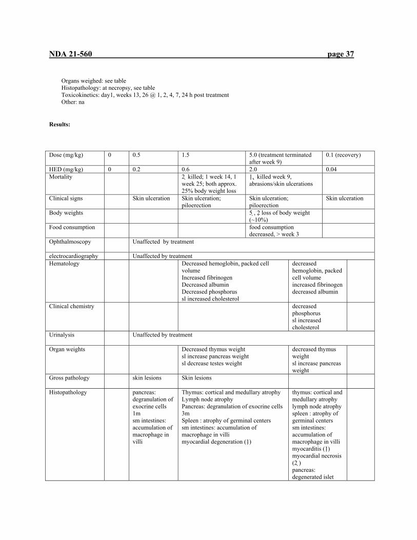
NDA 21-560 page 37
Organs weighed: see table Histopathology: at necropsy, see table Toxicokinetics: day1, weeks 13, 26 @ 1, 2, 4, 7, 24 h post treatment Other: na
Results: Dose (mg/kg) 0 0.5 1.5 5.0 (treatment terminated
after week 9) 0.1 (recovery)
HED (mg/kg) 0 0.2 0.6 2.0 0.04 Mortality 2 killed; 1 week 14, 1
week 25; both approx. 25% body weight loss
1 , killed week 9, abrasions/skin ulcerations
Clinical signs Skin ulceration Skin ulceration; piloerection
Skin ulceration; piloerection
Skin ulceration
Body weights 5 , 2 loss of body weight (~10%)
Food consumption food consumption decreased, > week 3
Ophthalmoscopy
Unaffected by treatment
electrocardiography Unaffected by treatment Hematology
Decreased hemoglobin, packed cell volume Increased fibrinogen Decreased albumin Decreased phosphorus sl increased cholesterol
decreased hemoglobin, packed cell volume increased fibrinogen decreased albumin
Clinical chemistry decreased phosphorus sl increased cholesterol
Urinalysis
Unaffected by treatment
Organ weights
Decreased thymus weight sl increase pancreas weight sl decrease testes weight
decreased thymus weight sl increase pancreas weight
Gross pathology
skin lesions Skin lesions
Histopathology
pancreas: degranulation of exocrine cells 1m sm intestines: accumulation of macrophage in villi
Thymus: cortical and medullary atrophy Lymph node atrophy Pancreas: degranulation of exocrine cells 3m Spleen : atrophy of germinal centers sm intestines: accumulation of macrophage in villi myocardial degeneration (1)
thymus: cortical and medullary atrophy lymph node atrophy spleen : atrophy of germinal centers sm intestines: accumulation of macrophage in villi myocarditis (1) myocardial necrosis (2 ) pancreas: degenerated islet
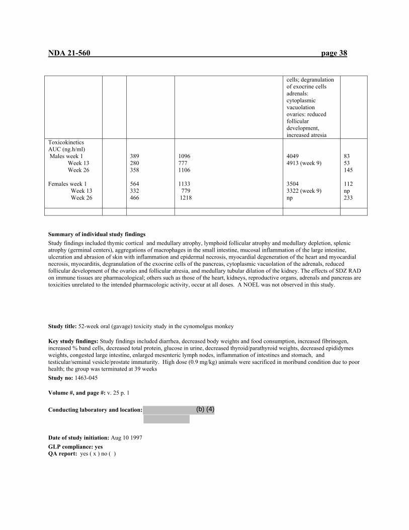
NDA 21-560 page 38
cells; degranulation of exocrine cells adrenals: cytoplasmic vacuolation ovaries: reduced follicular development, increased atresia
Toxicokinetics AUC (ng.h/ml) Males week 1 Week 13 Week 26 Females week 1 Week 13 Week 26
389 280 358 564 332 466
1096 777 1106 1133 779 1218
4049 4913 (week 9) 3504 3322 (week 9) np
83 53 145 112 np 233
Summary of individual study findings Study findings included thymic cortical and medullary atrophy, lymphoid follicular atrophy and medullary depletion, splenic atrophy (germinal centers), aggregations of macrophages in the small intestine, mucosal inflammation of the large intestine, ulceration and abrasion of skin with inflammation and epidermal necrosis, myocardial degeneration of the heart and myocardial necrosis, myocarditis, degranulation of the exocrine cells of the pancreas, cytoplasmic vacuolation of the adrenals, reduced follicular development of the ovaries and follicular atresia, and medullary tubular dilation of the kidney. The effects of SDZ RAD on immune tissues are pharmacological; others such as those of the heart, kidneys, reproductive organs, adrenals and pancreas are toxicities unrelated to the intended pharmacologic activity, occur at all doses. A NOEL was not observed in this study. Study title: 52-week oral (gavage) toxicity study in the cynomolgus monkey
Key study findings: Study findings included diarrhea, decreased body weights and food consumption, increased fibrinogen, increased % band cells, decreased total protein, glucose in urine, decreased thyroid/parathyroid weights, decreased epididymes weights, congested large intestine, enlarged mesenteric lymph nodes, inflammation of intestines and stomach, and testicular/seminal vesicle/prostate immaturity. High dose (0.9 mg/kg) animals were sacrificed in moribund condition due to poor health; the group was terminated at 39 weeks Study no: 1463-045
Volume #, and page #: v. 25 p. 1
Conducting laboratory and location:
Date of study initiation: Aug 10 1997 GLP compliance: yes QA report: yes ( x ) no ( )
(b) (4)

NDA 21-560 page 39
Drug, lot #, radiolabel, and % purity: X011 0937/X176 1297, na, Formulation/vehicle: aqueous solution, water Methods (unique aspects): none
Dosing: Species/strain: cynomolgus monkeys, #/sex/group or time point (main study): 4 Satellite groups used for toxicokinetics or recovery: na Age: 2-9 yrs Weight: 3.0-5.1 kg (males), 2.7-5.3 kg (females) Doses in administered units: 0.1, 0.3, 0.9 mg/kg Route, form, volume, and infusion rate: oral gavage, 5 ml/kg, na
Observations and times: Clinical signs: daily Body weights: weekly Food consumption: weekly Ophthalmoscopy: prior to study, study weeks 26, 39, 52 EKG: prior to study, study weeks 13, 26, 39, 52 Hematology: prior to study, study weeks 13, 26, 39, 52 Clinical chemistry: prior to study, study weeks 13, 26, 39, 52 Urinalysis: prior to study, study weeks 26, 39, 52 for 16 h Gross pathology: at necropsy Organs weighed: at necropsy, see table Histopathology: at necropsy, see table Toxicokinetics: day 1, weeks 26, 39, 52 @ 0, 1, 2, 4, 7, and 24 h post-dose Other: blood pressure, prior to study, study weeks 26, 39, 52
Results: Dose (mg/kg) 0 0.1 0.3 0.9 (terminated after 39 weeks) Mortality 1% , sacrificed
moribund day 225 (edema, hunched posture, poor physical condition)
1 %, sacrificed moribund, day 142 (chronic diarrhea, hunched posture, poor physical condition) 2 &, sacrificed day 183, day 258 (chronic diarrhea, emaciation, poor physical condition)
Clinical signs % diarrhea %,& diarrhea Body weights & sl decrease %,& sl decrease %,& sl decrease Food consumption
Decreases in animal sacrificed for morbidity prior to sac
Ophthalmoscopy
No treatment-related effect observed
Electrocardiography And blood pressure
No treatment-related effect observed

NDA 21-560 page 40
Hematology % % band cells increased % sl increased fibrinogen %,& %band cells increased
%,&increased fibrinogen %,& %band cells increased
Clinical chemistry
%decreased total protein % decreased total protein
Urinalysis 1& glucose 1& glucose Organ weights % decreased
thyroid/parathyroid % decreased epididymes
% decreased thyroid/parathyroid % decreased epididymes
% decreased thyroid/parathyroid % decreased epididymes
Gross pathology
1 % (moribund) ascites, hydropericardium, renal and hepatic enlargement
3 % congested lg intestine, mesenteric ln enlargement
1 %, 2& (moribund) mesenteric ln enlargement, congestion of intestinal vessels 1% lung, white-yellow focus 1&, cecal ulceration
Histopathology 3 %, 2&: acute inflammation lg intestine 2%: immaturity of testes (1% immaturity of prostate, seminal vesicles)
2& inflammation of colon /cecum 2%, 2&: stomach inflammation/erosion all %: testicular immaturity
Toxicokinetics AUC (ng.h/ml) Males Week 1 Week 26 Week 39 Week 52 Females Week 1 Week 26 Week 39 Week 52 Cmax (ng/ml) Males Week 1 Week 26 Week 39 Week 52 Females Week 1 Week 26 Week 39 Week 52
1.740 57.739 87.809 98.028 26.056 78.013 80.465 59.561 0.696 5.389 6.476 8.532 8.759 15.795 11.984 10.135
135.857 270.747 349.909 275.571 161.315 144.396 246.685 176.222 13.814 24.776 36.393 24.092 31.214 15.816 35.445 20.541
362.596 690.809 941.299 np 732.490 395.736 471.777 np 27.900 67.599 84.921 np 86.079 37.157 47.824 np
Summary of individual study findings: Study findings included diarrhea, decreased body weights and food consumption, increased fibrinogen, increased % band cells, decreased total protein, glucose in urine, decreased thyroid/parathyroid weights, decreased epididymes weights, congested large intestine, enlarged mesenteric lymph nodes, inflammation of intestines and stomach, and testicular/seminal vesicle/prostate immaturity. High dose (0.9 mg/kg) animals were sacrificed in moribund condition due to poor health; the group was terminated at 39 weeks. The findings of this study appear related to the immunosuppressive action of SDZ RAD and appear similar to the related drug rapamycin.

NDA 21-560 page 41
Study title: Toxicity study by intravenous (bolus) administration to HanIbm Wistar rats for 2 weeks
Key study findings: Rats receiving intravenous treatment for two weeks showed some signs of immunosuppression (decreased leukocytes, depletion of lymphocytes from thymic cortex) as well as increased cholesterol and triglycerides
Study no: SPM115/970022 (203-071)
Volume #, and page #: v. 27 p.1 Conducting laboratory and location: Date of study initiation: 10 Sept 1996
GLP compliance: yes QA report: yes ( x ) no ( )
Drug, lot #, radiolabel, and % purity: Y187 0895, na, 96.9% Formulation/vehicle: 2% microemulsion, 1 ml diluted with 3 ml Sandimmune placebo. 0.5 ml of this mixture added to 49.5 ml of 5% glucose solution Methods (unique aspects): none
Dosing: Species/strain: rat/hanIbm Wistar, #/sex/group or time point (main study): 10 Satellite groups used for toxicokinetics or recovery: 6/sex/group=toxicokinetic sampling Age: 35-42 days Weight: males, 174-209 g; females141-180 g Doses in administered units: 0.005, 0.01, 0.05 mg/kg
Route, form, volume, and infusion rate: intravenous, control, 0.05 mg/kg=1ml/kg; 0.005 mg/kg=0.1 ml/kg; 0.01mg/kg=0.2 ml/kg
Observations and times: Clinical signs: daily Body weights: one week before treatment, 1st day of treatment, twice weekly Food consumption: week of acclimation, weekly during treatment Ophthalmoscopy: before treatment, groups 1, 4 during week 2 of treatment EKG: not performed Hematology: week 2 Clinical chemistry: week 2 Urinalysis:week 2, for 16 h Gross pathology: at necropsy Organs weighed: at necropsy, see table Histopathology: at necropsy, see table Toxicokinetics: day 14, 5, 15, 30 min, 1, 4, 14 h after dosing Other: not performed
Results: Dose (mg/kg) 0 0.005 0.1 0.05 HED (mg/kg) 0 0.0003 0.0006 0.003 Mortality All rats survived until terminal sacrifice
(b) (4)
(b) (4)
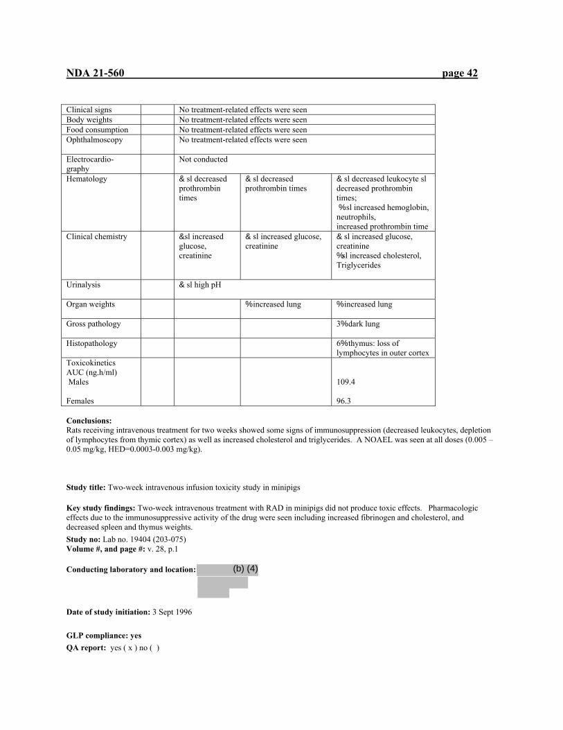
NDA 21-560 page 42
Clinical signs No treatment-related effects were seen Body weights No treatment-related effects were seen Food consumption No treatment-related effects were seen Ophthalmoscopy
No treatment-related effects were seen
Electrocardio-graphy
Not conducted
Hematology
& sl decreased prothrombin times
& sl decreased prothrombin times
& sl decreased leukocyte sl decreased prothrombin times; % sl increased hemoglobin, neutrophils, increased prothrombin time
Clinical chemistry &sl increased glucose, creatinine
& sl increased glucose, creatinine
& sl increased glucose, creatinine %sl increased cholesterol, Triglycerides
Urinalysis
& sl high pH
Organ weights
% increased lung % increased lung
Gross pathology
3% dark lung
Histopathology
6% thymus: loss of lymphocytes in outer cortex
Toxicokinetics AUC (ng.h/ml) Males Females
109.4 96.3
Conclusions: Rats receiving intravenous treatment for two weeks showed some signs of immunosuppression (decreased leukocytes, depletion of lymphocytes from thymic cortex) as well as increased cholesterol and triglycerides. A NOAEL was seen at all doses (0.005 –0.05 mg/kg, HED=0.0003-0.003 mg/kg). Study title: Two-week intravenous infusion toxicity study in minipigs
Key study findings: Two-week intravenous treatment with RAD in minipigs did not produce toxic effects. Pharmacologic effects due to the immunosuppressive activity of the drug were seen including increased fibrinogen and cholesterol, and decreased spleen and thymus weights. Study no: Lab no. 19404 (203-075) Volume #, and page #: v. 28, p.1
Conducting laboratory and location:
Date of study initiation: 3 Sept 1996
GLP compliance: yes QA report: yes ( x ) no ( )
(b) (4)
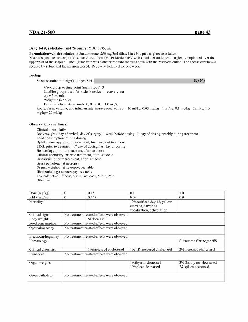
NDA 21-560 page 43
Drug, lot #, radiolabel, and % purity: Y187 0895, na, Formulation/vehicle: solution in Sandimmune, 250 mg/5ml diluted in 5% aqueous glucose solution Methods (unique aspects): a Vascular Access Port (VAP) Model GPV with a catheter outlet was surgically implanted over the upper part of the scapula. The jugular vein was catheterized into the vena cava with the reservoir outlet. The access canula was secured by suture and the incision closed. Recovery followed for one week.
Dosing: Species/strain: minipig/Gottingen SPF,
#/sex/group or time point (main study): 3 Satellite groups used for toxicokinetics or recovery: na Age: 3 months Weight: 5.6-7.5 kg Doses in administered units: 0, 0.05, 0.1, 1.0 mg/kg
Route, form, volume, and infusion rate: intravenous, control= 20 ml/kg, 0.05 mg/kg= 1 ml/kg, 0.1 mg/kg= 2ml/kg, 1.0 mg/kg= 20 ml/kg
Observations and times: Clinical signs: daily Body weights: day of arrival, day of surgery, 1 week before dosing, 1st day of dosing, weekly during treatment Food consumption: during dosing Ophthalmoscopy: prior to treatment, final week of treatment EKG: prior to treatment, 1st day of dosing, last day of dosing Hematology: prior to treatment, after last dose Clinical chemistry: prior to treatment, after last dose Urinalysis: prior to treatment, after last dose Gross pathology: at necropsy Organs weighed: at necropsy, see table Histopathology: at necropsy, see table Toxicokinetics: 1st dose, 5 min, last dose, 5 min, 24 h Other: na
Dose (mg/kg) 0 0.05 0.1 1.0 HED (mg/kg) 0 0.045 0.09 0.9 Mortality 1% sacrificed day 13, yellow
diarrhea, shivering, vocalization, dehydration
Clinical signs No treatment-related effects were observed Body weights Sl decrease Food consumption No treatment-related effects were observed Ophthalmoscopy
No treatment-related effects were observed
Electrocardiography No treatment-related effects were observed Hematology
Sl increase fibrinogen,%&
Clinical chemistry 1% increased cholesterol 1%, 1& increased cholesterol 2% increased cholesterol Urinalysis
No treatment-related effects were observed
Organ weights
1% thymus decreased 1% spleen decreased
3%, 2& thymus decreased 2& spleen decreased
Gross pathology
No treatment-related effects were observed
(b) (4)
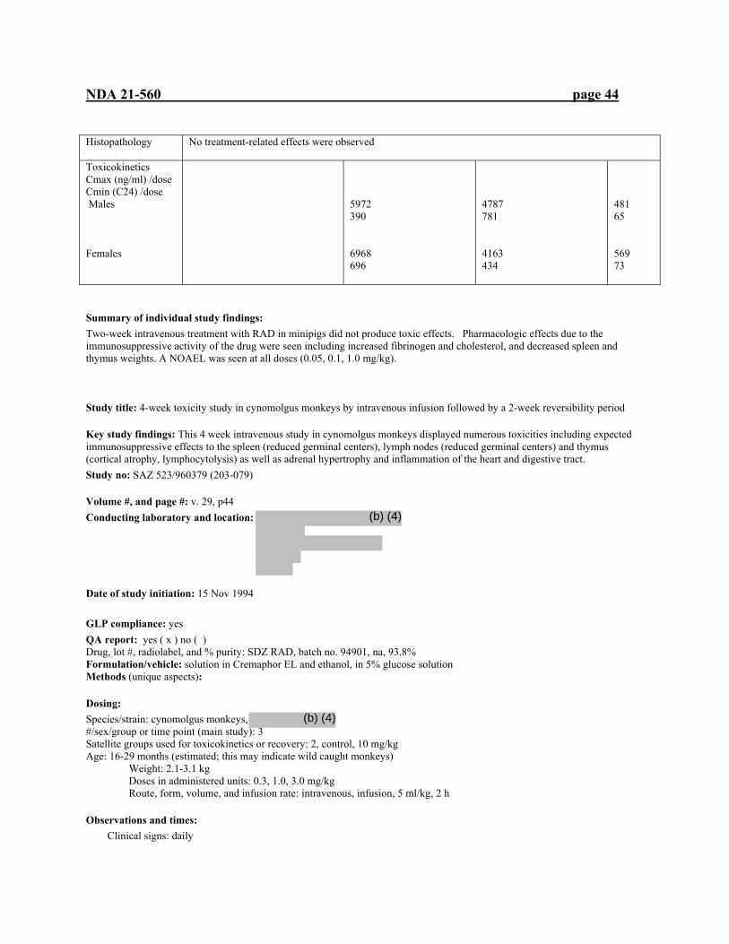
NDA 21-560 page 44
Histopathology
No treatment-related effects were observed
Toxicokinetics Cmax (ng/ml) /dose Cmin (C24) /dose Males Females
5972 390 6968 696
4787 781 4163 434
481 65 569 73
Summary of individual study findings: Two-week intravenous treatment with RAD in minipigs did not produce toxic effects. Pharmacologic effects due to the immunosuppressive activity of the drug were seen including increased fibrinogen and cholesterol, and decreased spleen and thymus weights. A NOAEL was seen at all doses (0.05, 0.1, 1.0 mg/kg). Study title: 4-week toxicity study in cynomolgus monkeys by intravenous infusion followed by a 2-week reversibility period
Key study findings: This 4 week intravenous study in cynomolgus monkeys displayed numerous toxicities including expected immunosuppressive effects to the spleen (reduced germinal centers), lymph nodes (reduced germinal centers) and thymus (cortical atrophy, lymphocytolysis) as well as adrenal hypertrophy and inflammation of the heart and digestive tract. Study no: SAZ 523/960379 (203-079)
Volume #, and page #: v. 29, p44 Conducting laboratory and location:
Date of study initiation: 15 Nov 1994
GLP compliance: yes QA report: yes ( x ) no ( ) Drug, lot #, radiolabel, and % purity: SDZ RAD, batch no. 94901, na, 93.8% Formulation/vehicle: solution in Cremaphor EL and ethanol, in 5% glucose solution Methods (unique aspects):
Dosing: Species/strain: cynomolgus monkeys, #/sex/group or time point (main study): 3 Satellite groups used for toxicokinetics or recovery: 2, control, 10 mg/kg Age: 16-29 months (estimated; this may indicate wild caught monkeys)
Weight: 2.1-3.1 kg Doses in administered units: 0.3, 1.0, 3.0 mg/kg Route, form, volume, and infusion rate: intravenous, infusion, 5 ml/kg, 2 h
Observations and times: Clinical signs: daily
(b) (4)
(b) (4)

NDA 21-560 page 45
Body weights: 1 week prior to dosing, twice weekly during treatment Food consumption: daily Ophthalmoscopy: predose, week 4, recovery EKG: predose, week 4, recovery Hematology: predose, week 4, recovery Clinical chemistry: predose, week 4, recovery Urinalysis: predose, week 4, recovery Gross pathology: at necropsy Organs weighed: at necropsy, see table Histopathology: at necropsy, see table Toxicokinetics: days 1, 28 @1, 3, 5, 7 and 24h post dosing Other: na
Results: Dose (mg/kg) 0 0.3 1.0 3.0 HED (mg/kg) 0 Mortality All animals survived until terminal sacrifice Clinical signs Huddled posture Huddled posture
Tremors during infusion Piloerection Loose stool&
Huddled posture Tremors during infusion Piloerection Loose stool
Body weights Weight loss Weight loss, 2/3 % 3/3& Weight loss-all Food consumption decreased decreased Ophthalmoscopy
No treatment-related effect
Electrocardiography No treatment-related effect Hematology
Increased leukocytes-& Increased fibrinogen
Increased leukocytes-all Increased fibrinogen
Increased leukocytes-all Increased fibrinogen Decreased PCV, Hb, RBC
Clinical chemistry
Increased plasma globulin Decreased albumin Elevated glucose-%
Increased plasma globulin Decreased albumin Elevated glucose Increased CPK Increased triglycerides
Urinalysis
& increased protein
Organ weights
Decreased thymus Decreased thymus Decreased thymus %decreased testes Increased adrenals
Gross pathology
Epi- and endocardial reddening-2/6 Red/pale foci on kidneys-1/6
Epi- and endocardial reddening-4/6 Red/pale foci on kidneys-2/6
Pale liver-3/6 Epi- and endocardial reddening-3/6 Reddening of stomach antrum 2/6 Red/pale foci on kidneys-2/6
Histopathology
Spleen:reduced germinal centers 5/6 Lymph nodes: reduced Germinal centers Thymus: cortical atrophy, lymphocytolysis Myocardial vacuolation/degeneration 3/6
Spleen:reduced germinal centers-5/6 Lymph nodes: reduced Germinal centers Thymus: cortical atrophy, lymphocytolysis Myocardial vacuolation/degeneration 4/6 Endocarditis 1/6 Myocarditis 2/6
Adrenals: hypertrophy of zona fasiculata and reticularis 6/6 Spleen:reduced germinal centers-6/6 Lymph nodes: reduced Germinal centers Thymus: cortical atrophy, lymphocytolysis Pancreas: acinar
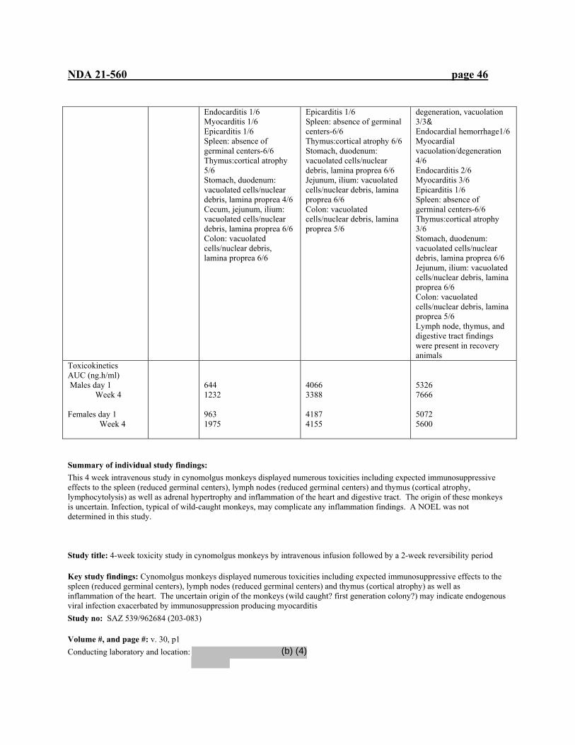
NDA 21-560 page 46
Endocarditis 1/6 Myocarditis 1/6 Epicarditis 1/6 Spleen: absence of germinal centers-6/6 Thymus:cortical atrophy 5/6 Stomach, duodenum: vacuolated cells/nuclear debris, lamina proprea 4/6 Cecum, jejunum, ilium: vacuolated cells/nuclear debris, lamina proprea 6/6 Colon: vacuolated cells/nuclear debris, lamina proprea 6/6
Epicarditis 1/6 Spleen: absence of germinal centers-6/6 Thymus:cortical atrophy 6/6 Stomach, duodenum: vacuolated cells/nuclear debris, lamina proprea 6/6 Jejunum, ilium: vacuolated cells/nuclear debris, lamina proprea 6/6 Colon: vacuolated cells/nuclear debris, lamina proprea 5/6
degeneration, vacuolation 3/3& Endocardial hemorrhage1/6 Myocardial vacuolation/degeneration 4/6 Endocarditis 2/6 Myocarditis 3/6 Epicarditis 1/6 Spleen: absence of germinal centers-6/6 Thymus:cortical atrophy 3/6 Stomach, duodenum: vacuolated cells/nuclear debris, lamina proprea 6/6 Jejunum, ilium: vacuolated cells/nuclear debris, lamina proprea 6/6 Colon: vacuolated cells/nuclear debris, lamina proprea 5/6 Lymph node, thymus, and digestive tract findings were present in recovery animals
Toxicokinetics AUC (ng.h/ml) Males day 1 Week 4 Females day 1 Week 4
644 1232 963 1975
4066 3388 4187 4155
5326 7666 5072 5600
Summary of individual study findings: This 4 week intravenous study in cynomolgus monkeys displayed numerous toxicities including expected immunosuppressive effects to the spleen (reduced germinal centers), lymph nodes (reduced germinal centers) and thymus (cortical atrophy, lymphocytolysis) as well as adrenal hypertrophy and inflammation of the heart and digestive tract. The origin of these monkeys is uncertain. Infection, typical of wild-caught monkeys, may complicate any inflammation findings. A NOEL was not determined in this study. Study title: 4-week toxicity study in cynomolgus monkeys by intravenous infusion followed by a 2-week reversibility period
Key study findings: Cynomolgus monkeys displayed numerous toxicities including expected immunosuppressive effects to the spleen (reduced germinal centers), lymph nodes (reduced germinal centers) and thymus (cortical atrophy) as well as inflammation of the heart. The uncertain origin of the monkeys (wild caught? first generation colony?) may indicate endogenous viral infection exacerbated by immunosuppression producing myocarditis Study no: SAZ 539/962684 (203-083)
Volume #, and page #: v. 30, p1 Conducting laboratory and location:
(b) (4)

NDA 21-560 page 47
Date of study initiation: 17 April 1996
GLP compliance: yes QA report: yes ( x ) no ( )
Drug, lot #, radiolabel, and % purity: SDZ RAD, Y187 0895 Formulation/vehicle: solution in Cremaphor EL and ethanol, in 5% glucose solution Methods (unique aspects):
Dosing: Species/strain: cynomolgus monkeys, #/sex/group or time point (main study): 3 Satellite groups used for toxicokinetics or recovery: 2 (control, 0.1 mg/kg) Age: 15-21 months (estimated; this may indicate wild caught monkeys) Weight: 2.0-3.1 kg Doses in administered units: 0.05, 0.1 mg/kg Route, form, volume, and infusion rate: intravenous, infusion, 5 ml/kg, 2 h
Observations and times: Clinical signs: daily Body weights: weekly during treatment, recovery Food consumption: weekly during treatment, recovery Ophthalmoscopy: prior to dosing, treatment week 4, recovery week 2 EKG: prior to dosing, treatment week 4, recovery week 2 Hematology: prior to dosing, treatment week 4, recovery week 2 Clinical chemistry: prior to dosing, treatment week 4, recovery week 2 Urinalysis: prior to dosing, treatment week 4, recovery week 2 (16 h collection period) Gross pathology: at necropsy Organs weighed: at necropsy, see table Histopathology: at necropsy, see table Toxicokinetics: days 1, 28; 0, 1, 3, 6, 22 h following infusion Other: na Results: Dose (mg/kg) 0 0.05 0.1 Recovery (control, 0.1
mg/kg) HED (mg/kg) 0 Mortality All animals survived until terminal sacrifice Clinical signs Huddled posture Huddled posture
Body weights Decrease 4/5%, 3/5 &
Food consumption Sl decrease Ophthalmoscopy
No treatment-related effect was observed
Electrocardiography
No treatment-related effect was observed
Hematology
decreased PT, %, week 4 increased fibrinogen, week 4
5/5 & increased leukocytes, week 4 decreased PT, %
(b) (4)
(b) (4)
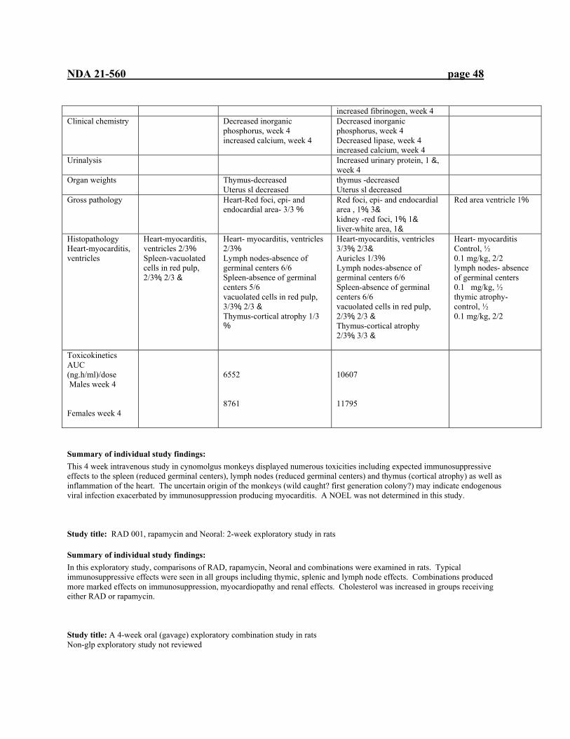
NDA 21-560 page 48
increased fibrinogen, week 4 Clinical chemistry Decreased inorganic
phosphorus, week 4 increased calcium, week 4
Decreased inorganic phosphorus, week 4 Decreased lipase, week 4 increased calcium, week 4
Urinalysis
Increased urinary protein, 1 &, week 4
Organ weights
Thymus-decreased Uterus sl decreased
thymus -decreased Uterus sl decreased
Gross pathology
Heart-Red foci, epi- and endocardial area- 3/3 %
Red foci, epi- and endocardial area , 1%, 3& kidney -red foci, 1%, 1& liver-white area, 1&
Red area ventricle 1%
Histopathology Heart-myocarditis, ventricles
Heart-myocarditis, ventricles 2/3% Spleen-vacuolated cells in red pulp, 2/3%, 2/3 &
Heart- myocarditis, ventricles 2/3% Lymph nodes-absence of germinal centers 6/6 Spleen-absence of germinal centers 5/6 vacuolated cells in red pulp, 3/3%, 2/3 & Thymus-cortical atrophy 1/3 %
Heart-myocarditis, ventricles 3/3%, 2/3& Auricles 1/3% Lymph nodes-absence of germinal centers 6/6 Spleen-absence of germinal centers 6/6 vacuolated cells in red pulp, 2/3%, 2/3 & Thymus-cortical atrophy 2/3%, 3/3 &
Heart- myocarditis Control, ½ 0.1 mg/kg, 2/2 lymph nodes- absence of germinal centers 0.1 mg/kg, ½ thymic atrophy- control, ½ 0.1 mg/kg, 2/2
Toxicokinetics AUC (ng.h/ml)/dose Males week 4 Females week 4
6552 8761
10607 11795
Summary of individual study findings: This 4 week intravenous study in cynomolgus monkeys displayed numerous toxicities including expected immunosuppressive effects to the spleen (reduced germinal centers), lymph nodes (reduced germinal centers) and thymus (cortical atrophy) as well as inflammation of the heart. The uncertain origin of the monkeys (wild caught? first generation colony?) may indicate endogenous viral infection exacerbated by immunosuppression producing myocarditis. A NOEL was not determined in this study. Study title: RAD 001, rapamycin and Neoral: 2-week exploratory study in rats
Summary of individual study findings: In this exploratory study, comparisons of RAD, rapamycin, Neoral and combinations were examined in rats. Typical immunosuppressive effects were seen in all groups including thymic, splenic and lymph node effects. Combinations produced more marked effects on immunosuppression, myocardiopathy and renal effects. Cholesterol was increased in groups receiving either RAD or rapamycin. Study title: A 4-week oral (gavage) exploratory combination study in rats Non-glp exploratory study not reviewed
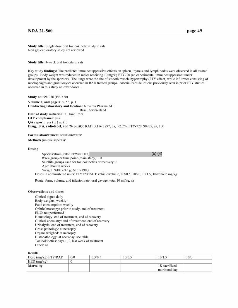
NDA 21-560 page 49
Study title: Single dose oral toxicokinetic study in rats Non glp exploratory study not reviewed Study title: 4-week oral toxicity in rats
Key study findings: The predicted immunosuppressive effects on spleen, thymus and lymph nodes were observed in all treated groups. Body weight was reduced in males receiving 10 mg/kg FTY720 (an experimental immunosuppressant under development by the sponsor). The lungs were the site of smooth muscle hypertrophy (FTY effect) while infiltrates consisting of macrophages and granulocytes occurred in RAD treated groups. Arterial/cardiac lesions previously seen in prior FTY studies occurred in this study at lower doses.
Study no: 991036 (BS-570) Volume #, and page #: v. 53, p. 1 Conducting laboratory and location: Novartis Pharma AG
Basel, Switzerland Date of study initiation: 21 June 1999 GLP compliance: yes QA report: yes ( x ) no ( ) Drug, lot #, radiolabel, and % purity: RAD, X176 1297, na, 92.2%; FTY-720, 98905, na, 100
Formulation/vehicle: solution/water Methods (unique aspects):
Dosing: Species/strain: rats/Crl:Wist Han, #/sex/group or time point (main study): 10 Satellite groups used for toxicokinetics or recovery: 6 Age: about 8 weeks Weight: %181-245 g, &135-190 g
Doses in administered units: FTY720/RAD: vehicle/vehicle, 0.3/0.5, 10/20, 10/1.5, 10/vehicle mg/kg
Route, form, volume, and infusion rate: oral gavage, total 10 ml/kg, na
Observations and times: Clinical signs: daily Body weights: weekly Food consumption: weekly Ophthalmoscopy: prior to study, end of treatment EKG: not performed Hematology: end of treatment, end of recovery Clinical chemistry: end of treatment, end of recovery Urinalysis: end of treatment, end of recovery Gross pathology: at necropsy Organs weighed: at necropsy Histopathology: at necropsy, see table Toxicokinetics: days 1, 2, last week of treatment Other: na
Results: Dose (mg/kg) FTY/RAD 0/0 0.3/0.5 10/0.5 10/1.5 10/0 HED (mg/kg) 0 Mortality 1& sacrificed
moribund day
(b) (4)

NDA 21-560 page 50
18 Clinical signs piloerection piloerection Body weights % decreased % decreased % decreased Food consumption % decreased-all study
& decreased wk 1, 2 % decreased-all study & decreased wk 1, 2
% decreased-all study & decreased wk 1, 2
Ophthalmoscopy
No treatment-related effects
EKG Not performed Hematology Lymphopenia Neutrophils RBC Hemoglobin Hematocrit
all inc, all inc, & inc, & inc, &
all inc, all inc, & inc, & inc, &
all inc, all inc, %, & inc, %, &
all inc, all inc, %
Clinical chemistry Bilirubin Serum urea Cholesterol Triglycerides $-, (-globulin Glucose Sodium Potassium Phosphate
Bilirubin-dec, & Cholesterol inc, % $-, (-globulin-dec, all
Serum urea-inc Bilirubin-dec, & Cholesterol inc, % Triglycerides inc, % Sodium-inc, % $-, (-globulin-dec, all
Serum urea-inc Bilirubin-dec, & Cholesterol inc, % Triglycerides inc, % Glucose dec, % Phosphate dec, % Sodium-inc, % Potassium-inc, % $-, (-globulin-dec, all
$-, (-globulin-dec, all
Urinalysis
% Sm spleen Oliguria, ketones
% Oliguria, ketones
Organ weights
Spleen sl inc, all Prostate sl dec Ovary sl dec Uterus sl dec
Lung inc, all Spleen sl inc, all Prostate sl dec Ovary sl dec Uterus sl dec
Lung inc, all Spleen sl inc, both Prostate sl dec Ovary sl dec Uterus sl dec Pituitary sl dec, both
Spleen sl inc, all
Gross pathology
Sm spleen Sm spleen Sm thymus Sm prostate
Sm spleen
Histopathology
Lungs:granulocyte infiltration Thymus: cortical atrophy, both Spleen: atrophy white pulp Lymph nodes: Atrophy
arteries; fibrinoid necrosis, periarteritis, arteriopathy, % Heart: necrotizing arteritis, arteriopathy, 2/10 % Lungs: sm muscle hypertrophy, both Interstitial collagenization, both
arteries; fibrinoid necrosis, periarteritis, arteriopathy, Both Heart: necrotizing arteritis, arteriopathy, 9/10 %, 6/10 & Lungs: sm muscle
Lungs: sm muscle hypertrophy, both Interstitial collagenization, both Alveolar macrophage infiltration
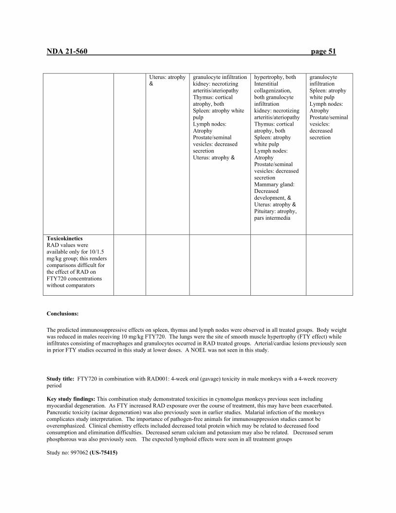
NDA 21-560 page 51
Uterus: atrophy &
granulocyte infiltration kidney: necrotizing arteritis/ateriopathy Thymus: cortical atrophy, both Spleen: atrophy white pulp Lymph nodes: Atrophy Prostate/seminal vesicles: decreased secretion Uterus: atrophy &
hypertrophy, both Interstitial collagenization, both granulocyte infiltration kidney: necrotizing arteritis/ateriopathy Thymus: cortical atrophy, both Spleen: atrophy white pulp Lymph nodes: Atrophy Prostate/seminal vesicles: decreased secretion Mammary gland: Decreased development, & Uterus: atrophy & Pituitary: atrophy, pars intermedia
granulocyte infiltration Spleen: atrophy white pulp Lymph nodes: Atrophy Prostate/seminal vesicles: decreased secretion
Toxicokinetics RAD values were available only for 10/1.5 mg/kg group; this renders comparisons difficult for the effect of RAD on FTY720 concentrations without comparators
Conclusions:
The predicted immunosuppressive effects on spleen, thymus and lymph nodes were observed in all treated groups. Body weight was reduced in males receiving 10 mg/kg FTY720. The lungs were the site of smooth muscle hypertrophy (FTY effect) while infiltrates consisting of macrophages and granulocytes occurred in RAD treated groups. Arterial/cardiac lesions previously seen in prior FTY studies occurred in this study at lower doses. A NOEL was not seen in this study. Study title: FTY720 in combination with RAD001: 4-week oral (gavage) toxicity in male monkeys with a 4-week recovery period Key study findings: This combination study demonstrated toxicities in cynomolgus monkeys previous seen including myocardial degeneration. As FTY increased RAD exposure over the course of treatment, this may have been exacerbated. Pancreatic toxicity (acinar degeneration) was also previously seen in earlier studies. Malarial infection of the monkeys complicates study interpretation. The importance of pathogen-free animals for immunosuppression studies cannot be overemphasized. Clinical chemistry effects included decreased total protein which may be related to decreased food consumption and elimination difficulties. Decreased serum calcium and potassium may also be related. Decreased serum phosphorous was also previously seen. The expected lymphoid effects were seen in all treatment groups Study no: 997062 (US-75415)
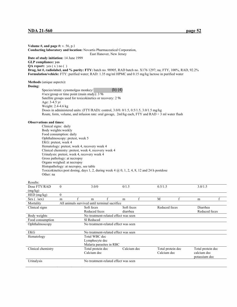
NDA 21-560 page 52
Volume #, and page #: v. 56, p.1 Conducting laboratory and location: Novartis Pharmaceutical Corporation, East Hanover, New Jersey Date of study initiation: 14 June 1999 GLP compliance: yes QA report: yes ( x ) no ( ) Drug, lot #, radiolabel, and % purity: FTY: batch no. 98905, RAD batch no. X176 1297; na; FTY, 100%, RAD, 92.2% Formulation/vehicle: FTY: purified water; RAD: 1.35 mg/ml HPMC and 0.15 mg/kg lactose in purified water Methods (unique aspects): Dosing:
Species/strain: cynomolgus monkey/ #/sex/group or time point (main study): 3 % Satellite groups used for toxicokinetics or recovery: 2 % Age: 3-4.5 yr Weight: 2.4-4.6 kg Doses in administered units: (FTY/RAD): control, 3.0/0. 0/1.5, 0.5/1.5, 3.0/1.5 mg/kg Route, form, volume, and infusion rate: oral gavage, 2ml/kg each, FTY and RAD + 3 ml water flush
Observations and times: Clinical signs: daily Body weights:weekly Food consumption: daily Ophthalmoscopy: pretest, week 5 EKG: pretest, week 4 Hematology: pretest, week 4, recovery week 4 Clinical chemistry: pretest, week 4, recovery week 4 Urinalysis: pretest, week 4, recovery week 4 Gross pathology: at necropsy Organs weighed: at necropsy Histopathology: at necropsy, see table Toxicokinetics:post dosing, days 1, 2, during week 4 @ 0, 1, 2, 4, 8, 12 and 24 h postdose Other: na Results: Dose FTY/RAD (mg/kg)
0 3.0/0 0/1.5 0.5/1.5 3.0/1.5
HED (mg/kg) 0 Sex ( /sex) m f m f m f M f m f Mortality All animals survived until terminal sacrifice Clinical signs Soft feces
Reduced feces Soft feces diarrhea
Reduced feces Diarrhea Reduced feces
Body weights No treatment-related effect was seen Food consumption Sl Reduced Ophthalmoscopy
No treatment-related effect was seen
EKG No treatment-related effect was seen Hematology
Total WBC dec Lymphocyte dec Malaria parasites in RBC
Clinical chemistry Total protein dec Calcium dec
Calcium dec Total protein dec Calcium dec
Total protein dec calcium dec potassium dec
Urinalysis
No treatment-related effect was seen
(b) (4)
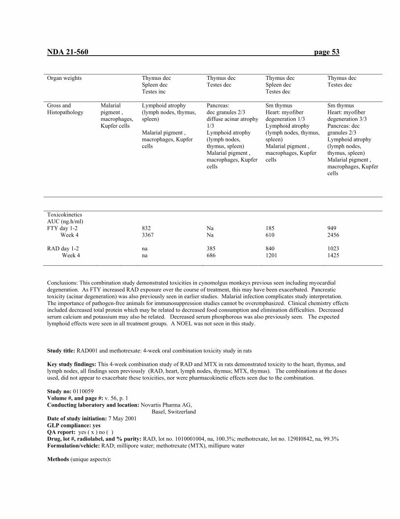
NDA 21-560 page 53
Organ weights
Thymus dec Spleen dec Testes inc
Thymus dec Testes dec
Thymus dec Spleen dec Testes dec
Thymus dec Testes dec
Gross and Histopathology
Malarial pigment , macrophages, Kupfer cells
Lymphoid atrophy (lymph nodes, thymus, spleen) Malarial pigment , macrophages, Kupfer cells
Pancreas: dec granules 2/3 diffuse acinar atrophy 1/3 Lymphoid atrophy (lymph nodes, thymus, spleen) Malarial pigment , macrophages, Kupfer cells
Sm thymus Heart: myofiber degeneration 1/3 Lymphoid atrophy (lymph nodes, thymus, spleen) Malarial pigment , macrophages, Kupfer cells
Sm thymus Heart: myofiber degeneration 3/3 Pancreas: dec granules 2/3 Lymphoid atrophy (lymph nodes, thymus, spleen) Malarial pigment , macrophages, Kupfer cells
Toxicokinetics AUC (ng.h/ml) FTY day 1-2 Week 4 RAD day 1-2 Week 4
832 3367 na na
Na Na 385 686
185 610 840 1201
949 2456 1023 1425
Conclusions: This combination study demonstrated toxicities in cynomolgus monkeys previous seen including myocardial degeneration. As FTY increased RAD exposure over the course of treatment, this may have been exacerbated. Pancreatic toxicity (acinar degeneration) was also previously seen in earlier studies. Malarial infection complicates study interpretation. The importance of pathogen-free animals for immunosuppression studies cannot be overemphasized. Clinical chemistry effects included decreased total protein which may be related to decreased food consumption and elimination difficulties. Decreased serum calcium and potassium may also be related. Decreased serum phosphorous was also previously seen. The expected lymphoid effects were seen in all treatment groups. A NOEL was not seen in this study. Study title: RAD001 and methotrexate: 4-week oral combination toxicity study in rats Key study findings: This 4-week combination study of RAD and MTX in rats demonstrated toxicity to the heart, thymus, and lymph nodes, all findings seen previously (RAD, heart, lymph nodes, thymus; MTX, thymus). The combinations at the doses used, did not appear to exacerbate these toxicities, nor were pharmacokinetic effects seen due to the combination. Study no: 0110059 Volume #, and page #: v. 56, p. 1 Conducting laboratory and location: Novartis Pharma AG, Basel, Switzerland Date of study initiation: 7 May 2001 GLP compliance: yes QA report: yes ( x ) no ( ) Drug, lot #, radiolabel, and % purity: RAD, lot no. 1010001004, na, 100.3%; methotrexate, lot no. 129H0842, na, 99.3% Formulation/vehicle: RAD; millipore water; methotrexate (MTX), millipure water Methods (unique aspects):
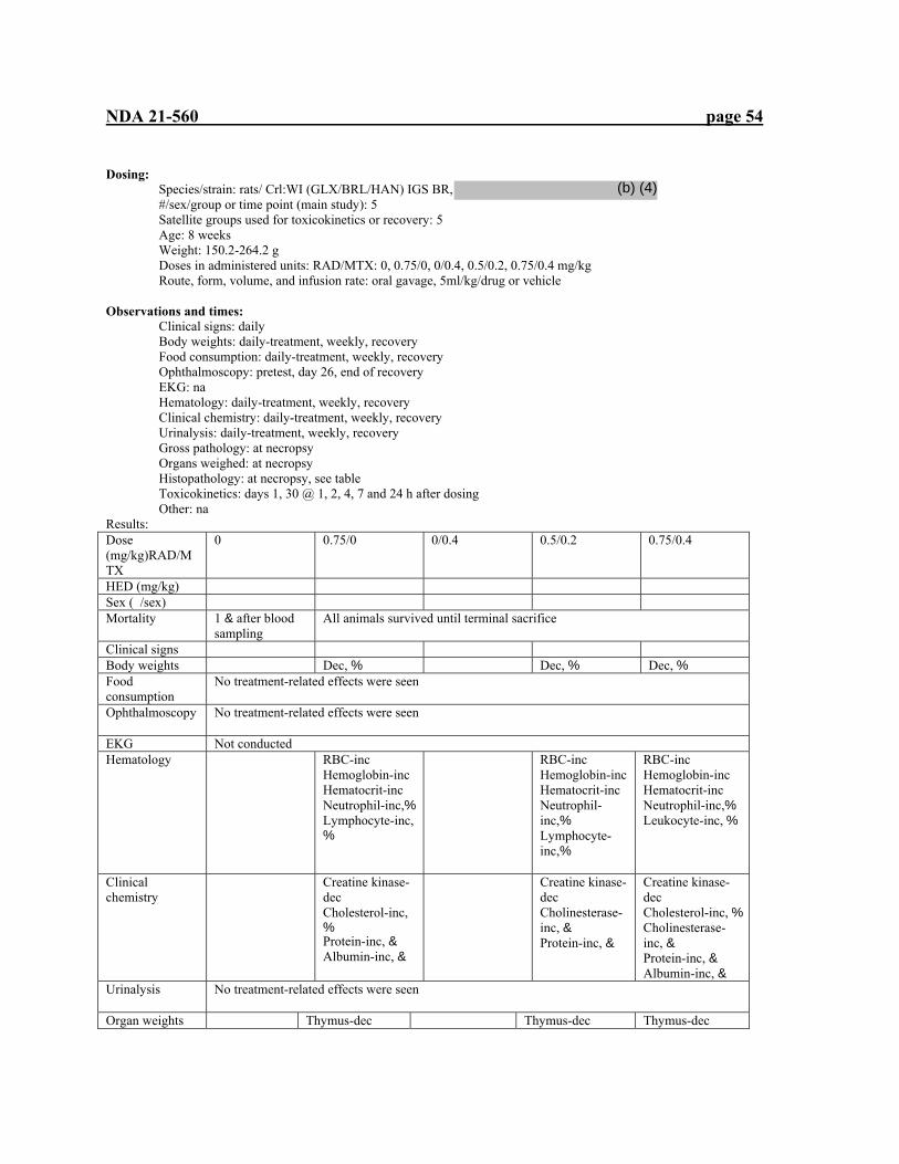
NDA 21-560 page 54
Dosing: Species/strain: rats/ Crl:WI (GLX/BRL/HAN) IGS BR, #/sex/group or time point (main study): 5 Satellite groups used for toxicokinetics or recovery: 5 Age: 8 weeks Weight: 150.2-264.2 g Doses in administered units: RAD/MTX: 0, 0.75/0, 0/0.4, 0.5/0.2, 0.75/0.4 mg/kg Route, form, volume, and infusion rate: oral gavage, 5ml/kg/drug or vehicle
Observations and times: Clinical signs: daily Body weights: daily-treatment, weekly, recovery Food consumption: daily-treatment, weekly, recovery Ophthalmoscopy: pretest, day 26, end of recovery EKG: na Hematology: daily-treatment, weekly, recovery Clinical chemistry: daily-treatment, weekly, recovery Urinalysis: daily-treatment, weekly, recovery Gross pathology: at necropsy Organs weighed: at necropsy Histopathology: at necropsy, see table Toxicokinetics: days 1, 30 @ 1, 2, 4, 7 and 24 h after dosing Other: na Results: Dose (mg/kg)RAD/MTX
0 0.75/0 0/0.4 0.5/0.2 0.75/0.4
HED (mg/kg) Sex ( /sex) Mortality 1 & after blood
sampling All animals survived until terminal sacrifice
Clinical signs Body weights Dec, % Dec, % Dec, % Food consumption
No treatment-related effects were seen
Ophthalmoscopy
No treatment-related effects were seen
EKG Not conducted Hematology
RBC-inc Hemoglobin-inc Hematocrit-inc Neutrophil-inc,% Lymphocyte-inc, %
RBC-inc Hemoglobin-inc Hematocrit-inc Neutrophil-inc,% Lymphocyte-inc,%
RBC-inc Hemoglobin-inc Hematocrit-inc Neutrophil-inc,% Leukocyte-inc, %
Clinical chemistry
Creatine kinase-dec Cholesterol-inc, % Protein-inc, & Albumin-inc, &
Creatine kinase-dec Cholinesterase-inc, & Protein-inc, &
Creatine kinase-dec Cholesterol-inc, % Cholinesterase-inc, & Protein-inc, & Albumin-inc, &
Urinalysis
No treatment-related effects were seen
Organ weights Thymus-dec Thymus-dec Thymus-dec
(b) (4)

NDA 21-560 page 55
Adrenals-inc, % Gross and Histopathology
Heart-myocardial degeneration, % Thymus: lymphocytolysis Mesenteric lymph node: germinal center-dec
Thymus: lymphocytolysis
Thymus: lymphocytolysis lymph node: germinal center-dec
Heart-myocardial degeneration, % Thymus: lymphocytolysis lymph node: germinal center-dec
Toxicokinetics Day 30 AUC (ng.h/ml) Males RAD MTX Females RAD MTX
26.1 38.4 22.4 38.4
14.6 45.9 23.9 39.6
23.0 59.8 18.2 32.4
Conclusions: This 4-week combination study of RAD and MTX in rats demonstrated toxicity to the heart, thymus, and lymph nodes, all findings seen previously (RAD, heart, lymph nodes, thymus; MTX, thymus). The combinations at the doses used, did not appear to exacerbate these toxicities, nor were pharmacokinetic effects seen due to the combination. As effects were seen in all treatment groups, a NOEL was not seen.
Study title: Comparative toxicity study in HanIbm Wistar rats with batches differing in by-product content
Key study findings: The immunosuppressive pharmacologic action of RAD was evident in the thymus, spleen, and lymph nodes. Immunosuppression may have allowed infection to result in myocarditis, otherwise it may be due to a direct toxic effect. Effects on the reproductive organs in both males (testes, prostate, epididymides, seminal vesicles) and females (uterus) appear to be a toxic effect on endocrine hormones. Lung eosinophilic deposits from macrophages may be due to immunosuppression or to a direct phospholipidosis effect. The eye effect of swelling/disruption of fibers in anterior cortex of the lens is similar to previously seen lens toxicity. The stressed RAD, presumed to contain the impurity (not quantified), did not appear to cause additional toxicities. At the stressed 0.25 mg/kg dose, uterine atrophy was seen but not in the unstressed RAD females. Otherwise existing toxicities were not exacerbated by the administration of stressed RAD.
Study no: 96/SPM091/0532 (203-076) Volume #, and page #: v. 65, p. 1 Conducting laboratory and location:
Date of study initiation: GLP compliance: yes QA report: yes ( x ) no ( ) Drug, lot #, radiolabel, and % purity: RAD, Y003 0196, 90.5%; Y254 1295, 82.2%; Y174 0895, 82.2% Formulation/vehicle: 2% microemulsion diluted with 5% glucose solution Methods (unique aspects):
Dosing: Species/strain: rats/HanIbm Wistar, #/sex/group or time point (main study): 10 Satellite groups used for toxicokinetics or recovery: 7 Age: 42 d
(b) (4)
(b) (4)
(b) (4)

NDA 21-560 page 56
Weight: 107-168 g Doses in administered units: RAD: 0.25, 5 mg/kg; stressed RAD 0.25, 5 mg/kg Route, form, volume, and infusion rate: oral gavage, total 5 ml/kg, na
Observations and times: Clinical signs: daily Body weights: twice weekly Food consumption: weekly Ophthalmoscopy: prior to study, day 13 EKG: not performed Hematology: day 14 Clinical chemistry: day 14 Urinalysis: day 10 Gross pathology: at necropsy Organs weighed: at necropsy Histopathology: at necropsy, see table Toxicokinetics: day 14, 0.5, 1, 3, 7, 24 h after dosing
Other: na Dose (mg/kg) 0 Vehicle
stressed 0.25 5.0 0.25
stressed 5.0 stressed
HED (mg/kg) 0 Mortality All animals survived until sacrifice Clinical signs No treatment-related signs were seen Body weights Dec gain Dec gain Food consumption Dec dec Ophthalmoscopy
No treatment-related signs were seen
EKG Not performed Hematology
PCV: inc HgB: inc RBC: inc���
PCV: inc HgB: inc RBC: inc�lymphocytes: dec������
PCV: inc HgB: inc RBC: inc���
Clinical chemistry
Triglycerides: inc Cholesterol: inc Sodium: inc, females
Amylase: inc Alk phos: dec Triglycerides: inc Cholesterol: inc Sodium: inc Phosphorus: dec Albumin: dec, males
Triglycerides: inc Cholesterol: inc
Amylase: inc Alk phos: dec Triglycerides: inc Cholesterol: inc Sodium: inc, females Phosphorus: dec Albumin:dec
Urinalysis No treatment-related signs were seen
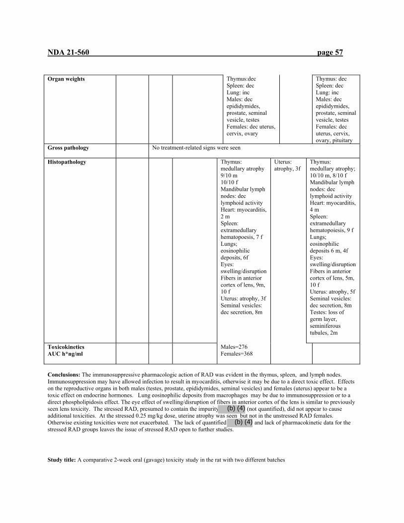
NDA 21-560 page 57
Organ weights
Thymus:dec Spleen: dec Lung: inc Males: dec epididymides, prostate, seminal vesicle, testes Females: dec uterus, cervix, ovary
Thymus: dec Spleen: dec Lung: inc Males: dec epididymides, prostate, seminal vesicle, testes Females: dec uterus, cervix, ovary, pituitary
Gross pathology
No treatment-related signs were seen
Histopathology
Thymus: medullary atrophy 9/10 m 10/10 f Mandibular lymph nodes: dec lymphoid activity Heart: myocarditis, 2 m Spleen: extramedullary hematopoesis, 7 f Lungs; eosinophilic deposits, 6f Eyes: swelling/disruption Fibers in anterior cortex of lens, 9m, 10 f Uterus: atrophy, 3f Seminal vesicles: dec secretion, 8m
Uterus: atrophy, 3f
Thymus: medullary atrophy; 10/10 m, 8/10 f Mandibular lymph nodes: dec lymphoid activity Heart: myocarditis, 4 m Spleen: extramedullary hematopoiesis, 9 f Lungs; eosinophilic deposits 6 m, 4f Eyes: swelling/disruption Fibers in anterior cortex of lens, 5m, 10 f Uterus: atrophy, 5f Seminal vesicles: dec secretion, 8m Testes: loss of germ layer, seminiferous tubules, 2m
Toxicokinetics AUC h*ng/ml
Males=276 Females=368
Conclusions: The immunosuppressive pharmacologic action of RAD was evident in the thymus, spleen, and lymph nodes. Immunosuppression may have allowed infection to result in myocarditis, otherwise it may be due to a direct toxic effect. Effects on the reproductive organs in both males (testes, prostate, epididymides, seminal vesicles) and females (uterus) appear to be a toxic effect on endocrine hormones. Lung eosinophilic deposits from macrophages may be due to immunosuppression or to a direct phospholipidosis effect. The eye effect of swelling/disruption of fibers in anterior cortex of the lens is similar to previously seen lens toxicity. The stressed RAD, presumed to contain the impurity (not quantified), did not appear to cause additional toxicities. At the stressed 0.25 mg/kg dose, uterine atrophy was seen but not in the unstressed RAD females. Otherwise existing toxicities were not exacerbated. The lack of quantified and lack of pharmacokinetic data for the stressed RAD groups leaves the issue of stressed RAD open to further studies.
Study title: A comparative 2-week oral (gavage) toxicity study in the rat with two different batches
(b) (4)
(b) (4)
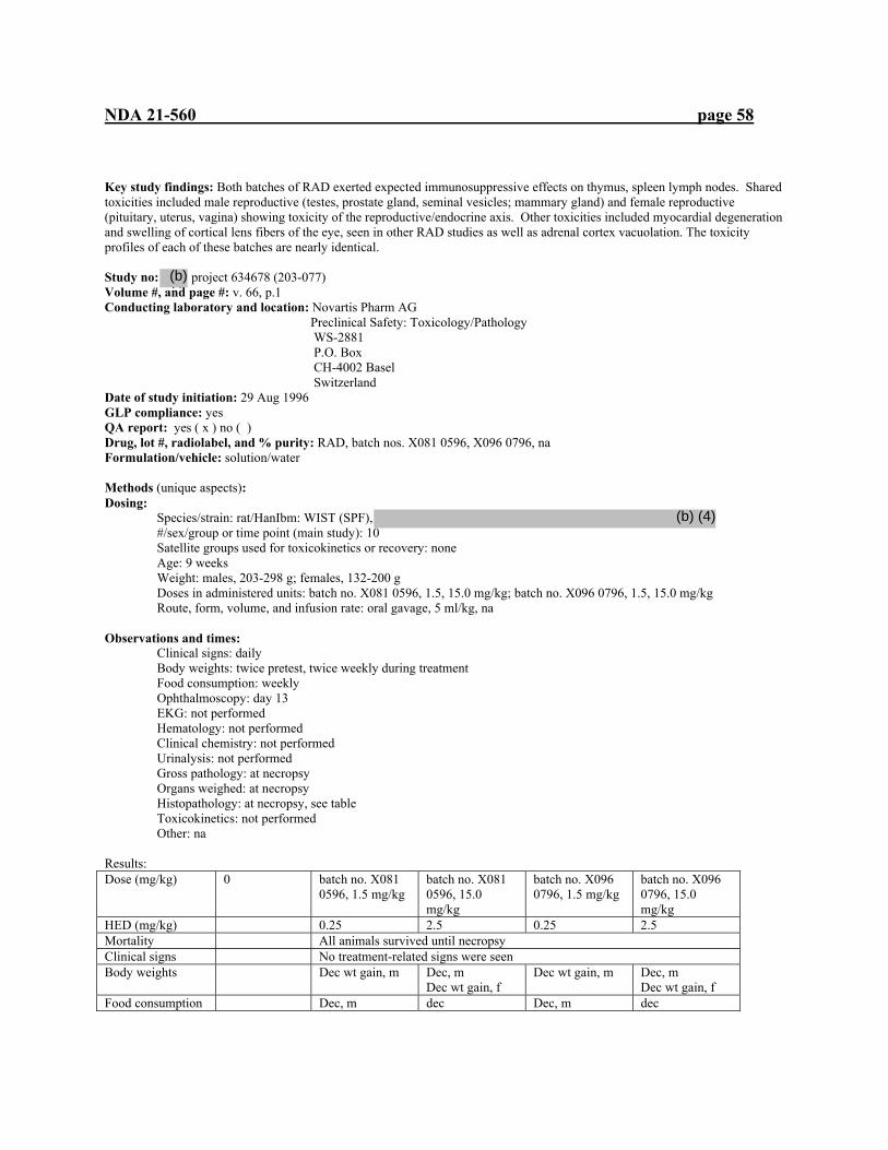
NDA 21-560 page 58
Key study findings: Both batches of RAD exerted expected immunosuppressive effects on thymus, spleen lymph nodes. Shared toxicities included male reproductive (testes, prostate gland, seminal vesicles; mammary gland) and female reproductive (pituitary, uterus, vagina) showing toxicity of the reproductive/endocrine axis. Other toxicities included myocardial degeneration and swelling of cortical lens fibers of the eye, seen in other RAD studies as well as adrenal cortex vacuolation. The toxicity profiles of each of these batches are nearly identical. Study no: project 634678 (203-077) Volume #, and page #: v. 66, p.1 Conducting laboratory and location: Novartis Pharm AG Preclinical Safety: Toxicology/Pathology WS-2881 P.O. Box CH-4002 Basel Switzerland Date of study initiation: 29 Aug 1996 GLP compliance: yes QA report: yes ( x ) no ( ) Drug, lot #, radiolabel, and % purity: RAD, batch nos. X081 0596, X096 0796, na Formulation/vehicle: solution/water Methods (unique aspects): Dosing:
Species/strain: rat/HanIbm: WIST (SPF), #/sex/group or time point (main study): 10 Satellite groups used for toxicokinetics or recovery: none Age: 9 weeks Weight: males, 203-298 g; females, 132-200 g Doses in administered units: batch no. X081 0596, 1.5, 15.0 mg/kg; batch no. X096 0796, 1.5, 15.0 mg/kg Route, form, volume, and infusion rate: oral gavage, 5 ml/kg, na
Observations and times: Clinical signs: daily Body weights: twice pretest, twice weekly during treatment Food consumption: weekly Ophthalmoscopy: day 13 EKG: not performed Hematology: not performed Clinical chemistry: not performed Urinalysis: not performed Gross pathology: at necropsy Organs weighed: at necropsy Histopathology: at necropsy, see table Toxicokinetics: not performed Other: na Results: Dose (mg/kg) 0 batch no. X081
0596, 1.5 mg/kg batch no. X081 0596, 15.0 mg/kg
batch no. X096 0796, 1.5 mg/kg
batch no. X096 0796, 15.0 mg/kg
HED (mg/kg) 0.25 2.5 0.25 2.5 Mortality All animals survived until necropsy Clinical signs No treatment-related signs were seen Body weights Dec wt gain, m Dec, m
Dec wt gain, f Dec wt gain, m Dec, m
Dec wt gain, f Food consumption Dec, m dec Dec, m dec
(b) (4)
(b) (4)
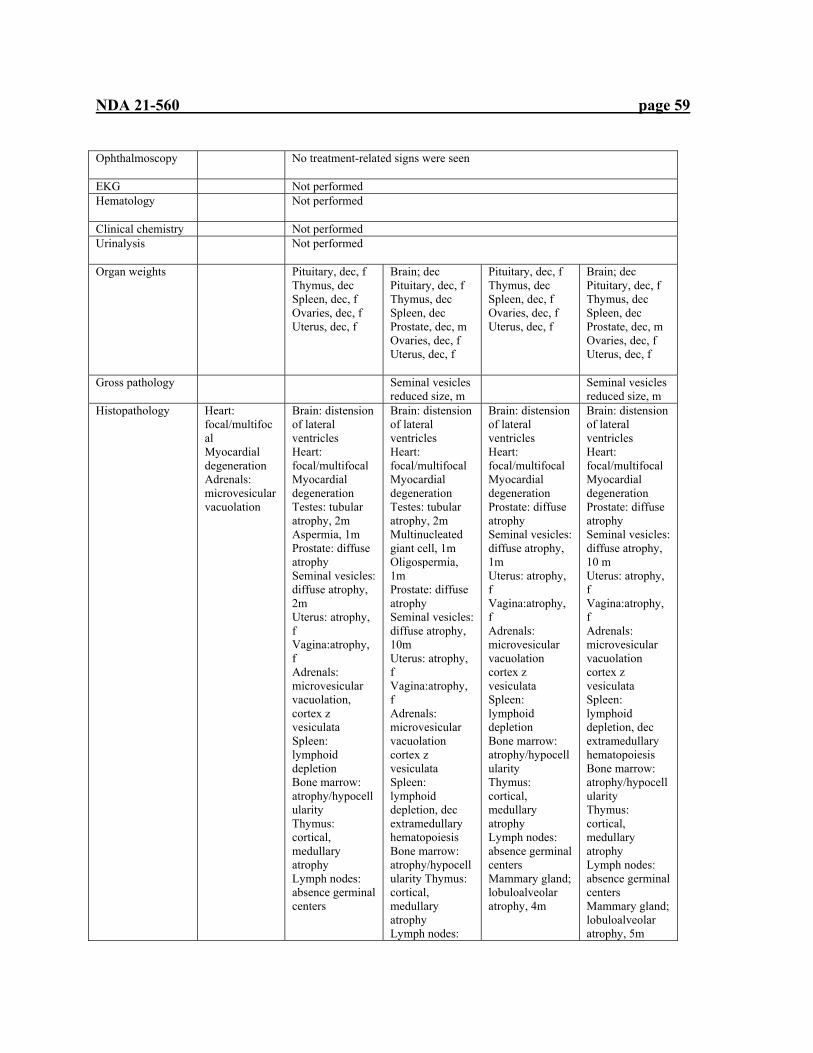
NDA 21-560 page 59
Ophthalmoscopy
No treatment-related signs were seen
EKG Not performed Hematology
Not performed
Clinical chemistry Not performed Urinalysis
Not performed
Organ weights
Pituitary, dec, f Thymus, dec Spleen, dec, f Ovaries, dec, f Uterus, dec, f
Brain; dec Pituitary, dec, f Thymus, dec Spleen, dec Prostate, dec, m Ovaries, dec, f Uterus, dec, f
Pituitary, dec, f Thymus, dec Spleen, dec, f Ovaries, dec, f Uterus, dec, f
Brain; dec Pituitary, dec, f Thymus, dec Spleen, dec Prostate, dec, m Ovaries, dec, f Uterus, dec, f
Gross pathology
Seminal vesicles reduced size, m
Seminal vesicles reduced size, m
Histopathology
Heart: focal/multifocal Myocardial degeneration Adrenals: microvesicular vacuolation
Brain: distension of lateral ventricles Heart: focal/multifocal Myocardial degeneration Testes: tubular atrophy, 2m Aspermia, 1m Prostate: diffuse atrophy Seminal vesicles: diffuse atrophy, 2m Uterus: atrophy, f Vagina:atrophy, f Adrenals: microvesicular vacuolation, cortex z vesiculata Spleen: lymphoid depletion Bone marrow: atrophy/hypocellularity Thymus: cortical, medullary atrophy Lymph nodes: absence germinal centers
Brain: distension of lateral ventricles Heart: focal/multifocal Myocardial degeneration Testes: tubular atrophy, 2m Multinucleated giant cell, 1m Oligospermia, 1m Prostate: diffuse atrophy Seminal vesicles: diffuse atrophy, 10m Uterus: atrophy, f Vagina:atrophy, f Adrenals: microvesicular vacuolation cortex z vesiculata Spleen: lymphoid depletion, dec extramedullary hematopoiesis Bone marrow: atrophy/hypocellularity Thymus: cortical, medullary atrophy Lymph nodes:
Brain: distension of lateral ventricles Heart: focal/multifocal Myocardial degeneration Prostate: diffuse atrophy Seminal vesicles: diffuse atrophy, 1m Uterus: atrophy, f Vagina:atrophy, f Adrenals: microvesicular vacuolation cortex z vesiculata Spleen: lymphoid depletion Bone marrow: atrophy/hypocellularity Thymus: cortical, medullary atrophy Lymph nodes: absence germinal centers Mammary gland; lobuloalveolar atrophy, 4m
Brain: distension of lateral ventricles Heart: focal/multifocal Myocardial degeneration Prostate: diffuse atrophy Seminal vesicles: diffuse atrophy, 10 m Uterus: atrophy, f Vagina:atrophy, f Adrenals: microvesicular vacuolation cortex z vesiculata Spleen: lymphoid depletion, dec extramedullary hematopoiesis Bone marrow: atrophy/hypocellularity Thymus: cortical, medullary atrophy Lymph nodes: absence germinal centers Mammary gland; lobuloalveolar atrophy, 5m

NDA 21-560 page 60
absence germinal centers Mammary gland; lobuloalveolar atrophy, 4m Eyes; swelling of cortical lens fibers
Eyes; swelling of cortical lens fibers
Toxicokinetics
Not performed
Conclusions: Both batches of RAD exerted expected immunosuppressive effects on thymus, spleen lymph nodes. Shared toxicities included male reproductive (testes, prostate gland, seminal vesicles; mammary gland) and female reproductive (pituitary, uterus, vagina) showing toxicity of the reproductive/endocrine axis. Other toxicities included myocardial degeneration and swelling of cortical lens fibers of the eye, seen in other RAD studies as well as adrenal cortex vacuolation. The toxicity profiles of each of these batches are nearly identical. No NOEL was seen in this study. Study title: 4-week oral toxicity study in rats (batch comparison) Key study findings: Both batches of RAD exerted expected immunosuppressive effects on thymus, spleen and lymph nodes. Other toxicities included male reproductive (testes, prostate gland, seminal vesicles, epididymides; mammary gland) and female reproductive (pituitary, ovaries, uterus, vagina) showed toxicity of the reproductive/endocrine axis. Other toxicities included myocardial degeneration and swelling of cortical lens fibers of the eye, seen in other RAD studies as well as adrenal cortex vacuolation. Batch 1 also had renal vasculopathy, seen in other rat studies. Otherwise, the toxicity profiles of each of these batches are nearly identical. Study no: 991094 Volume #, and page #: v. 66, p. 352 Conducting laboratory and location: Novartis Pharm AG Preclinical Safety: Toxicology/Pathology WS-2881 P.O. Box CH-4002 Basel Switzerland Date of study initiation: 10 Nov 1999 GLP compliance: yes QA report: yes ( x ) no ( ) Drug, lot #, radiolabel, and % purity: RAD, batch no. X033 0199 (batch 1), na, 92.9%; batch no. X153 1098 (batch 2), na, 96.9% Formulation/vehicle: dispersion of RAD in demineralized water Methods (unique aspects): Dosing:
Species/strain: rats? Crl:Wist Han, #/sex/group or time point (main study): 10 Satellite groups used for toxicokinetics or recovery: none Age: 7 weeks Weight: 112-184 g Doses in administered units: 0, 1.5(batch 1), 15 (day1)-5 (batch 1), 15 (day1)-5 (batch 2) mg/kg Route, form, volume, and infusion rate: oral gavage, 10 ml/kg, na
Observations and times:
(b) (4)

NDA 21-560 page 61
Clinical signs: daily Body weights: daily Food consumption: weekly Ophthalmoscopy: pretest, end of treatment EKG: not performed Hematology: day 23 Clinical chemistry: day 23 Urinalysis: day 23 Gross pathology: at necropsy Organs weighed: at necropsy Histopathology: at necropsy, see table Toxicokinetics: not performed Other: na Results: Dose (mg/kg) 0 1.5 mg/kg (batch 1) 15 (day1), 5 mg.kg
batch 1 15 (day1)-5 batch 2
HED (mg/kg) 0.25 25.0, 0.85 Mortality All rats survived until necropsy Clinical signs No treatment related signs were seen Body weights Dec body weight
gain Dec body weight gain
Dec body weight gain
Food consumption
Dec Dec Dec
Ophthalmoscopy
No treatment related signs were seen
EKG Not performed Hematology
RBCs: inc Hematocrit: inc Hemoglobin:inc Platelets: dec m Lymphocytes: dec Neutrophils: inc
RBCs: inc Hematocrit: inc Hemoglobin:inc Platelets: dec Lymphocytes: dec Neutrophils: inc
RBCs: inc Hematocrit: inc Hemoglobin:inc Platelets: dec Lymphocytes: dec Neutrophils: inc
Clinical chemistry
Cholesterol: inc, m Triglycerides: inc, m
Cholesterol: inc, m Triglycerides: inc, m
Cholesterol: inc, m Triglycerides: inc, m
Urinalysis
Leukocytes observed in urine of males
Organ weights
Thymus:dec Pituitary:dec
Thymus:dec Spleen: dec Pituitary:dec Adrenals:dec, f Testes:dec, m Prostate:dec, m Ovaries:dec,f Uterus:dec, f Brain:dec
Thymus:dec Spleen:dec Pituitary:dec Adrenals: dec, f Testes:dec, m Prostate:dec, m Ovaries:dec, f Uterus:dec, f Brain:dec
Gross pathology
Seminal Thymus:sm Thymus:sm Testes:sm Thymus:sm Prostate:sm Alopecia/scars: 1m
Thymus:sm testes:sm Thymus:sm Alopecia/scars: 3f
seminal vesicles: Heart: myocardial Heart: myocardial

NDA 21-560 page 62
Histopathology dec secretion kidney: hyaline droplet formation, m
degeneration Lungs:eosinophilic material in alveoli Thymus:cortical/ medullary atrophy lymph nodes: absence of germinal centers ovaries: dec follicles, hypertrophy of interstitial cells uterus: atrophy testes: atrophy epididymides:atrophy seminal vesicles: dec secretion mammaries: dec development, m salivary glands; depletion secretory granules eyes: swelling of lenticular fibers kidney: vasculopathy; hyaline droplet formation, m bone: depletion of cortical bone(femur) thyroid: inc sm follicles
degeneration Lungs:eosinophilic material in alveoli Thymus:cortical/medullary atrophy lymph nodes: absence of germinal centers ovaries: dec follicles, hypertrophy of interstitial cells uterus: atrophy testes: atrophy epididymides:atrophy seminal vesicles: dec secretion mammaries: dec development, m salivary glands; depletion secretory granules eyes: swelling of lenticular fibers kidney: hyaline droplet formation, m bone: depletion of cortical bone (femur) thyroid: inc sm follicles
Toxicokinetics
Not performed
CONCLUSIONS: Both batches of RAD exerted expected immunosuppressive effects on thymus, spleen and lymph nodes. Other toxicities included male reproductive (testes, prostate gland, seminal vesicles, epididymides; mammary gland) and female reproductive (pituitary, ovaries, uterus, vagina) showed toxicity of the reproductive/endocrine axis. Other toxicities included myocardial degeneration and swelling of cortical lens fibers of the eye, seen in other RAD studies as well as adrenal cortex vacuolation. Batch 1 also had renal vasculopathy, seen in other rat studies. Otherwise, the toxicity profiles of each of these batches are nearly identical. No NOEL was seen in this study. This study extends the findings of the 2-week study at 1.5 and 15 mg/kg to 1.5 and 5 mg/kg at 4 weeks.
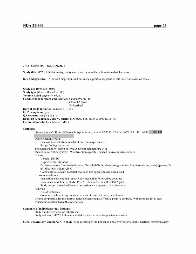
NDA 21-560 page 63
3.4.4 GENETIC TOXICOLOGY Study title: SDZ RAD 666: mutagenicity test using Salmonella typhimurium (batch control) Key findings: SDZ RAD (solid dispersion) did not cause a positive response in this bacterial reversion assay Study no: 10/96 (203-040) Study type (if not reflected in title): Volume #, and page #: v. 67, p. 1 Conducting laboratory and location: Sandoz Pharm Ltd. CH-4002 Basel
Switzerland Date of study initiation: January 31, 1996 GLP compliance: yes QA reports: yes ( x ) no ( ) Drug, lot #, radiolabel, and % purity: SDZ RAD 666, batch 95905, na, 95.4% Formulation/vehicle: solution, DMSO Methods:
Strains/species/cell line: Salmonella typhimurium, strains TA1535, TA97a, TA98, TA100, TA102
Dose selection criteria: Basis of dose selection: results of previous experiments Range finding studies: np
Test agent stability: stable in DMSO at room temperature 24 h Metabolic activation system: S9 rat liver homogenate, induced in vivo by Araclor 1254 Controls:
Vehicle: DMSO Negative controls: none Positive controls: 2-aminoanthracene, N-methyl-N-nitro-N-nitrosoguanidine, 9-aminoacridine, benz(a)pyrene, 2-nitrofluorene, mitomycin C Comments: a standard bacterial reversion test appears to have been used
Exposure conditions: Incubation and sampling times: 3 day incubation followed by counting Doses used in definitive study: 1562.5, 3125, 6250, 12500, 25000 :g/ml Study design: a standard bacterial reversion test appears to have been used
Analysis: No. of replicates: 3 Counting method: image analyzer counts of revertant bacterial colonies
Criteria for positive results: normal range solvent counts, effective positive controls, with response for at least concentration/strain twice that of controls
Summary of individual study findings:
Study validity: criteria for validity met Study outcome: SDZ RAD treatment did not meet criteria for positive reversion
Genetic toxicology summary: SDZ RAD (solid dispersion) did not cause a positive response in this bacterial reversion assay
(b) (4)
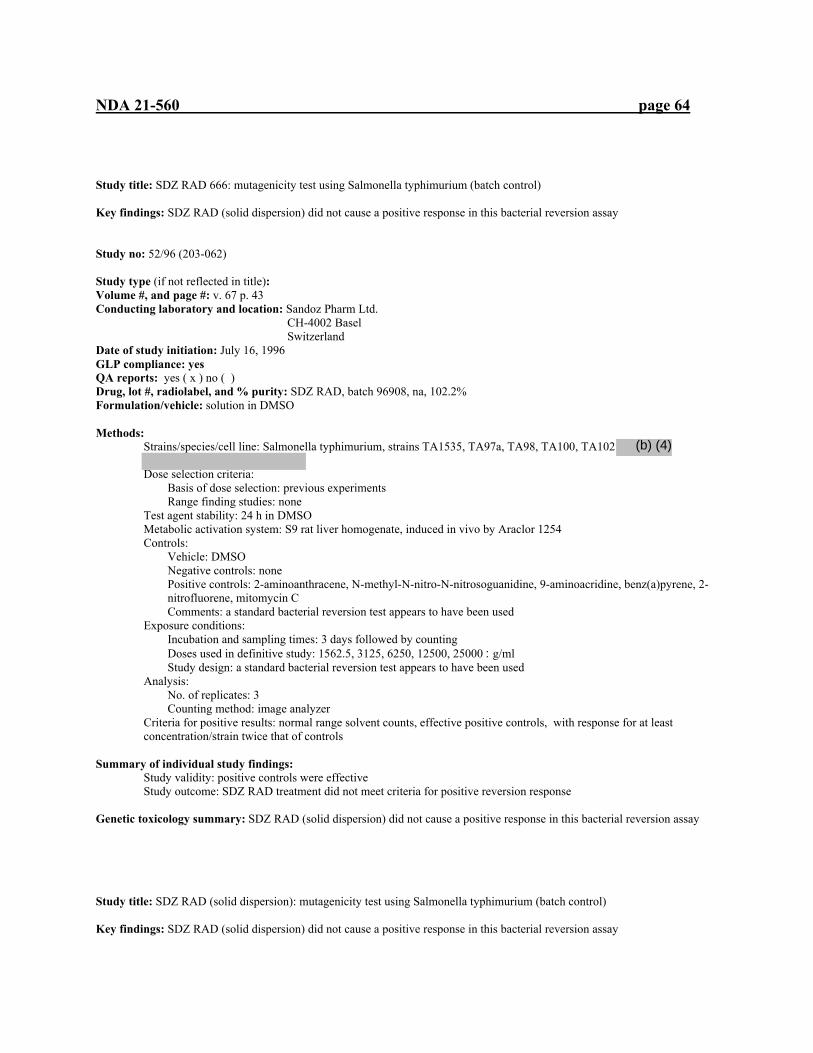
NDA 21-560 page 64
Study title: SDZ RAD 666: mutagenicity test using Salmonella typhimurium (batch control) Key findings: SDZ RAD (solid dispersion) did not cause a positive response in this bacterial reversion assay Study no: 52/96 (203-062) Study type (if not reflected in title): Volume #, and page #: v. 67 p. 43 Conducting laboratory and location: Sandoz Pharm Ltd. CH-4002 Basel
Switzerland Date of study initiation: July 16, 1996 GLP compliance: yes QA reports: yes ( x ) no ( ) Drug, lot #, radiolabel, and % purity: SDZ RAD, batch 96908, na, 102.2% Formulation/vehicle: solution in DMSO Methods:
Strains/species/cell line: Salmonella typhimurium, strains TA1535, TA97a, TA98, TA100, TA102
Dose selection criteria: Basis of dose selection: previous experiments Range finding studies: none
Test agent stability: 24 h in DMSO Metabolic activation system: S9 rat liver homogenate, induced in vivo by Araclor 1254 Controls:
Vehicle: DMSO Negative controls: none Positive controls: 2-aminoanthracene, N-methyl-N-nitro-N-nitrosoguanidine, 9-aminoacridine, benz(a)pyrene, 2-nitrofluorene, mitomycin C Comments: a standard bacterial reversion test appears to have been used
Exposure conditions: Incubation and sampling times: 3 days followed by counting Doses used in definitive study: 1562.5, 3125, 6250, 12500, 25000 :g/ml Study design: a standard bacterial reversion test appears to have been used
Analysis: No. of replicates: 3 Counting method: image analyzer
Criteria for positive results: normal range solvent counts, effective positive controls, with response for at least concentration/strain twice that of controls
Summary of individual study findings:
Study validity: positive controls were effective Study outcome: SDZ RAD treatment did not meet criteria for positive reversion response
Genetic toxicology summary: SDZ RAD (solid dispersion) did not cause a positive response in this bacterial reversion assay Study title: SDZ RAD (solid dispersion): mutagenicity test using Salmonella typhimurium (batch control) Key findings: SDZ RAD (solid dispersion) did not cause a positive response in this bacterial reversion assay
(b) (4)

NDA 21-560 page 65
Study no: 66/96 (203-063) Study type (if not reflected in title): Volume #, and page #: v. 67, p. 80 Conducting laboratory and location: Sandoz Pharm Ltd. CH-4002 Basel
Switzerland Date of study initiation: Aug 28, 1996 GLP compliance: yes QA reports: yes ( x ) no ( ) Drug, lot #, radiolabel, and % purity: SDZ RAD (solid dispersion), batch no. X096 0796, na, 104.3% Formulation/vehicle: solution in DMSO Methods:
Strains/species/cell line: Salmonella typhimurium, strains TA1535, TA97a, TA98, TA100, TA102
Dose selection criteria: Basis of dose selection: previous experiments Range finding studies: none
Test agent stability: 24 h at room temperature Metabolic activation system: S9 rat liver homogenate, induced in vivo by Araclor 1254 Controls:
Vehicle: DMSO Negative controls: none Positive controls: 2-aminoanthracene, N-methyl-N-nitro-N-nitrosoguanidine, 9-aminoacridine, benz(a)pyrene, 2-nitrofluorene, mitomycin C Comments: a standard bacterial reversion test appears to have been used
Exposure conditions: Incubation and sampling times: 3 day incubation followed by counting Doses used in definitive study: 14.54, 72.72, 363.60, 1818, 9090 :g/ml Study design: a standard bacterial reversion test appears to have been used
Analysis: No. of replicates: 3 Counting method: image analyzer
Criteria for positive results: normal range solvent counts, effective positive controls, with response for at least concentration/strain twice that of controls
Summary of individual study findings:
Study validity: positive controls were effective Study outcome: SDZ RAD did not meet criteria of an increased reversion rate (doubling)
Genetic toxicology summary: SDZ RAD (solid dispersion) did not cause a positive response in this bacterial reversion assay. Study title: RAD 001: mutagenicity test using Salmonella typhimurium (batch control) Key findings: SDZ RAD (solid dispersion) did not cause a positive response in this bacterial reversion assay Study no: 001801 (BS-482) Study type (if not reflected in title): Volume #, and page #: v67, p. 121 Conducting laboratory and location: Sandoz Pharm Ltd. CH-4002 Basel
(b) (4)
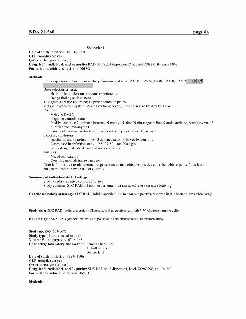
NDA 21-560 page 66
Switzerland Date of study initiation: Jan 26, 2000 GLP compliance: yes QA reports: yes ( x ) no ( ) Drug, lot #, radiolabel, and % purity: RAD 001 (solid dispersion 2%), batch X033 0199, na, 95.0% Formulation/vehicle: solution in DMSO Methods:
Strains/species/cell line: Salmonella typhimurium, strains TA1535, TA97a, TA98, TA100, TA102
Dose selection criteria: Basis of dose selection: previous experiments Range finding studies: none
Test agent stability: not tested; no precipitation on plates Metabolic activation system: S9 rat liver homogenate, induced in vivo by Araclor 1254 Controls:
Vehicle: DMSO Negative controls: none Positive controls: 2-aminoanthracene, N-methyl-N-nitro-N-nitrosoguanidine, 9-aminoacridine, benz(a)pyrene, 2-nitrofluorene, mitomycin C Comments: a standard bacterial reversion test appears to have been used
Exposure conditions: Incubation and sampling times: 3 day incubation followed by counting Doses used in definitive study: 12.5, 25, 50, 100, 200 :g/ml Study design: standard bacterial reversion assay
Analysis: No. of replicates: 3 Counting method: image analyzer
Criteria for positive results: normal range solvent counts, effective positive controls, with response for at least concentration/strain twice that of controls
Summary of individual study findings:
Study validity: positive controls effective Study outcome: SDZ RAD did not meet criteria of an increased reversion rate (doubling).
Genetic toxicology summary: SDZ RAD (solid dispersion) did not cause a positive response in this bacterial reversion assay Study title: SDZ RAD (solid dispersion) Chromosomal aberration test with V79 Chinese hamster cells Key findings: SDZ RAD (dispersion) was not positive in this chromosomal aberration study. Study no: Z63 (203-067) Study type (if not reflected in title): Volume #, and page #: v. 67, p. 148 Conducting laboratory and location: Sandoz Pharm Ltd. CH-4002 Basel
Switzerland Date of study initiation: Feb 9, 1996 GLP compliance: yes QA reports: yes ( x ) no ( ) Drug, lot #, radiolabel, and % purity: SDZ RAD solid dispersion, batch X0960796, na, 104.3% Formulation/vehicle: solution in DMSO Methods:
(b) (4)
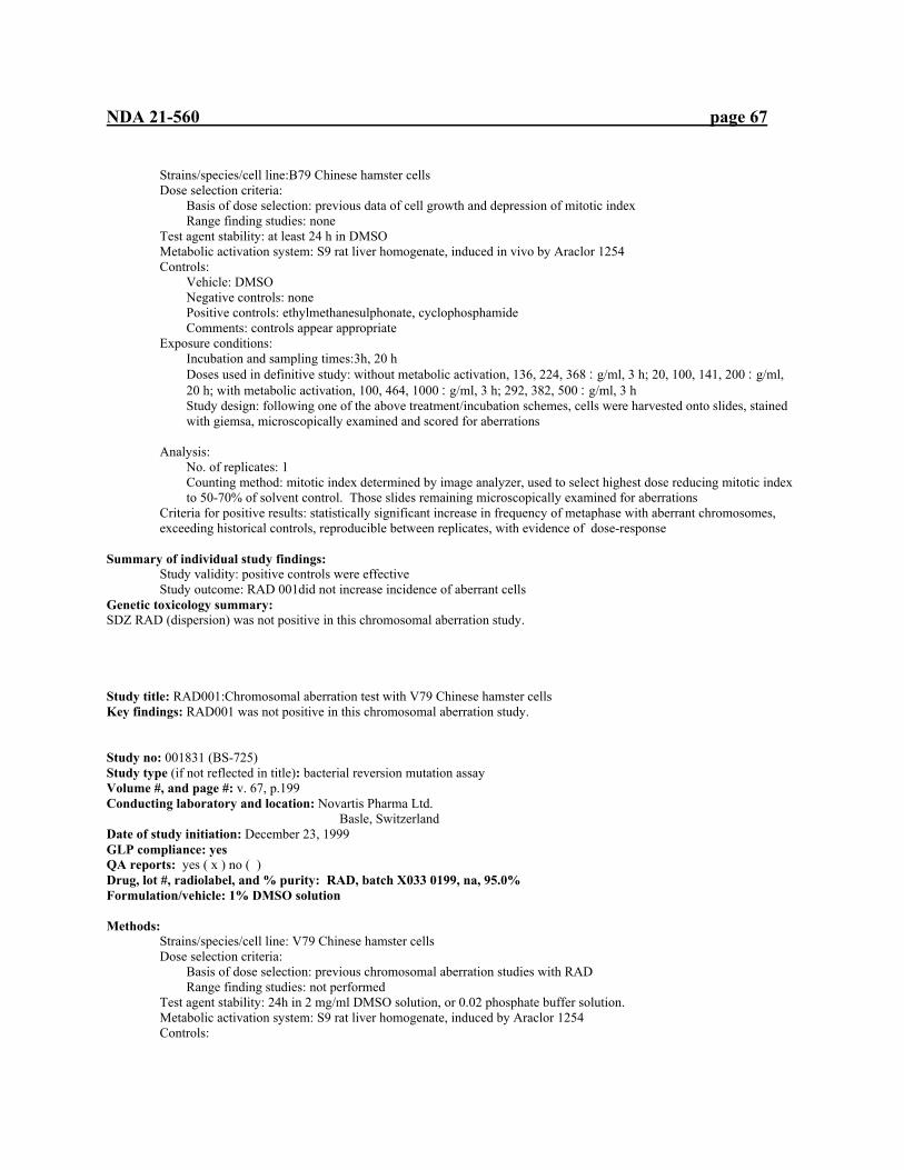
NDA 21-560 page 67
Strains/species/cell line:B79 Chinese hamster cells Dose selection criteria:
Basis of dose selection: previous data of cell growth and depression of mitotic index Range finding studies: none
Test agent stability: at least 24 h in DMSO Metabolic activation system: S9 rat liver homogenate, induced in vivo by Araclor 1254 Controls:
Vehicle: DMSO Negative controls: none Positive controls: ethylmethanesulphonate, cyclophosphamide Comments: controls appear appropriate
Exposure conditions: Incubation and sampling times:3h, 20 h Doses used in definitive study: without metabolic activation, 136, 224, 368 :g/ml, 3 h; 20, 100, 141, 200 :g/ml, 20 h; with metabolic activation, 100, 464, 1000 :g/ml, 3 h; 292, 382, 500 :g/ml, 3 h Study design: following one of the above treatment/incubation schemes, cells were harvested onto slides, stained with giemsa, microscopically examined and scored for aberrations
Analysis: No. of replicates: 1 Counting method: mitotic index determined by image analyzer, used to select highest dose reducing mitotic index to 50-70% of solvent control. Those slides remaining microscopically examined for aberrations
Criteria for positive results: statistically significant increase in frequency of metaphase with aberrant chromosomes, exceeding historical controls, reproducible between replicates, with evidence of dose-response
Summary of individual study findings:
Study validity: positive controls were effective Study outcome: RAD 001did not increase incidence of aberrant cells
Genetic toxicology summary: SDZ RAD (dispersion) was not positive in this chromosomal aberration study. Study title: RAD001:Chromosomal aberration test with V79 Chinese hamster cells Key findings: RAD001 was not positive in this chromosomal aberration study. Study no: 001831 (BS-725) Study type (if not reflected in title): bacterial reversion mutation assay Volume #, and page #: v. 67, p.199 Conducting laboratory and location: Novartis Pharma Ltd. Basle, Switzerland Date of study initiation: December 23, 1999 GLP compliance: yes QA reports: yes ( x ) no ( ) Drug, lot #, radiolabel, and % purity: RAD, batch X033 0199, na, 95.0% Formulation/vehicle: 1% DMSO solution Methods:
Strains/species/cell line: V79 Chinese hamster cells Dose selection criteria:
Basis of dose selection: previous chromosomal aberration studies with RAD Range finding studies: not performed
Test agent stability: 24h in 2 mg/ml DMSO solution, or 0.02 phosphate buffer solution. Metabolic activation system: S9 rat liver homogenate, induced by Araclor 1254 Controls:

NDA 21-560 page 68
Vehicle: 1% DMSO Negative controls: none Positive controls: ethyl methanesulphonate, cyclophophamide Comments: none
Exposure conditions: Incubation and sampling times: continuous, 20 h treatment; pulse, 3 h treatment+17 h incubation (with and without S9) Doses used in definitive study: Without activation: 3 h treatment+17 h incubation: 500, 1000, 2000 :g/ml 20 h treatment: 500, 1587.4, 2000 :g/ml 20 h treatment: 639, 793.7, 1000 :g/ml 20 h treatment: 500, 1259.9, 1587.4, 2000 :g/ml with activation: 3 h treatment+ 17 h incubation: 793.7, 1587.4, 2000 :g/ml 3 h treatment+ 17 h incubation: 630, 793.7, 1000 :g/ml 3 h treatment+ 17 h incubation: 793.7, 1259.9, 2000 :g/ml Study design: following one of the above treatment/incubation schemes, cells were harvested onto slides, stained with giemsa, microscopically examined and scored for aberrations
Analysis: No. of replicates: one Counting method: mitotic index determined by image analyzer, used to select highest dose reducing mitotic index to 50-70% of solvent control. Those slides remaining microscopically examined for aberrations
Criteria for positive results: statistically significant increase in frequency of metaphase with aberrant chromosomes, exceeding historical controls, reproducible between replicates, with evidence of dose-response.
Summary of individual study findings:
Study validity: positive controls were effective Study outcome: RAD 001did not increase incidence of aberrant cells
Genetic toxicology summary: RAD001 was not positive in this chromosomal aberration study. 3.4.5. Carcinogenicity Study title: Oncogenicity study by oral gavage administration to CD-1 mice for 104 weeks Key study findings: RAD administration for 104 weeks provided immunosuppression –related pathologies and previously seen reproductive effects with only one neoplastic finding in females, osteoma of the femur. Adequacy of the carcinogenicity study and appropriateness of the test model: The study appears to have achieved MTD as the high dose group (males and females) had weight loss in excess of 10% at final necropsy. Sufficient numbers lived to the terminal sacrifice to produce a valid study for statistical analysis. The 104-week oral mouse study is considered adequate by the executive carcinogenicity assessment committee; however, this protocol was not judged adequate by the executive CAC due to an insufficient MTD being set by the sponsor, who had already begun the study. The study was conducted in accordance with the protocol and provided sufficient histopathological data from the designated organs and tissues to evaluate both the non-neoplastic and neoplastic effects of everolimus at all dose levels including the zero-level controls. Evaluation of tumor findings: The statistical review of this study showed no positive linear trends for male mice and one positive trend for female mice. Femur (including joint) osteoma, a rare tumor, was significant in the females (p=0.021) compared with the combined control group but within the range of historical controls. Pairwise comparisons for this tumor were not significant for any of the dose groups compared to the combined control. None of the other observed neoplastic tumors were statistically significant by trend analysis or exact tests Study no.: SPM118/973229 (BS-418) Volume #, and page #: v.31-37, p 1
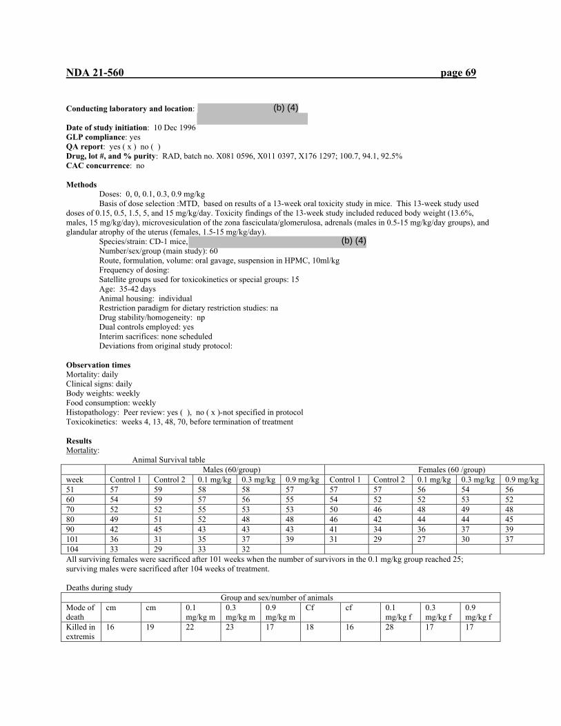
NDA 21-560 page 69
Conducting laboratory and location: Date of study initiation: 10 Dec 1996 GLP compliance: yes QA report: yes ( x ) no ( ) Drug, lot #, and % purity: RAD, batch no. X081 0596, X011 0397, X176 1297; 100.7, 94.1, 92.5% CAC concurrence: no Methods Doses: 0, 0, 0.1, 0.3, 0.9 mg/kg Basis of dose selection :MTD, based on results of a 13-week oral toxicity study in mice. This 13-week study used doses of 0.15, 0.5, 1.5, 5, and 15 mg/kg/day. Toxicity findings of the 13-week study included reduced body weight (13.6%, males, 15 mg/kg/day), microvesiculation of the zona fasciculata/glomerulosa, adrenals (males in 0.5-15 mg/kg/day groups), and glandular atrophy of the uterus (females, 1.5-15 mg/kg/day). Species/strain: CD-1 mice, Number/sex/group (main study): 60 Route, formulation, volume: oral gavage, suspension in HPMC, 10ml/kg Frequency of dosing: Satellite groups used for toxicokinetics or special groups: 15 Age: 35-42 days Animal housing: individual Restriction paradigm for dietary restriction studies: na Drug stability/homogeneity: np Dual controls employed: yes Interim sacrifices: none scheduled Deviations from original study protocol: Observation times Mortality: daily Clinical signs: daily Body weights: weekly Food consumption: weekly Histopathology: Peer review: yes ( ), no ( x )-not specified in protocol Toxicokinetics: weeks 4, 13, 48, 70, before termination of treatment Results Mortality: Animal Survival table Males (60/group) Females (60 /group) week Control 1 Control 2 0.1 mg/kg 0.3 mg/kg 0.9 mg/kg Control 1 Control 2 0.1 mg/kg 0.3 mg/kg 0.9 mg/kg 51 57 59 58 58 57 57 57 56 54 56 60 54 59 57 56 55 54 52 52 53 52 70 52 52 55 53 53 50 46 48 49 48 80 49 51 52 48 48 46 42 44 44 45 90 42 45 43 43 43 41 34 36 37 39 101 36 31 35 37 39 31 29 27 30 37 104 33 29 33 32 All surviving females were sacrificed after 101 weeks when the number of survivors in the 0.1 mg/kg group reached 25; surviving males were sacrificed after 104 weeks of treatment. Deaths during study Group and sex/number of animals Mode of death
cm cm 0.1 mg/kg m
0.3 mg/kg m
0.9 mg/kg m
Cf cf 0.1 mg/kg f
0.3 mg/kg f
0.9 mg/kg f
Killed in extremis
16 19 22 23 17 18 16 28 17 17
(b) (4)
(b) (4)

NDA 21-560 page 70
Found dead
8 9 4 1 1 4 6 1 2 0
Killed for humane reasons
3 3 1 4 4 8 9 7 11 6
total 27 31 27 28 22 30 31 36 30 23 Clinical signs: No treatment-related signs were seen through the study. Opthamology and hematology had no treatment-related effects. Body weights: Difference in body weight compared to controls Percent change from controls males Females 0.1 mg/kg 0.3 mg/kg 0.9 mg/kg 0.1 mg/kg 0.3 mg/kg 0.9 mg/kg Week 52 +2.5 +3.5 -10.5 +1.0 +6.5 -8.5 Week 78 +2.5 +4.5 -7.5 +3.0 -2.5 -14.5 Week 101 -3.0 +6.0 -9.5 -8.0 -10.5 -17.0 Week 104 +0.5 +6.0 -12.0 - - - Food consumption: There were no treatment-related differences in food consumption Gross pathology: Swellings/masses Group/sex Multiplicity number of animals total number mean weeks
with swellings of swellings of onset 0 1 2 3 4 or more Cm 47 7 5 0 1 13 21 47 Cm 45 6 7 0 2 15 29 52 0.1 mg/kg m 41 11 5 2 1 19 33 44 0.3 mg/kg m 48 6 1 5 0 12 23 53 0.9 mg/kg m 37 14 8 1 0 23 33 50 Cf 42 5 3 0 0 8 11 62 Cf 42 6 2 0 0 8 10 65 0.1 mg/kg f 45 4 0 0 1 5 8 50 0.3 mg/kg f 43 4 1 1 1 7 14 46 0.9 mg/kg f 60 0 0 0 0 0 0 --- Includes swellings which regressed or were not positively identified at necropsy. Histopathology: Non-neoplastic: Males c c 0.1 mg/kg 0.3 mg/kg 0.9 mg/kg Testes Flaccid 12/60 13/60 18/60 20/60 26/60

NDA 21-560 page 71
Tubular vacuolation
15 12 20 15 27
Degeneration of germinal epithelium
27 21 27 25 35
epididymides Hypospermia 13 11 15 8 24 Sciatic nerve Axonal degeneration
17 13 19 20 25
Females
thymus involution 11 10 7 11 19 Sciatic nerve Axonal degeneration
17 16 18 17 26
Neoplastic: 1st=Incidence of neoplastic tumors-scheduled and unscheduled sacrifices (including animals found dead) 2nd=Incidence of neoplastic tumors –animals sacrificed at 104 weeks 3rd=Total incidence of neoplastic tumors males
control 1
males Control2
0.1 mg/kg
0.3 mg/kg
0.9 mg/kg
Female Control1
female Control 2
0.1 mg/kg
0.3 mg/kg
0.9 mg/kg
Adrenal cortex Cortical adenoma 0/26
4/33 4/59
3/31 3/29 6/60
3/25 0/33 3/58
4/27 2/32 6/59
0/21 3/38 3/59
0/30 1/30 1/60
0/31 0/29 0/60
0/36 0/24 0/60
1/30 0/30 1/60
0/23 0/37 0/60
Neuroblastoma 0/26 0/33 0/59
1/31 0/29 1/60
0/25 0/33 0/58
0/26 0/33 0/59
0/21 0/38 0/59
0/30 0/30 0/60
0/31 0/29 0/60
0/36 0/24 0/60
0/30 0/30 0/60
0/23 0/37 0/60
Cortical adenocarcinoma
0/26 0/33 0/59
0/31 0/29 0/60
0/25 0/33 0/58
0/26 0/33 0/59
0/21 0/38 0/59
1/30 0/30 1/60
0/31 0/29 0/60
0/36 0/24 0/60
0/30 0/30 0/60
0/23 0/37 0/60
Adrenal medulla Pheochroma cytoma
0/26 1/33 1/59
1/31 0/29 1/60
0/25 0/33 0/58
0/27 0/32 0/59
0/21 0/38 0/59
0/30 1/30 1/60
0/31 0/29 0/60
0/36 0/24 0/60
0/30 0/30 0/60
0/23 0/37 0/60
brain astrocytoma 0/27
0/23 0/60
0/31 0/29 0/60
0/27 0/33 0/60
0/28 0/32 0/60
1/22 0/38 1/60
0/30 0/30 0/60
0/31 0/29 0/60
1/36 0/24 1/60
0/30 0/30 0/60
0/23 0/37 0/60
cecum Adenoma 0/27
0/33 0/60
0/31 0/29 0/60
0/26 0/33 0/59
0/28 0/32 0/60
0/22 0/38 0/60
0/30 1/30 0/60
0/31 0/29 0/60
0/35 0/24 0/59
0/30 0/30 0/60
0/23 0/37 0/60
femur osteoma 0/27
0/33 0/60
0/31 0/29 0/60
0/27 0/33 0/60
0/28 0/32 0/60
0/22 0/38 0/60
0/30 0/30 0/60
0/31 0/29 0/60
0/36 0/24 0/60
1/30 0/30 1/60
1/23 1/37 2/60
Hardarian gland Adenoma 0/27
3/33 3/60
3/31 2/29 5/60
5/27 3/32 8/59
0/28 3/32 3/60
2/22 3/38 5/60
2/30 1/30 3/60
4/31 4/29 8/60
1/36 2/24 3/60
2/30 2/30 4/60
2/23 1/37 3/60

NDA 21-560 page 72
Carcinoma 2/27 1/33 3/60
0/31 0/29 0/60
1/27 0/32 1/59
1/28 1/32 2/60
0/22 0/38 0/60
1/30 1/30 2/60
0/31 2/29 2/60
1/36 1/24 2/60
1/30 2/30 3/60
0/23 3/37 3/60
jejunum adenoma 0/25
0/33 0/58
0/30 0/29 0/59
0/24 0/33 0/57
0/28 0/32 0/60
0/21 0/38 0/59
0/30 0/30 0/60
0/31 0/29 0/60
0/25 0/24 0/59
0/30 0/30 0/60
0/23 1/37 1/60
carcinoma 0/25 1/33 1/58
0/30 0/29 0/59
0/24 0/33 0/57
0/28 0/32 0/60
0/21 0/38 0/59
0/30 0/30 0/60
0/31 0/29 0/60
0/25 0/24 0/59
0/30 0/30 0/60
0/23 0/37 0/60
kidneys Cortical adenoma 0/27
1/33 1/60
0/31 0/29 0/60
0/27 1/33 1/60
2/28 0/32 2/60
0/22 2/38 2/60
0/30 0/30 0/60
0/31 0/29 0/60
0/36 0/24 0/60
0/30 0/30 0/60
0/23 0/37 0/60
Cortical carcinoma 0/27 0/33 0/60
0/31 0/29 0/60
0/27 0/33 0/60
1/28 1/32 2/60
0/22 0/38 0/60
0/30 0/30 0/60
0/31 0/29 0/60
0/36 0/24 0/60
0/30 0/30 0/60
0/23 0/37 0/60
liver Hepatocellular Adenoma
3/27 12/33 15/60
4/31 4/29 8/60
0/27 4/33 4/60
3/28 9/32 12/60
2/22 8/38 10/60
0/30 1/30 1/60
0/31 1/29 1/60
0/36 2/24 2/60
1/30 1/30 2/60
0/23 1/37 1/60
Hemangioma 0/27 0/33 0/60
0/31 1/29 1/60
0/27 0/33 0/60
0/28 0/32 0/60
0/22 0/38 0/60
0/30 0/30 0/60
0/31 0/29 0/60
0/36 0/24 0/60
0/30 0/30 0/60
0/23 0/37 0/60
Hemangiosarcoma 1/27 0/33 1/60
0/31 1/29 1/60
0/27 0/33 0/60
1/28 0/32 1/60
0/22 0/38 0/60
0/30 1/30 1/60
0/31 0/29 0/60
0/36 0/24 0/60
0/30 1/30 1/60
0/23 0/37 0/60
Hepatocellular carcinoma
3/27 3/33 6/60
3/31 2/29 5/60
5/27 4/33 9/60
3/28 2/32 5/60
2/22 3/38 5/60
0/30 0/30 0/60
0/31 0/29 0/60
0/36 0/24 0/60
0/30 0/30 0/60
0/23 0/37 0/60
Cholangiocellular carcinoma
0/27 0/33 0/60
1/31 0/29 1/60
0/27 0/33 0/60
0/28 0/32 0/60
0/22 0/38 0/60
0/30 0/30 0/60
0/31 0/29 0/60
0/36 0/24 0/60
0/30 0/30 0/60
0/23 0/37 0/60
Lungs Pulmonary adenoma
1/27 6/33 7/60
5/31 9/29 14/60
3/27 11/33 14/60
7/28 7/32 14/60
3/22 11/38 14/60
4/30 6/30 10/60
3/31 3/29 6/60
3/36 3/24 6/60
2/30 6/30 8/60
1/23 6/37 7/60
Pulmonary carcinoma
3/27 4/33 7/60
7/31 2/29 9/60
5/27 0/33 5/60
3/28 0/32 3/60
2/22 0/38 2/60
0/30 0/30 0/60
0/31 0/29 0/60
2/36 0/24 2/60
1/30 1/30 2/60
1/23 0/37 1/60
Mammary area Cranial/caudal
carcinoma 0/27 0/33 0/60
1/31 0/29 0/60
0/27 0/33 0/60
0/28 0/32 0/60
0/22 0/38 0/60
1/30 2/30 3/60
3/31 0/29 3/60
1/36 0/24 0/60
0/30 0/30 0/60
0/23 0/37 0/60
ovaries adenoma 0/30
2/30 2/60
0/31 1/29 1/60
0/36 1/24 1/60
0/30 1/30 1/60
0/23 0/37 0/60
luteoma 0/30 0/30 0/60
0/31 0/29 0/60
0/36 1/24 1/60
0/30 1/30 1/60
0/23 2/37 2/60
Granulosa cell tumor-B
0/30 0/30 0/60
0/31 1/29 1/60
0/36 0/24 0/60
0/30 0/30 0/60
0/23 1/37 1/60

NDA 21-560 page 73
hemangioma 0/30 0/30 0/60
0/31 0/29 0/60
0/36 0/24 0/60
0/30 0/30 0/60
1/23 0/37 1/60
Granulosa cell tumor-M
0/30 0/30 0/60
0/31 0/29 0/60
0/36 1/24 1/60
0/30 0/30 0/60
0/23 0/37 0/60
pancreas Islet cell adenoma 0/27
0/33 0/60
0/31 0/29 0/60
0/26 0/33 0/59
0/28 1/32 1/60
0/22 0/38 0/60
0/30 0/30 0/60
0/30 0/29 0/59
0/26 0/24 0/60
0/30 0/30 0/60
0/23 0/37 0/60
pituitary adenoma 0/27
1/33 1/60
0/31 0/29 0/60
0/27 0/33 0/60
0/28 0/32 0/60
0/22 0/38 0/60
0/30 1/30 1/60
0/31 1/29 1/60
0/36 0/24 0/60
0/28 0/30 0/58
0/23 2/37 2/60
Seminal vesicles adenoma 0/33
0/27 0/60
0/31 0/29 0/60
0/27 0/33 0/60
0/28 0/32 0/60
0/22 2/38 2/60
spleen hemangiosarcoma 0/27
0/33 0/60
0/31 1/29 1/60
0/26 0/33 0/59
0/28 0/32 0/60
0/22 0/38 0/60
0/30 0/30 0/60
0/30 0/29 0/59
0/26 0/24 0/60
0/30 0/30 0/60
0/23 0/37 0/60
stomach Squamous cell papilloma
0/27 1/33 1/60
0/31 0/29 0/60
0/27 0/33 0/60
1/28 0/32 1/60
0/22 0/38 0/60
0/30 0/30 0/60
0/31 0/29 0/60
0/36 0/24 0/60
0/30 0/30 0/60
0/23 0/37 0/60
Submandibular salivary glands
Myoepithelial tumor
0/27 0/33 0/60
0/31 0/29 0/60
0/27 0/32 0/59
0/28 0/32 0/60
0/22 0/38 0/60
0/30 0/30 0/60
1/30 0/29 1/59
0/35 0/24 0/59
0/29 0/30 0/59
0/23 0/37 0/60
Testes Interstitial cell tumor
1/27 2/33 3/60
1/31 0/29 1/60
0/27 0/33 0/60
0/28 1/32 1/60
1/22 0/38 1/60
hemangiosarcoma
0/27 1/33 1/60
0/31 0/29 0/60
0/27 0/33 0/60
0/28 0/32 0/60
0/22 0/38 0/60
thymus thymoma 0/27
1/31 1/58
0/24 0/27 0/57
0/26 0/32 0/58
0/26 0/32 0/58
0/23 0/37 0/58
0/30 0/30 0/60
0/30 0/29 0/59
0/35 0/24 0/59
0/30 0/30 0/60
0/23 0/36 0/59
thyroid Follicular adenoma
0/26 0/33 0/59
1/31 1/29 2/60
0/27 0/33 0/60
1/28 1/32 1/60
0/22 0/38 0/60
0/30 0/30 0/60
0/31 1/29 1/60
0/36 0/24 0/60
0/29 0/30 0/59
0/23 0/37 0/60
Uterine cervix Stromal polyp 0/29
0/30 0/59
0/31 0/29 0/60
0/36 2/24 2/60
0/30 0/30 0/60
0/23 0/37 0/60
fibroma 0/29 0/30 0/59
0/31 0/29 0/60
1/36 0/24 1/60
0/30 0/30 0/60
0/23 0/37 0/60
leiomyoma
1/29 3/30 4/59
0/31 1/29 1/60
0/36 0/24 0/60
0/30 0/30 0/60
0/23 1/37 0/60

NDA 21-560 page 74
leiomyosarcoma 0/29 1/30 1/59
0/31 0/29 0/60
0/36 0/24 0/60
0/30 0/30 0/60
1/23 0/37 1/60
Stromal sarcoma 0/29 0/30 0/59
0/31 1/29 1/60
0/36 0/24 0/60
0/30 0/30 0/60
0/23 0/37 0/60
uterus Stromal polyp 0/30
4/30 4/60
3/31 2/29 5/60
0/36 3/24 3/60
0/30 1/30 1/60
1/23 3/37 4/60
fibroma 0/30 0/30 0/60
0/31 0/29 0/60
0/36 0/24 0/60
0/30 0/30 0/60
0/23 1/37 1/60
leiomyoma 0/30 3/30 3/60
0/31 1/29 1/60
0/36 0/24 0/60
0/30 0/30 0/60
0/23 1/37 1/60
leiomyosarcoma 0/30 0/30 0/60
0/31 0/29 0/60
0/36 0/24 0/60
0/30 0/30 0/60
0/23 0/37 0/60
adenocarcinoma 0/30 1/30 1/60
1/31 1/29 2/60
0/36 0/24 0/60
0/30 0/30 0/60
0/23 0/37 0/60
Endometrial sarcoma
2/30 0/30 2/60
0/31 0/29 0/60
0/36 0/24 0/60
0/30 0/30 0/60
0/23 0/37 0/60
abdomen Leimysarcoma 0/1
0/0 0/1
0/3 0/0 0/3
0/2 0/0 0/2
1/1 0/0 1/1
0/2 0/0 0/2
0/1 0/0 0/1
0/0 0/0 0/0
0/0 0/0 0/0
0/1 0/0 0/1
0/2 0/0 0/0
Buccal cavity papilloma 0/0
0/0 0/0
0/0 0/0 0/0
0/0 0/0 0/0
0/0 0/0 0/0
0/0 0/0 0/0
0/0 1/1 0/0
0/0 0/0 0/0
0/0 0/0 0/0
0/0 0/0 0/0
0/0 0/0 0/0
Hemopoietic tumor
Malignant lymphoma (unclassified)
0/27 1/33 1/60
1/31 0/29 1/60
1/27 0/33 1/60
0/28 0/32 0/60
0/22 0/38 0/60
0/30 0/30 0/60
1/31 0/29 1/60
0/36 0/24 0/60
0/30 0/30 0/60
1/23 0/37 1/60
Histiocytic sarcoma
1/27 0/33 1/60
1/31 0/29 1/60
0/27 0/33 0/60
0/28 0/32 0/60
3/22 0/38 3/60
5/30 0/30 5/60
1/31 3/29 4/60
3/36 1/24 4/60
3/30 1/30 4/60
1/23 1/37 2/60
Follicular center cell lymphoma
4/27 3/33 7/60
2/31 2/29 4/60
2/27 1/33 3/60
2/28 0/32 2/60
1/22 1/38 1/60
2/30 3/30 5/60
6/31 0/29 6/60
1/36 0/24 1/60
4/30 1/30 5/60
2/23 0/37 2/60
Granulocytic leukemia
1/27 0/33 1/60
0/31 0/29 0/60
0/27 0/33 0/60
1/28 0/32 1/60
1/22 1/38 1/60
1/30 0/30 1/60
1/31 0/29 1/60
1/36 0/24 1/60
2/30 0/30 2/60
3/23 0/37 3/60
Lymphocyttic/ lymphoblastic lymphoma
3/27 1/33 4/60
0/31 0/29 0/60
0/27 0/33 0/60
1/28 0/32 1/60
3/22 0/38 3/60
2/30 0/30 2/60
3/31 2/29 5/60
3/36 1/24 4/60
5/30 0/30 5/60
3/23 0/37 3/60

NDA 21-560 page 75
Pleiomorphic lymphoid lymphoma
0/27 0/33 0/60
0/31 0/29 0/60
0/27 0/33 0/60
0/28 0/32 0/60
0/22 0/38 0/60
0/30 0/30 0/60
0/31 0/29 0/60
0/36 0/24 0/60
1/30 0/30 1/60
0/23 0/37 0/60
Musculo-skeletal Sarcoma 0/3
0/1 0/4
0/6 0/1 0/7
1/5 0/2 1/7
0/2 0/0 0/2
0/1 0/1 0/2
1/1 0/0 1/1
0/3 0/2 0/5
0/3 0/2 0/5
0/2 0/0 0/2
0/1 ½ 1/3
osteosarcoma
0/0 0/0 0/0
1/6 0/0 1/6
0/5 0/0 0/5
0/2 0/0 0/2
0/1 0/0 0/1
0/1 0/0 0/1
1/3 0/0 1/3
0/3 0/0 0/3
½ 0/0 1/2
0/1 0/0 0/1
osteoma 0/0 0/1 0/1
0/0 0/1 0/1
0/0 0/2 0/2
0/0 0/0 0/0
0/0 0/1 0/1
0/0 0/0 0/0
0/0 0/2 0/2
0/0 0/2 0/2
0/0 0/0 0/0
0/0 ½ ½
peritoneum mesothelioma 0/0
0/0 0/0
1/3 0/0 1/3
0/0 0/0 0/0
0/0 0/0 0/0
0/0 0/0 0/0
0/0 0/0 0/0
0/1 0/0 0/1
0/0 0/0 0/0
0/0 0/0 0/0
0/0 0/0 0/0
skin Hemangiosarcoma # Examined at Necropsy not provided
1/6 ? 1/12
0/5 ? 0/13
0/9 ? 0/14
0/7 ? 0/11
0/10 ? 0/21
0/16 ? 0/17
0/6 ? 0/6
0/9 ? 0/9
0/11 ? 0/11
0/4 ? 0/4
Sarcoma # Examined at Necropsy not provided
0/6 ? 0/12
0/5 ? 0/13
0/9 ? 0/14
0/7 ? 0/11
0/10 ? 0/21
1/16 ? 1/17
0/6 ? 0/6
0/9 ? 0/9
0/11 ? 0/11
0/4 ? 0/4
thorax mesothelioma 0/2
0/0 0/2
1/3 0/0 1/3
0/1 0/0 0/1
0/0 0/0 0/0
0/0 0/0 0/0
0/1 0/0 0/1
0/1 0/0 0/1
0/2 0/0 0/2
0/0 0/0 0/0
0/0 0/0 0/0
Toxicokinetics: Dose (mg/kg) week sex AUC (0-24)
ng*h/ml 0.1 4 m 434.5 f 333.0 101 m 169.5 f 150.5 0.3 4 m 756.0 f 358.2 101 m 412.8 f 346.4 0.9 4 m 1633.3 f 2210.9 101 m 1377.8 f 3084.2 Conclusions: The statistical review of this study showed no dose mortality trends for the male and female mice. Exposure to drug can be considered adequate as more than 50% of mice were still alive at 89-90 weeks for both males and females. No positive linear trends were found for male mice and one positive trend for female mice. Femur (including joint) osteoma, a rare tumor, was significant in the females (p=0.021) compared with the combined control group. The incidence rate in this study was 1.64% in the middle dose group and 3.05% in the high dose group. However, when analyzed with historical control incidence rates this
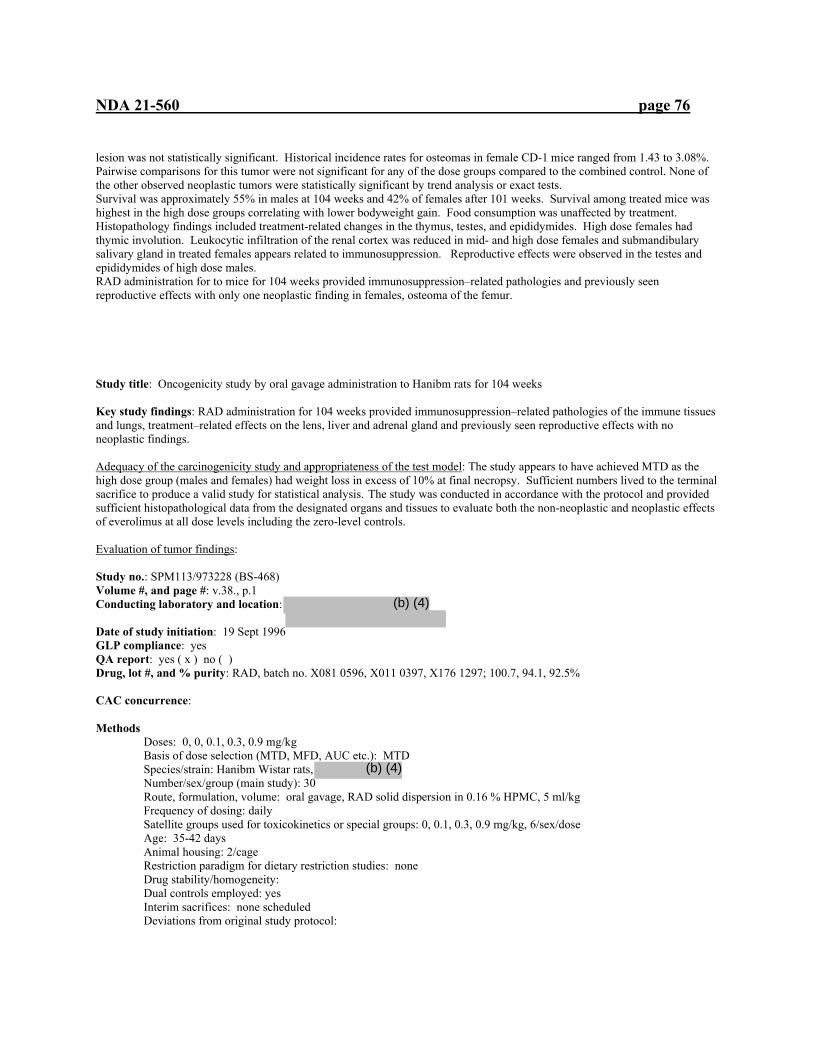
NDA 21-560 page 76
lesion was not statistically significant. Historical incidence rates for osteomas in female CD-1 mice ranged from 1.43 to 3.08%. Pairwise comparisons for this tumor were not significant for any of the dose groups compared to the combined control. None of the other observed neoplastic tumors were statistically significant by trend analysis or exact tests. Survival was approximately 55% in males at 104 weeks and 42% of females after 101 weeks. Survival among treated mice was highest in the high dose groups correlating with lower bodyweight gain. Food consumption was unaffected by treatment. Histopathology findings included treatment-related changes in the thymus, testes, and epididymides. High dose females had thymic involution. Leukocytic infiltration of the renal cortex was reduced in mid- and high dose females and submandibulary salivary gland in treated females appears related to immunosuppression. Reproductive effects were observed in the testes and epididymides of high dose males. RAD administration for to mice for 104 weeks provided immunosuppression–related pathologies and previously seen reproductive effects with only one neoplastic finding in females, osteoma of the femur.
Study title: Oncogenicity study by oral gavage administration to Hanibm rats for 104 weeks Key study findings: RAD administration for 104 weeks provided immunosuppression–related pathologies of the immune tissues and lungs, treatment–related effects on the lens, liver and adrenal gland and previously seen reproductive effects with no neoplastic findings. Adequacy of the carcinogenicity study and appropriateness of the test model: The study appears to have achieved MTD as the high dose group (males and females) had weight loss in excess of 10% at final necropsy. Sufficient numbers lived to the terminal sacrifice to produce a valid study for statistical analysis. The study was conducted in accordance with the protocol and provided sufficient histopathological data from the designated organs and tissues to evaluate both the non-neoplastic and neoplastic effects of everolimus at all dose levels including the zero-level controls. Evaluation of tumor findings: Study no.: SPM113/973228 (BS-468) Volume #, and page #: v.38., p.1 Conducting laboratory and location: Date of study initiation: 19 Sept 1996 GLP compliance: yes QA report: yes ( x ) no ( ) Drug, lot #, and % purity: RAD, batch no. X081 0596, X011 0397, X176 1297; 100.7, 94.1, 92.5% CAC concurrence: Methods Doses: 0, 0, 0.1, 0.3, 0.9 mg/kg Basis of dose selection (MTD, MFD, AUC etc.): MTD Species/strain: Hanibm Wistar rats, Number/sex/group (main study): 30 Route, formulation, volume: oral gavage, RAD solid dispersion in 0.16 % HPMC, 5 ml/kg Frequency of dosing: daily Satellite groups used for toxicokinetics or special groups: 0, 0.1, 0.3, 0.9 mg/kg, 6/sex/dose Age: 35-42 days Animal housing: 2/cage Restriction paradigm for dietary restriction studies: none Drug stability/homogeneity: Dual controls employed: yes Interim sacrifices: none scheduled Deviations from original study protocol:
(b) (4)
(b) (4)

NDA 21-560 page 77
Observation times Mortality: daily Clinical signs: daily Body weights: prior to treatment, start of treatment, weekly Food consumption: prior to treatment, start of treatment, weekly Histopathology: Peer review: yes ( ), no ( ) Toxicokinetics: weeks 4, 13, 26, and 48, 0.5, 1, 2, 4, 7, and 24 h after dosing Results Mortality: Animal Survival table Males (60/group) Females (60 /group) week Control 1 Control 2 0.1 mg/kg 0.3 mg/kg 0.9 mg/kg Control 1 Control 2 0.1 mg/kg 0.3 mg/kg 0.9 mg/kg 51 59 60 59 60 59 59 60 59 58 60 61 57 60 59 60 57 59 57 56 56 59 70 57 59 58 60 50 58 56 55 56 57 80 55 55 55 58 45 55 53 53 52 57 90 47 45 49 55 43 48 49 50 51 56 101 37 40 38 51 40 44 44 42 45 53 104 36 36 35 48 40 37 37 38 42 51 . Deaths during study Group and sex/number of animals Mode of death
cm cm 0.1 mg/kg m
0.3 mg/kg m
0.9 mg/kg m
Cf cf 0.1 mg/kg f
0.3 mg/kg f
0.9 mg/kg f
Killed in extremis
9 9 8 5 11 12 15 9 13 3
Found dead
0 3 1 0 0 2 0 3 1 1
Killed for humane reasons
15 12 16 7 9 9 8 10 4 5
total 24 24 25 12 20 23 23 22 18 9 Clinical signs: No treatment-related signs were seen through the study. Opthamology a: Ophthalmic examinations during weeks 53, 78 and 104 showed increased incidence of anterior suture line opacity in the lens for the 0.9 mg/kg group and during week 104 for the 0.3 mg/kg group. Increased incidence of capsular opacity was seen during week 104 in the 0.9 mg/kg group and males in the 0.3 mg/kg group. Increased incidence of anterior polar opacity of the lens was seen during weeks 52 and 78 for males of the 0.9 mg/kg group but not observed during week 104. Hematology: During week 104, males (0.3, 0.9 mg/kg) had slightly elevated packed cell volume, hemoglobin, red cell count and mean cell volume. Leukocyte and neutrophil counts were elevated for males receiving 0.1 mg/kg. Platelet counts were decreased for males and females receiving 0.9 mg/kg and males receiving 0.3 mg/kg. Body weights: Difference in body weight compared to controls Percent change from controls
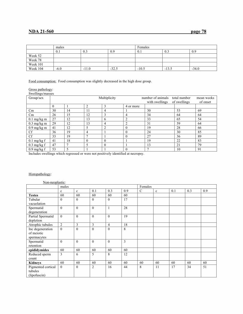
NDA 21-560 page 78
males Females 0.1 0.3 0.9 0.1 0.3 0.9 Week 52 Week 78 Week 101 Week 104 -6.0 -11.0 -32.5 -10.5 -13.5 -34.0 Food consumption: Food consumption was slightly decreased in the high dose group. Gross pathology: Swellings/masses Group/sex Multiplicity number of animals total number mean weeks
with swellings of swellings of onset 0 1 2 3 4 or more Cm 30 14 11 4 1 30 53 69 Cm 26 15 12 3 4 34 64 64 0.1 mg/kg m 27 12 13 6 2 33 65 54 0.3 mg/kg m 29 12 13 4 2 31 59 64 0.9 mg/kg m 41 12 5 2 0 19 28 66 Cf 36 19 4 1 0 24 30 85 Cf 33 19 7 1 0 27 36 89 0.1 mg/kg f 41 18 0 0 1 19 22 83 0.3 mg/kg f 47 7 5 0 1 13 21 79 0.9 mg/kg f 53 5 1 1 0 7 10 91 Includes swellings which regressed or were not positively identified at necropsy. Histopathology: Non-neoplastic: males Females c c 0.1 0.3 0.9 C c 0.1 0.3 0.9 Testes 60 60 60 60 60 Tubular vacuolation
0 0 0 0 17
Spermatid degeneration
0 0 0 1 28
Partial Spermatid depletion
0 0 0 0 19
Atrophic tubules 2 3 3 4 18 Inc degeneration of meiotic spermacytes
0 0 0 0 8
Spermatid retention
0 0 0 0 3
epididymides 60 60 60 60 60 Reduced sperm count
3 6 5 8 12
Kidneys 60 60 60 60 60 60 60 60 60 60 Pigmented cortical tubules (lipofuscin)
0 0 2 16 44 8 11 17 34 51

NDA 21-560 page 79
liver 60 60 60 60 60 60 60 60 60 60 Eosinophilic foci hepatocellular alteration
20 11 22 27 34 6 1 4 10 12
Clear cell foci 37 39 42 52 40 23 23 29 34 43 Portal tract senile change
5 1 5 12 15 5 7 8 19 6
Periacinar hepatocytic fatty vacuolation
0 2 2 6 6 2 0 0 1 2
lungs 60 60 60 60 60 60 60 60 60 60 Alveolar macrophages
0 9 6 10 31 6 4 4 4 15
Alveolar pneumonitis
7 0 6 21 32 6 6 1 3 5
Alveolar necrosis 0 0 0 0 9 0 0 0 0 1 Eosinophilic deposition
1 0 3 8 24 0 1 0 6 15
Pigment laden macrophages
0 0 0 0 0 0 1 1 2 10
Peribronchiolar lymphocytes
0 0 0 0 5 0 0 0 0 0
Mesenteric lymph nodes
60 60 60 60 60 60 59 60 60 60
Macrophage aggregation
26 25 44 45 44 10 10 22 23 13
Mast cells in sinuses
1 2 8 21 10 2 1 6 10 9
stomach 59 60 59 60 60 60 60 60 59 60 Proliferation of mucous neck cells
0 2 8 11 14 0 0 3 4 5
Hyperkeratosis/ Acanthosis of keratinized region
3 8 16 28 28 3 0 7 9 9
thymus 60 60 60 60 60 59 59 60 60 60 Atrophy 6 4 5 11 18 5 3 4 2 8 ovaries 60 60 60 60 60 Ovarian cyst 5 4 7 6 21 uterus 60 60 60 60 60 Squamous metaplasia
2 2 3 1 21
Hardarian glands 60 59 60 60 60 60 60 60 60 60 Chronic inflammation
3 2 3 10 14 6 2 6 4 11
Lachrymal glands
60 60 60 60 60 59 60 60 60 60
Inflammatory infiltration
17 18 25 39 43 7 4 7 3 6
Dilated acini 22 17 31 32 31 2 2 2 2 0 pancreas 60 60 60 60 60 59 60 60 60 60 Foci of lymphocytes
0 0 1 4 4 0 0 0 2 5
Skeletal muscle 60 60 60 60 60 60 60 60 60 60 Atrophy 2 0 0 11 14 2 2 1 3 4

NDA 21-560 page 80
Focal degeneration and necrosis
0 0 1 0 3 0 0 0 1 0
Chronic inflammation
3 1 2 23 24 0 0 1 3 10
Submandibular salivary glands
60 60 60 60 60 59 60 60 60 60
Lymphocytic infiltration
0 0 0 4 6 1 2 0 1 2
eyes 60 60 60 60 60 60 60 60 60 60 Lenticular degeneration
9 7 10 11 18 2 1 3 3 2
adrenals 60 60 60 60 60 60 60 60 60 60 Focal cortical hypertrophy
15 9 9 3 1 12 12 3 6 1
Focal cortical hyperplasia
6 3 4 0 0 6 8 5 3 0
Cortical fatty vacuolation
29 18 9 3 1 11 12 2 5 2
Kidney 60 60 60 60 60 60 60 60 60 60 Progressive senile nephropathy
46 50 38 27 12 43 45 33 38 33
Papillary hyperplasia
4 6 2 1 0 8 10 8 1 1
Mineralization of pelvic epithelium
25 27 22 10 4 45 50 49 42 39
mammary 59 60 60 60 60 60 60 60 60 60 Acinar hyperplasia 0 0 0 0 0 23 18 15 13 6 Ductular dilation 1 0 1 0 0 25 30 23 27 10 Neoplastic: 1st= Incidence of neoplastic tumors-scheduled and unscheduled sacrifices (including animals found dead) 2nd =Incidence of neoplastic tumors –animals sacrificed at 104 weeks 3rd =Total incidence of neoplastic tumors males Females
C c
0.1
0.3
0.9
C
c
0.1
0.3
0.9
Adrenal cortex Cortical adenoma 0/24
0/36 0/60
1/24 0/36 1/60
0/25 1/35 1/60
0/12 1/48 1/60
0/20 0/40 0/60
0/23 0/37 0/60
0/23 0/37 0/60
0/22 0/38 0/60
0/18 0/42 0/60
0/9 0/51 0/60
Adrenal medulla Pheochroma cytoma
2/24 2/36 4/60
0/24 0/36 0/60
0/25 0/35 0/60
0/12 0/48 0/60
0/20 0/40 0/60
0/23 0/37 0/60
0/23 1/37 1/60
0/22 1/38 1/60
1/18 0/42 1/60
0/9 1/50 1/59
ganglioneuroma 0/24 1/36 1/60
0/24 0/36 0/60
1/25 0/35 1/60
0/12 0/48 0/60
0/20 0/40 0/60
0/23 0/37 0/60
0/23 0/37 0/60
0/22 0/38 0/60
0/18 0/42 0/60
0/9 0/50 0/60
Brain Granular cell tumor-b
1/24 1/36 2/60
0/24 1/36 1/60
0/25 0/35 0/60
0/12 1/48 1/60
0/20 0/40 0/60
0/23 0/37 0/60
0/23 1/37 1/60
0/22 0/38 0/60
0/18 2/42 2/60
0/9 1/51 1/60
Oligodendroglioma 1/24 1/24 0/25 0/12 1/20 0/23 0/23 0/22 0/18 0/9

NDA 21-560 page 81
Malignant reticulosis
0/24 0/36 0/60
0/24 0/36 0/60
0/25 0/35 0/60
0/12 0/48 0/60
1/20 0/40 1/60
0/23 0/37 0/60
0/23 0/37 0/60
0/22 0/38 0/60
0/18 0/42 0/60
0/9 0/51 0/60
Astrocytoma 1/24 0/36 060
0/24 0/36 0/60
3/25 0/35 3/60
0/12 0/48 0/60
0/20 0/40 0/60
0/23 0/37 0/60
0/23 0/37 0/60
0/22 1/38 1/60
0/18 0/42 0/60
0/9 0/51 0/60
Mixed glioma 1/24 0/36 1/60
0/24 0/36 0/60
0/25 0/35 0/60
0/12 0/48 0/60
0/20 0/40 0/60
0/23 0/37 0/60
0/23 0/37 0/60
0/22 0/38 0/60
0/18 0/42 0/60
0/9 0/51 0/60
Granular cell tumor-m
0/24 0/36 0/60
1/24 0/36 1/60
0/25 0/35 0/60
0/12 0/48 0/60
0/20 0/40 0/60
0/23 0/37 0/60
0/23 0/37 0/60
0/22 1/38 1/60
1/18 0/32 1/60
0/9 0/51 0/60
Meningial sarcoma 0/24 0/36 0/60
0/24 0/36 0/60
0/25 0/35 0/60
0/12 0/48 0/60
0/20 0/40 0/60
0/23 0/37 0/60
0/23 0/37 0/60
0/22 0/38 0/60
1/18 0/32 1/60
0/9 0/51 0/60
Colon carcinoma 0/24
0/36 0/60
0/24 0/36 0/60
0/25 0/35 0/60
0/12 0/48 0/60
0/20 0/40 0/60
0/23 0/37 0/60
0/23 1/37 1/60
0/22 0/38 0/60
0/18 0/42 0/60
0/9 0/51 0/60
duodenum carcinoma 0/23
0/36 0/59
0/24 0/36 0/60
0/24 0/36 0/60
0/12 0/48 0/60
0/20 0/40 0/60
1/23 0/37 1/60
0/23 0/37 0/60
0/22 0/38 0/60
0/18 0/42 0/60
0/9 0/51 0/60
eyes Amelanotic melanoma
0/24 0/36 0/60
0/23 0/36 0/59
0/25 0/36 0/60
1/12 0/48 1/60
0/20 0/40 0/60
0/23 0/37 0/60
0/23 0/37 0/60
0/22 0/38 0/60
0/18 0/42 0/60
0/9 0/51 0/60
Jejunum leiomyoma 0/24
1/36 1/60
0/23 0/36 0/59
0/25 0/35 0/60
0/12 0/48 0/60
0/20 0/40 0/60
0/23 0/37 0/60
0/23 0/37 0/60
0/22 0/38 0/60
0/18 0/42 0/60
0/9 0/51 0/60
Kidneys Liposarcoma 0/24
0/36 0/60
0/24 0/36 0/60
0/25 0/35 0/60
0/12 0/48 0/60
0/20 0/40 0/60
0/23 0/37 0/60
0/23 0/37 0/60
0/22 0/38 0/60
1/18 0/42 1/60
0/9 0/51 0/60
Tubular sarcoma 1/24 0/36 1/60
0/24 0/36 0/60
0/25 0/35 0/60
0/12 0/48 0/60
0/20 0/40 0/60
0/23 0/37 0/60
0/23 0/37 0/60
0/22 0/38 0/60
0/18 0/42 0/60
0/9 0/51 0/60
Lymph node mesenteric
hemangioma 3/24 11/36 14/60
3/24 10/36 13/60
0/25 1/35 1/60
0/12 0/48 0/60
1/20 2/40 3/60
3/23 3/37 6/60
1/22 0/37 1/59
0/22 1/38 1/60
0/18 0/42 0/60
0/9 0/51 0/60
Liver Hepatocellular Adenoma
0/24 1/36 1/60
0/24 0/36 0/60
0/25 1/35 1/60
0/12 2/48 2/60
0/20 0/40 0/60
0/23 0/37 0/60
1/23 2/37 3/60
0/22 2/38 2/60
0/18 0/42 0/60
0/9 1/51 1/60
Lungs Pulmonary adenoma
0/24 0/36 0/60
0/24 2/36 2/60
0/25 0/35 0/60
0/12 0/48 0/60
0/20 1/40 1/60
0/23 0/37 0/60
0/23 0/37 0/60
0/22 0/38 0/60
0/18 1/42 1/60
0/9 0/51 0/60
Mammary Fibroadenoma 0/23
0/36 0/59
0/24 0/36 0/60
0/25 0/35 0/60
0/12 0/48 0/60
0/20 0/40 0/60
9/23 2/37 9/60
9/23 11/37 20/60
4/22 1/38 5/60
1/18 2/42 3/60
0/9 0/51 0/60

NDA 21-560 page 82
Adenoma 0/23 0/36 0/59
0/24 0/36 0/60
0/25 0/35 0/60
0/12 0/48 0/60
0/20 0/40 0/60
0/23 0/37 0/60
0/23 0/37 0/60
1/22 1/38 2/60
0/18 0/42 0/60
0/9 0/51 0/60
Carcinoma 0/23 0/36 0/59
0/24 0/36 0/60
0/25 0/35 0/60
0/12 0/48 0/60
0/20 0/40 0/60
1/23 2/37 3/60
0/23 1/37 1/60
0/22 1/38 1/60
1/18 1/42 2/60
1/9 1/51 2/60
Ovaries Granulosa cell tumor-b
0/23 1/37 1/60
0/23 0/37 0/60
0/22 1/38 1/60
0/18 0/42 0/60
0/9 0/51 0/60
Undifferentiated gonadal stromal tumor
0/23 0/37 0/60
0/23 2/37 2/60
0/22 0/38 0/60
0/18 0/42 0/60
1/9 0/51 1/60
Sertoli cell tumor
0/23 1/37 1/60
0/23 0/37 0/60
0/22 0/38 0/60
0/18 0/42 0/60
0/9 0/51 0/60
Tubulostromal tumor
0/23 1/37 1/60
0/23 1/37 1/60
0/22 0/38 0/60
0/18 0/42 0/60
0/9 1/51 1/60
Pancreas Islet cell adenoma 0/24
3/36 3/60
2/24 0/36 2/60
1/25 0/35 1/60
0/12 0/48 0/60
0/20 0/40 0/60
1/22 0/37 1/60
1/23 0/37 1/60
0/22 1/38 1/60
0/18 0/42 0/60
0/9 0/51 0/60
Islet cell carcinoma 1/24 1/24 0/25 0/12 0/20 0/22 0/23 0/22 0/18 0/9 Parathyroid adenoma 0/23
2/36 2/59
1/24 0/34 1/58
0/25 0/35 0/60
0/12 0/48 0/60
0/19 0/35 0/54
0/20 0/34 0/54
0/22 0/36 0/58
0/20 0/38 0/58
0/18 0/42 0/60
0/9 1/48 1/57
Pituitary Adenoma 9/24
8/36 17/60
14/24 8/36 22/60
9/25 12/34 21/59
7/12 7/48 14/60
0/20 5/40 5/60
16/23 16/37 32/60
14/23 17/37 31/60
15/22 20/38 35/60
11/18 20/42 31/60
3/9 11/51 12/60
ganglioneuroma 0/24 0/36 0/60
0/24 0/36 0/60
0/25 0/34 0/59
0/12 0/48 0/60
0/20 1/40 1/60
0/23 0/37 0/60
0/23 0/37 0/60
0/22 0/38 0/60
0/18 0/42 0/60
0/9 0/51 0/60
carcinoma 0/24 0/36 0/60
0/24 0/36 0/60
0/25 0/35 0/60
0/12 0/48 0/60
0/20 0/40 0/60
0/23 0/37 0/60
0/23 0/37 0/60
1/22 0/38 1/60
0/18 0/42 0/60
0/9 0/51 0/60
Prostate adenocarcinoma 0/24
0/36 0/60
1/24 0/36 0/60
0/25 0/35 0/60
0/12 0/48 0/60
0/20 0/40 0/60
Rectum carcinoma 0/24
1/36 1/60
0/24 0/36 0/60
0/25 0/35 0/60
0/12 0/48 0/60
0/20 0/40 0/60
0/23 0/37 0/60
0/23 0/37 0/60
0/22 0/38 0/60
0/18 0/42 0/60
0/9 0/51 0/60
Spleen Hemangiosarcoma 0/24
0/36 0/60
0/24 1/36 1/60
1/25 0/35 1/60
0/12 0/48 0/60
0/20 0/40 0/60
1/23 0/37 1/60
0/23 0/37 0/60
0/22 0/38 0/60
0/18 0/42 0/60
0/9 0/51 0/60
Submandibular gland

NDA 21-560 page 83
adenoma 0/24 1/36 1/60
0/24 0/36 0/60
0/25 0/35 0/60
0/12 0/48 0/60
0;20 0/40 0/60
0/22 0/37 0/59
0/23 0/37 0/60
0/22 0/38 0/60
0/18 0/42 0/60
0/9 0/51 0/60
fibrosarcoma 0/24 0/36 0/60
0/24 0/36 0/60
0/25 0/35 0/60
0/12 0/48 0/60
0/20 1/40 1/60
0/22 0/37 0/59
0/23 0/37 0/60
0/22 0/38 0/60
0/18 0/42 0/60
0/9 0/51 0/60
Testes Interstitial cell adenoma
0/24 1/36 1/60
0/24 1/36 1/60
0/25 1/35 1/60
0/12 0/48 0/60
0/20 0/40 0/60
Thymus Thymoma 0/24
1/36 1/60
1/24 4/36 5/60
2/25 1/35 3/60
0/12 1/48 1/60
0/20 0/40 0/60
2/23 6/36 8/59
3/22 8/37 11/59
2/22 6/36 8/59
0/18 4/42 4/60
0/9 3/51 3/60
Thymoma (epithelial)
0/24 0/36 0/60
0/24 0/36 0/60
0/25 0/35 0/60
0/12 0/48 0/60
0/20 0/40 0/60
1/23 0/36 1/59
0/22 0/37 0/59
0/22 0/36 0/58
0/18 0/42 0/60
0/9 1/51 1/60
Thyroids Follicular adenoma
2/24 3/36 5/60
0/24 7/35 7/59
1/25 4/35 5/60
0/12 3/48 3/60
0/20 1/40 1/60
1/22 1/37 2/59
0/23 1/37 1/59
0/22 0/38 0/60
0/18 0/42 0/60
0/9 2/51 2/60
Parafollicular cell adenoma
0/24 2/36 2/60
0/24 5/35 5/59
0/25 0/35 0/60
1/12 1/48 2/60
0/20 0/40 0/60
2/22 6/37 8/59
1/23 3/37 4/59
1/22 3/38 4/60
0/18 1/42 1/60
0/9 0/51 0/60
Parafollicular cell carcinoma
0/24 1/36 1/60
2/24 1/35 1/59
0/25 1/35 1/60
0/12 1/48 1/60
0/20 0/40 0/60
0/22 0/37 0/59
0/23 1/37 1/60
0/22 0/38 0/60
0/18 0/42 0/60
0/9 0/51 0/60
Follicular cell carcinoma
1/24 1/36 2/60
1/24 1/35 2/59
0/25 1/35 1/60
0/12 1/48 1/60
0/20 2/40 2/60
0/22 0/37 0/59
0/23 0/37 0/60
0/22 0/38 0/60
0/18 1/42 1/60
0/9 0/51 0/60
Urinary bladder Transitional cell carcinoma
1/24 0/36 1/60
0/24 0/36 0/60
0/25 0/35 0/60
0/12 0/47 0/59
0/20 0/40 0/60
0/23 0/37 0/60
0/23 0/37 0/60
0/22 0/38 0/60
0/18 0/42 0/60
0/9 0/51 0/60
Uterine cervix polyp 0/23
1/37 1/60
0/23 1/37 1/60
0/22 0/38 0/60
1/17 0/42 1/59
1/9 1/51 2/60
Uterus Adenoma 0/23
1/37 1/60
0/23 0/37 0/60
0/22 0/38 0/60
0/18 0/42 0/60
0/9 0/51 0/60
Adenocarcinoma 2/23 3/37 5/60
2/23 1/37 3/60
0/22 0/38 0/60
1/18 0/42 1/60
0/9 0/51 0/60
Endometrial sarcoma
1/23 0/23 0/22 0/18 0/9
schwannoma 0/23 0/37 0/60
0/23 0/37 0/60
0/22 0/38 0/60
1/18 0/42 1/60
0/9 0/51 0/60
polyp 3/23 8/37 11/60
1/23 3/37 4/60
1/22 7/38 8/60
1/18 6/42 7/60
0/9 0/51 0/60

NDA 21-560 page 84
Fallopian tubes Granular cell tumor 0/0
0/0 0/0
0/0 1/1 1/1
0/0 0/0 0/0
0/0 0/0 0/0
0/0 0/1 0/1
Vagina sarcoma 0/23
0/37 0/60
0/23 0/37 0/60
0/21 0/38 0/59
1/18 0/42 1/60
0/9 0/51 0/60
Abdomen Mesothelioma 0/1
0/0 0/1
0/0 0/0 0/0
0/1 0/0 0/1
0/0 0/0 0/0
0/0 0/0 0/0
0/1 0/0 0/1
0/2 0/0 0/2
0/0 0/0 0/0
0/3 0/0 0/3
1/1 0/0 1/1
schwannoma 0/1 0/0 0/1
0/0 0/0 0/0
1/1 0/0 1/1
0/0 0/0 0/0
0/0 0/0 0/0
0/1 0/0 0/1
0/2 0/0 0/2
0/0 0/0/ 0/0
0/3 0/0 0/3
0/1 0/0 0/1
Hemopoietic tumor
Malignant lymphoma (unclassified)
0/24 0/36 0/60
0/24 0/36 0/60
1/25 0/35 1/60
0/12 0/48 0/60
0/20 0/40 0/60
0/23 0/37 0/60
0/23 0/37 0/60
0/22 0/38 0/60
1/18 0/42 1/60
0/9 0/51 0/60
Large granular cell leukemia
0/24 0/36 0/60
1/24 0/36 1/60
1/25 0/35 1/60
0/12 0/48 0/60
0/20 1/40 1/60
0/23 0/37 0/59
0/23 0/37 0/60
0/22 0/38 0/60
1/18 0/42 1/60
0/9 0/51 0/60
Lymph node renal hemangioma 0/0
0/0 0/0
0/0 0/1 0/1
0/0 0/0 0/0
0/0 0/1 0/1
0/0 0/0 0/0
0/0 0/0 0/0
0/0 0/2 0/2
0/0 1/1 1/1
0/0 0/0 0/0
0/0 0/2 0/2
Lymph node inguinal
hemangioma 0/3 0/0 0/3
0/6 0/0 0/6
0/6 0/0 0/6
0/10 0/0 0/10
0/4 0/0 0/4
0/10 0/0 0/10
0/11 0/0 0/11
1/7 0/0 1/7
0/5 0/0 0/0
0/2 0/0 0/2
Musculo-skeletal ameloblastoma ½
0/0 1/2
0/1 0/0 0/1
0/2 0/0 0/2
0/3 0/0 0/3
0/1 0/0 0/1
0/1 0/0 0/1
0/4 0/0 0/4
0/2 0/0 0/2
0/0 0/0 0/0
0/1 0/0 0/1
Osteosarcoma ½ 0/0 1/2
0/1 0/0 0/1
0/2 0/0 0/2
0/3 0/0 0/3
1/1 0/0 1/1
0/1 0/0 0/1
0/4 0/0 0/4
0/2 0/0 0/2
0/0 0/0 0/0
0/1 0/0 0/1
Skin fibroma 0/9
0/20 0/29
0/14 0/19 0/33
0/10 0/23 0/33
0/5 0/25 0/30
0/8 0/13 0/21
0/7 0/7 0/14
0/4 1/5 1/9
0/5 0/5 0/10
0/3 0/7 0/10
0/1 0/4 0/5
Keratocanthoma 1/9 2/20 3/29
0/14 2/19 2/33
0/10 0/23 0/33
0/5 0/25 0/30
0/8 0/13 0/21
0/7 0/7 0/14
0/4 0/5 0/9
0/5 0/5 0/10
0/3 0/7 0/10
0/1 0/4 0/5
Papilloma 0/9 2/20 2/29
0/14 1/19 1/33
0/10 0/23 0/33
0/5 2/25 2/30
0/8 0/13 0/21
0/7 0/7 0/14
0/4 0/5 0/9
0/5 0/5 0/10
1/3 0/7 1/10
0/1 0/4 0/5
Schwannoma 0/9 1/20 1/29
1/14 0/19 1/33
0/10 0/23 0/33
0/5 0/25 0/30
0/8 0/13 0/21
0/7 0/7 0/14
0/4 0/5 0/9
0/5 0/5 0/10
1/3 0/7 1/10
0/1 0/4 0/5
Fibrosarcoma 0/9 0/20 0/29
1/14 0/19 1/33
1/10 0/23 1/33
0/5 0/25 0/30
0/8 0/13 0/21
0/7 1/7 1/14
¼ 0/5 1/9
1/5 0/5 1/10
1/3 0/7 1/10
0/1 0/4 0/5

NDA 21-560 page 85
Basal cell carcinoma
0/9 0/20 0/29
0/14 0/19 0/33
0/14 0/23 0/33
1/5 0/25 1/30
0/8 0/13 0/21
0/7 0/7 0/14
0/4 0/5 0/9
0/5 1/5 1/10
0/3 0/7 0/10
0/1 0/4 0/5
Squamous carcinoma
0/9 0/20 0/29
1/14 0/19 1/33
0/10 0/23 0/33
0/5 0/25 0/30
0/8 0/13 0/21
1/7 0/7 1/14
0/4 0/5 0/9
1/5 0/5 1/10
0/3 0/7 0/10
0/1 0/4 0/5
Hemangioma 0/9 0/20 0/29
0/9 0/20 0/29
0/14 0/23 0/33
0/5 0/25 0/30
0/8 0/13 0/21
0/7 1/7 1/14
0/4 0/5 0/9
0/5 0/5 0/10
0/3 0/7 0/10
0/1 0/4 0/5
Sarcoma 0/9 0/20 0/29
0/9 0/20 0/29
0/14 0/23 0/33
0/5 1/25 1/30
0/8 0/13 0/21
0/7 0/7 0/14
0/4 0/5 0/9
0/5 0/5 0/10
0/3 0/7 0/10
0/1 0/4 0/5
uretor papilloma 0/0
0/0 0/0
0/0 0/0 0/0
0/0 1/1 1/1
0/0 0/0 0/0
0/0 0/0 0/0
0/0 0/0 0/0
0/0 0/1 0/1
0/0 0/0 0/0
0/0 0/0 0/0
0/0 0/0 0/0
Toxicokinetics: Dose (mg/kg) week Sex AUC (0-24)
ng*h/ml 0.1 4 M 9.4 F 5.2 101 M 2.5 F 1.0 0.3 4 M 45.7 F 34.2 101 M 25.7 F 11.4 0.9 4 M 103.4 F 53.4 101 M 138.2 F 42.9 Conclusions: None of the observed neoplastic tumors were statistically significant by trend analysis or exact tests. No dose mortality trend were found for the male rats. A significant negative trend across treatment groups for mortality was found for female rats. Survival was approximately 58% in males and 62% of females after 104 weeks. Survival among treated rats was highest in the mid and high dose groups correlating with lower bodyweight gain. Food consumption was slightly decreased in the high dose group; the rest of the groups were unaffected by treatment. Histopathology findings included treatment-related changes in the testes, epididymides, ovaries and uterus in the 0.9 mg/kg group. Immunosuppression-related changes included thymic atrophy, inflammatory changes in the Harderian glands, mesenteric lymph nodes, lachrymal glands, lungs, pancreas, skeletal muscle and submandibular gland. In the lung, increased incidence of alveolar macrophages was found, with eosinophilic deposition and pigment-laden macrophages. In the liver, age-related effects such as increased incidence of senile portal liver tract changes in males receiving 0.3 and 0.9 mg/kg appear treatment-related. Axonal degeneration of the sciatic nerve in females receiving 0.9 mg/kg also treatment-related. Lens changes included anterior suture line opacity and increased incidence of lenticular degeneration in males at 0.9 mg/kg. Age-related effects of the adrenal cortex, focal hypertrophy, hyperplasia and fatty vacuolation, were reduced in treatment groups. RAD administration to rats for 104 weeks provided immunosuppression–related pathologies of the immune tissues and lungs, treatment –related effects on the lens, liver and adrenal gland and previously seen reproductive effects with no neoplastic findings.

NDA 21-560 page 86
3.4.5 REPRODUCTIVE TOXICOLOGY
Study title: An oral 13-week investigative fertility study in male rats with 13 weeks recovery Key study findings: Fertility of male rats was significantly decreased by RAD at 5.0 mg/kg , with histopathologic effects of the testes and epididymides. Sperm head count and testosterone were decreased, without full reversibility after 13-weeks of recovery. Unfortunately, drug levels were not determined at the end of recovery. Testicular atrophy was widely seen at 5.0 mg/kg part 1 rats but was not seen following recovery. Study no.: 7073R (203-094) Volume #, and page #: v. 58, p. 1 Conducting laboratory and location: Novartis Pharma AG, Basel, Switzerland Date of study initiation: May 13, 1996 GLP compliance: yes QA reports: yes ( x ) no ( ) Drug, lot #, radiolabel, and % purity: RAD, batch no. X035 0396, na, 83% Formulation/vehicle: solid dispersion in water/ hydroxymethyl cellulose solution Methods:
Species/strain: rat/ HanIbm:WIST, Doses employed: 0, 0.1, 0.5, 5.0 mg/kg Route of administration: oral gavage Study design: part 1: RAD administered orally to males daily for 10 weeks prior to mating, for a 3-week mating
period, until day 92 of study Part 2: RAD administered orally to males daily for 92 days, followed by a 13-week recovery period ,
including a 3-week mating period with sacrifice on day 183 of study. Females in both parts of study were untreated, paired with treated males and necropsied on day 16 post coitus and examined with fetuses
Number/sex/group: 20 Parameters and endpoints evaluated: male mating index, female mating index, female fecundity index, preimplantation loss, post implantation loss, sperm analysis, LH, FSH and testosterone determinations (blood and pituitary)
Results:
Mortality: 1%, 0.1 mg/kg, day 92 during blood sampling; 1%, 5.0 mg/kg, sacrificed moribund day 18 (piloerection, rapid breathing, prostration; reduced food consumption, loss of body weight days 11-15; at necropsy, lung discoloration may be due to misdosing) Clinical signs: skin lesions, 1%, 0.1 mg/kg, 1%, 0.5 mg/kg, 11%, 5.0 mg/kg Body weight: dec week 1, 5.0 mg/kg; dec week 1-13, 0.5 mg/kg; recovery reversed these decreases Food consumption: dec, %, 5.0 mg/kg part 1 and 2 throughout 13 week treatment, reversed in recovery Toxicokinetics:
0.1 mg/kg 0.5 mg/kg 5.0 mg/kg AUC (ng.h/ml) 9.9 52.0 414.8
In-life observations:
Part 1: Control 0.1 mg/kg 0.5 mg/kg 5.0 mg/kg Males mating 20/20 20/20 20/20 19/20 Males with pregnancy confirmed in female
19/20 20/20 19/20 0/19
Male fertility index 95 100 95 0 Part 2: Males mating 20 20 20 18 Males with pregnancy 20 19 18 13
(b) (4)
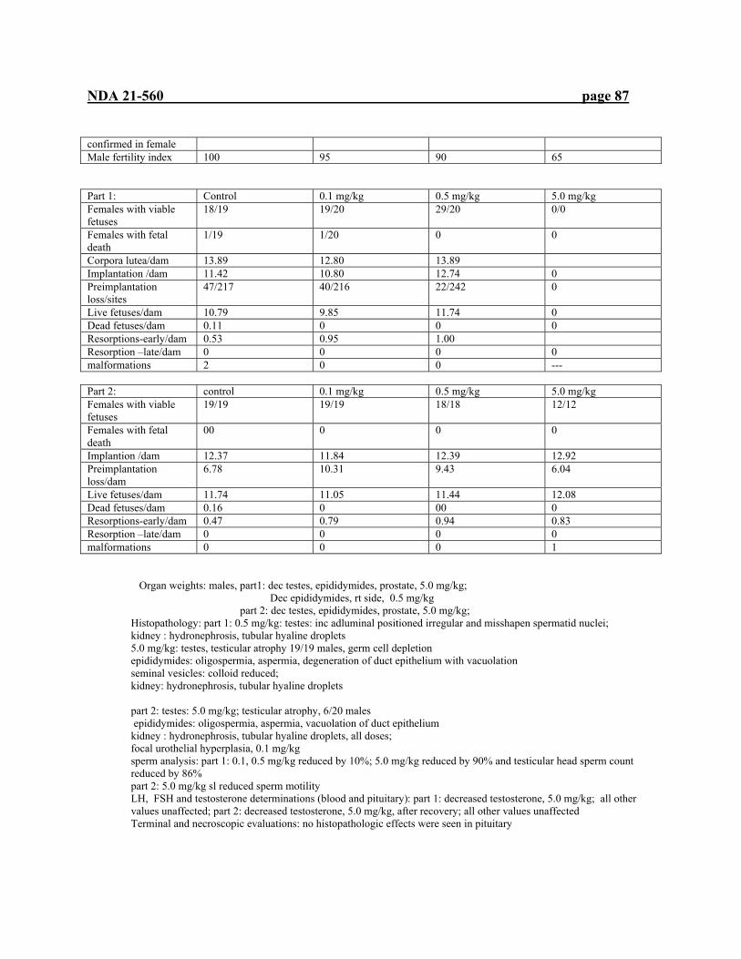
NDA 21-560 page 87
confirmed in female Male fertility index 100 95 90 65
Part 1: Control 0.1 mg/kg 0.5 mg/kg 5.0 mg/kg Females with viable fetuses
18/19 19/20 29/20 0/0
Females with fetal death
1/19 1/20 0 0
Corpora lutea/dam 13.89 12.80 13.89 Implantation /dam 11.42 10.80 12.74 0 Preimplantation loss/sites
47/217 40/216 22/242 0
Live fetuses/dam 10.79 9.85 11.74 0 Dead fetuses/dam 0.11 0 0 0 Resorptions-early/dam 0.53 0.95 1.00 Resorption –late/dam 0 0 0 0 malformations 2 0 0 ---
Part 2: control 0.1 mg/kg 0.5 mg/kg 5.0 mg/kg Females with viable fetuses
19/19 19/19 18/18 12/12
Females with fetal death
00 0 0 0
Implantion /dam 12.37 11.84 12.39 12.92 Preimplantation loss/dam
6.78 10.31 9.43 6.04
Live fetuses/dam 11.74 11.05 11.44 12.08 Dead fetuses/dam 0.16 0 00 0 Resorptions-early/dam 0.47 0.79 0.94 0.83 Resorption –late/dam 0 0 0 0 malformations 0 0 0 1
Organ weights: males, part1: dec testes, epididymides, prostate, 5.0 mg/kg; Dec epididymides, rt side, 0.5 mg/kg
part 2: dec testes, epididymides, prostate, 5.0 mg/kg; Histopathology: part 1: 0.5 mg/kg: testes: inc adluminal positioned irregular and misshapen spermatid nuclei; kidney : hydronephrosis, tubular hyaline droplets 5.0 mg/kg: testes, testicular atrophy 19/19 males, germ cell depletion epididymides: oligospermia, aspermia, degeneration of duct epithelium with vacuolation seminal vesicles: colloid reduced; kidney: hydronephrosis, tubular hyaline droplets part 2: testes: 5.0 mg/kg; testicular atrophy, 6/20 males epididymides: oligospermia, aspermia, vacuolation of duct epithelium kidney : hydronephrosis, tubular hyaline droplets, all doses; focal urothelial hyperplasia, 0.1 mg/kg sperm analysis: part 1: 0.1, 0.5 mg/kg reduced by 10%; 5.0 mg/kg reduced by 90% and testicular head sperm count reduced by 86% part 2: 5.0 mg/kg sl reduced sperm motility LH, FSH and testosterone determinations (blood and pituitary): part 1: decreased testosterone, 5.0 mg/kg; all other values unaffected; part 2: decreased testosterone, 5.0 mg/kg, after recovery; all other values unaffected Terminal and necroscopic evaluations: no histopathologic effects were seen in pituitary
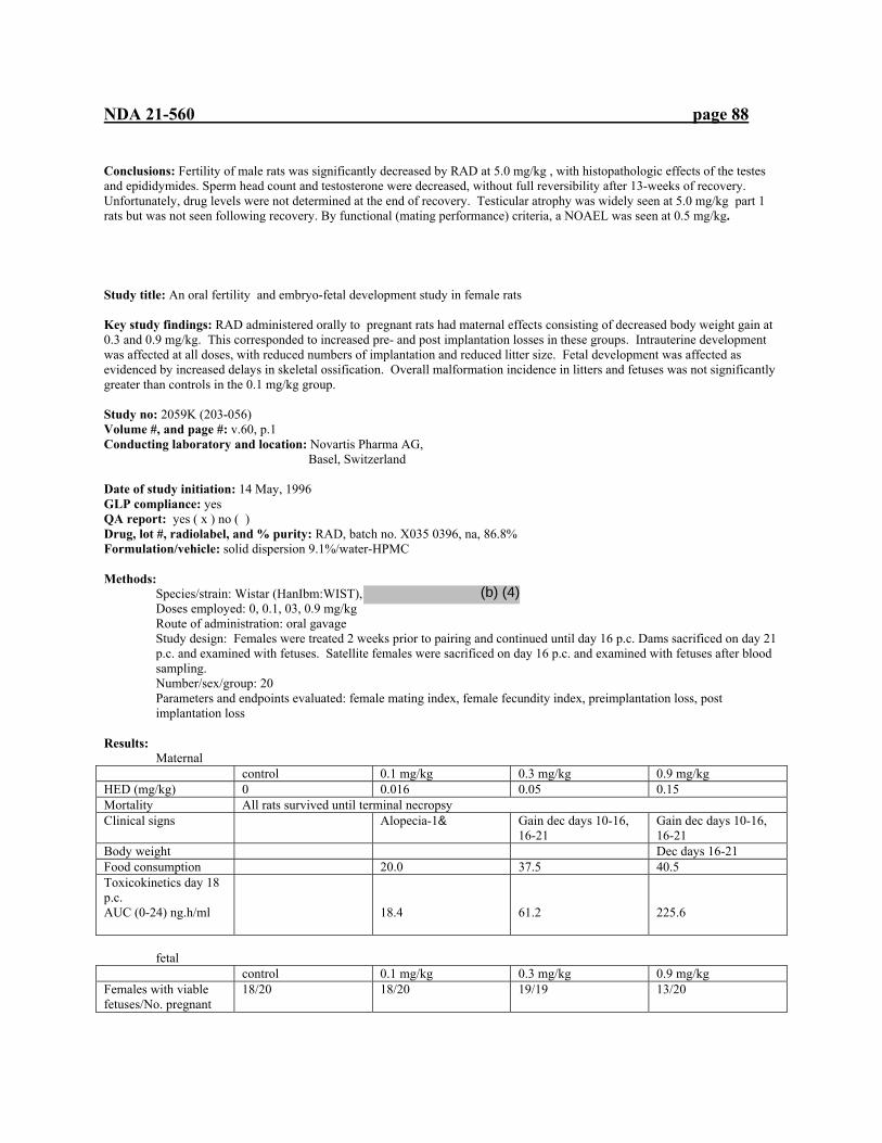
NDA 21-560 page 88
Conclusions: Fertility of male rats was significantly decreased by RAD at 5.0 mg/kg , with histopathologic effects of the testes and epididymides. Sperm head count and testosterone were decreased, without full reversibility after 13-weeks of recovery. Unfortunately, drug levels were not determined at the end of recovery. Testicular atrophy was widely seen at 5.0 mg/kg part 1 rats but was not seen following recovery. By functional (mating performance) criteria, a NOAEL was seen at 0.5 mg/kg. Study title: An oral fertility and embryo-fetal development study in female rats Key study findings: RAD administered orally to pregnant rats had maternal effects consisting of decreased body weight gain at 0.3 and 0.9 mg/kg. This corresponded to increased pre- and post implantation losses in these groups. Intrauterine development was affected at all doses, with reduced numbers of implantation and reduced litter size. Fetal development was affected as evidenced by increased delays in skeletal ossification. Overall malformation incidence in litters and fetuses was not significantly greater than controls in the 0.1 mg/kg group. Study no: 2059K (203-056) Volume #, and page #: v.60, p.1 Conducting laboratory and location: Novartis Pharma AG, Basel, Switzerland Date of study initiation: 14 May, 1996 GLP compliance: yes QA report: yes ( x ) no ( ) Drug, lot #, radiolabel, and % purity: RAD, batch no. X035 0396, na, 86.8% Formulation/vehicle: solid dispersion 9.1%/water-HPMC Methods:
Species/strain: Wistar (HanIbm:WIST), Doses employed: 0, 0.1, 03, 0.9 mg/kg Route of administration: oral gavage Study design: Females were treated 2 weeks prior to pairing and continued until day 16 p.c. Dams sacrificed on day 21 p.c. and examined with fetuses. Satellite females were sacrificed on day 16 p.c. and examined with fetuses after blood sampling. Number/sex/group: 20 Parameters and endpoints evaluated: female mating index, female fecundity index, preimplantation loss, post implantation loss
Results:
Maternal control 0.1 mg/kg 0.3 mg/kg 0.9 mg/kg HED (mg/kg) 0 0.016 0.05 0.15 Mortality All rats survived until terminal necropsy Clinical signs Alopecia-1& Gain dec days 10-16,
16-21 Gain dec days 10-16, 16-21
Body weight Dec days 16-21 Food consumption 20.0 37.5 40.5 Toxicokinetics day 18 p.c. AUC (0-24) ng.h/ml
18.4
61.2
225.6
fetal
control 0.1 mg/kg 0.3 mg/kg 0.9 mg/kg Females with viable fetuses/No. pregnant
18/20 18/20 19/19 13/20
(b) (4)
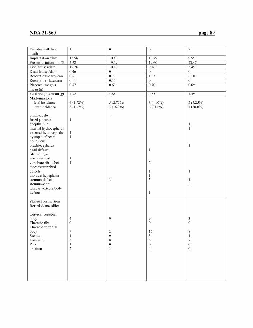
NDA 21-560 page 89
Females with fetal death
1 0 0 7
Implantation /dam 13.56 10.83 10.79 9.55 Preimplantation loss % 5.92 19.19 19.60 23.47 Live fetuses/dam 12.78 10.00 9.16 3.45 Dead fetuses/dam 0.06 0 0 0 Resorptions-early/dam 0.61 0.72 1.63 6.10 Resorption –late/dam 0.11 0.11 0 0 Placental weights mean (g)
0.67 0.69 0.70 0.69
Fetal weights mean (g) 4.82 4.88 4.63 4.59 Malformations fetal incidence litter incidence omphacoele fused placenta anopthalmia internal hydrocephalus external hydrocephalus dystopia of heart no truncus brachiocephalus head defects rib cartilage asymmetrical vertebrae rib defects thoracic/vertebral defects thoracic hypoplasia sternum defects sternum-cleft lumbar vertebra body defects
4 (1.72%) 3 (16.7%) 1 1 1 1 1
5 (2.75%) 3 (16.7%) 1 3
8 (4.60%) 6 (31.6%) 1 2 1 1 5 1
5 (7.25%) 4 (30.8%) 1 1 1 1 1 2
Skeletal ossification Retarded/unossified Cervical vertebral body Thoracic ribs Thoracic vertebral body Sternum Forelimb Ribs cranium
4 0 9 1 3 1 2
9 1 2 0 8 0 3
9 0 16 3 6 0 4
3 0 8 1 7 0 0

NDA 21-560 page 90
Summary of individual study findings: RAD administered orally to pregnant rats had maternal effects consisting of decreased body weight gain at 0.3 and 0.9 mg/kg. This corresponded to increased pre- and post implantation losses in these groups. Intrauterine development was affected at all doses, with reduced numbers of implantation and reduced litter size. Fetal development was affected as evidenced by increased delays in skeletal ossification. Overall malformation incidence in litters and fetuses was not significantly greater than controls in the 0.1 mg/kg group. A NOEL for fetal toxicity was observed at <0.1 mg/kg
Study title: An oral embryo-fetal development study in rabbits Key study findings: Maternal toxicity, demonstrated by reduced weight gain, food consumption, and mortality, was seen in the 0.2 and 0.8 mg/kg groups. The NOEL for maternal toxicity was 0.05 mg/kg. The maternal toxicity coincides with slightly increased late resorptions in the 0.8 mg/kg group. Fetal malformations were slightly (not statistically significant) increased in treated rabbits over those in controls. Study no: 203-074 Volume #, and page #: v.62, p.1 Conducting laboratory and location: Novartis Pharma AG, Basel, Switzerland Date of study initiation: 20 May 1996 GLP compliance: yes QA report: yes ( x ) no ( ) Drug, lot #, radiolabel, and % purity: RAD, batch no. X035 0396, na, 86.8% Formulation/vehicle: solid dispersion 9.1%/water-HPMC Methods:
Species/strain: female New Zealand hybrid rabbits (Crl:KBL (NZW) BR), Doses employed: 0, 0.05, 0.2, 0.8 mg/kg Route of administration: oral gavage Study design: Females were treated from day 6 post insemination to day 18 post insemination. Dams were sacrificed on day 29 p.i. and examined with fetuses. Satellite females were sacrificed on day 18 p.i. and examined with fetuses after blood sampling (0.5, 1, 2, 4 and 6 hr post dosing). Number/sex/group: control, 0.05 mg/kg, 20; 0.2, 0.8 mg/kg, 22; satellite for pk, 0.05 mg/kg, 4; 0.2 mg/kg, 2; 0.8 mg/kg, 3 Parameters and endpoints evaluated: female mating index, female fecundity index, preimplantation loss, post implantation loss
Maternal findings Dose 0 0.05 mg/kg 0.2 mg/kg 0.8 mg/kg HED (mg/kg) 0 0.025 0.100 0.400 Mortality 1, day 22 p.i., aborted 1, day 19, p.i. Clinical signs Reduced water
intake Reduced water intake 1 dam aborted, day 21 p.i.
Reduced water intake Reduced water intake
Body weight Sl red weight gain Weight loss, day6-18 p.i., overall dec weight gain during pregnancy
Food consumption Reduced during treatment
Toxicokinetics day 16 p.c. AUC (0-24) ng.h/ml
(b) (4)
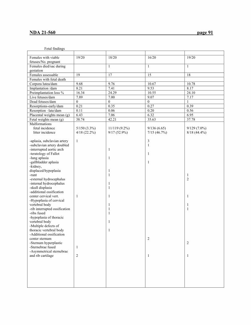
NDA 21-560 page 91
Fetal findings Females with viable fetuses/No. pregnant
19/20 18/20 16/20 19/20
Females died/sac during gestation
1 1 1
Females assessable 19 17 15 18 Females with fetal death Corpora lutea/dam 9.68 9.76 10.67 10.78 Implantation /dam 8.21 7.41 9.53 8.17 Preimplantation loss % 16.34 24.29 10.55 24.10 Live fetuses/dam 7.89 7.00 9.07 7.17 Dead fetuses/dam 0 0 0 1 Resorptions-early/dam 0.21 0.35 0.27 0.39 Resorption –late/dam 0.11 0.06 0.20 0.56 Placental weights mean (g) 6.43 7.06 6.32 6.95 Fetal weights mean (g) 38.74 42.21 35.63 37.78 Malformations fetal incidence litter incidence -aplasia, subclavian artery -subclavian artery doubled -interrupted aortic arch -teratology of Fallot -lung aplasia -gallbladder aplasia -kidney, displaced/hypoplasia -runt -external hydrocephalus -internal hydrocephalus -skull displasia -additional ossification center cervical vert. -Hypoplasia of cervical vertebral body -rib interrupted ossification -ribs fused -hypoplasia of thoracic vertebral body -Multiple defects of thoracic vertebral body -Additional ossification center sternum -Sternum hyperplastic -Sternebrae fused -Asymmetrical sternebrae and rib cartilage
5/150 (3.3%) 4/18 (22.2%) 1 1 1 2
11/119 (9.2%) 9/17 (52.9%) 1 1 1 1 1 1 1 1 1 1 1 1
9/136 (6.65) 7/15 (46.7%) 3 1 1 1 2 1
9/129 (7.0%) 8/18 (44.4%) 1 2 1 1 1 2 1
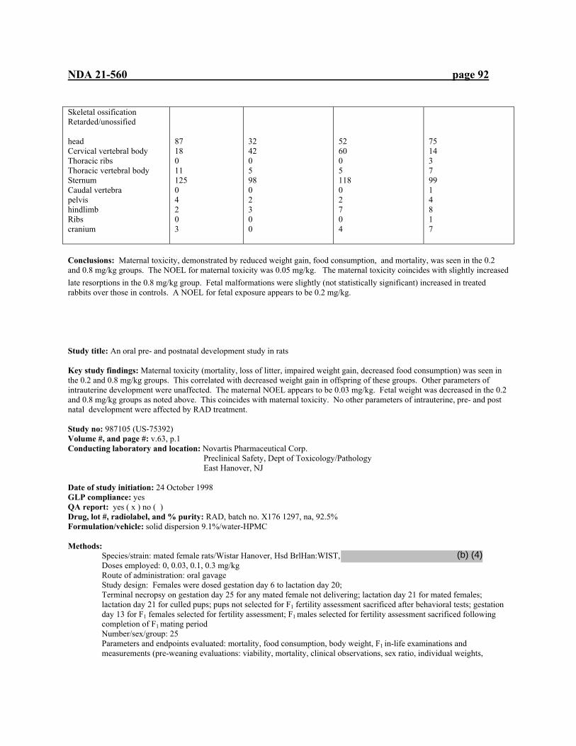
NDA 21-560 page 92
Skeletal ossification Retarded/unossified head Cervical vertebral body Thoracic ribs Thoracic vertebral body Sternum Caudal vertebra pelvis hindlimb Ribs cranium
87 18 0 11 125 0 4 2 0 3
32 42 0 5 98 0 2 3 0 0
52 60 0 5 118 0 2 7 0 4
75 14 3 7 99 1 4 8 1 7
Conclusions: Maternal toxicity, demonstrated by reduced weight gain, food consumption, and mortality, was seen in the 0.2 and 0.8 mg/kg groups. The NOEL for maternal toxicity was 0.05 mg/kg. The maternal toxicity coincides with slightly increased late resorptions in the 0.8 mg/kg group. Fetal malformations were slightly (not statistically significant) increased in treated rabbits over those in controls. A NOEL for fetal exposure appears to be 0.2 mg/kg. Study title: An oral pre- and postnatal development study in rats Key study findings: Maternal toxicity (mortality, loss of litter, impaired weight gain, decreased food consumption) was seen in the 0.2 and 0.8 mg/kg groups. This correlated with decreased weight gain in offspring of these groups. Other parameters of intrauterine development were unaffected. The maternal NOEL appears to be 0.03 mg/kg. Fetal weight was decreased in the 0.2 and 0.8 mg/kg groups as noted above. This coincides with maternal toxicity. No other parameters of intrauterine, pre- and post natal development were affected by RAD treatment. Study no: 987105 (US-75392) Volume #, and page #: v.63, p.1 Conducting laboratory and location: Novartis Pharmaceutical Corp. Preclinical Safety, Dept of Toxicology/Pathology
East Hanover, NJ Date of study initiation: 24 October 1998 GLP compliance: yes QA report: yes ( x ) no ( ) Drug, lot #, radiolabel, and % purity: RAD, batch no. X176 1297, na, 92.5% Formulation/vehicle: solid dispersion 9.1%/water-HPMC Methods:
Species/strain: mated female rats/Wistar Hanover, Hsd BrlHan:WIST, Doses employed: 0, 0.03, 0.1, 0.3 mg/kg Route of administration: oral gavage Study design: Females were dosed gestation day 6 to lactation day 20; Terminal necropsy on gestation day 25 for any mated female not delivering; lactation day 21 for mated females; lactation day 21 for culled pups; pups not selected for F1 fertility assessment sacrificed after behavioral tests; gestation day 13 for F1 females selected for fertility assessment; F1 males selected for fertility assessment sacrificed following completion of F1 mating period Number/sex/group: 25 Parameters and endpoints evaluated: mortality, food consumption, body weight, F1 in-life examinations and measurements (pre-weaning evaluations: viability, mortality, clinical observations, sex ratio, individual weights,
(b) (4)

NDA 21-560 page 93
righting reflex, pinna detached, and eye opening and post-weaning evaluations; mortality, clinical observations, acoustic startle, pupillary reflex, vaginal opening, preputial separation, passive avoidance-learning/acquisition, watermaze-learning and memory, open field motor activity, F1 mating, individual body weights)
Maternal dose 0 0.03 mg/kg 0.10 mg/kg 0.30 mg/kg HED (mg/kg) 0.005 0.017 0.050 Mortality All rats survived until terminal sacrifice Clinical signs Body weight Sl gain gestation days
6-9 Food consumption Inc days 6-9 Inc days 18-21 Necropsy findings 1 female (lost litter)
had no milk in mammary glands
Delivery data No treatment-related effects on number of corpora lutea, pre-, post implantation loss, fetal size, litter size or other maternal or developmental parameters
fetal
Body weights Dec days 28, 35, 42, 49, all; days 56, 63 &
Dec days 28, 35, 49 &
F1 in-life examinations and measurements (pre-weaning evaluations: viability, mortality, clinical observations, sex ratio, righting reflex, pinna detached, and eye opening and post-weaning evaluations; mortality, clinical observations, acoustic startle, pupillary reflex, vaginal opening, preputial separation, passive avoidance-learning/acquisition, watermaze-learning and memory, open field motor activity, F1 mating)
No treatment-related effect was seen in these studies
Conclusions: Maternal toxicity (mortality, loss of litter, impaired weight gain, decreased food consumption) was seen in the 0.2 and 0.8 mg/kg groups. This correlated with decreased weight gain in offspring of these groups. Other parameters of intrauterine development were unaffected. The maternal NOEL appears to be 0.03 mg/kg. Fetal weight was decreased in the 0.2 and 0.8 mg/kg groups as noted above. This appears to coincide with maternal toxicity. No other parameters of intrauterine, pre- and post natal development were affected by RAD treatment.. Due to the body weight effects at the 0.2 and 0.8 mg/kg doses, a fetal/F1 NOEL was seen at the 0.05 mg/kg dose.
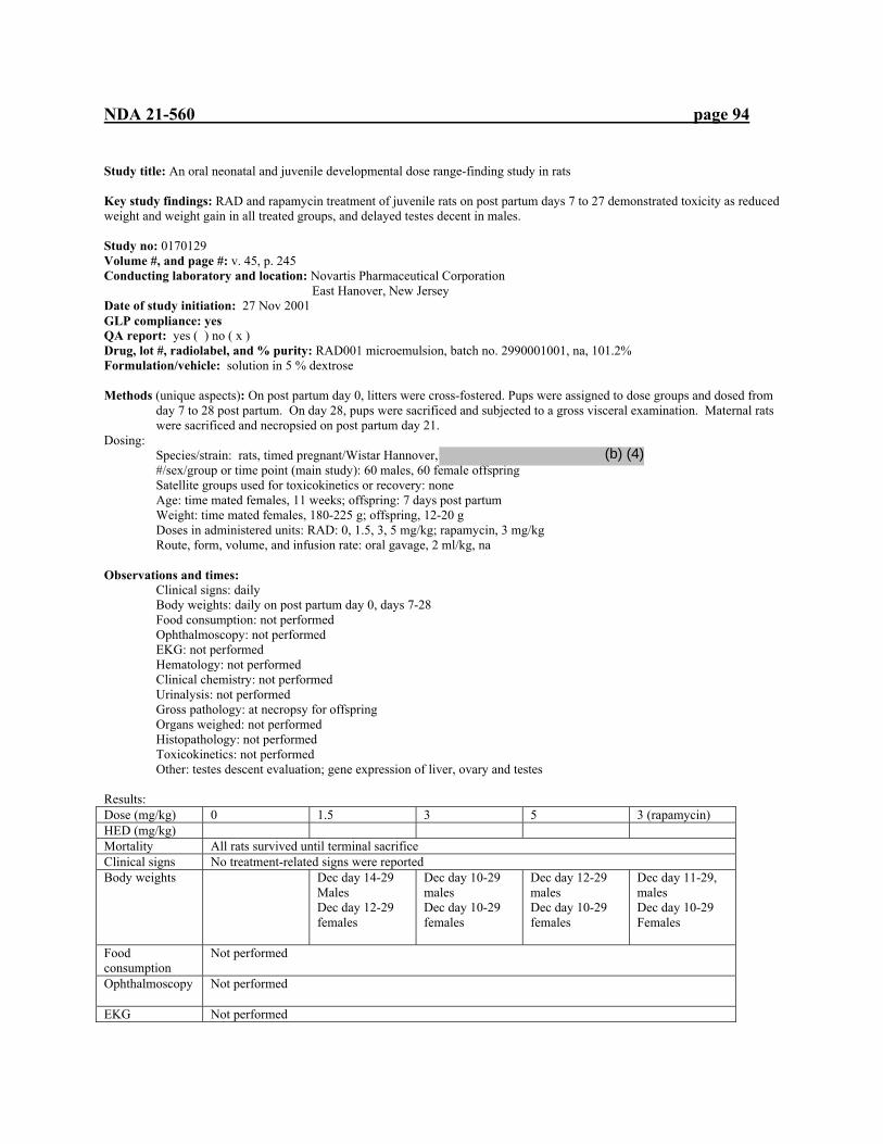
NDA 21-560 page 94
Study title: An oral neonatal and juvenile developmental dose range-finding study in rats Key study findings: RAD and rapamycin treatment of juvenile rats on post partum days 7 to 27 demonstrated toxicity as reduced weight and weight gain in all treated groups, and delayed testes decent in males. Study no: 0170129 Volume #, and page #: v. 45, p. 245 Conducting laboratory and location: Novartis Pharmaceutical Corporation East Hanover, New Jersey Date of study initiation: 27 Nov 2001 GLP compliance: yes QA report: yes ( ) no ( x ) Drug, lot #, radiolabel, and % purity: RAD001 microemulsion, batch no. 2990001001, na, 101.2% Formulation/vehicle: solution in 5 % dextrose Methods (unique aspects): On post partum day 0, litters were cross-fostered. Pups were assigned to dose groups and dosed from
day 7 to 28 post partum. On day 28, pups were sacrificed and subjected to a gross visceral examination. Maternal rats were sacrificed and necropsied on post partum day 21.
Dosing: Species/strain: rats, timed pregnant/Wistar Hannover, #/sex/group or time point (main study): 60 males, 60 female offspring Satellite groups used for toxicokinetics or recovery: none Age: time mated females, 11 weeks; offspring: 7 days post partum Weight: time mated females, 180-225 g; offspring, 12-20 g Doses in administered units: RAD: 0, 1.5, 3, 5 mg/kg; rapamycin, 3 mg/kg Route, form, volume, and infusion rate: oral gavage, 2 ml/kg, na
Observations and times: Clinical signs: daily Body weights: daily on post partum day 0, days 7-28 Food consumption: not performed Ophthalmoscopy: not performed EKG: not performed Hematology: not performed Clinical chemistry: not performed Urinalysis: not performed Gross pathology: at necropsy for offspring Organs weighed: not performed Histopathology: not performed Toxicokinetics: not performed Other: testes descent evaluation; gene expression of liver, ovary and testes Results: Dose (mg/kg) 0 1.5 3 5 3 (rapamycin) HED (mg/kg) Mortality All rats survived until terminal sacrifice Clinical signs No treatment-related signs were reported Body weights
Dec day 14-29 Males Dec day 12-29 females
Dec day 10-29 males Dec day 10-29 females
Dec day 12-29 males Dec day 10-29 females
Dec day 11-29, males Dec day 10-29 Females
Food consumption
Not performed
Ophthalmoscopy
Not performed
EKG Not performed
(b) (4)

NDA 21-560 page 95
Hematology
Not performed
Clinical chemistry
Not performed
Urinalysis
Not performed
Organ weights
Not performed
Gross pathology
No treatment-related findings were reported
Histopathology Not performed Testes decent evaluation, days 20-25
Fewer than controls, day 21
Fewer than controls, 20-21
Fewer than controls, day 20-22
Fewer than controls, day 20-22
Gene expression
Samples frozen, no further information reported
Toxicokinetics
Not performed
Conclusions: RAD and rapamycin treatment of juvenile rats on post partum days 7 to 27 demonstrated toxicity as reduced weight and weight gain in all treated groups, delayed testes decent in males. A NOEL was not established in this study. Study title: An oral neonatal and juvenile development study in rats with 13- and 26-week recovery periods Key study findings: At doses of 0.15, 0.5 and 1.5 mg/kg RAD and 1.5 mg/kg rapamycin (day 7 to 70 post partum) toxicities were observed at all doses including decreased body weight gain, decreased food consumption and delayed developmental landmarks. Weight gain and food consumption deficits were partially reversed following a recovery period of up top 26 weeks. The 1.5 mg/kg doses of RAD and rapamycin produced toxicities of similar severity including toxicities to the lens of the eye, prostate, blood and immune tissues, testes, epididymides, seminal vesicles, ovaries, uterus, and kidney. Reduced body weight, hyperlipidemia (males), renal mineralization remained after recovery at both doses. Study no.: 0270015 Volume #, and page #: electronic submission Conducting laboratory and location: Novartis Pharmaceutical Corp Hannover, NJ Date of study initiation: 22 Jan 2002 GLP compliance: yes QA reports: yes ( x) no ( ) Drug, lot #, radiolabel, and % purity: RAD001 2% microemulsion ; 2990001001, na, 101.7%; rapamycin, 2000014007, na, 101.6% Formulation/vehicle: RAD 2% microemulsion in 5% dextrose solution Methods:
Species/strain: rats/ timed-pregnant Wistar-Hannover female, Doses employed: RAD001: 0.15, 0.5, 1.5 mg/kg; rapamycin: 1.5 mg/kg Route of administration: oral gavage Study design:
(b) (4)
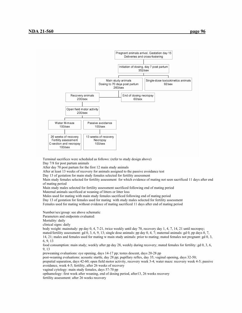
NDA 21-560 page 96
Terminal sacrifices were scheduled as follows: (refer to study design above) Day 7/8 for post partum animals After day 70 post partum for the first 12 main study animals After at least 13 weeks of recovery for animals assigned to the passive avoidance test Day 13 of gestation for main study females selected for fertility assessment Main study females selected for fertility assessment for which evidence of mating not seen sacrificed 11 days after end of mating period Main study males selected for fertility assessment sacrificed following end of mating period Maternal animals sacrificed at weaning of litters or litter loss Males used for mating with main study females sacrificed following end of mating period Day 13 of gestation for females used for mating with study males selected for fertility assessment Females used for mating without evidence of mating sacrificed 11 days after end of mating period Number/sex/group: see above schematic Parameters and endpoints evaluated: Mortality: daily clinical signs: daily body weight: mainstudy: pp day 0, 4, 7-21, twice weekly until day 70, recovery day 1, 4, 7, 14, 21 until necropsy; mated/fertility assessment: gd 0, 3, 6, 9, 13; single dose animals: pp day 0, 4, 7; maternal animals: gd 0, pp days 0, 7, 14, 21; males and females used for mating w main study animals: prior to mating; mated females not pregnant: gd 0, 3, 6, 9, 13 food consumption: main study; weekly after pp day 28, weekly during recovery; mated females for fertility: gd 0, 3, 6, 9, 13 preweaning evaluations: eye opening, days 14-17 pp; testes descent, days 20-28 pp post-weaning evaluations: acoustic startle, day 28 pp; pupillary reflex, day 35; vaginal opening, days 32-50; preputial separation, days 42-60; open field motor activity, recovery week 3-4; water maze: recovery week 4-5; passive avoidance, week 4-5; fertility, after 26 weeks of recovery vaginal cytology: main study females, days 57-70 pp opthamology: first week after weaning, end of dosing period, after13, 26 weeks recovery fertility assessment: after 26 weeks recovery
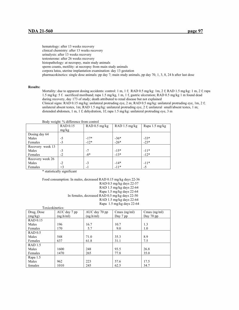
NDA 21-560 page 97
hematology: after 13 weeks recovery clinical chemistry: after 13 weeks recovery urinalysis: after 13 weeks recovery testosterone: after 26 weeks recovery histopathology: at necropsy, main study animals sperm counts, motility: at necropsy from main study animals corpora lutea, uterine implantation examination: day 13 gestation pharmacokinetics: single dose animals: pp day 7; main study animals, pp day 70, 1, 3, 8, 24 h after last dose
Results:
Mortality: due to apparent dosing accidents: control: 1 m, 1 f; RAD 0.5 mg/kg: 1m, 2 f; RAD 1.5 mg/kg: 1 m, 2 f; rapa 1.5 mg/kg: 5 f. sacrificed moribund; rapa 1.5 mg/kg, 1 m, 1 f, gastric ulceration; RAD 0.5 mg/kg 1 m found dead during recovery, day 173 of study; death attributed to renal disease but not explained Clinical signs: RAD 0.15 mg/kg: unilateral protruding eye, 2 m; RAD 0.5 mg/kg: unilateral protruding eye, 1m, 2 f; unilateral absent testes, 1m; RAD 1.5 mg/kg: unilateral protruding eye, 2 f; unilateral small/absent testes, 1 m; distended abdomen, 1 m, 1 f; dehydration, 1f; rapa 1.5 mg/kg: unilateral protruding eye, 3 m
Body weight: % difference from control
RAD 0.15 mg/kg
RAD 0.5 mg/kg RAD 1.5 mg/kg Rapa 1.5 mg/kg
Dosing day 64 Males Females
-5 -3
-17* -12*
-36* -26*
-33* -23*
Recovery week 13 Males Females
-3 -2
-7 -8*
-15* -13*
-11* -12*
Recovery week 26 Males Females
-2 +3
-3 -1
-14* -11*
-11* -5
* statistically significant Food consumption: In males, decreased RAD 0.15 mg/kg days 22-36 RAD 0.5 mg/kg days 22-57 RAD 1.5 mg/kg days 22-64 Rapa 1.5 mg/kg days 22-64 In females, decreased RAD 0.5 mg/kg days 22-50 RAD 1.5 mg/kg days 22-64 Rapa 1.5 mg/kg days 22-64 Toxicokinetics:
Drug, Dose (mg/kg)
AUC day 7 pp (ng.h/ml)
AUC day 70 pp (ng.h/ml)
Cmax (ng/ml) Day 7 pp
Cmax (ng/ml) Day 70 pp
RAD 0.15 Males Females
196 170
16.7 5.7
10.7 9.0
1.3 1.0
RAD 0.5 Males Females
548 637
71.0 61.8
35.3 31.1
8.9 7.5
RAD 1.5 Males Females
1600 1470
248 265
93.5 77.8
26.8 35.0
Rapa 1.5 Males females
962 1010
223 245
57.6 62.5
17.5 34.7

NDA 21-560 page 98
In-life observations:
Offspring: Pre-weaning observations: dec incidence of eye opening, females, RAD 1.5 mg/kg, rapa 1.5 mg/kg delayed testes descent, all treated groups Post-weaning observations:delayed preputial separation: all treated groups delayed vaginal opening: all treated groups
increased open field activity, RAD 0.15 (f) 0.5 (m), 1.5 (m+f) mg/kg, rapa (m+f)1.5 mg/kg increased time to criterion, water maze, males, RAD 0.5,1.5 mg/kg, rapa 1.5 mg/kg
Vaginal cytology: inc abnormal estrous cycles: rapa 1.5 mg/kg Opthalmology: males, females: RAD dose related inc zones of discontinuity, focal cataracts, treatment week 8, recovery week 13, 26; rapa 1.5 mg/kg similar to RAD 1.5 mg/kg Fertility assessment: no RAD or rapa effect on treated females; males, dec fertility, fecundity at RAD 1.5 mg/kg, rapa 1.5 mg/kg Hematology: inc WBC: RAD 0.15 mg/kg, 0.5 mg/kg (m)
Dec lymphocytes: RAD 1.5 mg/kg, rapa 1.5 mg/kg; sl inc during recovery, all treated groups Inc RBC, HGb, HCT; RAD 0.5, 1.5 mg/kg; rapa 1.5 mg/kg
Clinical biochemistry: dec serum protein, albumin: all treated males, females 1.5 mg/kg RAD and rapa; cholesterol and triglycerides: inc, all treated males; testosterone: dec, all treated groups; Creatinine: inc, 1-2 males in each treatment group Urinalysis: inc acid urine: males, RAD 0.5, 1.5 mg/kg, rapa 1.5 mg/kg
Terminal and necroscopic evaluations: Offspring:
Gross pathology: sm epididymides: rapa 1.5 mg/kg sm testes, prostate, seminal vesicles: all treated males
lung trauma, abdominal distension, gastric ulceration: rats found dead, probably due to gavage error
Histopathology: renal tubular mineralization: all treated groups Thymic lymphocytes, single cell necrosis: RAD 1.5 mg/kg, rapa 1.5 mg/kg
Summary of individual study findings: At doses of 0.15, 0.5 and 1.5 mg/kg RAD and 1.5 mg/kg rapamycin (day 7 to 70 post partum) toxicities were observed at all doses including decreased body weight gain, decreased food consumption and delayed developmental landmarks. Weight gain and food consumption deficits were partially reversed following a recovery period of up top 26 weeks. The 1.5 mg/kg doses of RAD and rapamycin produced similar toxicities of similar severity including toxicities to the lens of the eye, prostate, blood and immune tissues, testes, epididymides, seminal vesicles, ovaries, uterus, and kidney. Reduced body weight, hyperlipidemia (males), renal mineralization remained after recovery at both doses. Rapamycin produced higher incidence of small epididymides, testes, prostate and seminal vesicles than RAD at the 1.5 mg/kg dose. The small size of the male and female reproductive organs was not accompanied by related histopathology findings.
3.4.8 SPECIAL TOXICOLOGY STUDIES Study title: Comparative study of ophthalmic toxicity by oral gavage administration to CD rats and HanIbm rats for 4 weeks Key study findings: In CD-1 and HanIbm rats, the RAD high dose (5.0 mg/kg) exhibited frank toxicity to the lens of both strains as swelling and disruption of the fibers of the anterior cortex. Younger animals of both strains wee more susceptible to this toxicity. Myocarditis was exacerbated at both doses and both ages in both strains compared to the incidence in controls. Study no: 96/SPM098/0796 (203-068) Volume #, and page #: v. 44, p. 1 Conducting laboratory and location: (b) (4)

NDA 21-560 page 99
nd Date of study initiation: 1 April 1996 GLP compliance: yes QA report: yes ( x ) no ( ) Drug, lot #, radiolabel, and % purity: SDZ RAD microemulsion 2%, batch no. Y182 0895, na, 92.5% Formulation/vehicle: SDZ RAD2% microemulsion , diluted with 5% glucose solution Methods (unique aspects): none Dosing:
Species/strain: CD-1 rats, HanIbm Wistar rats, #/sex/group or time point (main study): 5/strain Satellite groups used for toxicokinetics or recovery: none Age: 6 weeks, 18 weeks (both CD-1 and Han Ibm) Weight: 6 week CD-1: 229-254g (males); 169-187g (females); 18 week CD-1: 466-526 g (males), 310-339g (females); 6 week HanIbm: 163-183g (males), 114-135g (females); 18 week HanIbm: 410-507g (males), 217-237g (females) Doses in administered units: 0, 0.5, 5.0 mg/kg Both strains, age groups Route, form, volume, and infusion rate: oral gavage, 5 ml/kg, na
Observations and times: Clinical signs: twice daily Body weights: pretreatment, weekly Food consumption: weekly Ophthalmoscopy: pretreatment, day 29 of treatment, the eyes of each rat were examined using an indirect opthalmoscope in the following structures: palpebrae and adjacent structures, conjunctiva, cornea and sclera, anterior chamber and iris, lens and vitreous and ocular fundus. EKG: not performed Hematology: not performed Clinical chemistry: not performed Urinalysis: not performed Gross pathology: at necropsy Organs weighed: at necropsy Histopathology: at necropsy, only eyes examined Toxicokinetics: not performed Other: na Results:
CD-1 HanIbm 6 week 18 week 6 week 18 week
Dose (mg/kg) 0.5 5.0 0.5 5.0 0.5 5.0 0.5 5.0 HED (mg/kg) 0.084 0.84 0.084 0.84 0.084 0.84 0.084 0.84 Mortality 1 male
died week 4 no clin signs
Clinical signs No treatment-related signs were seen Body weights loss Loss Food consumption
dec dec dec Dec
Ophthalmoscopy Lesions-swelling and disruption, fibers of anterior cortex of lens
5/5 males 5/5 females
3/5 males 4/5 males 4/5 females
(b) (4)
(b) (4)(b) (4)
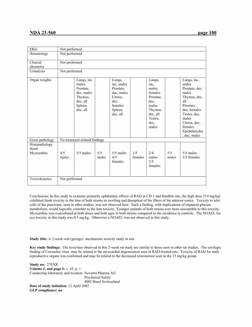
NDA 21-560 page 100
EKG Not performed Hematology
Not performed
Clinical chemistry
Not preformed
Urinalysis
Not performed
Organ weights
Lungs, inc males Prostate, dec, males Thymus, dec, all Spleen, dec, all
Lungs, inc, males Prostate, dec, males Uterus, dec, females Spleen, dec, all
Lungs, inc, males, females Prostate, dec, males Thymus, dec, all Testes, dec, males
Lungs, inc, males Prostate, dec, males Thymus, dec, all Pituitary , dec, females Testes, dec, males Uterus, dec, females Epididimydes, dec, males
Gross pathology No treatment-related findings Histopathology Heart Myocarditis
4/5 males
5/5 males
5/5 males
5/5 males 4/5 females
1/5 females
2/4 males 2/5 females
5/5 males
5/5 males 3/5 females
Toxicokinetics
Not performed
Conclusions: In this study to examine primarily ophthalmic effects of RAD in CD-1 and HanIbm rats, the high dose (5.0 mg/kg) exhibited frank toxicity to the lens of both strains as swelling and disruption of the fibers of the anterior cortex. Toxicity to islet cells of the pancrease, seen in other studies, was not observed here. Such a finding, with implications of impaired glucose metabolism, would logically correlate to the lens toxicity. Younger animals of both strains wee more susceptible to this toxicity. Myocarditis was exacerbated at both doses and both ages in both strains compared to the incidence in controls. The NOAEL for eye toxicity in this study was 0.5 mg/kg. Otherwise a NOAEL was not observed in this study. Study title: A 2-week oral (gavage) mechanistic toxicity study in rats Key study findings: The toxicities observed in this 2-week rat study are similar to those seen in other rat studies. The serologic finding of Coxsackie virus may be related to the myocardial degeneration seen in RAD-treated rats. Toxicity of RAD for male reproductive organs was confirmed and may be related to the decreased testosterone seen in the 15 mg/kg group. Study no: 27EXR Volume #, and page #: v. 45, p. 1 Conducting laboratory and location: Novartis Pharma AG Preclinical Safety
4002 Basel Switzerland Date of study initiation: 12 April 2002 GLP compliance: no
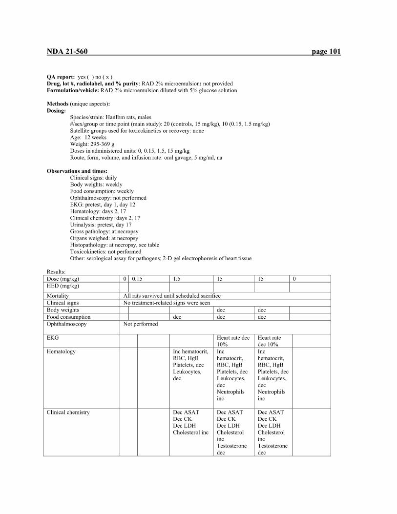
NDA 21-560 page 101
QA report: yes ( ) no ( x ) Drug, lot #, radiolabel, and % purity: RAD 2% microemulsion: not provided Formulation/vehicle: RAD 2% microemulsion diluted with 5% glucose solution Methods (unique aspects): Dosing:
Species/strain: HanIbm rats, males #/sex/group or time point (main study): 20 (controls, 15 mg/kg), 10 (0.15, 1.5 mg/kg) Satellite groups used for toxicokinetics or recovery: none Age: 12 weeks Weight: 295-369 g Doses in administered units: 0, 0.15, 1.5, 15 mg/kg Route, form, volume, and infusion rate: oral gavage, 5 mg/ml, na
Observations and times: Clinical signs: daily Body weights: weekly Food consumption: weekly Ophthalmoscopy: not performed EKG: pretest, day 1, day 12 Hematology: days 2, 17 Clinical chemistry: days 2, 17 Urinalysis: pretest, day 17 Gross pathology: at necropsy Organs weighed: at necropsy Histopathology: at necropsy, see table Toxicokinetics: not performed Other: serological assay for pathogens; 2-D gel electrophoresis of heart tissue Results: Dose (mg/kg) 0 0.15 1.5 15 15 0 HED (mg/kg)
Mortality All rats survived until scheduled sacrifice Clinical signs No treatment-related signs were seen Body weights dec dec Food consumption dec dec dec Ophthalmoscopy
Not performed
EKG Heart rate dec 10%
Heart rate dec 10%
Hematology
Inc hematocrit, RBC, HgB Platelets, dec Leukocytes, dec
Inc hematocrit, RBC, HgB Platelets, dec Leukocytes, dec Neutrophils inc
Inc hematocrit, RBC, HgB Platelets, dec Leukocytes, dec Neutrophils inc
Clinical chemistry Dec ASAT Dec CK Dec LDH Cholesterol inc
Dec ASAT Dec CK Dec LDH Cholesterol inc Testosterone dec
Dec ASAT Dec CK Dec LDH Cholesterol inc Testosterone dec

NDA 21-560 page 102
Urinalysis
No treatment-related findings reported
Organ weights
Gross pathology
No treatment-related findings reported
Histopathology Thymus Medullary Atrophy Cortical atrophy Heart Myocardial degeneration Lungs Alveolar macrophages Eyes Fiber swelling differential or focal Adrenal cortex Vacuolation Mammary atrophy Testes Germ cell degeneration Vacuoles/seminal epithelium Giant multinucleated cells Tubular atrophy Prostate Atrophy Seminal vesicles Atrophy Sternum –bone marrow depletion
1/10 1/10
5/10 1/10 1/10
10/10 9/10 10/10 10/10 2/10 9/10 1/10 1/10 5/10 2/10
10/10 10/10 10/10 10/10 10/10 9/10 6/10 10/10 7/10 1/10 2/10 9/10 8/10 5/10
10/10 8/10 7/10 10/10 9/10 10/10 6/10 10/10 9/10 3/10 3/10 7/10 9/10 8/10
3/9 1/10 2/10 1/10 1/10
2-d gel electrophoresis heart tissue
Tubulin-$, "-1-antiproteinase, apolipoprotein E precursor, transeferrin dec Apolipoprotein E inc
serology Antibody against Coxsackie virus B3 Toxicokinetics
Not performed
Conclusions: The toxicities observed in this 2-week rat study are similar to those seen in other rat studies. The serologic finding of Coxsackie virus may be related to the myocardial degeneration seen in RAD-treated rats. Toxicity of RAD for male reproductive organs was confirmed and may be related to the decreased testosterone seen in the 15 mg/kg group. Without a basis for the 2-D gel electrophoresis findings, they are of unknown value. Study title: Assessment of contact hypersensitivity to RAD001 in the albino guinea pig (maximization test) Key study findings: RAD did not cause contact hypersensitivity Study no: project 246792 (BS-812) (b) (4)
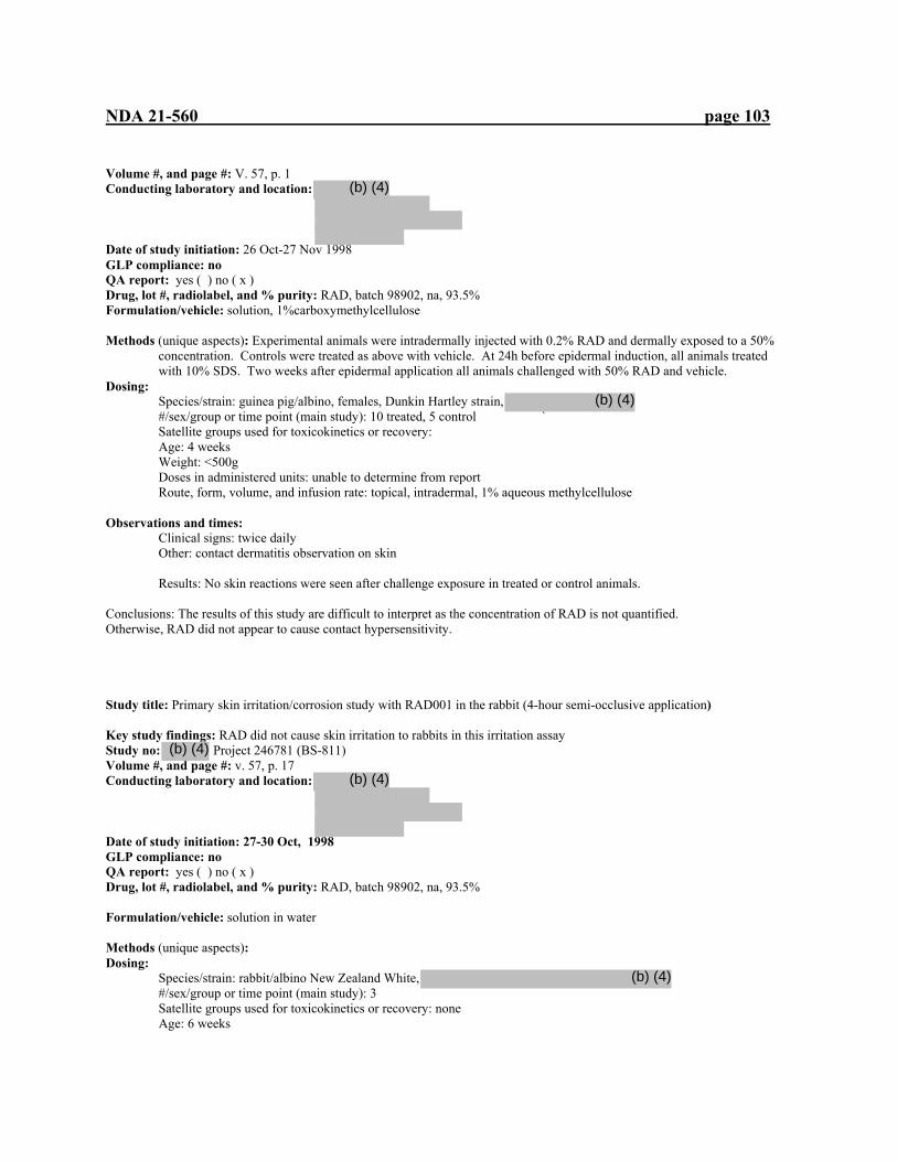
NDA 21-560 page 103
Volume #, and page #: V. 57, p. 1 Conducting laboratory and location: 3 Date of study initiation: 26 Oct-27 Nov 1998 GLP compliance: no QA report: yes ( ) no ( x ) Drug, lot #, radiolabel, and % purity: RAD, batch 98902, na, 93.5% Formulation/vehicle: solution, 1%carboxymethylcellulose Methods (unique aspects): Experimental animals were intradermally injected with 0.2% RAD and dermally exposed to a 50%
concentration. Controls were treated as above with vehicle. At 24h before epidermal induction, all animals treated with 10% SDS. Two weeks after epidermal application all animals challenged with 50% RAD and vehicle.
Dosing: Species/strain: guinea pig/albino, females, Dunkin Hartley strain, #/sex/group or time point (main study): 10 treated, 5 control ` Satellite groups used for toxicokinetics or recovery: Age: 4 weeks Weight: <500g Doses in administered units: unable to determine from report Route, form, volume, and infusion rate: topical, intradermal, 1% aqueous methylcellulose
Observations and times: Clinical signs: twice daily Other: contact dermatitis observation on skin
Results: No skin reactions were seen after challenge exposure in treated or control animals. Conclusions: The results of this study are difficult to interpret as the concentration of RAD is not quantified. Otherwise, RAD did not appear to cause contact hypersensitivity. Study title: Primary skin irritation/corrosion study with RAD001 in the rabbit (4-hour semi-occlusive application) Key study findings: RAD did not cause skin irritation to rabbits in this irritation assay Study no: Project 246781 (BS-811) Volume #, and page #: v. 57, p. 17 Conducting laboratory and location: Date of study initiation: 27-30 Oct, 1998 GLP compliance: no QA report: yes ( ) no ( x ) Drug, lot #, radiolabel, and % purity: RAD, batch 98902, na, 93.5% Formulation/vehicle: solution in water Methods (unique aspects): Dosing:
Species/strain: rabbit/albino New Zealand White, #/sex/group or time point (main study): 3 Satellite groups used for toxicokinetics or recovery: none Age: 6 weeks
(b) (4)
(b) (4)
(b) (4)
(b) (4)
(b) (4)

NDA 21-560 page 104
Weight: <3.5 kg Doses in administered units: 0.5 g RAD Route, form, volume, and infusion rate: dermal patch, 4 h
Observations and times: Clinical observations: twice daily Other: skin irritation/corrosion observation on skin at 1, 24, 48, and 72 h after application Results: No skin irritation was observed after 72h of skin application of RAD. Conclusions: RAD did not cause skin irritation to rabbits in this irritation assay.
Histopathology Inventory for NDA 21560 Study Species Dog 2 wk
Mini-pig
4 wk mini- pig
28-day cyno
26 wk cyno
52 wk cyno
2 week rat iv
2 week minipig iv
Adrenals X* X* X* X* X* X* X* Aorta x X x x x x x Bone Marrow smear x x Bone (femur) x X x x x x Brain X* X* X* X* X* X* X* Cecum X x x x x x Cervix x X* x x Colon X x x x x Duodenum X x X x x x x x Epididymis X X* X* X* X* x Esophagus X x X x x x Eye x X x x x x x Fallopian tube x Gall bladder x X x x x x Gross lesions X x x Harderian gland Heart X* X* X* X* X* X* X* Ileum x X x x x x x Injection site x Jejunum x X x x x x x Kidneys X* X* X* X* X* X* X* Lachrymal gland Larynx Liver X* X* X* X* X* X* X* Lungs X x X X* X* x X* X Lymph nodes, cervical Lymph nodes mandibular x X x x x x X Lymph nodes, mesenteric x X x x x x X Mammary Gland x X x x x X Nasal cavity Optic nerves x X x x x X Ovaries X* X* X* X* X* X* X* Pancreas X* X* X* X* x x X
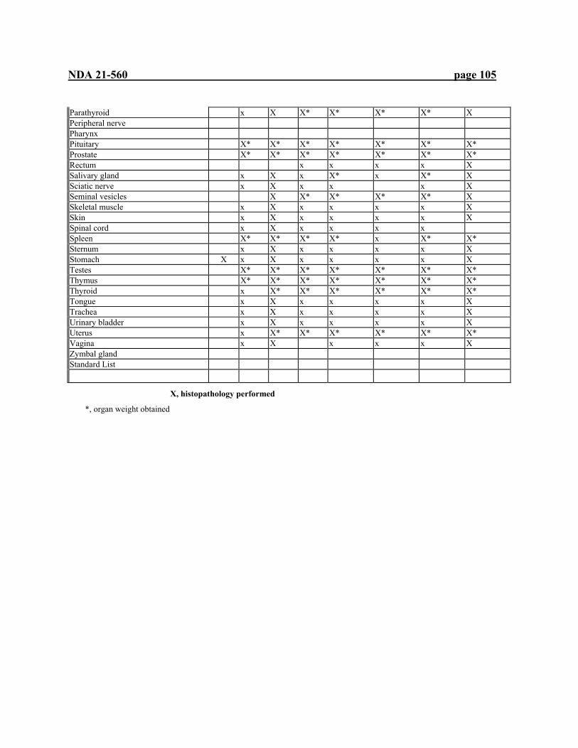
NDA 21-560 page 105
Parathyroid x X X* X* X* X* X Peripheral nerve Pharynx Pituitary X* X* X* X* X* X* X* Prostate X* X* X* X* X* X* X* Rectum x x x x X Salivary gland x X x X* x X* X Sciatic nerve x X x x x X Seminal vesicles X X* X* X* X* X Skeletal muscle x X x x x x X Skin x X x x x x X Spinal cord x X x x x x Spleen X* X* X* X* x X* X* Sternum x X x x x x X Stomach X x X x x x x X Testes X* X* X* X* X* X* X* Thymus X* X* X* X* X* X* X* Thyroid x X* X* X* X* X* X* Tongue x X x x x x X Trachea x X x x x x X Urinary bladder x X x x x x X Uterus x X* X* X* X* X* X* Vagina x X x x x X Zymbal gland Standard List
X, histopathology performed
*, organ weight obtained
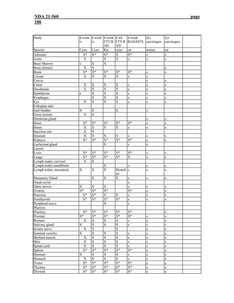
NDA 21-560 page 106
Study 4 week iv
4 week iv
4 weekFTY/RAD
4 wk FTY/RAD
4 week RAD/MTX
2yr carcinogen
2yr carcinogen
Species Cyno Cyno Rat cyno rat mouse rat Adrenals X* X* X* X X* x x Aorta X X X x x x Bone Marrow x X X Bone (femur) X X x Brain X* X* X* X* X* x x Cecum X X X X x x Cervix x x Colon X X X X x x x Duodenum X X X X x x x Epididymis x X X X x x x Esophagus X X X x x x Eye X X X X x x x Fallopian tube Gall bladder X X X x Gross lesions X X Harderian gland x x Heart X* X* X* X* X* x x Ileum X X X X x x x Injection site X X Jejunum X X X X x x x Kidneys X* X* X* X* X* x x Lachrymal gland X x x Larynx Liver X* X* X* X* X* x x Lungs X* X* X* X* X x x Lymph nodes, cervical X X Lymph nodes mandibular X x x x Lymph nodes, mesenteric X X X Bronch
ial x x x
Mammary Gland X X X x x x Nasal cavity x Optic nerves X X X x x x Ovaries X* X* X* X* x x Pancreas X* X* X X x x x Parathyroid X* X* X* X* x x x Peripheral nerve X x Pharynx Pituitary X* X* X* X* X* x Prostate X* X* X* X* X* x x Rectum X X X X x x x Salivary gland X X X X x x x Sciatic nerve X X X x x Seminal vesicles X X X X x x x Skeletal muscle X X X X x x x Skin X X X X x x x Spinal cord X X X X x x x Spleen X* X* X* X* X* x x Sternum X X X X x x x Stomach X X X X x x x Testes X* X* X* X* X* x x Thymus X* X* X* X* X* x x Thyroid X* X* X* X* X* x x
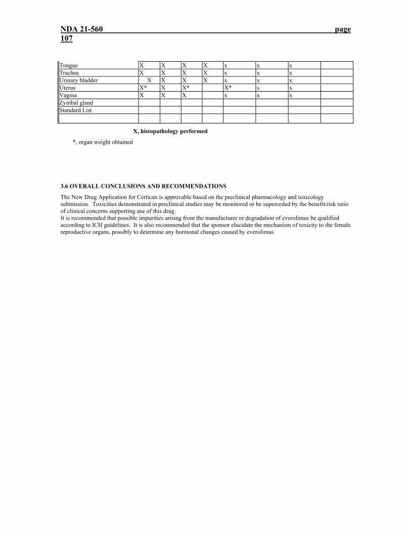
NDA 21-560 page 107
Tongue X X X X x x x Trachea X X X X x x x Urinary bladder X X X X x x x Uterus X* X X* X* x x Vagina X X X x x x Zymbal gland Standard List
X, histopathology performed
*, organ weight obtained
3.6 OVERALL CONCLUSIONS AND RECOMMENDATIONS
The New Drug Application for Certican is approvable based on the preclinical pharmacology and toxicology submission. Toxicities demonstrated in preclinical studies may be monitored or be superceded by the benefit/risk ratio of clinical concerns supporting use of this drug. It is recommended that possible impurities arising from the manufacturer or degradation of everolimus be qualified according to ICH guidelines. It is also recommended that the sponsor elucidate the mechanism of toxicity to the female reproductive organs, possibly to determine any hormonal changes caused by everolimus

---------------------------------------------------------------------------------------------------------------------This is a representation of an electronic record that was signed electronically andthis page is the manifestation of the electronic signature.--------------------------------------------------------------------------------------------------------------------- /s/---------------------Steve Kunder10/20/03 05:03:10 PMPHARMACOLOGIST
Kenneth Hastings10/21/03 03:42:47 PMPHARMACOLOGIST
Shukal Bala10/21/03 04:05:30 PMMICROBIOLOGIST


