Application of solution NMR spectroscopy for characterizing (and optimizing) mAb formulations Alexander “Sasha” Golovanov Manchester Institute of Biotechnology, The University of Manchester, UK [email protected]
Transcript
Application of solution NMR spectroscopy for characterizing
(and optimizing) mAb formulations
Alexander “Sasha” Golovanov
Manchester Institute of Biotechnology, The University of Manchester, UK [email protected]
Introduction • Generally, all proteins (including mAbs) may self-associate or aggregate, especially at
very high concentration => particle formation, opalescence, phase-separation
• Addition of co-solutes (excipients) may enhance protein stability, and decrease aggregation - the whole purpose of formulation process
• But how should you select excipients, which physical measurables to use as criteria to “choose the best”?
• Many biophysical methods require sample dilution, but best to measure “in situ”
• Solution NMR spectroscopy is sensitive to monomeric content and can detect transient self-association of proteins, even as large as mAbs, in different formulations
• Each traditional protein NMR study begins anyway from “sample condition optimisation” to achieve the highest monomeric content <-> spectra of best quality
Manchester Institute of Biotechnology, The University of Manchester, UK [email protected]
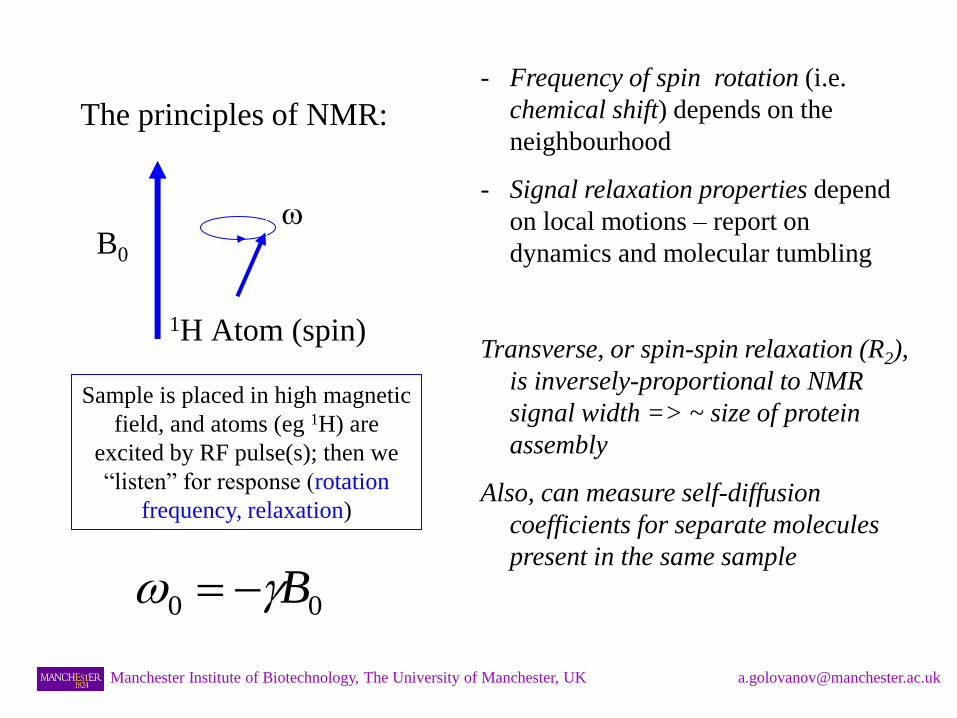
The principles of NMR:
- Frequency of spin rotation (i.e.
chemical shift) depends on the
neighbourhood
- Signal relaxation properties depend
on local motions – report on
dynamics and molecular tumbling
Transverse, or spin-spin relaxation (R2),
is inversely-proportional to NMR
signal width => ~ size of protein
assembly
Also, can measure self-diffusion
coefficients for separate molecules
present in the same sample
B0 w
1H Atom (spin)
Sample is placed in high magnetic
field, and atoms (eg 1H) are
excited by RF pulse(s); then we
“listen” for response (rotation
frequency, relaxation)
00 Bw
Manchester Institute of Biotechnology, The University of Manchester, UK [email protected]
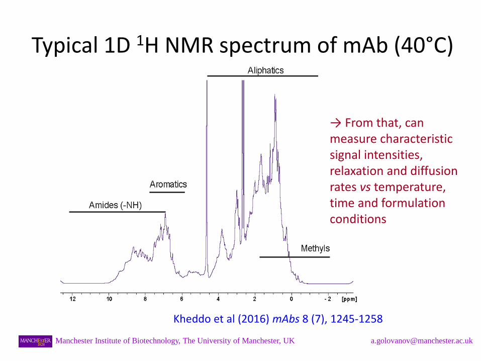
Typical 1D 1H NMR spectrum of mAb (40°C)
Kheddo et al (2016) mAbs 8 (7), 1245-1258
→ From that, can measure characteristic signal intensities, relaxation and diffusion rates vs temperature, time and formulation conditions
Manchester Institute of Biotechnology, The University of Manchester, UK [email protected]
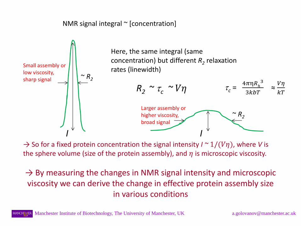
NMR signal integral ~ [concentration]
Here, the same integral (same concentration) but different R2 relaxation rates (linewidth)
R2 ~ c ~ 𝑉𝜂
→ So for a fixed protein concentration the signal intensity I ~ 1/(𝑉𝜂), where V is the sphere volume (size of the protein assembly), and 𝜂 is microscopic viscosity.
→ By measuring the changes in NMR signal intensity and microscopic viscosity we can derive the change in effective protein assembly size
in various conditions
I I
~ R2
~ R2
Manchester Institute of Biotechnology, The University of Manchester, UK [email protected]
Small assembly or low viscosity, sharp signal
Larger assembly or higher viscosity, broad signal
c = 4𝜋η𝑅
ℎ3
3𝑘𝑏𝑇 ≈
𝑉𝜂
𝑘𝑇
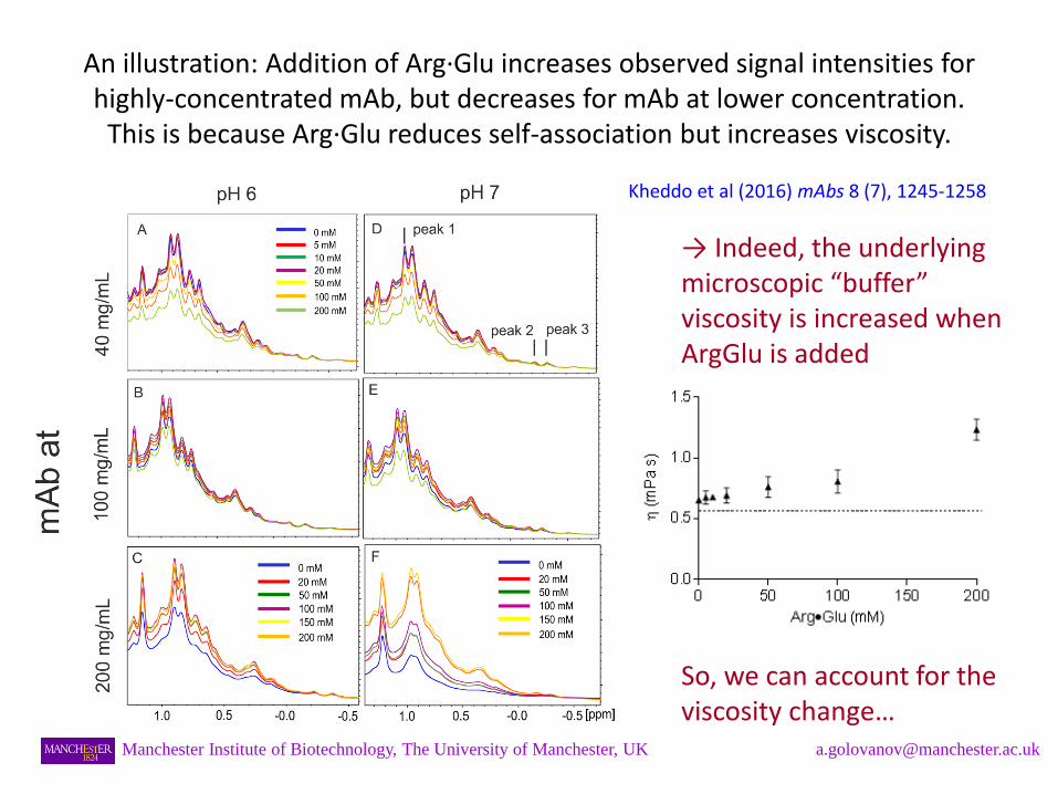
An illustration: Addition of Arg·Glu increases observed signal intensities for highly-concentrated mAb, but decreases for mAb at lower concentration.
This is because Arg·Glu reduces self-association but increases viscosity.
→ Indeed, the underlying microscopic “buffer” viscosity is increased when ArgGlu is added
So, we can account for the viscosity change…
Kheddo et al (2016) mAbs 8 (7), 1245-1258
Manchester Institute of Biotechnology, The University of Manchester, UK [email protected]
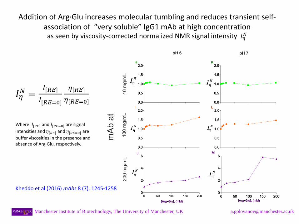
Addition of Arg·Glu increases molecular tumbling and reduces transient self-association of “very soluble” IgG1 mAb at high concentration
as seen by viscosity-corrected normalized NMR signal intensity 𝐼𝜂𝑁
𝐼𝜂𝑁 =
𝐼[𝑅𝐸]
𝐼[𝑅𝐸=0]
𝜂[𝑅𝐸]
𝜂[𝑅𝐸=0]
Where 𝐼[𝑅𝐸] and 𝐼[𝑅𝐸=0]
are signal
intensities and 𝜂[𝑅𝐸] and 𝜂[𝑅𝐸=0]
are
buffer viscosities in the presence and absence of Arg·Glu, respectively.
Kheddo et al (2016) mAbs 8 (7), 1245-1258
Manchester Institute of Biotechnology, The University of Manchester, UK [email protected]
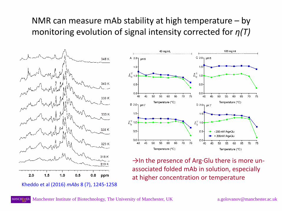
NMR can measure mAb stability at high temperature – by monitoring evolution of signal intensity corrected for η(T)
→In the presence of Arg·Glu there is more un-associated folded mAb in solution, especially at higher concentration or temperature
Kheddo et al (2016) mAbs 8 (7), 1245-1258
Manchester Institute of Biotechnology, The University of Manchester, UK [email protected]
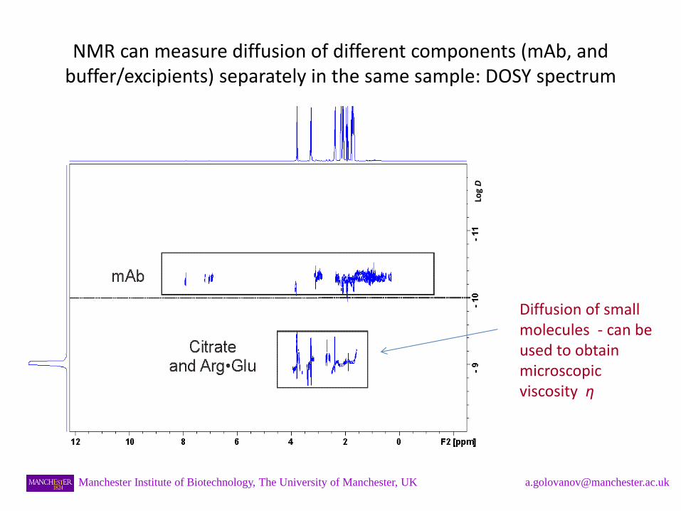
NMR can measure diffusion of different components (mAb, and buffer/excipients) separately in the same sample: DOSY spectrum
Log D
Manchester Institute of Biotechnology, The University of Manchester, UK [email protected]
Diffusion of small molecules - can be used to obtain microscopic viscosity η
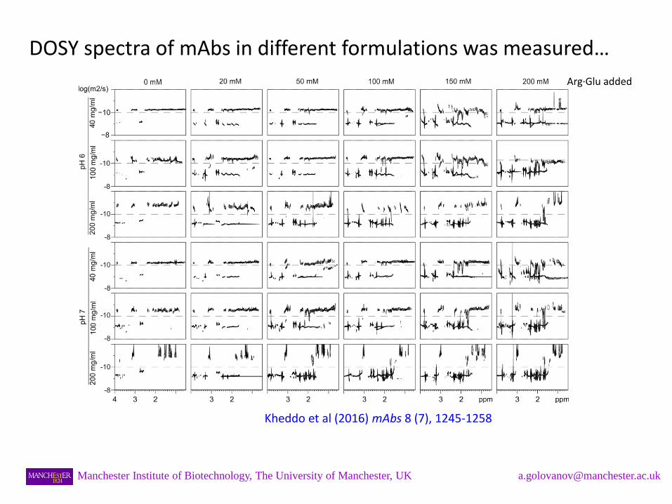
DOSY spectra of mAbs in different formulations was measured…
Manchester Institute of Biotechnology, The University of Manchester, UK [email protected]
Kheddo et al (2016) mAbs 8 (7), 1245-1258
Arg·Glu added
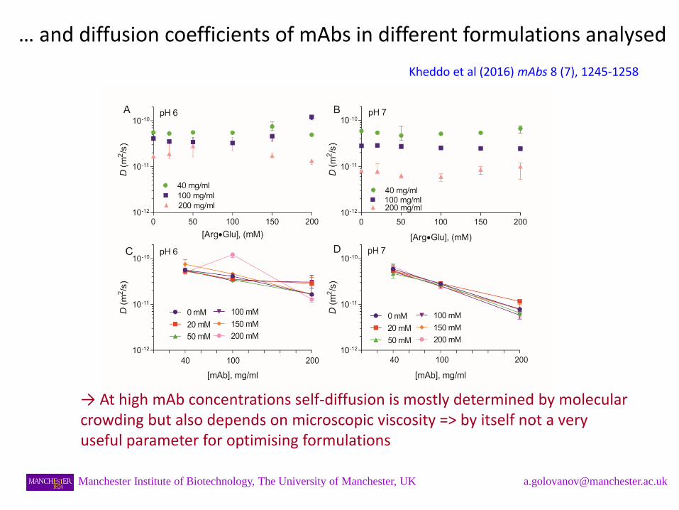
… and diffusion coefficients of mAbs in different formulations analysed
→ At high mAb concentrations self-diffusion is mostly determined by molecular crowding but also depends on microscopic viscosity => by itself not a very useful parameter for optimising formulations
Kheddo et al (2016) mAbs 8 (7), 1245-1258
Manchester Institute of Biotechnology, The University of Manchester, UK [email protected]
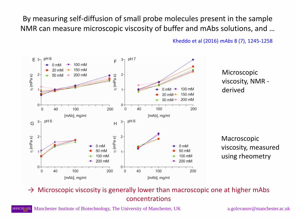
By measuring self-diffusion of small probe molecules present in the sample NMR can measure microscopic viscosity of buffer and mAbs solutions, and …
Kheddo et al (2016) mAbs 8 (7), 1245-1258
Manchester Institute of Biotechnology, The University of Manchester, UK [email protected]
Microscopic viscosity, NMR - derived
Macroscopic viscosity, measured using rheometry
→ Microscopic viscosity is generally lower than macroscopic one at higher mAbs concentrations
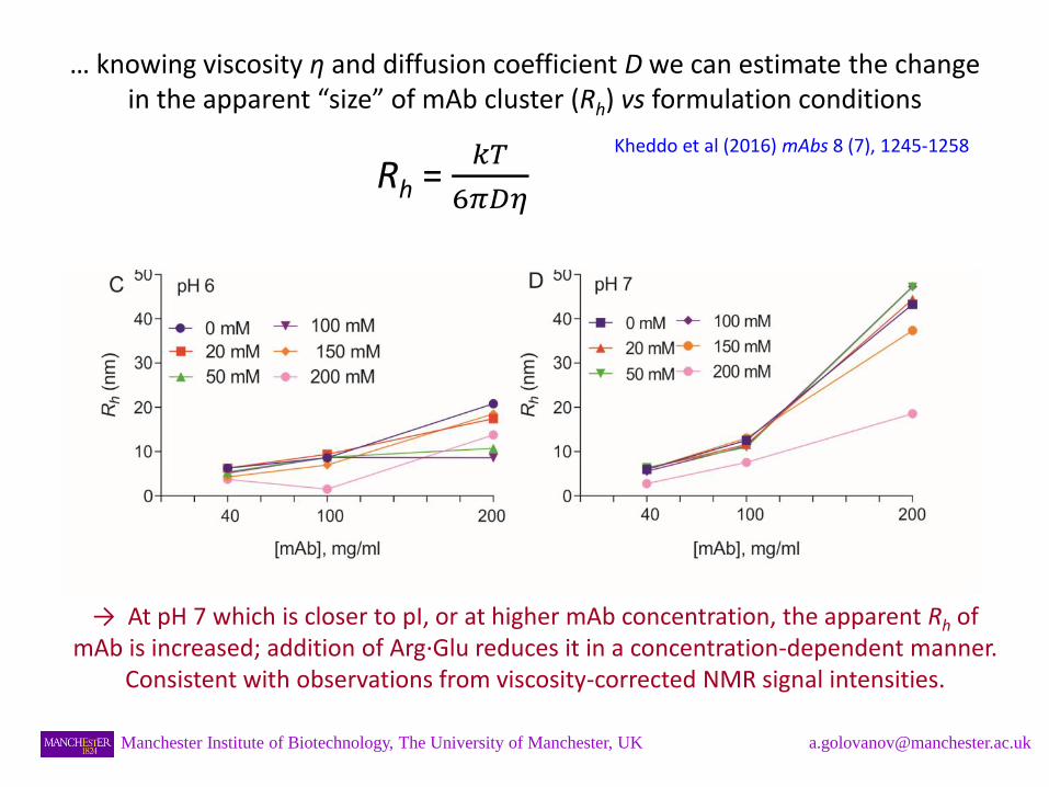
… knowing viscosity η and diffusion coefficient D we can estimate the change in the apparent “size” of mAb cluster (Rh) vs formulation conditions
Rh = 𝑘𝑇
6𝜋𝐷𝜂
→ At pH 7 which is closer to pI, or at higher mAb concentration, the apparent Rh of mAb is increased; addition of Arg·Glu reduces it in a concentration-dependent manner.
Consistent with observations from viscosity-corrected NMR signal intensities.
Kheddo et al (2016) mAbs 8 (7), 1245-1258
Manchester Institute of Biotechnology, The University of Manchester, UK [email protected]
Conclusions
• NMR signal intensity of mAb is a sensitive reporter for its state in solution: self-association, aggregation, loss of monomer, melting etc can be monitored, dependent on formulation conditions.
• Other NMR-derived parameters, such as diffusion coefficients of mAbs and excipients, provide further valuable information.
• NMR can work with non-diluted highly-concentrated mAb formulations.
• Increasing evidence that addition of relatively small concentrations of Arg·Glu (≤200 mM) often can stabilise mAb formulations better than Arg·HCl.
Manchester Institute of Biotechnology, The University of Manchester, UK [email protected]
Acknowledgements
Manchester’s team:
- Priscilla Kheddo (BBSRC BRIC CASE Studentship)
- Jack Bramham (BBSRC CASE Studentship)
- Matt Cliff
- Rebecca Dearman
MedImmune’s team (Cambridge, UK):
- Christopher van der Walle
- Shahid Uddin
- Malgorzata Tracka
- Jonathan Armer
Manchester Institute of Biotechnology, The University of Manchester, UK [email protected]