51
Applied Motion A Analysis 1

Applied Motion AnalysisApplied Motion Analysis
1

WalkingWalkingWalking
2
Walking

What we know?
• Usin a motion analysis method we know the position of the sensors or the anatomical points.
Which anatomical points are needed?Which anatomical points are needed?
Usin a motion analysis method we know the position of the sensors
Which anatomical points are needed?Which anatomical points are needed?
3
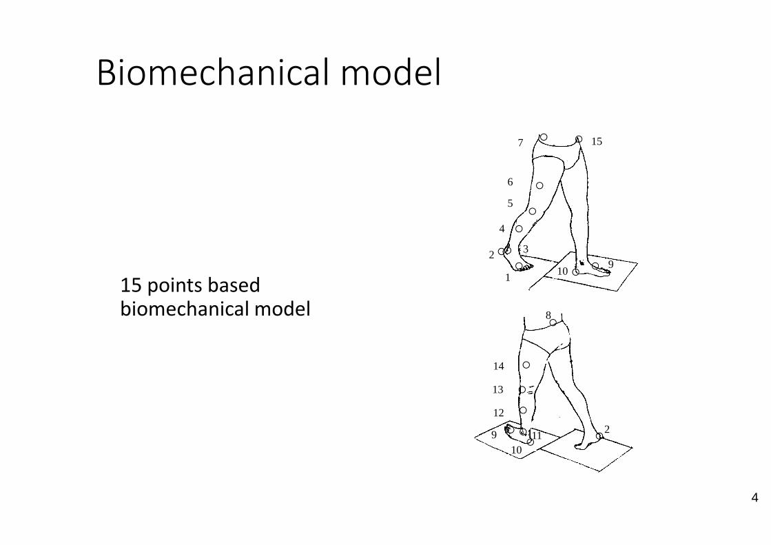
Biomechanical model
15 points based 15 points based biomechanical model
Biomechanical model
4
1
2 3
5
7
6
15
910
4
1
11
8
10
910
12
13
14
2

Biomechanical model
22 points based 22 points based biomechanical model
Biomechanical model
1
23
4-6
7
8-10
11
12
13
22
5
13
212
13
14
15-17
18
19-21
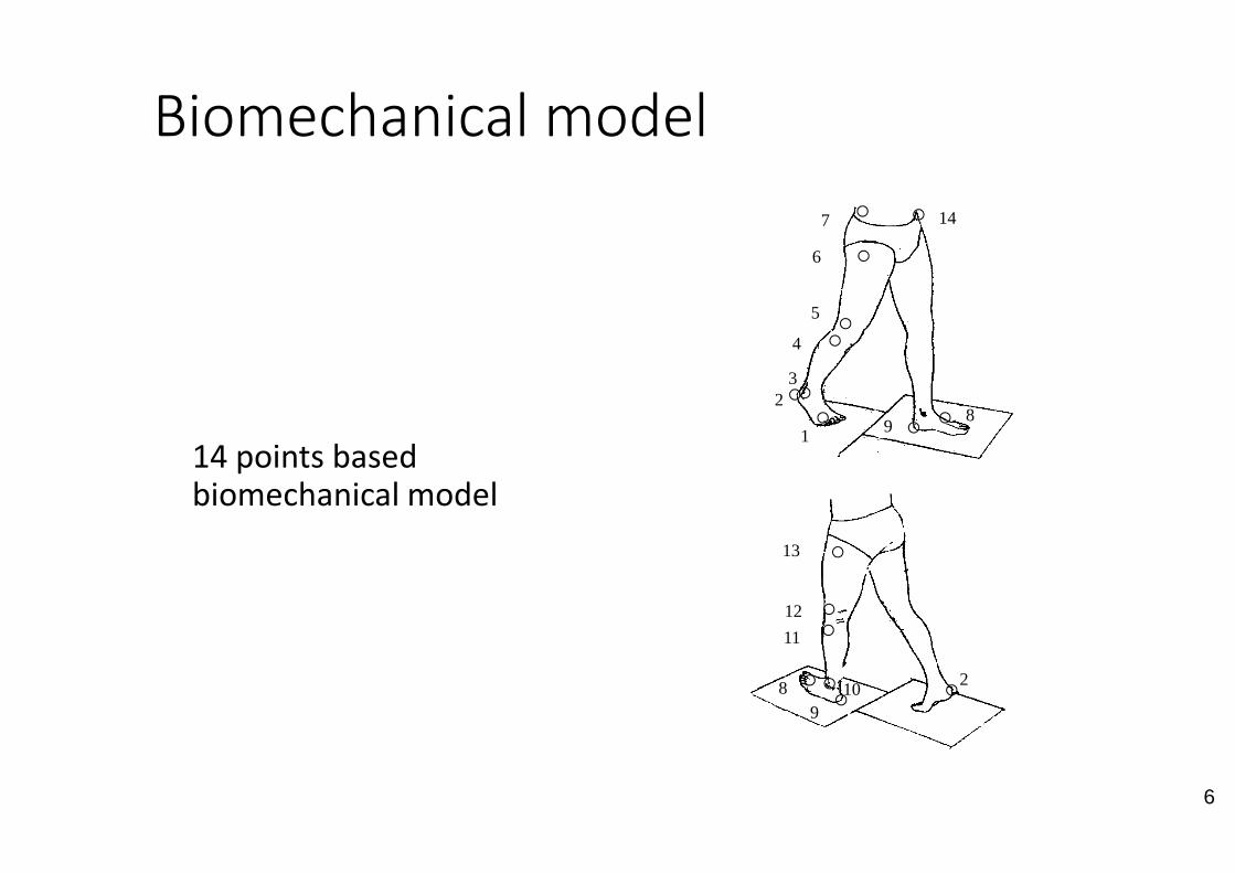
Biomechanical model
14 points based 14 points based biomechanical model
Biomechanical model
4
1
23
5
7
6
14
89
6
1
1089
11
12
13
2

Biomechanical model
19 points based 19 points based
biomechanical model
Biomechanical model
5
23
5
9
8
18
10
4
16
11
13
7
1112
14
15
17
21
19
What should we calculate from the spatial position of the anatomical points?
What should we calculate from the spatial position of the anatomical points?
8

Displacement, time like parameters
• Displacement-time parameters
• Length of the step
• Length of stepcycle
• Width of stepcycle Wid
th o
f
Width of stepcycle
• Length of swinging phase
• Length of double support phase
Displacement, time like parameters
Length of step
Wid
th o
f ste
p
Wid
th o
f st
epcy
cle
9
Length of step
Length of stepcyle

Angle like parameters
• Angle-like parameters
• Knee-angle
• Hip angle
180 - α
Knee angle
10
180 + β
Hip angle

Angle-like parameters
Swaying of pelvic girdle
Rotation of the pelvic girdle
Inclination ofThe pelvic girdle
like parameters
Rotation of the
Inclination ofThe pelvic
Defintion od the local coordinate system
Turning of the pelvic
11
Turning of the pelvic girdle around the axises like a rigid body

LCA LCP
Defomation like parameters
Relative ligament
Between specified anatomical pointsmaximum value of normalized High priority: ligament injuriesvarus-valgus knee
LCL LCM
Defomation like parameters
Relative ligament-point deformation
12
anatomical pointsvalue of normalized displacement.
High priority: ligament injuries

Deformation like parameters
Relative hip
Donáth: Anatómiai atlasz
Deformation like parameters
Between ASIS (anterior superior iliac spine) and the great trochanter
Relative hip-point changing
13
the great trochanterNormalized valued of the highest displacementImportant:
hip-jointendoprsosthesis

Motions of the upper limbMotions of the upper limbMotions of the upper limb
14
Motions of the upper limb

Method
Measurement triplet for the motion of the scaplulaMeasurement triplet for the motion of the scaplula
15
Vacuum: rigid contactAcromion: smallest skin movement
visible by the measurement headmeasurable during the motion

What we know?
• Usin a motion analysis method we know the position of the sensors or the anatomical points.
Which anatomical points are needed?Which anatomical points are needed?
Usin a motion analysis method we know the position of the sensors
Which anatomical points are needed?Which anatomical points are needed?
16

Biomechancal model
The developed 16 points based biomechanical modelThe developed 16 points based biomechanical model
Anatomical points onthe bones of theshoulder joint and at
17
shoulder joint and atleast three anatomicalpoints of the lowerarm should beinvestigated

The investigated motion
Armlifting in the plane of the scapula
The investigated motion
18
Armlifting in the plane of the scapula

What should we calculate from the spatial position of the anatomical points?
What should we calculate from the spatial position of the anatomical points?
19

Parameters – Spatial angles
HE the angle betweeen thetrunk and the humerus(humerus elevation)
ST the angle between theST the angle between thetrunk and the scapula(scapulo-thorocalis angle)
GH the spatial angle betweenthe humerus and thescapula (glenohumeralisszög)
Spatial angles
20

Angle-like parameters
Parametes of the angle
changing
The difference between the initial and the present angle
• Eliminating antropometric properties
• Dynamics is not measurable
Scapulothorocalis and
glenohumeralis rythmThe scapulothorocalis and glenohumeralis angle in the function of elevation
21
• dinamika nem jellemezhető
• Durng the h

The determination of rotation point
B
vB
rAB
Rigid bodiesare supposed-Determinationof the angularvelocity of thelower and
rAB
y
ACx
z
vA
vCrAC
ωf
lower andupper arm canbe calculatedfrom thevelocities ofthreemeasuredpoints
vC
vA
The determination of rotation point
A
vA
rAC
C
vC
vA
The angular velocity joint
ϖf-ϖa
Determination of
22
B
rAB
vB
ωa
Determination of the helical axis and the rotation point
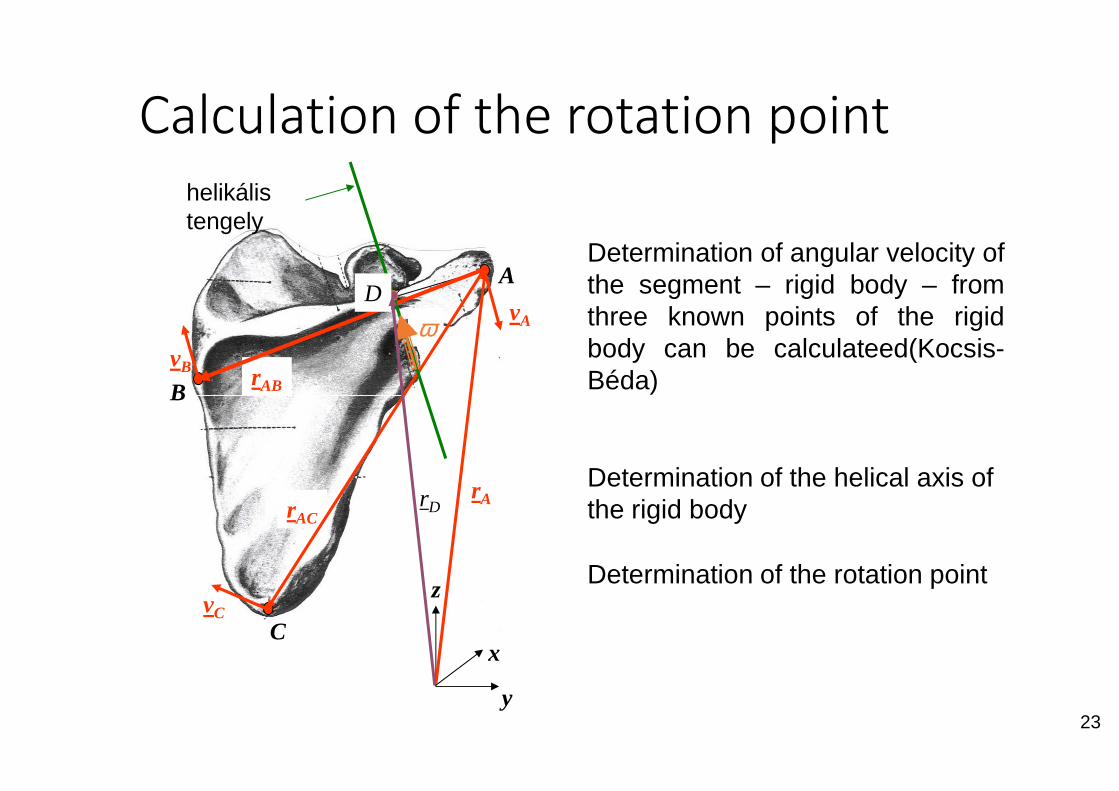
Calculation of the rotation point
A
B
vA
vB rAB
ω
helikálistengely
D
y
B
Cx
z
rA
vC
rACrD
Calculation of the rotation point
Determination of angular velocity ofthe segment – rigid body – fromthree known points of the rigidbody can be calculateed(Kocsis-Béda)
23
Determination of the helical axis of the rigid body
Determination of the rotation point
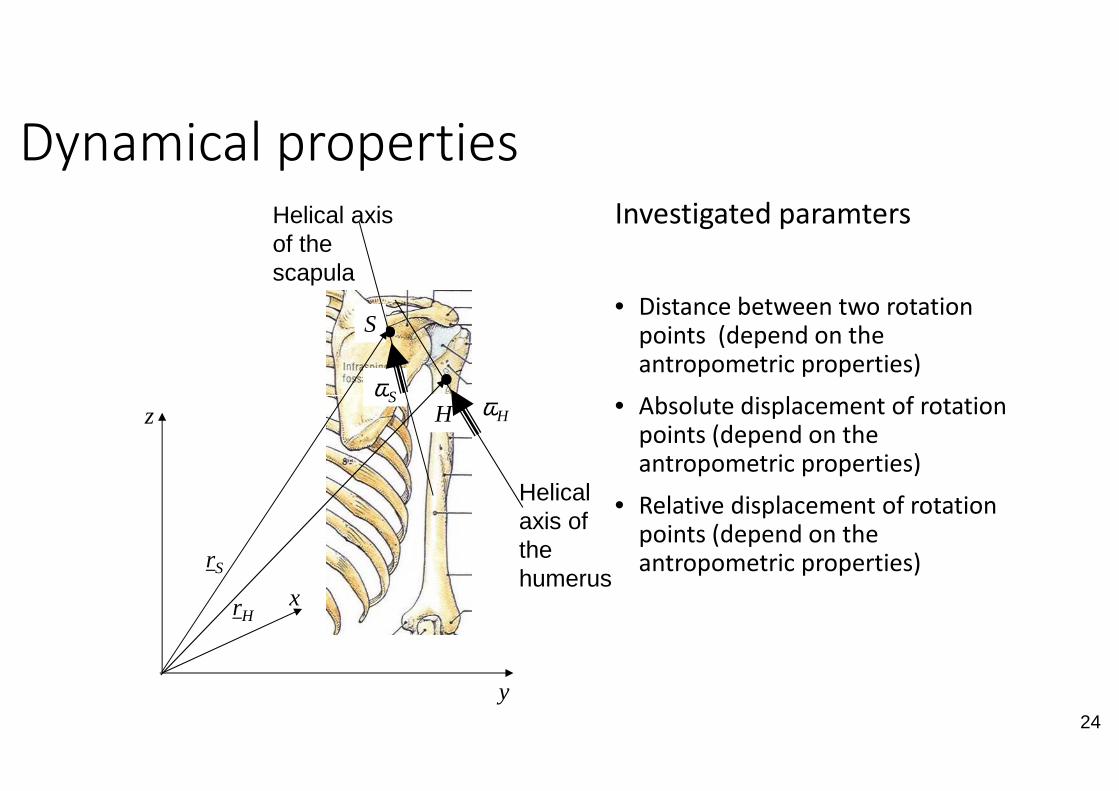
Dynamical properties
ωωS
Helical axis of the scapula
S
y
z
x
ωHH
rH
Helical axis of the humerus
ωS
rS
Investigated paramters
• Distance between two rotation points (depend on the antropometric properties)
• Absolute displacement of rotation
24
• Absolute displacement of rotation points (depend on the antropometric properties)
• Relative displacement of rotation points (depend on the antropometric properties)
Helical axis of
humerus

Measuring of muscle activityMeasuring of muscle activityMeasuring of muscle activity
25
Measuring of muscle activity

EMG
• EMG=elektromyography
• Electrode potential changing of the skeletal muscle between two points
• Recorded figure: elektromiogramm
• Types:Surface (superficial muscle groups)
Types:• Surface (superficial muscle groups)
• Needle (some muscle, deep muscle, painfull
• Based the method of wireing : monoplar, bipolar
• Fields of application:• Distinguish the nerve and muscle based paresis
• Work, sports and orthopedic disorders usually
Electrode potential changing of the skeletal muscle between two points
26
full, sterilization, hard-hitting)
monoplar, bipolar
Distinguish the nerve and muscle based paresis
Work, sports and orthopedic disorders usually have effects pn the activation sequence

What we know?
• Using superficial electromyography (EMG) the electrode potential changing of muscles are measured in time. (electromyogram).
Which muscles should be measured??Which muscles should be measured??
Using superficial electromyography (EMG) the electrode potential changing of muscles are measured in time. (electromyogram).
Which muscles should be measured??Which muscles should be measured??
27

Walking –lower limb
On surface EMG (elektromyograph) activation detection
• m. vastus med.
• m. vastus lat.• m. vastus lat.
• m. rectus femoris
• m. biceps fem.
• m. adductor longus
• m. gluteus medius
• m. gastrocnemius med.
• m. gastrocnemius lat.
elektromyograph)
28
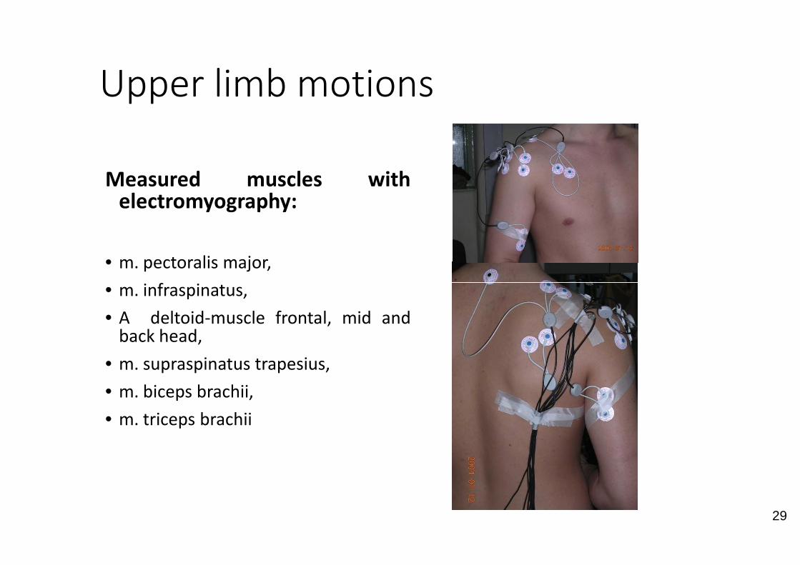
Upper limb motions
Measured muscles withelectromyography:
• m. pectoralis major,
• m. infraspinatus,• m. infraspinatus,
• A deltoid-muscle frontal, mid andback head,
• m. supraspinatus trapesius,
• m. biceps brachii,
• m. triceps brachii
Upper limb motions
with
29
and

Muscle activity parametersm. vastus lat.
-400.0
-200.0
0.0
200.0
400.0
0.0 200.0 400.0 600.0 800.0 1000.0 1200.0
[mV
]
time [msec]
EMG creating envelope graph with rms (root mean square) methodAnalysis of 6 gait cyclesnormalization of the average of the maximum values
m.quadriceps lat.
0.00.20.40.60.81.01.21.4
0% 20% 40% 60% 80% 100%
Nor
mál
t ért
ék
30
Lépésciklus százalékbanStep-cycle in percentage
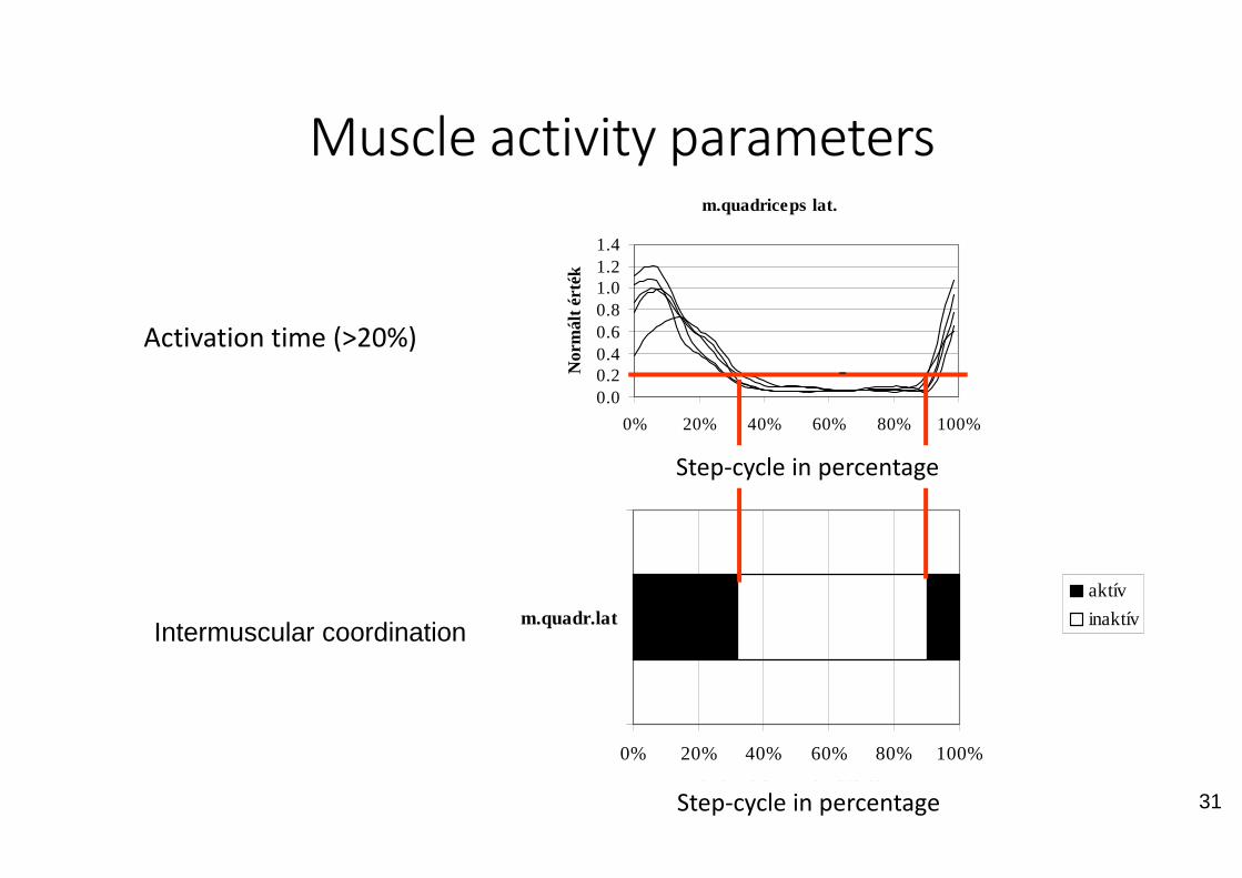
Activation time (>20%)
0.00.20.40.60.81.01.21.4
Nor
mál
t ért
ék
Muscle activity parameters
0%
m.quadr.latIntermuscular coordination
m.quadriceps lat.
0% 20% 40% 60% 80% 100%
Muscle activity parameters
31
Lépésciklus százalékban
0% 20% 40% 60% 80% 100%
Lépésciklus százalékában
aktív
inaktív
Step-cycle in percentage
Step-cycle in percentage

Conduction of the measurement I.
• Equipments
• Fixing devices
• Electrodes (superficial or nailed)
• Validation of the • Validation of the measurements (in general it is automatic)
• Finding measured muscles
Conduction of the measurement I.
32
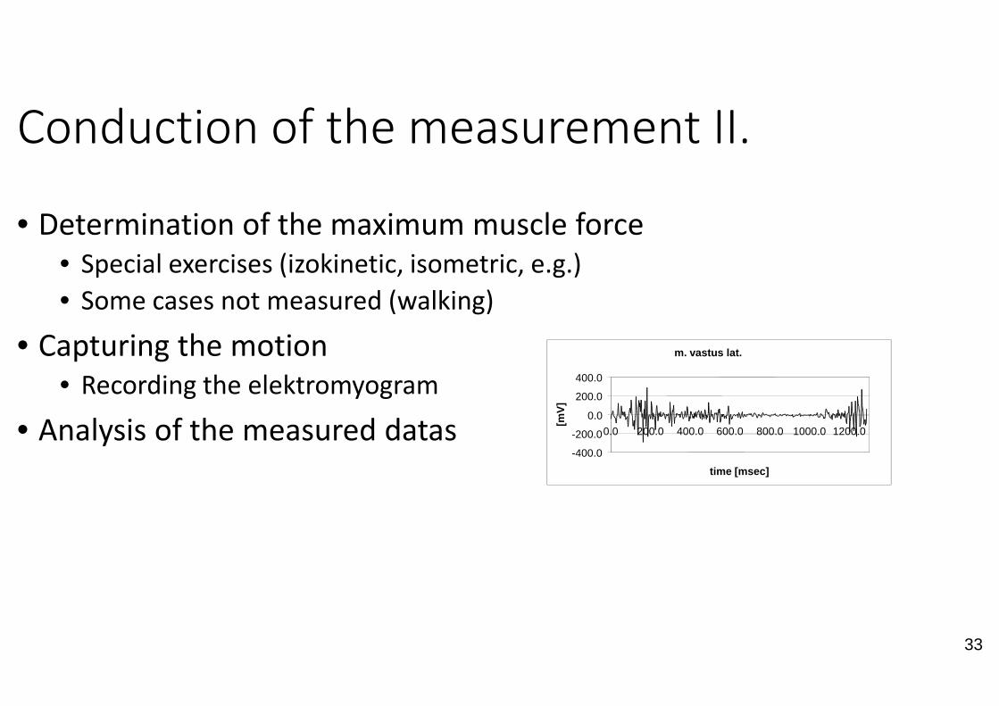
Conduction of the measurement II.
• Determination of the maximum muscle force
• Special exercises (izokinetic, isometric, e.g.)
• Some cases not measured (walking)
• Capturing the motion• Capturing the motion
• Recording the elektromyogram
• Analysis of the measured datas
Conduction of the measurement II.
Determination of the maximum muscle force
Special exercises (izokinetic, isometric, e.g.)
m. vastus lat.
33
m. vastus lat.
-400.0
-200.0
0.0
200.0
400.0
0.0 200.0 400.0 600.0 800.0 1000.0 1200.0
time [msec]
[mV
]

Analysis
• Analysis of raw electromyogramm
• contraction rate
• In rest state (no electrical activity)
• Moderate contraction
• Maximum contraction (normalization require maximum muscle strength)• Maximum contraction (normalization require maximum muscle strength)
• Record the changes of disease
• Denervation potential (equal contraction)
• Polyphasic potentials ("do not belong there" contractions)
Maximum contraction (normalization require maximum muscle strength)
34
Maximum contraction (normalization require maximum muscle strength)
potential (equal contraction)
potentials ("do not belong there" contractions)
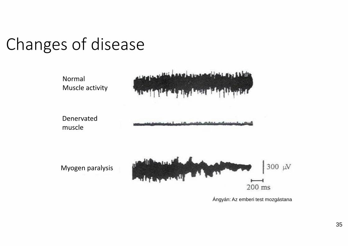
Changes of disease
Normal
Muscle activity
Denervated
muscle
Myogen paralysis
muscle
35
Ángyán: Az emberi test mozgástana
Processing I.• Signal-processing:
• Absolute value computing
• Filtering
• Processing:• Time based processing:
• Norming
• With the maximal value of a special exercise (standarding, defined for each • With the maximal value of a special exercise (standarding, defined for each muscle, generally with elementary motions)
• With the maximum value of the given motion
• With the avarage value of the maximal values of the given motion(walking)
• With the maximal value of more given motion
• Avarage computing (Root-mean square)
• Frequency based processing (determination of frequency properties):
• Avarage frequency
• Median frequency
With the maximal value of a special exercise (standarding, defined for each
36
With the maximal value of a special exercise (standarding, defined for each muscle, generally with elementary motions)
With the maximum value of the given motion
With the avarage value of the maximal values of the given motion-cycles
With the maximal value of more given motion
mean square)
Frequency based processing (determination of frequency properties):

Time based processing II.
-400.0
-200.0
0.0
200.0
400.0
0.0 200.0 400.0
time [msec]
[mV
]
40%Motioncycle in %
Nor
mal
ized
d va
lue
Time based processing II.
600.0 800.0 1000.0 1200.0
time [msec]
Raw diagram
37
40% 60% 80% 100%Motioncycle in %
Envelope graph
Reaction-force measurementReaction-force measurementforce measurement
38
force measurement

Reaction force
Measured valuesComputed values
39
F1: Heel strikeF2: Whole footF3: Heel liftingTi : Times pof phases
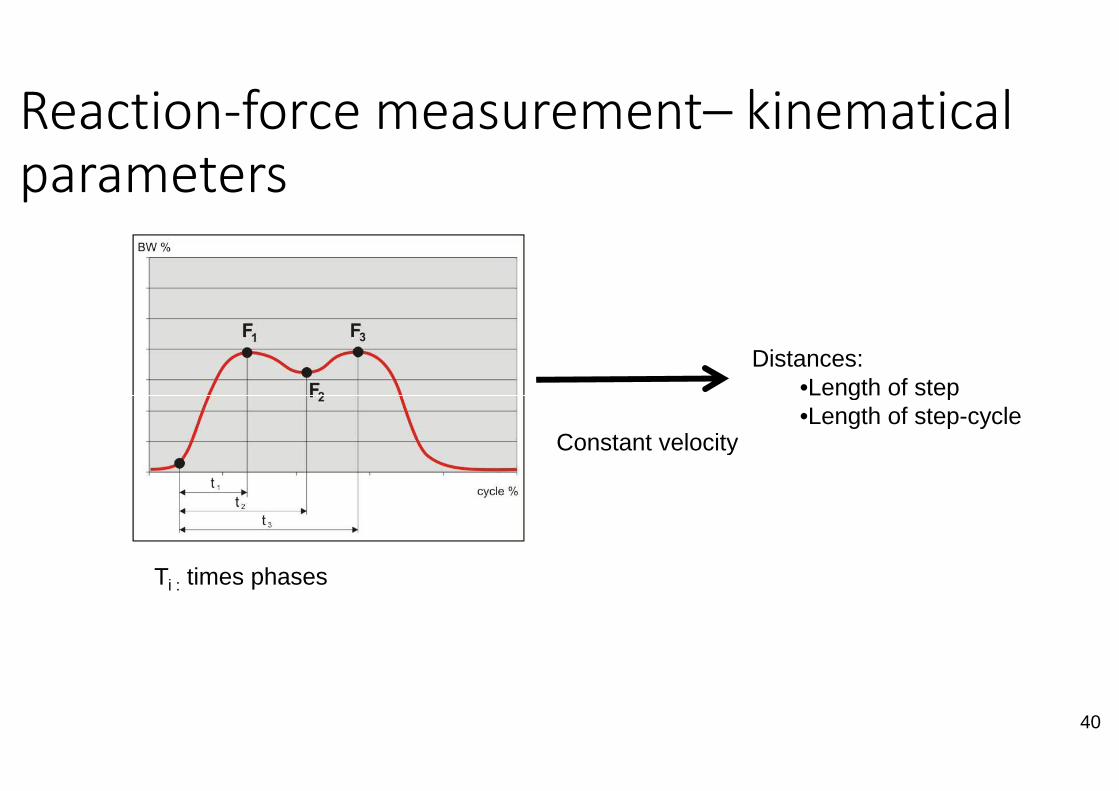
Reaction-force measurement
parameters
Ti : times phases
Constant velocity
force measurement– kinematical
Distances:•Length of step
40
Constant velocity
•Length of step•Length of step-cycle

Foot-pressure distributionFoot-pressure distributionpressure distribution
41
pressure distribution

Static examination
Investigation the pressure under the foot
Special force measurement plate
42
Special force measurement plate

Determined parameters
Determined parameters
Pressure distributio
Center of gravity
43
Center of gravity

Applicability
• Investigation of the foot force distribution
• Effect of flat-feet shoe insert
• Effects of other diseases (diabetes, stroke,..)
• Stability investigation• Stability investigation
• Problem of closed and open eyes –eye
Investigation of the foot force distribution
Effects of other diseases (diabetes, stroke,..)
44
eye-stabilization

Dynamical investigations
45
Balance investigationsBalance investigationsBalance investigations
46
Balance investigations
Balance investigation I.
• Types:
• Static (closed or open eyes):
• Investigation of foot force distribution
• Investigation of head motion (Romberg
• Dynamic:• Dynamic:
• Walking through a beam
• Investigation of head motion walking in one place with closed eyes
• Special:
• Proprioception (motion coordination)
Romberg-probe: standing for 1 minute with close eyes)
47
Investigation of head motion walking in one place with closed eyes (Unterberger)
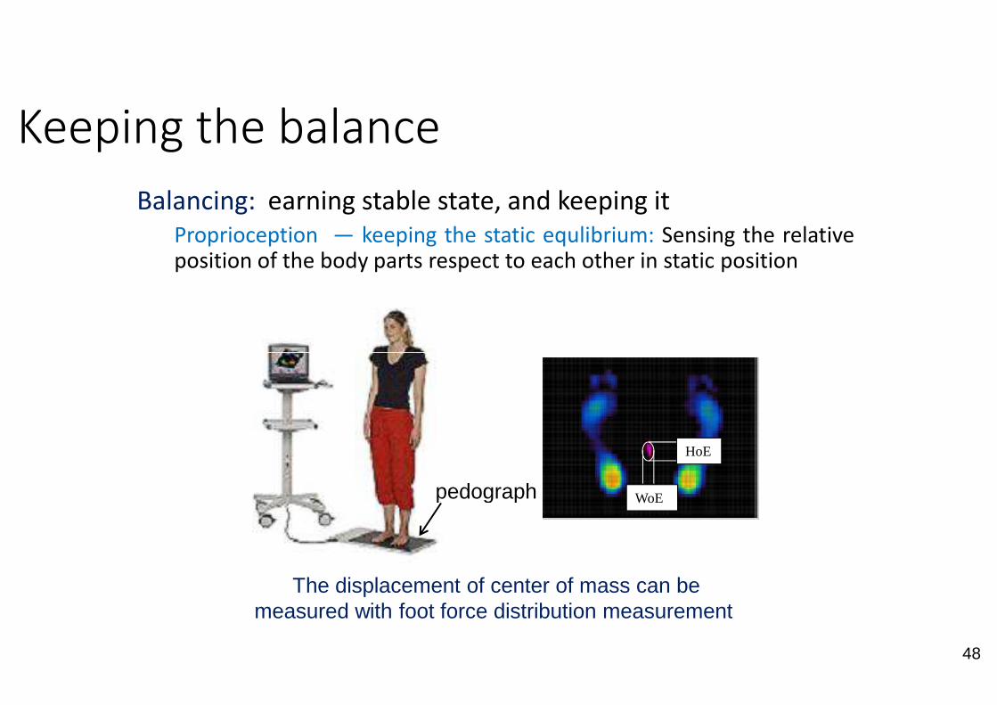
Keeping the balance
Balancing: earning stable state,
Proprioception ― keeping the staticposition of the body parts respect
The displacement of center of mass can be measured with foot force distribution measurement
pedograph
state, and keeping it
static equlibrium: Sensing the relativerespect to each other in static position
48
The displacement of center of mass can be measured with foot force distribution measurement
HoE
WoEpedograph
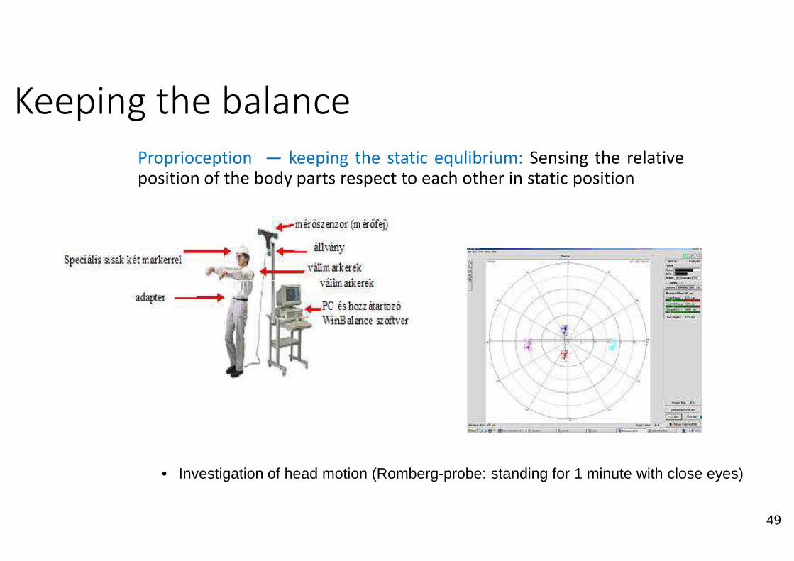
Keeping the balance
Proprioception ― keeping the staticposition of the body parts respect
• Investigation of head motion (Romberg
static equlibrium: Sensing the relativeto each other in static position
49
Investigation of head motion (Romberg-probe: standing for 1 minute with close eyes)

Keeping the balanceKinesthesis – keeping the dynamic equlibrium
moving body-parts respect to each other
• Investigation of head motion walking in one place with closed eyes (Unterberger)
equlibrium: The relative position ofother
50
Investigation of head motion walking in one place with closed eyes (Unterberger)

Advantages
• Well known methods
Disadvantages:
• stability analysis
• Mainly analyze the effects of neurological
• Small shocks to the escalator,'attacks' are not modeled
• After a sudden change of directionon moving ground is not analyzed
neurological problems
the bumpy streets, the streets, pets
direction the keeping of balance and walkinganalyzed
51


