*Corresponding author email: [email protected]Symbiosis Group Symbiosis www.symbiosisonline.org www.symbiosisonlinepublishing.com Approximation of the CoM Estimate Raymond Chong* Department of Physical Therapy, Georgia Regents University, Georgia, USA Journal of Exercise, Sports & Orthopedics Open Access Case Report Introduction Increasing sophistication in kinematics technology has enhanced the precision of measurements in time series analyses of human walking [1]. Depending on the nature of the scientific inquiry, however, and to the extent that small errors can be tolerated when using a simplified procedure, increasing the level of precision may not necessarily yield a corresponding increase in information deemed to be relevant [2]. In clinical settings for example, time and logistical considerations often times override the need to be as ultra-precise as technology can provide [3]. We demonstrate here two simplified methods to estimate the vertical trajectory of the body center of mass (CoM) during walking. One used a point on the mid-shoulder and the other on the navel region. The two methods were then compared to a full- body CoM estimate using a full-body model to test the hypothesis that similar CoM traces can be obtained compared to the full- body CoM estimate. Methods Data was obtained with consent from a 68 year-old healthy woman with no musculoskeletal disorders that may affect gait abnormalities. The study was approved by the institutional review board. The subject walked barefooted. A 19-marker 12-segment full-body CoM model: bilateral (fifth metatarsal) styloid process, posterior calcaneus, lateral malleolus, lateral femoral condyle, greater trochanter, acromion process, olecranon process, (wrist) styloid process, (squamous) temporal bone and chin (single)[4]. Table 1 was used to compare against two single- marker CoM estimates, one at the mid-shoulder (virtual) point and the other at the navel region (Figure 1). The 6-camera PEAK Motus system (PEAK Performance Technologies Inc, Boulder CO) was used to capture the kinematic data. The data was sampled at 120 Hz and underwent a 5th-order Butterworth filtering routine. 76 sampled points making up the second and third steps (right heel to left heel) from quiet stance were extracted for analyses. The two-tailed non-parametric Spearman r correlation test was Abstract The extent to which small errors from simplified methods of measurements can be tolerated may be useful for clinical studies where time and logistical considerations often override the need to be very precise. We demonstrate here two kinematic methods to approximate the vertical trajectory of the body center of mass (CoM) during walking: the mid-shoulder and navel regions compared to a full-body CoM model. The navel marker achieved a near-perfect correlation with the CoM model for displacement and velocity. In the acceleration data, deviations from the CoM model for both the navel and mid-shoulder markers were observed around the points of inflection. The estimation of CoM displacement and velocity using the navel marker appears to be adequate for clinical use in comparing between subjects, while CoM acceleration may be more suited for studying the relative change between conditions in the same subject. Keywords: Center of mass; Gait; Human; Kinematics; Motion analysis; Walking Received: February 24, 2014; Accepted: April 16, 2014; Published: April 18, 2014 *Corresponding author: Raymond Chong, Department of Physical Therapy, Georgia Regents University, Augusta, Georgia, USA, Tel: 706.721.2141; Fax: 706.721.3209; E-mail: [email protected]Segment % mass head 8.1 trunk 49.7 arm 2.8 * 2 forearm 2.2 * 2 thigh 10 * 2 leg 4.65 * 2 foot 1.45 * 2 Total 100 Table 1: Center of mass values for simple and complex segments. Figure 1: CoM approximation: 1) 19-marker 12-segment model (white), mid-shoulder (blue) and naval (red).
Department of Physical Therapy, Georgia Regents University, Georgia, USA
Journal of Exercise, Sports & Orthopedics Open AccessCase Report
IntroductionIncreasing sophistication in kinematics technology has
enhanced the precision of measurements in time series analyses of human walking [1]. Depending on the nature of the scientific inquiry, however, and to the extent that small errors can be tolerated when using a simplified procedure, increasing the level of precision may not necessarily yield a corresponding increase in information deemed to be relevant [2]. In clinical settings for example, time and logistical considerations often times override the need to be as ultra-precise as technology can provide [3].
We demonstrate here two simplified methods to estimate the vertical trajectory of the body center of mass (CoM) during walking. One used a point on the mid-shoulder and the other on the navel region. The two methods were then compared to a full-body CoM estimate using a full-body model to test the hypothesis that similar CoM traces can be obtained compared to the full-body CoM estimate.
MethodsData was obtained with consent from a 68 year-old healthy
woman with no musculoskeletal disorders that may affect gait abnormalities. The study was approved by the institutional review board. The subject walked barefooted. A 19-marker
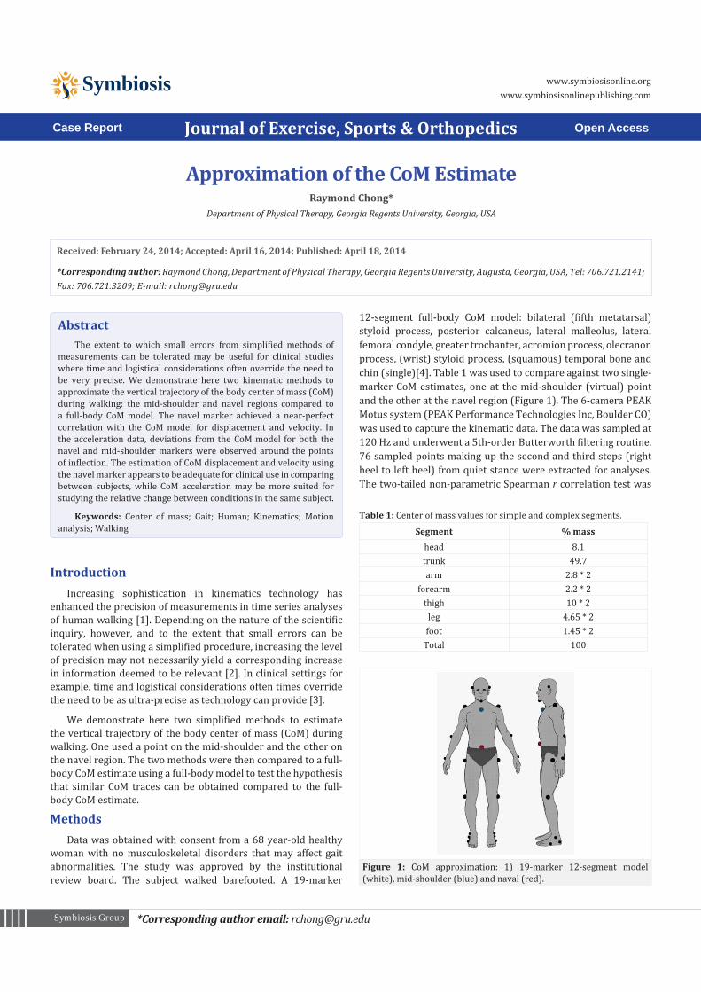
12-segment full-body CoM model: bilateral (fifth metatarsal) styloid process, posterior calcaneus, lateral malleolus, lateral femoral condyle, greater trochanter, acromion process, olecranon process, (wrist) styloid process, (squamous) temporal bone and chin (single)[4]. Table 1 was used to compare against two single-marker CoM estimates, one at the mid-shoulder (virtual) point and the other at the navel region (Figure 1). The 6-camera PEAK Motus system (PEAK Performance Technologies Inc, Boulder CO) was used to capture the kinematic data. The data was sampled at 120 Hz and underwent a 5th-order Butterworth filtering routine. 76 sampled points making up the second and third steps (right heel to left heel) from quiet stance were extracted for analyses. The two-tailed non-parametric Spearman r correlation test was
AbstractThe extent to which small errors from simplified methods of
measurements can be tolerated may be useful for clinical studies where time and logistical considerations often override the need to be very precise. We demonstrate here two kinematic methods to approximate the vertical trajectory of the body center of mass (CoM) during walking: the mid-shoulder and navel regions compared to a full-body CoM model. The navel marker achieved a near-perfect correlation with the CoM model for displacement and velocity. In the acceleration data, deviations from the CoM model for both the navel and mid-shoulder markers were observed around the points of inflection. The estimation of CoM displacement and velocity using the navel marker appears to be adequate for clinical use in comparing between subjects, while CoM acceleration may be more suited for studying the relative change between conditions in the same subject.
Keywords: Center of mass; Gait; Human; Kinematics; Motion analysis; Walking
Received: February 24, 2014; Accepted: April 16, 2014; Published: April 18, 2014
*Corresponding author: Raymond Chong, Department of Physical Therapy, Georgia Regents University, Augusta, Georgia, USA, Tel: 706.721.2141; Fax: 706.721.3209; E-mail: [email protected]
Segment % masshead 8.1trunk 49.7arm 2.8 * 2
forearm 2.2 * 2thigh 10 * 2
leg 4.65 * 2foot 1.45 * 2
Total 100
Table 1: Center of mass values for simple and complex segments.
Figure 1: CoM approximation: 1) 19-marker 12-segment model (white), mid-shoulder (blue) and naval (red).
Page 2 of 3Citation: Chong R (2014) Approximation of the CoM Estimate. J Exerc Sports Orthop 1(2): 1-3. DOI: http://dx.doi.org/10.15226/2374-6904/1/2/00107
conducted to test the hypothesis that the distribution curve of the vertical CoM displacement, velocity and acceleration of the mid-shoulder and navel markers are similar to the full-body CoM model.
ResultsFigure 2 shows the vertical displacement, velocity and
acceleration traces of the three methods of CoM estimates over one step. The average speed and step length over the step was 0 .91 m/s and 0.58 m, respectively. Peak-to-peak vertical CoM displacement was about 3.2 cm.
The navel marker was more consistent than the mid-shoulder marker in tracking with the CoM vertical displacement (rmid-shoulder
= 0.997 and rnavel = 0.998) and velocity (rmid-shoulder = 0.995 and rnavel =0 .997). The mid-shoulder marker deviated from the full model by up to 0.03 m/s during the early swing phase and up to 0.024 m/s during the late swing phase. In the acceleration data, the mid-
shoulder and navel markers deviated from the full CoM model by up to 0.55 and 0.31m/s2, respectively in the period before mid-swing as the CoM approached maximum vertical displacement (acceleration rmid-shoulder = 0.955 and rnavel = 0.986).
DiscussionThe trunk is not entirely rigid during walking. There are
movements around the pelvic region and shoulder region [5]. Thus, the decreased accuracy in the CoM estimate using the mid-shoulder and navel markers becomes more prominent with differentiations of the displacement data. One reason why the navel marker tracked more closely with the full CoM model than the mid-shoulder marker is that the navel marker is very near to where the CoM of the body is. The sacral region has also been used to estimate CoM motions [6,7].
When utmost precision in kinematic measurements is not the highest priority, a balance needs to be sought between preserving the rigor of scientific enquiry and the physical demands imposed by the experimental setup on the subject, particularly when the kinematic technology is combined with surface electromyography and force plate recordings. The ability to obtain data from the simplified methods that is as similar as possible to the full-body model preserves the construct validity of causes and effects associated with the study of human walking. Generalization should be confirmed at least from visual inspection of the traces to ensure that the operational definition of CoM estimate is comparable using the simpler or full model methods.
Since the mid-shoulder and navel CoM displacements tracked almost perfectly with the full CoM model throughout the gait cycle, either one of the single markers can be used in place of the full model to compare across subjects. Interpretation of the navel marker from the first derivative of its displacement (i.e., velocity) also appears to be valid. The navel and mid-shoulder markers may also accurately represent the full CoM model in estimating vertical acceleration except at the regions around the points of inflection. Then the single markers would likely be more suited for use in repeated measures designs where one is interested in relative change between two conditions [8,9] provided the change in speed and/or acceleration is not too large.
References1. Sutherland DH (2002) The evolution of clinical gait analysis. Part II
kinematics. Gait Posture 16(2): 159-179.
2. Zijlstra W, Hof AL (2003) Assessment of spatio-temporal gait parameters from trunk accelerations during human walking. Gait Posture 18(2): 1-10.
3. Alexander NB (1996) Using technology-based techniques to assess postural control and gait in older adults. Clin Geriatr Med 12(4): 725-744.
4. Winter DA (2005) Biomechanics and motor control of human movement. In: John Wiley and Sons (3rd edn), Anthropometry. New York, USA, pp. 59-85.
5. Kerrigan DC, Della Croce U, Marciello M, Riley PO (2000) A refined view of the determinants of gait: significance of heel rise. Arch Phys Med Rehabil 81(8): 1077-1080.
Figure 2: Frame-by-frame (120 Hz) traces of three methods of CoM es-timates across one step (heel-to-heel) along the sagittal plane.
Page 3 of 3Citation: Chong R (2014) Approximation of the CoM Estimate. J Exerc Sports Orthop 1(2): 1-3. DOI: http://dx.doi.org/10.15226/2374-6904/1/2/00107
6. Gard SA, Miff SC, Kuo AD (2004) Comparison of kinematic and kinetic methods for computing the vertical motion of the body center of mass during walking. Hum Mov Sci 22(6): 597-610.
7. Saini M, Kerrigan DC, Thirunarayan MA, Duff-Raffaele M (1998) The vertical displacement of the center of mass during walking: A comparison of four measurement methods. J Biomech Eng 120(1): 133-139.
8. Chong RK, Chastan N, Welter ML, Do MC (2009) Age-related changes in the center of mass velocity control during walking. Neurosci Lett 458(1): 23-27.
9. Chong RK, Chiu FC, Lee KH, Do MC (2009) An instance of reduced center of mass displacement: The Ba Gua Zhang walking gait. Percept Mot Skills 109(3): 646-648.