504 PROFESSOR E. RAY LANKESTER. LIMTILUS an ARACHNID. By E. RAY LANKESTER, M.A., F.R.S., Jodrell Professor of Zoology in University College, London. (With Plates XXVIII and XXIX). A. INTRODUCTION AND BIBLIO- GRAPHY. B. COMPARISON OF LIMULUS AND SCOEPIO. § a. Nervous system. § b. Skeleton. I. Tergites. IT. Appendages. III. Sternites. IV. The common charac- ters of the lamelligerous appendages of Scorpio aud Limulus. V. Hypothesis as to their mode of origin. VI. Entosternite. § c. Alimentary canal. | d. Vascular system. § e. Generative glands. C. THE EURYPTJSRINA AS A CON- NECTING LINK. D. REVIEW OP OPINIONS OP MODEKN AUTHORITIES AS TO THE AFFINITIES OF LIMULTJS. E. CONCLUSIONS: LIMULTJS AND THE ANCESTRY OF TRA- CHEATJS ARTHROFODA. A. INTRODUCTION AND BIBLIOGRAPHY. THE prevailing opinion among zoologists at the present day, with regard to the affinities of the King Crab, is that it must be regarded as one of the Crustacea. Even when this view is not fully accepted the King Crab is placed in a special position of isolation and its relationship with Crus- tacea strongly insisted upon, whilst more remote affinity with the Arachnida is grudgingly admitted. My friend Edouard Van Beneden, of Li£ge, is the only zoologist who has definitely taken a divergent line, and has frankly endorsed the instinctive perception of Straus Durk- heim in declaring that Limulus is no Crustacean, but simply and unreservedly an Arachnid. Ed. Van Beneden bases his opinion upon embryological data. I have elsewhere ex- pressed my full concurrence in that opinion, but the grounds upon which my conclusion rests are not solely embryological —they have reference to the structure of the adult Limulus and Scorpion. In the following pages I hope to show that Limulus is best understood as an aquatic scorpion, and the Scorpion and its allies as terrestrial modifications of the King Crab. My views on this subject were formed some eight years ago, and I have to acknowledge the kindness of Mr. Car- rington, F.L.S., of the Royal Westminster Aquarium, by which I have been enabled-to dissect and make histological study of perfectly fresh specimens of Limulus sent to me in the living state.
Transcript
5 0 4 PROFESSOR E. RAY LANKESTER.
LIMTILUS an ARACHNID. By E. RAY LANKESTER, M.A.,F.R.S., Jodrell Professor of Zoology in UniversityCollege, London. (With Plates XXVIII and XXIX).
A. INTRODUCTION AND BIBLIO-GRAPHY.
B. COMPARISON OF LIMULUS ANDSCOEPIO.
§ a. Nervous system.§ b. Skeleton.
I. Tergites.IT. Appendages.III. Sternites.IV. The common charac-
ters of the lamelligerousappendages of Scorpioaud Limulus.
V. Hypothesis as to theirmode of origin.
VI. Entosternite.§ c. Alimentary canal.| d. Vascular system.§ e. Generative glands.
C. THE EURYPTJSRINA AS A CON-NECTING LINK.
D. REVIEW OP OPINIONS OP MODEKNAUTHORITIES AS TO THEAFFINITIES OF LIMULTJS.
E. CONCLUSIONS: LIMULTJS ANDTHE ANCESTRY OF T R A -CHEATJS ARTHROFODA.
A. INTRODUCTION AND BIBLIOGRAPHY.
THE prevailing opinion among zoologists at the presentday, with regard to the affinities of the King Crab, is thatit must be regarded as one of the Crustacea. Even whenthis view is not fully accepted the King Crab is placed in aspecial position of isolation and its relationship with Crus-tacea strongly insisted upon, whilst more remote affinitywith the Arachnida is grudgingly admitted.
My friend Edouard Van Beneden, of Li£ge, is the onlyzoologist who has definitely taken a divergent line, and hasfrankly endorsed the instinctive perception of Straus Durk-heim in declaring that Limulus is no Crustacean, but simplyand unreservedly an Arachnid. Ed. Van Beneden bases hisopinion upon embryological data. I have elsewhere ex-pressed my full concurrence in that opinion, but the groundsupon which my conclusion rests are not solely embryological—they have reference to the structure of the adult Limulusand Scorpion. In the following pages I hope to show thatLimulus is best understood as an aquatic scorpion, and theScorpion and its allies as terrestrial modifications of theKing Crab.
My views on this subject were formed some eight yearsago, and I have to acknowledge the kindness of Mr. Car-rington, F.L.S., of the Royal Westminster Aquarium, bywhich I have been enabled-to dissect and make histologicalstudy of perfectly fresh specimens of Limulus sent to me inthe living state.
LIMULUS AN AEAOHNID. 505
It is not desirable at the outset to follow the history ofthe discussion relative to the zoological position of Limulus.Those who desire to become acquainted with the most im-portant contributions to the subject should consult thememoirs of Anton Dohrn and A. S. Packard, who have givenvery ample references to the literature of the subject.
I shall here give in alphabetical order a list of the chiefworks referred to in the following pages, the number at-tached to an author's name when cited, having reference tothe number in the present list. After I have put forwardthe facts and inferences with reference to the structure andaffinities of Limulus which appear to me to be well-estab-lished, I shall briefly review the various opinions whichhave been advanced by recent writers of authority.
Special Memoirs.
1. DOHRN, Anton. "Bau und Entwickelung der Arthropoden," 'Jen-aische Zeitschrift,' Bd. vi, 1871-
2. GEGENBATJR, Carl. " Auatomische Untersuchung eines Limulus,"' Abhandlungen der naturforschenden Gesellschaft in Halle/ 1858.
3. GRENACHER. " Unters. uber das Sehorgan der Artbropoden," 1879.4. LANKESTER, E. Ray. "Mobility of the Spermatozoa of Limulus,"
' Quart. Journ. of Micr. Science,' 1878.5. MILNE-EDWARDS, Alphonse. "Recherches snr l'Anatomie des
6. NEWPORT, George. "Nervous and Circulatory Systems in Myria-poda and Macrourous Aiachnida," 'Philos. Transactions of theRoyal Society,' part ii, 1813.
7. OWEN, Richard. "Anatomy of the King Crab," 'Transactions ofthe Lmnsean Society of London,' 1873.
8. PACKARD, A. S., Junr. " The Development of Limulns polypbemus,"'Memoirs of the Boston Society of Natural History,' 1872.
9. PACKARD, A. S, Junr. "The Anatomy, Histology, and Embryo-logy of Limulus polyphemus," 'Anniversary Memoirs ol theBoston Society of Natural History, 1880.
10. VAN BEUEDEN, Edouard. " De la place qui les Limules doiventoccuper dans la classification des Arthropodes," Soci6t6 Entomo-logique de Belgique, October, 1871 (translated in the 'Annalsand Mag. of Natural History,' 1872).
11. VAN DEK HOEVKN. 'Recherches sur l'Histoire Nalurelle etl'Anatomie des Limules,' Leyden, 1838.
12. WALCOT, C. D. " The Trilobite," ' Bulletin of the Museum ofComparative Zoology at Harvard College,' vol. viii, April, 1881.
13. WOODWARD, Henry. " A Monograph of the British Fossil Mero-stomata," ' Palasontographical Society of London,' I860—1878.
Text-books of Zoology.
14. CLATJS, Carl. ' Grundziige der Zoologie,' fourth edition, first volume,third part, p. 638, 1880.
5 0 6 PROFESSOR E. RAY LANKESTER.
15. GEGBNBAUK, Carl. 'Elements of Comparative Anatomy,' English /translation, 1878, p. 230. /
16. HTJXLEY, Thos. H. 'The Anatomy of Invertebrated Animals/ /1S77, p. 260 and p. 374. {
17. HAECKEL, Ernst. ' Generelle Morphologie,' vol. ii, p. xc.18. OWEN, "Richard, 'Lectures on Invertebrate Animals,' 1843, p. 181.19. BTBAUS DDBKHEIM in Appendix to the sixth volume of the French
translation of Meckel's ' Comparative Anatomy,' 1829.
Embryology of Arachnida.20. BALPOTTB, F. M. " Notes on the Development of the Araneina,"
' Quart. Journ. of Micr. Science,' vol. xx, 1880.21. METSCHNIKOFF, Elias. " Bmbryologie des Scorpions," ' Zeitschriffc
fur Wiss. Zoologie,' Bd. xxi, 1870.
As I am about to endorse the conclusion arrived at by aneminent naturalist of the first half of this century, viz.Straus Durkheim, it will be well to give here at once thegrounds upon which he based that conclusion.
Straus Durkheim maintained that Limulus should beclassified with the Arachuida, but the publication of hisviews on the subject appears never to have taken a verydefinite or satisfactory form. In fact the only record ofStraus Durkheim's teaching on this subject which I canfind is in the French translation of Meokel's 'Genera lTreatise on Comparative Anatomy.' MM. Riester andAlph. Sanson carried out this translation, and added manynotes in the form of appendices to each volume. At theend (p. 497) of the sixth volume, which bears the date1829-1830, there is a note headed, " S u r l'appareil locomo-teur passif des Arachnides," which appears to be an abstractof a memoir ' On the Comparative Anatomy of the Arach-nida, ' read to the Academy of Sciences, June 1st, 1829, butnever, I believe, published. M. Straus Durkheim com-municated its contents to MM. Riester and Sanson. Fromthis note I submit a few extracts. The authors commence," L a classe des Arachnides, dans laquelle M. Straus corn-prend le genre Liinule, formant a lui-seul un ordre designe"sous le nom de GNATHOPODES, et dont il isole les P Y C N O -GONIDES qu'il renvoie aux Crustace's, offre dans la disposi-tion de son squelette et d i s muscles qui en nieuvent lesdiverses pieces, des par t icular i ty tellement t r a n c h e s qu'onne peut y m^connaitre un type different. C'est de cesquelette que sont tire's les traits principaux propres hcharacteriser la classe des arachnides en general, et quiconsiste dans la disposition des pattes rayonnant sur unsternum commun, dans la presence d'un sternum cartila-gineux interieur, dans I'absence d'anlennes."
i
\
LIMULUS AN ARACHNID. 5 0 7
The Arachnida are then divided into three orders, " lespulmonaires, les branchiferes, et les tracheens," but it is notexplained whether the term " gnathopodes" is to be regardedas simply a synonym of the order "branchiferes."
With regard to the internal sternum, the citation of theviews of M. Straus runs as follows:—"Dans l'interieur duthorax de tous les arachnides, k l'exception peut-etre desacarides dont la plupart des especes sont trop petites pourqu'on puisse les disse*quer et connaltre leur organisation, ontrouve une piece cartilagineuse diversement configuredsuivant les families^ et place'e dans le thorax au-dessus dusternum. Cette piece, EL laquelle convient le nom de sternuminterieur est maintenue librement par le moyen de plusieursmuscles qui se soudent de diffe'rents points de sa surface surle bouclier, ou sur le sternum exterieur auquel ils se fixent.Elle sert en outre de point d'insertion k un certain nombrede muscles des pattes."
Since the time when Straus Durkheim put forward theseviews a mass of knowledge has accumulated which hastended to throw light on the affinities of Limulus. Of mostimportance has been the discovery of the complete form ofthe body of the palaeozoic scorpion-like Arthropods knownas the Eurypterina, and the quite recent (1873) thoroughinvestigation of the nervous system of Limulus, by AlphonseMilne-Edwards, and further, the investigation of the de-velopmental history of Scorpio, by Metschnikoff, and ofLimulus, by Dohrn and by Packard. The gradual growthof the recognition of the Arachnidan affinities of Limulusduring the last twenty-five years is obvious enough, and yetall systematic writers, and all who have especially discussedthe question, continue to classify Limulus among the Crus-tacea whilst speculating as to the possible derivation ofthe Arachnida from that form, or else place Limulus in adistinct group, neither Crustacean nor Arachnidan.
I shall endeavour to show in the following pages thatthere is a much closer agreement of parts between Limulusand the Arachnida (especially Scorpio) than has been hithertoadmitted by any one writer, even by Straus himself.It appears to me that the full extent of the agree-ment between Limulus and the Arachnida has never yetbeen stated, for whilst this or that observer has recognisedone set of facts he has overlooked or misinterpreted another,and thus undervalued the indications of affinity between thetwo forms which he had admitted to exist. That the KingCrab is as closely related to the Scorpion as is the Spider
VOL, XXI. —NEW SER. h L
5 0 8 PROFESSOR E. RAY LANKESTER.
has for years been an open secret, which has escaped noticeby something like fatality.
B. COMPARISON OF LIMULUS AND SCORPIO.
The Arachnid which comes nearest in structure to Limulusis the Scorpion. In some few points the Spiders and, yetagain, the Phrynidas are more closely similar to Limulusthan is that animal. I shall proceed, systematically, througha comparison of the skeletal and chief internal organs ofLimulus with those of Scorpio, pointing out where othergenera of living Arachnida come into closer agreement withthe former than does the Scorpion.
§ a. NERVOUS SYSTEM,—As the view which may beadopted in regard to the agreement or distinctness of appa-rently corresponding parts in Limulus and Scorpio depends,to a considerable extent, on the indications afforded by thenervous system, it will be as well to proceed at once tostate what is now known with regard to that system inboth Limulus and Scorpio.
For a long time our knowledge of the nervous system ofLimulus was very defective, owing to the fact that onlybadly preserved spirit-specimens had been dissected. Henceit has been held by Van der Hoeven (11) and by Owen (7)that the nerves which supply the first two pairs of appen-dages take their origin from a nervous mass in front of theoesophagus. Dohrn (1) and Huxley (16), on the other hand,have stated that only the nerves to the first pair of appen-dages are prse-oesophageal in origin. It was reserved forM. Alphonse Milne-Edwards (5) to demonstrate by the dis-section of perfectly fresh specimens of Limulus the truearrangement of these parts. I am able, from my own dissec-tion of a fresh specimen of the same animal, to confirmM. Milne-Edward's description, though I must say that suchconfirmation is a mere formality, since the beautiful memoirin which that author has published his results bears through-out unmistakable evidence of care and accuracy.
With regard to the nervous system of Scorpio, we are notin the same favourable position. No zoologist, so far as Iam aware, has studied the nervous system, or, indeed, anyof the viscera of Scorpio by the aid of fresh specimens, andI cannot but expect that some very important modifications,in accepted conclusions, may result from a renewed investi-gation of the anatomy of that animal carried out upon freshlykilled individuals. Nor has the nervous system of the adultScorpion been studied by the aid of the microscope, in regardto which deficiency we are in the same difficulty so far as
4
LIMULUS AN ARACHNID. 5 0 9t
Limulus is concerned in spite of Packard's recent work ins, that direction (9) ; in fact, the comparative anatomy of the
\ nervous, system of Arthropoda generally has yet to be placedon a firm histological basis, and uutil this is done we mustnot attach a very great importance to the results of simpledissection. With regard to the naked-eye appearance ofthe nervous system of Scorpion, -we have, however, theexceedingly careful work of George Newport (6), which isworthy of all confidence, and what is of more importancewe have certain embryological data furnished by the investi-gations of Metschnikoff (21) and of Balfour (20). Theobservations of the latter zoologist relate to the Araneina,but may fairly be considered as confirmatory of those ofMetschnikoff.
The central nervous system of Limulus consists, accordingto M. Alph. Milne-Edwards, (A) of a distinctly emarginatedbrain or cerebral mass which I have elsewhere proposed tocall the ARCHI-CEREBRUM,1 and of two strands of nervoustissue, which embrace the oesophagus and unite behind it, soas to form (B) an oval CESOPHAGEAL COLLAR, being continuedbackward from their point of union along the ventral surfaceof the animal as (c) the ABDOMINAL CORD to a point somedistance in front of the anus. The limbs of the collar areunited by from three to eight transverse commissures infront of their point of union with one another and behindthe oesophagus. From the archi-cerebrum are given off fivenerves only, namely, those to the ocelli, to the compoundeyes, and to the frontal integument. From the cesophagealcollar a great number of nerves radiate, including those tothe first as well as to all the other pediform appendages,and also the nerves to the chilaria (or metathoracic sternites)and to the genital operculum. We find a distinct nerve toeach appendage, and a number of large tegumentary nervesalso given off from the cesophageal collar. It is important tonote that the pair of nerves to the genital operculum isderived from this region and not from the cord-like prolonga-tion of the united strands of the collar. It is also importantto observe that at present we have no knowledge of the exist-ence of distinct ganglia or enlarged masses of nerve-cells inthe cesophageal collar, so that it is not possible to infer fromany such fact of structure how many ganglia corresponding
*y to an equal number of segments are represented by thej cesophageal collar. M. Alphonse Milne-Edwards, who holds
the "chilaria" to be the equivalents of the Scorpion's " pec-1 This Journal, April, 1881. 'On the Appendages and on the Nervous
System of Jpus cancri/ormis.'i
510 PROFESSOR a. RAY LANKESTER.
tiniform organs," considers that eight pairs of ganglia arethus represented, a pair for each of the walking legs, a pair ^for the chilaria, and a pair for the genital operculum. The"chilaria" appear to me (as explained below) to be simply"sternites," and not related to the Scorpion's '*• combs j " andand I should therefore consider only seven pairs of segmen-tal ganglia to be represented in the Cesophageal collar. Thehistory of development is not yet quite definitely ascertained,but it should decide this point, and should show, supposingthe views which I am about to advocate are correct, thatthere is no ganglionic enlargeraent of the cord correspondingto the " chilaria," whilst the ganglonic enlargement fromwhich the genital operculum is innervated should at first bemore distinctly abdominal in position, and at a later periodbecome fused with the six ganglion-pairs corresponding tothe pediform appendages,
The third portion of the central nervous system ofLimulus distinguished as the ABDOMINAL COED, stretchesfrom the oesophageal collar into the abdominal region, andgives off no nerves over a space equalling half its totallength; it then enlarges and gives origiu to a series of fivegroups of nerves, of which the first four correspond to andsupply the four first pairs of branchial feet, whilst the fifthsupplies not only the fifth pair of branchial feet, but also thepraeanal and perianal regions and the postanal spine. Asto the disposition of nerve^cells in this abdominal cord wehave no information, that is to say, as to whether it ispossible anatomically to define separate ganglia in connec-tion with the five groups of nerves in its hinder part, or inany region in front of them.
A very important relation between the arteries of Limulusand the main nerve trunks was first indicated by Owen (18),but more fully elucidated by Alphonse Milne-Edwards. Thisconsists in the ensheathing of the cesophageal collar and ofthe abdominal cord in an actual arterial trunk j not onlythis but many of the larger nerves (those to the limbs) areensheathed also by branches of the same arterial trunk.M. Milne-Edwards has pointed out that this arrangement ismost nearly approached in Scorpio, and has recognised theremarkable agreements between the arterial system of thetwo animals—to which reference will be made further on— 'though he nevertheless is led by other considerations which /are, I think, erroneous, to refuse to Limulus a position iamong the Arachnida.
"When we compare the nervous system of Scorpio, as faras it has been made known by Newport and Metschnikoff,
i
tIMtJLUS AN ARACHNID. 5 1 1
\ with that of Limulus we find portions precisely eorrespond-\ ing to the three main regions above distinguished in the
latter animal. Anteriorly we have (A) a cerebral masssupplying the central and marginal eyes with nerves, (B) alarge oesophageal collar,, from which radiate the nerves tothe appendages and some other parts, and (c) an abdominalcord which terminates in the fourth of the narrow prseanalsegments of the body.
When we look into details a little more closely we findsome very obvious differences between these regious as pre-sented in the Scorpion on the one hand and in Limulus onthe other. But it must be remembered, in regard to thesedifferences, that we have no account of the Scorpion's nerve-centres derived from the dissection of fresh specimens, norof the actual arrangement of nerve-cells and nerve-fibres asrevealed by microscopic examination.
In the first place the brain and the oesophageal collar ofScorpio are more intimately fused with one another thanare the corresponding parts of Limulus. Moreover, thecesophageal collar is relatively more massive, and exhibitsbut a small perforation for the passage of the very narrowoesophagus. Instead of being bridged over behind the oeso-phagus by transverse commissures, as in Limulus, the twohalves of the collar appear to be flattened out here andfused with one another. It is possible that a more accurate
; knowledge of this region in Scorpio might show structurerepresenting the transverse commissures of Limulus.
A long tract of the most anterior portion of the abdo-minal cord in Scorpio, as in Limulus, gives off no nerves.But in accordance with the elongated form and well-marked segmentation of the hinder region of the body, wefind that after this first tract there are, in Scorpio, seven
' well-marked ganglia placed at intervals on the cord, themost anterior of them sending off nerves to the third pair oflung-sacs, but to nothing in front of this.
With regard to the actual origin of nerves, it has alwaysbeen stated that the first pair of appendages of Scorpioreceive each a nerve from the prae-cesophageal ganglion. Ifthis were absolutely the case it would mark a considerabledifference between Scorpio and Limulus. But as a matter
LN of fact mere inspection of Newport's drawing is sufficient to/ show that the nerves to the chelicerse of the Scorpion have
a lateral position embracing the true " archi-cerebrum,"which supplies the lateral and central eyes between them,
i and whatever may be the result to be obtained in the futureL by microscopic sections or study of fresh specimens, we have
5 1 2 PROFESSOR E. RAY LANKESTEK.
the important embryological fact due to MetschnikofF (and /confirmed for other Arachnida by Balfour) that the nerve-ganglion mass from which the nerve to the chelicera on eachside takes its origin is quite independent of the archi-cere-brum, and in the embryo is placed behind the latter, and tothe side of the oesophagus right and left. This seems to mesufficient to justify a complete assimilation of the two regionsin Scorpio and Limulus, the difference being merely that post-embryonic fusion of the archi-cerebrum and lateral gangliahas proceeded a little further in Scorpio than in Limulus.
From the collar, then, in Scorpio, as in Limulus, thenerves to all six of the pediform appendages take theirorigin. But the agreement extends even further than this,for the nerves to that region of the Scorpion's body whichcorresponds with the genital operculum of Limulus alsoproceed from the oesophageal collar. The attraction (if Imay use the term) of nerve origins to the oesophageal collarappears to have proceeded further in the Scorpion than inLimulus, for, whereas, in Limulus, the first and remainingfour pairs of branchial feet are supplied from the abdominalcord, in Scorpio those parts, which for reasons to be givenbelow, I consider to represent the first, second, and third ofthe branchial feet of Limulus, all appear to receive theirnerves from the oesophageal collar, so that it is not untilwe come to the representatives of the fourth pair of bran- ,,chial feet of Limulus (viz. the third pair of lung-books, aee [below) that we find in the. Scorpion a nerve supply fromthe abdominal cord. This phenomenon of the travellingforward and concentration of nerve origins and their con-nected ganglia is one sufficiently familiar in various groupsof animals. The fact of the dislocation in this way of thenerve supply of the genital operculum of Limulus aboveremarked on, receives illustration by the still further carry-ing out of the same process in Scorpio.
The difference in the disposition of the nerve orgins (suchas it is) in regard to the hinder part of the abdominal cordin the two animals receives its explanation from the differ-ence of general form and segmentation of the hinder regionof the body which they respectively exhibit.
It appears, then, that there is when the most recent resultsof anatomical and of embryological observation are taken ,*into consideration, no important difference between the Jcentral nervous system of Limulus and of Scorpio, and more '•especially it is to be noted for the purpose which we havenext in view, viz. that of comparing the skeleton and ap-pendages of the two animals, that there is not a difference of
i
V L1MULUS AN ARACHNID. 513
origin in the large nerves supplying the appendages, or thegenital or the respiratory region, which can forbid us fromunreservedly accepting as exactly representing one another,parts, which on the ground of numerical sequence, appear toreciprocally correspond.
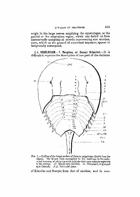
§ b. SKELETON.—I. Tergites, or Dorsal Sclerites.—It isdifficult to separate the description of one part of the skeleton
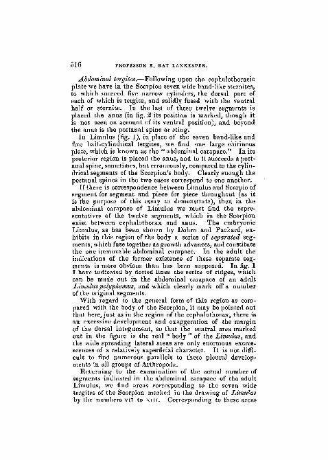
FIG. 1.—Outline of the tergal surface of famulus polyphemus (drawn from theobject). The dotted lines correspond to the markings on the abdo-minal carapace, which in the adult indicate what were separate segmentsin the embryo, oti'. Simple eyes (mesial), oe. Compound, or groupedeyes (lateral). P.A. Post-anal spine.
of Limulus and Scorpio from that of another, and in com-
514 PROFESSOR E. RAY LANKESTER.
mencing with the tergal elements, we must necessarily refersimultaneously to the general disposition of the appendages.
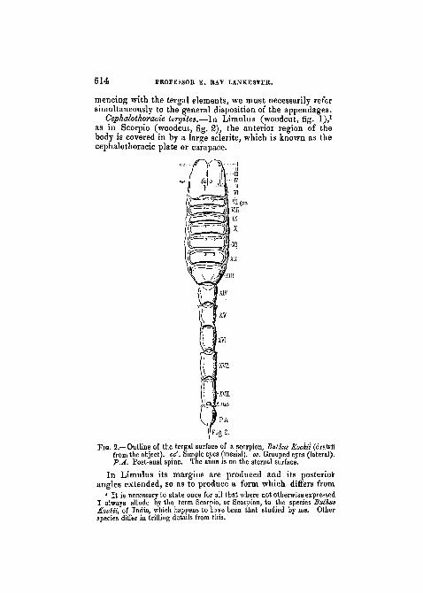
as in Scorpio (woodcut, fig. 2), the anterior region of thebody is covered in by a large sclerite, which is known as thecephalothoracic plate or carapace.
XIV
XV]
XVQ
ig 2-
Fio. 2.—Outline of the tergal surface of a scorpion, Bulhus fcochii (drawn"from the object), oc'. Simple eyes (mesial), oc. Grouped eyes (lateral).P.A. Post-anal spine. The anus is on the sternal surface.
I n Limulus its margins are produced and its posteriorangles extended, so as to produce a form which differs from
1 It is necessary to state once for all that where not otherwise expressedI always allude by the term Scorpio, or Scorpion, to the species ButhuaKochii, of India, which happens to have been that studied by me. Otherspecies differ in trifling details from this.
A
LIMULUS AN ARACHNID. 515
i that Seen in the Scorpion, but in essential points there isremarkable agreement. In both the carapace carries twopaired groups of eyes. Nearer the middle line is a single pairof simple eyes (pcr), which in Scorpio have an almost centralposition; more laterally placed (quite laterally in Scorpio)is a group, on either side, of simple eyes (oc), which inLimulus are $o closely aggregated as to form what is oftencalled " a compound eye." The compound eyes of Limulushave, however, been shown by Grenadier (3) to differ verymuch in structure from the compound eyes of either Crus-tacea or Insects, to which they have usually been asssimi-lated. They are more correctly interpreted—as the com-parison with Scorpio would suggest—as an aggregation ofsimple eyes. Such an aggregation (varying, according tothe genus, in number from two to five) we find in a lesscompact form than in Limulus on the right and left side ofthe Scorpion's cephalothoracic tergite.• In both Limulus and Scorpio the cephalothoracic tergitecovers in an area corresponding to the six leg-like appen-dages which are present in both animals, and may thereforebe considered as representing six coalesced tergites (i tovi).In Limulus the genital operculum which follows upon thelegs, and also the metathoracic sternites or chilaria which liebetween it and the bases of the last pair of legs, have beenby some morphologists regarded as also indicating segments
j which should be reckoned to the cephalothorax, and accord-v\ ingly eight coalesced tergites have been supposed to con-
stitute the carapace of the King Crab, whilst only six can bereckoned for the Scorpion. In reality, however, the chilariaare not appendages at all, as is proved by their late appear-ance in development (Packard, 8) and their form ; they aresimply sternites corresponding to the pentagonal sterniteplaced between the bases of the last pair of legs in Scorpio(woodcut, fig. 5). As to the genital operculum of Limulus,though in the adult it is in some measure adherent to the re-gion of the cephalothorax, yet it has a tergal area correspond-ing to it in the abdominal carapace, and in the embryonicLimulus is clearly seen to belong to that region, and not tothe cephalothorax. The innervation of the genital operculumfrom the oesophageal nerve-collar has, as already pointed
i out, no weight as an argument in favour of the association ofY that coalesced pair of appendages with the cephalothorax,
( for on the very same grounds it would be necessary to asso-ciate a large part of the middle region of the Scorpion'sbody (as far as and inclusive of the second pair of pulmonarysacs) with the cephalothorax.
51G PKOFESSOK E. RAY LANKESTEtt.
Abdominal tergites.—Following upon the cephalothoracic /plate we have in the Scorpion seven wide band-like sternites,to which succeed five narrow cylinders, the dorsal part ofeach of which is tergite, and solidly fused with the ventralhalf or sternite. In the last of these twelve segments isplaced the anus (in fig. 2 its position is marked, though itis not seen on account of its ventral position), and beyondthe anus is the postanal spine or sting.
In Limulus (fig. 1), in place of the seven band-like andfive half-cylindrical tergites, we find one large chitinousplate, which is known as the " abdominal carapace." In itsposterior region is placed the anus, and to it succeeds a post-anal spine, sometimes, but erroneously, compared to the cylin-drical segments of the Scorpion's body. Clearly enough thepostanal spines in the two cases correspond to one another.
If there is correspondence between Limulus and Scorpio ofsegment for segment and piece for piece throughout (as itis the purpose of this essay to demonstrate), then in theabdominal carapace of Limulus we must find the repre-sentatives of the twelve segments, which in the Scorpionexist between cephalothorax and anus. The embryonicLimulus, as has been shown by Dohrn and Packard, ex-hibits in this region of the body a series of separated seg-ments, which fuse together as growth advances, and constitutethe one immovable abdominal carapace. In the adult the .•-indications of the former existence of these separate seg- jments is more obvious than has been supposed. In fig. 1 /I have indicated by dotted lines the series of ridges, whichcan be made out in the abdominal carapace of an adultLimuluspolyp/iemus, and which clearly mark off a numberof the original segments.
With regard to the general form of this region as com-pared with the body of the Scorpion, it may be pointed outthat here, just as iu the region of the cephalothorax, there isan excessive development and exaggeration of the marginof the dorsal integument, so that the central area markedout in the figure is the real " body" of the Limulus, andthe wide spreading lateral areas are only enormous excres-scences of a relatively superficial character. It is not diffi-cult to find numerous parallels to these pleural develop-ments in all groups of Arthropoda.
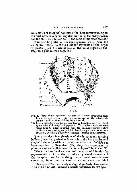
Returning to the examination of the actual number of j'segments indicated in the abdominal carapace of the adult >Limulus, we find areas corresponding to the seven widetergites of the Scorpion marked in the drawing of Limulusby the numbers vn to XIII. Corresponding to these areas
LIMULUS AN ARACHNID. 517
L
are a series of marginal processes, the first corresponding tothe first area, is a mere angular process of the integument ,but the six which follow are in the form of movable spines.1
Corresponding also to the six segments which hear thesix spines (that is, to the six hinder segments of the sevenin question) are a series of pits in the axial region of thetergum, a pair in each segment.
Fig 3.
TIG. 3.—View of the abdominal carapace of Limulus polyphemus frombelow, the soft sternal region and appendages of the anterior sixsegments and the viscera having been removed.
The figures vn to xn upon the drawing (drawn from the object) are placedby the sides of the tergal entapophyses. The continuation of the sameseries (xiii to xvni) is placed upon the chitinized sternal surfaceof the uusegmented region, which in Limulus represents the seventhabdominal and the five cylindrical prseanal segments of the Scorpion.
These are deep invaginations of the integument forminghollow processes, pushed as it were into the body cavity andclothed internally with cartilage, the structure of which hasbeen described by Gregenbaur (2); they give attachment tomuscles and are well termed " entapophyses " by Owen (7).
When we look in the abdominal carapace of Limulus forrepresentatives of the five cylindrical prseanal segments ofthe Scorpion, we find nothing but a broad smooth areaextending from the marking which indicates the hind
1 These spines I have seen slowly moving, independently of one another,in the living King Crab, indicating a separate musculature for each spine.
6 1 8 PROFESSOR E. RAY LANKESTEB. i
border of the thirteenth segment (seventh of the abdominal /series) to the soft membrane which forms the hinge of thepostanal spine.
In the embryo Limulus, however, this area is further seg-mented. We do not find the five segments of the Scorpion,but we find two of which (as segments) no indication is leftin the adult, and the foremost of these carries a movablespine on each side like those in front of it.
The anterior margin of the segment or tract of the bodywhich carries the anus appears to be uniformly in Arthro-poda, and in some other segmented animals, the part fromwhich new segments grow and become individualised, andit is to this tract of the body including its prse- and post-anal regions that the name " telson " is applicable as, forexample, in the Lobster. It not unfrequently happens thatthis segment-producing region does not produce the fullnumber of segments in given examples of an Arthropodousclass, which is characteristic of the majority or of the morefully segmented members of the class. Thus, both inCrustacea and Arachnida we find numerous forms with areduced number of abdominal segments.1 Usually, however,as in the spiders, the embryo exhibits at some time of itsdevelopment the full complement of segments, the hinder-most of which subsequently become obliterated by fusion oratrophy. Limulus so far conforms to this plan as to show fthe segmental potentiality of its praeanal area, but fails to jexhibit to the observer the full complement of segments even }as a temporary arrangement of its living substance.
Accordingly the whole area posterior to the ridge mark-ing the posterior border of the thirteenth segment maybe regarded in Limulus as belonging to the " telson," orarea of potential segmentation, a certain reservation beingobserved in respect to the one or two minute segmentswhich appeared and disappeared in this region in theembryo.
We may, when comparing this condition of things withthat exhibited by the Scorpion, either consider the telsonicarea and spine of Limulus as representing the five cylin-drical segments and the sting of the Scorpion in an unseg-mented state, or we may insist rather upon the actualitythan the protentiality, and identify the telson or fifth of the ,cylindrical segments of the Scorpion (viz. that carrying the Aanus), and the postanal spine with the telsonic area and Vspine of Limulus, whilst regarding the four anterior cylin-
1 Note also the evanescent character of the three last segments of Thely-phonus (fig. 12).
LIMULUS AN ARACHNID. 5 1 9
drical segments of the Scorpion as something over and aboveand not developed in Limulus at all.
It seems, however, probable from the evidence of extinctf forms, as well as from the abortive segmentation of the
embryo, that Limulus is not derived from an ancestor inwhich the telsonic area was as limited in its production ofsegments as it is in Limulus itself, but on the contrary, thatthe ancestor of Limulus had the full complement of seg-ments (and possibly more) which is seen in Scorpio and theEurypterina. In that case the praeanal area and spine ofLimulus would not merely be an area representing the fivecylindrical segments and sting of Scorpio in potentiality,but would be the actual representative of those segmentsgradually reduced and fused in the course of an historicprocess of change.
II . Appendages.—At each stage of the comparison betweenLimulus and Scorpio, the proofs of the intimate affinity ofthe two animals become more convincing, since we find thatthe view which it is necessary to adopt in order to makeone set of structures agree closely in the two animals, isprecisely the view which it is necessary to adopt, when a
, second set are considered, in order to make agreementi possible.\ We have just dealt with the tergites and have found an( exact correspondence of piece for piece, with the exception
that four prseanal segments are suppressed or five fused inLimulus which are discretely present in Scorpio. In order
I to admit such an agreement of piece for piece as to tergites,' we have to reject the view that the chilaria and the genital( operoulum represent segments belonging to the cephaloj thoracic tergite, for in that case the cephalothorax of! Scorpio would be a fusion of six, whilst that of Limulus
•• '• would be a fusion of eight pieces.When we come to examine the sternites, we shall find
t that the exclusion of the chilaria from the series of appen-dages is exactly what is required in order to identify thesternites of Limulus with those of Scorpio, and the removal
L of the genital operculum . of Limulus from the cephalo-m thorax makes its identity with the genital operculum ofI Scorpio even more obvious than it would otherwise be.V The six pairs of appendages of the cephalothorax of
Limulus may be compared one by one with the six pairs ofScorpio.
Cepkalothoracic appendage, No. /.—We have alreadydisposed of the obstacle which has been always raisedhitherto when the chelicerse of the Scorpion have been
520 PHOFESSOtt E. RAY LANRESTER.
J
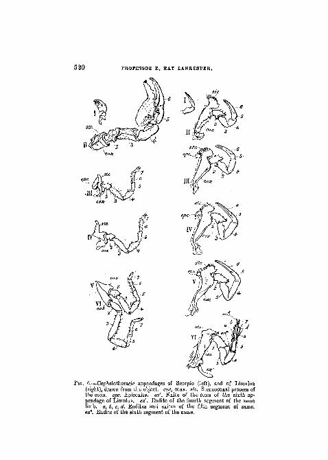
FIG. 4.—Cephalothoracic appendages of Scorpio (left), and of Limulus(right), drawn from the object, cox, coxa. stc. Steruocoxal process ofthe coxa. epc. Epicoxite. ex'. Exite of the coxa of the sixth ap-pendage of Limulus. en'. Endite of the fourth segment of the samelimb, a, 6, c, d. Endites and exiles of the fifth segment of same.e>t6. Endite of the sixth segment of the same.
LIMULUS AN ARACHNID. 5 2 1
assimilated to the chelicerse or first pair of limbs of Limulus.Instead of there being a difference as to innervation wehave seen that there is a real identity.
In Limulus, each of the first pair of appendages is ashort pair of nippers (woodcut, fig. 4, I, right) composed ofthree sclerites; at the base of the two appendages andbetween them and the mouth is placed an ovate sternite, thecamerostome or upper lip. (Plate XXVIII , fig. 4).
In Scorpio (woodcut, fig. 4, I left) a similarly small pairof appendages is found similarly composed of three sclerites,and similarly overhanging an oval " camerostome."
Ceph. thor. app., No. II.—In this and the following leg-like appendages of Limulus six chief sclerites are developed,the basal one or " coxa " being much enlarged, and its in-terior border produced into a well-marked process providedwith tooth-like hairs. The arrangement of the limbs aroundthe mouth and the central sternite which follows it (pmstin PI. XXVIII, fig. 4), is such that the processes of thecoxae of all ten limbs act together as manducatory organs.The process of the coxa may be called " the sterno-coxalprocess " (stc. in the woodcut, fig. 4). The second cephalo-thoracic appendage in the female Limulus polyphemus is likethe third, fourth, and fifth, a chela—that is to say,the penulti-mate sclerite is produced so as to form with the last scleritea pair of nippers. In the male this is not the case, thesecond pair of appendages being thicker and heavier thanin the female, and the penultimate joint not prolonged. Theform of appendage seen in the male L. polyphemus in thisposition is similar to appendages seen in other Arachnidathan Scorpio, viz. Thelyphonus (woodcut, fig. 12).
The second pair of appendages in the Scorpion is likethat of the female Limulus, but relatively larger. It con-sists of six sclerites as in Limulus, and has a sterno-coxalprocess on its coxa, which acts with its fellow of the oppositeside as a jaw (woodcut, fig 4, n ) .
Cephalo-thoracic appendage, No. III.—In Limulus poly-phemus this has, in both sexes, the same form as has thesecond appendage in the female. It is similarly composedof six sclerites, but in addition to these we find a distinctmovable sclerite developed on the median border of the coxa.This sclerite may be termed the " epicoxite" (woodcut,fig. 4, m , epc, right). The epicoxite is a remarkablefeature, and is not easily paralleled among Arthropoda.The basal " endite " of the limbs of the Crustacean Apus issimilar to it, and perhaps derived from a common ancestralorigin.
5 2 2 PROFESSOR E. RAY LANKESTER.
In Scorpio the third cephalothoracic appendage is in theform of a walking leg, and as such has seven scjerites. Itis a remarkable fact that in Limulus the sixth cephalo-thoracic appendage, which is non-chelate, also presentsseven axial sclerites (woodcut, fig. 4, vi, right), so that theScorpion's ambulatory limbs do not depart from the possi-bilities of Limulus in developing axial sclerites beyond thenumber six. It is also important to notice in this connectionthat the Arachnida exhibit a great variability in the numberof joints present in their legs. Thelyphonus develops a four-jointed "tarsus " at the end of the five proximal segmentsof its ambulatory limbs (woodcut, fig. 12), whilst Galeodespresents a curious increase in the number of segments inthe proximal region of its hinder limbs (woodcut, fig. 10).
The most important feature in which the third and sub-sequent cephalothoracic limbs of Scorpio resemble those ofLimulus is in the great development of the coxffi. Thesterno-coxal process is present on the third and fourthcephalothoracic appendages, and is even larger relatively thanin Limulus. In the third and fourth limbs it is free, overlyinga very soft minute sternal region belowthe mouth, and playingwith its fellow of the opposite side the part of an ingestiveorgan for the mouth. The narrow cleft between the opposedsterno-coxal processes probably acts by capillary attractionin the taking up of such food as the blood of other animals.
The coxse of the fifth and sixth appendages of Scorpiohave, on the other hand, no free sterno-coxal process.
The great enlargement of the coxse of these four pairs ofappendages, and their encroachment upon the median area,is accompanied by, and related to, the suppression of anyrepresentative of the sternal sclerite {pmst., fig. 4,PI. XXVIII) which is present in Limulus. The coxa? ofthe third pair and of the fourth pair meet one another inthe middle ventral line, but are separated by soft membrane.The coxse of the fifth and sixth pairs do not meet theirfellows in the middle line, but are kept apart by the wedge-shaped extremity of a sternite (met. in woodcut, fig. 8).They differ from the coxae of the third and fourth pairs in thatthe fifth is adherent to the sixth (woodcut, fig. 4, v, vi, left.)
The base of the third appendage in Scorpio exhibits adevelopment internal to the sterno-coxal process, whichcorresponds to, and probably represents, the " epicoxite " ofLimulus. This is in the form of a movable plate (woodcut,fig. 4, in , epc, left), which presents parallel ridges on itssurface.
Cephalothoracic appendage, No. IV.—Appendage No. iv
i
LIMULUS AN ARACHNID. 5 2 3
in Limulus closely resembles No. in . As in No. in , anepicoxite is present.
The corresponding appendage of Scorpio has been alreadymentioned. It has seven joints and a large sterno-coxalprocess, but no epicoxite, such as occurs in the limb next infront of it.
Ce§> halo thoracic appendage, No. V.—In Limulus thisresembles Nos. in and iv, like them having an epicoxite.
In Scorpio, No. v, is a seven-jointed ambulatory limb,with large coxa fused to the coxa of the next followingappendage, but devoid of sterno-coxal process.
Cephalothoracic appendage, No. VI.—In Limulus this isthe characteristic digging limb, unlike in the special modifica-tion of its parts and their remarkable function (for whichsee the citations of Lockwood and of Lloyd in ' Owen'sMemoir,' No. 7) any other arthropod appendage.
In structure it is remarkable for exhibiting the feature ofsecondary movable arthrites diverging from the axis of thelimb, unusual in Arthropoda other than the Crustacea. Sevenaxial sclerites or segments can be distinguished, the coxabeing large, as in the oiher limbs, but devoid of an epicoxite.On the other hand, whilst the " endite " is thus absent, an" exite " is developed in the form of a flattened elongatedpiece articulated to the external border of the coxa (wood-cut fig. 4, vi ex' right).
The second and third segments of the axis are devoid ofapophyses, but the fourth bears a large spine-like articulatedendite. The fifth joint of the axis carries four flattenedapophyses (endites and exites), which are articulated andcapable of active movement. The sixth joint bears one arti-culated endite, and, further, the short terminal seventh orultimate segment of the axis, which is relatively muchlonger in newly hatched individuals than in the adult.
The sixth cephalothoracic appendage in Scorpio is quitesimilar to the three preceding walking legs. Its large coxais fused to that of the fifth appendage of the same side. Thespinous outgrowths on the sixth and seventh segments ofthis and the other legs are in character somewhat similar tothe more highly developed apophyses of the digging limb ofLimulus.
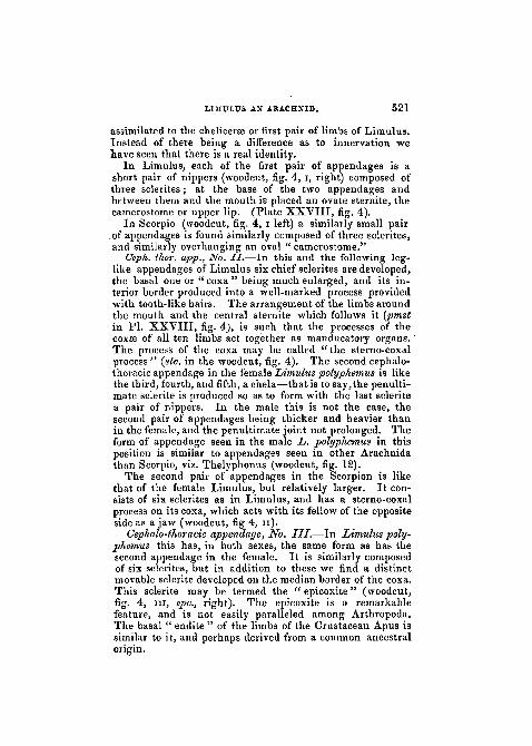
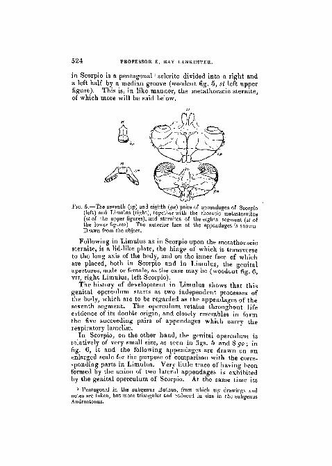
The seventh pair of appendages or genital operculum.—InLimulus lying between the bases of the sixth pair of cepha-lothoracic appendages is a pair of sclerites, the chilaria ofOwen, actually the metathoracic division of the sternum(woodcut fig. 5,st. right), which belongs to the segment carry-ing the sixth pair of appendages. Precisely similar in position
VOX.. X.XI. — NEW SElt. M M
524 PROFESSOR E. RAY LANKESTER.
in Scorpio is a pentagonal' sclerite divided into a right anda left half by a median groove (woodcut fig. 5, st left upperfigure). This is, in like manner, the metathoracic sternite,of which more will be said below.
JIG. 5.—The seventh (op) and eighth (ga) pairs of appendages of Scorpio(left) and Limulus (right), together with the thoracic metasternites(st of the upper figures), and sternites of the eighth segment (si ofthe lower figures). The anterior face of the appendages is shownDrawn from the object.
Following in Limulus as in Scorpio upon the metathoracicsternite, is a lid-like plate, the hinge of which is transverseto the long axis of the body, and on the inner face of whichare placed, both in Scorpio and in Limulus, the genitalapertures, male or female, as the case may be (woodcut fig. 6,VII, right Limulus, left Scorpio).
The history of development in Limulus shows that thisgenital operculum starts as two independent processes ofthe body, which are to be regarded as the appendages of theseventh segment. The operculum retains throughout lifeevidence of its double origin, and closely resembles in formthe five succeeding pairs of appendages which carry therespiratory lamellae.
In Scorpio, on the other hand, the genital operculum isrelatively of very small size, as seen in figs. 5 and 8 go; infig. 6, it and the following appendages are drawn on anenlarged scale for the purpose of comparison with the corre-sponding parts in Limulus. Very little trace of having beenformed by the union of two lateral appendages is exhibitedby the genital operculum of Scorpio. At the same time its
1 Pentagonal in the subgenus Buthus, from which my drawings andnotes are taken, but more triangular and reduced in size iu the subgeuusAndroctonus.
LIMULUS AN ARACHNID. 525
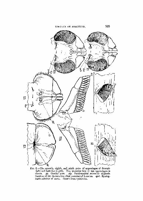
FIG. 6.—The seventh, eighth, and ninth pairs of appendages of Scorpio(left) and Litnulus (right). The posterior face of the appendages isshown, gp. Genital pore. stg. Parabranchial muscular stigmata(tendons of the thoraco-branchial muscles) of Limulus. epsl. Epistig-matic sclerites of same. Drawn from specimens.
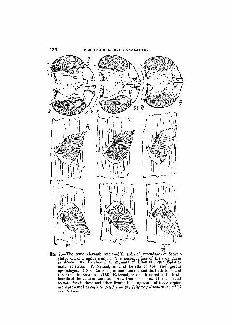
526 PKOFESSOK E. EAY LANKESTKR.
matic sclerites. I'. Mediad, or first lamella of the lamelligerousappendages. H.30. External, or one hundred and thirtieth lamella ofthe same in Scorpio 1150 External or one hundred and fiftiethappendages. H.30. External, or one hundred and thirtieth lamella ofthe same in Scorpio. 1150. External, or one hundred and fiftiethlamella of the same in Limulus. Draw from specimens. It is importantto note that in these and other figures the lung-books of the Scorpionare represented as entirely freed from the delicate pulmonary sac whichinvests them
pinvests them.
LIMULUS AN ARACHNID. 527
bifid margin speaks of such an origin, and, as a matter offact, such appears to be its embryological history.
I shall here quote a passage from ' Balfour's Embryology,'recounting Metschnikoff's observations upon the existenceof rudiments of appendages in the segments of the Scorpion'sbody following upon the cephalothorax with its six pairs oflimbs. The observations have great importance, not only inreference to the genital operculum but also in regard to thepulmonary sacs and their " branchial books" which arefound in succeeding segments.
Balfour says, " Rudimentary appendages appear on thesix segments behind the ambulatory legs. . . . They persistonly on the second segment, where they appear to form thecomb-like organs or pectines. The last abdominal segment,i.e. that next the tail, is without provisional appendages.The embryonic tail is divided into six segments, includingthe telson. The lungs are formed by paired invaginations,the walls of which subsequently become plicated, on thefour last segments, which bear rudimentary limbs, andsimultaneously with the disappearance of the rudimentarylimbs " (' Comp. Embryology,' vol. i, p. 359).
Hence it appears that, in Scorpio, in front of the comb.-likeorgans, that is to say, in the position subsequently occupied bythe genital operculum, there is in the embryo, as in that ofLimulus, a pair of rudimentary appendages. We know that inLimulus these grow together to form the genital operculum.It is in the very highest degree probable that the same historyobtains for the similarly related genital operculum of Scorpio.
In discussing the tergites, it has already been pointed outthat the genital operculum corresponds to a separate band-like tergite in Scorpio (vn, in woodcut, fig. 2), and to anemarginated area on the anterior border of the abdominalcarapace of Limulus (vn, in woodcut, fig. 1), which is moredistinctly marked in the embryo.
The eighth pair of appendages.—In Scorpio we find, on theventral surface corresponding with the eighth tergite (six ter-gitesbeing reckoned to the cephalothorax) a pairof appendagescarrying fine lamellae set like the teeth of a comb along the in-ferior margin (woodcuts fig. 5 ga, left, and fig. 6 VIII, left; seealso Plate XXVIII). They are developed from the secondpair of rudimentary abdominal appendages of the embryo.
In Limulus, in the corresponding position, we find a pairof appendages, the first of a series of five pairs (woodcuts fig. 5ga, right, and fig. 6 VIII, right). The appendages of the twosides, as in the case of the genital operculum, do not, divergefrom one another but are directed tovvards one another and
5 2 8 PROFESSOR E. RAY LANKESTER.
united across the middle line by a soft plate-like fold of thesternal integument; the result being that a plate-like body isformed from two originally distinct right and left appendages.On the under surface of each of the combined appendagesa series of very delicate lamellse is found corresponding tothe lamelliform teeth of the Scorpion's comb-like organs.
Ninth, tenth, eleventh, and twelfth appendages.—InLimulus, corresponding to the tergal areas marked ix, x, xi,xn, we find a series of pairs of appendages precisely similarto that belonging to the eighth segment.
In Scorpio it will be remembered that in the embryorudimentary appendages appear corresponding to the firstsix abdominal segments, or the seventh, eighth, ninth, tenth,eleventh, and twelfth of the whole body. Of these the firstpair we have seen, become in all probability the genital oper-culum; the second pair are known to become the " pectines j "the pairs on the ninth, tenth, eleventh, and twelfth segmentsdisappear, as the lung sacs on those segments develop by aprocess of invagination.
They disappear, but only from view. It has not beenshown by actual observation, but it cannot well be doubted,that these rudimentary appendages sink within the lung-invaginations, and become the lamelligerous appendageswhich are found in them in the adult Scorpion.
The four pairs of stigmata on the ventral surface of theninth, tenth, eleventh, and twelfth segments of the Scor-pion's body (woodcut, fig. 8) lead into sacs, each of whch con-tains, concealed within it, an appendage consisting of an axisbearing a series of delicate lamellae (woodcuts, figs. 6 and7, ix, x, xi, xn , left).
Each of these concealed appendages is strictly comparablein structure to one of the comb-like organs of the eighthsegment, the axis corresponding to the axis, and the delicatelamellse to the teeth of the comb.
Thus, then, we find five pairs of lamelligerous appendageson the five segments of the Scorpion's body uumbered 8, 9,10, 11, 12, of which the first pair is external, and accordinglymodified, whilst the next four are sunk below the surface,and accordingly modified. In Limulus, on the exactly cor-responding segments, namely, those numbered 8, 9, 10, 11,12, we find five pairs of lamelligerous appendages, but theseare all external, and all alike modified for the purposes ofaquatic respiration.1
1 Latreille, though holding the Limuli to be Crustacea, and not Aracli-niaa, was the first to insist on the branchia-like character of the Scorpion'slung-books
LIMULTJS AN ARACHNID. 529
Furthermore, it is important to notice that in Scorpioneither in the embryo nor at any other time does the seventhabdominal segment (thirteenth of the whole series) carry apair of appendages, nor do any of the subsequent cylindricalsegments. Similarly in Linmlus no appendages or rudi-ments of appendages are to be detected after the last pairof lamelligerous organs—the twelfth of the whole series.
The segmented region, devoid of appendages in theScorpion, is represented by an unsegmented region devoidof appendages in the King Crab.
Before entering into a more minute comparison of thelamelligerous appendages of the Scorpion with those ofLimulus, with the object of establishing the identity oforigin of the two series by the detection of agreementbetween them in details of structure, it will be most con-venient to examine another series of skeletal elements,namely, the sternites.
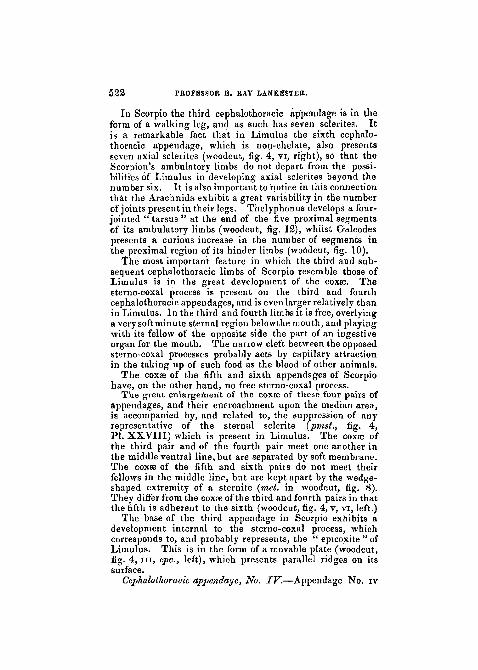
III. Sternites.—In Limulus,inthe cephalo-thoracic region,we find that the integument of the sternal area, though to alarge extent soft and devoid of hard chitinous plates, yetpresents here and there well-marked sclerites. On thesub-frontal area, a small discoidal piece, the sub-frontalsclerite is found (PI. XXVIII, fig. 4, sf). Between themouth and the bases of the first pair of appendages a muchmore important sclerite occurs, to which the term used byLatreille for the similarly placed sclerite in Arachnida, viz.(carnerostome), may be used.
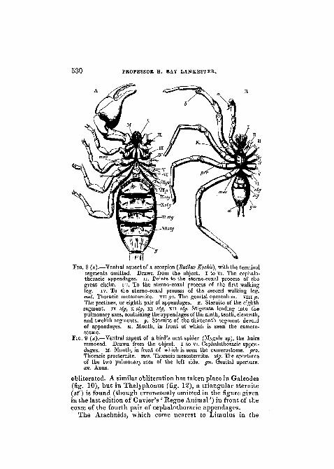
In the Scorpion (fig. 8, in front of the mouth to which theline M points) a similar tubercular sclerite is found. Thereis advantage in not merely designating this piece " labium/'since there is but little ground for holding it to be equivalenteither to the labrum of Insecta or to that of Crustacea.
In the Spider Mygale (fig. 9) and in Galeodes (figs. 10and 11, cam), this same piece is observed, attaining a remark-able development in the latter.
When we come to the region behind the mouth, we findin Limulus a large median sclerite extending from thepharynx backward. It lies between the bases of the third,fourth, fifth, and sixth pairs of cephalothoracic appendages.On account of its position, it may be termed the thoracicpromeso-sternite (PI. XXVIII, fig. A,pmst), since it appearsto represent elements which, in other Arachnida, are markedoff as distinct prosternite and mesosternite.
In Scorpio we find nothing corresponding to this piece. Bythe enlargement and mesiad production of the coxee of thefour hinder cephalothoracic appendages it lias been as it were
530 PROFESSOR E. RAY LANKESTER.
FIG. 8 (A).—Ventral aspect of a scorpion {Buthus Kochit), with the terminalsegments omitted. Drawn from the object. I to vi. The cephalo-thoracic appendages, n. Points to the sterno-coxal process of thegreat chelae, in. To the sterno-coxal process of the first walkingleg. iv. To the sterno-coxal process of the second walking leg.met. Thoracic metasternite. vn go. The genital operculum. viiijt).The pectines, or eighth pair of appendages, x. Sternite of the eighthsegment, ix stg, x stg, xi stg, xn stg. Stigmata leading into thepulmonary sacs, containing the appendages of the ninth, tenth, eleventh,and twelfth segments, y. Sternite of the thirteenth segment devoidof appendages, M. Mouth, in front of which is seen the camero-stome.
PIG. 9 (B).—Ventral aspect of a bird's nest spider (Mygale sp), the hairsremoved. Drawn from the object. I to vi. Cephalothoracic appen-dages. M. Mouth, in front of which is seen the camerostome. pro.Thoracic prosternite. rues. Thoracic mesosternite. stg. The aperturesof the two pulmonary sacs of the left side. gn. Genital aperture.an. Anus.
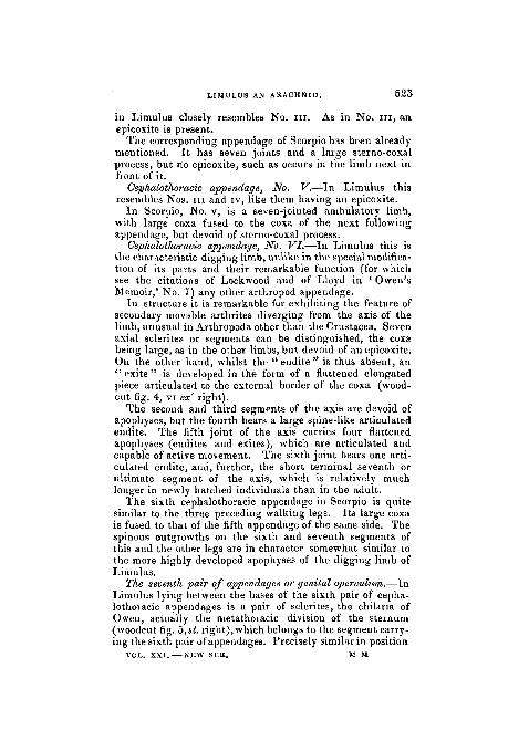
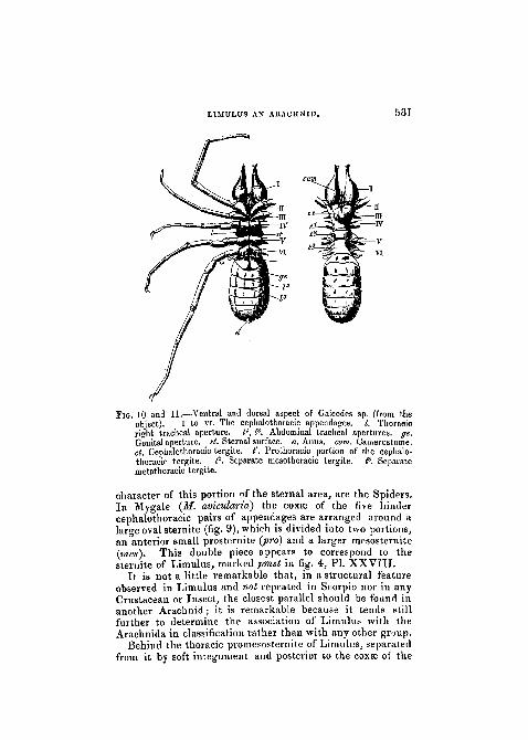
obliterated. A similar obliteration has taken place in Galeodes(fig. 10), but in The]yphonus (fig. 12), a triangular sternite(sO is found (though erroneously omitted in the figure givenin the last edition of Cuvier's ' Regne Animal'') in front of thecoxje of the fourth pair of cephalothoracic appendages.
The Arachnids, which come nearest to Limulus in the
LIMULUS AN ARACHNID. 531
PIG. 10 and 11.—Ventral and dorsal aspect of Galeodes sp. (from theobject). I to VI. The ceplialothoracie appendages. /. Thoracicright tracheal aperture. P, I3, Abdominal tracheal apertures, ge.Genital aperture, at. Sternal surface, a. Anus. cam. Camerostome.cl. Cephalothoracic tergite. t'. Prothoracic portion of the ceplialo-thoracic tergite. P. Separate mesothoraoic tergite. P. Separatemetathoracic tergite.
character of this portion of the sternal area, are the Spiders.In Mygale (M. avicularia) the coxse of the five hindercephalothoracic pairs of appendages are arranged around alarge oval sternite (fig. 9), which is divided into two portions,an anterior small prosternite {pro) and a larger mesosternite(mes). This double piece appears to correspond to thesternite of Limulus, marked pmst in fig. 4, PL XXVIII.
It is not a little remarkable that, in a structural featureobserved in Limulus and not repeated in Scorpio nor in anyCrustacean or Insect, the closest parallel should be found inanother Arachnid; it is remarkable because it tends stillfurther to determine the association of Limulus with theArachnida in classification rather than with any other group.
Behind the thoracic promesosteniite of Limulus, separatedfrom it by soft integument and posterior to the coxae of the
532 PROFESSOR E. RAY JLANKESTER.
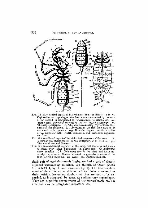
FIG. 12 (A).—Ventral aspect of Thelyphonus (from the object). I to vi.Cephalothoracic appendages; the first, which is concealed by the coxaof the second, is represented as removed from its attachment, sic.Sterno-coxal process of the coxa of the left second appendage. s(l.Thoracic prosternite. sf. Thoracic metasternite. vn to xvm. Seg-ments of the abdomen. 1,1. Apertures of the right lung sacs in theninth and tenth segments, msg. Muscular stigmata on the sternitesof the tenth, eleventh, twelfth, thirteenth, and fourteenth segments.an. Anus.
Fi&. 13 (B).—Dorsal aspect of the abdominal segments of the same. p.Muscular pits corresponding to the entapophyses of Limulus. pa/The jointed postanal filament.
FIG. 14 (c).—Abdominal segments of the same, with the terga and visceradissected away (after Blanchard). n. Nerve cord. ng. Abdominalnerve ganglion. 1,1. Pulmonary sacs in the ninth and tenth seg-ments, m, m, tn, m. Muscles attached to muscular stigmata of thefour following segments, an. Anus. pa/. Postanal filament.
sixth pair of cephalothoracic limbs, we find a pair of closely-opposed upstanding sclerites, the chilaria of Owen (metstPL XXVIII, fig. 4, and woodcut, fig. 4). The late develop-ment of these pieces, as determined by Packard, as well astheir position, leaves no doubt that they are not to be re-garded, as is supposed by some, as rudimentary appendages.They are a paired development of the metathoiacic sternalarea and may be designated metasternites.
\
LIMULUS AN ARACHNID. 5 3 3
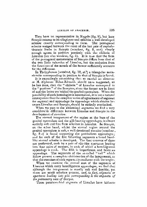
They have no representative in Mygale (fig. 9), but hereScorpio returns to its allegiance and exhibits a well-developedsclerite exactly corresponding to them. The pentagonalsclerite wedged between the coxae of the last pair of cephalo-thoracic limbs in Scorpio (woodcut, fig. 8, met) clearlyenough agrees in position precisely with the chilaria ofLimulus (see also woodcut, fig. 5). It is true that the formof the pentagonal metasternite of Scorpio differs from that ofthe two little tubercles of Limulus, but the exclusion fromthe functions of the mouth of the former sufficiently accountsfor the difference.
In Thelyphonus (woodcut, fig. 12, st?) a triangular meta-sternite corresponding in position to that of Scorpio is found.
It is exceedingly astonishing that so careful an observeras M. Alphonse Milne-Edwards should have suggested, ashe has done, that the "chilaria" of Limulus correspond tothe " pectines " of the Scorpion, since the former are in frontof and the latter are behind the genital operculum. When thepossibility of such homologies is entertained, it is but a naturalconsequence that the complete series of agreements of segmentfor segment and appendage for appendage which obtains be-tween Limulus and Scorpio, should be entirely overlooked.
When we pass to the abdominal segments we find a veryconsiderable difference between Limulus and Scorpio in thedevelopment of sternites.
The sternal integument of the region at the base of thegenital operculum and the gill-bearing appendages, is almostentirely soft and free from sclerites in Limulus. In Scorpio,on the other hand, whilst the sternal region around thegenital operculum is soft, a well-developed stemite (woodcut,fig. 8 a;) is found supporting the pectiniform appendages;and for each of the five following segments a broad band-like sternal sclerite is developed. The four anterior of theseare perforated, each by a pair of slit-like apertures leadinginto four pairs of recesses, in each of which a lamelligerousappendage is sunk. The fifth is imperforate, and bears noappendage. The segments of the so-called " tail" whichfollow present a complete chitinisation of the integument, sothat the sternites of each segment is confluent with the tergite.
When we examine the sternal area of the segments ofLimulus which carry lamelligerous appendages, we find thatalthough the integument is mostly soft and flexible, yetthere are small sclerites present, and, in fact, stigmata orapertures leading into pits corresponding to the stigmata ofthe pulmonary sacs of Scorpio.
These parabranchial stigmata of Limulus have hitherto
5 3 4 PROFESSOR E. RAY LANKESTER.
escaped observation.1 They are found on the posterior faceof the median sternal elevation or lobe which unites the twolateral elements or appendages which go to form one of thedouble lamelligerous organs of that animal (Plate XXVIII,fig. 10 stg, and woodcuts, figs. 6 and 7 sty). The lips of thestigma are chitinised, and the opening leads into a funnel-likecavity with chitinised walls. The sternal integument furthershows one or two small sclerites, the " epistigmatic sclerites "(epst), by the side of the stigma. These stigmata occur inthe position mentioned, not only at the bases of the appen-dages of the four segments corresponding to those whichcarry the pulmonary stigmata in the Scorpion, namely, theninth, tenth, eleventh, and twelfth, but also at the base ofthe appendages of the eighth segment, which represent thepectines of the Scorpion, and at the base of the genital oper-culum. They are connected with the attachment of a seriesof powerful muscles, the thoraco-branchials, which, takingtheir origin in the thorax, are inserted into the integumentright and left at the base of each of the six pairs of abdo-minal appendages. The function of these muscles is clearlyenough to agitate this series of plate-like organs, either forthe purpose of respiration or for that of locomotion, probablyfor both simultaneously.
The fact that the insertion of a muscle into the integu-ment of Limulus is connected with a " cupping " of the areaof attachment is remarkable but not without parallel. Theseries of dorsal entapophyses have a precisely similar signi-ficance, and in other Arachnida, e.g. Thelyphonus (fig. 12msg fig. 13 p, and fig. 14 m), we find an identicalarrangement on both ventral and dorsal surface, the stig-mata being, however, much shallower than in Limulus.
I am not aware of the occurrence of such " muscularstigmata " in any other Arthropoda than the Arachnida, atany rate, of stigmata comparable to those of Limulus.Usually the tendons of muscles are in Avthropoda formedby solid fibrous extensions of the subepidermic layers ofthe integument.
The tendons or processes connected with the parabranchi'alstigmata, and with the dorsal entapophyses of Limulus, are byno means entirely formed by the invaginated epidermis and itschitinous product. The tissue below the epidermis is deve-loped in a very special manner, and forms part of an endo-skeleton which in the thoracic region giyes rise to a veryremarkable internal sternum or entosternite. The struc-
1 I communicated an account of their occurrence and probable signi-ficance to the Royal Society on May 20th, 1881.
LTMULUS AN ARACHNID. 5 3 5
ture of this deep skeletal tissue has been investigated byGegenbaur, who has shown that it may have the formeither of a fibrous or of a more distinctly cartilaginousmodification of the connective tissue into which it gradu-ally passes, and from which, on the other hand, is developedin other regions a series of vascular channels constitutingthe capillaries, veins, and arteries. On the present occasionI do not propose to enter into histological details withregard to Limulus, but I may just mention that whilst thehollow entapophyses are invested on their visceral surfaceby a richly developed cartilaginous modification of the con-nective substance, with a well developed capsular arrange-ment of the intercellular substance, the funnel-like invagi-nations connected with the parabranchial stigmata areclothed and continued by a fibrous tissue not unlike thetendon of Vertebrata. The same tendon-like tissue alsoforms the entosternite.
In Plate XXVIII, fig. 11, the internal connection of thepair of parabranchial stigmata of a lamelligerous appendage-pair of Limulus is drawn. The integument has been dis-sected away from the whole of the anterior face of theappendages and their uniting sternal bridge, so as to showthe inner aspect of the integument of the posterior face.The pouch-like character of the invaginations into whichthe stigmata lead and the attachment of the thoraco-branchial muscle is thus exhibited. In fig. 13, PI.XXVIII, one ®f the funnel-like tendons, consisting inter-nally of chitin borne on epidermis, and externally of fibroustissue, is shown in an isolated condition. It is possible tointroduce a probe into the funnel to the depth of an inch,the axial cavity of invagination extending to that distance.Th» funnel-like pouch of Limulus thus constituted, I con-sider to be the homologue (that is, the genetic representa-tive or homogen) of the pulmonary sac of Scorpion.
It will now be convenient to give, in a tabular form, a sum-mary of the view which has been set forth in the precedingpages. Having thus exposed what I conceive to be the legiti-mate conception of the morphological relations of Limulusand Scorpio, I shall endeavour to justify, by a closerexamination, the identification (which forms an essentialpart of it) of the pectines of the Scorpion and its fourpairs of book-like organs sunk in recesses of the integu-ment with the five pairs of lamelligerous appendages ofLimulus.
(The tabular statement is given on the next page.)
536 PROFESSOR E. RAY 1ANKESTER.
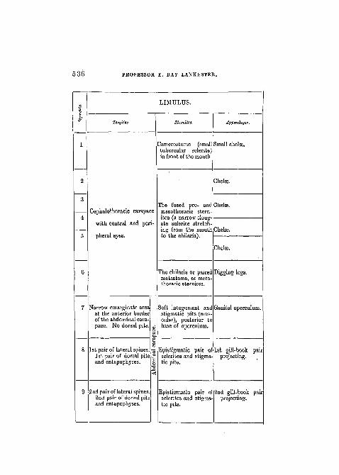
LIMTJLUS.
Tcrgitcs. Appendages.
2amerostome (smaltubercular sclerite)in front of the mouth
Small chelae.
Chela
Cephalothoracic carapace
with central and peri-
pheral eyes.
Tbe fused pro- andmesothoraoie stern-ites (a narrow elong-ate sclerite stretch-ing from the mouthto the chilaria).
Jhelse.
The chilaria or paired Digging legs,metastoma, or meta-thoraoic sternites.
Narrow emarginate areaat the anterior borderof the abdominal cara-pace. No dorsal pits.
1st pair of lateral spines.1st pair of dorsal pitsand entapophyses.
2nd pair of lateral spines.2nd pair of dorsal pitsand entapophyses.
Soft integument and Genital operculumstigmatic pits (mus-cular), posterior tobase of operculum.
Epistigmatic pairsclerites and stigmatic pits.
of 1st gill-book pairprojecting.
Epistigmatic pair osclerites and stigma-tic pits.
2nd gill-book parprojecting.
LIMTJLUS AN ARACHNID. 537
E!>
1
2
3
4
5
6
7
8
9
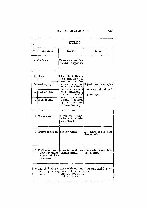
SCOEPIO.
Appendages.
Chelicerse.
jhelffi.
talking legs.
talking legs.
Walking legs.
Walking legs.
Genital operculum.
Pectinse, or pair ocomb-like organsmodified gill-boolprojecting.
1st gill-book paisunk in pulmonarysacs.
SUrnitm.
3amerostome (of La-treille), or upper lip.
Obliterated by the me-siad extension of theCOXSB of the fourwalking legs; thetwo anterior movable,the two posteriorfixed. (In Mygaleadistinctly markedsmall prothoracicsternite is followedby a large oval meso-thoracic sternite.)
Pentagonal elongatesclerite or metatho-racic sternite.
Soft integument.
Separate small rectangular sclerite.
Aseoarate broad transverse sclerite witlstigmata leading topulmonary sacs.
Ttrgita.
Cephalothoracic carapace
with central and peri-
pheral eyes.
A separate narrow band-like sclerite.
A separate narrow band-like sclerite.
A separate band-like scle-rite.
538 PROFESSOR E. RAY LANKESTER.
10
11
3rd pair of lateral spines3rd pair of dorsal pitsand entapophyses.
4th pair of lateral spines4th pair of dorsal pitsand entapophyses.
5th pair of lateral spines5th pair of dorsal pitsand entapophyses.
13
15
16
17
18
LIMULUS.
TergUes.
6th pair of lateral spines.6th pair of dorsal pitsand entapophyses.
Only in the embryo thissegment is separate,and has a 7th pair oilateral spines.
Only in the embryo thissegment is indicated.
These three segmentsare never expressedand are representedby the prreanal re-gion of the telson.
Post-anal spine.
Epistigmatic pair o'sclerites and stigma-tic pits.
Epistigmatic pairsclerites and stigmatic pits.
of 4th
Epistigmatic pair ofsclerites and stigma-tic pits.
None.
Noue.Large and solid scle-
rite forming the ster-
num of the "Telson,"
i.e. of the prse-anal None,
region of potential
segmentation, which None,
includes a soft inva-
ginate area on which
opens the ANUS.
Appendagei
3rd gill ok pairprojp .ng.
gill-book pairprojecting.
ith gill-book paiiprojecting.
None.
None.
L I M U L U S AN A R A C H N I D . 539
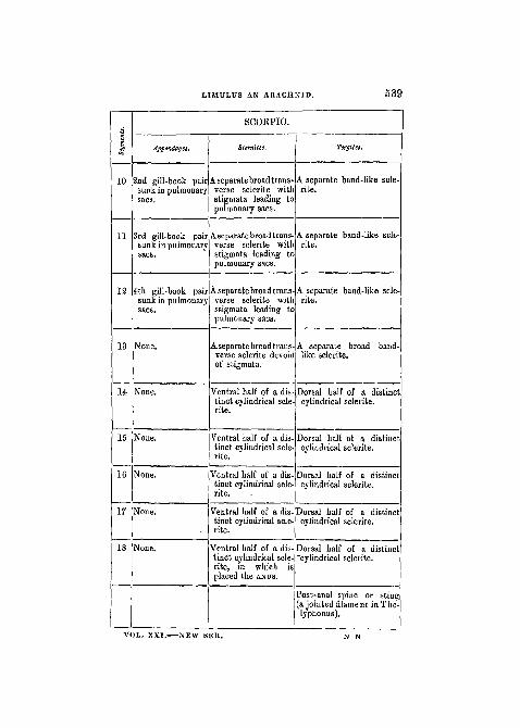
43
10
11
12
13
14
15
16
17
18
SCOBPIO.
Jppcndagti.
2nd gill-book pairsunk in pulmonarysacs.
3rd gill-book pairsunk in pulmonarysacs.
4th gill-book pairsunk in pulmonarysacs.
None.
None.
None.
None.
None.
None.
Sternites.
A separate broad trans-verse sclerite withstigmata leading topulmonary sacs.
A separate broad trans-verse sclerite withstigmata leading topulmonary saes.
A separate broad trans-verse sclerite withstigmata leading topulmonary sacs.
A separate broad trans-verse sclerite devoidof stigmata.
Ventral half of a dis-tinct cylindrical scle-rite.
Ventral half of a dis-tinct cylindrical scle-rite.
"Ventral half of a dis-tinct cylindrical scle-rite.
Ventral half of a dis-tinct cylindrical scle-rite.
Ventral half of a dis-tinct cylindrical scle-rite, in which isplaced the ANUS.
Tergites.
A separate band-like scle-rite.
A separate band-like scle-rite.
A separate band-like scle-rite.
A separate broad band-like sclerite.
Dorsal half of a distinctcylindrical sclerite.
Dorsal half of a distinctcylindrical sclerite.
Dorsal half of a distinctcylindrical sclerite.
Dorsal half of a distinctcylindrical sclerite.
Dorsal half of a distinct"cylindrical sclerite.
Post-anal spine or sting(a jointed filament inThe-lyphonus).
VOL. XXI. NEW SEK.
i
5 4 0 PROFESSOR E. RAY LANKESTER.
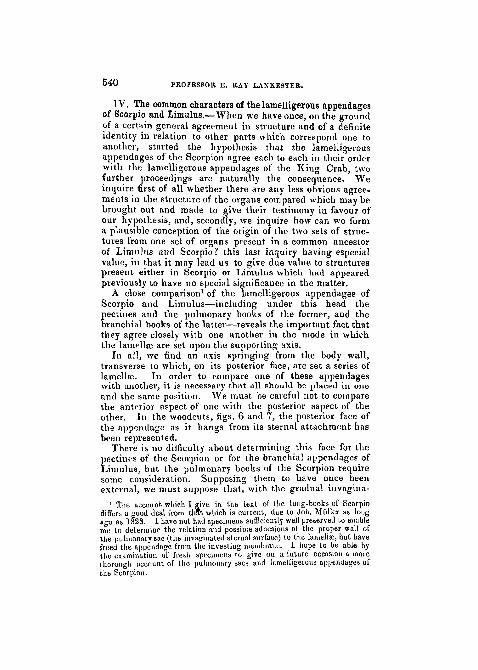
TV. The common characters of the lamelligerous appendagesof Scorpio and Limulus.—When we have once, on the groundof a certain general agreement in structure and of a definiteidentity in relation to other parts which correspond one toanother, started the hypothesis that the lamelligerousappendages of the Scorpion agree each to each in their orderwith the lamelligerous appendages of the King Crab, twofurther proceedings are naturally the consequence. Weinquire first of all whether there are any less obvious agree-ments in the structure of the organs compared which may bebrought out and made to give their testimony in favour ofour hypothesis, and, secondly, we inquire how can we forma plausible conception of the origin of the two sets of struc-tures from one set of organs present in a common ancestorof Limulus iind Scorpio? this last inquiry having especialvalue, in that it may lead us to give due value to structurespresent either in Scorpio or Limulus which had appearedpreviously to have no special significance in the matter.
A close comparison1 of the lamelligerous appendages ofScorpio and Limulus—including under this head thepectiues and the pulmonary books of the former, and thebranchial books of the latter—reveals the important fact thatthey agree closely with one anotiier in the mode in whichthe lamollse are set upon the supporting axis.
In all, we find an axis springing from the body wall,transverse to which, on its posterior face, are set a series oflamellae. In order to compare one of these appendageswith another, it is necessary that sill should be placed in oneand the same position. We must be careful not to comparethe anterior aspect of one with the posterior aspect of theother. In the woodcuts, figs. 6 and 7, the posterior face ofthe appendage as it hangs from its sternal attachment hasbeen represented.
There is no difficulty about determining this face for thepectines of the Scorpion or for the branchial appendages ofLimulus, but the pulmonary books of the Scorpion requiresome consideration. Supposing them to have once beenexternal, we must suppose that, with the gradual invagina-
1 The account which I give in the text of the lung-books of Scorpiodiffers a good deal from thft which is current, due to Joh. Miiller as longago as 1828. I have not had specimens sufficiently well preserved to enableme to determiue the relation and possible adhesions of the proper wall ofthe pulmonary sac (the invaginated sternal surface) to the lamella:, but havefreed the appendage from the investing membrane. I hope to be able bythe examination of fresh specimens to give on a future occasion a morethorough account of the pulmonary sacs and lamelligerous appendages ofthe Scorpion.
L
LIMULUS AN ARACHNID. 5*1
tion of their surface of attachment, they have become moreand more deflected into the cavity of invagination, movingon their fixed base at first backwards, then upwards, andfinally forwards. As we now find them (in a spirit speci-men !), on viewing the inner surface of the ventral scleritesby removing the terga and viscera, they can be rotated ontheir hinge line so that they may be made to lie prone for-wards, exposing the stigma or opening of the pulmonaryrecess posteriorly, as in PI. XXVIII, fig. 1 a, or they may bemade to lie prone in the reverse direction, hiding from viewthe stigma, as shown in PI. XXVIII , fig. 2 a, and in thewoodcuts, figs. 6 and 7. The position which correspondswith that of the external appendages the peotines and thebranchial organs of Limulus, when viewed from the posteriorface, so as to show (in the case of Limulus) the lamellae, isthat in which the lung-book is directed backward so as tohide the stigmatic aperture and is looked at from within theScorpion's body, that is> by dissecting off the tcrga, viscera,and muscles.
When the pectines, lung-books, and branchial books arethus placed we find that the lamella; are not set precisely atright angles upon the axes, but obliquely, so that there isan imbrication of the successive lamellae. In all three it isthe proximal lamella which is uppermost (see PI. XVIII, fig.& and 2a I' and fig. 10 I'). The imbrication is identical in all.
As to number of lamellae, we find in the pecten of ButhusKochii eighteen (in other scorpions there are more or less);in the lung-books of some scorpions1 as many as 130, and inthe Limulus gill-book as many as 150. These numbersvary slightly, increasing with growth in ah probability.
As to structure of lamella?, those of the pecten ate moresolid and strongly chitinised than those of the other twoorgans, but are, nevertheless, true lamellse flattenedtransversely.
Those of the lung-books are exceedingly delicate platescomposed of two closely approximated membranes, betweenwhich the blood circulates ; they are, in fact, flattened bags.They carry on their free margins a few chitinous spinules(PI. XXVIII , fig. 8). The lamella; of the -gill-books ofLimulus are similarly delicate flattened bags with a setosefree border. I am not able to institute any comparisonof the histological structure of the lamellae of the Scorpion'slung-book with that of the King Crab's gill-book, foralthough I have been able to work out that of the latter on
1 I believe the form in which I counted these to be a species of Androc-tomis.
5 4 2 PROFESSOR E. RAY LANKESTER.
fresh material, no such opportunity has yet presented itselfof investigating the Scorpion.
As to the shape of the lamellae, those of the pecten arenarrower and relatively thicker than those of the lung-booksor gill-books ; the whole eighteen are also more nearlyequal to one another in size and shape. In the lung-booksthe shape differs at the two extremities of the series a little,and in size the proximal laminse are much larger than thedistal. The average shape may be described as that of abroad scythe-blade (PI. XXVIII , fig. 8) with a narrow basesupport (a b). The lamellae of the gill-books of Limulus,on the other hand, are approximately semicircular in shape,with a wide base of origin [a b in fig. 9, PI. XXVIII).Moreover an important difference, which is explained bythe convergence in place of divergence of the axis of thelimb relatively to the mid line of the body, is seen when thelamellae of the gill-book and of the lung-book are compared,in the fact that in the gill-book the proximal laminae arethe smallest (PL XXVIII, fig. 10 V), whilst in the lung-book they are the largest.
Further comparison of the grouping and form of thelamellae is facilitated by the figures on PI. XXVIII, wherefig. 1 and fig. 1 a, fig. £ and figs. 2 a and 10, fig. 3 andfigs. 3 a and S b, give representations of the three varieties oflamelligerous appendages in a series of identical positions.Fig. 1 should, for comparison with fig. 1 a, be looked at byinverting the plate.
The axes which support the lamellae in the three varietiesof lamelligerous limb differ much from one another, but in amanner directly corresponding with obvious functions.
The pecten has a large free axis firmly chitinised, imper-fectly divided into two joints. It is flattened by antero-posterior pressure. The function of the pecten is notactually known, but it appears to be tactile. It is notrespiratory, and the Scorpion is of terrestrial habit; henceits comparatively solid character and protective developmentof chitin.
The gill-book of Limulus is supported on an axis, whichis flattened by dorso-ventral pressure, protection being thusafforded to the otherwise naked and very delicate lamellae.It is not free except at its extremity, where it exhibits aCointing of separate chitinous plates. Its base is very wide,and is attached, not to a flat sternal surface, but to an out-standing sternal lobe, which extends between the bases offellow-appendages, and gives rise to a teat-like soft processin the median line (PI. XXVIII , md, fig. 10). The charac-
i LIMULUS AN ARACHNID. 543
ter of this axis is obviously an adaptation to the branchialfunction of the lamellae combined with a locomotorfunction.
The lung-book of Scorpio has no locomotor function, andit is protected by the recess of the sternum, in which it lies.It is not tactile, nor is it exposed to desiccation and roughusage, as are the pectines. It is specialised for respiratorypurposes. The axis is exceedingly small and simple, forthe greater part of its length. adherent to the invaginatedsternal wall, leaving, however, a small free distal portion(see PI. XXVIII, fig. 2 a). Its walls are quite free fromchitinisation, and of great delicacy. It is little else than ahorizontal vascular tube supporting the lamelliform bagsinto which its cavity leads (PI. XXVIII, 2 a, b).
Though the axis is here reduced to its simplest ex-pression, it is not possible to overlook in it the representativeof the vertically compressed chitinised axis of the pecten,and of the horizontally compressed chitinised axis of thegill-book.
V. Hypothesis as to the mode of origin of the three varietiesof lamelligerous appendages in Scorpio and Limulus.—Theview which I have advanced in this memoir as to the prac-tical identity of the gill-books of Limulus and the lung-books of Scorpio implicitly contains the affirmation that eitherthe structures of Limulus have been derived from those ofScorpio, or those of Scorpio from those of Limulus, or thata third (now extinct) form has given rise to both Limulusand Scorpio. Further, it is to be observed that suchextinct form might be more like to Limulus than toScorpio, or vice versa, in respect of any particular element ofstructure.
To make a long story as short as possible I may say that,without prejudicing the recognition of the (as I think) well-established morphological identities above pointed out, wemay best explain their existence by assuming that an aquaticform breathing the dissolved oxygen of the water inhabitedby it, by means of book-like gills, was the common ancestorof Limulus and of Scorpio. From the book-like gills of this--cestral form the broad series of Limulus and the narrower
ig-books, as well as the pectines or combs of the Scorpion,ye been derived. The form of the book-like gills of this
Arachnidan ancestor was probably something intermediatebetween the three existent modifications of it, and best con-ceived of, perhaps, by imagining the teeth of the Scorpion's" pectinate organs" to become soft and flattened andincreased in number (see PL XXIX, fig. 1).
544l PROFESSOR E. RAY LANKESTER.
To obtain from these the Limulus gill we have but tosuppose certain definite changes of dimension, the imbricationand character of the lamellae, and their external positionremaining unaltered (PI. XXIX, figs. 2 a and 3 a).
To arrive at the book-lungs of the Scorpion, we have toimagine the ventral surface on each side in close proximityto the short appendages carrying the gill-books—to havebecome deeply cupped or depressed, so that two series ofcup-like pits should be formed, a right and a left, a pairbeing placed in each segment, corresponding to each pair ofgill-books. Each cup must have become so large in areaand so deep as to embrace within its limits the relativelysmall adjacent gill-book (XXIX, fig. 2 b). Further, whenonce the gill-book had been involved in this cup-like de-pression, the walls of the cup must have tended to growtogether so as to form a pulmonary chamber with only anarrow slit-like opening to the exterior (PI. XXIX, fig. 3 b),and part passu with this closing in of the cupped area, andthe protection of the respiratory lamellae, the Arachnid musthave acquired the power of leaving the water and of breathingthe atmospheric oxygen admitted to the damp chamberformed by the cave-like areas of depression.
Whilst framing such a hypothetical account of the way inwhich the transition from naked "gill-book" to in-sunken" lung-book" could have taken place, one naturallyasks—" Is it not somewhat gratuitous to assume thatcupped arese should form conveniently by the side ofthe gill-books of the aquatic ancestor, so as to be ready toincrease in size, and ultimately draw into themselves, as itwere, the gill-books ?" " Is there," we are led on further toask, " any known instance in Arachnida of the formation ofcupped arese on the chitinous surface of the body ? If so,can we show in what mechanical relation they are formed ?And, lastly, can it be demonstrated that such mechanicalrelation probably existed in connection with the gill-booksof the assumed common ancestor of Limulus and Scorpio?"If all these questions can be affirmatively answered, thenour hypothesis as to the transition of the aquatic Arachnidto the pulmonate condition acquires great plausibility.
The answer to these questions appears to me to have morethan ordinary interest, since the formation of cupped areseon the chitinous surface of the body and the mechanicalrelations connected with their formation have, as pointed outa few pages back, come to light as demanded by the hypo-thesis. They exist in Limulus itself and in Thelyphonus.In Limulus there are two great muscles, a right and a left,
J
LIMU.LUS AN ARACHNID. 5 4 5
inserted into the soft ventral integument near the base, ofeach double gill-plate. These muscles serve (together withothers that enter the appendage itself) by their contractionsto move the gill-plates in the water and so aid in aquaticrespiration. The position of the insertion of each muscularmass is marked by a deep funnel-like depression of theintegument. From the external surface this depressionappears as a " stigma," which we have already described asthe parabranchial stigma. The funnel-like depression has anarrow mouth which is often as much as half an inch inlength. Internally the invaginated cuticle stands up as aflexible tendon clothed with fibrous tissue and giving attach-ment to the muscle already mentioned.
In Limulus we find a pair of these " muscle-stigmata,"right and left behind the genital operculum, and a pair(right and left) behind each of the lamelliform fused appen-dages which carry the gill-books.
We have only to suppose the appendages carrying thegill-books not to have fused as yet in the middle line, andthe muscular stigmata to have become greatly developed(perhaps by increased development of the muscle aiding inaquatic respiration when the appendage itself grew smalland therefore less efficient) and we have at once the gill-booksinking within the area of the stigmatic pit, PI. XXIX,fig. 2b.
A very important feature in the supposed further develop-ment is the correspondence of the atrophy of the muscle(which atrophy is required to fit in with our hypothesis, andto convert the muscle-pit into a pulmonary sac) with thechanges in the structures which would necessarily resultwere the physiological conditions gradually to become suchas to favour aerial in place of aquatic respiration. Theviolent agitation of the gills by the muscle attached to thestigmatic pit would become useless, supposing an exposureof the gill-lamellae to the atmosphere became by degreeshabitual with the ancestral Arachnidan. In proportion asthese hypothetical creatures acquired the habit of aerialrepiration—the deepening and arching in of the stigmaticpit would be favoured, and the atrophy and final disappear-ance of the muscle which was attached to its inner surface,and mechanically brought it into existence, would also bedirectly promoted.
A further confirmation of the view now advanced is foundin the remarkable East Indian Arachnid Thelyphonus. ThisArachnid has not four pairs of lung-sacs like Scorpio, butonly two pairs, corresponding to the two foremost lungs of
5 4 6 PROFESSOR E. RAY LANKESTER,
Scorpio, and to the second and third gill-book-pairs ofLimulus (woodcut, figs. 12, 13, 14). Nevertheless, as wehave seen in a previous section of this Essay, the four seg-ments of the abdomen posterior to these are each marked bya pair of shallow stigmata placed in line with the orifices ofthe pulmonarg sacs of the two anterior segments, msg. Whenthe internal structure corresponding to these parts is exa-mined, it is found that a large muscle (similar to the simi-larly placed muscle of Limulus) is inserted into each of thefour right and four left stigmata in the segments posteriorto the pulmonary sacs (woodcut fig. 14, m.~) The two seg-ments into which the two pairs of pulmonary sacs are sunk,have no such muscles. The pulmonary sacs are, therefore,to all appearance, enlarged muscular stigmata, from whichtheir former muscles have disappeared by disuse andatrophy.
VI. The Entosternite.—Leaving now the comparison ofsegments and appendages, which is undoubtedly the mostimportant element in determining our judgment as to theaffinity of Limulus with Scorpio, we come to the considera-tion of a number of other structures, which we shall findsome more and some less favorable to the hypothesis of aclose relationship between the two animals.
Connected with the exoskeleton and locomotor system isthat remarkable development of an endoskeleton in theArachnida, which Straus Durkheim put forward in thepassage cited at the commencement of this article, as oneof the leading characteristics of the class and one of theobvious features in which Limulus shows itself to be a trueArachnid.
As already remarked, in speaking of the entapophyses andparabranchial stigmata, Limulus shows a marked tendencyto the development of cartilage and fibro-cartilage by themodification of its connective substance at certain points andin certain areas.
The most striking result of this tendency, is the formationof a large fibro-cartilagiuous plate which lies in the cephalo-thoracic region between the alimentary canal above and thenerve collar below, and unconnected by hard parts with anyportion of the exoskeleton. It is represented of the naturalsize as seen from the ventral (sternal) aspect in fig. 7,PL XXVIII . It has been previously figured by Van derHoeveu (12), whose figure is not very accurate, and is copiedby Owen (7).
This body is the base of origin of a large number ofmuscles, and may be regarded as an enlargement and
LIMULUS AN ARACHNID 5 4 7
interlacing of their respective tendons. In the figure,nothing but the skeletal structure, cleaned of its muscles, isrepresented.
In Scorpio, a similarly shaped loose skeletal piece isound, which gives attachment to muscles in the same way
and has a similar relation to the ventral nerve-mass andartery, by which in Scorpio it is perforated (PI. XXVIII,figs. 6 and 6 a). The corresponding body in Mygale is(PI. XXVIII, figs. 5, 5 a) more closely similar in form tothat of Limulus than is that of Scorpio.
In order to make a close comparison of these Entoster-nites, it will be necessary to determine exactly the insertionsof the muscles to which they give origin; and further, toascertain how far the histological structure of those of Scor-pio and Mygale agrees with that of Limulus. The resultsof this investigation I hope to make the subject of a futurepublication. In the meantime the close correspondence ingeneral character of the three Entosternites figured onPL XXVIII cannot escape notice, and fully justifies theimportance which Straus Durkheim attached to them. Thetwo pairs of tendinous outgrowths right and left of thecentral plate in Limulus correspond with the three pairsseen in Mygale, whilst the deep anterior notch in the lattercorresponds with the shallower excavation in Limulus, inwhich the number 7 is placed in the drawing, and in whichin the animal itself the bend of the alimentary canal isplaced, the mouth being actually below the central regionof the plate, so that the alimentary canal passes first for-wards beneath the plate and is then reflected so as to passbackwards whilst resting on the upper surface of the plate.
Whilst of this as of so many other structures of theArachnida (such as the lung-books, &c.) which have beencompared in the present memoir with structures in Limulus,a renewed and critical examination is absolutely needed,yet we have sufficient ground, even in our presentincomplete knowledge, for concluding that the agreementas to them presented by the two animals is a very closeone.
In no Crustacean is a free entosternite at all similar tothe organ under discussion known. The apodemes of thesternal surface of Decapodous Crustacea do not resemble itin form though of a similar function. The nearest approachto it is seen in the rod-like skeletal organ found in theabdomen of Lepidoptera, and described by Leydig (' Baudes Thierischen Korpers,' Atlas, pi. vi, fig. 1). Itsshape and position are very different, however, from the
5 4 8 PROFESSOR E. RAY LANKESTKR.
entosternites of Liraulus and other Arachnida. It agreeswith these only so far as that it is a free internal skeletalpiece.
In the abdominal region of Limulus small loose fibro-cartilageB, similar in nature to the Entosternite, are found,connected with the tendons of muscles. I have isolatedfour such pieces. They are mentioned by Straus Durkheim.