Architecture of Eph receptor clusters Juha P. Himanen a,1 , Laila Yermekbayeva b,1 , Peter W. Janes c,1 , John R. Walker b,1 , Kai Xu a , Lakmali Atapattu c , Kanagalaghatta R. Rajashankar d , Anneloes Mensinga c , Martin Lackmann c,2 , Dimitar B. Nikolov a,2 , and Sirano Dhe-Paganon b,e,2 a Structural Biology Program, Memorial Sloan-Kettering Cancer Center, 1275 York Avenue, New York, NY 10065; b Structural Genomics Consortium, University of Toronto, 101 College Street, Toronto, ON M5G 1L7, Canada; c Department of Biochemistry and Molecular Biology, Monash University, Victoria 3800, Australia; d Advanced Photon Source, Argonne National Laboratory, 9700 S. Cass Avenue, Argonne, IL 60439; and e Department of Physiology, University of Toronto, 101 College Street, Toronto, ON M5G 1L7, Canada Edited* by Dinshaw J. Patel, Memorial Sloan-Kettering Cancer Center, New York, NY, and approved April 14, 2010 (received for review March 29, 2010) Eph receptor tyrosine kinases and their ephrin ligands regulate cell navigation during normal and oncogenic development. Signaling of Ephs is initiated in a multistep process leading to the assembly of higher-order signaling clusters that set off bidirectional signaling in interacting cells. However, the structural and mechanistic details of this assembly remained undefined. Here we present high-resolu- tion structures of the complete EphA2 ectodomain and complexes with ephrin-A1 and A5 as the base unit of an Eph cluster. The struc- tures reveal an elongated architecture with novel Eph/Eph interac- tions, both within and outside of the Eph ligand-binding domain, that suggest the molecular mechanism underlying Eph/ephrin clus- tering. Structure-function analysis, by using site-directed muta- genesis and cell-based signaling assays, confirms the importance of the identified oligomerization interfaces for Eph clustering. cell-cell attraction and repulsion ∣ Eph receptor clustering E ph receptors and their ephrin ligands control a diverse array of cell–cell interactions during patterning of the nervous, skeletal, and vascular systems (1, 2). Upon ephrin binding, the Eph kinase initiates “forward” signaling into receptor-expressing cells, and the ephrin cytoplasmic tail triggers “reverse” signaling into ligand-expressing cells. Ephs and ephrins are divided into two subclasses (A and B) on the basis of their affinities for each other. With some exceptions, EphA receptors (EphA1–A10) bind to A-class ephrins (ephrin-A1–A6), whereas EphB receptors (EphB1–B6) interact with the B-subclass ephrins (ephrin-B1– B3). Given that Ephs and ephrins are membrane-bound, their interaction occurs only at sites of cell–cell contact. In the absence of cell–cell interactions, they exist in loosely associated microdo- mains, which become more compact and well-ordered when Eph/ ephrin complexes assemble to generate clearly defined signaling centers (2). The extracellular Eph region contains a conserved N-terminal ligand-binding domain (LBD), an adjacent cysteine-rich domain (CRD) (3), followed by two fibronectin repeats (FN3) (Fig. S1). The cytoplasmic Eph region encompasses a regulatory juxtamem- brane region connecting the kinase domain, a sterile α motif do- main, and a postsynaptic density protein (PSD95), Drosophila disc large tumor suppressor (DlgA), and zonula occludens-1 pro- tein (zo-1) binding motif. All ephrins possess a 20 KDa extra- cellular receptor-binding domain; B-type ephrins also contain a short cytoplasmic region. Several crystal structures of complexes between the minimal Eph and ephrin-binding Eph domains have been reported (reviewed in ref. 4). The crystal structure of the complexed EphB2 and ephrin-B2 binding domains revealed two contact surfaces that are involved in the assembly of Eph/ephrin tetramers: an expansive high-affinity ephrin-binding channel, likely responsible for the initial interaction, and a smaller interface that mediates a lower affinity Eph-ephrin contact with an adjacent ephrin molecule (4). In addition to these structurally defined ligand–receptor interactions, several observations revealed that additional protein interfaces are important for the generation and function of Eph/ephrin signaling centers at points of cell-cell contact: First, the EphB2-ephrinB2 crystal structure suggests a propensity of in- dividual Eph/ephrin complexes to assemble, via direct Eph/Eph contacts, into heterooligomeric clusters. Furthermore, earlier findings indicated that Eph-Eph interactions via a C-terminal ec- todomain region outside the LBD are critical for Eph function during development (3). Presumably such Eph-Eph interfaces act to recruit non-ligand-bound Eph receptors into Eph clusters (5). In addition, a random mutagenesis survey of the EphA3 ectodomain revealed that binding to ephrinA5 requires an inter- action site located in the CRD. Although having only a modest contribution to ligand-binding affinity, mutation of this region severely effected receptor phosphorylation and recruitment of signaling molecules (6). Last, a recent report suggested that ephrin-A5 may also interact via the EphA3 fibronectin III repeats (7). To determine the structural basis of Eph clustering and explore underlying receptor–receptor and receptor–ligand interactions, we determined structures of the complete EphA2 ectodomain, alone and in complexes with its cognate ligands ephrin-A5 and ephrin-A1, the latter of which was recently used to elaborate spa- tiomechanical concepts related to EphA2 clustering (8). These crystal structures, supported by cell-based functional studies, show that the CRD mediates Eph/Eph interactions in the assembly of signaling-competent EphA2/ephrin clusters. Results and Discussion Overall Structures. We crystallized and determined protein struc- tures comprising the whole—or parts of the—EphA2 extracellular region, either in their apo forms (complete EphA2 ectodomain and LBD) or bound to their ephrin ligands ephrin-A1 or -A5 (LBD, LBD-CRD, and LBD-CRD-nFN3, Table 1). The structure of a full-length Ephrin receptor ectodomain (Fig. 1A) reveals an extended structure of tightly packed domains spanning 146 × 52 × 55 Å(Fig. S2). The fold and atomic details of the Eph CRD, encompassing ∼114 amino acids (residues 201–314) immediately C-terminal to the LBD, can be subdivided into two domains, an N-terminal domain similar to complement control module/short complement regulator domain, and a TNF receptor like CRD. The N-terminal half (201–260) includes five antiparallel β-strands arranged as a β-sandwich, whereby the first residue of the fold (Cys201) is disulfide-anchored to the end of the fourth β-strand (Fig. S3). A Dali search (9) reveals the top Protein Data Bank (PDB) Author contributions: J.P.H., M.L., D.B.N., and S.D.-P. designed research; J.P.H., L.Y., P.W.J., J.R.W., K.X., L.A., K.R.R., and A.M. performed research; M.L. and D.B.N. contributed new reagents/analytic tools; J.P.H., P.W.J., J.R.W., M.L., D.B.N., and S.D.-P. analyzed data; and J.R.W., M.L., D.B.N., and S.D.-P. wrote the paper. The authors declare no conflict of interest. *This Direct Submission article had a prearranged editor. Freely available online through the PNAS open access option. 1 J.P.H., L.Y., P.W.J., and J.R.W. contributed equally to this work. 2 To whom correspondence should be addressed. E-mail: [email protected]. edu.au, [email protected], or [email protected]. This article contains supporting information online at www.pnas.org/lookup/suppl/ doi:10.1073/pnas.1004148107/-/DCSupplemental. 10860–10865 ∣ PNAS ∣ June 15, 2010 ∣ vol. 107 ∣ no. 24 www.pnas.org/cgi/doi/10.1073/pnas.1004148107
Transcript
Architecture of Eph receptor clustersJuha P. Himanena,1, Laila Yermekbayevab,1, Peter W. Janesc,1, John R. Walkerb,1, Kai Xua, Lakmali Atapattuc,Kanagalaghatta R. Rajashankard, Anneloes Mensingac, Martin Lackmannc,2,Dimitar B. Nikolova,2, and Sirano Dhe-Paganonb,e,2
aStructural Biology Program, Memorial Sloan-Kettering Cancer Center, 1275 York Avenue, New York, NY 10065; bStructural Genomics Consortium,University of Toronto, 101 College Street, Toronto, ON M5G 1L7, Canada; cDepartment of Biochemistry and Molecular Biology, Monash University,Victoria 3800, Australia; dAdvanced Photon Source, Argonne National Laboratory, 9700 S. Cass Avenue, Argonne, IL 60439; and eDepartment ofPhysiology, University of Toronto, 101 College Street, Toronto, ON M5G 1L7, Canada
Edited* by Dinshaw J. Patel, Memorial Sloan-Kettering Cancer Center, New York, NY, and approved April 14, 2010 (received for review March 29, 2010)
Eph receptor tyrosine kinases and their ephrin ligands regulate cellnavigation during normal and oncogenic development. Signalingof Ephs is initiated in a multistep process leading to the assemblyof higher-order signaling clusters that set off bidirectional signalingin interacting cells. However, the structural and mechanistic detailsof this assembly remained undefined. Herewe present high-resolu-tion structures of the complete EphA2 ectodomain and complexeswith ephrin-A1 and A5 as the base unit of an Eph cluster. The struc-tures reveal an elongated architecture with novel Eph/Eph interac-tions, both within and outside of the Eph ligand-binding domain,that suggest the molecular mechanism underlying Eph/ephrin clus-tering. Structure-function analysis, by using site-directed muta-genesis and cell-based signaling assays, confirms the importanceof the identified oligomerization interfaces for Eph clustering.
cell-cell attraction and repulsion ∣ Eph receptor clustering
Eph receptors and their ephrin ligands control a diverse arrayof cell–cell interactions during patterning of the nervous,
skeletal, and vascular systems (1, 2). Upon ephrin binding, theEph kinase initiates “forward” signaling into receptor-expressingcells, and the ephrin cytoplasmic tail triggers “reverse” signalinginto ligand-expressing cells. Ephs and ephrins are divided intotwo subclasses (A and B) on the basis of their affinities for eachother. With some exceptions, EphA receptors (EphA1–A10) bindto A-class ephrins (ephrin-A1–A6), whereas EphB receptors(EphB1–B6) interact with the B-subclass ephrins (ephrin-B1–B3). Given that Ephs and ephrins are membrane-bound, theirinteraction occurs only at sites of cell–cell contact. In the absenceof cell–cell interactions, they exist in loosely associated microdo-mains, which become more compact and well-ordered when Eph/ephrin complexes assemble to generate clearly defined signalingcenters (2).
The extracellular Eph region contains a conserved N-terminalligand-binding domain (LBD), an adjacent cysteine-rich domain(CRD) (3), followed by two fibronectin repeats (FN3) (Fig. S1).The cytoplasmic Eph region encompasses a regulatory juxtamem-brane region connecting the kinase domain, a sterile α motif do-main, and a postsynaptic density protein (PSD95), Drosophiladisc large tumor suppressor (DlgA), and zonula occludens-1 pro-tein (zo-1) binding motif. All ephrins possess a 20 KDa extra-cellular receptor-binding domain; B-type ephrins also containa short cytoplasmic region.
Several crystal structures of complexes between the minimalEph and ephrin-binding Eph domains have been reported(reviewed in ref. 4). The crystal structure of the complexed EphB2and ephrin-B2 binding domains revealed two contact surfaces thatare involved in the assembly ofEph/ephrin tetramers: an expansivehigh-affinity ephrin-binding channel, likely responsible for theinitial interaction, and a smaller interface that mediates a loweraffinity Eph-ephrin contact with an adjacent ephrin molecule (4).
In addition to these structurally defined ligand–receptorinteractions, several observations revealed that additional proteininterfaces are important for the generation and function ofEph/ephrin signaling centers at points of cell-cell contact: First,
the EphB2-ephrinB2 crystal structure suggests a propensity of in-dividual Eph/ephrin complexes to assemble, via direct Eph/Ephcontacts, into heterooligomeric clusters. Furthermore, earlierfindings indicated that Eph-Eph interactions via a C-terminal ec-todomain region outside the LBD are critical for Eph functionduring development (3). Presumably such Eph-Eph interfacesact to recruit non-ligand-bound Eph receptors into Eph clusters(5). In addition, a random mutagenesis survey of the EphA3ectodomain revealed that binding to ephrinA5 requires an inter-action site located in the CRD. Although having only a modestcontribution to ligand-binding affinity, mutation of this regionseverely effected receptor phosphorylation and recruitment ofsignaling molecules (6). Last, a recent report suggested thatephrin-A5 may also interact via the EphA3 fibronectin III repeats(7). Todetermine the structural basis ofEph clustering and exploreunderlying receptor–receptor and receptor–ligand interactions,we determined structures of the complete EphA2 ectodomain,alone and in complexes with its cognate ligands ephrin-A5 andephrin-A1, the latter of which was recently used to elaborate spa-tiomechanical concepts related to EphA2 clustering (8). Thesecrystal structures, supported by cell-based functional studies,show that theCRDmediatesEph/Eph interactions in the assemblyof signaling-competent EphA2/ephrin clusters.
Results and DiscussionOverall Structures. We crystallized and determined protein struc-tures comprising the whole—or parts of the—EphA2 extracellularregion, either in their apo forms (complete EphA2 ectodomainand LBD) or bound to their ephrin ligands ephrin-A1 or -A5(LBD, LBD-CRD, and LBD-CRD-nFN3, Table 1). The structureof a full-length Ephrin receptor ectodomain (Fig. 1A) reveals anextended structure of tightly packed domains spanning 146 × 52 ×55 Å (Fig. S2).
The fold and atomic details of the Eph CRD, encompassing∼114 amino acids (residues 201–314) immediately C-terminalto the LBD, can be subdivided into two domains, an N-terminaldomain similar to complement control module/short complementregulator domain, and a TNF receptor like CRD. The N-terminalhalf (201–260) includes five antiparallel β-strands arranged as aβ-sandwich, whereby the first residue of the fold (Cys201) isdisulfide-anchored to the end of the fourth β-strand (Fig. S3).A Dali search (9) reveals the top Protein Data Bank (PDB)
Author contributions: J.P.H., M.L., D.B.N., and S.D.-P. designed research; J.P.H., L.Y., P.W.J.,J.R.W., K.X., L.A., K.R.R., and A.M. performed research; M.L. and D.B.N. contributednew reagents/analytic tools; J.P.H., P.W.J., J.R.W., M.L., D.B.N., and S.D.-P. analyzed data;and J.R.W., M.L., D.B.N., and S.D.-P. wrote the paper.
The authors declare no conflict of interest.
*This Direct Submission article had a prearranged editor.
Freely available online through the PNAS open access option.1J.P.H., L.Y., P.W.J., and J.R.W. contributed equally to this work.2To whom correspondence should be addressed. E-mail: [email protected], [email protected], or [email protected].
This article contains supporting information online at www.pnas.org/lookup/suppl/doi:10.1073/pnas.1004148107/-/DCSupplemental.
*Ordered and modeled.†Highest resolution shell shown in parenthesis.‡Rfree calculated with 5% of the data.
Fig. 1. EphA2/ephrin-A1 (5) structures, structural alignment, and ligand binding. (A) Left, backbone representation of superimposed structures. Right, allstructures are shown from the same perspective in ribbon format. Each structure is labeled according to its PDB code and differentially colored. (B) TheEphA2-EphrinA1 high-affinity heterodimer interface. Stereoscopic view of the interface with domains labeled and shown in ribbon format. Ligand residuesthat are within 4 Å of the LBD are shown as sticks; LBD residues as lines. Water molecules in the vicinity are shown as red spheres, hydrogen bonds as blackdashed lines.
Himanen et al. PNAS ∣ June 15, 2010 ∣ vol. 107 ∣ no. 24 ∣ 10861
BIOCH
EMISTR
Y
hit with this domain is that of 2Z3R with a Z score of 4.5, an rmsdof 2.7 Å over 56 Cα atoms, with 13% sequence identity, includingthe cysteines involved in the disulphide bridges matching the Ephsequences 201–247 and 230–260. The C-terminal half of the EphCRD comprises two β-strands and six tightly packed randomcoils, including four disulfide bridges, the first of which anchorsthe first residue of this half to the sixth CRD β-strand. It resem-bles the TNF receptor CRD, closely matching the Death Recep-tor 5 (PDB ID 2H9G) with a Z score of 4.9, an rmsd of 2.4 over 46Cα atoms, and 17% sequence identity, including conservation ofthree disulphide bridges (262–273, 276–290, and 293–307). Thetwo CRD halves are tightly packed against each other, with theN- and C-terminal residues of the CRD occurring on oppositesides of the long axis of the domain. Apart from some negativelycharged patches, the CRD surface is predominantly neutral.
The N-terminal fibronectin-type-3 domain (nFN3) adopts atypical immunoglobulin-like fold (Fig. 1A and Fig. S4A), mostclosely homologous to Integrin Beta-4 (1QG3) and Plectin-1(3F7P) FN3 (rmsd ∼2.0 Å over 91 Cα atoms, DALI search).Of note, lack of significant binding clefts at either ends of thedomain suggests that nFN3 does not bind small-molecule ligands(Fig. S4B). Although cFN3 has the same topology as nFN3, it isstructurally distinct with an rmsd of 7.7 Å extending over 68 Cα
atoms (Fig. S4A): Its β3-β4 and β5-β6 loops are split open toreveal an aromatic-lined cleft that might represent a membrane-surface binding pocket (Fig. S4B). Structural homologs to thecFN3 include Neural Cell Adhesion Molecule 2, Fibronectin,and Tenascin, with rmsd values from 1.6 to 2.0 over 82 Cα atoms.
The elongated architecture of EphA2 is stabilized by extensiveinterdomain interactions. The first and last LBD domain residues(25–27 and 199–200) together with the β5-β6 loop form an inter-action surface with the CRD, which buries 1;211 Å2 surface areaand is stabilized by six hydrogen or salt bonds (Fig. S5A). Like-wise, a buried 701 Å2 interface and a salt bridge stabilizes theCRD–nFN3 interaction (Fig. S5B). The association betweennFN3 and cFN3 seems more flexible and, apart from a single hy-drogen bond, not stabilized by buried protein surfaces.
High-Affinity Eph/Ephrin Heterodimer.FunctionalEph/ephrin signal-ing clusters assemble from high-affinity Eph/ephrin heterodimers,which aggregate into heterotetramers and higher-order oligomers(1). The two EphA2/ephrin-A1 and one EphA2/ephrin-A5 com-plexes elucidated in our study show strikingly similar structuralarrangements (Fig. 1A, alignment): The two EphA2/ephrin-A5heterodimers in the asymmetric unit of the corresponding crystalsdiffer only by an rmsd of 0.3Å between equivalent Cα positions. Asexpected, high-affinity EphA2/ephrin-A1/5 interactions involve
only the N-terminal globular LBD (Fig. 1B). Overall, theEphA2/ephrin-A1/5 heterodimers are very similar to knownEph/ephrin structures involving only the Eph LBD (4, 9). Thehigh-affinity ligand/receptor interface centers around the G-Hloop of ephrin-A1 or -A5, which is inserted in a channel on the sur-face of EphA2 (Fig. 1B and Fig. S6). Four antiparallel β-strandsdefine the two sides of the channel and two strands line its back.The ligand binds by attaching the side of its β-sandwich to theoutside surface of the channel and inserting its longG-H loop intothe channel, which then becomes buttressed by a receptor loopclosing in from the top. The binding is dominated by van derWaalscontacts between two predominantly hydrophobic surfaces, be-cause the ligand buries Gln109, Phe111, Thr112, Pro113,Phe114, Thr115, Leu116, and Gly117 (Fig. S6). Gln109 interactsnot only with the sides of the channel but also with Phe100 andPro101 from the long EphA2 loop at the top of the interface.Pro113 is in direct contact with the Cys70-Cys188 disulfide bridgein EphA2. Adjacent to the channel/G-H-loop interactions, a sec-ond, structurally separate, contact area encompasses the ephrin-A1/5 docking site along the upper surface of the receptor.Here theephrin β-sandwich interacts via a network of hydrogen bonds andsalt bridges (Eph-ephrin: Arg103-Glu119; Arg159-Asp86; Asp53-Lys107) (Fig. S6).
Comparisons of Bound and Unbound EphA2-Ectodomain (ECD) andEphrinA5. Interestingly, the overall structure of the ephrin-boundEphA2-ECD is very similar to that of the unbound protein, withan rmsd between equivalent Cα positions of 0.9 Å (Fig. 1A). In-deed, the most significant conformational changes involve loopswithin the ephrin-binding interface. The fact that there is littleconformational differences in the various crystal lattices impliesa very rigid rod-like architecture of the Eph ectodomain, at leastin the region encompassing the LBD, CRD, and nFN3, which isnot modulated by ephrin binding. Likewise, ephrin-A5/1 does notundergo significant structural rearrangements upon EphA2 bind-ing and can be superimposed onto the structure of the unboundmolecule with rmsd between equivalent Cα positions of ∼0.4 Å.The only significant conformational changes upon complex for-mation involve the rearrangement of the Eph-binding (G-H)loop, which becomes structurally complementary to the ephrin-binding channel on the Eph-LBD surface.
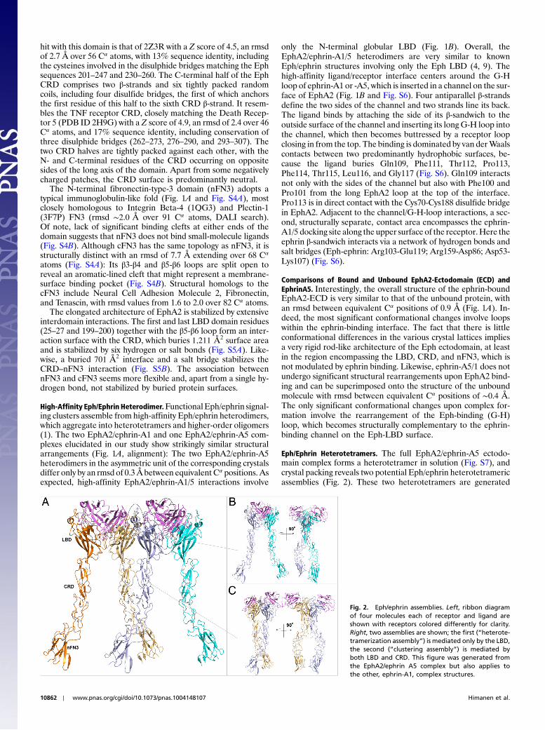
Eph/Ephrin Heterotetramers. The full EphA2/ephrin-A5 ectodo-main complex forms a heterotetramer in solution (Fig. S7), andcrystal packing reveals two potential Eph/ephrin heterotetramericassemblies (Fig. 2). These two heterotetramers are generated
Fig. 2. Eph/ephrin assemblies. Left, ribbon diagramof four molecules each of receptor and ligand areshown with receptors colored differently for clarity.Right, two assemblies are shown; the first (“heterote-tramerization assembly”) is mediated only by the LBD,the second (“clustering assembly”) is mediated byboth LBD and CRD. This figure was generated fromthe EphA2/ephrin A5 complex but also applies tothe other, ephrin-A1, complex structures.
10862 ∣ www.pnas.org/cgi/doi/10.1073/pnas.1004148107 Himanen et al.
via two distinct EphA2/EphA2 interfaces and, when combined,would generate a continuous Eph/ephrin assembly (Fig. 2A).
The first of these heterotetramers is generated by Eph-Ephand Eph-ephrin interactions that encompass only Eph residueswithin the LBD. Indeed this Eph-LBD/ephrin heterotetrameris also observed in all our complex structures (Figs. 2B and 3).Architecturally, these EphA2-LBD/ephrinA1/5 complexes aresimilar to the circular heterotetramers observed in the EphB2-
LBD/ephrin-B2 structure (10), although the precise interfacesand interactions are quite distinct. Indeed, whereas Eph-Ephcontacts are not observed in the EphB2-LBD/ephrin-B2 tetra-mer, both Eph/ephrin and Eph/Eph interactions facilitateEphA2/ephrinA1/5 tetramers (Fig. 3). Moreover, ephrin glycosy-lation units may also contribute to this interaction (Fig. 3C andPDB ID code 3MBW). The total surface area buried in theEphA2/ephrinA1/5 heterotetramers (2;088 Å2) is somewhatsmaller than the surface area buried in the EphB2/ephrin-B2 het-erotetramers (2;532 Å2). Because the presence of Eph/ephrincontacts suggests that the formation of this heterotetramericcomplex is dependent on ephrin binding, and because the archi-tecturally similar B-class assembly has been referred to as “lowaffinity Eph/ephrin heterotetramers” (1), we will also refer tothese EphA2/ephrinA1/5 and EphA2/EphA2 interfaces simplyas the “heterodimerization” interfaces. This assembly is seenin all three of our EphA2/ephrin structures, even in the absenceof the CRD domain.
The second heterotetrameric assembly (Fig. 2B) is generatedonly via Eph-Eph interactions, suggesting that its formation is notdependent on ephrin binding. In fact, these interactions areconserved in all CRD-containing structures. We will therefore re-fer to these Eph/Eph interactions and interfaces as “clustering.”The EphA2/ephrin clustering interactions involve two distinctEph/Eph interfaces—one in the LBD (Fig. 4 A and B) andone in the CRD (Fig. 4C).
The LBD-mediated clustering interface is almost entirely po-lar, involving several salt bridges and hydrogen bonds (Fig. 4B).At the center of this Eph-Eph interface Lys116 from one of theEph molecules makes a salt bridge with Glu117 and Asp104 fromthe other, whereas the side chain of Thr144 hydrogen-bondswith the main chain carbonyl of Pro147. Because the interfaceis twofold symmetric, the reverse salt bridge and hydrogen bondare also present (Fig. 4). The CRD-mediated clustering interfaceinvolves a leucine-zipper-like assembly with a large number ofvan der Waals interactions. The hydrophobic zipper is formedby residues Pro221, Leu223, Leu254, Val255, and Ile257 (Fig. 4C).Upon formation of the clustering interface, approximately850 Å2 of surface area is buried in each Eph molecule—largeenough for this interface to be considered biologically relevant.Moreover, the interaction surfaces are composed of conservedresidues across human Eph receptors, further underlying itspotential functional significance (Fig. S8).
Interestingly, both the Eph-Eph heterodimerization andclustering interfaces are present in the free Eph-ECD crystals
Fig. 3. Eph/ephrin heterotetramerization assembly. (A) Ribbon and (B) sur-face representations of the LBD-ephrin-A5 heterotetramer with each domaincolored differently. (C) Detailed stereoscopic view of the 2∶2 assembly.
Fig. 4. Clusteringassembly. (A)Ribbondisplayof theclusteringassemblywitheachEphA2moleculecoloreddifferently; (B)detailedstereoscopicviewoftheLBD-mediatedclustering interaction.Middle, ribbondiagramoftwomoleculesof theunboundEphA2—dimer in lightorangeandpalegreen. (C)DetailedviewofCRD-mediated clustering. Residues that mediate clustering are shown in stick format and labeled. Dashes represent van der Waals or hydrogen bond interactions.
Himanen et al. PNAS ∣ June 15, 2010 ∣ vol. 107 ∣ no. 24 ∣ 10863
and in all complex structures (Eph-LBD-CRD-nFN3/ephrin-A5and Eph-LBD-CRD/ephrin-A1). This observation suggests highaffinity and that the continuous Eph-ECD/Eph-ECD assembliesare formed independent of ephrin binding at high enough Ephconcentrations.
Role of the Eph/Eph Interfaces for Stability and Function of Eph/EphrinSignaling Clusters. To compare the relative contributions of recep-tor–receptor interactions within the LBD and CRD of EphA2,we designed GFP-tagged EphA2 deletion mutants lacking eitherdomain: ΔLBD is truncated between residues 28–198, and inΔCRD residues 201–325 are replaced by a Gly-Ser-Gly-Ser lin-ker. We tested these mutants functionally, by transfecting mutantor WT EphA2-GFP cDNAs into HEK293 cells and analyzedtheir capacity to support ligand-independent Eph kinase activa-tion that is induced upon transient overexpression. Immunoblotanalysis of anti-GFP immunoprecipitated receptors demon-strated markedly reduced relative phosphorylation levels of bothdeletion mutants compared to WT EphA2, confirming the invol-vement of both domains in Eph-Eph clustering (Fig. 5A). Impor-tantly, ligand-independent activation of the ΔCRD mutant wasmost strongly affected, confirming its critical role in Eph signalinginitiation as was suggested previously for EphA3 (3, 5).
To evaluate the contribution of ligand-independent clusteringto EphA2 activation, we interrogated EphA2 point mutants in acell-based EphA2 phosphorylation assay. The mutants were de-signed to substitute hydrophobic residues in the leucine-zipper-like clustering interface predicted from the crystal structure withpositively charged ones. They included Arg substitutions at posi-tions Leu223 (single mutation), Leu223 and Leu254 (doublemutation), or Leu223, Leu254, and Val255 (triple mutation).Analysis of HEK293Tcell clones stably expressing WTor mutantEphA2 by flow cytometry confirmed that all of these exogenousreceptors were expressed on the cell surface at similar levels andwere capable of ephrin-A5 binding (Fig. S9). Anti-phospho-tyrosineWestern blot analysis of ephrin-A5-Fc stimulated cells re-vealedsignificantly reducedactivationofEphA2mutant comparedto WT-EphA2: As expected, double and triple substitutionsaffected activation stronger than single substitutions (Fig. 5B).In contrast, substitutions of charged residues within the LBD re-gion of the clustering interface did not affect ephrin-inducedEphA2phosphorylation, suggesting that increased hydrophobicityof the interfacemaycompensate for lossof the salt bridgecausedbythesemutations (Fig. 5B). Together, these findings in live cells con-firm the relevance of the Eph–Eph interactions observed in thecrystal structures for the formation of functional signaling clusters.
To directly assess the effect of EphA2 point mutations onclustering via the CRD but in the absence of LBD-mediated in-teractions, we transfected HEK293Tcell clones, expressingWTorL-R-mutated EphA2 with GFP-tagged EphA2 lacking the ligand-binding domain (ΔLBD-EphA2-GFP). This “reporter” allowedus to monitor coclustering via its CRD with ephrin-A5-boundWT EphA2 or with the L-R-substitution mutants. Anti-GFPWestern blot analysis of ephrin-bound EphA2 demonstrated thatthe relative level of coprecipitated GFP-tagged reporter wasnotably reduced in cells expressing L-R-substituted EphA2receptors, indicating that CRD interface point mutations, mostprominently the triple Arg substitution, disrupt the ability forCRD-mediated clustering (Fig. 5C).
We validated the functional importance of these findings byanalyzing with confocal fluorescence microscopy in HEK293Tcell clones the recruitment of ephrin-binding-compromisedΔLBD-EphA2-GFP to full-length WT or mutant receptors.Localized recruitment and clustering of ΔLBD-EphA2-GFP toAlexa594ephrinA5-coated beads added to the cells was discern-ible from GFP fluorescence marking the outline of the beads:Thus, in cells expressing full-lengthWTEphA2, but not in control(293) cells, bead-associated GFP fluorescence confirmed a robust
Eph/Eph interaction via the intact CRD (Fig. 5D). By comparison,cells expressing triple L-R EphA2 point mutants and, to a lesserextent, double Arg substitutions revealed significantly reducedGFP fluorescence around the ephrin-coated beads (Fig. 5D),
Fig. 5. Cellular studies. (A) Immunoprecipitates from HEK293 cells trans-fected with increasing amounts of GFP-tagged WT EphA2, ΔLBD-EphA2,or ΔCRD-EphA2 were immunoblotted with α-EphA2 and α-phosphotyrosineantibodies. Densitometry quantified EphA2 phosphorylation relative toEphA2 expression, by using samples with most similar EphA2 levels (lanes2, 5, and 8). (B) Activation of WT and mutated EphA2 in HEK293 cells by pre-clustered ephrin-A5. Single ¼ L223R, double ¼ L223R;L254R, and triple ¼L223R;L254R;V255R. (C) HEK293 cells stably expressing WT or point mutatedEphA2, or control cells, were transfected with ΔLBD-EphA2-GFP (ΔLBD) andstimulated with clustered ephrinA5-Fc. Protein A sepharose pull-downs ofephrinA5-Fc associated receptors were Western blotted with α-EphA2 andα-GFP antibodies. The graph shows the amount of ΔLBD pulled down(anti-GFP blot) via association with full-length EphA2, relative to full-lengthEphA2, quantified by densitometry. (D) Parental HEK293 cells or derivedclones stably expressingWT EphA2 or EphA2 point mutants as indicated weretransfected with ΔLBD-EphA2-GFP. Alexa594-ephrinA5 conjugated Dyna-beads were added to the cells for 5 min before cultures were rinsed andfixed for microscopy. Panels show representative images of EphA2-GFPand Alexa594-ephrinA5 fluorescence, white arrows indicating beads withrecruited EphA2-GFP, yellow arrows indicating beads in contact with cellslacking recruitment. Insets show higher magnification images of boxed re-gions. The average proportion of beads in contact with cells that recruitedΔLBD-EphA2-GFP was determined for each cell line (WT EphA2, triple ordouble mutant or control cells) and is shown in the graph (�SEM).
10864 ∣ www.pnas.org/cgi/doi/10.1073/pnas.1004148107 Himanen et al.
confirming that residues 223, 254, and 255 of EphA2 mediatereceptor–receptor interactions and facilitate ephrin-independentreceptor clustering in intact cells.
Implications for Eph Signaling InitiationLigand-induced activation of receptor tyrosine kinases (RTKs) isa tightly regulated process that is not yet well understood at themolecular level. It seems likely that the different RTK familiesutilize different molecular mechanisms for coupling ligandbinding with activation of the catalytic kinase domain. The recep-tor structures reported here reveal that the extracellular region ofEph RTKs are composed of four individual structural domainsthat together fold into a unique rod-like structure. The Ephectodomain is rigid, and ligand binding does not cause significantconformational changes in any of the individual domains orsignificant structural rearrangements between them. Thus, ligand-induced conformational changes in the receptor extracellulardomain do not seem to be the molecular mechanism drivingEph signal transduction.
Our structures reveal that the Eph CRD is a unique protein-interaction/dimerization module, which cooperates with ligand-dependent clustering to mediate the assembly of continuousoligomers, a process that could also happen independently ofligand binding at high receptor concentrations. We confirmedthis concept by structure-based mutagenesis in combinationwith cell-based signaling and receptor-visualization assays, alsoexplaining the previously observed recruitment of non-ligand-bound receptors into signaling clusters (5). The presence at thecell surface of highly ordered receptor assemblies is a uniquefeature of the Eph receptors and has not been observed in anyother receptor kinase family.
Another unique characteristic of Eph/ephrin signaling is thedependence of Eph activation and downstream signalling onmembrane-attached and preculstered ephrin ligands. ContinuousEph/Eph and Eph/ephrin assemblies in our crystals therefore sug-gest that the function of ephrin ligands might be to increase localreceptor concentration so that ordered Eph/ephrin assembliescan be formed on the cell surface. Indeed, it has been shown thattreatment of cells with antibodies recognizing the Eph ectodo-main can also induce Eph receptor activation and initiation ofdownstream signaling (11).
Finally, EphA2 clustering has been associated with tissueinvasion by cancer cells. Indeed, nearly half of human breastcancers overexpressed the receptor (12). Our studies confirmthat, at high concentrations, Eph receptors could cluster indepen-dent of ligand, potentially leading to transforming phenotypes.
SummaryEphrin-dependent Eph receptor clustering and subsequentdownstream signaling cause cytoskeleton reorganization thatleads to the contact-dependent cell–cell attraction or repulsionthat is involved in tissue patterning (13). Our study reveals thestructure of the functional Eph/ephrin assemblies at the cellsurface and suggests a mechanism for Eph receptor clusteringand activation that involves bivalent homotypic interactionsbetween the LBD and CRD domains in neighboring receptors.Previously, we demonstrated that the CRD plays a critical rolein the formation of Eph/ephrin “signalosomes” (3, 5) and nowreveal the specific molecular regions involved, as well as the un-derlying structural mechanisms. Focusing on EphA2, we deter-mined a series of EphA2 ectodomain structures containing theCRD, including that of the full EphA2 extracellular region, bothalone and in complex with A-class ephrin ligands. The sameCRD-mediated EphA2 assemblies are observed under all differ-ent crystallization conditions and space groups, in both the pre-sence and absence of bound ligand. Importantly, we documentthe physiological relevance of the proposed activation mechanismby using structure-based mutagenesis in a variety of cell-basedsignaling systems, including EphA2 phosphorylation and cell-surface localization and clustering.
Plasmid construction, host-cell growth, protein purification,crystallization, structure determination, and cell-culture studiesare described in SI Methods.
Note Added in Proof.While this manuscript was under consideration, Y. Jones andcolleagues published similar structure findings (23).
ACKNOWLEDGMENTS. We thank Christine Butler for cloning plasmids, AlmaSeitova for generating recombinant baculovirus, Linda Hii and DorotheaRobev for generating EphA2 mutants, and Yehuda Goldgur for help withdata collection and analysis. This work was supported by National Institutesof Health Grants NS38486 (to D.B.N.) and GM75886 (to J.P.H.) and NationalHealth and Medical Research Council Grant 487922 (to M.L.). The NE-CATbeamlines are supported by Award RR-15301 from the National Center forResearch Resources at the National Institutes of Health. Argonne AdvancedPhoton Source use is supported by the United States Department of Energyunder Contract DE-AC02-06CH11357. The Structural Genomics Consortium isa registered charity (#1097737) that receives funds from the CanadianInstitutes for Health Research, the Canadian Foundation for Innovation,Genome Canada through the Ontario Genomics Institute, GlaxoSmithKline,Karolinska Institutet, the Knut and AliceWallenberg Foundation, the OntarioInnovation Trust, the Ontario Ministry for Research and Innovation, Merck &Co., Inc., the Novartis Research Foundation, the Swedish Agency for Innova-tion Systems, the Swedish Foundation for Strategic Research, and theWellcome Trust.
1. Himanen JP, Nikolov DB (2003) Eph signaling: A structural view. Trends Neurosci26:46–51.
2. Pasquale EB (2005) Eph receptor signalling casts a wide net on cell behaviour. Nat RevMol Cell Biol 6:462–475.
3. Lackmann M, et al. (1998) Distinct subdomains of the EphA3 receptor mediate ligandbinding and receptor dimerization. J Biol Chem 273:20228–20237.
4. Himanen JP, Saha N, Nikolov DB (2007) Cell-cell signaling via Eph receptors andephrins. Current Opin Cell Biol 19:534–542.
5. Wimmer-Kleikamp SH, Janes PW, Squire A, Bastiaens PI, Lackmann M (2004)Recruitment of Eph receptors into signaling clusters does not require ephrin contact.J Cell Biol 164:661–666.
6. Smith FM, et al. (2004) Dissecting the EphA3/Ephrin-A5 interactions using a novelfunctional mutagenesis screen. J Biol Chem 279:9522–9531.
7. Carvalho RF, et al. (2006) Silencing of EphA3 through a cis interaction with ephrinA5.Nat Neurosci 9:322–330.
8. Salaita K, et al. (2010) Restriction of receptor movement alters cellular response:Physical force sensing by EphA. Science 327:1380–1385.
9. Himanen JP, et al. (2009) Ligand recognition by A-class Eph receptors: Crystal structuresof the EphA2 ligand-binding domain and the EphA2/ephrin-A1 complex. EMBO Rep10:722–728.
10. Himanen JP, et al. (2001) Crystal structure of an Eph receptor-ephrin complex. Nature414:933–938.
11. Vearing C, et al. (2005) Concurrent binding of anti-EphA3 antibody and ephrin-A5amplifies EphA3 signaling and downstream responses: Potential as EphA3-specifictumor-targeting reagents. Cancer Res 65:6745–6754.
12. Lackmann M, Boyd AW (2008) Eph, a protein family coming of age: More confusion,insight, or complexity?. Sci Signal 1:re2.
13. Carter N, Nakamoto T, Hirai H, Hunter T (2002) EphrinA1-induced cytoskeletalre-organization requires FAK and p130(cas). Nat Cell Biol 4:565–573.
14. Otwinowski Z, Minor W (1997) Processing of X-ray diffraction data collected inoscillation mode. Method Enzymol 276:307–326.
15. McCoy AJ, et al. (2007) Phaser crystallographic software. J Appl Crystallogr 40:658–674.16. Emsley P, Cowtan K (2004) Coot: Model-building tools for molecular graphics.
Acta Crystallogr D 60:2126–2132.17. Murshudov GN, Vagin AA, Dodson EJ (1997) Refinement of macromolecular structures
by the maximum-likelihood method. Acta Crystallogr D 53:240–255.18. Brunger AT, et al. (1998) Crystallography & NMR system: A new software suite for
macromolecular structure determination. Acta Crystallogr D 54:905–921.19. Project CC (1994) The CCP4 suite: Programs for X-ray crystallography. Acta Crystallogr
D 50:760–763.20. Painter J, Merritt EA (2006) TLSMD web server for the generation of multi-group TLS
models. J Appl Crystallogr 39:109–111.21. Davis IW, Murray LW, Richardson JS, Richardson DC (2004) MOLPROBITY: Structure
validation and all-atom contact analysis for nucleic acids and their complexes. NucleicAcids Res 32:W615–W619.