37
The new insight in 0steoarthritis management Arif Soemarjono, M.D. FACSM Bandung Musculoskeletal Rehabilitation Care Jakarta FlexFree Musculoskeletal Rehabilitation Clinic

The new insight in 0steoarthritis management
Arif Soemarjono, M.D. FACSM
Bandung Musculoskeletal Rehabilitation CareJakarta FlexFree Musculoskeletal Rehabilitation Clinic

Osteoarthritis
- The most common joint disease cause disability around the world.
- In the US > 3 million people have symptomatic knee OA and half of those < 65 yrs old. (NHIS 2018)
- Impact : ADL (Activities of Daily Living), occupational, mental health, cost and societal burden, increase risk for CV disease.
Vina RE, Kwoh CK. Epidemiology of Osteoarthritis: Literature Update. Curr Opin Rheumatol. 2018 March;30(2):160-67

Osteoarthritis
• Comorbidities
The prevalence of OA increase steadily with age. As a result, disease such as CV, hypertension, diabetes and obesity are common comorbidities in patients with OA.
These comorbidities add considerably to the costs and complexity of the treatment of patients with OA.
Gabriel SE, Crowson CS, O’fallon WM. Comorbidity in arthritis. J Rheumatol 1999;26:2475-9

Osteoarthritis
Joints Commonly Affected by Osteoarthritis
- Spinal apophyseal joints- Proximal interphalangeal (PIP) joints- Distal interphalangeal (DIP) joints- Carpometacarpal (CMC) joints- First metatarsophalangeal (MTP) joint- Hip- Knee (tibiofemoral)- Patellofemoral joints
Sharma L: Local factors in osteoarthritis, Curr Opin Rheumatol 13:441-446, 2001.

Osteoarthritis risk factors
1. Older age
2. Gender; female > male
3. Genetics
4. Obesity and metabolic syndrome
5. Diet/Vitamins; Vit D, dietary fibers, soy milk
6. Bone Mass Density (BMD); high BMD> risks
7. Knee alignment; varus, valgus
8. Ligament laxity (MCL, LCL, ACL, PCL)
9. Muscle strength (quadriceps)
10. Proprioception
11. Repetitive physical activity
12. Injury
13. Surgery (meniscetomy)
14. Synovitis
15. Bone marrow, cartilage and meniscal abnormalities from radiographic findings
Vina RE,Kwoh CK. Epidemiology of Osteoarthritis: Literature update. Curr OpinRheumatol.2018March;(30):160-67

Relation of Obesity with OA
- strongest risk factors for knee OA
- less strongly associated with hip OA
- obesity not only associated with weight-bearing activities that cause cartilage breakdown but also with non-weight-bearing joints secondary to different systemic factors.
- elevated blood glucose and C-reactive protein (high sensitive assays) are associated with the risk of knee OA and its progression in women.
- the link between obesity and OA seems stronger in women than in men.
Pottie P, Presle N, Terlain B, et al.: Obesity and osteoarthritis: More complex than predicted! Ann Rheum Dis 65:1403-1405.2006
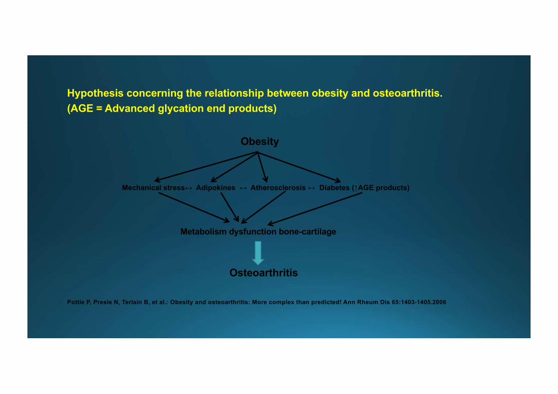
Hypothesis concerning the relationship between obesity and osteoarthritis. (AGE = Advanced glycation end products)
Obesity
Mechanical stress↔ Adipokines ↔ Atherosclerosis ↔ Diabetes (↑AGE products)
Metabolism dysfunction bone-cartilage
Osteoarthritis
Pottie P, Presle N, Terlain B, et al.: Obesity and osteoarthritis: More complex than predicted! Ann Rheum Dis 65:1403-1405.2006

OsteoarthritisCategories of OA
Primary OA- idiopathic, results in articular changes although the etiological basis for the disease is unknown.
- the most common type
Secondary OA- Caused by underlying factors that accelerate age-related degeneration of cartilage.
- These factors include : sequele of inflammatory arthritis (ankylosing spondylitis, rheumatoid arthritis, septic arthritis etc), metabolic disease (diabetes, cristal associated arthritis e.g. Gout, calcium phyrophosphate dihydrate arthropathy, pseudogout, acromegaly, hemachromatosis,etc), congenital and acquired joint surface incongruities that accelerate damage to the cartilage (developmental hip dislocation, LLD, legg-calve perthes disease, slipped femoral epiphysis etc.), hipermobility syndrome, traumatic events (major joint trauma, sports related injury, chronic joint injury, joint surgery)

OsteoarthritisDiagnostic criterias for OA
1. Clinical findings2. Clinical and radiological findings
Useful clinical features of OA that could be used in routine clinical practice:
Tissue predominantly involved : cartilageClinical features : affects hips, knees, spine, ankles, DIP, PIP, and MCP
joints, joint pain, malalignment, decreased proprioception, muscle weakness, stiffness, restricted motion, early : focal cartilaginous lesions, end stage : loss of cartilage, bone on bone.
Radiographic features : osteophytes at joint margins, joint space narrowing, subchondral sclerosis and cyst.

Osteoarthritis
1. knee pain for most days or prior months.2. crepitus on active joint motion.3. morning stiffness lasting ≤30min.4. age ≥38yr5. bony enlargement of knee on examination.
(OA if the Items are present 1,2,3,4 or 1,2,5 or 1,4,5)

Classic Radiographic Appearance of Primary OA
1. Unequal joint space narrowing
2. Eburnation (hard, white appearance) of subchondral bone
3. Osteophyte formation
4. Subchondral cysts
Kellgren-Lawrence Classification of Radiological Joint Changes in Osteoarthritis
Grade 0 : No features
Grade 1 : doubtful: minute osteophyte, doubtful significance
Grade 2 : minimal: definite osteophyte, unimpaired joint space
Grade 3 : moderate: moderate diminution of joint space
Grade 4 : severe: greatly impaired joint space with sclerosis of subchondral
bone
Kellgren JH, Lawrence JS: Radiological assesment of osteoarthrosis, Ann Rheum Dis 16:494-501, 1957.
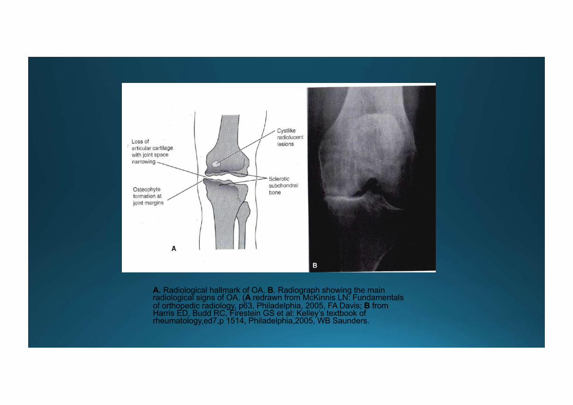
A. Radiological hallmark of OA. B. Radiograph showing the main radiological signs of OA. (A redrawn from McKinnis LN: Fundamentals of orthopedic radiology, p63, Philadelphia, 2005, FA Davis; B from Harris ED, Budd RC, Firestein GS et al: Kelley’s textbook of rheumatology,ed7,p 1514, Philadelphia,2005, WB Saunders.

Role of Bone in OsteoarthritisPresented by Clifton O. Bingham III, MD.www.medscape.org/education

OsteoarthritisOrigins of joint pain in patients with OA
Tissue Mechanism of pain
Subchondral bone Medullary hypertension, microfractures
Osteophytes Stretching of nerve endings in periosteum
Enthesis Inflammation
Joint capsule Inflammation, distension
Periarticular muscle Spasm
Synovium Inflammation
Hunter DJ, Niu J, Zhang Y, et al. Altered perfusion and venous hypertension is present in regions of bone affected by BMLs in knee OA. Osteoarthritis Cartilage.2007;15(suppl C):C171.

Hemodynamic abnormality (medullary hypertension) as the source of the pain in OA
The medullary hypertension happened in the subchondral bone.
The mechanism of medullary hypertension :
Thickened subchondral trabeculae (sclerosis) and subchondral cyst
Distortion of the medullary blood flow
Increase intraosseous pressure and intraosseous stasis (venous congestion)
Decrease oxygen tension increase CO2 tension increase lactate concentration
Pain
Hunter DJ, Niu J, Zhang Y, et al. Altered perfusion and venous hypertension is present in regions of bone affected by BMLs in knee OA. Osteoarthritis Cartilage.2007;15(suppl C):C171.

Osteophytes as the source of the pain in OA
Osteophytes
• a bony proliferations at the joint margins and in the floor of cartilage lessions are partly responsible for the pain and restriction of joint movement in OA.
• synthesize cartilage with significant amounts of type I collagen and nonaggregating proteoglycans.
• increase joint surface available for load bearing, they may contribute to some cases of regression of the early osteoarthritis cartilage changes.
• occur as a result of penetration of blood vesels into the basal layers of degenerating cartilage, or as a result of abnormal healing of stress fractures in the subchondral trabeculae near the joint margins and caused pain due to stretching of the nerve endings in periosteum.
• Biochemically are induced by the anabolic growth factor TGF-ᵦ.
Uchino M, Izumi T, Tominaga T, et al: Growth factor expression in the osteophytes of the human femoral head in OA. Clin
Ortho 377:119-125,2000.
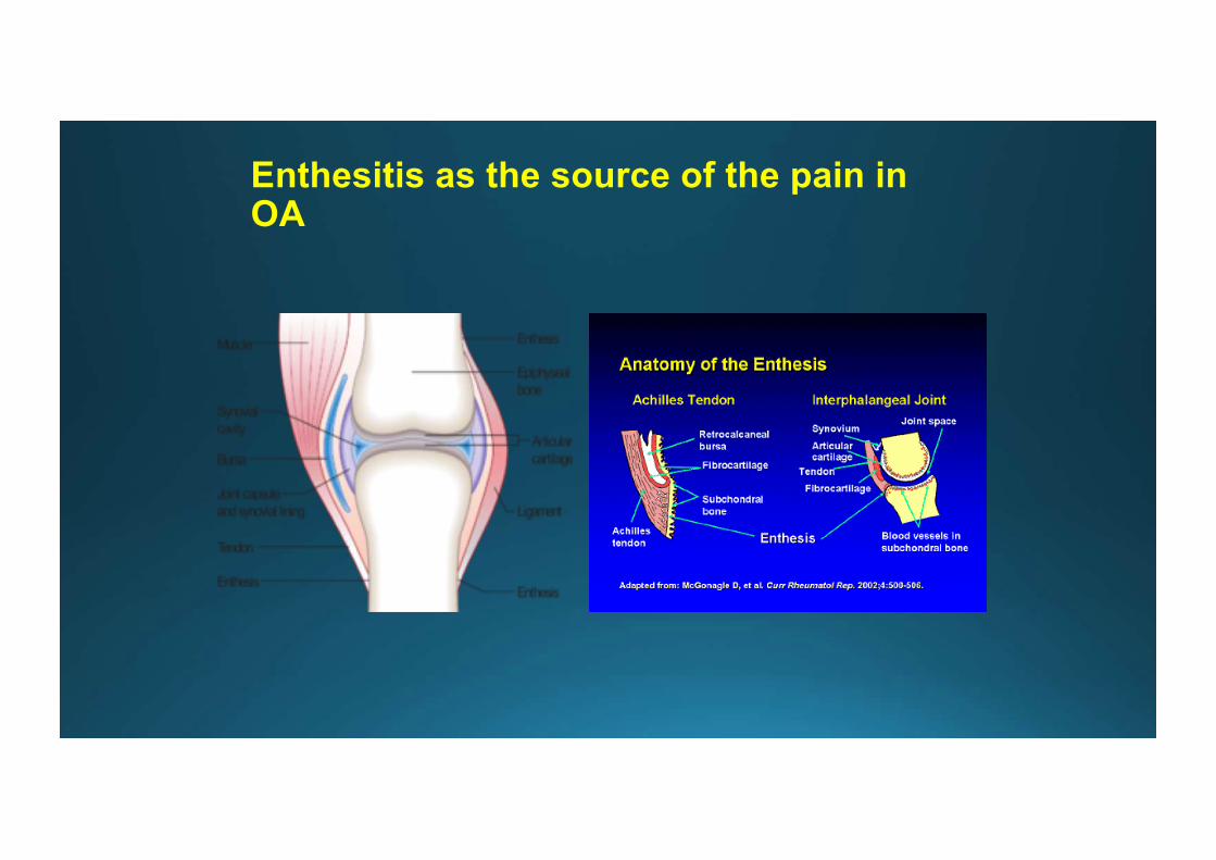
Enthesitis as the source of the pain in OA

A. Normal Appearance of achilles tendon, B. Enthesitis at achilles tendon (insertional achilles tendinosis), C. Enthesitis at achilles tendion with retrocalcaneal bursitis (insertional achilles tendinosis with bursitis retrocalcaneal). The property of arif soemarjono 2010.

DEFINING OSTEOARTHRITIS: WHAT IT IS, AND WHAT IT IS NOT OA is an organ failure
- Once though of as a cartilage or bone disease, now osteoarthritis is considered as an organ failure.
- Joint can fail because of a primary problem in any of its tissues : ligaments, meniscus, subchondral bone, periarticular muscle, synovium, nerves or articular cartilage there are many causes of OA, OA has no common pathophysiological pathway but only a final common end stage.
- The inflammatory changes in OA are secondary and are caused by particulate and soluble breakdown of cartilage and bone.
Brandt KD, Dieppe P, Radin EL. Ethiopathogenesis of osteoarthritis. Rheum Dis Clin North Am. 2008;34:531-559. Brandt KD, Dieppe P, Radin EL. Commentary: is it useful to subset “primary” osteoarthritis? A critique based on evidence regarding the ethiopathogenesis of osteoarthritis. Semin Arthritis Rheum. 2009;39:81-95.

Role of Bone in OsteoarthritisPresented by Clifton O. Bingham III, MD.www.medscape.org/education

Role of Bone in OsteoarthritisPresented by Clifton O. Bingham III, MD.www.medscape.org/education

Role of Bone in OsteoarthritisPresented by Clifton O. Bingham III, MD.www.medscape.org/education

Role of Bone in OsteoarthritisPresented by Clifton O. Bingham III, MD.www.medscape.org/education

DEFINING OSTEOARTHRITIS: WHAT IT IS, AND WHAT IT IS NOT
OA is not just a “cartilage disease”
- Poor correlation between the severity of cartilage loss (as reflected, eg, by radiographic joint-space narrowing) and theseverity of symptoms. (1)
- Patients with knee OA who underwent an osteotomy, improvement 2 years later was unrelated to whether the articular surfacewas now hyaline cartilage or fibrocartilage or was denuded relative to that seen at a baseline arthroscopy-ie, the cartilagehistology did not seem to affect the patient’s clinical status. (2)
- The loss of cartilage haven’t helped understanding of the etiopatogenesis of OA, a concern different from that of thepathogenesis of articular damage caused by cytokines, tissue-degrading enzymes, and toxic oxygen radicals / ReactiveOxygen Species (ROS).
1.Cibere J: Do we need radiographs to diagnose osteoarthritis? Best Pract Res Clin Rheumatol 20:27-38,2006.
2. Bergenudd H, Johnell O et.al. The articular cartilage after osteotomy for medial gonarthrosis:biopsies after 2 years in 19
cases. Acta Orthop Scan. 1992;63:413-416.

Proinflammatory cytokines that frequently cause articular damage in OA: (1)
- NO (Nitric Oxide)- TNF-α- IL-1
Tissue-degrading enzymes that cause articular damage in OA: (2)
� The matrix metalloproteinase (MMP) degrade proteoglycans (aggrecanases) and collagen (collagenases).
� Collagenases breakdown type II collagen (especially MMP-1, MMP-8 and MMP-28) and proteoglycans (MMP-3), aggrecanases also known as ADAMTS( A Disintegrin and Metalloproteinase with Thrombospondin Motifs) mediate aggrecan degradation in cartilage.
Reactive Oxigen Species that cause articular damage in OA: (3)
� Higher oxidative stress , either due to increased extent of lipid peroxidation or due to decreased levels of antioxidants.
� synovial cavity damage correlates with fluctuating oxygen pressure in the joint, overproduction of free radicals and lack of oxygen-processing enzymes and free radical scavenging molecules.
1.Maroudas A, Katz EP, Wachtel EJ, et al: Physiochemical properties and functional behavior of normal and osteoarthritic human
cartilage. In Schleyerbach R, Kuettner KE, Hascall VC (eds): Articular Cartilage Biochemistry. New york, Raven Press,
1986,pp311-330.
2. Kevorkian L, Young DA, Darrah C, et al: Expression profiling of metalloproteinase and their inhibitors in cartilage. Arthritis
Rheum 50:131-141,2004.
3. Maneesh M, Jayalekshmi H, Summa T, et al: Evidence for oxidative stress in osteoarthritis. Indian Journal of Clinical
Biochemistry,2005,20(1), 129-130.

OA is not ‘a degenerative joint disease’
� OA should not be considered a degenerative joint disease because the cartilage and bone cells essentially are normal and, if high levels of intra-articular stress are reduced, retain the capacity to restore the damaged tissue to normal. (1)
� OA reflects a repair process intended to contain joint damage caused by local mechanical problem. Under the appropriate conditions (correction of abnormal stress, joint motion, and establishment of source of cells), joints with OA can heal-with structural and symptomatic improvement. (1)
� Not all articular damage progresses to OA. Non progressive cartilage degeneration are asymptomatic and do not lead to joint-space narrowing or bony changes of OA on radiography. (2)
1. Radin EL, Burr DB. Hypothesis: joints can heal. Semin Arthritis Rheum. 1984;13:293-302. 2. Hirsch C. The pathogenesis of chondromalacia of the patella: a physical, histologic and chemical study. Acta Chir Scand. 1994;90(suppl 83)

OA is not merely due to aging process of joint tissues
- Although the prevalence of OA clearly increases with age and age is the major risk factor for OA, nearly one-third of knee joints of human tissue donors who were in their seventh through ninth decade and had no history of arthritis showed no evidence of OA.
- The increased prevalence of OA in older persons most likely reflects a gradual accumulation of microdamage to the joint over a lifetime rather than a direct consequence of the aging of joint tissues.
Loeser RF, Shakoor N. Aging or osteoarthritis: which is the probelm? Rheum Dis Clin North Am.2003;29:653-673.
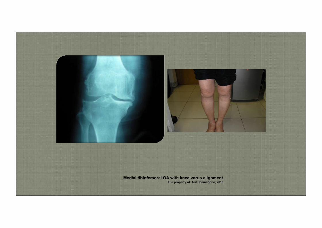
Medial tibiofemoral OA with knee varus alignment. The property of Arif Soemarjono, 2010.

� There is no known cure for osteoarthritis.
� Treatment goals include :- reducing pain- maintaining/improving joint mobility- limiting functional impairment- improving helath-related quality of life- prevention of structural progression- educating patients about the nature of the disorder andits management
(Kvien TK. The burden of osteoarthritis and the associated benefits and risks of current therapeutic
options:Optimising the management of osteoarthritis emerging therapies.EULAR satelite
symposium,2010;13-18.)

Osteoarthritis management

� Viscosupplementation is the use of intra-articular injections of high elastoviscous solutions of hyaluronan to treat arthritis.
� Intra-articular injection of hyaluronic acid now is considered as a slow-acting drug for the treatment of symptoms of OA.
� In OA both the concentration and the molecular weight of intra-articular endogenous hyaluronic acid are decreased, which reduces the viscoelasticity of synovial fluid. (molecular weight of endogenous hyaluronic acid in healthy synovial fluid is about 3-4X10 6 D)
� The rationale for intra-articular injection of hyaluronic acid :
- restored the viscoelasticity of synovial fluid- augment the flow of synovial fluid- normalize the synthesis and inhibit the degradation of endogenous hyaluronic
acid- relieve joint pain and inflammation
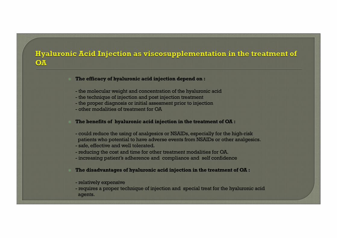
� The efficacy of hyaluronic acid injection depend on :
- the molecular weight and concentration of the hyaluronic acid- the technique of injection and post injection treatment- the proper diagnosis or initial assesment prior to injection- other modalities of treatment for OA
� The benefits of hyaluronic acid injection in the treatment of OA :
- could reduce the using of analgesics or NSAIDs, especially for the high-risk patients who potential to have adverse events from NSAIDs or other analgesics.
- safe, effective and well tolerated.- reducing the cost and time for other treatment modalities for OA. - increasing patient’s adherence and compliance and self confidence
� The disadvantages of hyaluronic acid injection in the treatment of OA :
- relatively expensive- requires a proper technique of injection and special treat for the hyaluronic acid agents.

� The contraindication for hyaluronic injection in the treatment of OA :
- systemic or local infection (including TBC)- malignancy- thrombophlebitis, severe vein insufficiency or coagulation disorders- known allergic to the hyaluronic agents or its substrate- large effusion and signs of synovitis
� The complications of hyaluronic injection :
- injection site pain- joint pain could occurs a few hours to 2 days after the injection- joint swollen, effusion, in most cases resolve spontaneously in a few days. Rest,
cold packs, analgesics, NSAIDs and occasionally corticosteroid injections or arthrocentesis to remove the effusion have been helpful.
- crystal arthritis with tissue swelling due to misplacement of the injections.
� No alteration of blood or urine tests related to hyaluronic acid injection.
� No interaction with drugs taken by the patients for coincident conditions are known, nor are they expected because intra-articular hyaluronic has no systemic pharmcological activity.
Mark E, Adams, Andre J, Lussier, et al. A Risk-Benefit Assessment of Injections of Hyalluronan and its Derivatives in the Treatment of Osteoarthritis of the Knee. Drug Safety, 2000 Aug;23(2):115-130.

� Key points that are relevant to a contemporary definition of OA :
The clinical presentation- There are multiple causes- The most common clinical presentation, garden-variety OA, may result from a wide variety of
insults to the joint.
Ethiopathogenesis- Common, garden-variety OA is initiated by a mecahnical insult to the joint- OA is a manifestation of attempts to heal the joint and to ameliorate the abnormal
biomechanics of the joint- The OA process may cause joint pain but often is succesful in leading to a stable, painless joint
Radiology- Radiographic changes of OA are extremely common in the patient population- Many persons with X-ray films showing severe changes of OA are asymptomatic- Radiographic progression of OA usually is slow and may cease completely for years.

� Mechanical abnormalities play a central role in the etiopathogenesis of the structural abnormalities and symptoms of knee OA and that if the excessive load borne by a joint with OA is redistributed, joint can heal.
� In efforts to find new OA therapies, a search for the genes that control the shape of joints or neuromuscular coordination or otherwise influence joint loading may be more fruitful than a search for genes that regulate the synthesis or breakdown of articular cartilage.
� Management of OA should consist of pharmacological and non pharmacological treatment concomitantly, could not be separated, and patient education is the first priority before commencing other treatment.

THANK YOU FOR YOUR ATTENTION


