Arl13b-regulated cilia activities are essential forpolarized radial glial scaffold formationHolden Higginbotham, University of North CarolinaJiami Guo, University of North CarolinaYukako Yokota, University of North CarolinaNicole L. Umberger, Emory UniversityChen-Ying Su, Emory UniversityJingjun Li, University of North CarolinaNisha Verma, University of North CarolinaJoshua Hirt, University of North CarolinaVladimir Ghukasyan, University of North CarolinaTamara Caspary, Emory University
Only first 10 authors above; see publication for full author list.
Journal Title: Nature NeuroscienceVolume: Volume 16, Number 8Publisher: Nature Publishing Group | 2013-08-01, Pages 1000-U44Type of Work: Article | Post-print: After Peer ReviewPublisher DOI: 10.1038/nn.3451Permanent URL: https://pid.emory.edu/ark:/25593/rscjp
Final published version: http://dx.doi.org/10.1038/nn.3451
Arl13b-regulated activities of primary cilia are essential for the formation of the polarized radial glial scaffold
Holden Higginbotham1,*, Jiami Guo1,*, Yukako Yokota1, Nicole L. Umberger2, Chen-Ying Su2, Jingjun Li1, Nisha Verma1, Joshua Hirt1, Tamara Caspary2,^, and E. S. Anton1,^
1UNC Neuroscience Center and the Department of Cell and Molecular Physiology, University of North Carolina School of Medicine, Chapel Hill, North Carolina 27599
2Department of Human Genetics, Emory University School of Medicine, Atlanta, Georgia 30322
Abstract
The construction of cerebral cortex begins with the formation of radial glia. Once formed,
polarized radial glial cells divide either symmetrically or asymmetrically to balance appropriate
production of progenitor cells and neurons. Upon birth, neurons use the processes of radial glia as
scaffolding for oriented migration. Radial glia thus provide an instructive structural matrix to
coordinate the generation and placement of distinct groups of cortical neurons in the developing
cerebral cortex. Here we show that Arl13b, a cilia-specific small GTPase mutated in Joubert
syndrome patients, is critical for the initial formation of the polarized radial progenitor scaffold.
Through developmental stage-specific deletion of Arl13b in mouse cortical progenitors, we found
that early neuroepithelial deletion of ciliary Arl13b leads to a reversal in the apical-basal polarity
of radial progenitors and aberrant neuronal placement. Arl13b modulates ciliary signaling
necessary for radial glial polarity. Our findings demonstrate that Arl13b signaling in primary cilia
is important for the initial formation of a polarized radial glial scaffold and suggest that disruption
of this process may contribute to aberrant neurodevelopment and brain abnormalities in Joubert
The cerebral cortex emerges as a result of coordinated unfolding of radial progenitor
formation, neurogenesis, neuronal migration, post-migratory neuronal differentiation and
Users may view, print, copy, download and text and data- mine the content in such documents, for the purposes of academic research, subject always to the full Conditions of use: http://www.nature.com/authors/editorial_policies/license.html#terms^Correspondence to: E. S. Anton, UNC Neuroscience Center and the Department of Cell and Molecular Physiology, The University of North Carolina School of Medicine, Chapel Hill, NC 27599, [email protected], PHONE: 919-843-6114, FAX: 919-966-1844.*Contributed equally
Author ContributionsE.A., H.H., J.G., Y.Y., and T.C. designed the experiments and supervised the project. H.H., J.G., Y.Y., N.L.U., C.S., J.L., and N.V. conducted the experiments and analyzed the data. E.A., H.H., J.G., and T.C. wrote the manuscript.
HHS Public AccessAuthor manuscriptNat Neurosci. Author manuscript; available in PMC 2014 February 01.
Published in final edited form as:Nat Neurosci. 2013 August ; 16(8): 1000–1007. doi:10.1038/nn.3451.
Author M
anuscriptA
uthor Manuscript
Author M
anuscriptA
uthor Manuscript
connectivity. The formation of polarized radial glia is the first step in the construction of
cerebral cortex1. Once formed, polarized radial glial cells can divide either symmetrically or
asymmetrically. Symmetric radial glial divisions occur primarily during the early stages of
cortical development to expand the radial glial population. Asymmetric divisions of radial
glia result in a daughter neuron and either a radial glial cell or an intermediate precursor.
Temporal regulation of the ratio of asymmetric to symmetric divisions of radial glia is
critical for balancing appropriate neuronal production with progenitor maintenance2–4. Once
born, new neurons use radial processes of radial glia as permissive and instructive
scaffolding for oriented migration towards their target locations. Polarized radial glia in
essence provide an instructive structural matrix to coordinate the generation and placement
of distinct groups of cortical neurons in the developing cerebral cortex1, 5–8.
The initial establishment of apical-basal polarity of radial glial cells is an essential step in
the orderly unfolding of cerebral cortical organization. In mice, between embryonic days 9
and 12 (E9 and E12), an undifferentiated sheet of neuroepithelial cells in the telencephalic
vesicles transforms into radial progenitors. During this transformation, the psuedostratified
neuroepithelial cells stop their interkinetic nuclear migration, which spans their entire
apical-basal axis, begin to express specific radial progenitor markers (e.g., BLBP, Glast, and
vimentin) and initiate a restricted pattern of basal-apical interkinetic nuclear movement
limited to the boundaries of the ventricular zone. Importantly, the cell soma of radial
progenitors remain apically positioned within the ventricular zone, with a long basal process
extending towards the pial surface, thus enabling the orderly generation and guidance of
new neurons in the cerebral cortex. In spite of its importance for the proper unfolding of
cortical development, little is known about what determines the initial emergence of apical-
basal polarity of the radial glial scaffold in the developing cerebral cortex.
Intriguingly, primary cilia, the microtubule-based, slender projections from cells, are always
localized to the apical region of a radial glial cell. The significance of cilia function for
cerebral cortical development and function is evident in developmental brain disorders such
as Joubert, Meckel-Gruber, orofaciodigital and Bardet-Biedl syndromes (commonly referred
to as ciliopathies), where disrupted cilia function and the resulting changes in cortical
formation may underlie cognitive deficits and mental retardation9–16. Mouse mutations that
affect ciliogenesis also lead to hydrocephalus, disruptions in neurogenesis and brain tumor
formation9, 12, 17–24. Primary cilia, unique in their ability to function as sensors and
conveyors of critical signals in a complex environment, may play a guiding role in the
establishment of apical-basal polarity of the radial glial scaffold.
To examine this hypothesis, we used a mouse genetic model in which cilia are present, but
their function is impaired. Arl13b, a small GTPase of the Arf/Arl family, mutated in Joubert
syndrome patients, is specifically localized to cilia and controls the microtubule-based,
ciliary axoneme structure10, 25. Deletion of Arl13b impairs a cilium’s ability to convey
critical extracellular signals such as Sonic hedgehog (Shh)25. Importantly, this provides us a
unique entry point for understanding the role of cilia in radial progenitor development
because, in contrast to most cilia null mutants (e.g., intraflagellar transport (IFT) mutants)
that ablate cilia formation and all related biological signalling11–16, 20, Arl13b mutants
impair but do not destroy cilia and their downstream pathways. Thus, genetic manipulation
Higginbotham et al. Page 2
Nat Neurosci. Author manuscript; available in PMC 2014 February 01.
Author M
anuscriptA
uthor Manuscript
Author M
anuscriptA
uthor Manuscript
of Arl13b function provides an efficient model to disrupt the signaling functions of cilia and
assay the resultant effect. By conditionally deleting Arl13b at distinct stages of cortical
progenitor development, we show that primary cilia activity involving Arl13b is necessary
for the initial establishment of the apical-basal polarity of the radial glial scaffold. Loss of
Arl13b and the resultant loss of cilia function lead to a reversal of the apical-basal polarity
of radial progenitors in the developing cerebral wall. As a result, the laminar organization of
neurons in cerebral cortex is drastically disrupted. These findings demonstrate that primary
cilia activity plays an essential role in the initial establishment of a properly polarized radial
glial scaffold, which is necessary for constructing a normal cerebral cortex.
Results
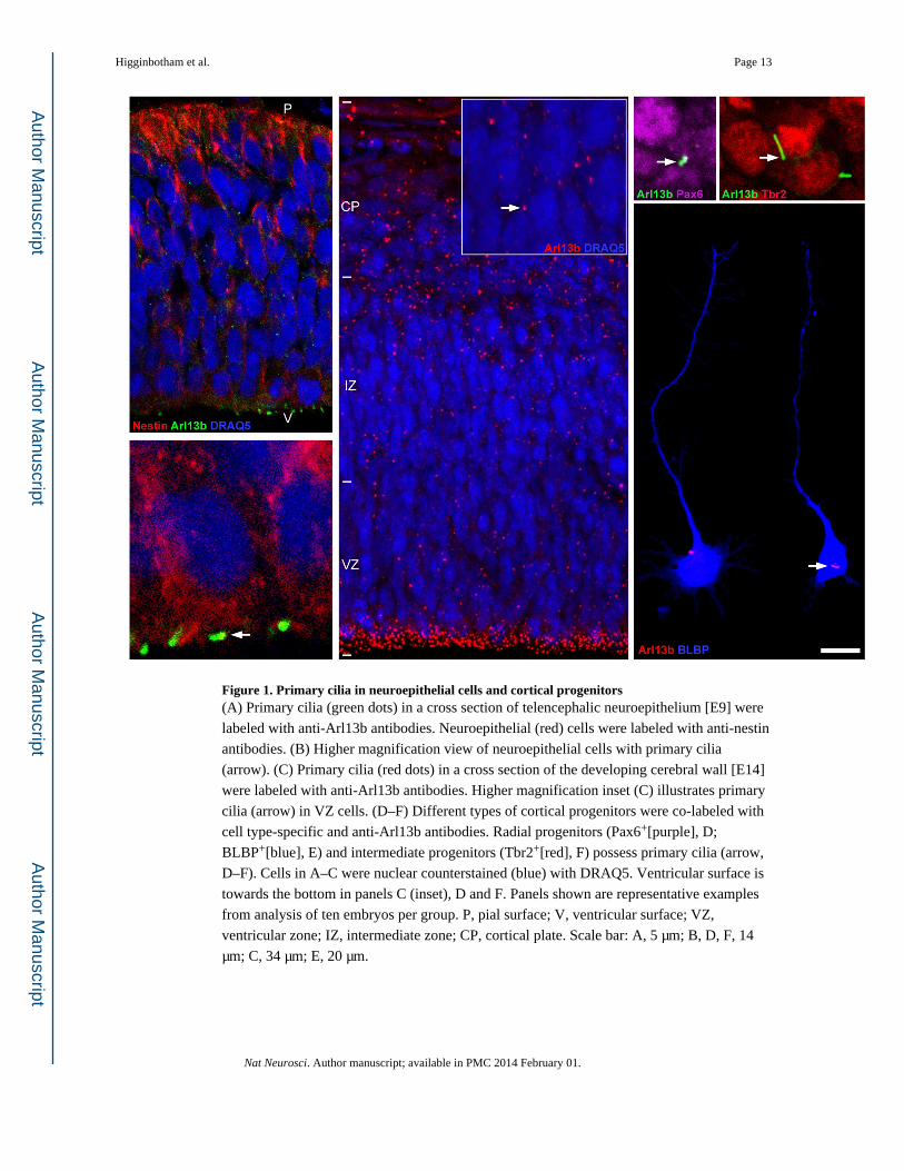
Cilia-specific expression of Arl13b in radial progenitors
To map the expression of Arl13b in neuroepithelial cells and developing radial glia, we
immunolabeled E9 telencephalic neuroepithelium (Figure 1A, B) and radial progenitors
(Figure 1C–E) from E14 telencephalon with anti-Arl13b antibody. Arl13b expression is
localized to the primary cilium of both neuroepithelial and radial progenitor cells (Figure 1
A–F). In radial progenitors, Arl13b+ cilia were always found in the apical, cell soma
domains of these cells (Figure 1E). Further, intermediate progenitors derived from radial
glial cells also display primary cilia (Figure 1D, F).
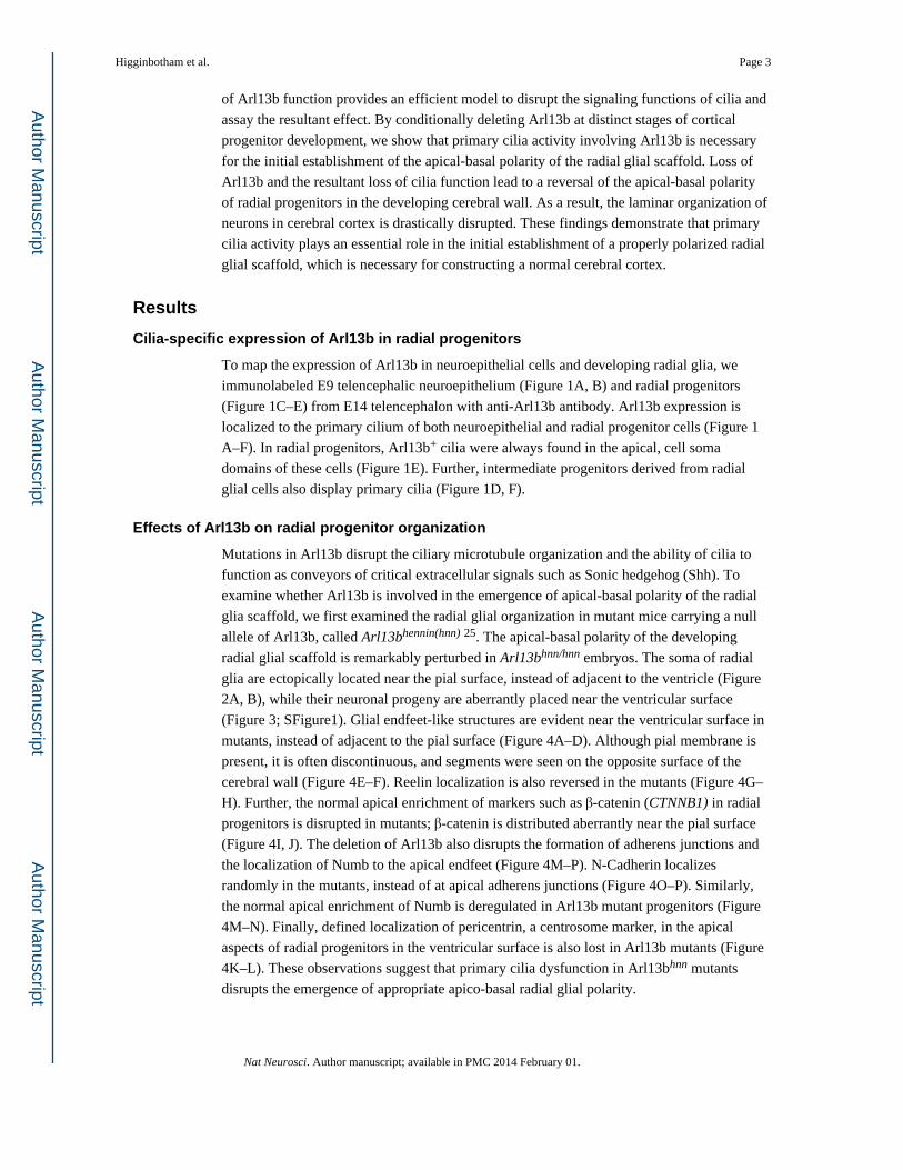
Effects of Arl13b on radial progenitor organization
Mutations in Arl13b disrupt the ciliary microtubule organization and the ability of cilia to
function as conveyors of critical extracellular signals such as Sonic hedgehog (Shh). To
examine whether Arl13b is involved in the emergence of apical-basal polarity of the radial
glia scaffold, we first examined the radial glial organization in mutant mice carrying a null
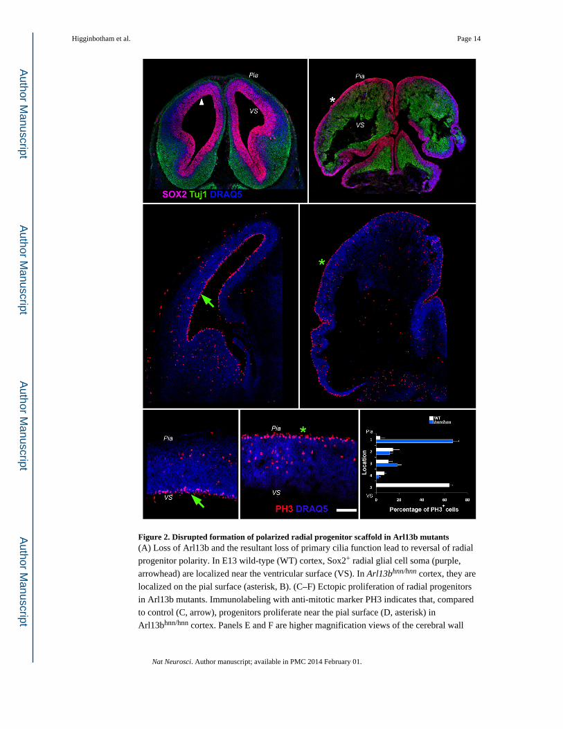
allele of Arl13b, called Arl13bhennin(hnn) 25. The apical-basal polarity of the developing
radial glial scaffold is remarkably perturbed in Arl13bhnn/hnn embryos. The soma of radial
glia are ectopically located near the pial surface, instead of adjacent to the ventricle (Figure
2A, B), while their neuronal progeny are aberrantly placed near the ventricular surface
(Figure 3; SFigure1). Glial endfeet-like structures are evident near the ventricular surface in
mutants, instead of adjacent to the pial surface (Figure 4A–D). Although pial membrane is
present, it is often discontinuous, and segments were seen on the opposite surface of the
cerebral wall (Figure 4E–F). Reelin localization is also reversed in the mutants (Figure 4G–
H). Further, the normal apical enrichment of markers such as β-catenin (CTNNB1) in radial
progenitors is disrupted in mutants; β-catenin is distributed aberrantly near the pial surface
(Figure 4I, J). The deletion of Arl13b also disrupts the formation of adherens junctions and
the localization of Numb to the apical endfeet (Figure 4M–P). N-Cadherin localizes
randomly in the mutants, instead of at apical adherens junctions (Figure 4O–P). Similarly,
the normal apical enrichment of Numb is deregulated in Arl13b mutant progenitors (Figure
4M–N). Finally, defined localization of pericentrin, a centrosome marker, in the apical
aspects of radial progenitors in the ventricular surface is also lost in Arl13b mutants (Figure
4K–L). These observations suggest that primary cilia dysfunction in Arl13bhnn mutants
disrupts the emergence of appropriate apico-basal radial glial polarity.
Higginbotham et al. Page 3
Nat Neurosci. Author manuscript; available in PMC 2014 February 01.
Author M
anuscriptA
uthor Manuscript
Author M
anuscriptA
uthor Manuscript
Neuroepithelial cells undergo interkinetic nuclear movement (INM) throughout the apico-
basal extent of the neuroepithelial layer as they transform into polarized radial progenitors.
Normal patterns of INM result in the apical localization of PH3+, M-phase radial progenitors
in the wild-type telencephalon. The significant misplacement of PH3+ progenitors towards
the basal surface in Arl13b mutant telencephalon (Figure 2C–F) indicates that disrupted
INM may have contributed to the aberrant radial progenitor organization in Arl13b mutants.
Arl13b–deficient radial progenitors ectopically divide at or near the pial surface, instead of
adjacent to the ventricular surface (Figure 2C–F).
Because radial progenitors actively proliferate and support the migration of their neuronal
progeny, we asked how the disrupted polarity in Arl13b mutants might affect the dual
function of radial glia as neural precursors and migratory guides of newly generated
neurons. As new neurons resulting from radial glial division are guided to distinct laminar
positions in the developing cortex, we next examined neuronal migration and positioning in
Arl13bhnn mice. Analysis of newly generated cortical neurons with different neuron-specific
markers indicates profound disruption of neuronal organization (Figure 3, SFigure 1). In
control cortices, immunolabeling of different newly generated cortical neurons with Tuj-1,
anti-calretinin, anti-reelin or anti-Tbr1 antibodies indicates that they migrate normally and
are positioned appropriately within the emerging cortical plate; however, neuronal migration
to distinct laminar positions is completely disrupted in the mutants (Figure 3, SFigure 1).
Arl13b mutants have enlarged lateral ventricles, but a thin, distinguishable midline is present
(SFigure 1). Corresponding to the reversed apical-basal polarity of radial progenitors,
mutant neurons are ectopically placed near the ventricular surface. Often these neurons form
tuber-like clusters within the developing cerebral wall (Figure 3B–F). The disrupted polarity
in Arl13b mutant radial glial scaffold likely affected the subsequent migration and
positioning of these cortical neurons. Axial MRI scans through the embryos further illustrate
the profound perturbance in normal cerebral cortical structural organization resulting from
Arl13b deletion (SFigure 2, Movies 1, 2).
Primary cilia affects the development of radial glia
To further define the role of primary cilia function in the formation and function of the
polarized radial glial scaffold, we conditionally inactivated Arl13b at three developmental
stages: just prior to the formation of radial glia from neuroepithelial cells (at E9, using
Foxg1-Cre); when the radial glial population is actively expanding (at E10.5, using Nestin-
Cre); and when radial progenitors are actively differentiating to generate and guide new
neurons (at E13.5, using hGFAP-Cre [SFigure 3A]). To do this, we crossed the Arl13b
floxed allele (Arl13blox/lox) to Arl13b+/hnn mice heterozygous for the respective Cre
allele26–28. We verified the loss of Arl13b signal in progenitors following Cre-mediated
recombination in these lines on a cell-by-cell basis using immunolabeling with an anti-
Arl13b antibody (SFigure 3B–C E–F, H–I,). We further confirmed this by immunoblot of
whole brain lysates showing a reduction in Arl13b following Cre-mediated recombination of
Arl13b floxed alleles (SFigure 3D, G, J, K). In Arl13blox/hnn; Foxg1-Cre mice, where
deletion is initiated at E9 in the neuroepithelium, we found abnormal organization of the
radial glial scaffold. Radial progenitors formed, but their apical-basal polarity was lost
(Figure 5A–B). As a result, neuronal positioning and layer formation is also perturbed in
Higginbotham et al. Page 4
Nat Neurosci. Author manuscript; available in PMC 2014 February 01.
Author M
anuscriptA
uthor Manuscript
Author M
anuscriptA
uthor Manuscript
these mutants (Figure 5C–H). Further, other brain structures, such as the hippocampus,
where radial progenitor polarity is essential for its normal formation, are severely malformed
following the disruption of primary cilia function in neuroepithelial cells (Figure 5I–J).
To examine whether Arl13b–regulated primary cilia activity after the formation of the
polarized radial glial scaffold is necessary for the ongoing maintenance of radial glial
polarity, we used either Nestin-Cre or hGFAP-Cre to inactivate Arl13b. Nestin-Cre initiates
deletion at E10.5, soon after the initial establishment of radial progenitors, whereas hGFAP-
Cre begins to delete at E13.5, well after the formation of polarized radial glial scaffold. We
found that radial progenitor organization is normal in both Arl13blox/hnn; Nestin-Cre and
Arl13blox/hnn; hGFAP Cre brains (Figure 5K–N), indicating that disruption of primary cilia
function, via the deletion of Arl13b, is not necessary to maintain the polarized organization
of radial progenitors once it formed.
To further confirm the requirement for Arl13b in the initial formation and organization of
radial glial scaffold, we combined a tamoxifen-inducible Cre line, CAGG-CreERTM, with the
Arl13b floxed allele and injected the pregnant dams with tamoxifen to control the timing of
Arl13b deletion in the embryos29. We deleted Arl13b from E6.5 and analyzed the
telencephalic neuroepithelium at E9.5 (SFigure 4C–G). E6.5 injection deletes Arl13b in
neuroepithelial cells prior to the formation of radial progenitors. Deletion of Arl13b was
confirmed as before with immunohistochemical and immunoblot analysis (SFigure 4E–F, I).
Early loss of Arl13b did not affect neuroepithelial organization at E9.5 (SFigure 4).
Consistently, telencephalic neuroepithelial organization in E9.5 Arl13bhnn/hnn mutants is
similar to wild-type (SFigure 4A, B). To examine the effect of Arl13b deletion after the
formation of radial glia, we deleted Arl13b from E10.5 and analyzed the radial glial scaffold
at E12.5 (SFigure 4G, H, J). As with the observations made earlier with different Nestin and
hGFAP Cre lines, deletion of Arl13b after the formation of radial glia did not affect the
organization of the radial glial scaffold. Lastly, we also deleted Arl13b from E6.5 and
analyzed radial glial scaffold at E14. This early deletion of Arl13b in neuroepithelium, lead
to the disruption of polarized radial glial scaffold formation and cortical neuronal
organization, similar to Arl13bhnn/hnn mutants (SFigure 4M–U). Radial glial polarity is
reversed and consequently neuronal laminar formation is disrupted and neurons formed
ectopic tuber- like clusters in the cerebral wall (SFigure 4M–R). Together, the
neuroepithelial/radial progenitor-specific inactivation of Arl13b at different developmental
stages suggests that Arl13b–mediated primary cilia function is critical for the initial
establishment of the apical-basal polarity in cortical radial progenitors.
Altered dynamics of cilia in Arl13b mutant progenitors
To assess the cell biological basis of the disrupted apico-basal polarity of radial glial
scaffolding in Arl13bhnn mutants, we first performed real-time observations of primary cilia
activity in radial progenitors in the embryonic cortex. We labeled primary cilia in radial
progenitors with 5Htr6-GFP electroporation and live-imaged GFP+ cilia activity in radial
progenitors. These live-imaging observations demonstrate that primary cilia in wild-type
progenitors extend, retract, branch and remodel in the ventricular zone (Figure 6A, C,
SFigure 5, Movies 3, 4 and 5). Further, inter-cilia contacts between adjacent progenitors are
Higginbotham et al. Page 5
Nat Neurosci. Author manuscript; available in PMC 2014 February 01.
Author M
anuscriptA
uthor Manuscript
Author M
anuscriptA
uthor Manuscript
also evident (Movie 3). In contrast, primary cilia of Arl13b–deficient radial progenitors are
shorter and do not actively remodel (Figure 6B, C; Movie 6). To complement the real-time
imaging of radial progenitor primary cilia activity in the ventricular zone, we also measured
cilia orientation and length in fixed tissue. In wild-type radial progenitors, the average length
of primary cilia is 3.45 ± 0.27 µm (n=30), and at any point in time a significant majority of
them were oriented towards the ventricular surface (SFigure 5E–F). In contrast, Arl13b
electroporation, cortices were removed from the embryos, coronally sectioned (200 µm) in a
vibratome (Leica VT 1000S), mounted on nucleopore membrane filters and maintained in
DMEM/10% FBS at 37°C/5% CO2. In some experiments, dissected halves of embryonic
cortices were whole-bath electroporated with specific DNA mix prior to slicing as described
in Yokota et al. 2007. Within 24 hours, XFP+ primary cilia in the medio-dorsal region of the
cerebral cortex were repeatedly imaged at 5–10 minute intervals using an Olympus inverted
microscope attached to a FluoView confocal laser scanning system and a live-cell
incubation chamber. Changes in cilia length, angle of tip rotation and number of branching
events (branching index) were measured during one hour of observation.
Statistical analysis
Statistical analyses used for each experiment are provided in the figure legends or text.
GraphPad or Excel was used for data analysis. Two-tailed Student’s t-test and One way
ANOVA with Tukey-Kramer multiple comparison test were performed using GraphPad.
Data distribution was assumed to be normal. Data were collected and processed blindly. No
statistical methods were used to predetermine sample size. But sample sizes are similar to
those described in previous related publications36, 38, 43–45.
Supplementary Material
Refer to Web version on PubMed Central for supplementary material.
Acknowledgements
This research was supported by NIH grants MH060929 to E.S.A. and NS056380 to T.C., a NARSAD Young Investigator Award to H.H., a NIH predoctoral training grant to N.L.U. T32GM008490 and by the confocal imaging core of an NINDS institutional center core grant. We thank A- S. Lamantia, L. Pevny, M. Deshmukh and W. Snider for helpful comments and C. T. Strauss for editing.
References
1. Rakic P. Specification of cerebral cortical areas. Science. 1988; 241:170–176. [PubMed: 3291116]
2. Miyata T, Kawaguchi A, Okano H, Ogawa M. Asymmetric inheritance of radial glial fibers by cortical neurons. Neuron. 2001; 31:727–741. [PubMed: 11567613]
3. Noctor SC, Martinez-Cerdeno V, Ivic L, Kriegstein AR. Cortical neurons arise in symmetric and asymmetric division zones and migrate through specific phases. Nat Neurosci. 2004; 7:136–144. [PubMed: 14703572]
4. Gotz M, Huttner WB. The cell biology of neurogenesis. Nat Rev Mol Cell Biol. 2005; 6:777–788. [PubMed: 16314867]
5. Manzini MC, Walsh CA. What disorders of cortical development tell us about the cortex: one plus one does not always make two. Curr Opin Genet Dev. 2011; 21:333–339. [PubMed: 21288712]
6. Tabata H, Kanatani S, Nakajima K. Differences of migratory behavior between direct progeny of apical progenitors and basal progenitors in the developing cerebral cortex. Cereb Cortex. 2009; 19:2092–2105. [PubMed: 19150920]
7. Valiente M, Marin O. Neuronal migration mechanisms in development and disease. Curr Opin Neurobiol. 2010; 20:68–78. [PubMed: 20053546]
8. Yokota Y. Radial glial dependent and independent dynamics of interneuronal migration in the developing cerebral cortex. PLoS ONE. 2007; 2:e794. [PubMed: 17726524]
9. Oh EC, Katsanis N. Cilia in vertebrate development and disease. Development. 2012; 139:443–448. [PubMed: 22223675]
Higginbotham et al. Page 10
Nat Neurosci. Author manuscript; available in PMC 2014 February 01.
Author M
anuscriptA
uthor Manuscript
Author M
anuscriptA
uthor Manuscript
10. Cantagrel V, et al. Mutations in the cilia gene ARL13B lead to the classical form of Joubert syndrome. Am J Hum Genet. 2008; 83:170–179. [PubMed: 18674751]
11. Eggenschwiler JT, Anderson KV. Cilia and developmental signaling. Annu Rev Cell Dev Biol. 2007; 23:345–373. [PubMed: 17506691]
12. Hildebrandt F, Benzing T, Katsanis N. Ciliopathies. N Engl J Med. 2011; 364:1533–1543. [PubMed: 21506742]
13. Lancaster MA, Gleeson JG. The primary cilium as a cellular signaling center: lessons from disease. Curr Opin Genet Dev. 2009; 19:220–229. [PubMed: 19477114]
14. Louvi A, Grove EA. Cilia in the CNS: The Quiet Organelle Claims Center Stage. Neuron. 2011; 69:1046–1060. [PubMed: 21435552]
15. Reiter JF. A cilium is not a cilium is not a cilium: signaling contributes to ciliary morphological diversity. Dev Cell. 2008; 14:635–636. [PubMed: 18477443]
16. Scholey JM, Anderson KV. Intraflagellar transport and cilium-based signaling. Cell. 2006; 125:439–442. [PubMed: 16678091]
17. Tissir F, et al. Lack of cadherins Celsr2 and Celsr3 impairs ependymal ciliogenesis, leading to fatal hydrocephalus. Nat Neurosci. 2010; 13:700–707. [PubMed: 20473291]
18. Han YG, et al. Dual and opposing roles of primary cilia in medulloblastoma development. Nat Med. 2009; 15:1062–1065. [PubMed: 19701203]
19. Wong SY, et al. Primary cilia can both mediate and suppress Hedgehog pathway-dependent tumorigenesis. Nat Med. 2009; 15:1055–1061. [PubMed: 19701205]
20. Han YG, Alvarez-Buylla A. Role of primary cilia in brain development and cancer. Curr Opin Neurobiol. 2010; 20:58–67. [PubMed: 20080044]
21. Wilson SL, Wilson JP, Wang C, Wang B, McConnell SK. Primary cilia and Gli3 activity regulate cerebral cortical size. Dev Neurobiol. 2011
22. Breunig JJ, et al. Primary cilia regulate hippocampal neurogenesis by mediating sonic hedgehog signaling. Proc Natl Acad Sci U S A. 2008; 105:13127–13132. [PubMed: 18728187]
23. Amador-Arjona A, et al. Primary cilia regulate proliferation of amplifying progenitors in adult hippocampus: implications for learning and memory. J Neurosci. 2011; 31:9933–9944. [PubMed: 21734285]
24. Willaredt MA, et al. A crucial role for primary cilia in cortical morphogenesis. J Neurosci. 2008; 28:12887–12900. [PubMed: 19036983]
25. Caspary T, Larkins CE, Anderson KV. The graded response to Sonic Hedgehog depends on cilia architecture. Dev Cell. 2007; 12:767–778. [PubMed: 17488627]
26. Hebert JM, McConnell SK. Targeting of cre to the Foxg1 (BF-1) locus mediates loxP recombination in the telencephalon and other developing head structures. Dev Biol. 2000; 222:296–306. [PubMed: 10837119]
27. Tronche F, et al. Disruption of the glucocorticoid receptor gene in the nervous system results in reduced anxiety. Nat Genet. 1999; 23:99–103. [PubMed: 10471508]
28. Zhuo L, et al. hGFAP-cre transgenic mice for manipulation of glial and neuronal function in vivo. Genesis. 2001; 31:85–94. [PubMed: 11668683]
29. Su C-Y, Bay SN, Mariani LE, Hillman MJ, Caspary T. Temporal deletion of Arl13b reveals that a mispatterned neural tube corrects cell fate over time. Development. 2012; 122(139):2760–2768.
30. Mukhopadhyay S, Lu Y, Shaham S, Sengupta P. Sensory signaling-dependent remodeling of olfactory cilia architecture in C. elegans. Dev Cell. 2008; 14:762–774. [PubMed: 18477458]
31. Kim J, et al. Functional genomic screen for modulators of ciliogenesis and cilium length. Nature. 2010; 464:1048–1051. [PubMed: 20393563]
32. Lehtinen MK, et al. The cerebrospinal fluid provides a proliferative niche for neural progenitor cells. Neuron. 2011; 69:893–905. [PubMed: 21382550]
33. Zhu D, Shi S, Wang H, Liao K. Growth arrest induces primary-cilium formation and sensitizes IGF-1-receptor signaling during differentiation induction of 3T3-L1 preadipocytes. J Cell Sci. 2009; 122:2760–2768. [PubMed: 19596798]
34. Christensen ST, Clement CA, Satir P, Pedersen LB. Primary cilia and coordination of receptor tyrosine kinase signalling. J. Pathol. 2012; 226:172–184. [PubMed: 21956154]
Higginbotham et al. Page 11
Nat Neurosci. Author manuscript; available in PMC 2014 February 01.
Author M
anuscriptA
uthor Manuscript
Author M
anuscriptA
uthor Manuscript
35. Sawamoto K, et al. New neurons follow the flow of cerebrospinal fluid in the adult brain. Science. 2006; 311:629–632. [PubMed: 16410488]
36. Higginbotham H, Eom T, Mariani LE, Bachleda A, Hirt J, Gukassyan V, Cusack CL, Lai C, Caspary T, Anton ES. Arl13b in primary cilia regulates the migration and placement of interneurons in the developing cerebral cortex. Dev Cell. 2012; 122:2760–2768.
37. Cevik S, et al. Joubert syndrome Arl13b functions at ciliary membranes and stabilizes protein transport in Caenorhabditis elegans. J Cell Biol. 2010; 188:953–969. [PubMed: 20231383]
38. Horner VL, Caspary T. Disrupted dorsal neural tube BMP signaling in the cilia mutant Arl13b hnn stems from abnormal Shh signaling. Dev Biol. 2011; 355:43–54. [PubMed: 21539826]
39. Wilsch-Brauninger M, Peters J, Paridaen JT, Huttner WB. Basolateral rather than apical primary cilia on neuroepithelial cells committed to delamination. Development. 2012; 139:95–105. [PubMed: 22096071]
40. Sahara S, O'Leary DD. Fgf10 regulates transition period of cortical stem cell differentiation to radial glia controlling generation of neurons and basal progenitors. Neuron. 2009; 63:48–62. [PubMed: 19607792]
41. Siegenthaler JA, Pleasure SJ. We have got you 'covered': how the meninges control brain development. Curr Opin Genet Dev. 2011; 21:249–55. [PubMed: 21251809]
42. Brancati F, Dallapiccola B, Valente EM. Joubert syndrome and related disorders. Orphanet Journal of Rare Diseases. 2010; 5:20. [PubMed: 20615230]
43. Yokota Y, Ring C, Cheung R, Pevny L, Anton ES. Nap1-regulated neuronal cytoskeletal dynamics is essential for the final differentiation of neurons in cerebral cortex. Neuron. 2007; 54:429–445. [PubMed: 17481396]
44. Schmid RS, et al. Neuregulin 1-erbB2 signaling is required for the establishment of radial glia and their transformation into astrocytes in cerebral cortex. Proc Natl Acad Sci U S A. 2003; 100:4251–4256. [PubMed: 12649319]
45. Yokota Y, Kim W-Y, Chen Y, Wang X, Stanco A, Komuro Y, Snider WD, Anton ES. The adenomatous polyposis coli protein is an essential regulator of radial glial polarity and construction of the cerebral cortex. Neuron. 2009; 61:42–56. [PubMed: 19146812]
46. Berbari NF, Johnson AD, Lewis JS, Askwith CC, Mykytyn K. Identification of ciliary localization sequences within the third intracellular loop of G protein-coupled receptors. Mol Biol Cell. 2008; 19:1540–1547. [PubMed: 18256283]
Higginbotham et al. Page 12
Nat Neurosci. Author manuscript; available in PMC 2014 February 01.
Author M
anuscriptA
uthor Manuscript
Author M
anuscriptA
uthor Manuscript
Figure 1. Primary cilia in neuroepithelial cells and cortical progenitors(A) Primary cilia (green dots) in a cross section of telencephalic neuroepithelium [E9] were
labeled with anti-Arl13b antibodies. Neuroepithelial (red) cells were labeled with anti-nestin
antibodies. (B) Higher magnification view of neuroepithelial cells with primary cilia
(arrow). (C) Primary cilia (red dots) in a cross section of the developing cerebral wall [E14]
were labeled with anti-Arl13b antibodies. Higher magnification inset (C) illustrates primary
cilia (arrow) in VZ cells. (D–F) Different types of cortical progenitors were co-labeled with
cell type-specific and anti-Arl13b antibodies. Radial progenitors (Pax6+[purple], D;
D–F). Cells in A–C were nuclear counterstained (blue) with DRAQ5. Ventricular surface is
towards the bottom in panels C (inset), D and F. Panels shown are representative examples
from analysis of ten embryos per group. P, pial surface; V, ventricular surface; VZ,
ventricular zone; IZ, intermediate zone; CP, cortical plate. Scale bar: A, 5 µm; B, D, F, 14
µm; C, 34 µm; E, 20 µm.
Higginbotham et al. Page 13
Nat Neurosci. Author manuscript; available in PMC 2014 February 01.
Author M
anuscriptA
uthor Manuscript
Author M
anuscriptA
uthor Manuscript
Figure 2. Disrupted formation of polarized radial progenitor scaffold in Arl13b mutants(A) Loss of Arl13b and the resultant loss of primary cilia function lead to reversal of radial
Nat Neurosci. Author manuscript; available in PMC 2014 February 01.
Author M
anuscriptA
uthor Manuscript
Author M
anuscriptA
uthor Manuscript
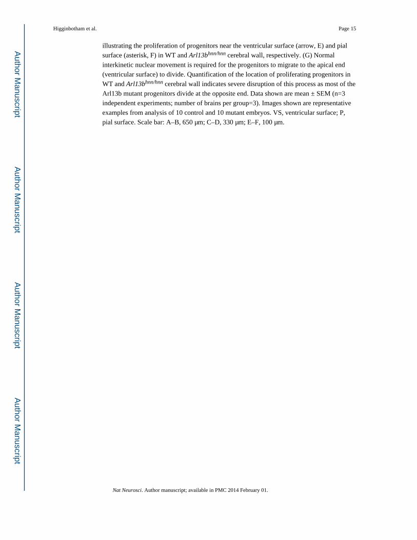
Figure 3. Disrupted cortical layer formation in Arl13b mutants(A–D) The reversal of radial progenitor polarity affects neuronal migration and layer
formation. Instead of the layer-like organization of different types of cortical neurons (red
[Calretinin+]; green [Tuj-1+]) in E13 wild-type cortex (A), neurons in Arl13bhnn/hnn cortex
(E13) migrate aberrantly and are placed ectopically (B–D). Neurons are frequently found
near the ventricular surface (arrowheads, C, D) away from the cortical plate and often form
tuber-like clusters (asterisk, B and C). (E–F) Co-immunolabeling of E13 WT (E) and mutant
(F) cortices with radial progenitor-specific RC2 antibodies (red) and early generated, deeper
Higginbotham et al. Page 16
Nat Neurosci. Author manuscript; available in PMC 2014 February 01.
Author M
anuscriptA
uthor Manuscript
Author M
anuscriptA
uthor Manuscript
layer neuronal marker Tbr1 (green) further illustrates the disrupted radial progenitor scaffold
formation (arrowhead, F) and ectopic neuronal placement (asterisk, F) in Arl13b mutant
cortex (F). Sections in A–D were nuclear counterstained (blue) with DRAQ5. Pial (P) and
ventricular surface (V) are indicated. Images shown are representative examples from
analysis of 10 control and 10 mutant embryos. Scale bar: A–D, 275 µm; E–F, 130 µm.
Higginbotham et al. Page 17
Nat Neurosci. Author manuscript; available in PMC 2014 February 01.
Author M
anuscriptA
uthor Manuscript
Author M
anuscriptA
uthor Manuscript
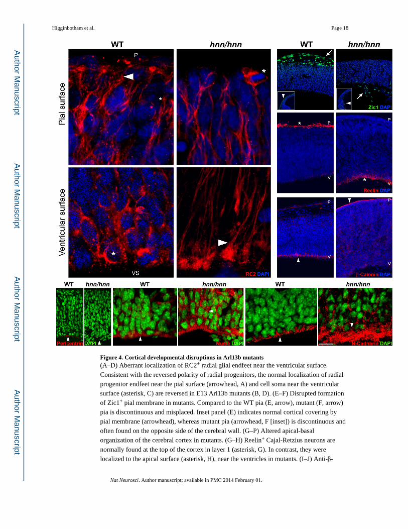
Figure 4. Cortical developmental disruptions in Arl13b mutants(A–D) Aberrant localization of RC2+ radial glial endfeet near the ventricular surface.
Consistent with the reversed polarity of radial progenitors, the normal localization of radial
progenitor endfeet near the pial surface (arrowhead, A) and cell soma near the ventricular
surface (asterisk, C) are reversed in E13 Arl13b mutants (B, D). (E–F) Disrupted formation
of Zic1+ pial membrane in mutants. Compared to the WT pia (E, arrow), mutant (F, arrow)
pia is discontinuous and misplaced. Inset panel (E) indicates normal cortical covering by
pial membrane (arrowhead), whereas mutant pia (arrowhead, F [inset]) is discontinuous and
often found on the opposite side of the cerebral wall. (G–P) Altered apical-basal
organization of the cerebral cortex in mutants. (G–H) Reelin+ Cajal-Retzius neurons are
normally found at the top of the cortex in layer 1 (asterisk, G). In contrast, they were
localized to the apical surface (asterisk, H), near the ventricles in mutants. (I–J) Anti-β-
Higginbotham et al. Page 18
Nat Neurosci. Author manuscript; available in PMC 2014 February 01.
Author M
anuscriptA
uthor Manuscript
Author M
anuscriptA
uthor Manuscript
catenin prominently labels the apical surface of progenitors (arrowhead, I), adjacent to the
ventricles in WT cortex. In contrast, β-catenin labeling is evident in the opposite, basal
surface (arrowhead, J) in mutants. Further, apical localizations of pericentrin (K), Numb (M)
and N-Cadherin (O) are disrupted in mutants (L, N, P; compare arrowheads). P, pial surface;
V, ventricular surface. Images are representative examples from analysis of 7 control and 7
mutant embryos. Scale bar: A–D, 17 µm; E–F, 110 µm; G, I, 190 µm; H, J, 300 µm; K–L, 60
µm; M–P, 30 µm.
Higginbotham et al. Page 19
Nat Neurosci. Author manuscript; available in PMC 2014 February 01.
Author M
anuscriptA
uthor Manuscript
Author M
anuscriptA
uthor Manuscript
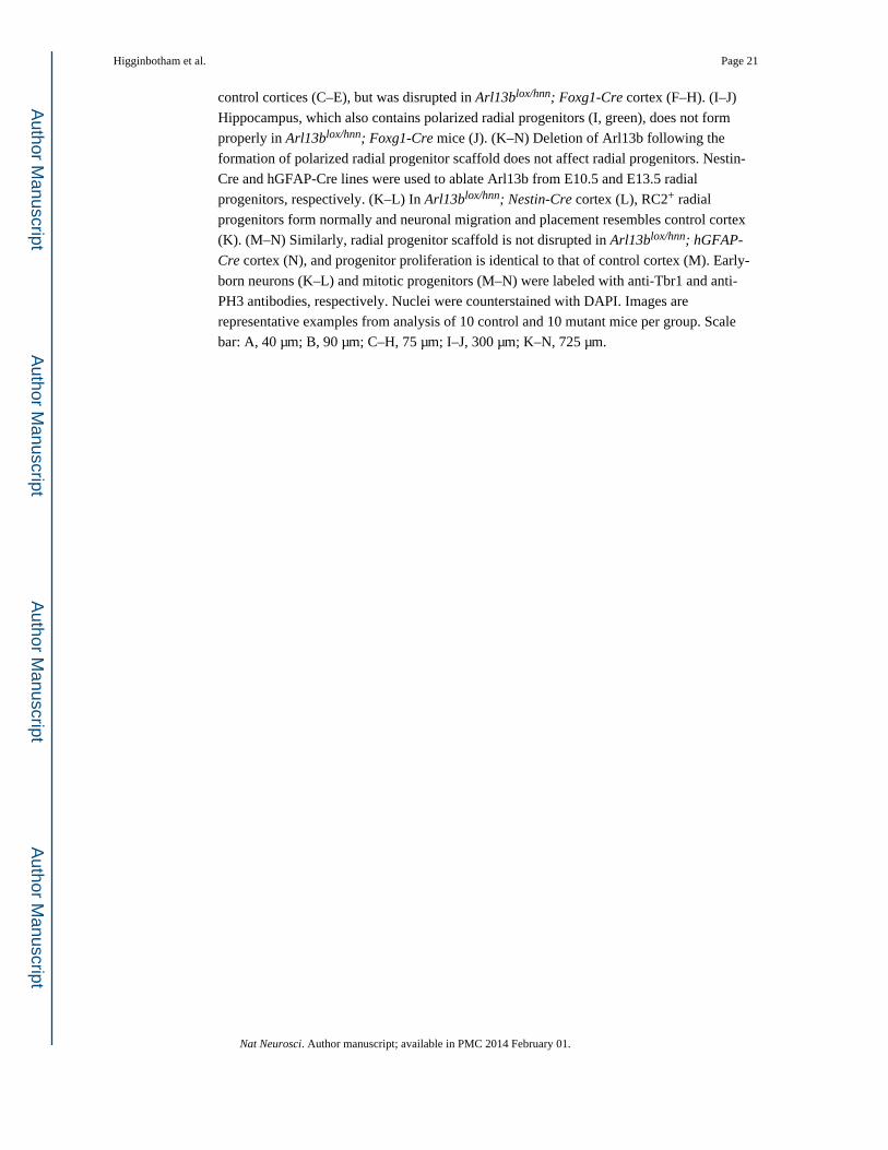
Figure 5. Arl13b deletion in neuroepithelial cells disrupts radial progenitor scaffold organization and laminar organization of neurons in cerebral cortex(A–B) Normal radial progenitor scaffold (RC2+) and progenitor proliferation (PH3+) in E13
control cerebral wall. (B) Loss of polarized radial progenitor organization and ectopic
proliferation in E13 Arl13blox/hnn; Foxg1-Cre cortex following deletion of Arl13b in
neuroepithelial cells. (C–H) Deeper and upper-layer neurons in E16 cortices from control
and Arl13blox/hnn; Foxg1-Cre mice were labeled with anti-Tbr1, -Ctip2 (Bcl11b) and -Cux1
antibodies. Characteristic laminar organization of different classes of neurons was evident in
Higginbotham et al. Page 20
Nat Neurosci. Author manuscript; available in PMC 2014 February 01.
Author M
anuscriptA
uthor Manuscript
Author M
anuscriptA
uthor Manuscript
control cortices (C–E), but was disrupted in Arl13blox/hnn; Foxg1-Cre cortex (F–H). (I–J)
Hippocampus, which also contains polarized radial progenitors (I, green), does not form
properly in Arl13blox/hnn; Foxg1-Cre mice (J). (K–N) Deletion of Arl13b following the
formation of polarized radial progenitor scaffold does not affect radial progenitors. Nestin-
Cre and hGFAP-Cre lines were used to ablate Arl13b from E10.5 and E13.5 radial
progenitors, respectively. (K–L) In Arl13blox/hnn; Nestin-Cre cortex (L), RC2+ radial
progenitors form normally and neuronal migration and placement resembles control cortex
(K). (M–N) Similarly, radial progenitor scaffold is not disrupted in Arl13blox/hnn; hGFAP-
Cre cortex (N), and progenitor proliferation is identical to that of control cortex (M). Early-
born neurons (K–L) and mitotic progenitors (M–N) were labeled with anti-Tbr1 and anti-
PH3 antibodies, respectively. Nuclei were counterstained with DAPI. Images are
representative examples from analysis of 10 control and 10 mutant mice per group. Scale
bar: A, 40 µm; B, 90 µm; C–H, 75 µm; I–J, 300 µm; K–N, 725 µm.
Higginbotham et al. Page 21
Nat Neurosci. Author manuscript; available in PMC 2014 February 01.
Author M
anuscriptA
uthor Manuscript
Author M
anuscriptA
uthor Manuscript
Figure 6. Altered primary cilia dynamics in Arl13b mutants(A, B). Time-lapse imaging of 5-Htr6-labeled cilia illustrates that in WT progenitors (A),
primary cilium changes its length, shape, orientation, and sometimes forms branches
(arrowhead, A). In contrast, such dynamism is absent in Arl13b mutant cilium (B). Time
interval between each panel is 6 minutes. Scale bar: 1 µm.
Higginbotham et al. Page 22
Nat Neurosci. Author manuscript; available in PMC 2014 February 01.
Author M
anuscriptA
uthor Manuscript
Author M
anuscriptA
uthor Manuscript
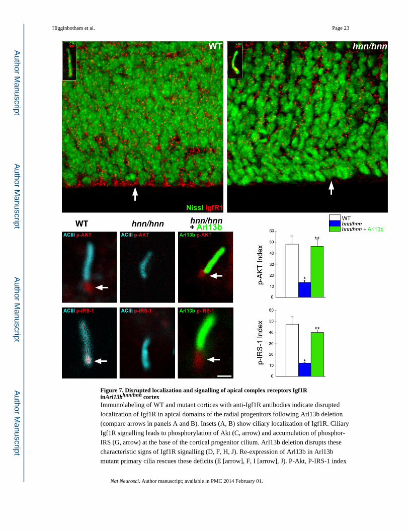
Figure 7. Disrupted localization and signalling of apical complex receptors Igf1R inArl13bhnn/hnn cortexImmunolabeling of WT and mutant cortices with anti-Igf1R antibodies indicate disrupted
localization of Igf1R in apical domains of the radial progenitors following Arl13b deletion
(compare arrows in panels A and B). Insets (A, B) show ciliary localization of Igf1R. Ciliary
Igf1R signalling leads to phosphorylation of Akt (C, arrow) and accumulation of phosphor-
IRS (G, arrow) at the base of the cortical progenitor cilium. Arl13b deletion disrupts these
characteristic signs of Igf1R signalling (D, F, H, J). Re-expression of Arl13b in Arl13b
mutant primary cilia rescues these deficits (E [arrow], F, I [arrow], J). P-Akt, P-IRS-1 index
Higginbotham et al. Page 23
Nat Neurosci. Author manuscript; available in PMC 2014 February 01.
Author M
anuscriptA
uthor Manuscript
Author M
anuscriptA
uthor Manuscript
indicates percentage of cilia with p-Akt or P-IRS-1 accumulation at the base of the cilium.
Data shown are mean ± SEM. Number of cilia analyzed: p-AKT group-WT (77), hnn/hnn