34
| Date post: | 22-Jan-2018 |
| Category: |
Health & Medicine |
| Upload: | magdy-elmasry |
| View: | 235 times |
| Download: | 0 times |

Reversible Cardiomyopathies
Cardiomyopathies have many etiological factors that
can result in severe structural and functional
dysregulation.
Fortunately, there are several potentially reversible
cardiomyopathies that are known to improve when the
root etiological factor is addressed.

Summary of common reversible cardiomyopathies and proposed mechanisms.

Arrhythmia-induced cardiomyopathy(AIC)
FOUR KEY QUESTIONS
What is AIC?
What is the pathophysiology of AIC?
How do I recognize the patient with AIC ?
How do I manage AIC?

AIC is a condition characterized by
either a tachyarrhythmia
(tachycardia - induced cardiomyopathy)
or frequent ventricular ectopy
(PVC - induced cardiomyopathy)
that results in LV dysfunction and heart failure.
The hallmark of this condition is partial or complete reversibility
once arrhythmiacontrol is achieved.
What is arrhythmia-induced cardiomyopathy ?
AIC can be classified into 2 categories:
One in which arrhythmia is the sole reason for
ventricular dysfunction
(pure or arrhythmia-induced)
And another in which the arrhythmia exacerbates
ventricular dysfunction and/or worsens HF in a
patient with concomitant heart disease
(impure or arrhythmia-mediated)

A 62-year-old man without significant past medical history presented with new onset HF symptoms. His ECG on presentation revealed a wide complex tachycardia and an echo demonstrated a LVEFof 10–15 % with normal LV wall thickness and a moderately dilated LV cavity. An EPS was performed, which made the diagnosis of atrio ventricular reciprocating tachycardia (AVRT) with a concealed left lateral accessory pathway, which was successfully abated. A follow-up echo one month later demonstrated an improved LVEF to 35–40 % and an echo performed one year later demonstrated normal LV wall thickness, cavity size and systolic function

Key features of an arrhythmia that will result in AIC are:
rapid ventricular rates, an irregular rhythm, and asynchronous myocardial
contraction, however not all need to be present to result in AIC
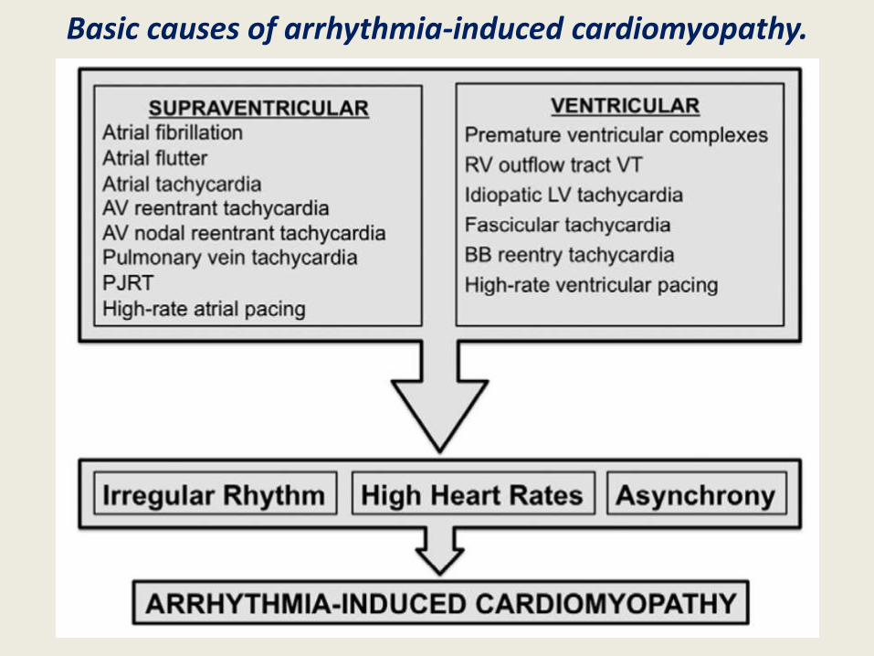
Basic causes of arrhythmia-induced cardiomyopathy.
Primary arrhythmia or primary cardiomyopathy
The chicken-egg dilemma
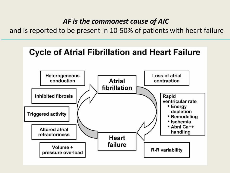
AF is the commonest cause of AICand is reported to be present in 10-50% of patients with heart failure

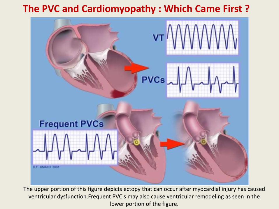
The upper portion of this figure depicts ectopy that can occur after myocardial injury has caused ventricular dysfunction.Frequent PVC's may also cause ventricular remodeling as seen in the
lower portion of the figure.
The PVC and Cardiomyopathy : Which Came First ?

Characteristics of primary cardiomyopathy versus
tachycardia-induced cardiomyopathy

A 12-lead ECG shows LBBB morphology PVCs with inferior axis in bigeminy pattern.(RVOT origin)
(PVC-LBBB)

A 12-lead ECG shows RBBB morphology PVCs with superior axis in trigeminy pattern.
(PVC-RBBB)
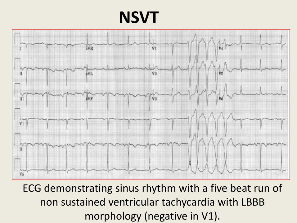
ECG demonstrating sinus rhythm with a five beat run of non sustained ventricular tachycardia with LBBB
morphology (negative in V1).
NSVT
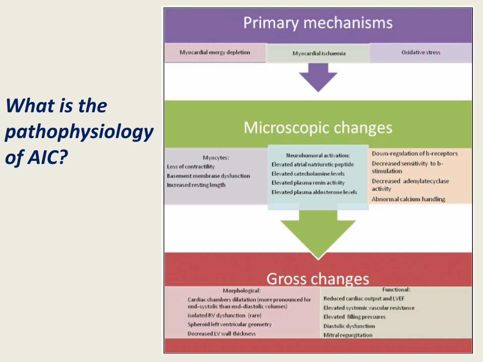
What is the pathophysiology of AIC?

How do I recognize the patient with AIC?We are frequently faced with patients in clinical practice who have the
combination of arrhythmia and heart failure, however teasing out which came first (the old chicken or the egg question) is not easy.
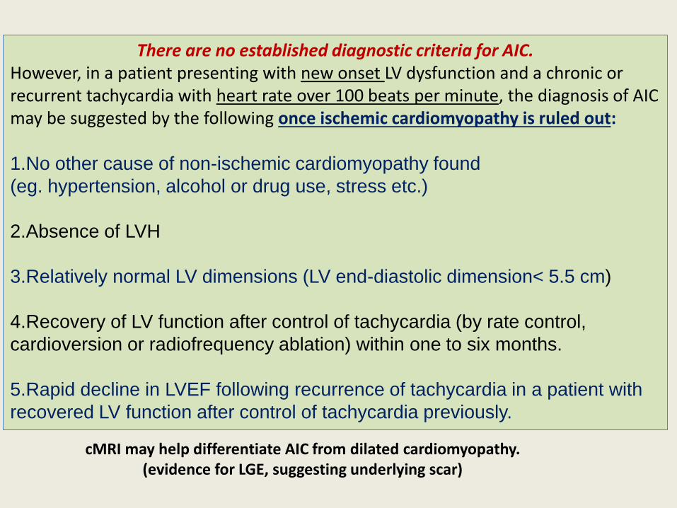
There are no established diagnostic criteria for AIC. However, in a patient presenting with new onset LV dysfunction and a chronic or recurrent tachycardia with heart rate over 100 beats per minute, the diagnosis of AIC may be suggested by the following once ischemic cardiomyopathy is ruled out:
1.No other cause of non-ischemic cardiomyopathy found
(eg. hypertension, alcohol or drug use, stress etc.)
2.Absence of LVH
3.Relatively normal LV dimensions (LV end-diastolic dimension< 5.5 cm)
4.Recovery of LV function after control of tachycardia (by rate control,
cardioversion or radiofrequency ablation) within one to six months.
5.Rapid decline in LVEF following recurrence of tachycardia in a patient with
recovered LV function after control of tachycardia previously.
cMRI may help differentiate AIC from dilated cardiomyopathy. (evidence for LGE, suggesting underlying scar)

How do I manage AIC?
PRINCIPLES OF MANAGEMENT
AIC management should focus on concerted attempts to eliminate or control the arrhythmia,
with the goal of improving symptoms, reversing LV dysfunction , and preventing arrhythmia recurrence

AIC Associated with specific Arrhythmias in adult
Atrial fibrillation
AF is the most common cause of AIC in adults
Management of AF consists of rate and/or rhythm control

Atrial flutter
Atrial flutter is more difficult to rate control than AF
Catheter ablation to eliminate atrial flutter is recommended when AIC is suspected.
For those in whom catheter ablation is not feasible or desired, cardioversion with antiarrhythmic therapy or
aggressive rate control should be used.
Supraventricular tachycardias
A curative strategy by catheter ablation should be pursued whenever possible as first-line
therapy for SVT-mediated AIC.

PVC-induced cardiomyopathy.
How many PVC's are too much? PVC burden
The most prominent predictor of cardiomyopathy in patients
with frequent PVCs appears to be the daily burden of PVCs.
There appears to be a threshold burden of approximately
10,000 PVCs/day for developing AIC.
Ventricular function can improve if the PVC burden is
reduced to <5,000/day
Therapy for PVC-mediated AIC should be targetedat suppressing or eliminating the PVCs and shouldinclude antiarrhythmic therapy and catheter ablation.
Catheter ablation has emerged as the definitive therapy for PVC-mediated AIC, with success rates ranging from 70% to 90%

Yong-Mei Cha et al. Circ Arrhythm
Electrophysiol. 2012;5:229-236
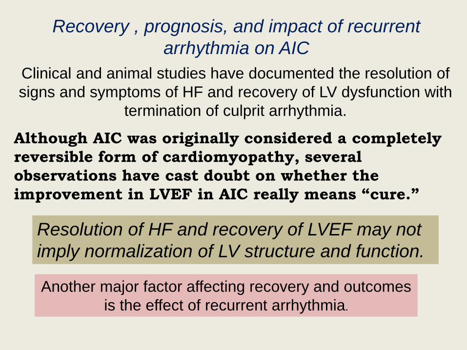
Recovery , prognosis, and impact of recurrent
arrhythmia on AIC
Clinical and animal studies have documented the resolution of
signs and symptoms of HF and recovery of LV dysfunction with
termination of culprit arrhythmia.
Although AIC was originally considered a completely
reversible form of cardiomyopathy, several
observations have cast doubt on whether the
improvement in LVEF in AIC really means “cure.”
Resolution of HF and recovery of LVEF may not
imply normalization of LV structure and function.
Another major factor affecting recovery and outcomes
is the effect of recurrent arrhythmia.

Risk of sudden death
Long-term survival of patients with
AIC following arrhythmia resolution is
likely; however, concerns remain.
Sudden cardiac death has been
reported in patients with AIC following
symptom recovery and LVEF
normalization

From arrhythmia occurrence to heart failure.

Overview of the Current Understanding of AIC,
From Mechanisms to Management and
Prognosis
(J Am Coll Cardiol 2015;66:1714–28)

Schematic Illustration of the Natural History and Pathophysiology of Rapid Pacing–Induced Dilated Cardiomyopathy and Heart Failure
CENTRAL ILLUSTRATION

Overview of the Current Understanding of AIC,
From Mechanisms to Management and
Prognosis

Overview of the Current Understanding of AIC,
From Mechanisms to Management and
Prognosis

When confronting a patient with heart failure and any kind of tachyarrhythmias :
AF and uncontrolled ventricular rates Frequent PVCs
Arrhythmia-induced Cardiomyopathy
or
Arrhythmia-aggravated Cardiomyopathy
Clinicians should focus on eliminating the arrhythymiawith catheter ablation and “attempt careful and aggressive control of rate and rhythm”
Conclusion


