64
ANATOMY OF ABDOMINAL AORTA Dr Fahad Shafi PG 1 ST YEAR
| Date post: | 14-Aug-2015 |
| Category: |
Health & Medicine |
| Upload: | fahad-shafi |
| View: | 15 times |
| Download: | 0 times |

ANATOMY OF ABDOMINAL AORTA
Dr Fahad Shafi
PG 1ST YEAR Deptt of
radiodiagnosis & imaging
2
ABDOMINAL AORTA
The abdominal aorta begins at the aortic hiatus of the diaphragm, anterior to the lower border of vertebra TXII.
It descends through the abdomen, anterior to the vertebral bodies, and by the time it ends at the level of vertebra LIV it is slightly to the left of midline.
The terminal branches of the abdominal aorta are the two common iliac arteries.

3
Anterior branches of the abdominal aorta
The abdominal aorta has anterior, lateral, and posterior branches as it passes through the abdominal cavity.

4
The three anterior branches supply the gastrointestinal viscera: the celiac trunk the superior mesenteric and
the inferior mesenteric arteries.

5


7
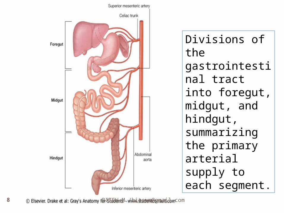
The primitive gut tube can be divided into :
A. foregut, B. midgut, and C. hindgut regions.
The boundaries of these regions are directly related to the areas of distribution of the three anterior branches of the abdominal aorta.
BITEW M./[email protected]
Divisions of the gastrointestinal tract into foregut, midgut, and hindgut, summarizing the primary arterial supply to each segment.
9
The foregut
The foregut begins with the abdominal esophagus and ends just inferior to the major duodenal papilla, midway along the descending part of the duodenum.
It includes the abdominal esophagus, stomach, duodenum (superior to the major papilla), liver, pancreas, and gallbladder.
The spleen also develops in relation to the foregut region. The foregut is supplied by the celiac trunk.

10
Divisions of the gastrointestinal tract into foregut, midgut, and hindgut, summarizing the primary arterial supply to each segment.
11
The midgut
The midgut begins just inferior to the major duodenal papilla, in the descending part of the duodenum, and ends at the junction between the proximal two-thirds and distal one-third of the transverse colon.
It includes the duodenum (inferior to the major duodenal papilla), jejunum, ileum, cecum, appendix, ascending colon, and the right two-thirds of the transverse colon.
The midgut is supplied by the superior mesenteric artery.

12
Divisions of the gastrointestinal tract into foregut, midgut, and hindgut, summarizing the primary arterial supply to each segment.

13
The hindgut The hindgut begins just before the left
colic flexure (the junction between the proximal two-thirds and distal one-third of the transverse colon) and ends midway through the anal canal.
It includes the left one-third of the transverse colon, descending colon, sigmoid colon, rectum, and upper part of the anal canal.
The hindgut is supplied by the inferior mesenteric artery

BITEW M./[email protected]
Divisions of the gastrointestinal tract into foregut, midgut, and hindgut, summarizing the primary arterial supply to each segment.

15
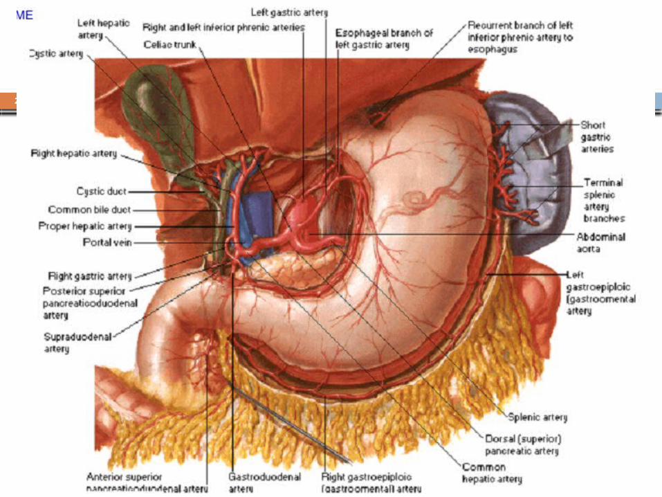
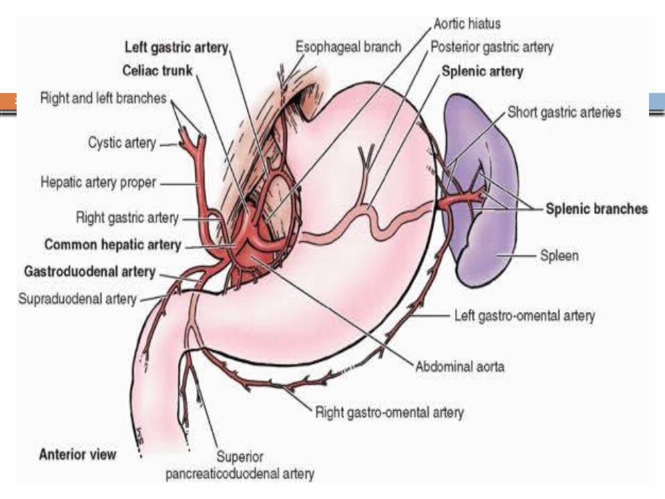
Celiac trunk The celiac trunk is the anterior branch of
the abdominal aorta supplying the foregut.
It arises from the abdominal aorta immediately below the aortic hiatus of the diaphragm, anterior to the upper part of vertebra LI.
It immediately divides into the:
A. left gastric, B. splenic, and C. common hepatic arteries.


18
A. Left gastric artery
The left gastric artery is the smallest branch of the celiac trunk.
The left gastric artery itself turns to the right and ascends along the lesser curvature of the stomach in the lesser omentum.
It supplies both surfaces of the stomach in this area and anastomoses with the right gastric artery
It ascends to the cardioesophageal junction and sends esophageal branches upward to the abdominal part of the esophagus.
Some of these branches continue through the esophageal hiatus of the diaphragm and anastomose with esophageal branches from the thoracic aorta.
19
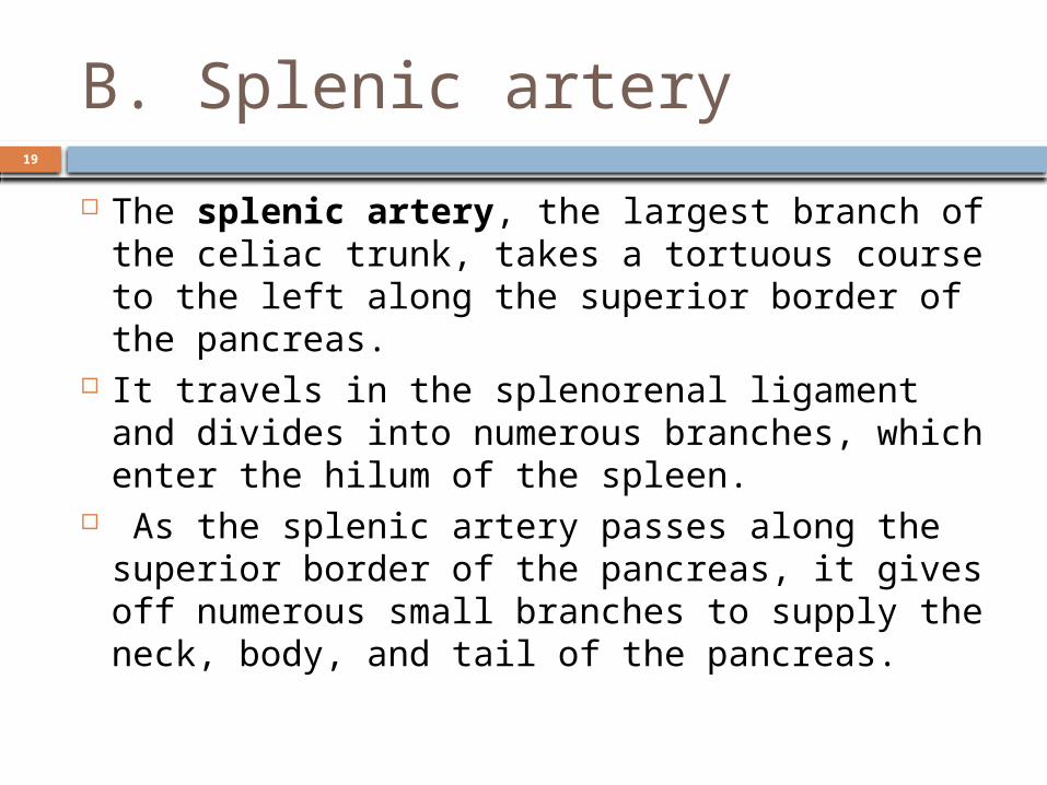
B. Splenic artery
The splenic artery, the largest branch of the celiac trunk, takes a tortuous course to the left along the superior border of the pancreas.
It travels in the splenorenal ligament and divides into numerous branches, which enter the hilum of the spleen.
As the splenic artery passes along the superior border of the pancreas, it gives off numerous small branches to supply the neck, body, and tail of the pancreas.
20
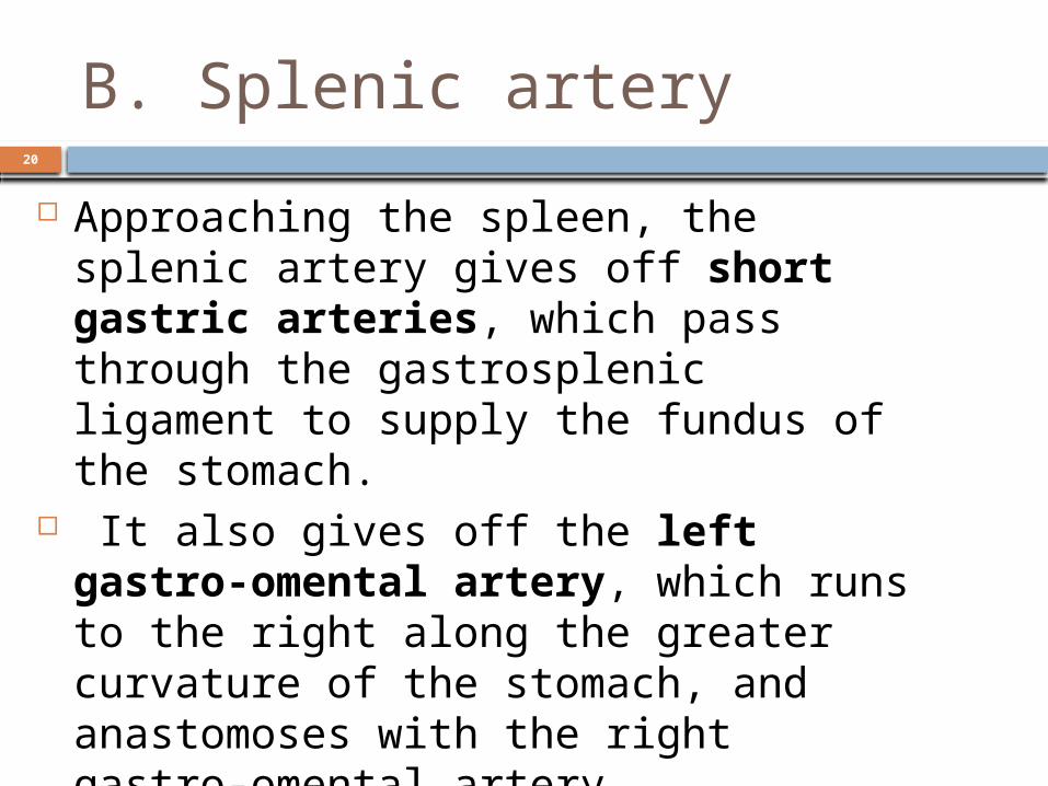
B. Splenic artery
Approaching the spleen, the splenic artery gives off short gastric arteries, which pass through the gastrosplenic ligament to supply the fundus of the stomach.
It also gives off the left gastro-omental artery, which runs to the right along the greater curvature of the stomach, and anastomoses with the right gastro-omental artery.
22
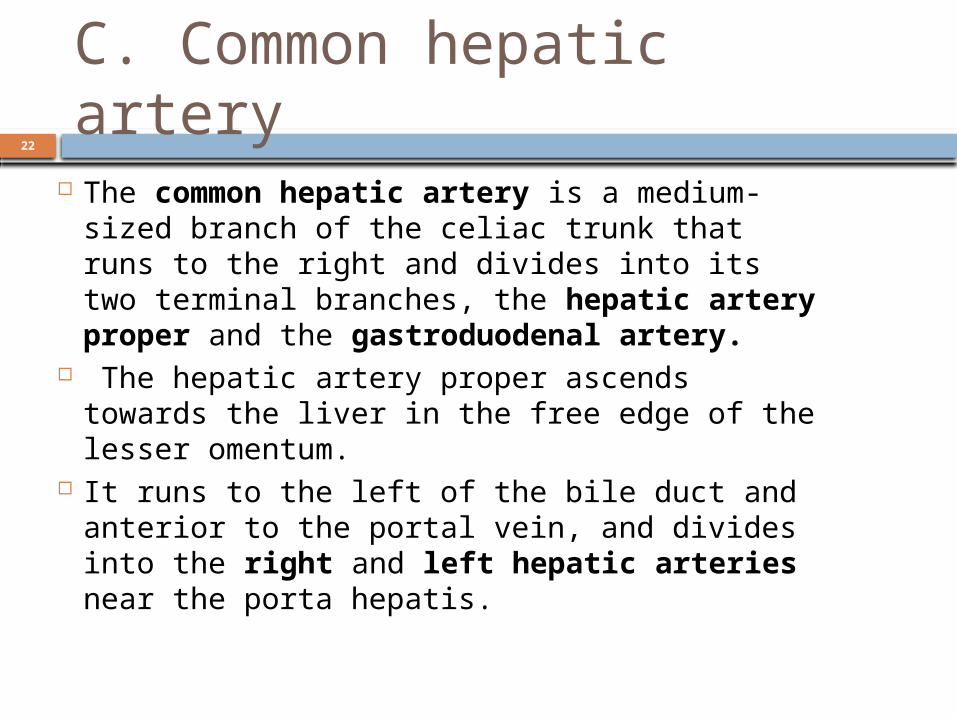
C. Common hepatic artery
The common hepatic artery is a medium-sized branch of the celiac trunk that runs to the right and divides into its two terminal branches, the hepatic artery proper and the gastroduodenal artery.
The hepatic artery proper ascends towards the liver in the free edge of the lesser omentum.
It runs to the left of the bile duct and anterior to the portal vein, and divides into the right and left hepatic arteries near the porta hepatis.

25
CONT’D
As the right hepatic artery nears the liver, it gives off the cystic artery to the gallbladder.
The gastroduodenal artery may give off the supraduodenal artery before descending posterior to the superior part of the duodenum.
Reaching the lower border of the superior part of the duodenum, the gastroduodenal artery divides into its terminal branches, the right gastro-omental artery and the superior pancreaticoduodenal artery
28
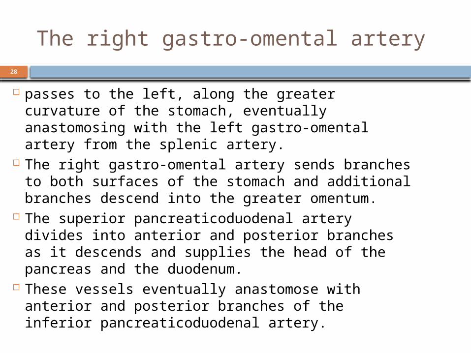
The right gastro-omental artery
passes to the left, along the greater curvature of the stomach, eventually anastomosing with the left gastro-omental artery from the splenic artery.
The right gastro-omental artery sends branches to both surfaces of the stomach and additional branches descend into the greater omentum.
The superior pancreaticoduodenal artery divides into anterior and posterior branches as it descends and supplies the head of the pancreas and the duodenum.
These vessels eventually anastomose with anterior and posterior branches of the inferior pancreaticoduodenal artery.

29
Superior mesenteric artery
The superior mesenteric artery is the anterior branch of the abdominal aorta supplying the midgut.
It arises from the abdominal aorta immediately below the celiac artery, anterior to the lower part of vertebra LI.
30
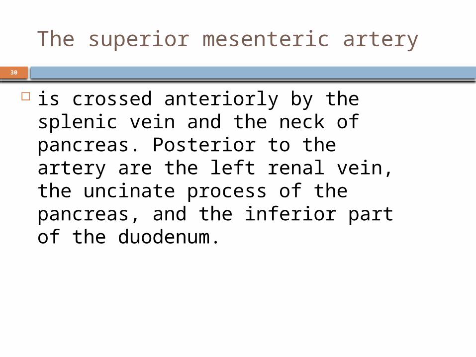
The superior mesenteric artery
is crossed anteriorly by the splenic vein and the neck of pancreas. Posterior to the artery are the left renal vein, the uncinate process of the pancreas, and the inferior part of the duodenum.

31
INFERIOR PANCREATICO DUODENAL ARTERY
The inferior pancreaticoduodenal artery is the first branch of the superior mesenteric artery.
It divides immediately into anterior and posterior branches, which ascend on the corresponding sides of the head of the pancreas.
Superiorly, these arteries anastomose with anterior and posterior superior pancreaticoduodenal arteries.
This arterial network supplies the head and uncinate process of the pancreas and the duodenum

32
33
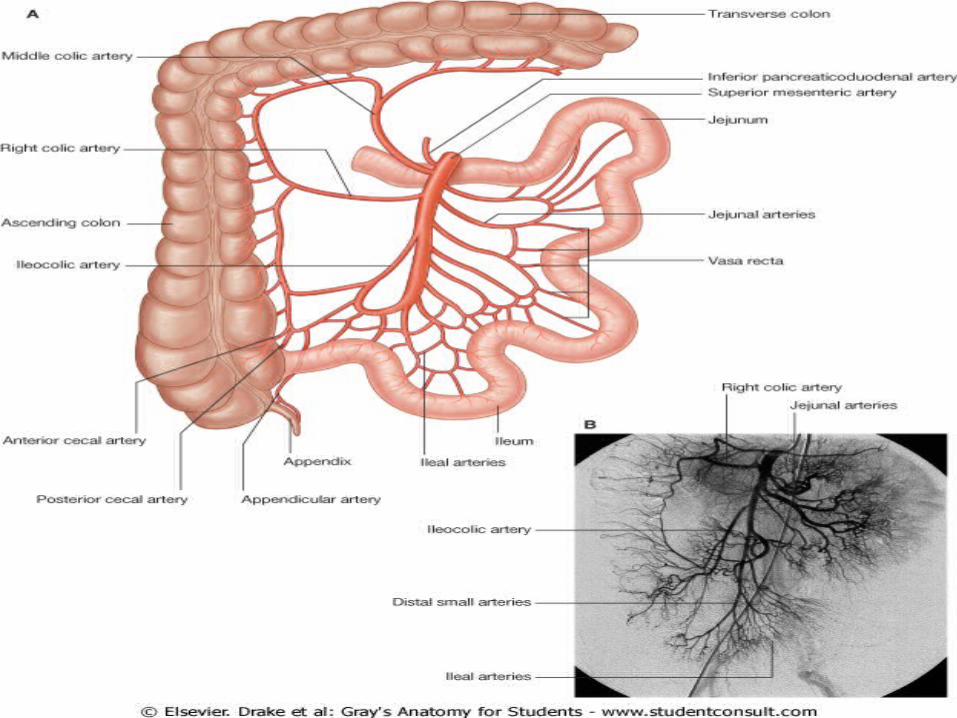
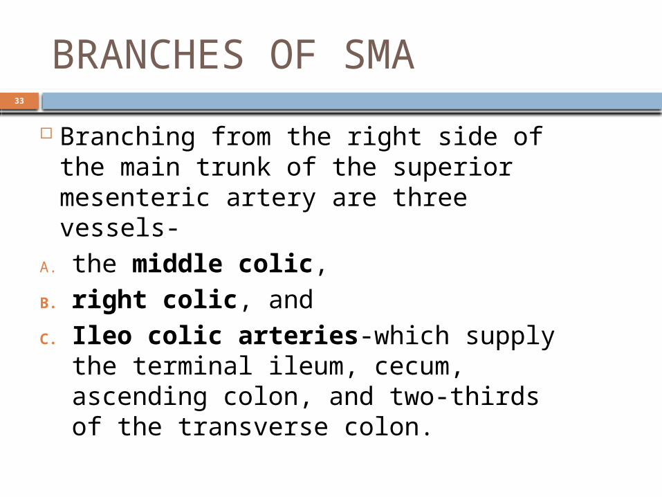
BRANCHES OF SMA
Branching from the right side of the main trunk of the superior mesenteric artery are three vessels-
A. the middle colic, B. right colic, and C. Ileo colic arteries-which supply
the terminal ileum, cecum, ascending colon, and two-thirds of the transverse colon.

34
MIDDLE COLIC ARTERY
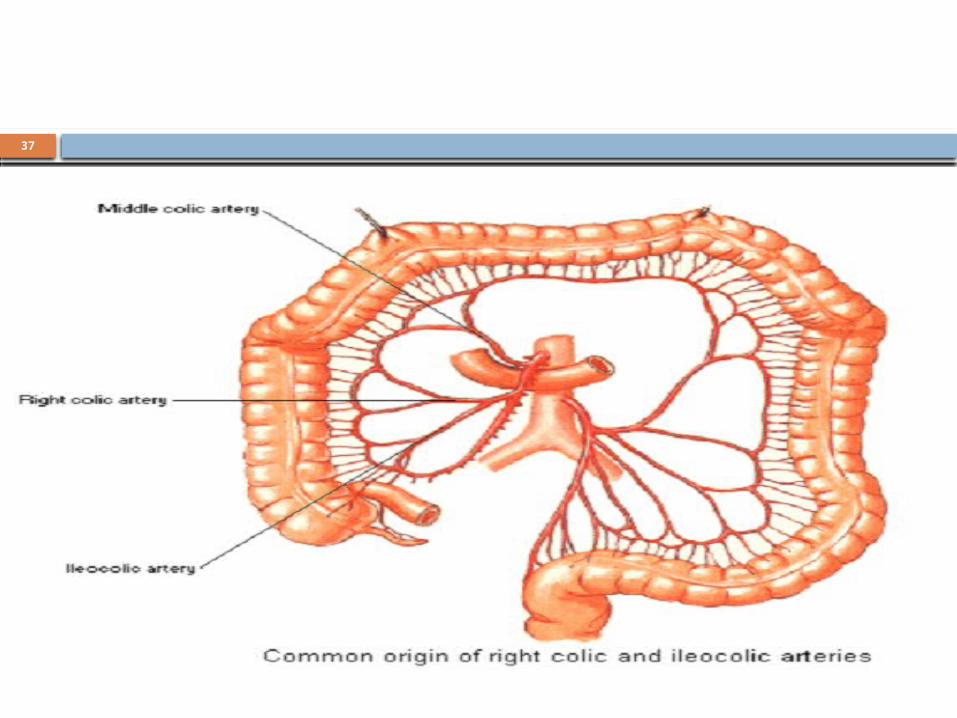
The middle colic artery is the first of the three branches from the right side of the main trunk of the superior mesenteric artery.
Arising as the superior mesenteric artery emerges from beneath the pancreas, the middle colic artery enters the transverse mesocolon and divides into right and left branches.
The right branch anastomoses with the right colic artery while the left branch anastomoses with the left colic artery, which is a branch of the inferior mesenteric artery.
36
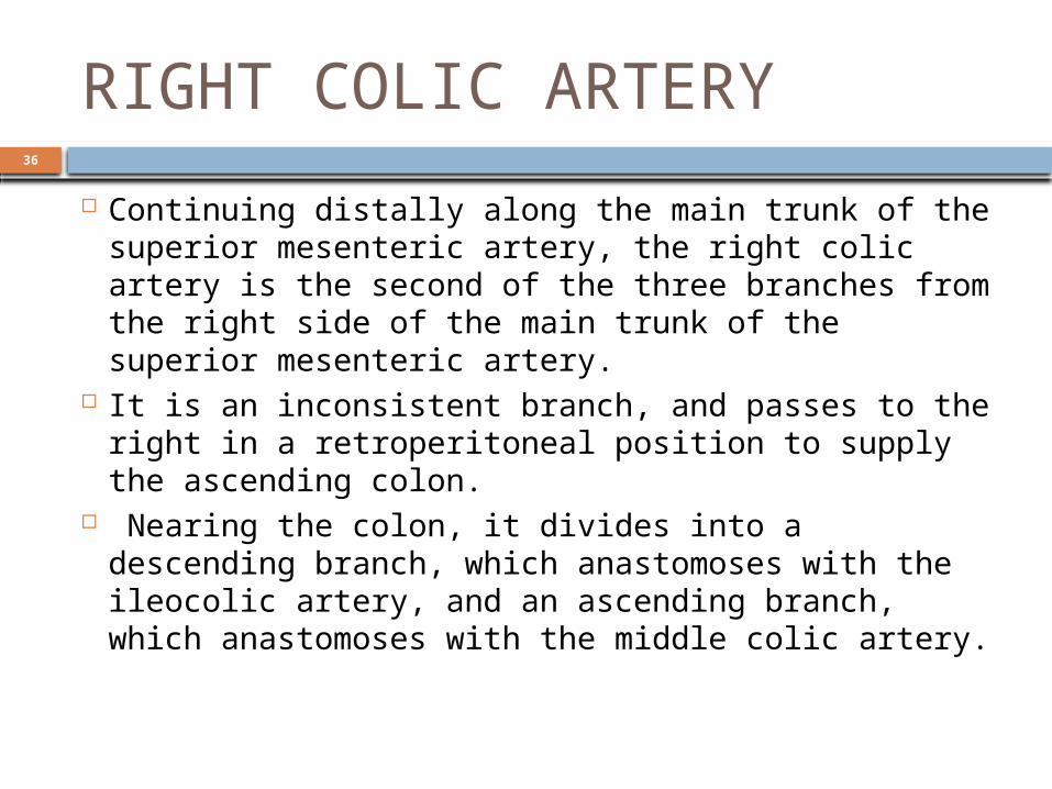
RIGHT COLIC ARTERY
Continuing distally along the main trunk of the superior mesenteric artery, the right colic artery is the second of the three branches from the right side of the main trunk of the superior mesenteric artery.
It is an inconsistent branch, and passes to the right in a retroperitoneal position to supply the ascending colon.
Nearing the colon, it divides into a descending branch, which anastomoses with the ileocolic artery, and an ascending branch, which anastomoses with the middle colic artery.
37

38
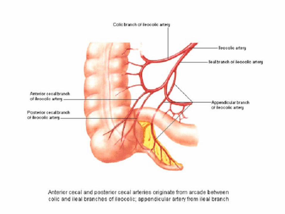
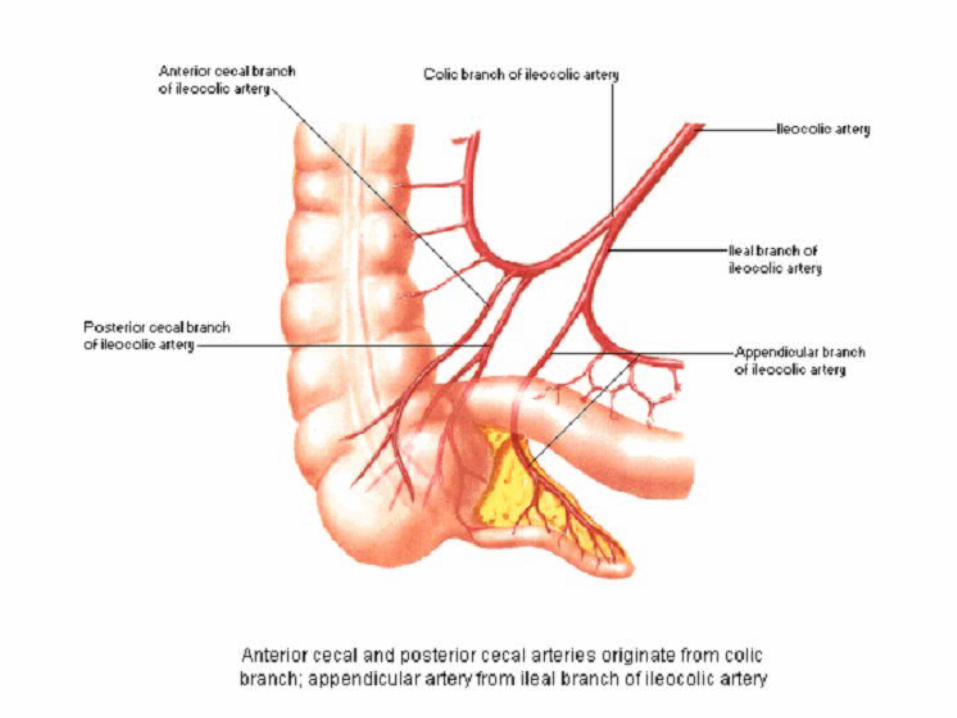
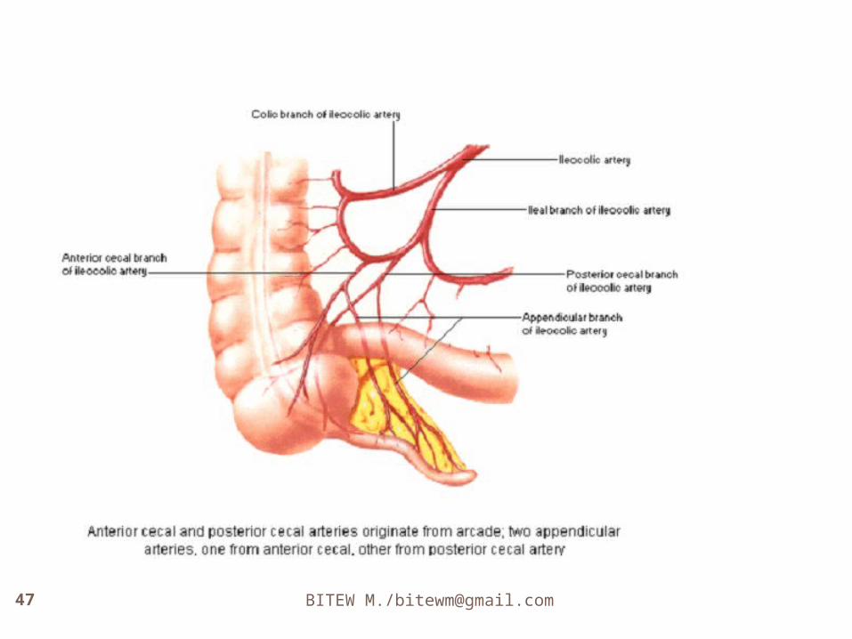
Ileocolic artery
The final branch arising from the right side of the superior mesenteric artery is the ileocolic artery.
This passes downward and to the right towards the right iliac fossa where it divides into superior and inferior branches:
the superior branch passes upward along the ascending colon to anastomose with the right colic artery;
the inferior branch continues towards the ileocolic junction dividing into colic, cecal, appendicular, and ileal branches.

39
Jejunal and ileal arteries
Arising on the left is a large number of jejunal and ileal arteries supplying the jejunum and most of the ileum.
These branches leave the main trunk of the artery, pass between two layers of the mesentery, and form anastomosing arches or arcades as they pass outward to supply the small intestine.
The number of arterial arcades increases distally along the gut.
40
Jejunal and ileal arteries There may be single and then double arcades in
the area of the jejunum, with a continued increase in the number of arcades moving into and through the area of the ileum.
Extending from the terminal arcade are vasa recta (straight arteries), which provide the final direct vascular supply to the walls of the small intestine.
The vasa recta supplying the jejunum are usually long and close together, forming narrow windows visible in the mesentery.
The vasa recta supplying the ileum are generally short and far apart, forming low broad windows.

The specific pattern of distribution and origin of these branches is variable


46
BITEW M./[email protected]

49
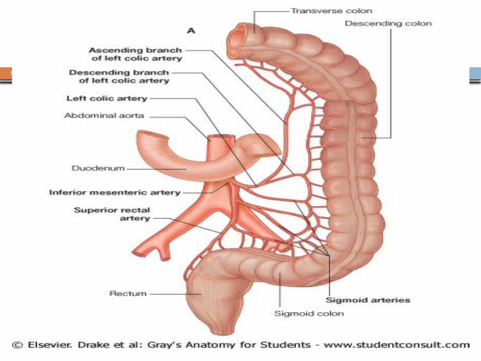
Inferior mesenteric artery
The inferior mesenteric artery is the anterior branch of the abdominal aorta that supplies the hindgut.
It is the smallest of the three anterior branches of the abdominal aorta and arises anterior to the body of vertebra LIII.
Initially, the inferior mesenteric artery descends anteriorly to the aorta and then passes to the left as it continues inferiorly.
Its branches include the left colic artery, several sigmoid arteries, and the superior rectal artery.

51
Left colic artery
The left colic artery is the first branch of the inferior mesenteric artery.
It ascends retroperitoneally, dividing into ascending and descending branches:
the ascending branch passes anteriorly to the left kidney, then enters the transverse mesocolon, and passes superiorly to supply the upper part of the descending colon and the distal part of the transverse colon, and anastomoses with branches of the middle colic artery;
the descending branch passes inferiorly, supplying the lower part of the descending colon and anastomoses with the first sigmoid artery.
53
Sigmoid arteries
The sigmoid arteries consist of two to four branches, which descend to the left, in the sigmoid mesocolon, to supply the lowest part of the descending colon and the sigmoid colon.
These branches anastomose superiorly with branches from the left colic artery and inferiorly with branches from the superior rectal artery.

55
Superior rectal artery The terminal branch of the inferior mesenteric artery is
the superior rectal artery. This vessel descends into the pelvic cavity in the
sigmoid mesocolon, crossing the left common iliac vessels.
Opposite vertebra SIII, the superior rectal artery divides. The two terminal branches descend on each side of the rectum, dividing into smaller branches in the wall of the rectum.
These smaller branches continue inferiorly to the level of the internal anal sphincter, anastomosing along the way with branches from the middle rectal arteries (from the internal iliac artery) and the inferior rectal arteries (from the internal pudendal artery).
57
LATERAL BRANCHES
Renal arteries The renal arteries normally arise off the side of the
abdominal aorta, immediately below the superior mesenteric artery, and supply the kidneys with blood. Each is directed across the crus of the diaphragm, so as to form nearly a right angle with the aorta,
Due to the position of the aorta, the inferior vena cava, and the kidneys in the body, the right renal artery is normally longer than the left renal artery.
The right passes behind the inferior vena cava, the right renal vein, the head of the pancreas, and the descending part of the duodenum.
The right is somewhat lower than the left; it lies behind the left renal vein, the body of the pancreas and the splenic vein, and is crossed
58
Middle suprarenal vein The middle suprarenal arteries
(middle capsular arteries; suprarenal arteries) are two small vessels which arise, one from either side of the abdominal aorta, opposite the superior mesenteric artery.
They pass laterally and slightly upward, over the crura of the diaphragm, to the suprarenal glands, where they anastomose with suprarenal branches of the inferior phrenic and renal arteries
59
Gonadal areteries gonadal artery is a generic term for a
paired artery, with one arising from the abdominal aorta for each gonad
Lumbar arteries lumbar arteries are arteries located in
the lumbar region. The lumbar arteries are in parallel with the intercostals.
They are usually four in number on either side, and arise from the back of the aorta, opposite the bodies of the upper four lumbar vertebrae

60
OTHER BRANCHES
INFERIOR PHRENIC ARTERY The inferior phrenic arteries are two
small vessels, which supply the diaphragm but present much variety in their origin.
They may arise separately from the front of the aorta, immediately above the celiac artery, or by a common trunk, which may spring either from the aorta or from the celiac artery. Sometimes one is derived from the aorta, and the other from one of the renal arteries; they rarely arise as separate vessels from the aorta.

61
62
MEDIAN SACRAL ARTERY
median sacral artery (or middle sacral artery) is a small vessel that arises posterior to the abdominal aorta and superior to its bifurcation.
It descends in the middle line in front of the fourth and fifth lumbar vertebræ, the sacrum and coccyx, ending in the glomus coccygeum (coccygeal gland).
63
COMMON ILIAC ARTERY
The abdominal aorta terminates at the level of L4 by dividing into the two common iliac arteries

64
The end
Thank you for your attention