Linköping University Medical Dissertations No. 1610 Arthroplasty in Elbow Fracture Treatment Jens Nestorson Division of Orthopaedic Surgery Department of Clinical and Experimental Medicine Faculty of Health Sciences Linköping University Sweden Linköping 2018
Transcript
Linköping University Medical DissertationsNo. 1610
Arthroplasty in Elbow Fracture Treatment
Jens Nestorson
Division of Orthopaedic SurgeryDepartment of Clinical and Experimental Medicine
Faculty of Health SciencesLinköping University
Sweden
Linköping 2018
Supervisor
Lars Adolfsson, MD, PhD, ProfessorDivision of Orthopaedic SurgeryDepartment of Clinical and Experimental MedicineLinköping University, Linköping
Co-supervisors
Carl Ekholm, MD, PhD, Ass. ProfessorDepartment of Orthopaedic SurgeryInstitute of Clinical SciencesSahlgrenska Academy, Gothenburg
Ola Wahlström, MD, PhD, ProfessorDepartment of Clinical and Experimental MedicineLinköping University, Linköping
Faculty opponent
Simon Lambert BSc FRCS FRCSEdOrthConsultant Orthopaedic SurgeonUniversity College London HospitalUnited Kingdom
Examination board
Göran Sjödén, MD, PhD, Ass. ProfessorDepartment of Clinical Science and Education, SödersjukhusetKarolinska Institutet, Stockholm
Simin Mohseni, Ass. ProfessorDepartment of Clinical and Experimental MedicineLinköping University, Linköping
Anders Kalén, MD, PhD, Ass. ProfessorDivision of Orthopaedic SurgeryDepartment of Clinical and Experimental MedicineLinköping University, Linköping
Paper I-III reprinted with permission Printed by LIU-Tryck, Linköping 2018-01-17
ISBN 978-91-7685-357-3 ISSN 0345-0082
In some ways we feel that we are as confused as ever, but we think we are confused on a higher level and about more important things.
Prof. Earl C. Kelly 1951 in “The Workshop Way of Learning”
Abstract Open reduction and internal fixation is the treatment of choice for distal humeral fractures. Stable fixation is required to allow early mobilisation and to reduce the risk of poor functional results. In an elderly patient with osteoporotic bone and with a comminuted intra-articular fracture stable internal fixation can be difficult to achieve. In these cases elbow arthroplasty is an option.
An irreparable radial head fracture can be treated by excision or replacement. The indications for the respective procedure are unclear since reports include an array of different associated soft-tissue and bony injuries.
The aim of this thesis was to evaluate the use, compli-cation rates and functional outcome of elbow arthroplasty as primary treatment for complex distal humeral fractures and assess the usefulness of radial head replacement in Mason IV fracture dislocations.
The nationwide use of primary arthroplasty for a distal humeral fracture between 1999 and 2014 was examined using three different registers. The survival rates in relation to prosthetic desing, age and sex were investigated using Cox regression analysis and number of adverse events recorded.
In total 405 patients were treated with primary arthroplasty for a distal humeral fracture. The mean age at surgery was 75 years and the mean observation time was 67 months. Eighteen patients had undergone revision surgery and another 26 patients suffered an adverse event, 24 of which required secondary surgery.
Increasing age reduced the risk for revision and there was no significant difference in survival between total- and hemi arthroplasty. The cumulative survival rate at 5 years was 99% (CI 98-100) and at 10 years 90% (CI 85-96). Elbow arthroplasty as primary treatment for distal humeral fractures produced reliable results with regards to revision surgery and adverse events.
18 patients, age 19-79 years, treated with radial head replacement, and 14 patients, age 29-70 years, treated with radial head resection, for a Mason IV fracture dis-location were retrospectively reviewed.
There were no significant differences in functional outcome in patients treated with replacement or excision for a Mason IV fracture dislocation. The rate of second-ary surgery was higher in patients treated with replace-ment and ulno-humeral osteoarthritis was more pro-nounced in patients treated with radial head excision but follow-up was longer in these patients. Functional results were not improved by using radial head arthroplasty for Mason IV fracture dislocation. Secondary osteoarthritis is a concern in patients treated with excision but did not affect functional outcome after a mean follow-up time of 108 months.
A b s t r Ac t
6
Sammanfattning på svenska
Frakturer på nedre delen av överarmen (distala humerus) som engagerar armbågsleden behandlas i regel med opera-tion. Frakturfragmenten hålls på plats med plattor och skruvar så att man inom någon vecka kan börja röra på armbågen och därmed förhindra stelhet. Hos äldre med benskörhet kan en stabil fixation vara svårt att åstad-komma då skelettets hållfasthet är nedsatt. I dessa fall kan man överväga att ersätta armbågsleden med en protes vilket visat sig fungera väl i andra leder, exempelvis höft och axel. Även vid frakturer på övre delen av strålbenet (caput radi) som inte går att fixera med plattor och/eller skruvar, kan protes (caput radi protes) vara ett alternativ. Det är dock oklart vid vilka skador denna metod behövs.
Denna avhandling utvärderar värdet av armbågs-proteser vid behandling av akuta frakturer på distala humerus och caput radi i samband med att armbågen har hoppat ur led (luxerat).
I delarbete I och II har den efterföljande funktionen studerats hos patienter som blivit behandlade med en så kallad halvprotes, där endast det frakturerade benet ersätts med protes, på grund av en komplicerad fraktur. Studierna har genomförts med hjälp av enkäter, mätningar av rörelse omfång samt röntgen för att värdera om övriga delar av leden eventuellt blivit påverkade av protesen.
I Sverige har vi kvalitetsregister inom vården. Ett sådant är Svenska Armbågsregistret där patienter registreras när de har blivit opererade med en armbågsprotes. För att hitta alla patienter som blivit opererade med en armbågsprotes på grund av fraktur har vi även använt Socialstyrelsens register och kontaktat alla ortopedkliniker i Sverige.
I delarbete IV jämfördes de olika registren för att identi-fiera alla dessa patienter och därefter undersöktes hur stor andel av patienterna som blivit om-opererade pga komp-
likationer relaterade till protesen och om protesen har behövt bytas ut.
För att studera värdet av protes vid fraktur på caput radi vid samtidig armbågsluxation utgick vi från två olika sjukhus, Linköping och Malmö, som behandlat denna skada på två olika sätt.
I Linköping ersattes det frakturerade caput radi med en protes och i Malmö tog man endast bort caput radi. I båda fallen reparerades det yttre ledbandet. Efter operationen blev man gipsad i 3-4 veckor. De två behand-lings-grupperna fick svara på enkäter, undersöktes avseende rörelseomfång och stabilitet samt röntgades för att undersöka förekomst av eventuell ledpåverkan och proteslossning.
Denna avhandling visar att armbågsprotes som behandling vid akuta frakturer på distala humerus fungerar bra i ett kort- och medellångt perspektiv. Funk-tionen efter att man har blivit opererad med en halvpro-tes är god men med en nedsatt rörlighet. Proteslossning samt revision är ovanligt. Resultaten är jämförbara med andra typer av behandling så som operation med plattor och skruvar och så kallade helproteser (där både det skadade och övre delen av armbågsbenet byts ut). Caput radi protes visade sig inte innebära några fördelar jämfört med enbart borttagandet av caput radi avseende rörlighet och funktion. Det var dock fler reoperationer i gruppen opererade med protes men graden av ledyte-slitage i armbågsleden var större i gruppen där radius-huvudet tagits bort.
s A m m A n fAt t n i n g på s v e n s k A
10
List of Papers I The Kudo humeral component as primary hemiarthroplasty in distal humeral fractures. J Shoulder Elbow Surg, 2012. 21(4): p. 451-5. Adolfsson L. and Nestorson J.
II Hemiarthroplasty for irreparable distal humeral fractures: medium-term follow-up of 42 patients. Bone Joint J, 2015. 97-B(10): p. 1377-84.
Nestorson J., Ekholm C., Etzner M. and Adolfsson L.
III A radial head prosthesis appears to be unnecessary in Mason-IV fracture dislocation. Acta Orthop, 2017. 88(3): p. 315-319.
Nestorson, J., Josefsson PO. and Adolfsson L.
IV Arthroplasty as primary treatment for distal humeral fractures produces reliable results with regards to revisions and adverse events. A registry based study.
Nestorson J., Rahme H., Adolfsson L Manuscript (submitted)
List of abbreviations DASH Disabilities of Arm Shoulder and Hand
MEPS Mayo Elbow Performance Score
ORIF Open Reduction Internal Fixation
PROM Patient Related Outcome Measures
Contents
1 Introduction 21
2 Anatomy of the elbow 25
The bones 25Joints 28Blood supply 28Nerves 29Elbow stability 29
3 Epidemiology 33
Distal humeral fractures 33Radial head fractures 34
4 Classification of elbow fractures 37
Distal humeral fractures 37Radial head fractures 39
5 Instruments for assessment of elbow function 43
6 Treatment of intra-articular distal humeral fractures 49
7 Development of elbow arthroplasty 57
8 Arthroplasty in distal humeral fracture treatment 61
9 Treatment of radial head fractures 65
10 Development and use of radial head prostheses 69
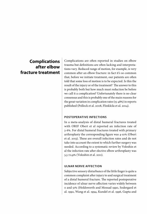
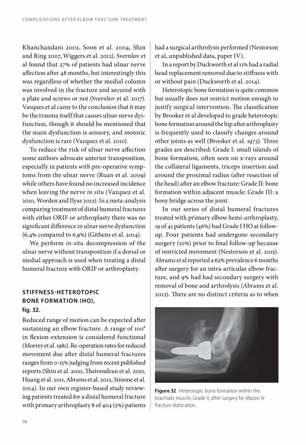
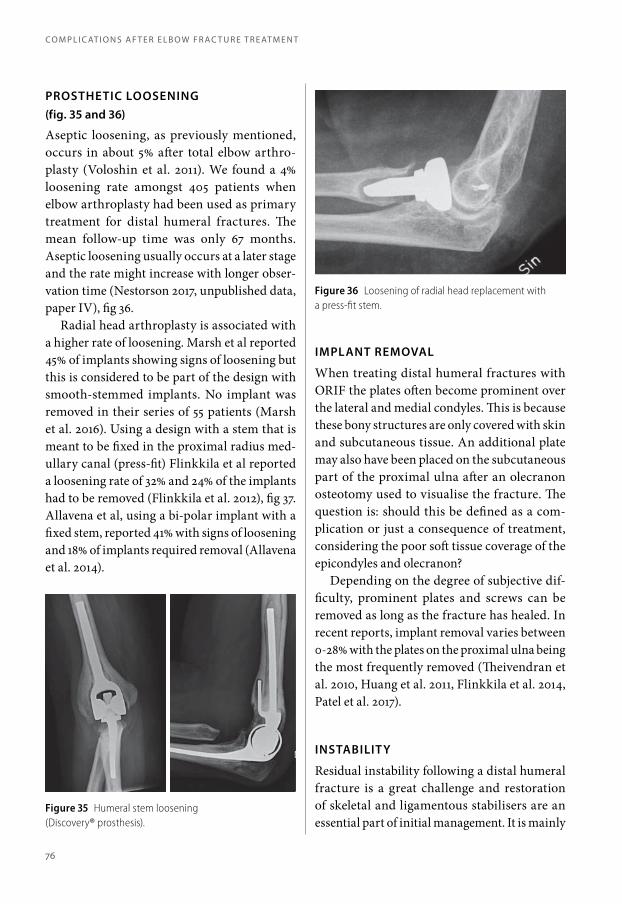
11 Complications after elbow fracture treatment 73
12 Aims 81
13 Patients 83
14 Methods 85
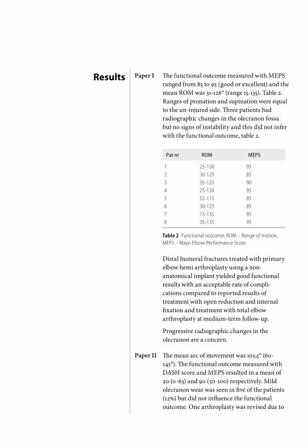
15 Results 89
16 Discussion 95
Treatment of distal humeral fractures 95Treatment of radial head fractures 96
17 Conclusions 99
18 Future research 101
19 Acknowledgments 105
20 References 109
21 Papers 123
co n t e n t s
18
Introduction Historically, the treatment of elbow fractures has largely been non-surgical and resulting stiffness and mal-union has been a frequent problem. The development of x-ray imaging, anaesthesia techniques and anti-bacterial treat-ment made surgical intervention feasible (Kozanek et al. 2014). Initial surgical attempts gave rather poor results due to inadequate fixation techniques combined with prolonged immobilisation (Aitken and Rorabeck 1986).
Vital to the outcome after sustaining an elbow injury is early mobilization. A prerequisite for early mobilization is to achieve good reduction, a stable fixation of the frac-ture and to secure elbow stability by addressing soft tissue injuries (i.e lateral and medial ligaments) when necessary. Malaligned articular surfaces and instability will lead to restricted motion and may lead to secondary osteoarthritis that could be difficult to manage (Ring and Jupiter 2000, Ring et al. 2004).
Better understanding of the biomechanics and improve-ment of implants used to secure fracture fragments have gradually led to better functional outcomes but still with reduced range of motion and complications rates ranging from 15-48% (Pollock et al. 2008, Flinkkila et al. 2014).
Arthroplasty was initially used to treat secondary changes at the elbow after previous fracture, mal-union and/or osteoarthritis (Mellen and Phalen 1947). It was not until the 90’s results were published regarding total arthro-plasty used as a primary procedure for distal humeral fractures (Cobb and Morrey 1997).
Today surgical treatment predominates in the treatment of complex distal humeral and displaced radial head frac-tures but the optimal method of treatment in the individ-ual case is still uncertain.
Occasionally, stable internal fixation cannot be achieved
and instead of prolonged immobilisation that often leads to poor functional outcome, primary arthroplasty is an option.
There are several reports on functional outcome and complications in distal humeral fractures treated with total elbow arthroplasty (Cobb and Morrey 1997, Gambirasio et al. 2001, Kamineni and Morrey 2004). Fractures of the neck of femur and proximal humeral fractures can be treated with hemi-arthroplasty (i.e. replacing the fractured bone only). This concept has also been used in elbow fractures, but pre-viously as a secondary procedure and reported in small series or case reports (Mellen and Phalen 1947, Macausland 1954, Barr and Eaton 1965). In 2005 Parsons et al described the use of hemi arthroplasty as primary treatment and presented the short-term outcome of four patients (Parsons 2005). In 2006, Adolfsson and Hammer presented the results of four more patients (Adolfsson and Hammer 2006).
Paper I and II retrospectively investigated the functional outcome of hemi-arthroplasty as primary treatment for distal humeral fractures using one or the other of two prosthetic designs. Paper IV investigated the survival of arthro-plasty (both total- and hemi-arthroplasty) when
used as primary treatment for distal humeral fractures and the rate of adverse events using data from three Swedish register sources.
The use of radial head arthroplasty for a comminuted radial head fracture is also well documented but reports include many types of elbow injury involving a radial head frac-ture (Popovic et al. 2000, Grewal et al. 2006, Flinkkila et al. 2012, Marsh et al. 2016). Out-come following a radial head fracture depends to a great extent on the degree of associated fractures and/or soft tissue injury. Since pros-thetic loosening and secondary surgery rates in patients treated with radial head replacement are not negligible the question is if the radial head must always be replaced. There are reports of radial head fractures combined with elbow dislocation (Mason IV) treated with radial head resection and ligament repair (Sanchez-Sotelo et al. 2000, Herbertsson et al. 2009). The aim in paper III was to evaluate the need for radial head replacement in the specific context of Mason IV fracture dislocations by comparing the functional and radiographic results of two treatment strategies, radial head replacement or radial head resection.
i n t r o d u c t i o n
22
Anatomy of the elbow
tHe bonesHUMERUS
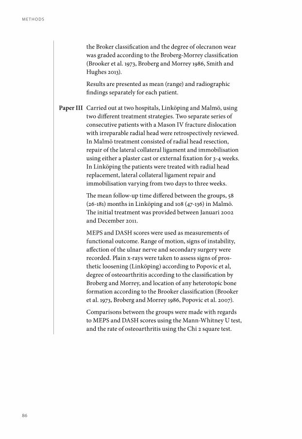
The distal part of the humerus consists of the trochlea, capitellum and the lateral and medial epicondyles. The lateral and medial columns support these structures. Between the columns are the olecranon fossa dorsally and the coronoid fossa anteriorly. The joint-line has a slight internal rotation, valgus tilt and anterior angulation (approximately 6, 7 and 30 degrees respectively) in relation to the humeral shaft, fig 1 and 2.
The trochlea articulates with the greater sigmoid notch of the olecranon and the capitellum articulates with the radial head. The depth of the trochlear notch in the coronal plane varies unrelated to the size of the distal humerus and the trochlear notch angle ranges between 125 and 155 degrees (Giannicola et al. 2016), fig 3. The capitellum is not spherical but has a somewhat ellipsoid shape with a greater radius of curvature in the medial-lateral plane (Sabo et al. 2011).
The lateral epicondyle is the point of origin of the common extensors and the lateral collateral ligament. The medial epicondyle is the origin of the common flexors and the medial collateral ligament.
ULNA
The proximal ulna has a varus angulation in relation to the shaft. Totlis et al reported a mean angle of 8 (range 2-16) degrees in 100 (200 bones) Caucasian specimens while Windisch et al reported a mean of 18 (range 11-28) degrees in 63 elbow specimens (Windisch et al. 2007, Totlis et al. 2014). The apex is located 8 cm (range 6-12) distal to the olecranon tip (Totlis et al. 2014), fig 4. There is also an anterior angulation of about 8 (range 2-14) degrees starting 8 (range 5-12) cm from the olecranon tip (Totlis et al. 2014), fig 5. The olecranon has two articular surfaces: the greater sigmoid notch that artic-ulates with the trochlea, and the lesser sigmoid notch that articulates with the radial head. The coronoid process contains the tip, base and the sublime tubercle. Distal to the coronoid process laterally is the supinator crest.
The sublime tubercle medially and the supi-nator crest laterally are the ulnar insertions of the collateral ligaments.
The triceps inserts on the proximal dorsal aspect of the olecranon and the brachialis muscle inserts just distal to the coronoid tip.
Figure 1 Distal humerus. A – the valgus angle of the rotational axis in relation to the humeral shaft.
Figure 2 Distal humerus, lateral view showing the anterior angulation of distal humerus.
Figure 3 A – Trochlear notch angle.
A n Ato m y o f t H e e l b o w
26
A n Ato m y o f t H e e l b o w
RADIUS
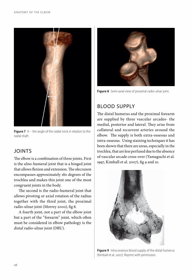
The proximal radius is made up of the radial head and the radial neck. The radial head is slightly oval and varies considerably in size and height, fig 6. In a study by Kuhn et al the maximum median radial head diameter was 25 (range 20-34) mm and the minimum diameter 23 (range 18-30) mm. The median height was 11 (range 7-17) mm (Kuhn et al. 2012), fig 6. The radial neck has a mean 13 (range 4-22) degrees angle to the radial shaft, fig 7. There is also a variation in the length of the neck with a mean of 13 (range 9-19) mm (Van Riet et al. 2004). The radial head is kept in place in relation to the lesser sigmoid notch on the ulna by the annular ligament. The ligament has its origin at the supi-nator crest of the ulna and inserts on the lateral aspect of the coronoid. The annular ligament and the lateral collateral ligament make up the lateral ligament complex. Distal to the radial neck is the radial tuberosity where the biceps tendon inserts.
Figure 4 A – varus angulation of proximal ulna.
Figure 5 A – anterior angulation of the proximal ulna. Sometimes referred to as PUDA (Proximal Ulna Dorsal Angulation).
FIgur 6 Proximal radius. A – radial head height, B – radial neck length.
27
JointsThe elbow is a combination of three joints. First is the ulno-humeral joint that is a hinged joint that allows flexion and extension. The olecranon encompasses approximately 180 degrees of the trochlea and makes this joint one of the most congruent joints in the body.
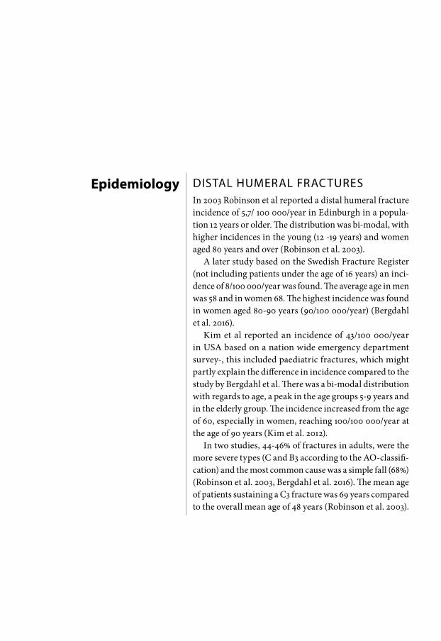
The second is the radio-humeral joint that allows pivoting or axial rotation of the radius together with the third joint, the proximal radio-ulnar joint (Morrey 2000), fig 8.
A fourth joint, not a part of the elbow joint but a part of the “forearm” joint, which often must be considered in elbow pathology is the distal radio-ulnar joint (DRU).
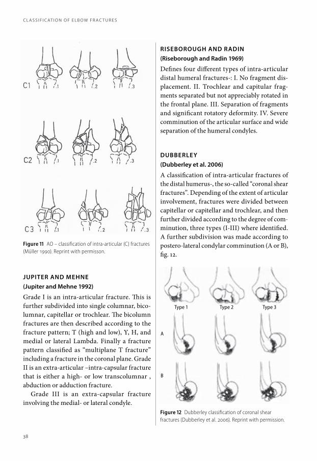
blood supplyThe distal humerus and the proximal forearm are supplied by three vascular arcades- the medial, posterior and lateral. They arise from collateral and recurrent arteries around the elbow. The supply is both extra-osseous and intra-osseous. Using staining techniques it has been shown that there are areas, especially in the trochlea, that are less perfused due to the absence of vascular arcade cross-over (Yamaguchi et al. 1997, Kimball et al. 2007), fig 9 and 10.
Figure 7 A – the angle of the radial neck in relation to the radial shaft.
Figure 8 Semi-axial view of proximal radio-ulnar joint.
Figure 9 Intra osseous blood supply of the distal humerus (Kimball et al. 2007). Reprint with permission.
A n Ato m y o f t H e e l b o w
28
A n Ato m y o f t H e e l b o w
nervesThe ulnar nerve runs posteromedially and lies directly above the capsule and the medial col-lateral ligament behind the medial epicondyle. It then continues distally in between the two heads of the flexor carpi ulnaris muscle.
The median nerve runs anteriorly medial to the biceps tendon on top of the brachialis muscle. It is accompanied by the brachial artery and veins and continues distally under the lacer-tus fibrosus and pronator teres muscle.
The radial nerve runs ventrally medial to the proximal part of the brachioradialis muscle origin and continues distally underneath the common extensors and the brachioradialis muscle above the joint capsule covering the radial head. At the level of the radial neck it divides into a superficial sensory branch and the deep posterior interosseous nerve that continues postero-laterally between the deep and super-ficial portions of the supinator muscle.
elbow stAbilityThe stability of the elbow depends on three main factors-, osseous configuration, static stabilisers and dynamic stabilisers.
The osseous configuration of the olecranon and the trochlea makes the joint highly stable in flexion and extension. The coronoid process acts as a buttress against axial dislocation and together with the sublime tubercle it also pro-tects against varus and rotational instability. The radio-capitellar joint also contributes in preventing axial and rotational instability with its “ball-in-socket” shape, although the articular surfaces are relatively flat.
The static stabilizers consist of the lateral and medial collateral ligaments together with the anterior capsule. Laterally the lateral collateral ligament originates from the lateral epicondyle at the centre of rotation. One part, the radial collateral ligament fuses with the annular ligament, and the other part, the lateral ulnar collateral ligament, inserts on the proximal part of the supinator crest on the ulna. The lateral ulnar collateral ligament is considered the most important part as regards elbow stability, pre-venting postero-lateral rotatory instability and is also a restraint to varus loading (O’Driscoll et al. 1991). The annular ligament keeps the radial head in place in relation to ulna (proximal radio-ulnar joint) together with the interos-seous membrane.
Medially the medial collateral ligament has three parts: anterior, posterior and the trans-verse. The anterior part causes most clinical problems. It originates from the inferior border of the medial epicondyle, slightly posterior to the rotational axis, and inserts on the sublime tubercle of the ulna. Morrey et al showed that the medial collateral ligament was more important than the radial head regarding valgus stability of the elbow (Morrey et al. 1991).
Figure 10 Extra-osseous blood supply of the dorsal and ventral aspect of distal humerus (Yamaguchi et al. 1997). Reprint with permission.
29
The dynamic stabilizers are the muscles crossing the elbow. Dorsally the triceps muscle and anteriorly the biceps and brachialis muscle act by compressing mainly the ulno-humeral joint. Laterally the brachioradialis muscle and the common extensors compress the radio-hu-
meral joint and also resist varus instability. In the same manner the pronator teres muscle and the common flexors compress the ulno-humeral joint preventing valgus forces (An 2000, Morrey 2000, de Haan et al. 2011, Chalmers 2017).
A n Ato m y o f t H e e l b o w
30
Epidemiology distAl HumerAl frActuresIn 2003 Robinson et al reported a distal humeral fracture incidence of 5,7/ 100 000/year in Edinburgh in a popula-tion 12 years or older. The distribution was bi-modal, with higher incidences in the young (12 -19 years) and women aged 80 years and over (Robinson et al. 2003).
A later study based on the Swedish Fracture Register (not including patients under the age of 16 years) an inci-dence of 8/100 000/year was found. The average age in men was 58 and in women 68. The highest incidence was found in women aged 80-90 years (90/100 000/year) (Bergdahl et al. 2016).
Kim et al reported an incidence of 43/100 000/year in USA based on a nation wide emergency department survey-, this included paediatric fractures, which might partly explain the difference in incidence compared to the study by Bergdahl et al. There was a bi-modal distribution with regards to age, a peak in the age groups 5-9 years and in the elderly group. The incidence increased from the age of 60, especially in women, reaching 100/100 000/year at the age of 90 years (Kim et al. 2012).
In two studies, 44-46% of fractures in adults, were the more severe types (C and B3 according to the AO-classifi-cation) and the most common cause was a simple fall (68%) (Robinson et al. 2003, Bergdahl et al. 2016). The mean age of patients sustaining a C3 fracture was 69 years compared to the overall mean age of 48 years (Robinson et al. 2003).
rAdiAl HeAd frActuresThe incidence of radial head fractures ranges from 28-55/100 000/year. Generally, men are younger than women when sustaining a radial head fracture-, around 40 years compared to 52 in women. Approximately 12% of fractures
are the more severe forms- Mason III and IV. Of all patients with a radial head fracture 40% have an associated injury (additional fracture or soft tissue injury) and the most commonly associated injury is a coronoid fracture (Kaas et al. 2010, Duckworth et al. 2012).
e p i d e m i o lo g y
34
Classification of elbow fractures
The optimal fracture classification system should indicate how to treat a fracture-, as well as what complications and also to some extent, what functional outcome to expect (Burstein 1993, Martin and Marsh 1997, Audige et al. 2004). Since there are quite a number of elbow fracture classifications systems, one may draw the conclusion that the perfect one is yet to be presented. The classification systems presented below are the most commonly used in the orthopaedic literature.
distAl HumerAl frActuresAO (Arbeitsgemeinschaft für Osteosynthesefragen) (Müller 1990)
The AO-classification first divides fractures in to extra-articular (A), partially articular (B) and intra artic-ular (C). These are then further subdivided into three categories (1-3) depending on complexity and yet a third category level (1-3) according to degree of comminution (ie number of fragments), Fig. 11.
JUPITER AND MEHNE(Jupiter and Mehne 1992)
Grade I is an intra-articular fracture. This is further subdivided into single columnar, bico-lumnar, capitellar or trochlear. The bicolumn fractures are then described according to the fracture pattern; T (high and low), Y, H, and medial or lateral Lambda. Finally a fracture pattern classified as “multiplane T fracture” including a fracture in the coronal plane. Grade II is an extra-articular –intra-capsular fracture that is either a high- or low transcolumnar , abduction or adduction fracture.
Grade III is an extra-capsular fracture involving the medial- or lateral condyle.
RISEBOROUGH AND RADIN(Riseborough and Radin 1969)
Defines four different types of intra-articular distal humeral fractures-: I. No fragment dis-placement. II. Trochlear and capitular frag-ments separated but not appreciably rotated in the frontal plane. III. Separation of fragments and significant rotatory deformity. IV. Severe comminution of the articular surface and wide separation of the humeral condyles.
DUBBERLEY(Dubberley et al. 2006)
A classification of intra-articular fractures of the distal humerus-, the so-called “coronal shear fractures”. Depending of the extent of articular involvement, fractures were divided between capitellar or capitellar and trochlear, and then further divided according to the degree of com-minution, three types (I-III) where identified. A further subdivision was made according to postero-lateral condylar comminution (A or B), fig. 12.
Figure 11 AO – classification of intra-articular (C) fractures (Müller 1990). Reprint with permisson.
Figure 12 Dubberley classification of coronal shear fractures (Dubberley et al. 2006). Reprint with permission.
Type 3Type 2Type 1
A
B
c l A s s i f i c At i o n o f e l b o w f r Ac t u r e s
38
c l A s s i f i c At i o n o f e l b o w f r Ac t u r e s
rAdiAl HeAd frActuresMASON(Mason 1954)
The most commonly used classification system, originally based on X-ray findings. Type I is a fissure or marginal sector fracture without dis-placement. Type II marginal sector fracture with displacement. Type III is a comminuted fracture involving the whole radial head. Johnston added type IV-, a radial head fracture in combination with an elbow dislocation (Johnston 1962). Broberg and Morrey further added fractures of the radial neck (Morrey 1985), fig. 13. Hotchkiss modified the classification system to account for concomitant injuries to the soft tissues sur-rounding the elbow and bony injury to the ulna. This was to assist in treatment decision-making since these associated injuries are considered to have a significant impact on functional outcome (Hotchkiss 1997).
Figure 13 Modification of the Mason-Johnston classification of radial head and neck fractures (Morrey 1985). Reprint with permission.
CHARALAMBOUS CLASSIFICATION(Charalambous et al. 2011)
Charalambous presented a classification in 2011 dividing the fractures of the radial head and neck into four different categories depending on degree of comminution and involvement of the radial neck and further subdivided into un-dis-placed or displaced fractures. This to recognize the possibility of comminuted partly articular fractures and the possibility of partly articular fractures involving any part of the radial head rather than simply its lateral part, fig 14.
THE AO CLASSIFICATION(Müller et al. 1990)
A little more complex and takes into account the proximal part of both the radius and ulna and is therefore not frequently used to describe isolated fractures of the proximal radius, fig. 15.
CLASSIFICATION OF CORONOID FRACTURES
Several patho-mechanisms can lead to fractures of the radial head and cause associated injuries that may have great importance regarding treat-ment and prognosis. One associated injury that is of particular importance for outcome when associated with a radial head fracture is the cor-onoid fracture, which is why the classification of these fractures is included here.
39
Figure 14 Charalambous classification of radial head fractures (Rhyou et al. 2013). Reprint with permission.
O’DRISCOLL(O’Driscoll et al. 2003)
A further development of radiographic classi-fication based on CT-scans was introduced to account for the patho-mechanics involved and to guide treatment. This classification empha-sizes the importance of the different parts of the coronoid process in relation to elbow stability, fig 17. Three types 1, 2 and 3. Type 1 and 2 are further sub-divided depending on which facet of the coronoid that is involved, 1:1 or 1:2, 2:1,2 or 3.
REGAN-MORREY(Regan and Morrey 1989)
The most commonly used due to its simplicity and only requires a plain lateral x-ray.Grade I involves only the tip of the coronoid process, Grade II 50% or less and Grade III more than 50%, fig 16.
A potential draw-back of the Regan-Morrey classification system is that it does not take in to account which facet of the coronoid that is involved. Small fragments might be considered as benign according to this classification. A frac-ture involving the medial side of the coronoid can, if not treated properly, lead to a postero-me-dial rotatory instability or varus instability with rapid onset of secondary ostheoarthritis (O’Driscoll et al. 2003).
c l A s s i f i c At i o n o f e l b o w f r Ac t u r e s
40
c l A s s i f i c At i o n o f e l b o w f r Ac t u r e s
Figure 15 AO – classification of proximal forearm fractures (Müller 1990). Reprint with permission.
Figure 17 O’Driscoll classification of coronoid fractures. Types subdivided depending on the size or part of the coronoid that is involved (O’Driscoll et al. 2003).
FIgure 16 The Regan-Morrey classification of coronoid fractures (Regan and Morrey 1989). Reprint with permission
41
Instruments for assessment of
elbow function
DASH (DISABILIES OF ARM, SHOULDER AND HAND) SCORE
The DASH score is a self-administered outcome instru-ment to evaluate disabilities or symptoms from the upper extremity in daily life and social activities over the past week, fig. 18. Although it is designed to encompass the entire arm it has been found to be a reliable instrument for evalu ation of elbow disorders (Turchin et al. 1998).
The basic form contains 30 items. Each item is graded in five categories ranging from no difficulty/no symptoms to unable to perform/severe symptoms. It was developed by Hudak et al and later validated in Swedish by Atroshi et al (Hudak et al. 1996, Atroshi et al. 2000). The score ranges from 0 to 100 where 0 is an upper extremity with no functional limitations. A key feature (and maybe a limitation) of DASH is that it does not take into account what arm that is predominately used when performing the task asked for in the item. This can sometimes been confusing for the person answering the questions and may influence the score.
At least 27 items must be answered to calculate a score. This is sometimes not achieved since, many elderly, for example, do not perform the activities asked about and therefore do not answer the item.
QUICK-DASH
As stated above it can sometimes be difficult to obtain complete answers using the DASH. In an attempt to make it easier for patients answer-ing questionnaires and to increase the rate of complete DASH scores Beaton et al developed a shorter version of DASH containing 11 ques-tions, Quick-DASH (Beaton et al. 2005). The precision of Quick-DASH has been reported similar to DASH in upper extremity disorders (Gummesson et al. 2006).
MEPS(Mayo Elbow Performance Score)
The original score was developed by Morrey et al in 1985 as an evaluation of elbow function (Morrey et al. 1985). It has been revised and the version used in the present studies combines questions regarding pain (45 points) and ability to perform certain daily activities (25 points) with clinical measurement of range of move-ment (20 points) and elbow stability (10 points) (Morrey and An 2000). The score ranges from 0 to 100 where 100 represent an elbow with normal function. It has never been validated, in English or Swedish, but has been compared with the vali dated ASES-E score (see below) and found to be reliable to assess non-surgical treatment (Cusick et al. 2014). Scores above 90 are rated as excellent, 75-89 as good, 60-74 fair and below 60 poor.
NO DIFFICULTY
MILD DIFFICULTY
MODERATE DIFFICULTY
SEVERE DIFFICULTY
UNABLE
1. Open a tight or new jar. 1 2 3 4 5
10. Carry a shopping bag or briefcase. 1 2 3 4 5
17. Recreational activities which require little effort (e.g., cardplaying, knitting, etc.)
1 2 3 4 5
Figure 18 Example from the DASH questionnaire.
Figure 19 Mayo Elbow Performance Score (Morrey and An 2000).
PAIN
None 45p
Mild 30p
Moderate 15p
Severe 0p
RANGE OF MOVEMENT
>100° 20p
50-100° 15p
<50° 5p
STABILITY
Stable 10p
Moderate instability 5p
Gross instability 0p
FUNCTION OF THE ELBOW (Yes- able to perform, 5p each. No-not able to perform)
Comb hair yes no
Feed self yes no
Hygiene yes no
Do shirt yes no
Do shoes yes no
i n s t r u m e n t s f o r A s s e s s m e n t o f e l b o w f u n c t i o n
44
i n s t r u m e n t s f o r A s s e s s m e n t o f e l b o w f u n c t i o n
It has been a very popular instrument to assess elbow function and was therefore included in papers presented in this thesis allowing com-parisons of our findings with previous publi-cations, fig. 19.
ASES-E(American Shoulder and Elbow Surgeons- Elbow Assessment Form)
In 1999 the American Shoulder and Elbow Sur-geons adopted a standardized elbow assessment form (King et al. 1999). It is also a form that com-bines self-assessment (pain, activities of daily life and satisfaction) with clinical assessment (range of movement, instability, strength and clinical signs). Validated in English in 2002 but it has not been validated in Swedish (Michener et al. 2002).
OES(Oxford Elbow Score)
This is a self-evaluation form with twelve items that addresses elbow function, pain and social-psychological aspects (four items each) (Dawson et al. 2008). The maximum score is 60 representing an elbow with complete function and no pain. It has been validated in English to evaluate outcome after elbow surgery (Dawson et al. 2008). It has not been validated in Swedish.
JUPITER
The Jupiter score is dependent on the range of movement, pain and degree of disability. All three categories are graded excellent, good, fair or poor (Jupiter et al. 1985). It was later revised to include measurement and limits of the range of movement to better distinguish between various outcomes (Holdsworth and Mossad 1990). The lowest score in either category is used to give the overall score.
SF-36(Mos 36-item Short-Form Health Survey)
Constructed by Ware and Sherbourne in 1992 (Ware and Sherbourne 1992) to detect medi-cally and socially relevant differences in health status between groups and changes with time. It was translated and validated in Swedish 1995 (Sullivan et al. 1995).
PREE(Patient Related Elbow Evaluation questionnaire)
A self-evaluation questionnaire with questions regarding pain in different situations and activi-ties of daily living including personal hygiene (MacDermid 2001). The original questionnaire has not been translated into Swedish. There is a score based on PREE to evaluate patients with lateral elbow pain (Patient Related Tennis Elbow Evaluation, PRTEE) (Rompe et al. 2007). This version has been translated and validated in Sweden (Nilsson et al. 2008).
ROM(Range Of Movement)
There are several ways to measure range of motion, from hand-held goniometers to active markers and video analysis. Two measurements of motion were made in the present studies (I-III), the flexion-extension arc and pronation- supination arc, both with the aid of a handheld goniometer. In the clinical setting the relia-bility of this method has been found to be good (Rothstein et al. 1983, Fish and Wingate 1985).
STRENGTH/POWER(JAMAR)
Strength has frequently been used to evaluate lateral and medial epicondyalgia and to monitor effects of treatment of these disorders (De Smet
45
et al. 1998). We found no reference in English validating grip strength as a measurement of outcome in distal humeral fractures but it is often used for this purpose. Measurement is often made according to the method described by Mathiowetz et al (Mathiowetz et al. 1984) with the elbow in 90 degrees of flextion and the forearm in a neutral position of rotation. The average of three attempts yields the best reliability with regards to test-retest and com-parison with the un-injured side (Mathiowetz et al. 1984, Radpasand 2009). Either maximum strength or maximum pain-free grip strength may be measured. Some authors argue that pain-free grip strength is the preferred out-
come measurement since it is more sensitive to change and better reflects the clinical outcome (Stratford and Levy 1994, Smidt et al. 2002, Bisset et al. 2006).
ULNAR NERVE AFFECTION
In the papers included in this thesis anamnes-tic and clinical signs were used to assess any dysfunction of the ulnar nerve. Three levels of affection were recorded: intermittent sensory affection, permanent sensory affection, and atrophy and weakness of the intrinsic muscles of the hand (McGowan 1950).
i n s t r u m e n t s f o r A s s e s s m e n t o f e l b o w f u n c t i o n
46
Treatment of intra-articular distal
humeral fractures
CONSERVATIVE TREATMENT AND TRACTION
Before the development of small plates and screws con-servative treatment was the method of choice, sometimes with an initial period of traction. Treatment could be divided into three categories; sling and early movement, cast immobilization and initial traction followed by cast immobilization.
Sling and early movement was described in the 19th century by Sir H O Thomas and later Sir Robert Jones (Eastwood 1937). Finding the results of these early studies was not possible but using the same method Brown and Morgan(Brown and Morgan 1971) reported an elbow with little pain, reasonable range of motion (mean 100 degrees) and full forearm rotation in a series of ten patients with Riseborough and Radin types III and IV fractures, after a mean follow-up of two and a half years.
CAST IMMOBILIZATION
Closed manipulation under general anaesthesia followed by cast immobilization in 90 degrees of flexion for four to six weeks was standard practice in the past. This treat-ment usually yielded a healed fracture but a very stiff elbow (Jupiter 2000). Miller presented seven cases treated with plaster (four patients after a brief period of traction) (Miller 1964). No patient complained of pain but the average range of movement in flexion/extension was only 47 degrees. No mentioning of pronation or supination was made.
The method is occasionally used today in elderly and in patients with severe co-morbidity. Pidhorz et al examined 56 patients (34 patients retrospectively reviewed and 22 patients followed prospectively) with a mean age of 84,7 years treated with a cast for six to eight weeks (Pidhorz
et al. 2013). In the retrospective series, with 26 intra-articular fractures (B and C according to AO) the mean MEPS was 83, the quick-DASH score 31,3 and there was a mean arc of movement of 81 degrees. In the prospective series, with 12 intra-articular fractures (B and C) the mean MEPS was 86, the quick-DASH score 34,4 and there was a mean arc of movement 94 degrees. Malunion was frequent as might be expected and there were three non-unions. The degree of osteoarthritis increased with time. Aitken et al reviewed a series of 40 patients with a mean age of 73,5 years treated with initial cast changed to a simple sling within 14 days (Aitken et al. 2015). 19 extra-articular (A), 7 partly articular (B) and 14 intra-articular (C) fractures were included. At final follow-up the 20 patients that were still alive had a mean Oxford Elbow Score of 30 and a DASH of 38. There were fifteen non-unions, in the initial group of forty patients, of which five underwent secondary surgery.
TRACTION
The most common means of traction was by placing a pin through the olecranon and the arm suspended with the patient lying supine in bed. Additional traction could be applied in order to align the shaft, this usually achieved by a sling around the upper arm to reduce dorsal displace-ment of the condyles(Smith 1950). Traction was maintained for 2-3 weeks followed by further immobilization in a cast or with a splint for another 2-3 weeks. Of 40 patients treated in this manner, the results of 18 patients was published by Wade and Batdorf (Wade and Batdorf 1961). They divided patients who had been treated with traction into two groups, primary traction (31 patients) or delayed traction (9 patients). In the primary group 57% had good and 36% fair results, and in the secondary group 75% had good and 25% had fair results. Their definition of “good” was anatomical alignment and normal function
with “fair” being “detectable alterations exists in function and appearance though the extremity remains satisfactory for use”. It should also be mentioned that patients of all ages were included and the results were not analysed in relation to different age groups. In summary they concluded that traction yielded satisfactory results and that treatment should be initiated early.
Traction treatment, however, was controver-sial and Sir Watson-Jones stated that it combined the worst features of all forms of treatment. “It’s uncomfortable, unwise and unnecessary” (Watson-Jones 1960).
MINI-INVASIVE SURGERY COMBINED WITH IMMOBILIZATION
Blind nailing was advocated by Miller (Miller 1936). The method combines traction and manipulation to reduce the fracture and then with at least three K-wires securing the reduc-tion. X-rays were then taken and patients taken back to the theatre if reduction was poor or wires were not adequately positioned. Cast immobi-lisation was applied for four to six weeks. No results were reported but the author stated it was a better option than purely conservative treatment or open reduction.
Evans described a technique dealing with Y-type distal humeral fractures using a small incision over either the lateral or medial con-dyle. Using a finger he was able to reduce the condyles and thereafter secure the reduction with a screw (Evans 1953). In this way making the fracture “only” extra articular and managed the remaining supracondylar fracture compo-nent with a cast for three weeks. When reporting a series of 6 patients he came to the conclusion that this method addressed the essential frac-ture displacement and that “ one should be able to rely on 60 degrees of movement, complete stability and adequate muscle power”.
Lansinger and Måre reported a series of 16
t r e At m e n t o f i n t r A - A r t i c u l A r d i s tA l H u m e r A l f r Ac t u r e s
50
t r e At m e n t o f i n t r A - A r t i c u l A r d i s tA l H u m e r A l f r Ac t u r e s
patients treated with wire/pin fixation. Twelve patients had a poor outcome and x-rays showed early loss of reduction in five (Lansinger and Mare 1982).
SURGICAL TREATMENT – OPEN REDUCTION INTERNAL FIXATION
According to Lecestre Dr Albin Lambotte stated in 1913 that “Almost all fractures of the elbow should undergo surgery, which is the only way of perfect repair (Lecestre et al. 1979). The opposite opinion was held by Sir Watson-Jones, who stated that open reduction and internal fixation only yielded serious joint stiffness (Watson-Jones 1960).
In the early days open reduction and inter-nal fixation was performed using wires/pins or solitary screws. This technique was not stable enough to allow early mobilization and was always combined with a cast for four to six weeks. This may explain the fact that the outcome of this early form of surgical inter-vention was poor and complication rates high (Keon-Cohen 1966, Riseborough and Radin 1969). It wasn’t until the late 60’s and early 70’s that reports were published favouring open reduction and internal fixation (Cassebaum 1969, Johansson and Olerud 1971, Burri et al. 1975, Scharplatz and Allgower 1975). This was further emphasised by the results published by Zagorski et al in the 80’s, where 76% of patients treated with internal fixation had good or excellent results compared to patients treated non-surgically in whom satisfactory results were obtained in only 8% (Zagorski et al. 1986).
It was recognised that stable fixation was of utmost importance for a surgical intervention to succeed and yield better clinical results with an acceptable complication rate (Lansinger and Mare 1982). The grounds for this was the development of plates and screws to facilitate the fixation of small intra-articular fragments
and the possibility of securing the condyles to the columns and shaft (Heim and Pfeiffer 1982). Jupiter et al published a retrospective review of 34 patients treated according to the above principles and 27 patients had a func-tional outcome rated excellent or good. There were three non-unions, five with ulnar nerve affection. Three patients suffered re-fracture, heterotopic bone formation and deep infection respectively (Jupiter et al. 1985). In 1986 Aitken et al showed that early physiotherapy is a key factor in obtaining acceptable results regardless of treatment, and that immobilisation for more than four weeks results in poorer functional outcome (Aitken and Rorabeck 1986). They recommended stable internal fixation for the management of most distal humeral fractures as well as early mobilisation. This was further emphasized during the 90’s but the complication rate remained at an unacceptable level and fur-ther implant development came during the same decade (Holdsworth and Mossad 1990, Sodergard et al. 1992, Helfet and Schmeling 1993, John et al. 1994, Jupiter 1995, Ring and Jupiter 1999). The principles behind the new implant designs were to minimize plates’ contact with bone and to reduce the effect on the periosteal blood supply and provide the option of locking screws into the plate to obtain an angle stable fixation (Korner et al. 2003, Korner et al. 2004). In the laboratory set-ting these new implants improved stability of the fixation but had not been clinically evaluated for the treatment of distal humeral fractures when introduced. It was not until the introduction of pre-contoured plates (anatomical plates), that had the option of angular stability, that clinical results begun to be reported. Two case series including a total of 52 patients showed good/excellent results in 79% of cases (Greiner et al. 2008, Reising et al. 2009). No reports compar-ing locking and non-locking screws in distal humeral fracture surgery have been published.
51
Consensus at the turn of the century was that operative treatment of intra-articular distal humeral fractures should be internally fixed with screws and two plates but there was dis-agreement regarding placement of the plates. Two predominant methods were advocated; the “standard” AO technique with a medial plate along the medial epicondyle and ridge and one dorso-lateral plate (90-degree construct) or plates placement along the medial and lateral epicondyles and columns (180-degree con-struct) as proposed by O’Driscoll (O’Driscoll 2005), fig 20.
Biomechanical studies favoured the 180- degree construct but also stated that a 90-degree construct yielded satisfactory stability (Arnander et al. 2008, Stoffel et al. 2008). Shin et al pro-spectively randomized 38 patients with an intra- articular distal humeral fracture to either 90- or 180-degree construct plate fixation (Shin et al. 2010). No statistically significant differences were found but there were two cases of non-
union in the 90–degree construct group and none in the 180-degree group.
As of today there are anatomically shaped implants for both constructs and depending on fracture pattern, either a 90- or 180-degree con-struct can be used to optimise fracture stability. Whether angularly stable implants improves outcome has yet to be determined.
RESULTS, HAVE THEY IMPROVED?
It is difficult to compare results over time since methods of evaluation have varied as well as the severity of fractures included but the following are examples:
The Fifties (evans 1953, bickel and perry 1963, miller 1964)
The above studies included 41 patients with an intra-articular distal humeral fracture. Three patients were 15 years or younger. Thirty treated with open reduction and internal fixation and a cast, and 11 with a closed technique. According to Bickel and Perry a good result was a stable elbow with no deformity, range of motion of at least 60 degrees and acceptance of mild pain during heavy use. Thirty-eight patients eval-uated using the above criteria 26 patients had a good result.
The Sixties (brown and morgan 1971, Johansson and olerud 1971)
Twenty-two patients evaluated in these two studies. Ten were treated non-surgically (Brown and Morgan) and 12 with open reduction and internal fixation (Johansson and Olerud). No patient complained of pain in either study and all patients rated good had an arc of flexion/extension of at least 70 degrees. By the Bickel and Perry definition 18 patients had a good result.
Figure 20 Internal fixation with a 180-degree construct. Exposure through an olecranon osteotomy.
t r e At m e n t o f i n t r A - A r t i c u l A r d i s tA l H u m e r A l f r Ac t u r e s
52
t r e At m e n t o f i n t r A - A r t i c u l A r d i s tA l H u m e r A l f r Ac t u r e s
The Seventies(lansinger and mare 1982, shetty 1983, Jupiter et al. 1985)
A total of 65 patients included in the three stud-ies above. Compared to previous decades, the criteria for reaching a good result were higher although Lansinger and Måre were somewhat more tolerant regarding range of motion. These were: an arc of movement of at least 100 degrees: only mild pain after heavy use: and minimum disability. Fifty-seven patients had surgery and internal fixation. Plates and screws were more frequently used. Of these 57 patients, 51 had a good result or better.
Of the 8 patients treated conservatively three patients had a good result.
Complications were also mentioned more specifically: 7 cases of non-unions, 2 deep infec-tions, 1 olecranon osteotomy that didn’t unite, 2 cases of superficial skin necrosis, 5 ulnar and 1 median nerve affection, and finally 1 patient with extensive heterotopic bone formation.
The Eighties(letsch et al. 1989, Holdsworth and mossad 1990)
Holdsworth and Mossad included 57 patients, mean age 36 (13-83) years. An excellent result was defined as an arc of movement of more than 115 degrees, no pain and no disability. A good result was an arc of movement of more than 90 degrees, slight pain and minimal disability. There were 26 excellent and 18 good results. Nine patients were rated fair and 4 rated poor.
One patient needed bone grafting due to non-union, one had a superficial infection, three olecranon osteotomy non-unions, and four patients had wires removed from the olecranon. Bridging the 70’s and 80’s, Letsch et al reported a subgroup of 40 patients with C-fractures of the distal humerus. Six patients had very good, 21 good, 9 fair and 4 had poor results.
As regards complications, there were seven superficial and one deep infections, eight loos-ening of implants. Thirty-five patients had ulnar nerve problems ranging from palsy to slight sen-sory deficit; all but four had spontaneous regres-sion of symptoms at follow-up. One patient had a non-union of the fracture and 3 delayed unions of the olecranon osteotomy. Apart from these, one patient developed severe heterotopic bone formation.
The Nineties(mckee et al. 2000, gofton et al. 2003)
Comparisons are harder to make. In the nine-ties various scores were becoming popular and the criteria used in defining a good or excellent result are hard to distinguish. The 48 patients representing the Nineties in the studies above were all evaluated with DASH. The first study reported a mean DASH score of 20 (range 0-52) and the second 12 (0-38). The score 0 means that there are no limitations in activity or pain due to an injury to the upper extremity.
The mean arc of movement was 108 (range 55-140) and 122 (19+/- 12 to 142+/- 6) respectively.
The mean score for the parameter bodily pain in the SF-36 questionnaire was 70 (i.e. little pain) compared to age- and sex- matched controls that had 71.
Complications included 4 non-unions, 10 patients with subsequent surgery to remove hardware or contracture and three infections, 1 deep and 2 superficial. Nine patients suffered transient nerve problems: 8 ulnar and 1 radial nerve.
Since the turn of the century(celli et al. 2008, flinkkila et al. 2014)
A thorough assessment was performed on 45 patients in the papers above. According to MEPS 38 patients had an excellent or good result. (a minimum of 76 points). Thirty-nine
53
patients had a flexion/extension arc of at least 100 degrees. Mean DASH score in Flinkkila’s study was 26 (range 0-77) compared to a popu-lation mean of 10.
Complications included 3 deep infections, 13 subsequent procedures to remove plates and/or screws, 4 with transient ulnar nerve affections, and 1 with extensive heterotopic bone formation.
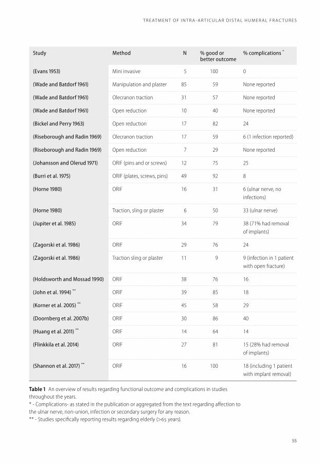
summary of these results over seven decades, table 1.
It is difficult to draw conclusions, especially when there are very few studies presenting results using a specific method and the fracture types treated varies substantially and are pre-sented in small numbers. Comparative studies are lacking and according to a Cochrane review there are no or insufficient evidence regarding
surgical treatment or not and if any method is superior over another (Wang et al. 2013).
In later studies the criteria used to define good result were of much higher standard and, with the surgical techniques used today most patients end up with a functional elbow with little or no pain. In general it appears that in recent years most patients have benefited from open reduc-tion and internal fixation. On the other hand, however, quite a few complications still occur, especially in the elderly population sustaining a complex intra-articular distal humeral fracture (Gofton et al. 2003, Obert et al. 2013).
The use of arthroplasty has therefore gained popularity as the primary treatment of distal humeral fractures in selected patients and this is the subject of the present doctoral thesis.
t r e At m e n t o f i n t r A - A r t i c u l A r d i s tA l H u m e r A l f r Ac t u r e s
54
t r e At m e n t o f i n t r A - A r t i c u l A r d i s tA l H u m e r A l f r Ac t u r e s
Study Method N % good or better outcome
% complications *
(Evans 1953) Mini invasive 5 100 0
(Wade and Batdorf 1961) Manipulation and plaster 85 59 None reported
(Wade and Batdorf 1961) Olecranon traction 31 57 None reported
(Wade and Batdorf 1961) Open reduction 10 40 None reported
(Jupiter et al. 1985) ORIF 34 79 38 (71% had removal of implants)
(Zagorski et al. 1986) ORIF 29 76 24
(Zagorski et al. 1986) Traction sling or plaster 11 9 9 (infection in 1 patient with open fracture)
(Holdsworth and Mossad 1990) ORIF 38 76 16
(John et al. 1994) ** ORIF 39 85 18
(Korner et al. 2005) ** ORIF 45 58 29
(Doornberg et al. 2007b) ORIF 30 86 40
(Huang et al. 2011) ** ORIF 14 64 14
(Flinkkila et al. 2014) ORIF 27 81 15 (28% had removal of implants)
(Shannon et al. 2017) ** ORIF 16 100 18 (including 1 patient with implant removal)
Table 1 An overview of results regarding functional outcome and complications in studies throughout the years.* - Complications- as stated in the publication or aggregated from the text regarding affection to the ulnar nerve, non-union, infection or secondary surgery for any reason.** - Studies specifically reporting results regarding elderly (>65 years).
55
Development of elbow arthroplasty
At the time of their publication, Street and Stevens found eleven reports on elbow arthroplasty performed on a total of thirty patients (Street and Stevens 1974). Most of the prostheses used were made of metal but acrylic (Mellen and Phalen), fig. 21 and 22, and nylon (Macausland) was also used (Mellen and Phalen 1947, Macausland 1954).
Figure 21 Acrylic hemi-arthroplasty used by Mellen and Phalen, frontal view. (Mellen and Phalen 1947). Reprint with permission.
Figure 22 Prosthesis used by Mellen and Phalen (Mellen and Phalen 1947). Lateral view. Reprint with permission.
The follow-up period were short: the average being less than a year excluding one case-report with a follow-up of thirteen years (Johnson and Schlein 1970). The largest series included twelve patients and was presented by Dee (Dee 1972). The initial prostheses were designed either as surface replacements or creating a hinge for the proximal ulna. This worked reasonably well in patients with preserved bone-stock and intact or reconstructable collateral ligaments, but not in patients with a highly unstable elbow due to loss of bone, due to trauma or rheumatoid arthritis (Dee 1975). Not being happy with the designs available, Dee constructed a hinged total elbow replacement made of chrome-cobalt, and was one of the first to use acrylic cement for fixation.
Street and Stevens presented an arthroplasty design that replaced only the articular surface of the distal humerus (Street and Stevens 1974), fig. 23. This was used in nine patients (10 elbows), 3 with rheumatoid arthritis, 2 due to ankylosis and 5 with post-traumatic arthritis. In their series the hemi-arthroplasty technique worked reasonably well for patients with post-traumatic conditions, but not in the elbow joint affected by arthritic disease.
Souter noted a high incidence of signs of loosening after implantation of hinged metallic
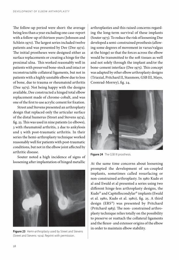
arthroplasties and this raised concerns regard-ing the long-term survival of these implants (Souter 1973). To reduce the risk of loosening Dee developed a semi-constrained prosthesis (allow-ing some degrees of movement in varus/valgus at the hinge) so that the forces across the elbow would be transmitted to the soft tissues as well and not solely through the implant and/or the bone-cement interface (Dee 1975). This concept was adapted by other elbow arthroplasty designs (Triaxial, Pritchard II, Stanmore, GSB III, Mayo, Coonrad-Morrey), fig. 24.
At the same time concerns about loosening prompted the development of un-coupled implants, sometimes called resurfacing or non-constrained arthroplasty. In 1980 Kudo et al and Ewald et al presented a series using two different hinge-less arthroplasty designs, the Kudo® and Capitellocondylar® implants (Ewald et al. 1980, Kudo et al. 1980), fig. 25. A third design (ERS®) was presented by Pritchard (Pritchard 1983). The non- constrained arthro-plasty technique relies totally on the possibility to preserve or reattach the collateral ligaments and the flexor- and extensor origins of the elbow in order to maintain elbow stability.
Figure 23 Hemi-arthroplasty used by Street and Stevens (Street and Stevens 1974). Reprint with permission.
Figure 24 The GSB III prosthesis.
d e v e lo p m e n t o f e l b o w A r t H r o p l A s t y
58
d e v e lo p m e n t o f e l b o w A r t H r o p l A s t y
In a series of 43 patients, Lowe et al had 8 patients with some degree of instability using a non- constrained implant but only one of these required revision (Lowe et al. 1984).
The semi-constrained hinged arthroplasty and non-constrained arthroplasty techniques described above are those that prevail today. Increased knowledge of the biomechanics of
Figure 25 The Kudo prosthesis.
the elbow has led to further improvements in prosthesis design and materials used. Significant loosening is still a problem and occurs in about 5% regardless of design and elbow instability is more frequent when using a non-constrained implant according to a review by Voloshin et al (Voloshin et al. 2011).
59
Arthroplasty in distal humeral
fracture treatment
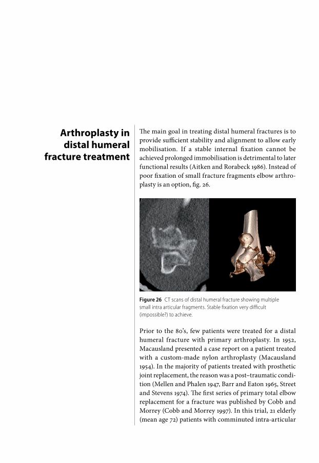
The main goal in treating distal humeral fractures is to provide sufficient stability and alignment to allow early mobilisation. If a stable internal fixation cannot be achieved prolonged immobilisation is detrimental to later functional results (Aitken and Rorabeck 1986). Instead of poor fixation of small fracture fragments elbow arthro-plasty is an option, fig. 26.
Figure 26 CT scans of distal humeral fracture showing multiple small intra articular fragments. Stable fixation very difficult (impossible?) to achieve.
Prior to the 80’s, few patients were treated for a distal humeral fracture with primary arthroplasty. In 1952, Macausland presented a case report on a patient treated with a custom-made nylon arthroplasty (Macausland 1954). In the majority of patients treated with prosthetic joint replacement, the reason was a post–traumatic condi-tion (Mellen and Phalen 1947, Barr and Eaton 1965, Street and Stevens 1974). The first series of primary total elbow replacement for a fracture was published by Cobb and Morrey (Cobb and Morrey 1997). In this trial, 21 elderly (mean age 72) patients with comminuted intra-articular
distal humeral fractures were treated with primary elbow arthroplasty (Coonrad-Morrey prosthesis). All patients had excellent or good functional results according to the Mayo Elbow Performance Score. One patient was revised (prosthetic fracture due to a fall). An early meta- analysis comparing series of elderly patients treated with either ORIF or arthroplasty con-cluded that primary arthroplasty produced results comparable to ORIF in the elderly. Of note is that at the time of analysis there had been only 46 patients that had been treated with primary arthroplasty and the longest follow- up was four years (Obremskey et al. 2003). Frankle et al published a retrospective review comparing ORIF and TEA, 12 patients in each group (Frankle et al. 2003). They concluded that TEA was preferable in elderly patients with poor bone-stock and co-morbidites. This study was not included in the meta-analysis by Obremskey et al.
In 2009 the first (and only) randomised trial comparing primary total arthroplasty and ORIF in elderly patients with a comminuted intra- articular distal humeral fractures was published (McKee et al. 2009). In this trial, 25% of the patients randomised to ORIF were converted to arthroplasty during the surgical procedure due to inability to achieve stable fixation. The number of complications was equal in the two groups and there was no statistically significant difference regarding the DASH-score. The Mayo Elbow Performance Score was significantly better in the arthroplasty group throughout the follow-up period. They recommended the use of arthroplasty in elderly patients with a commi-nuted intra-articular distal humeral fracture.
In the studies by Frankle et al and McKee et al, patients with arthroplasty had short fol-low-up times (52 and 24 months respectively), and loosening of the prosthesis is usually seen later. However, since the mortality rate within a 10-year period in this population is relatively
high revision is seldom necessary (Nestorson et al, unpublished data, paper IV).
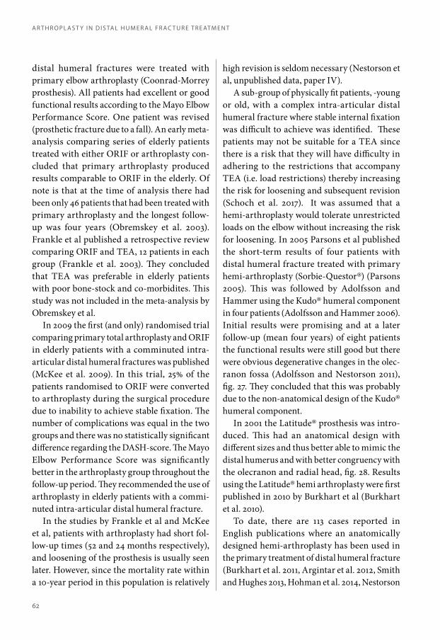
A sub-group of physically fit patients, -young or old, with a complex intra-articular distal humeral fracture where stable internal fixation was difficult to achieve was identified. These patients may not be suitable for a TEA since there is a risk that they will have difficulty in adhering to the restrictions that accompany TEA (i.e. load restrictions) thereby increasing the risk for loosening and subsequent revision (Schoch et al. 2017). It was assumed that a hemi-arthroplasty would tolerate unrestricted loads on the elbow without increasing the risk for loosening. In 2005 Parsons et al published the short-term results of four patients with distal humeral fracture treated with primary hemi-arthroplasty (Sorbie-Questor®) (Parsons 2005). This was followed by Adolfsson and Hammer using the Kudo® humeral component in four patients (Adolfsson and Hammer 2006). Initial results were promising and at a later follow-up (mean four years) of eight patients the functional results were still good but there were obvious degenerative changes in the olec-ranon fossa (Adolfsson and Nestorson 2011), fig. 27. They concluded that this was probably due to the non-anatomical design of the Kudo® humeral component.
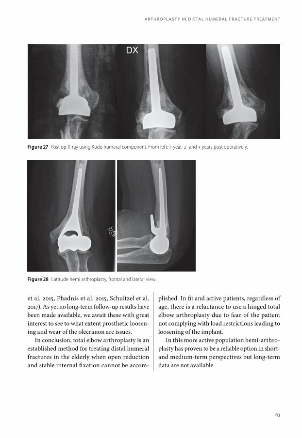
In 2001 the Latitude® prosthesis was intro-duced. This had an anatomical design with different sizes and thus better able to mimic the distal humerus and with better congruency with the olecranon and radial head, fig. 28. Results using the Latitude® hemi arthroplasty were first published in 2010 by Burkhart et al (Burkhart et al. 2010).
To date, there are 113 cases reported in English publications where an anatomically designed hemi-arthroplasty has been used in the primary treatment of distal humeral fracture (Burkhart et al. 2011, Argintar et al. 2012, Smith and Hughes 2013, Hohman et al. 2014, Nestorson
A r t H r o p l A s t y i n d i s tA l H u m e r A l f r Ac t u r e t r e At m e n t
62
A r t H r o p l A s t y i n d i s tA l H u m e r A l f r Ac t u r e t r e At m e n t
et al. 2015, Phadnis et al. 2015, Schultzel et al. 2017). As yet no long-term follow-up results have been made available, we await these with great interest to see to what extent prosthetic loosen-ing and wear of the olecranon are issues.
In conclusion, total elbow arthroplasty is an established method for treating distal humeral fractures in the elderly when open reduction and stable internal fixation cannot be accom-
plished. In fit and active patients, regardless of age, there is a reluctance to use a hinged total elbow arthroplasty due to fear of the patient not complying with load restrictions leading to loosening of the implant.
In this more active population hemi-arthro-plasty has proven to be a reliable option in short- and medium-term perspectives but long-term data are not available.
Figure 27 Post op X-ray using Kudo humeral component. From left: 1 year, 2- and 3 years post operatively.
Figure 28 Latitude hemi arthroplasty, frontal and lateral view.
63
Treatment of radial head fractures
There is little controversy regarding the conservative man-agement of non-displaced radial head fractures (Mason I), a principle that has not changed over many decades (Bohrer 1933, Ruchelsman et al. 2013). The displaced simple radial head fracture (Mason II) without mechanical block of forearm rotation can also be managed conservatively with good results (Akesson et al. 2006). Lindenhovius et al found that patients treated with open reduction and internal fixation had less degenerative changes in their injured elbow than those treated conservatively but com-plications and over all functional results did not warrant surgery (Lindenhovius et al. 2009).
Simple fractures with displaced fragments (Mason II) impeding forearm rotation have previously been success-fully treated with partial or total resection (sometimes at a later date) depending on the size of the fragment (Mason 1954, Watson-Jones 1960). As with distal humeral fractures internal fixation methods have improved and the preferred treatment of displaced simple radial head fractures is now internal fixation using screws, headless screws or resorb-able pins (Duckworth et al. 2011).
More complex fractures where the radial head is split into multiple fragments (Mason III) are more difficult to treat, and the management is mainly governed by associ-ated injuries. The aim of treatment is to maintain a stable elbow and forearm with preserved range of movement in all directions.
In a radial head fracture combined with elbow disloca-tion (Mason IV) fig. 29, the stability of the elbow is jeop-ardised because of concomitant ligament injury, usually both lateral and medial, and sometime associated injury to the flexor- and extensor origins.
In cases with no other bony injury, this can be treated with excision of the radial head, repair of the lateral collateral ligament and immo-bilization for 3-4 weeks in a cast making sure that ulno-humeral congruence is maintained. This ensures healing of the medial collateral ligament, and valgus stability will be re-estab-lished as long as the flexor muscle origins are intact. Repair of the lateral ligament will prevent postero- lateral rotatory instability(O’Driscoll et al. 1991). When keeping to the treatment princi-ples above, late elbow instability is a rare event but an increased valgus carrying angle develops over time in patients treated with radial head resection (Sanchez-Sotelo et al. 2000, Antuna et al. 2010). Furthermore the frequency of degen-erative changes is high and proximal migration of the radius is seen but functional results are reportedly good in most cases (Herbertsson et
al. 2009, Antuna et al. 2010, Karlsson et al. 2010, Nestorson et al. 2017).
When scouring the literature, most Mason IV fracture dislocations with a non-recon-structable radial head fracture are treated with radial head replacement and re-attachment of ligaments (Doornberg et al. 2007a, Flinkkila et al. 2012, Marsh et al. 2016). A problem when interpreting the results of these studies is that several different types of injury to the elbow and forearm with associated radial head fracture are included. There is only one study that has com-pared outcomes between replacement and radial head resection in the treatment of true Mason IV fracture dislocations and it did not show any statistical difference regarding functional outcome (Nestorson et al. 2017).
A comminuted radial head fracture (Mason III) and disruption of the inter-osseous mem-brane between the radius and ulna causes longi-tudinal forearm instability and incongruence of the distal radio-ulnar joint leading to wrist pain if the length of the radius cannot be preserved. Curr and Coe described the injury in 1946 but it is named after another author (Essex-Lopresti) who described the consequence of radial head resection in these circumstances (Curr and Coe 1946, Essex-Lopresti 1951). This clinical obser-vation prompted the development of radial head replacement to prevent proximal migration of the radius after resection of the radial head.
To achieve this Speed developed “ferrule caps” to replace the radial head (Speed 1941). The follow-up period in this publication was short, but later studies have confirmed reduction in morbidity at the wrist by reducing proximal migration of the radius using a radial head replacement (Edwards and Jupiter 1988, Knight et al. 1993).
Another patho-mechanic scenario is frac-tures of both the radial head and the coronoid in combination with elbow dislocation, the so called “terrible triad” (Hotchkiss 1996). Not
Figure 29 Mason IV fracture dislocation. Radial head fracture with concomitant dislocation of the elbow.
t r e At m e n t o f r A d i A l H e A d f r Ac t u r e s
66
t r e At m e n t o f r A d i A l H e A d f r Ac t u r e s
only must the radial head fracture and the soft tissue injury be addressed but also, in many cases, the coronoid fracture. Historically these injuries have had a poor outcome (Bakalim 1970, Josefsson et al. 1989, Heim 1998, Ring et al. 2002a). Depending on the size of the fragment and part of the coronoid involved, different aspects of elbow instability must be consid-ered and the radial head fracture addressed if posterior, valgus or postero-lateral stability is in jeopardy. The anterior part of the coronoid protects against posterior dislocation of the elbow, the medial part (sublime tubercle) resists varus load and together with the medial collat-eral ligament protects against postero-medial rotatory instability. The lateral part together with the radial head resists valgus instability (Ring and Jupiter 1998). Small fragments from the tip of the olecranon (Regan-Morrey I) are of minor relevance for elbow stability but avulsion is an indicator of anterior soft tissue trauma that sometimes needs to be addressed in cases of gross instability, i.e. when capsule, ligaments and muscle-origins are all torn. If the coronoid fragment is larger (Regan-Morrey II) there is some evidence for leaving the coronoid fragment without internal fixation as long as the radial head can be preserved or replaced (Ring et al. 2002a, Papatheodorou et al. 2014). A frac-ture involving the base of the coronoid and the
sublime tubercle (Regan-Morrey III) needs to be treated with open reduction and internal fixa-tion to reduce the risk for posterior-, varus- and posterio-medial rotatory instability, (Regan and Morrey 1989, Ring 2006).
Stable internal fixation of the coronoid is difficult to achieve and the fixation must be protected in some way while the fracture heals to reduce the risk for future elbow instability. Stable fixation of the radial head fracture or, if repair is not possible, a radial head replacement will reduce the load on the coronoid and protect a fracture fixation (Pugh et al. 2004).
In extreme cases, instability might ensue despite primary triple stabilisation (radial head, coronoid and collateral ligaments) of the elbow. In such cases external fixation, either hinged or static, might be required to keep the elbow congruent (McKee et al. 1998, Pugh et al. 2004). It cannot be determined whether instability is due to sub-optimal surgery in combination with injuries to the dynamic stabilizers (extensors, flexors, brachialis, biceps and triceps) or with temporary palsy of these muscles, since this aspect has not specifically been investigated. In a severely unstable dislocation without frac-ture, detachment of the ligaments, capsule and both the flexor and extensor origin have been described probably explaining the gross insta-bility (Adolfsson et al. 2017).
67
Development and use of radial head prostheses
As mentioned above, Speed used “ferrule caps” to replace the radial head (Speed 1941). Several materials have been used since: acrylic (Cherry 1953, Edwards and Rostrup 1960), silicone (Mackay et al. 1979, Swanson et al. 1981) and vitallium (Knight et al. 1993). Silicone was found not to withstand the forces across the radio-capitellar joint and the predominant radial head prostheses are now made of chrome-cobalt or titanium alloys (Carn et al. 1986).
There are at present several designs available, most of them modular with different sizes regarding stem, radial neck length and radial head diameter, to accommodate for the anatomic variations between patients. Two concepts exist regarding the stem: a smooth stem that is supposed to be able to rotate within the remaining medullary canal of the proximal radius (e.x Evolve®) which together with the head replacement works as a “true” spacer (King and Patterson 2001). The second concept is to have a securely fixed stem, either by press-fit (e.g. Acumed Ana-tomical Radial Head ®), fig 30 and 31, screw expansion (MoPyc®) or cement (Judet®) in an attempt to mimic the normal anatomy.
There are also two concepts regarding the connection between the stem and the radial head: rigid (mono-polar) or floating (bi-polar where the head is able to move in relation to the stem). Usually both concepts have a built-in angle between the stem and neck as with the native proximal radius of approximately 15°. Anatomical studies have been made on mono-polar and bi-polar implants, and none is able to replicate the physiological kinematics of the radial head (Wegmann et al. 2014, Shannon et al. 2015).
When considering reports related to radial head replacement, the reader must bear in mind that focus has been on the implant, but there is a host of associated injuries where the common denominator is a radial head fracture. Residual elbow instability is hard to define and is rarely included in the assessment reported. The out-comes that have been presented vary but usually include range of motion, pain, strength and later PROM’s and aggregated scores (MEPS).
In recent years three reports have been pub-lished with regards to these different concepts. Flinkkila et al reviewed 42 patients with a “press-fit” monopolar implant after a mean of 50 months. Of these 37 had an x-ray and 31 patients a clinical assessment. They found a mean MEPS of 86 and a DASH score of 23, residual insta-bility was not mentioned. Nine implants had been removed and a further three showed signs of loosening (Flinkkila et al. 2012).
Allavena et al presented the result of 22 patients with a bipolar implant after a mean follow-up of 50 months. The mean MEPS was 79, quick-DASH score 21, and 3 patients had residual postero-lateral instability, and four implants had been removed during the observation time (Allavena et al. 2014).
Finally Marsh et al presented their results of 51 patients with a smooth-stemmed mono-polar implant after a mean follow-up of 8 years. The mean MEPS was 91, residual instability was not
Figure 30 Press-fit radial head replacement (Acumed, Anatomical Radial Head®). Frontal view. Two suture-anchors in the lateral epicondyle for reattachment of lateral collateral ligament.
Figure 31 Lateral view.
d e v e lo p m e n t A n d u s e o f r A d i A l H e A d p r o s t H e s e s
70
d e v e lo p m e n t A n d u s e o f r A d i A l H e A d p r o s t H e s e s
reported, 25 implants showed signs of radio-graphic loosening (part of the design) but no implant had been removed (Marsh et al. 2016).
A meta-analysis by Heijink et al did not show
any difference in overall complication rates or elbow function with regards to the implant designs used (Heijink et al. 2016).
71
Complications after elbow
fracture treatment
Complications are often reported in studies on elbow trauma but definitions are often lacking and interpreta-tions vary. Reduced range of motion, for example, is very common after an elbow fracture: in fact it’s so common that, before we initiate treatment, our patients are often told that some loss of motion is to be expected. Is this the result of the injury or of the treatment? The answer to this is probably both but how much must reduction be before we call it a complication? Unfortunately there is no clear consensus and this is probably one of the main reasons for the great variation in complication rates (15-48%) in reports published (Pollock et al. 2008, Flinkkila et al. 2014).
POSTOPERATIVE INFECTIONS
In a meta-analysis of distal humeral fractures treated with ORIF Obert et al reported an infection rate of 3-6%. For distal humeral fractures treated with primary arthroplasty the corresponding figure was 4-10% (Obert et al. 2013). These are overall infection rates and do not take into account the extent to which further surgery was needed. According to a systematic review by Voloshin et al the infection rate after elective elbow arthroplasty was 3,3 ±2,9% (Voloshin et al. 2011).
ULNAR NERVE AFFECTION
Subjective sensory disturbance of the little finger is quite a common complaint after injury to and surgical treatment of a distal humeral fracture. The reported postoperative incidence of ulnar nerve affection varies widely between 0 and 51% (Holdsworth and Mossad 1990, Sodergard et al. 1992, Wang et al. 1994, Kundel et al. 1996, Gupta and
Khanchandani 2002, Soon et al. 2004, Shin and Ring 2007, Wiggers et al. 2012). Svernlov et al found that 27% of patients had ulnar nerve affection after 48 months, but interestingly this was regardless of whether the medial column was involved in the fracture and secured with a plate and screws or not (Svernlov et al. 2017). Vasques et al came to the conclusion that it may be the trauma itself that causes ulnar nerve dys-function, though it should be mentioned that the main dysfunction is sensory, and motoric dysfunction is rare (Vazquez et al. 2010).
To reduce the risk of ulnar nerve affection some authors advocate anterior transposition, especially in patients with pre-operative symp-toms from the ulnar nerve (Ruan et al. 2009) while others have found no increased incidence when leaving the nerve in situ (Vazquez et al. 2010, Worden and Ilyas 2012). In a meta-analysis comparing treatment of distal humeral fractures with either ORIF or arthroplasty there was no significant difference in ulnar nerve dysfunction (6,4% compared to 6,9%) (Githens et al. 2014).
We perform in-situ decompression of the ulnar nerve without transposition if a dorsal or medial approach is used when treating a distal humeral fracture with ORIF or arthroplasty.
STIFFNESS-HETEROTOPIC BONE FORMATION (HO),fig. 32.
Reduced range of motion can be expected after sustaining an elbow fracture. A range of 100° in flexion-extension is considered functional (Morrey et al. 1981). Re-operation rates for reduced movement due after distal humeral fractures ranges from 0-15% judging from recent published reports (Shin et al. 2010, Theivendran et al. 2010, Huang et al. 2011, Abrams et al. 2012, Simone et al. 2014). In our own register-based study review-ing patients treated for a distal humeral fracture with primary arthroplasty 8 of 404 (2%) patients
had a surgical arthrolysis performed (Nestorson et al, unpublished data, paper IV).
In a report by Duckworth et al 11% had a radial head replacement removed due to stiffness with or without pain (Duckworth et al. 2014).
Heterotopic bone formation is quite common but usually does not restrict motion enough to justify surgical intervention. The classification by Brooker et al developed to grade heterotopic bone formation around the hip after arthroplasty is frequently used to classify changes around other joints as well (Brooker et al. 1973). Three grades are described: Grade I: small islands of bone formation, often seen on x-rays around the collateral ligaments, triceps insertion and around the proximal radius (after resection of the head) after an elbow fracture: Grade II: bone formation within adjacent muscle: Grade III: a bony bridge across the joint.
In our series of distal humeral fractures treated with primary elbow hemi-arthroplasty, 19 of 41 patients (46%) had Grade I HO at follow- up. Four patients had undergone secondary surgery (10%) prior to final follow-up because of restricted movement (Nestorson et al. 2015). Abrams et al reported a 62% prevalence 6 months after surgery for an intra-articular elbow frac-ture, and 9% had had secondary surgery with removal of bone and arthrolysis (Abrams et al. 2012). There are no distinct criteria as to when
Figure 32 Heterotopic bone formation within the brachialis muscle, Grade II, after surgery for Mason IV fracture dislocation.
co m p l i c At i o n s A f t e r e l b o w f r Ac t u r e t r e At m e n t
74
co m p l i c At i o n s A f t e r e l b o w f r Ac t u r e t r e At m e n t
secondary surgery should be done because of stiffness, it depends on the functional demands of the patients.
LOSS OF REDUCTION, MAL-UNION AND NON-UNION(fig. fig 33 and 34).
Early loss of reduction appears to have been more common before the development of pre-contoured plate implants. Sodergard et al reported early loss of fixation in 16 of 96 patients with a mean age of 55 (Sodergard et al. 1992). Twenty-one years later, Clavert et al reported a series of 53 patients older than 65 years where 5 patients suffered early loss of reduction (Clavert et al. 2013). Huang et al reported no loss of reduc-tion in a series of 23 patients aged 65 or older (Huang et al. 2011).
Non-union occurs in about 8% but does not always require secondary surgery (Obert et al. 2013, Flinkkila et al. 2014, Simone et al. 2014). When surgery is needed, bone-grafting with revision plating will often lead to union (Helfet et al. 2003). In elderly patients, arthroplasty revi-sion may be an option (Cil et al. 2008, Smith and Hughes 2013).
Mal-union is quite frequent. In the French SOFCOT series from 2012, 50% of patients treated with internal fixation had intra-articu-lar (definition not stated) or extra-articular mal-union defined as >15° in the frontal plane and 10° in the sagittal plane (Obert et al. 2013).
Figure 32 Collapse of the distal humeral articular surface, frontal view.
Figure 34 Lateral view.
75
PROSTHETIC LOOSENING(fig. 35 and 36)
Aseptic loosening, as previously mentioned, occurs in about 5% after total elbow arthro-plasty (Voloshin et al. 2011). We found a 4% loosening rate amongst 405 patients when elbow arthroplasty had been used as primary treatment for distal humeral fractures. The mean follow-up time was only 67 months. Aseptic loosening usually occurs at a later stage and the rate might increase with longer obser-vation time (Nestorson 2017, unpublished data, paper IV), fig 36.
Radial head arthroplasty is associated with a higher rate of loosening. Marsh et al reported 45% of implants showing signs of loosening but this is considered to be part of the design with smooth-stemmed implants. No implant was removed in their series of 55 patients (Marsh et al. 2016). Using a design with a stem that is meant to be fixed in the proximal radius med-ullary canal (press-fit) Flinkkila et al reported a loosening rate of 32% and 24% of the implants had to be removed (Flinkkila et al. 2012), fig 37. Allavena et al, using a bi-polar implant with a fixed stem, reported 41% with signs of loosening and 18% of implants required removal (Allavena et al. 2014).
IMPLANT REMOVAL
When treating distal humeral fractures with ORIF the plates often become prominent over the lateral and medial condyles. This is because these bony structures are only covered with skin and subcutaneous tissue. An additional plate may also have been placed on the subcutaneous part of the proximal ulna after an olecranon osteotomy used to visualise the fracture. The question is: should this be defined as a com-plication or just a consequence of treatment, considering the poor soft tissue coverage of the epicondyles and olecranon?
Depending on the degree of subjective dif-ficulty, prominent plates and screws can be removed as long as the fracture has healed. In recent reports, implant removal varies between 0-28% with the plates on the proximal ulna being the most frequently removed (Theivendran et al. 2010, Huang et al. 2011, Flinkkila et al. 2014, Patel et al. 2017).
INSTABILIT Y
Residual instability following a distal humeral fracture is a great challenge and restoration of skeletal and ligamentous stabilisers are an essential part of initial management. It is mainly
Figure 36 Loosening of radial head replacement with a press-fit stem.
co m p l i c At i o n s A f t e r e l b o w f r Ac t u r e t r e At m e n t
76
co m p l i c At i o n s A f t e r e l b o w f r Ac t u r e t r e At m e n t
associated with non- or mal-unions of single column fractures, type B according to the AO classification, (Jupiter 1993). In our follow-up of 42 patients treated with primary hemi- arthroplasty one patient had secondary surgery (reconstruction of the lateral ulnar collateral ligament) because of instability (Nestorson et al. 2015).
Publications on patients with an injury related to the elbow (dislocation, terrible triad, trans-olecranon fracture) with concomitant radial head fracture often omit separate report-ing of instability assessment. Instead it is usually included as a component of a score (one item in MEPS) and only the overall score is presented. In a review of radial head arthroplasty, of 21 publications found only 13 had assessed stabil-ity and no residual instability reported in these studies (Kaur et al. 2014). In our series com-paring the results of radial head resection and radial head arthroplasty for Mason IV fracture dis locations one patient in the arthroplasty group had increased elbow joint laxity but was subjectively stable, and two patients treated with radial head resection complained of instability (Nestorson et al. 2017).
It is not possible to distinguish from the literature the extent to which treatment of the radial head fracture matters concerning risk for residual instability, but this is an area of controversy. Some authors recommend radial head arthroplasty for all fractures that are not amenable for conservative treatment or ORIF, so as to reduce the risk for future instability and proximal migration of the radius (Ruchelsman et al. 2013, Allavena et al. 2014, Duckworth et al. 2014, Heijink et al. 2016).
Regardless of treatment good results are probably the result of a better understanding of the biomechanics of the elbow i.e. that not only the radial head fracture but also associated frac-tures and soft tissue injuries must be addressed in the treatment of complex elbow injuries.
SECONDARY OSTEOARTHRITIS
Secondary arthritis may occur in intra-articu-lar fractures treated with ORIF or partial joint replacement but is not a concern when total arthroplasty is used.
The degree of arthritis and how fast it devel-ops depends on the quality of reduction of the joint surfaces and congruence of an implant, but can occur even after perfect reduction and fixa-tion (Gofton et al. 2003, Doornberg et al. 2007b). Damage to the sub-chondral blood-supply can lead to avascular necrosis and collapse of the joint surface months after injury and treat-ment. In a series reported by Theivendran et al 6 patients of 16 (38%) had secondary osteoarthri-tis and Huang et al reported 14% after 35 and 51 months follow-up respectively (Theivendran et al. 2010, Huang et al. 2011).
Using non-anatomical implant attrition was seen in 3 out of 8 patients after a mean follow-up of four years (in two patients as early as after two years) (Adolfsson and Nestorson 2012). The problem of olecranon wear appeared to be less: 12% showing mild radiographic signs of reduc-tion of cartilage thickness after a slightly shorter follow-up (mean of 34 months) when using an anatomical implant.
In patients treated with radial head resection the rate of ulno-humeral osteoarthritis is quite high. Antuna et al reported 100% mild to mod-erate secondary ulno-humeral osteoarthritis after a mean follow-up of 25 years (Antuna et al. 2010). No series with such a long follow-up after radial head replacement is available but secondary osteoarthritis in the ulno-humeral joint has been reported in several publications using different implant designs (Grewal et al. 2006, Doornberg et al. 2007a, Marsh et al. 2016). Attrition of the capitellum has also been reported, in some cases associated with lateral elbow pain necessitating removal of the implant (Flinkkila et al. 2012, Allavena et al. 2014, Marsh et al. 2016).
77
Secondary osteoarthritis in our series was less frequent in the group treated with radial head replacement but the mean follow-up time was shorter compared to the group treated with radial head resection (Nestorson et al. 2017).
MECHANICAL FAILURE OF ARTHROPLAST Y IMPLANTS(hinge or bushings)
Clinically significant wear of bushings, leading to revision, occurs in about 2% (Voloshin et al. 2011). This is unusual in the first five years and is more frequent in patients younger than sixty years treated with TEA for secondary osteoar-thritis (Throckmorton et al. 2010). Polyethylene debris can also lead to aseptic loosening as pre-
viously described in hip and knee arthroplasty (Harris 2001, Goldberg et al. 2008). Disassembly or fracture of the implant is rare. Platschke et al reported less then 1% prosthetic fracture and dissasembly rate and Voloshin et al reported a prosthetic fracture rate of 2,9% and disassembly rate of 2,3% in their reviews (Voloshin et al. 2011, Plaschke et al. 2014). A higher rate of prosthetic fracture (6%) was reported by Throckmorton et al in a series of patients treated with TEA for secondary osteoarthritis (Throckmorton et al. 2010).
According to Fevang et al patients treated with total elbow arthroplasty for post-traumatic changes had a 5,8-times higher risk for revision (for any reason) than patients treated for inflam-matory arthritis (Fevang et al. 2009).
co m p l i c At i o n s A f t e r e l b o w f r Ac t u r e t r e At m e n t
78
Aims The overall aim was to evaluate the use of arthroplasty as primary treatment of comminuted distal humeral fractures and the treatment of comminuted radial head fractures with associated elbow dislocation (Mason IV). Specific aims were:
I: To evaluate the medium-term functional and radiographic outcome of hemi-arthroplasty as primary treatment of comminuted intra-articular distal humeral fractures using a non-anatomical prosthesis (Kudo®).
II: To evaluate the medium-term functional and
radiographic outcome of hemi-arthroplasty as primary treatment of comminuted intra-articular distal humeral fractures using an anatomical shaped implant (Latitude®).
IV: To investigate the use of arthroplasty in Sweden as
primary treatment of distal humeral fractures with regards to revision and adverse event rates.
III: To evaluate the functional and radiographic outcome after either radial head resection or radial head replacement in comminuted radial head fractures with associated elbow dislocation (Mason IV).
Patients Paper I Eight women (mean age 79) consecutively treated for a distal humeral fracture with primary hemi-arthroplasty. Inclusion criteria were: living independently: ability to adhere to post-operative restrictions: x-ray showing osteopenia: and an intra-articular distal humeral fracture where stable internal fixation was deemed impossible to achieve. At the latest follow-up two patients had died and one suffered from dementia.
Paper II Forty-two consecutive patients treated at three different hospitals (Linköping University Hospital, Sahlgrenska University Hospital and Varberg Hospital) with primary hemi-arthro-plasty due to an intra-articular distal humeral fracture. Mean age was 72 (range 56-84) years, 39 women and 3 men. The inclusion criteria were the same as in Paper I.
Paper III Eighteen consecutive patients with Mason IV fracture dislocation treated with radial head replacement and lateral ligament repair at Linköping University Hospital. Median age 56 years (19-79).
Fourteen consecutive patients with Mason IV fracture dislocation treated with radial head resection and lateral ligament repair at Malmö University Hospital. Median age 50 (29-70). Inclusion criteria were: irreparable radial head fracture with concomitant elbow dislocation: a coronoid fracture no higher than Regan- Morrey Grade I. Patients complaining of wrist pain at presentation were excluded.
Paper IV A register-based study identifying 405 patients (406 elbows) treated with primary arthroplasty for distal humeral fracture. Mean age at surgery was 75 (SD 10) years: 365 women and 40 men.
Methods Paper I Between 2004 and 2007 eight women with an intra-articular distal humeral fracture, where stable internal fixation was deemed impossible, were consecutively treated with primary hemi-arthroplasty using the humeral component of the Kudo® prosthesis. A resting splint was used post operatively and range of motion and load gradually increased under the supervision of a physiotherapist. No restrictions were applied after three months.
After a mean follow-up of 4 (2,5-6) years clinical and radiographic evaluation was performed.
Measurements included range of motion and MEPS was used to assess functional outcome. Results presented separately for each patient.
The degree of secondary osteoarthritis (mainly changes in the olecranon fossa) was recorded according to the Broberg-Morrey classification (Broberg and Morrey 1986).
Paper II A prospective consecutive series of 42 patients with a distal humeral fracture, not suitable for ORIF, treated with primary hemi arthro-plasty (Latitude®) between January 2007 and September 2012. Post-operative protocol as in Paper I. Mean follow-up of 34 months (24-61). The fractures were classified according to the AO classification using the initial x-rays and CT scans.
Clinical examination included assessment of range of motion, stability of the elbow joint, and ulnar nerve function. The MEPS and DASH scores were used to grade functional outcome. Plain x-rays were taken and signs of prosthetic loosening were classified according to the protocol adapted by Smith and Hughes, heterotopic bone formation according to
the Broker classification and the degree of olecranon wear was graded according to the Broberg-Morrey classification (Brooker et al. 1973, Broberg and Morrey 1986, Smith and Hughes 2013).
Results are presented as mean (range) and radiographic findings separately for each patient.
Paper III Carried out at two hospitals, Linköping and Malmö, using two different treatment strategies. Two separate series of consecutive patients with a Mason IV fracture dislocation with irreparable radial head were retrospectively reviewed. In Malmö treatment consisted of radial head resection, repair of the lateral collateral ligament and immobilisation using either a plaster cast or external fixation for 3-4 weeks. In Linköping the patients were treated with radial head replacement, lateral collateral ligament repair and immobilisation varying from two days to three weeks.
The mean follow-up time differed between the groups, 58 (26-181) months in Linköping and 108 (47-136) in Malmö. The initial treatment was provided between Januari 2002 and December 2011.
MEPS and DASH scores were used as measurements of functional outcome. Range of motion, signs of instability, affection of the ulnar nerve and secondary surgery were recorded. Plain x-rays were taken to assess signs of pros-thetic loosening (Linköping) according to Popovic et al, degree of osteoarthritis according to the classification by Broberg and Morrey, and location of any heterotopic bone formation according to the Brooker classification (Brooker et al. 1973, Broberg and Morrey 1986, Popovic et al. 2007).
Comparisons between the groups were made with regards to MEPS and DASH scores using the Mann-Whitney U test, and the rate of osteoarthritis using the Chi 2 square test.
m e t H o d s
86
m e t H o d s
Paper IV Three different registers were used to identify patients, who had been treated with primary elbow arthroplasty for a distal humeral fracture:
Swedish Elbow Arthroplasty Register, Swedish Board of Health and Welfare (Socialstyrelsen) inpatient register, and local registers at all the orthopaedic departments in Sweden. The initial treatment observation period was January 1999 until end of June 2014. To reduce the risk of not identifying patients suffering an adverse event related to the elbow, any re-admission to an orthopaedic department was sought for in the above three registers between January 1999 and end of March 2016. Any discrepancies were sorted by checking the individual patient’s hospital files.
Using prosthetic survival as the dependent variable, the potential relation of patient age and sex, or prosthetic design was examined using Cox regression analysis with Kaplan-Meier plots. Comparison of implant survival between hemi- and total elbow arthroplasty was performed for the Latitude® system since this was the predominant system used for both modalities. Rates of revision and adverse events at institutions having performed more or less than 10 procedures were reported. Infections not necessitating revision of the arthroplasty, peri-prosthetic fractures and secondary surgery due to instability or stiffness were reported as separate, adverse events.
87
Results Paper I The functional outcome measured with MEPS ranged from 85 to 95 (good or excellent) and the mean ROM was 31-126° (range 15-135). Table 2. Ranges of pronation and supination were equal to the un-injured side. Three patients had radiographic changes in the olecranon fossa but no signs of instability and this did not infer with the functional outcome, table 2.
Pat nr ROM MEPS
12345678
25-13030-12535-12525-13055-11530-12515-13535-135
9585909585859595
Distal humeral fractures treated with primary elbow hemi arthroplasty using a non- anatomical implant yielded good functional results with an acceptable rate of compli-cations compared to reported results of treatment with open reduction and internal fixation and treatment with total elbow arthroplasty at medium-term follow-up.
Progressive radiographic changes in the olecranon are a concern.
Paper II The mean arc of movement was 105,5° (60-145°). The functional outcome measured with DASH score and MEPS resulted in a mean of 20 (0-63) and 90 (50-100) respectively. Mild olecranon wear was seen in five of the patients (12%) but did not influence the functional outcome. One arthroplasty was revised due to
Table 2 Functional outcome. ROM – Range of motion, MEPS – Mayo Elbow Performance Score
aseptic loosening, four patients had undergone resection of heterotopic bone formation to increase range of movement and one patient lateral ligament reconstruction for recurrent instability. Three patients had ulnar nerve affection, one was a multi-trauma patient with a “floating” elbow, one had pre-existing diabetes mellitus and associated polyneuro-pathy, and the third had ulnar symptoms prior to surgery.
Distal humeral fractures deemed not to be suitable for ORIF, and thus primarily treated with an anatomically shaped hemi-arthroplasty have predictable short- to medium- term outcomes. One can expect some loss of motion, especially in extension, and the rate of secondary surgery and complications do not differ significantly compared to other methods of treating distal humeral fractures.
Paper III There was no significant difference in functional outcome between the groups treated with radial head resection (Group M) and that with radial head replacement (Group L). The median MEPS in Group M was 100 (50-100) and the median DASH score 12 (0-44). The corresponding results were 85 (65-100) and 13 (0-63) in Group L.
The median arc of movement in Group M was 127,5° (105-150) and in Group L 130° (95-155). Two patients had reduced forearm rotation in Group M and nine in Group L of which one developed a forearm synostosis.
No patient in either group had secondary surgery due to instability. Two patients had secondary surgery due to stiffness in Group M and four patients in Group L had additional surgery in a total of seven times, three because of prosthetic loosening and one due to synostosis.
There were more osteoarthritic changes in Group M but the follow-up time in this group was longer. Prosthetic loosening was seen in four patients in Group L but this did not cause symptoms justifying revision.
The use of a radial head implant did not improve the functional results after sustaining a Mason IV fracture dislocation. Follow-up time differed making conclusion regarding secondary osteoarthritic changes difficult.
r e s u lt s
90
r e s u lt s
Paper IV A total of 405 patients were identified from three different register sources, (Swedish Elbow Arthroplasty Register, National Board of Health and Welfares inpatient Register and local registers at all Swedish orthopaedic departments). No register had complete coverage regarding primary surgery. The patients were treated at 29 different hospitals of which 14 had 10 procedures or more. The mean age at surgery was 75 (SD 10) years: 365 women and 40 men. Of these, 186 patients died during the observation period.
Six different implants were used of which two were used once only and these were excluded from further analysis, table 3.
Table 3 Overview of implants and demographics.
Implant N Mean age Gender (male) Year used
C-M 144 77 12 1999-2014
Discovery 79 76 10 2005-2014
GSB 36 75 6 2000-2012
Kudo total 25 73 2 1999-2008
Kudo hemi 9 78 0 2004-2009
Latitude total 35 72 4 2008-2014
Latitude Hemi 76 72 6 2008-2014
Total elbow arthroplasty was used in 319 patients and hemi-arthroplasty in 85. In recent years the number of elbow arthroplasties for distal humeral fracture has remained steady at around 40 each year. The number of total arthroplasties has decreased and the number of hemi-arthroplasties has increased correspondingly, fig. 36.
91
45
40
35
30
25
20
15
10
5
01999 2000 2001 2002 2003 2004 2005 2006 2007
Year (2014 �rst 6 months)
2008 2009 2010 2011 2012 2013 2014
Th e overall cumulative 5-year survival rate was 99% (CI 98-100%) and the ten-year rate 90% (CI 85-96%). Notable is that 46% of the patients died under the observation period and only 45 patients were followed for 10 years or more. Increasing age reduced the risk for revision.
Th ere were 18 revisions in 18 patients, 16 aft er total arthro-plasty and 2 aft er hemi arthroplasty. Seventeen patients with total arthroplasty and 9 patients with hemi arthroplasty suff ered an adverse event or undervent secondary surgery with infection, stiff ness and peri-prosthetic fracture being the most common indications.
Primary elbow arthroplasty as treatment for distal humeral fractures produces a reliable outcome in terms of acceptable revision and adverse event rates.
Figure 36 Number of arthroplasties per year as primary treatment for distal humeral fracture. Blue- total number, red- total elbow arthroplasies, green- hemi-arthroplasties. NB! 2014 only the fi rst 6 months.
r e s u lt s
92
treAtment of distAl HumerAl frActuresPrimary elbow arthroplasty for distal humeral frac-tures have been investigated in three papers included in this thesis. Arthroplasty in general produces reliable results with regards to revision and adverse event rates. With regards to hemi-arthroplasty the functional out-come has also been assessed and is comparable to other methods, such as ORIF and total elbow arthroplasty, for treating distal humeral fractures in short- to medium- term follow-up.
A basic principle when comparing different methods of treating a fracture is that studies have applied the same injury classification systems. In the case of the most com-plex distal humeral fractures, C3 and B3 according to AO, recent studies on internal fixation reports good functional results with an acceptable complication rate (Flinkkila et al. 2014, Tarallo et al. 2015, Shannon et al. 2017). In our study including 42 patients treated with primary hemi- arthroplasty 38% of the fractures could not be classified according to the AO or the Jupiter classification systems. This was mainly due to the fact that intra-articular frac-ture lines existed in all three planes, which is not taken into account in existing classification systems. Retrospec-tive comparisons are thus difficult to perform: fractures included in various reports might not be the same, and their results should be interpreted accordingly. There is only one prospective randomised trial comparing ORIF with arthroplasty as primary treatment for intra-articu-lar distal humeral fractures. They reported a 25% intra- operative crossover to total elbow arthroplasty in patients allocated to ORIF since stable internal fixation could not be achieved (McKee et al. 2009). This is in line with what we
Discussion
have experienced and supports the notion that not all fractures are suited for internal fixation and that primary arthroplasty may be the best option for these patients.
There are several reports on the use of total elbow arthroplasty as primary treatment for distal humeral fractures and it has become an established method (Kamineni and Morrey 2004, Kalogrianitis et al. 2008, Antuna et al. 2012). The overall complication rate is compa-rable to total arthroplasty used for rheumatoid arthritis elbows and significantly lower than total arthroplasty used for post-traumatic sequelae (Voloshin et al. 2011).
Some surgeons recommend load restrictions after implantation of a total elbow prosthesis in order to reduce the risk of loosening or mechani-cal failure of the implant (Barlow et al. 2013). In patients with low functional demands this is probably not a problem but elderly patients that remain highly active and younger patients that are still working are likely to find these restrictions most inconvenient and will proba-bly have poor compliance. The benefit of hemi- arthroplasty is that there are no load restrictions after the initial post-operative period, and has therefore been suggested to be a better option in patients with high functional demands (Parsons 2005). We have reported the outcome and com-plications of hemi-arthroplasty in 50 patients, 8 using the Kudo® humeral implant and 42 the Latitude® implant. The range of motion, func-tional outcome assessed by MEPS and DASH scores and complication rates compared well with recent reports using other methods to treat distal humeral fractures regardless of fracture severity (Mansat et al. 2013, Sorensen et al. 2014, Tarallo et al. 2015, Shannon et al. 2017).
The method is still controversial since pub-lications on its use are still sparse. All in all the outcomes of about 130 patients have been reported in English journals but no long-term follow-up results are available. (Burkhart et al.
2011, Adolfsson and Nestorson 2012, Smith and Hughes 2013, Hohman et al. 2014, Heijink et al. 2015, Nestorson et al. 2015, Phadnis et al. 2015, Smith et al. 2016, Schultzel et al. 2017).
Early wear of the olecranon was seen using the Kudo® humeral component. However, when using a more anatomical design (Latitude®) the amount of wear was seen to be less at medi-um-term follow-up (Adolfsson and Nestorson 2012, Nestorson et al. 2015).
A meta-analysis comparing total- vs hemi arthroplasty reported a higher incidence of second ary surgery after hemi arthroplasty, mainly due to the complications of olecranon osteotomy, but there were no significant differ-ences in functional outcome at short- to medi-um-term follow-up (Rangarajan et al. 2017). This was not confirmed in our own register-based study that showed no significant difference in revision rate or adverse events leading to second-ary surgery (Nestorson et al, unpublished data, paper IV). The difference may be explained by the fact that olecranon osteotomy has not been part of the procedure in our series.
Due to the lack of long-term data on both hemi-arthroplasties and total arthroplasties long-term comparisons cannot be made.
treAtment of rAdiAl HeAd frActuresThe results of previous studies on elbow injuries with a concomitant irreparable radial head frac-ture are difficult to interpret since they include an array of different soft-tissue and bony inju-ries. The main focus has been on function and complications related to the radial head implant and not the specific injury treated. For many years, radial head resection was the primary method of treating comminuted radial head fractures. When the problem of secondary wrist pain was brought to light (axial forearm insta-
d i s c u s s i o n
96
d i s c u s s i o n
bility) and the importance of preserving the radial length was recognized the use of radial head replacement was begun (Speed 1941). With improvements made in recent decades regarding surgical technique and implants, restoration of the radial head using internal fixation is an option but outcomes after ORIF in severely comminuted radial head fractures has not been encouraging with high failure rates (Ring et al. 2002b, Li and Chen 2014).
Residual elbow instability is a concern after sustaining a fracture dislocation of the elbow but this depends more on the associated bony and soft tissue injuries than the radial head fracture per se (Josefsson et al. 1989, Morrey et al. 1991). Resection of the radial head and lateral collateral ligament repair in Mason IV fracture dislocation has been included in several reports and appears to have yielded good functional results with a low frequency of residual insta-bility, but secondary osteoarthritic changes are seen in most patients (Broberg and Morrey 1987, Sanchez-Sotelo et al. 2000, Herbertsson et al. 2009, Karlsson et al. 2010).
Studies on the use of radial head replacement (not only in Mason IV fracture dislocations) report low incidences of residual instability,
Allavena et al, however, reported 27% early postero-lateral subluxations managed by reat-tachment of the lateral collateral ligament and/or external fixation leading to a stable elbow at later follow-up. This emphasises the importance of lateral ligament repair. Ulno-humeral oste-oarthritis has been seen in 27-50% of patients treated with radial head replacement and re- operation rates range from 4-31% (Flinkkila et al. 2012, Allavena et al. 2014, Marsh et al. 2016). Prosthetic loosening is a frequent indication for secondary surgery. In in-vitro studies have shown that perfect restoration of the normal anatomy is not possible using implants availa-ble today, and the incongruence produced leads to eccentric loads on the implant and that may cause loosening (Yian et al. 2008, Wegmann et al. 2014, Shannon et al. 2015).
Our own results confirm the low incidence of residual instability regardless of replacement or not of the radial head in the treatment of Mason IV fracture dislocations. Osteoarthritis was more common in patients treated with radial head resection and secondary surgery more frequently seen in patients treated with radial head replacement (Nestorson et al. 2017).
97
Conclusions Internal fixation is the treatment of choice for distal humeral fractures as long as a stable enough fixation can be achieved enabling early mobilisation. When this is not possible primary elbow arthroplasty produces reliable results regarding revision rates and adverse events leading to secondary surgery. There is no difference in survival rates between hemi- and total elbow arthroplasty. As yet, functional outcome of these methods in terms of ROM, and MEPS, and DASH scores has been comparable but for most patients follow-up has been less than ten years. The decision regarding the use of total- or hemi arthro-plasty depend on the ability to restore elbow stability. Reconstruction of the medial and lateral columns and the preservation of the collateral ligaments is a prerequisite for hemi-arthroplasty.
As long as the lateral collateral ligament injury is addressed and the ulno-humeral congruency is main-tained during the initial healing period, residual elbow instability is rare following treatment of a Mason IV frac-ture dislocation with an irreparable radial head fracture. Functional outcome is not improved by replacement of the radial head.
Future research TREATMENT OF DISTAL HUMERAL FRACTURE
The treatment of distal humeral fractures with internal fixation has improved over the years and stable fixation can now be achieved even in more complex fractures. Is there a place for arthroplasty? Taking into consideration the intra-osseous blood supply of the distal humerus and the forces that act upon it, it appears likely that certain frac-ture patterns are more prone to poor healing and avascular necrosis of the joint surface. To further investigate this aspect, an agreement on how to classify these fractures is mandatory, and may even call for a new classification based on CT-scans and not plain x-rays as of today. Only then will there be a possibility to compare different methods of treatment.
If there are distal humeral fractures that should be treated with primary arthroplasty, should we use hemi- or total elbow arthroplasty?
No long-term data are available on hemi- or total elbow replacement used as primary treatment for distal humeral fractures and as of yet, no prospective comparison studies have appeared in the literature.
Finally the use of registers should not be forgotten. In some respects the procedures are maybe regarded as experi mental and should be followed over time.
TREATMENT OF RADIAL HEAD FRACTURE
Prospective comparison studies on the treatment of radial head fracture are needed to answer the questions: What methods may be used in the treatment of different types of elbow injuries with concomitant radial head fracture, and is there an implant design that is superior? To address these questions we need to start to discuss the classification
of the injury here as well. As of now fractures of the radial head with associated proximal ulnar fracture are usually classified separately (i.e. Mason and Regan-Morrey classification) but with regards to patho-mechanics and treatment the injuries must be addressed as one entity. A classification based on this would make future comparisons easier.
The major problem is how to obtain a cohort large enough to satisfy the power requirement for meaningful comparison. Although many of the more complex elbow injuries are referred to a tertiary trauma centre for treatment, the number of patients treated each year is low. Multi-centre studies are needed if prospective comparisons are to be made.
There are a few reports that provide long-term results and few patients have a follow-up exceeding 10 years. Big multi-centre studies are difficult but long-term follow up of patients previously reported are reasonable to achieve.
Stiffness is frequently seen after elbow inju-ries. Is has been shown that early mobilization is important to reduce the risk and that NSAID may reduce the formation of heterotopic bone formation but are there other factors to be con-sidered? Can changes in physiotherapy yield better outcomes and are there other adjuvant treatments that can be beneficial?
f u t u r e r e s e A r c H
102
Acknowledgments Professor Lars Adolfsson
For your friendship, perseverance, patience and support in every possible way, in short: being the perfect mentor.
Ass. Professor Carl Ekholm
For difficult questions and smart remarks in an attempt to make me think for myself.
Professor Ola Wahlström
Louring in the background with a never-end-ing positivism.
Professor Per Aspenberg
Keeping an open door and scientific guidance in times of distress.
Ass. Professor Per Olof Josefsson
For showing that there is not just one way of doing things.
Dr Mikael Etzner
Being the best of us at keeping track of patients and follow protocols.
Ass. Professor Björn Salomonsson
Guidance in the word of register based research
Ass Professor Hans Rahme
Support during final hours of my scientific education
Johan Scheer, Hanna Björnsson Hallgren, Ida Dånmark and Bo Tillander
The best companions one could ever have. Pushing, listening and arguing when needed.
All colleagues at Dep. of Orthopaedics, Linköping University Hospital
Creating an air open for discussions and opinions but most importantly being good friends.
Peter Cox
Making this thesis and paper II readable (if it cannot be understood it is my fault).
Helena Jansson
Saving me from hours of frustration and work.
Holger Nestorson
For the encouraging words “are you not done yet!” meaning “don’t worry, it all goes well in the end”.
Lisa Nestorson
Tack!
Joel and Fredrik
For not pouring sticky beverages or stuffing chocolate in to my computer.
Elin
Being my wife, greatest supporter and keeping my priorities straight.
Ac k n o w l e d g m e n t s
106
References Abrams GD, Bellino MJ, Cheung EV. Risk factors for development of heterotopic ossification of the elbow after fracture fixation. J Shoulder Elbow Surg. 2012; 21 (11): 1550-4.
Adolfsson L, Hammer R. Elbow hemiarthroplasty for acute reconstruction of intraarticular distal humerus fractures: a preliminary report involving 4 patients. Acta Orthop. 2006; 77 (5): 785-7.
Adolfsson L, Nestorson J. The Kudo humeral component as primary hemiarthroplasty in distal humeral fractures. J Shoulder Elbow Surg. 2011.
Adolfsson L, Nestorson J. The Kudo humeral component as primary hemiarthroplasty in distal humeral fractures. J Shoulder Elbow Surg. 2012; 21 (4): 451-5.
Adolfsson LE, Nestorson JO, Scheer JH. Extensive soft tissue lesions in redislocated after simple elbow dis locations. J Shoulder Elbow Surg. 2017; 26 (7): 1294-7.
Aitken GK, Rorabeck CH. Distal humeral fractures in the adult. Clin Orthop Relat Res. 1986; (207): 191-7.
Aitken SA, Jenkins PJ, Rymaszewski L. Revisiting the ‘bag of bones’: functional outcome after the conservative management of a fracture of the distal humerus. Bone Joint J. 2015; 97-b (8): 1132-8.
Akesson T, Herbertsson P, Josefsson PO, Hasserius R, Besjakov J, Karlsson MK. Displaced fractures of the neck of the radius in adults. An excellent long-term outcome. J Bone Joint Surg Br. 2006; 88 (5): 642-4.
Allavena C, Delclaux S, Bonnevialle N, Rongieres M, Bonnevialle P, Mansat P. Outcomes of bipolar radial head prosthesis to treat complex radial head fractures in 22 patients with a mean follow-up of 50 months. Orthop Traumatol Surg Res. 2014; 100 (7): 703-9.
An K-N, Morrey B.F. Biomechanics of the Elbow. In: The Elbow and its Disorders. (Ed. Morrey B). W.B Saunders Company. Philadelphia 2000.
Antuna SA, Laakso RB, Barrera JL, Espiga X, Ferreres A. Linked total elbow arthroplasty as treatment of distal humerus fractures. Acta Orthop Belg. 2012; 78 (4): 465-72.
Antuna SA, Sanchez-Marquez JM, Barco R. Long-term results of radial head resection following isolated radial head fractures in patients younger than forty years old. J Bone Joint Surg Am. 2010; 92 (3): 558-66.
Argintar E, Berry M, Narvy SJ, Kramer J, Omid R, Itamura JM. Hemiarthroplasty for the treatment of distal humerus fractures: short-term clinical results. Orthopedics. 2012; 35 (12): 1042-5.
Arnander MW, Reeves A, MacLeod IA, Pinto TM, Khaleel A. A biomechanical comparison of plate configuration in distal humerus fractures. J Orthop Trauma. 2008; 22 (5): 332-6.
Atroshi I, Gummesson C, Andersson B, Dahlgren E, Johansson A. The disabilities of the arm, shoulder and hand (DASH) outcome questionnaire: reliability and validity of the Swedish version evaluated in 176 patients. Acta Orthop Scand. 2000; 71 (6): 613-8.
Audige L, Bhandari M, Kellam J. How reliable are reliability studies of fracture classifications? A systematic review of their methodologies. Acta Orthop Scand. 2004; 75 (2): 184-94.
Bakalim G. Fractures of radial head and their treatment. Acta Orthop Scand. 1970; 41 (3): 320-31.
Barlow JD, Morrey BF, O’Driscoll SW, Steinmann SP, Sanchez-Sotelo J. Activities after total elbow arthroplasty. J Shoulder Elbow Surg. 2013; 22 (6): 787-91.
Barr JS, Eaton RG. Elbow reconstruction with a new prosthesis to replace the distal end of the humerus. A case report. J Bone Joint Surg Am. 1965; 47 (7): 1408-13.
Beaton DE, Wright JG, Katz JN, Upper Extremity Collaborative G. Development of the QuickDASH: comparison of three item-reduction approaches. J Bone Joint Surg Am. 2005; 87 (5): 1038-46.
Bergdahl C, Ekholm C, Wennergren D, Nilsson F, Moller M. Epidemiology and patho-anatomical pattern of 2,011 humeral fractures: data from the Swedish Fracture Register. BMC Musculoskelet Disord. 2016; 17: 159.
Bickel WE, Perry RE. Comminuted fractures of the distal humerus. JAMA. 1963; 184: 553-7.
Bisset LM, Russell T, Bradley S, Ha B, Vicenzino BT. Bilateral sensorimotor abnormalities in unilateral lateral epicondylalgia. Arch Phys Med Rehabil. 2006; 87 (4): 490-5.
Bohrer JV. Fractures of the Head and Neck of the Radius. Ann Surg. 1933; 97 (2): 204-8.
Broberg MA, Morrey BF. Results of delayed excision of the radial head after fracture. J Bone Joint Surg Am. 1986; 68 (5): 669-74.
Broberg MA, Morrey BF. Results of treatment of fracture-dislocations of the elbow. Clin Orthop Relat Res. 1987; (216): 109-19.
Brooker AF, Bowerman JW, Robinson RA, Riley LH, Jr. Ectopic ossification following total hip replacement. Incidence and a method of classification. J Bone Joint Surg Am. 1973; 55 (8): 1629-32.
Brown RF, Morgan RG. Intercondylar T-shaped fractures of the humerus. Results in ten cases treated by early mobilisation. J Bone Joint Surg Br. 1971; 53 (3): 425-8.
Burkhart KJ, Muller LP, Schwarz C, Mattyasovszky SG, Rommens PM. [Treatment of the complex intraarticular fracture of the distal humerus with the latitude elbow prosthesis]. Oper Orthop Traumatol. 2010; 22 (3): 279-98.
r e f e r e n c e s
110
r e f e r e n c e s
Burkhart KJ, Nijs S, Mattyasovszky SG, Wouters R, Gruszka D, Nowak TE, et al. Distal humerus hemiarthroplasty of the elbow for comminuted distal humeral fractures in the elderly patient. J Trauma. 2011; 71 (3): 635-42.
Burri C, Henkemeyer H, Spier W. Results of operative treatment of intraarticular fractures of the distal humerus. Acta Orthop Belg. 1975; 41 (2): 227-34.
Burstein AH. Fracture classification systems: do they work and are they useful? J Bone Joint Surg Am. 1993; 75 (12): 1743-4.
Carn RM, Medige J, Curtain D, Koenig A. Silicone rubber replacement of the severely fractured radial head. Clin Orthop Relat Res. 1986; (209): 259-69.
Cassebaum WH. Open reduction of T & Y fractures of the lower end of the humerus. J Trauma. 1969; 9 (11): 915-25.
Celli A, Donini MT, Minervini C. The use of pre-contoured plates in the treatment of C2-C3 fractures of the distal humerus: clinical experience. Chir Organi Mov. 2008; 91 (2): 57-64.
Chalmers PN, Chamberlain A.M. Biomecanics of the Elbow. In: The Unstable Elbow An Evidence Based Approach to Evaluation and Management. (Ed. Tashjian RZ). Springer 2017; 13-26.
Charalambous CP, Stanley JK, Mills SP, Hayton MJ, Hearnden A, Trail I, et al. Comminuted radial head fractures: aspects of current management. J Shoulder Elbow Surg. 2011; 20 (6): 996-1007.
Cherry JC. Use of acrylic prosthesis in the treatment of fracture of the head of the radius. J Bone Joint Surg Br. 1953; 35-B (1): 70-1.
Cil A, Veillette CJ, Sanchez-Sotelo J, Morrey BF. Linked elbow replacement: a salvage procedure for distal humeral nonunion. J Bone Joint Surg Am. 2008; 90 (9): 1939-50.
Clavert P, Ducrot G, Sirveaux F, Fabre T, Mansat P. Outcomes of distal humerus fractures in patients above 65 years of age treated by plate fixation. Orthop Traumatol Surg Res. 2013; 99 (7): 771-7.
Cobb TK, Morrey BF. Total elbow arthroplasty as primary treatment for distal humeral fractures in elderly patients. J Bone Joint Surg Am. 1997; 79 (6): 826-32.
Curr JF, Coe WA. Dislocation of the inferior radio-ulnar joint. Br J Surg. 1946; 34: 74-7.
Cusick MC, Bonnaig NS, Azar FM, Mauck BM, Smith RA, Throckmorton TW. Accuracy and reliability of the Mayo Elbow Performance Score. J Hand Surg Am. 2014; 39 (6): 1146-50.
Dawson J, Doll H, Boller I, Fitzpatrick R, Little C, Rees J, et al. The development and validation of a patient-reported questionnaire to assess outcomes of elbow surgery. J Bone Joint Surg Br. 2008; 90 (4): 466-73.
de Haan J, Schep NW, Eygendaal D, Kleinrensink GJ, Tuinebreijer WE, den Hartog D. Stability of the elbow joint: relevant anatomy and clinical implications of in vitro biomechanical studies. Open Orthop J. 2011; 5: 168-76.
De Smet L, Van Ransbeeck H, Fabry G. Grip strength in tennis elbow : long-term results of operative treatment. Acta Orthop Belg. 1998; 64 (2): 167-9.
Dee R. Total replacement arthroplasty of the elbow for rheumatoid arthritis. J Bone Joint Surg Br. 1972; 54 (1): 88-95.
Dee R. Elbow replacement with the R. Dee prosthesis. Acta Orthop Belg. 1975; 41 (4): 477-83.
Doornberg JN, Parisien R, van Duijn PJ, Ring D. Radial head arthroplasty with a modular metal spacer to treat acute traumatic elbow instability. J Bone Joint Surg Am. 2007a; 89 (5): 1075-80.
111
Doornberg JN, Ring DC. Fracture of the antero-medial facet of the coronoid process. J Bone Joint Surg Am. 2006; 88 (10): 2216-24.
Doornberg JN, van Duijn PJ, Linzel D, Ring DC, Zurakowski D, Marti RK, et al. Surgical treatment of intra-articular fractures of the distal part of the humerus. Functional outcome after twelve to thirty years. J Bone Joint Surg Am. 2007b; 89 (7): 1524-32.
Dubberley JH, Faber KJ, Macdermid JC, Patterson SD, King GJ. Outcome after open reduction and internal fixation of capitellar and trochlear fractures. J Bone Joint Surg Am. 2006; 88 (1): 46-54.
Duckworth AD, Clement ND, Jenkins PJ, Aitken SA, Court-Brown CM, McQueen MM. The epidemiology of radial head and neck fractures. J Hand Surg Am. 2012; 37 (1): 112-9.
Duckworth AD, Watson BS, Will EM, Petrisor BA, Walmsley PJ, Court-Brown CM, et al. Radial head and neck fractures: functional results and predictors of outcome. J Trauma. 2011; 71 (3): 643-8.
Duckworth AD, Wickramasinghe NR, Clement ND, Court-Brown CM, McQueen MM. Radial head replacement for acute complex fractures: what are the rate and risks factors for revision or removal? Clin Orthop Relat Res. 2014; 472 (7): 2136-43.
Eastwood WJ. The T-shaped Fracture of the Lower End of Humerus. Journal of Bone and Joint Surgery. 1937; 19: 364.
Edwards GE, Rostrup O. Radial head prosthesis in the management of radial head fractures. Can J Surg. 1960; 3: 153-5.
Essex-Lopresti P. Fractures of the radial head with distal radio-ulnar dislocation; report of two cases. J Bone Joint Surg Br. 1951; 33B (2): 244-7.
Evans EM. Supracondylar-Y fractures of the humerus. J Bone Joint Surg Br. 1953; 35-B (3): 371-5.
Ewald FC, Scheinberg RD, Poss R, Thomas WH, Scott RD, Sledge CB. Capitellocondylar total elbow arthroplasty. J Bone Joint Surg Am. 1980; 62 (8): 1259-63.
Fevang BT, Lie SA, Havelin LI, Skredderstuen A, Furnes O. Results after 562 total elbow replacements: a report from the Norwegian Arthroplasty Register. J Shoulder Elbow Surg. 2009; 18 (3): 449-56.
Fish DR, Wingate L. Sources of goniometric error at the elbow. Phys Ther. 1985; 65 (11): 1666-70.
Flinkkila T, Kaisto T, Sirnio K, Hyvonen P, Leppilahti J. Short- to mid-term results of metallic press-fit radial head arthroplasty in unstable injuries of the elbow. J Bone Joint Surg Br. 2012; 94 (6): 805-10.
Flinkkila T, Toimela J, Sirnio K, Leppilahti J. Results of parallel plate fixation of comminuted intra-articular distal humeral fractures. J Shoulder Elbow Surg. 2014; 23 (5): 701-7.
Frankle MA, Herscovici D, Jr., DiPasquale TG, Vasey MB, Sanders RW. A comparison of open reduction and internal fixation and primary total elbow arthroplasty in the treatment of intraarticular distal humerus fractures in women older than age 65. J Orthop Trauma. 2003; 17 (7): 473-80.
Gambirasio R, Riand N, Stern R, Hoffmeyer P. Total elbow replacement for complex fractures of the distal humerus. An option for the elderly patient. J Bone Joint Surg Br. 2001; 83 (7): 974-8.
Giannicola G, Scacchi M, Sedati P, Gumina S. Anatomical variations of the trochlear notch angle: MRI analysis of 78 elbows. Musculoskelet Surg. 2016; 100 (Suppl 1): 89-95.
r e f e r e n c e s
112
r e f e r e n c e s
Githens M, Yao J, Sox AH, Bishop J. Open Reduction and Internal Fixation Versus Total Elbow Arthroplasty for the Treatment of Geriatric Distal Humerus Fractures: A Systematic Review and Meta-Analysis. J Orthop Trauma. 2014; 28 (8): 481-8.
Gofton WT, Macdermid JC, Patterson SD, Faber KJ, King GJ. Functional outcome of AO type C distal humeral fractures. J Hand Surg Am. 2003; 28 (2): 294-308.
Goldberg SH, Urban RM, Jacobs JJ, King GJ, O’Driscoll SW, Cohen MS. Modes of wear after semiconstrained total elbow arthroplasty. J Bone Joint Surg Am. 2008; 90 (3): 609-19.
Greiner S, Haas NP, Bail HJ. Outcome after open reduction and angular stable internal fixation for supra-intercondylar fractures of the distal humerus: preliminary results with the LCP distal humerus system. Arch Orthop Trauma Surg. 2008; 128 (7): 723-9.
Grewal R, MacDermid JC, Faber KJ, Drosdowech DS, King GJ. Comminuted radial head fractures treated with a modular metallic radial head arthroplasty. Study of outcomes. J Bone Joint Surg Am. 2006; 88 (10): 2192-200.
Gummesson C, Ward MM, Atroshi I. The shortened disabilities of the arm, shoulder and hand questionnaire (QuickDASH): validity and reliability based on responses within the full-length DASH. BMC Musculoskelet Disord. 2006; 7: 44.
Gupta R, Khanchandani P. Intercondylar fractures of the distal humerus in adults: a critical analysis of 55 cases. Injury. 2002; 33 (6): 511-5.
Harris WH. Wear and periprosthetic osteolysis: the problem. Clin Orthop Relat Res. 2001; (393): 66-70.
Heijink A, Kodde IF, Mulder PG, Veltman ES, Kaas L, van den Bekerom MP, et al. Radial Head Arthroplasty: A Systematic Review. JBJS Rev. 2016; 4 (10).
Heijink A, Wagener ML, de Vos MJ, Eygendaal D. Distal humerus prosthetic hemiarthroplasty: midterm results. Strategies Trauma Limb Reconstr. 2015; 10 (2): 101-8.
Heim U. [Combined fractures of the radius and the ulna at the elbow level in the adult. Analysis of 120 cases after more than 1 year]. Rev Chir Orthop Reparatrice Appar Mot. 1998; 84 (2): 142-53.
Heim U, Pfeiffer KM. General Techniques of Internal Fixation Using the SFS. In: Small Fragment Set Manual: Technique Recommended by the ASIF Group. Springer Berlin Heidelberg. Berlin, Heidelberg 1982; 27-49.
Helfet DL, Kloen P, Anand N, Rosen HS. Open reduction and internal fixation of delayed unions and nonunions of fractures of the distal part of the humerus. J Bone Joint Surg Am. 2003; 85-a (1): 33-40.
Helfet DL, Schmeling GJ. Bicondylar intraarticular fractures of the distal humerus in adults. Clin Orthop Relat Res. 1993; (292): 26-36.
Herbertsson P, Hasserius R, Josefsson PO, Besjakov J, Nyquist F, Nordqvist A, et al. Mason type IV fractures of the elbow: a 14- to 46-year follow-up study. J Bone Joint Surg Br. 2009; 91 (11): 1499-504.
Hohman DW, Nodzo SR, Qvick LM, Duquin TR, Paterson PP. Hemiarthroplasty of the distal humerus for acute and chronic complex intra-articular injuries. J Shoulder Elbow Surg. 2014; 23 (2): 265-72.
Holdsworth BJ, Mossad MM. Fractures of the adult distal humerus. Elbow function after internal fixation. J Bone Joint Surg Br. 1990; 72 (3): 362-5.
Horne G. Supracondylar fractures of the humerus in adults. J Trauma. 1980; 20 (1): 71-4.
Hotchkiss RN. Fractures and Dislocations of the Elbow. In: Fractures in Adults. (Ed. Rockwood C, Green D, Bucholz R, Heckman J). Lippincott Raven. Philadelphia 1996; 980-1.
113
Hotchkiss RN. Displaced Fractures of the Radial Head: Internal Fixation or Excision? J Am Acad Orthop Surg. 1997; 5 (1): 1-10.
Huang JI, Paczas M, Hoyen HA, Vallier HA. Functional outcome after open reduction internal fixation of intra-articular fractures of the distal humerus in the elderly. J Orthop Trauma. 2011; 25 (5): 259-65.
Hudak PL, Amadio PC, Bombardier C. Development of an upper extremity outcome measure: the DASH (disabilities of the arm, shoulder and hand) [corrected]. The Upper Extremity Collaborative Group (UECG). Am J Ind Med. 1996; 29 (6): 602-8.
Johansson H, Olerud S. Operative treatment of intercondylar fractures of the humerus. J Trauma. 1971; 11 (10): 836-43.
John H, Rosso R, Neff U, Bodoky A, Regazzoni P, Harder F. Operative treatment of distal humeral fractures in the elderly. J Bone Joint Surg Br. 1994; 76 (5): 793-6.
Johnson EW, Jr., Schlein AP. Vitallium prosthesis for the olecranon and proximal part of the ulna. Case report with thirteen-year follow-up. J Bone Joint Surg Am. 1970; 52 (4): 721-4.
Johnston GW. A follow-up of one hundred cases of fracture of the head of the radius with a review of the literature. Ulster Med J. 1962; 31: 51-6.
Josefsson PO, Gentz CF, Johnell O, Wendeberg B. Dislocations of the elbow and intraarticular fractures. Clin Orthop Relat Res. 1989; (246): 126-30.
Jupiter JB. Complex fractures of the distal part of the humerus and associated complications. Instr Course Lect. 1995; 44: 187-98.
Jupiter JB, Mehne DK. Fractures of the distal humerus. Orthopedics. 1992; 15 (7): 825-33.
Jupiter JB, Morrey, B F. Fractures of the distal humerus in adults. In: The Elbow and Its Disorders. (Ed. Morrey BF). W.B. Saunders. Philadelphia 1993; 353.
Jupiter JB, Morrey, B F. Adult Trauma. Fractures and Dislocations. In: The Elbow and its Disorders. (Ed. Morrey BF). W.B Saunders Company. Philadelphia 2000; 293-329.
Jupiter JB, Neff U, Holzach P, Allgower M. Intercondylar fractures of the humerus. An operative approach. J Bone Joint Surg Am. 1985; 67 (2): 226-39.
Kaas L, van Riet RP, Vroemen JP, Eygendaal D. The epidemiology of radial head fractures. J Shoulder Elbow Surg. 2010; 19 (4): 520-3.
Kalogrianitis S, Sinopidis C, El Meligy M, Rawal A, Frostick SP. Unlinked elbow arthroplasty as primary treatment for fractures of the distal humerus. J Shoulder Elbow Surg. 2008; 17 (2): 287-92.
Kamineni S, Morrey BF. Distal humeral fractures treated with noncustom total elbow replace ment. J Bone Joint Surg Am. 2004; 86-A (5): 940-7.
Karlsson MK, Herbertsson P, Nordqvist A, Besjakov J, Josefsson PO, Hasserius R. Comminuted fractures of the radial head. Acta Orthop. 2010; 81 (2): 224-7.
Kaur MN, MacDermid JC, Grewal RR, Stratford PW, Woodhouse LJ. Functional outcomes post-radial head arthroplasty: a systematic review of literature. Shoulder Elbow. 2014; 6 (2): 108-18.
Keon-Cohen BT. Fractures at the elbow. J Bone Joint Surg Am. 1966; 48 (8): 1623-39.
Kim SH, Szabo RM, Marder RA. Epidemiology of humerus fractures in the United States: nationwide emergency department sample, 2008. Arthritis Care Res (Hoboken). 2012; 64 (3): 407-14.
Kimball JP, Glowczewskie F, Wright TW. Intraosseous blood supply to the distal humerus. J Hand Surg Am. 2007; 32 (5): 642-6.
King GJ, Patterson SD. Metallic radial head arthroplasty. Tech Hand Up Extrem Surg. 2001; 5 (4): 196-203.
r e f e r e n c e s
114
r e f e r e n c e s
King GJ, Richards RR, Zuckerman JD, Blasier R, Dillman C, Friedman RJ, et al. A standardized method for assessment of elbow function. Research Committee, American Shoulder and Elbow Surgeons. J Shoulder Elbow Surg. 1999; 8 (4): 351-4.
Knight DJ, Rymaszewski LA, Amis AA, Miller JH. Primary replacement of the fractured radial head with a metal prosthesis. J Bone Joint Surg Br. 1993; 75 (4): 572-6.
Korner J, Diederichs G, Arzdorf M, Lill H, Josten C, Schneider E, et al. A biomechanical evalu-ation of methods of distal humerus fracture fixation using locking compression plates versus conventional reconstruction plates. J Orthop Trauma. 2004; 18 (5): 286-93.
Korner J, Lill H, Muller LP, Hessmann M, Kopf K, Goldhahn J, et al. Distal humerus fractures in elderly patients: results after open reduction and internal fixation. Osteoporos Int. 2005; 16 Suppl 2: S73-9.
Korner J, Lill H, Müller LP, Rommens PM, Schneider E, Linke B. The LCP-concept in the operative treatment of distal humerus fractures – biological, biomechanical and surgical aspects. Injury. 2003; 34: 20-30.
Kozanek M, Bartonicek J, Chase SM, Jupiter JB. Treatment of distal humerus fractures in adults: a historical perspective. J Hand Surg Am. 2014; 39 (12): 2481-5.
Kudo H, Iwano K, Watanabe S. Total replacement of the rheumatoid elbow with a hingeless prosthesis. J Bone Joint Surg Am. 1980; 62 (2): 277-85.
Kuhn S, Burkhart KJ, Schneider J, Muelbert BK, Hartmann F, Mueller LP, et al. The anatomy of the proximal radius: implications on fracture implant design. J Shoulder Elbow Surg. 2012; 21 (9): 1247-54.
Lansinger O, Mare K. Intercondylar T-fractures of the humerus in adults. Arch Orthop Trauma Surg. 1982; 100 (1): 37-42.
Lecestre P, Dupont JY, Lortat Jacob A, Ramadier JO. [Severe fractures of the lower end of the humerus in adults (author’s transl)]. Rev Chir Orthop Reparatrice Appar Mot. 1979; 65 (1): 11-23.
Letsch R, Schmit-Neuerburg KP, Sturmer KM, Walz M. Intraarticular fractures of the distal humerus. Surgical treatment and results. Clin Orthop Relat Res. 1989; (241): 238-44.
Li N, Chen S. Open reduction and internal-fixation versus radial head replacement in treatment of Mason type III radial head fractures. Eur J Orthop Surg Traumatol. 2014; 24 (6): 851-5.
Lindenhovius AL, Felsch Q, Ring D, Kloen P. The long-term outcome of open reduction and internal fixation of stable displaced isolated partial articular fractures of the radial head. J Trauma. 2009; 67 (1): 143-6.
Lowe LW, Miller AJ, Allum RL, Higginson DW. The development of an unconstrained elbow arthroplasty. A clinical review. J Bone Joint Surg Br. 1984; 66 (2): 243-7.
Macausland WR. Replacement of the lower end of the humerus with a prosthesis; a report of four cases. West J Surg Obstet Gynecol. 1954; 62 (11): 557-66.
MacDermid JC. Outcome evaluation in patients with elbow pathology: issues in instrument development and evaluation. J Hand Ther. 2001; 14 (2): 105-14.
Mackay I, Fitzgerald B, Miller JH. Silastic replace ment of the head of the radius in trauma. J Bone Joint Surg Br. 1979; 61-B (4): 494-7.
Mansat P, Nouaille Degorce H, Bonnevialle N, Demezon H, Fabre T. Total elbow arthroplasty for acute distal humeral fractures in patients over 65 years old - results of a multicenter study in 87 patients. Orthop Traumatol Surg Res. 2013; 99 (7): 779-84.
115
Marsh JP, Grewal R, Faber KJ, Drosdowech DS, Athwal GS, King GJ. Radial Head Fractures Treated with Modular Metallic Radial Head Replacement: Outcomes at a Mean Follow-up of Eight Years. J Bone Joint Surg Am. 2016; 98 (7): 527-35.
Martin JS, Marsh JL. Current classification of fractures. Rationale and utility. Radiol Clin North Am. 1997; 35 (3): 491-506.
Mason ML. Some observations on fractures of the head of the radius with a review of one hundred cases. Br J Surg. 1954; 42 (172): 123-32.
Mathiowetz V, Weber K, Volland G, Kashman N. Reliability and validity of grip and pinch strength evaluations. J Hand Surg Am. 1984; 9 (2): 222-6.
McGowan A. The results of transposition of the ulnar nerve for traumatic ulnar neuritis. J Bone Joint Surg Br. 1950; 32-B (3): 293-301.
McKee MD, Bowden SH, King GJ, Patterson SD, Jupiter JB, Bamberger HB, et al. Management of recurrent, complex instability of the elbow with a hinged external fixator. J Bone Joint Surg Br. 1998; 80 (6): 1031-6.
McKee MD, Veillette CJ, Hall JA, Schemitsch EH, Wild LM, McCormack R, et al. A multicenter, prospective, randomized, controlled trial of open reduction--internal fixation versus total elbow arthroplasty for displaced intra-articular distal humeral fractures in elderly patients. J Shoulder Elbow Surg. 2009; 18 (1): 3-12.
McKee MD, Wilson TL, Winston L, Schemitsch EH, Richards RR. Functional outcome following surgical treatment of intra-articular distal humeral fractures through a posterior approach. J Bone Joint Surg Am. 2000; 82-A (12): 1701-7.
Mellen RH, Phalen GS. Arthroplasty of the elbow by replacement of the distal portion of the humerus with an acrylic prosthesis. J Bone Joint Surg Am. 1947; 29 (2): 348-53.
Michener LA, McClure PW, Sennett BJ. American Shoulder and Elbow Surgeons Standardized Shoulder Assessment Form, patient self-report section: reliability, validity, and responsiveness. J Shoulder Elbow Surg. 2002; 11 (6): 587-94.
Miller OL. Blind nailing of the T-fracture of the lower end of humerus which involves the joint. j Bone Joint Surg. 1936; 21: 933-8.
Miller WE. COMMINUTED FRACTURES OF THE DISTAL END OF THE HUMERUS IN THE ADULT. J Bone Joint Surg Am. 1964; 46: 644-57.
Morrey B. Anatomy of the Elbow Joint. In: The Elbow and It’s Disorders. W.B Saunders Company. Philadelphia 2000; 13-42.
Morrey B, An K, Chao E. Functional evaluation of the elbow. In: The Elbow and Its Disorders. (Ed. Morrey B). W.B. Saunders. Philadelphia 1985; 73.
Morrey B, An K-N. Functional Evaluation of the Elbow. In: Morrey B, ed The Elbow and its Disorders 3rd edition Philadelphia, WB Saunders Company. 2000; 81-2.
Morrey BF. Radial head fracture. In: The Elbow and Its Disorders. (Ed. Morrey BF). W.B. Saunders. Philadelphia 1985; 355.
Morrey BF, Askew LJ, Chao EY. A biomechanical study of normal functional elbow motion. J Bone Joint Surg Am. 1981; 63 (6): 872-7.
Morrey BF, Tanaka S, An KN. Valgus stability of the elbow. A definition of primary and secondary constraints. Clin Orthop Relat Res. 1991; (265): 187-95.
Müller ME, Nazarian S, Koch P, Schatzker J. Comprehensive Classification of Fractures of Long Bones. Berlin, Springer-Verlag. 1990.
Nestorson J, Ekholm C, Etzner M, Adolfsson L. Hemiarthroplasty for irreparable distal humeral fractures: medium-term follow-up of 42 patients. Bone Joint J. 2015; 97-B (10): 1377-84.
r e f e r e n c e s
116
r e f e r e n c e s
Nestorson J, Josefsson PO, Adolfsson L. A radial head prosthesis appears to be unnecessary in Mason-IV fracture dislocation. Acta Orthop. 2017; 88 (3): 315-9.
Nilsson P, Baigi A, Marklund B, Mansson J. Cross-cultural adaptation and determination of the reliability and validity of PRTEE-S (Patientskattad Utvardering av Tennisarmbage), a questionnaire for patients with lateral epicondylalgia, in a Swedish population. BMC Musculoskelet Disord. 2008; 9: 79.
Obert L, Ferrier M, Jacquot A, Mansat P, Sirveaux F, Clavert P, et al. Distal humerus fractures in patients over 65: complications. Orthop Traumatol Surg Res. 2013; 99 (8): 909-13.
Obremskey WT, Bhandari M, Dirschl DR, Shemitsch E. Internal fixation versus arthro-plasty of comminuted fractures of the distal humerus. J Orthop Trauma. 2003; 17 (6): 463-5.
Papatheodorou LK, Rubright JH, Heim KA, Weiser RW, Sotereanos DG. Terrible triad injuries of the elbow: does the coronoid always need to be fixed? Clin Orthop Relat Res. 2014; 472 (7): 2084-91.
Parsons M, O’Brian, JR, Hughes, JS. Elbow Hemiarthroplasty for Acute and Salvage Reconstruction of Intra-articular Distal Huerus Fractures. Techniques in Shoulder and Elbow Surgery. 2005; 6 (2): 87-97.
Patel J, Motwani G, Shah H, Daveshwar R. Outcome after internal fixation of intraarticular distal humerus (AO type B & C) fractures: Preliminary results with anatomical distal
Phadnis J, Banerjee S, Watts AC, Little N, Hearnden A, Patel VR. Elbow hemiarthro-plasty using a “triceps-on” approach for the management of acute distal humeral fractures. J Shoulder Elbow Surg. 2015; 24 (8): 1178-86.
Pidhorz L, Alligand-Perrin P, De Keating E, Fabre T, Mansat P. Distal humerus fracture in the elderly: does conservative treatment still have a role? Orthop Traumatol Surg Res. 2013; 99 (8): 903-7.
Plaschke HC, Thillemann TM, Brorson S, Olsen BS. Implant survival after total elbow arthro-plasty: a retrospective study of 324 procedures performed from 1980 to 2008. J Shoulder Elbow Surg. 2014; 23 (6): 829-36.
Pollock JW, Faber KJ, Athwal GS. Distal humerus fractures. Orthop Clin North Am. 2008; 39 (2): 187-200, vi.
Popovic N, Gillet P, Rodriguez A, Lemaire R. Fracture of the radial head with associated elbow dislocation: results of treatment using a floating radial head prosthesis. J Orthop Trauma. 2000; 14 (3): 171-7.
Popovic N, Lemaire R, Georis P, Gillet P. Midterm results with a bipolar radial head prosthesis: radiographic evidence of loosening at the bone-cement interface. J Bone Joint Surg Am. 2007; 89 (11): 2469-76.
Pugh DM, Wild LM, Schemitsch EH, King GJ, McKee MD. Standard surgical protocol to treat elbow dislocations with radial head and coronoid fractures. J Bone Joint Surg Am. 2004; 86-A (6): 1122-30.
117
Radpasand M. Combination of manipulation, exercise, and physical therapy for the treatment of a 57-year-old woman with lateral epicondy-litis. J Manipulative Physiol Ther. 2009; 32 (2): 166-72.
Rangarajan R, Papandrea RF, Cil A. Distal Humeral Hemiarthroplasty Versus Total Elbow Arthroplasty for Acute Distal Humeral Fractures. Orthopedics. 2017; 40 (1): 13-23.
Regan W, Morrey B. Fractures of the coronoid process of the ulna. J Bone Joint Surg Am. 1989; 71 (9): 1348-54.
Reising K, Hauschild O, Strohm PC, Suedkamp NP. Stabilisation of articular fractures of the distal humerus: early experience with a novel perpendicular plate system. Injury. 2009; 40 (6): 611-7.
Rhyou IH, Kim KC, Kim KW, Lee JH, Kim SY. Collateral ligament injury in the displaced radial head and neck fracture: correlation with fracture morphology and management strategy to the torn ulnar collateral ligament. J Shoulder Elbow Surg. 2013; 22 (2): 261-7.
Ring D. Fractures of the coronoid process of the ulna. J Hand Surg Am. 2006; 31 (10): 1679-89.
Ring D, Jupiter JB. Fracture-dislocation of the elbow. J Bone Joint Surg Am. 1998; 80 (4): 566-80.
Ring D, Jupiter JB. Complex fractures of the distal humerus and their complications. J Shoulder Elbow Surg. 1999; 8 (1): 85-97.
Ring D, Jupiter JB. Reconstruction of post-traumatic elbow instability. Clin Orthop Relat Res. 2000; (370): 44-56.
Ring D, Jupiter JB, Zilberfarb J. Posterior dislocation of the elbow with fractures of the radial head and coronoid. J Bone Joint Surg Am. 2002a; 84-A (4): 547-51.
Ring D, Quintero J, Jupiter JB. Open reduction and internal fixation of fractures of the radial head. J Bone Joint Surg Am. 2002b; 84-A (10): 1811-5.
Ring D, Tavakolian J, Kloen P, Helfet D, Jupiter JB. Loss of alignment after surgical treatment of posterior Monteggia fractures: salvage with dorsal contoured plating. J Hand Surg Am. 2004; 29 (4): 694-702.
Riseborough EJ, Radin EL. Intercondylar T fractures of the humerus in the adult. A compa-rison of operative and non-operative treatment in twenty-nine cases. J Bone Joint Surg Am. 1969; 51 (1): 130-41.
Robinson CM, Hill RM, Jacobs N, Dall G, Court-Brown CM. Adult distal humeral metaphyseal fractures: epidemiology and results of treatment. J Orthop Trauma. 2003; 17 (1): 38-47.
Rompe JD, Overend TJ, MacDermid JC. Validation of the Patient-rated Tennis Elbow Evaluation Questionnaire. J Hand Ther. 2007; 20 (1): 3-10; quiz 1.
Rothstein JM, Miller PJ, Roettger RF. Goniometric reliability in a clinical setting. Elbow and knee measurements. Phys Ther. 1983; 63 (10): 1611-5.
Ruan HJ, Liu JJ, Fan CY, Jiang J, Zeng BF. Incidence, management, and prognosis of early ulnar nerve dysfunction in type C fractures of distal humerus. J Trauma. 2009; 67 (6): 1397-401.
Ruchelsman DE, Christoforou D, Jupiter JB. Fractures of the radial head and neck. J Bone Joint Surg Am. 2013; 95 (5): 469-78.
Sabo MT, McDonald CP, Ng J, Ferreira LM, Johnson JA, King GJ. A morphological analysis of the humeral capitellum with an interest in prosthesis design. J Shoulder Elbow Surg. 2011; 20 (6): 880-4.
Sanchez-Sotelo J, Romanillos O, Garay EG. Results of acute excision of the radial head in elbow radial head fracture-dislocations. J Orthop Trauma. 2000; 14 (5): 354-8.
Scharplatz D, Allgower M. Fracture-dislocations of the elbow. Injury. 1975; 7 (2): 143-59.
r e f e r e n c e s
118
r e f e r e n c e s
Schoch BS, Werthel JD, Sanchez-Sotelo J, Morrey BF, Morrey M. Total elbow arthroplasty for primary osteoarthritis. J Shoulder Elbow Surg. 2017; 26 (8): 1355-9.
Schultzel M, Scheidt K, Klein CC, Narvy SJ, Lee BK, Itamura JM. Hemiarthroplasty for the treatment of distal humeral fractures: midterm clinical results. J Shoulder Elbow Surg. 2017; 26 (3): 389-93.
Shannon HL, Deluce SR, Giles JW, Johnson JA, King GJ. The effect of radial head implant shape on radiocapitellar kinematics during in vitro forearm rotation. J Shoulder Elbow Surg. 2015; 24 (2): 258-64.
Shannon SF, Wagner ER, Houdek MT, Mascarenhas D, Pensy RA, Eglseder WA. Osteosynthesis of AO/OTA 13-C3 distal humeral fractures in patients older than 70 years. J Shoulder Elbow Surg. 2017.
Shetty S. Surgical treatment of T and Y fractures of the distal humerus. Injury. 1983; 14 (4): 345-8.
Shin R, Ring D. The ulnar nerve in elbow trauma. J Bone Joint Surg Am. 2007; 89 (5): 1108-16.
Shin SJ, Sohn HS, Do NH. A clinical comparison of two different double plating methods for intra-articular distal humerus fractures. J Shoulder Elbow Surg. 2010; 19 (1): 2-9.
Simone JP, Streubel PN, Sanchez-Sotelo J, Morrey BF. Low transcondylar fractures of the distal humerus: results of open reduction and internal fixation. J Shoulder Elbow Surg. 2014; 23 (4): 573-8.
Smidt N, van der Windt DA, Assendelft WJ, Deville WL, Korthals-de Bos IB, Bouter LM. Corticosteroid injections, physiotherapy, or a wait-and-see policy for lateral epicondylitis: a randomised controlled trial. Lancet. 2002; 359 (9307): 657-62.
Smith FM. Traction and suspension in the treatment of fractures. Surg Clin North Am. 1950; 31 (2): 545-60.
Smith GC, Bayne G, Page R, Hughes JS. The clinical outcome and activity levels of patients under 55 years treated with distal humeral hemiarthroplasty for distal humeral fractures: minimum 2-year follow-up. Shoulder Elbow. 2016; 8 (4): 264-70.
Smith GC, Hughes JS. Unreconstructable acute distal humeral fractures and their sequelae treated with distal humeral hemiarthroplasty: a two-year to eleven-year follow-up. J Shoulder Elbow Surg. 2013; 22 (12): 1710-23.
Sodergard J, Sandelin J, Bostman O. Postoperative complications of distal humeral fractures. 27/96 adults followed up for 6 (2-10) years. Acta Orthop Scand. 1992; 63 (1): 85-9.
Soon JL, Chan BK, Low CO. Surgical fixation of intra-articular fractures of the distal humerus in adults. Injury. 2004; 35 (1): 44-54.
Sorensen BW, Brorson S, Olsen BS. Primary total elbow arthroplasty in complex fractures of the distal humerus. World J Orthop. 2014; 5 (3): 368-72.
Souter WA. Arthroplasty of the elbow with parti-cular reference to metallic hinge arthroplasty in rheumatoid patients. Orthop Clin North Am. 1973; 4 (2): 395-413.
Speed K. Ferrule caps for the head of the radius. Surgery, gynecology & obstetrics. 1941; 73: 845-50.
Stoffel K, Cunneen S, Morgan R, Nicholls R, Stachowiak G. Comparative stability of perpendicular versus parallel double-locking plating systems in osteoporotic comminuted distal humerus fractures. J Orthop Res. 2008; 26 (6): 778-84.
Stratford PW, Levy DR. Assessing Valid Change over Time in Patients with Lateral Epicondylitis at the Elbow. Clinical Journal of Sport Medicine. 1994; 4 (2): 88-91.
Street DM, Stevens PS. A humeral replacement prosthesis for the elbow: results in ten elbows. J Bone Joint Surg Am. 1974; 56 (6): 1147-58.
119
Sullivan M, Karlsson J, Ware JE, Jr. The Swedish SF-36 Health Survey--I. Evaluation of data quality, scaling assumptions, reliability and construct validity across general populations in Sweden. Soc Sci Med. 1995; 41 (10): 1349-58.
Svernlov B, Nestorson J, Adolfsson L. Subjective ulnar nerve dysfunction commonly following open reduction, internal fixation (ORIF) of distal humeral fractures and in situ decom-pression of the ulnar nerve. Strategies Trauma Limb Reconstr. 2017; 12 (1): 19-25.
Swanson AB, Jaeger SH, La Rochelle D. Comminuted fractures of the radial head. The role of silicone-implant replacement arthroplasty. J Bone Joint Surg Am. 1981; 63 (7): 1039-49.
Tarallo L, Mugnai R, Adani R, Zambianchi F, Costanzini CA, Catani F. Shear fractures of the distal humerus: Is the use of intra-articular screws a safe treatment? Musculoskelet Surg. 2015; 99 (3): 217-23.
Theivendran K, Duggan PJ, Deshmukh SC. Surgical treatment of complex distal humeral fractures: functional outcome after internal fixation using precontoured anatomic plates. J Shoulder Elbow Surg. 2010; 19 (4): 524-32.
Throckmorton T, Zarkadas P, Sanchez-Sotelo J, Morrey B. Failure patterns after linked semiconstrained total elbow arthroplasty for posttraumatic arthritis. J Bone Joint Surg Am. 2010; 92 (6): 1432-41.
Totlis T, Anastasopoulos N, Apostolidis S, Paraskevas G, Terzidis I, Natsis K. Proximal ulna morphometry: which are the “true” anatomical preshaped olecranon plates? Surg Radiol Anat. 2014; 36 (10): 1015-22.
Turchin DC, Beaton DE, Richards RR. Validity of observer-based aggregate scoring systems as descriptors of elbow pain, function, and disability. J Bone Joint Surg Am. 1998; 80 (2): 154-62.
Van Riet RP, Van Glabbeek F, Neale PG, Bimmel R, Bortier H, Morrey BF, et al. Anatomical considerations of the radius. Clin Anat. 2004; 17 (7): 564-9.
Vazquez O, Rutgers M, Ring DC, Walsh M, Egol KA. Fate of the ulnar nerve after operative fixation of distal humerus fractures. J Orthop Trauma. 2010; 24 (7): 395-9.
Voloshin I, Schippert DW, Kakar S, Kaye EK, Morrey BF. Complications of total elbow replace ment: a systematic review. J Shoulder Elbow Surg. 2011; 20 (1): 158-68.
Wade FV, Batdorf J. Supracondylar fractures of the humerus: a twelve year review with follow-up. J Trauma. 1961; 1: 269-78.
Wang KC, Shih HN, Hsu KY, Shih CH. Intercondylar fractures of the distal humerus: routine anterior subcutaneous transposition of the ulnar nerve in a posterior operative approach. J Trauma. 1994; 36 (6): 770-3.
Wang Y, Zhuo Q, Tang P, Yang W. Surgical inter-ventions for treating distal humeral fractures in adults. Cochrane Database Syst Rev. 2013; 1: Cd009890.
Ware JE, Jr., Sherbourne CD. The MOS 36-item short-form health survey (SF-36). I. Conceptual framework and item selection. Med Care. 1992; 30 (6): 473-83.
Watson-Jones R. Injuries of the Elbow. In: Fractures and Joint Injuries. (Ed. Watson-Jones R). E.& S. Livingstone Ltd. Edinburgh and London 1960; 508-58.
Wegmann K, Hain MK, Ries C, Neiss WF, Muller LP, Burkhart KJ. Do the radial head prosthesis components fit with the anatomical structures of the proximal radioulnar joint? Surg Radiol Anat. 2014.
Wiggers JK, Brouwer KM, Helmerhorst GT, Ring D. Predictors of diagnosis of ulnar neuropathy after surgically treated distal humerus fractures. J Hand Surg Am. 2012; 37 (6): 1168-72.
r e f e r e n c e s
120
r e f e r e n c e s
Windisch G, Clement H, Grechenig W, Tesch NP, Pichler W. The anatomy of the proximal ulna. J Shoulder Elbow Surg. 2007; 16 (5): 661-6.
Worden A, Ilyas AM. Ulnar neuropathy following distal humerus fracture fixation. Orthop Clin North Am. 2012; 43 (4): 509-14.
Yamaguchi K, Sweet FA, Bindra R, Morrey BF, Gelberman RH. The extraosseous and intra-osseous arterial anatomy of the adult elbow. J Bone Joint Surg Am. 1997; 79 (11): 1653-62.
Yian E, Steens W, Lingenfelter E, Schneeberger AG. Malpositioning of radial head prostheses: an in vitro study. J Shoulder Elbow Surg. 2008; 17 (4): 663-70.
Zagorski JB, Jennings JJ, Burkhalter WE, Uribe JW. Comminuted intraarticular fractures of the distal humeral condyles. Surgical vs. nonsurgical treatment. Clin Orthop Relat Res. 1986; (202): 197-204.
121
Papers
The papers associated with this thesis have been removed for
copyright reasons. For more details about these see: