15 Arthroscopic Treatment of Suprascapular Nerve Neuropathy Dipit Sahu, Robert Fullick and Laurent Lafosse Alps Surgery Institute, Clinique Generale Annecy, Annecy, France 1. Introduction Compression of SupraScapular Nerve (SSN) was first described by Thomas in 1936 (Thomas 1936). However, Thompson and Kopell described SupraScapular Nerve (SSN) entrapment occurring at the transverse scapular notch (Thompson and Kopell 1959). Aiello et al differentiated between entrapment of this nerve at the Suprascapular notch and entrapment at the Spinoglenoid notch (Aiello, Serra et al. 1982). The incidence and prevalence of this condition has not been conclusively reported. A metanalysis by Zehetgruber showed there were 88 reports of this condition from 1959 to 2001 (Zehetgruber, Noske et al. 2002). However during the past decade there have been increasing awareness of this condition, leading to a higher reporting of SupraScapular Neuropathy. The senior author (LL) was the first to report the results of Arthroscopic SupraScapular Nerve release in 2006 in a series of 10 patients with mean follow up of 15 months (Lafosse, Tomasi et al. 2007). Reported prevalence of this entity has been reported as 7%-10%. Most of the reported incidence is in overhead athletes like volleyball player, athletic population (12-33%) (Ferretti, Cerullo et al. 1987; Witvrouw, Cools et al. 2000). 2. Anatomy and pathophysiology Suprascapular Nerve arises from the upper trunk of Brachial Plexus with contributions from C5 and C6 nerve roots. Occasionally it also derives from C4 nerve root. 2.1 Course The nerve initially runs posterior to the clavicle and then across the superior border of scapula and enters the Suprascapular Notch traversing under the transverse scapular ligament. The artery accompanying the nerve travels above the ligament. The transverse scapular ligament forms the roof of the notch. After exiting the suprascapular notch, the nerve runs medial to the supraglenoid tubercle and posterior glenoid rim. The nerve angles around the spine of scapula and then along with the artery runs in the spinoglenoid notch under the inferior transverse scapular ligament. This ligament has been described by different researchers (Demaio, Drez et al. 1991; Cummins, Messer et al. 2000) (Ticker, Djurasovic et al. 1998). 2.1.1 Suprascapular notch The Suprascapular notch is a major site of SupraScapular Nerve Compression. The Suprascapular notch has been classified into 6 different types based on the morphology by www.intechopen.com
Transcript
15
Arthroscopic Treatment of Suprascapular Nerve Neuropathy
Dipit Sahu, Robert Fullick and Laurent Lafosse Alps Surgery Institute, Clinique Generale Annecy, Annecy,
France
1. Introduction
Compression of SupraScapular Nerve (SSN) was first described by Thomas in 1936 (Thomas 1936). However, Thompson and Kopell described SupraScapular Nerve (SSN) entrapment occurring at the transverse scapular notch (Thompson and Kopell 1959). Aiello et al differentiated between entrapment of this nerve at the Suprascapular notch and entrapment at the Spinoglenoid notch (Aiello, Serra et al. 1982). The incidence and prevalence of this condition has not been conclusively reported. A metanalysis by Zehetgruber showed there were 88 reports of this condition from 1959 to 2001 (Zehetgruber, Noske et al. 2002). However during the past decade there have been increasing awareness of this condition, leading to a higher reporting of SupraScapular Neuropathy. The senior author (LL) was the first to report the results of Arthroscopic SupraScapular Nerve release in 2006 in a series of 10 patients with mean follow up of 15 months (Lafosse, Tomasi et al. 2007). Reported prevalence of this entity has been reported as 7%-10%. Most of the reported incidence is in overhead athletes like volleyball player, athletic population (12-33%) (Ferretti, Cerullo et al. 1987; Witvrouw, Cools et al. 2000).
2. Anatomy and pathophysiology
Suprascapular Nerve arises from the upper trunk of Brachial Plexus with contributions from C5 and C6 nerve roots. Occasionally it also derives from C4 nerve root.
2.1 Course
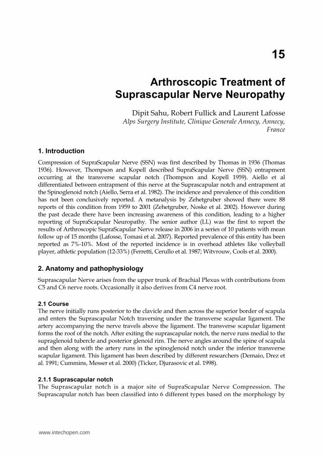
The nerve initially runs posterior to the clavicle and then across the superior border of scapula and enters the Suprascapular Notch traversing under the transverse scapular ligament. The artery accompanying the nerve travels above the ligament. The transverse scapular ligament forms the roof of the notch. After exiting the suprascapular notch, the nerve runs medial to the supraglenoid tubercle and posterior glenoid rim. The nerve angles around the spine of scapula and then along with the artery runs in the spinoglenoid notch under the inferior transverse scapular ligament. This ligament has been described by different researchers (Demaio, Drez et al. 1991; Cummins, Messer et al. 2000) (Ticker, Djurasovic et al. 1998).
2.1.1 Suprascapular notch
The Suprascapular notch is a major site of SupraScapular Nerve Compression. The Suprascapular notch has been classified into 6 different types based on the morphology by
www.intechopen.com
Applications of EMG in Clinical and Sports Medicine 226
Fig. 1. 3D representation of SSN and artery and their relationships with the TSL.
Rengachary et al (Rengachary, Burr et al. 1979; Bayramoglu, Demiryurek et al. 2003). The existence of different morphologies of the notch has been corroborated by more investigators. The greatest risk of nerve entrapment is in a small scapular notch with a thick Transverse scapular ligament
2.1.2 Transverse scapular ligament
The transverse scapular ligament forms the ceiling of the Suprascapular notch. This ligament may hypertrophy and lead to stenosis within the notch. There have also been reports of calcification of the ligament which causes nerve entrapment. A sling effect mechanism of injury of Suprascapular nerve has been described in which in certain anatomical variants of the Suprascapular notch, the transverse ligament cause nerve irritation and compression during certain movement of the limb (Rengachary, Neff et al. 1979).
2.1.3 Spinoglenoid ligament
Spinoglenoid ligament, also known as inferior transverse scapular ligament is a more common site for compression of the Suprascapular Nerve. This ligament originates on the spine of the scapula and inserts on the superior margin of glenoid neck as a bilaminar structure. The spinoglenoid ligament has been classified into two types: Type I -thin band, Type 2 is a well formed ligament. The spinoglenoid ligament is dynamic because of its insertion into the posterior Glenohumeral joint capsule. In certain positions of the arm, like adduction and internal rotation, this ligament may tighten due to tensioning of the capsule and compress the nerve.
www.intechopen.com
Arthroscopic Treatment of Suprascapular Nerve Neuropathy 227
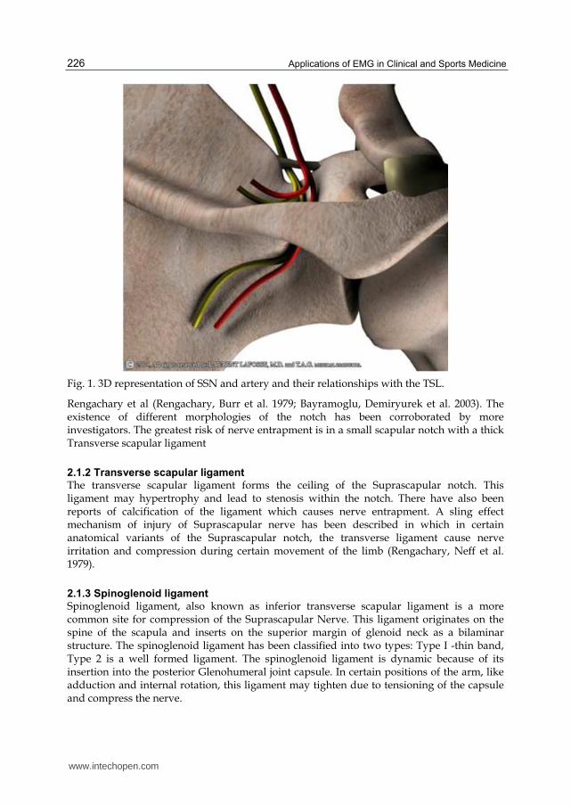
Fig. 2. Suprascapular nerve under the cut Superior transverse ligament and the artery running above.
www.intechopen.com
Applications of EMG in Clinical and Sports Medicine 228
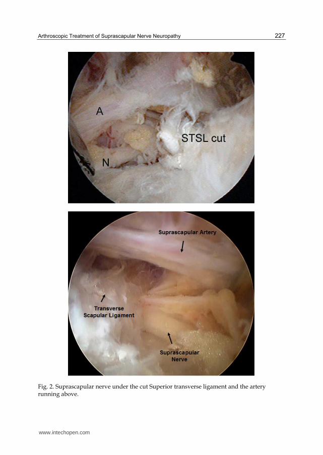
Fig. 3. 3D view of distal SSN passing around the spine of the scapula.
2.2 Distribution
The suprascapular nerve is predominantly motor nerve, but it also has nerve supply to skin and joints. Some reports by Ritchie et al and Matsumoto et al also suggest it may supply 70 % of sensations to the skin of the shoulder (Brown, James et al. 1988).
2.2.1 Muscles
The Nerve supplies two motor branches to the Supraspinatus before proceeding to the Spinoglenoid notch. After entering the Spinoglenoid Notch, the nerve supplies 2 - 4 motor branches to the Infraspinatus. Lesions of the nerve in the Suprascapular notch affect both Supraspinatus and Infraspinatus. While lesions in the Spinoglenoid notch only affect the Infraspinatus.
2.2.2 Joints
Cadaveric studies have demonstrated that Suprascapular nerve has a sensory branch to the Glenohumeral and acromioclavicular joint and coracoacromial ligament.
3. Etiology, physical examinaton and indication for arthroscopic release
3.1 Etiology
SSN compression neuropathy can primary or secondary.
3.1.1 Primary SSN neuropathy
Primary compression Neuropathy is due to repetitive trauma and microtrauma. In overhead
athletes there maybe a dynamic compression by spinoglenoid ligament when shoulder is in
www.intechopen.com
Arthroscopic Treatment of Suprascapular Nerve Neuropathy 229
a position of overhead throwing. There may also be compression at the suprascapular notch
or spinoglenoid notch by soft tissue tumor, bone tumor, cyst secondary to capsular or
labrum injury. Stenosis of the suprascapular or spinoglenoid notch, with a calcified or
hypertrophied transverse scapular ligament or a spinoglenoid ligament may predispose to
SSN neuropathy.
Other causes of Suprascapular nerve entrapment may include:
Compression by anterior coracoscapular ligaments
Compression by edge of hypertrophied infrascapular muscle
Compression by omohyoid muscle
Glenohumeral dislocation
Viral neuritis
Penetrating injuries to the shoulder
Posterior surgical approach to the scapula
3.1.2 Secondary SSN neuropathy
Secondary SSN neuropathy is associated with a massive rotator cuff tear. A retracted
massive rotator cuff tear predisposes the nerve to traction injury at the suprascapular notch.
According to Albritton et al, as the rotator muscles retract, the angle between the
suprascapular nerve and its first motor branch reduces, which leads to increased tension in
the nerve (Albritton, Graham et al. 2003).
Some studies have found correlation between presence of SSN neuropathy and massive
retracted rotator cuff tear (Mallon, Wilson et al. 2006). Some cadaveric studies have reported
that a lateral advancementof rotator cuff of more than 3 cm may increase the risk of
neurovascular twisting or injury (Warner, Krushell et al. 1992). However there have been
other reports of lateral advancement safe length to vary from 1 cm to 3 cm and there is no
consensus as to the safe level of lateral advancement (Greiner, Golser et al. 2003).
3.2 Physical examination and electrodiagnostic tests
3.2.1 Physical examination
A thorough physical examination includes examination of the cervical spine, both shoulders
to detect tenderness around posterior shoulder, muscle atrophy, strength of shoulder in
external rotation.
Supraspinatus and Infraspinatus atrophy may point towards an involvement of the nerve in
the Suprascapular / Spinoglenoid notch. There may be tenderness to palpation posterior to
the clavicle with injury to the nerve in the Suprascapular fossa. External rotation weakness
may also point towards an involvement of the Infraspinatus. Cross body adduction test
which puts the spinoglenoid ligament under tension may be a good indicator of the nerve
involvement in the spinoglenoid notch.
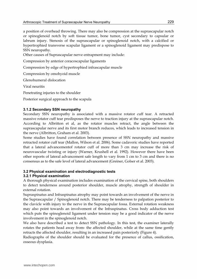
We also have described a test to detect SSN pathology. In this test, the examiner laterally
rotates the patients head away from- the affected shoulder, while at the same time gently
retracts the affected shoulder, resulting in an increased pain posteriorly (Figure 4).
Radiographs of the shoulder should be evaluated for the presence of callus, ossification,
osseous dysplasia.
www.intechopen.com
Applications of EMG in Clinical and Sports Medicine 230
Fig. 4. Clinical test to detect Suprascapular Nerve Neuropathy.
Other imaging modalities (Ultrasound, CT scan, MRI) may give a better idea about the presence of tumor, compressing cysts, lesions in course of nerve.
3.2.2 Role of electromyography Electromyography studies are indicated when there is an unexplained shoulder pain. However the sensitivity and specificity of Electromyography has been debated by various authors. Electromyography of a damaged nerve may show reduced amplitude of motor potentials, increased spontaneous activity, fibrillations, polyphasic activity, etc. It may show denervation of the supraspinatous or infraspinatous muscle with resultant fibrillation and sharp waves. EMG is also useful in confirming traumatic lesions of SSN. In our study of 100 patients, we performed EMG pre operatively and at 6 months and 2 years postoperatively. A positive EMG, however, is not always the necessary criterion for diagnosing SSN neuropathy. We, believe that SSN pathology is a dynamic process and not always demonstrable on an EMG. An EMG however, may be indicated in unexplained posterior shoulder pain. EMG in retracted rotator cuff tears have shown to be abnormal suggesting Suprascapular nerve pathology.
3.3 Indications for arthroscopic release
Our Indications for Arthroscopic Release: 1. Patients with weakness of Infraspinatus/Supraspinatus with or without positive EMG 2. Patients with a thickened or ossified ligament on assessment during arthroscopic
rotator cuff repair 3. Patients who present with posterior shoulder pain and a positive SSN test with or
without positive EMG 4. space occupying lesion compressing SSN 5. reduction of large/massive rotator cuff tear 6. compression due to anatomical variants
www.intechopen.com
Arthroscopic Treatment of Suprascapular Nerve Neuropathy 231
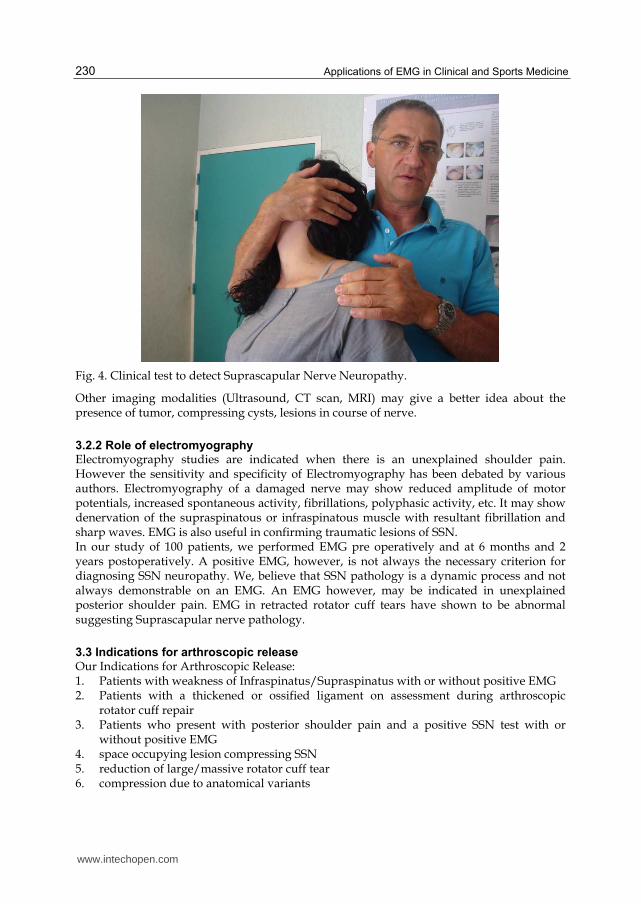
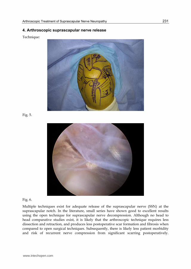
4. Arthroscopic suprascapular nerve release
Technique:
Fig. 5.
Fig. 6.
Multiple techniques exist for adequate release of the suprascapular nerve (SSN) at the suprascapular notch. In the literature, small series have shown good to excellent results using the open technique for suprascapular nerve decompression. Although no head to head comparative studies exist, it is likely that the arthroscopic technique requires less dissection and retraction, and produces less postoperative scar formation and fibrosis when compared to open surgical techniques. Subsequently, there is likely less patient morbidity and risk of recurrent nerve compression from significant scarring postoperatively.
www.intechopen.com
Applications of EMG in Clinical and Sports Medicine 232
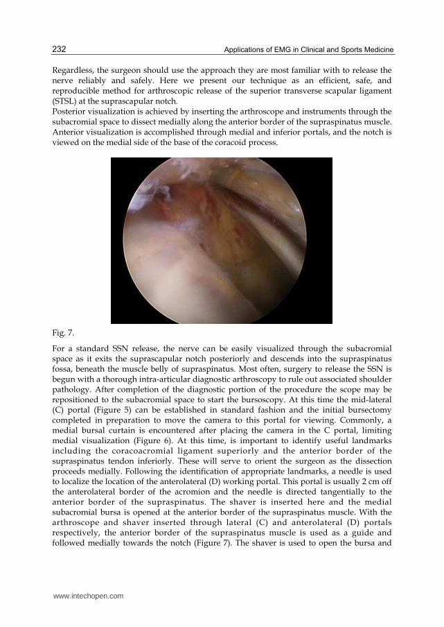
Regardless, the surgeon should use the approach they are most familiar with to release the nerve reliably and safely. Here we present our technique as an efficient, safe, and reproducible method for arthroscopic release of the superior transverse scapular ligament (STSL) at the suprascapular notch. Posterior visualization is achieved by inserting the arthroscope and instruments through the subacromial space to dissect medially along the anterior border of the supraspinatus muscle. Anterior visualization is accomplished through medial and inferior portals, and the notch is viewed on the medial side of the base of the coracoid process.
Fig. 7.
For a standard SSN release, the nerve can be easily visualized through the subacromial space as it exits the suprascapular notch posteriorly and descends into the supraspinatus fossa, beneath the muscle belly of supraspinatus. Most often, surgery to release the SSN is begun with a thorough intra-articular diagnostic arthroscopy to rule out associated shoulder pathology. After completion of the diagnostic portion of the procedure the scope may be repositioned to the subacromial space to start the bursoscopy. At this time the mid-lateral (C) portal (Figure 5) can be established in standard fashion and the initial bursectomy completed in preparation to move the camera to this portal for viewing. Commonly, a medial bursal curtain is encountered after placing the camera in the C portal, limiting medial visualization (Figure 6). At this time, is important to identify useful landmarks including the coracoacromial ligament superiorly and the anterior border of the supraspinatus tendon inferiorly. These will serve to orient the surgeon as the dissection proceeds medially. Following the identification of appropriate landmarks, a needle is used to localize the location of the anterolateral (D) working portal. This portal is usually 2 cm off the anterolateral border of the acromion and the needle is directed tangentially to the anterior border of the supraspinatus. The shaver is inserted here and the medial subacromial bursa is opened at the anterior border of the supraspinatus muscle. With the arthroscope and shaver inserted through lateral (C) and anterolateral (D) portals respectively, the anterior border of the supraspinatus muscle is used as a guide and followed medially towards the notch (Figure 7). The shaver is used to open the bursa and
www.intechopen.com
Arthroscopic Treatment of Suprascapular Nerve Neuropathy 233
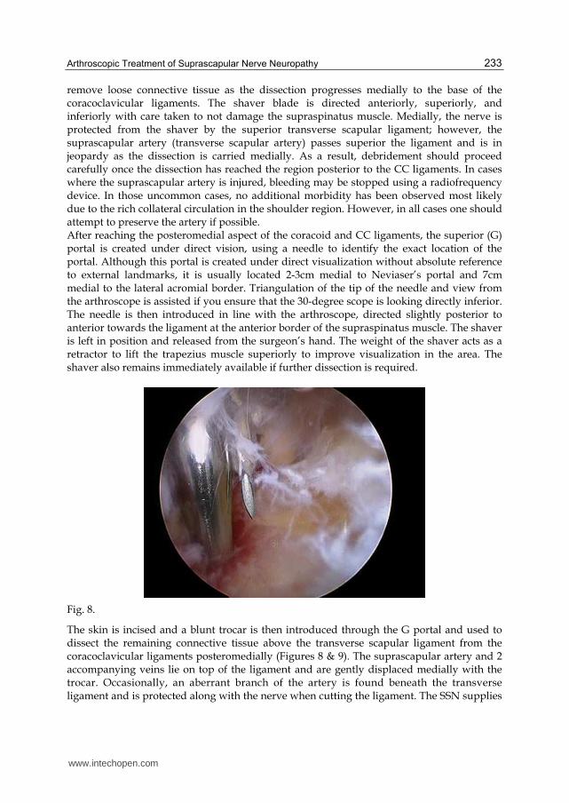
remove loose connective tissue as the dissection progresses medially to the base of the coracoclavicular ligaments. The shaver blade is directed anteriorly, superiorly, and inferiorly with care taken to not damage the supraspinatus muscle. Medially, the nerve is protected from the shaver by the superior transverse scapular ligament; however, the suprascapular artery (transverse scapular artery) passes superior the ligament and is in jeopardy as the dissection is carried medially. As a result, debridement should proceed carefully once the dissection has reached the region posterior to the CC ligaments. In cases where the suprascapular artery is injured, bleeding may be stopped using a radiofrequency device. In those uncommon cases, no additional morbidity has been observed most likely due to the rich collateral circulation in the shoulder region. However, in all cases one should attempt to preserve the artery if possible. After reaching the posteromedial aspect of the coracoid and CC ligaments, the superior (G) portal is created under direct vision, using a needle to identify the exact location of the portal. Although this portal is created under direct visualization without absolute reference to external landmarks, it is usually located 2-3cm medial to Neviaser’s portal and 7cm medial to the lateral acromial border. Triangulation of the tip of the needle and view from the arthroscope is assisted if you ensure that the 30-degree scope is looking directly inferior. The needle is then introduced in line with the arthroscope, directed slightly posterior to anterior towards the ligament at the anterior border of the supraspinatus muscle. The shaver is left in position and released from the surgeon’s hand. The weight of the shaver acts as a retractor to lift the trapezius muscle superiorly to improve visualization in the area. The shaver also remains immediately available if further dissection is required.
Fig. 8.
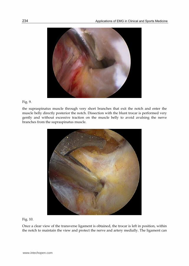
The skin is incised and a blunt trocar is then introduced through the G portal and used to dissect the remaining connective tissue above the transverse scapular ligament from the coracoclavicular ligaments posteromedially (Figures 8 & 9). The suprascapular artery and 2 accompanying veins lie on top of the ligament and are gently displaced medially with the trocar. Occasionally, an aberrant branch of the artery is found beneath the transverse ligament and is protected along with the nerve when cutting the ligament. The SSN supplies
www.intechopen.com
Applications of EMG in Clinical and Sports Medicine 234
Fig. 9.
the supraspinatus muscle through very short branches that exit the notch and enter the
muscle belly directly posterior the notch. Dissection with the blunt trocar is performed very
gently and without excessive traction on the muscle belly to avoid avulsing the nerve
branches from the supraspinatus muscle.
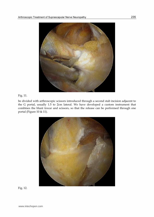
Fig. 10.
Once a clear view of the transverse ligament is obtained, the trocar is left in position, within the notch to maintain the view and protect the nerve and artery medially. The ligament can
www.intechopen.com
Arthroscopic Treatment of Suprascapular Nerve Neuropathy 235
Fig. 11.
be divided with arthroscopic scissors introduced through a second stab incision adjacent to the G portal, usually 1.5 to 2cm lateral. We have developed a custom instrument that combines the blunt trocar and scissors, so that the release can be performed through one portal (Figure 10 & 11).
Fig. 12.
www.intechopen.com
Applications of EMG in Clinical and Sports Medicine 236
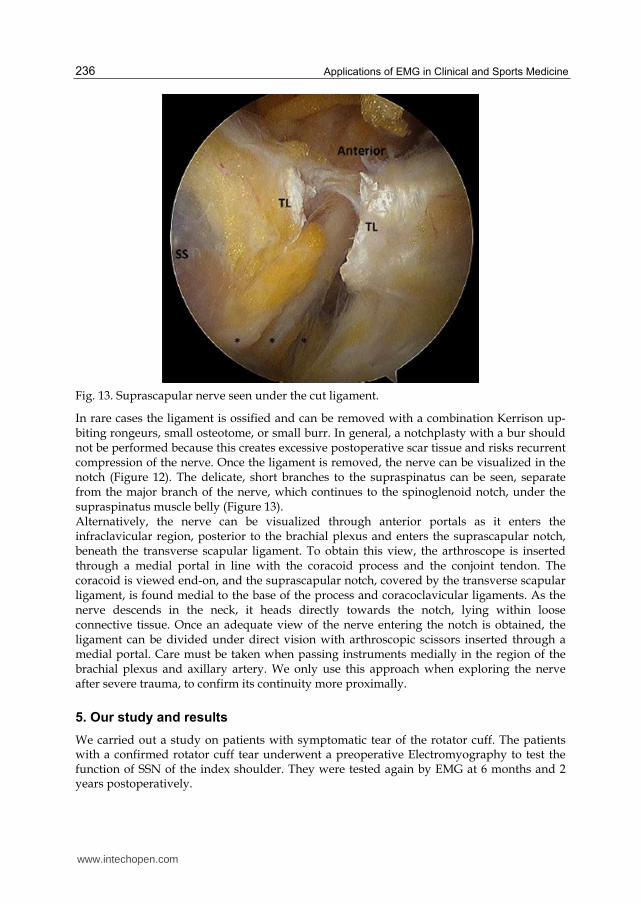
Fig. 13. Suprascapular nerve seen under the cut ligament.
In rare cases the ligament is ossified and can be removed with a combination Kerrison up-biting rongeurs, small osteotome, or small burr. In general, a notchplasty with a bur should not be performed because this creates excessive postoperative scar tissue and risks recurrent compression of the nerve. Once the ligament is removed, the nerve can be visualized in the notch (Figure 12). The delicate, short branches to the supraspinatus can be seen, separate from the major branch of the nerve, which continues to the spinoglenoid notch, under the supraspinatus muscle belly (Figure 13). Alternatively, the nerve can be visualized through anterior portals as it enters the infraclavicular region, posterior to the brachial plexus and enters the suprascapular notch, beneath the transverse scapular ligament. To obtain this view, the arthroscope is inserted through a medial portal in line with the coracoid process and the conjoint tendon. The coracoid is viewed end-on, and the suprascapular notch, covered by the transverse scapular ligament, is found medial to the base of the process and coracoclavicular ligaments. As the nerve descends in the neck, it heads directly towards the notch, lying within loose connective tissue. Once an adequate view of the nerve entering the notch is obtained, the ligament can be divided under direct vision with arthroscopic scissors inserted through a medial portal. Care must be taken when passing instruments medially in the region of the brachial plexus and axillary artery. We only use this approach when exploring the nerve after severe trauma, to confirm its continuity more proximally.
5. Our study and results
We carried out a study on patients with symptomatic tear of the rotator cuff. The patients with a confirmed rotator cuff tear underwent a preoperative Electromyography to test the function of SSN of the index shoulder. They were tested again by EMG at 6 months and 2 years postoperatively.
www.intechopen.com
Arthroscopic Treatment of Suprascapular Nerve Neuropathy 237
There were three groups of patients. Group 1 Those who had positive EMG and underwent cuff repair only. Group 2 had positive EMG and underwent cuff repair along with SSN release. Group 3 had normal EMG and underwent cuff repair only. We had 100 patients with average age of 58 (+/- 9.2) years. According to EMG studies, 50 pateints demonstrated normal EMG findings of SSN and 27 patients demonstrated abnormalities in EMG (23 were unclear). Accordng to the postoperative EMG studies, 81% of patients demonstrated normal electrodiagnostic findings of SSN and 9% demonstrated abnormalities in EMG. 45 patients demonstrated the same normal level as preoperative, 22 patients were better than pre operative and 5 patients remained pathologic. In subgroup 1, 9 patients out of group 1 postoperatively demonstrated normal EMG, 4 remained at the same pathologic level, even though SSN release had been performed, constant scores improved in all the patients. In subgroup 2, all 11 patients post operatively demonstrated a normal EMG, despite no release of SSN being performed. In subgroup 3, patients with a preoperative normal EMG, at the time of postoperative EMG. All showed normal EMG.
6. Discussion and overview
SSN neuropathy may have a primary etiology or may also be found in massive retracted rotator cuff. Several Authors have now identified association of SSN neuropathy with massive rotator cuff tear. Senior Author (L. L) in 2006 reported 9 of 10 patients who underwent SSN arthroscopic decompression without rotator cuff disease had excellent outcomes. 7 of these patients also had complete normalization of latency in motor fibres of SSN in EMG / NCV. The results in this study convincingly showed an improvement in Nerve conduction velocity after SSN decompression via the arthroscopic techniques (Lafosse, Tomasi et al. 2007). Costouros et al reported 6 patients with documented SSN pathology associated with massive cuff tear and found improvement in EMG function in all (Costouros, Porramatikul et al. 2007). SSN neuropathy may be treated conservatively, but the results have been poor as shown by Callahan et al. In their series, 76% of patients required surgery eventually (Callahan, Scully et al. 1991). Operative techniques may include open nerve release or Arthroscopic Nerve release. Open Suprascapular nerve release has been reported to give good results. In a series of 39 patients, 28 showed improvement of supraspinatus muscle strength to grade 4 (Kim, Murovic et al. 2005). However, Arthroscopic techniques of SSN release have now gained popularity due to the fact that they are less invasive, afford better visualization and require less operative time. Arthroscopically, the SSN can be visualized and released posteriorly (Lafosse, Tomasi et al. 2007) or anteriorly (Reineck and Krishnan 2009) to the coracoclavicular ligaments. Our unpublished data suggests EMG improves post operatively in patients undergoing massive rotator cuff repair. However, there were also 4 patients in subgroup 1 in whom the EMG stayed at the same pathologic level. In our center we do not evaluate every patient of rotator cuff tear by an EMG. We do however release the SSN in massive rotator cuff tears as we believe that a retracted rotator cuff tear does put tension on SSN and releasing the Superior transverse scapular ligament releases the tension on the nerve. Albritton et al also
www.intechopen.com
Applications of EMG in Clinical and Sports Medicine 238
proved in their study that suprascapular nerve injury may result from a retraction of a torn rotator cuff. They proved that retraction of Supraspinatus changed the course of suprascapular nerve as it passed through the notch (Albritton, Graham et al. 2003). We also assess the ligament as needed and if the ligament is found to be ossified or under tension, we go ahead and release the ligament. In a study by Shah et al, 21 out of 24 patients who presented with deep posterior shoulder pain had EMG/NCV findings of denervation of SSN and 17 of the 24 patients showed an improvement in pain and improvement in ASES scores after SSN decompression (Shah, Butler et al.). They also concluded that there may be a subset of patients with negative electro diagnostic studies presenting with unexplained posterior shoulder pain who may respond to SSN release. Warner et al also showed that lateral reduction of rotator cuff tears of more than 3 cm during reconstruction may result in significant tension on the Supra Scapular Nerve and entrapment by the Transverse Scapular Ligament (Warner, Krushell et al. 1992).
7. Summary and our recommendation
To summarise, there are three main advantages of arthroscopic release of SSN over the traditional open technique. First it provides superior visualization of neurovascular structures and the transverse scapular ligament. The SSN is as small as 2 mm and is therefore sometimes difficult to appreciate via open exposures. The small diameter of the SSN is easily visualized with the magnification afforded by the arthroscope and permits easy release of the ligament. Secondly the arthroscopic release of the SSN is significantly less invasive and does not involve detachment of the trapezius insertion. Thirdly the operative time is considerable less and patient has significantly less pain post operatively. We believe that by releasing the TSL, particularly in large chronically retracted tears, we improve SSN mobility both in the retracted state and for the reduction of the cuff and therefore enhancing the chances of neuromuscular recovery.
8. EMG/NCV
EMG/NCV testing and evaluation are very user dependent, and an electro physiologist skilled in evaluation of the SSN is needed for accurate interpretation of the results. Positive EMG/NCVs are helpful; however, a negative EMG/NCV is not a contraindication to SSN release. Similarly, our study on massive tears showed ~27% positive EMG studies. Although no significant difference in pre/post-op EMGs has been found and controversy exists regarding SSN release in the repair of large-massive cuff tears, our opinion is the procedure can be completed quickly and reproducibly without subjecting the patient to significant additional risks.
9. References
Aiello, I., G. Serra, et al. (1982). "Entrapment of the suprascapular nerve at the spinoglenoid notch." Ann Neurol 12 (3): 314-6.
www.intechopen.com
Arthroscopic Treatment of Suprascapular Nerve Neuropathy 239
Albritton, M. J., R. D. Graham, et al. (2003). "An anatomic study of the effects on the suprascapular nerve due to retraction of the supraspinatus muscle after a rotator cuff tear." J Shoulder Elbow Surg 12 (5): 497-500.
Bayramoglu, A., D. Demiryurek, et al. (2003). "Variations in anatomy at the suprascapular notch possibly causing suprascapular nerve entrapment: an anatomical study." Knee Surg Sports Traumatol Arthrosc 11 (6): 393-8.
Brown, D. E., D. C. James, et al. (1988). "Pain relief by suprascapular nerve block in gleno-humeral arthritis." Scand J Rheumatol 17 (5): 411-5.
Callahan, J. D., T. B. Scully, et al. (1991). "Suprascapular nerve entrapment. A series of 27 cases." J Neurosurg 74 (6): 893-6.
Costouros, J. G., M. Porramatikul, et al. (2007). "Reversal of suprascapular neuropathy following arthroscopic repair of massive supraspinatus and infraspinatus rotator cuff tears." Arthroscopy 23 (11): 1152-61.
Cummins, C. A., T. M. Messer, et al. (2000). "Suprascapular nerve entrapment." J Bone Joint Surg Am 82 (3): 415-24.
Demaio, M., D. Drez, Jr., et al. (1991). "The inferior transverse scapular ligament as a possible cause of entrapment neuropathy of the nerve to the infraspinatus. A brief note." J Bone Joint Surg Am 73 (7): 1061-3.
Ferretti, A., G. Cerullo, et al. (1987). "Suprascapular neuropathy in volleyball players." J Bone Joint Surg Am 69 (2): 260-3.
Greiner, A., K. Golser, et al. (2003). "The course of the suprascapular nerve in the supraspinatus fossa and its vulnerability in muscle advancement." J Shoulder Elbow Surg 12 (3): 256-9.
Kim, D. H., J. A. Murovic, et al. (2005). "Management and outcomes of 42 surgical suprascapular nerve injuries and entrapments." Neurosurgery 57 (1): 120-7; discussion 120-7.
Lafosse, L., A. Tomasi, et al. (2007). "Arthroscopic release of suprascapular nerve entrapment at the suprascapular notch: technique and preliminary results." Arthroscopy 23 (1): 34-42.
Mallon, W. J., R. J. Wilson, et al. (2006). "The association of suprascapular neuropathy with massive rotator cuff tears: a preliminary report." J Shoulder Elbow Surg 15 (4): 395-8.
Reineck, J. R. and S. G. Krishnan (2009). "Subligamentous suprascapular artery encountered during arthroscopic suprascapular nerve release: a report of three cases." J Shoulder Elbow Surg 18 (3): e1-3.
Rengachary, S. S., D. Burr, et al. (1979). "Suprascapular entrapment neuropathy: a clinical, anatomical, and comparative study. Part 2: anatomical study." Neurosurgery 5 (4): 447-51.
Rengachary, S. S., J. P. Neff, et al. (1979). "Suprascapular entrapment neuropathy: a clinical, anatomical, and comparative study. Part 1: clinical study." Neurosurgery 5 (4): 441-6.
Shah, A. A., R. B. Butler, et al. "Clinical outcomes of suprascapular nerve decompression." J Shoulder Elbow Surg.
Thomas, A. (1936)."La Paralysie du muscle sous-pineux. " Presse Med 64: 1283-4. Thompson, W. A. and H. P. Kopell (1959). "Peripheral entrapment neuropathies of the upper
extremity." N Engl J Med 260 (25): 1261-5.
www.intechopen.com
Applications of EMG in Clinical and Sports Medicine 240
Ticker, J. B., M. Djurasovic, et al. (1998). "The incidence of ganglion cysts and other variations in anatomy along the course of the suprascapular nerve." J Shoulder Elbow Surg 7 (5): 472-8.
Warner, J. P., R. J. Krushell, et al. (1992). "Anatomy and relationships of the suprascapular nerve: anatomical constraints to mobilization of the supraspinatus and infraspinatus muscles in the management of massive rotator-cuff tears." J Bone Joint Surg Am 74 (1): 36-45.
Witvrouw, E., A. Cools, et al. (2000). "Suprascapular neuropathy in volleyball players." Br J Sports Med 34 (3): 174-80.
Zehetgruber, H., H. Noske, et al. (2002). "Suprascapular nerve entrapment. A meta-analysis." Int Orthop 26 (6): 339-43.
www.intechopen.com
Applications of EMG in Clinical and Sports MedicineEdited by Dr. Catriona Steele
ISBN 978-953-307-798-7Hard cover, 396 pagesPublisher InTechPublished online 11, January, 2012Published in print edition January, 2012
InTech ChinaUnit 405, Office Block, Hotel Equatorial Shanghai No.65, Yan An Road (West), Shanghai, 200040, China
Phone: +86-21-62489820 Fax: +86-21-62489821
This second of two volumes on EMG (Electromyography) covers a wide range of clinical applications, as acomplement to the methods discussed in volume 1. Topics range from gait and vibration analysis, throughposture and falls prevention, to biofeedback in the treatment of neurologic swallowing impairment. The volumeincludes sections on back care, sports and performance medicine, gynecology/urology and orofacial function.Authors describe the procedures for their experimental studies with detailed and clear illustrations andreferences to the literature. The limitations of SEMG measures and methods for careful analysis arediscussed. This broad compilation of articles discussing the use of EMG in both clinical and researchapplications demonstrates the utility of the method as a tool in a wide variety of disciplines and clinical fields.
How to referenceIn order to correctly reference this scholarly work, feel free to copy and paste the following:
Dipit Sahu, Robert Fullick and Laurent Lafosse (2012). Arthroscopic Treatment of Suprascapular NerveNeuropathy, Applications of EMG in Clinical and Sports Medicine, Dr. Catriona Steele (Ed.), ISBN: 978-953-307-798-7, InTech, Available from: http://www.intechopen.com/books/applications-of-emg-in-clinical-and-sports-medicine/arthroscopic-treatment-of-suprascapular-nerve-neuropathy