Original: November 20, 2014 Page 1 of 3 ARVC OPERATIONS MANUAL ANGIOGRAPHY PROCESSING AND TRANSFER OVERVIEW Angiography (angio) study is required, if performed per standard of care, for all subjects enrolled in the ARVC study. If angiography is done for diagnosis purposes at the enrolling center after consent form is signed (not previously done by outside source), they should be done using the standardized ARVC protocols in this manual. If angio was performed prior to enrollment, a copy of the latest test CD and or a copy of the test CD that was done for ARVC diagnosis purposes, should be sent along with the written report with a completed Form 4F and Form 4A. Any new angiogram done prior to a specific follow –up visit must be sent to the angio core lab as soon as it is done and the required CRFs should be completed in the subsequent yearly visit event in the ARVC electronic data entry system. ANGIOGRAPHY PROCESSING AND PROCEDURES If the angio is performed after subject signs the study consent form, we recommend using the angio procedures in this manual. The angiographic features of arrhythmogenic right ventricular cardiomyopathy (ARVC) include global and/or regional function and morphological abnormalities of the right ventricle. Cardiomegaly, localized akinetic or dyskinetic bulges, outpouchings, dilatation of the infundibulum, trabecular hypertrophy and/or disarray with deep fissures are morphological findings in ARVC. The following protocol for RV angiography is designed to illustrate these abnormalities and to perform quantitative measurements of RV volumes, ejection fraction, and regional contraction. If angiogram is performed after subject signs consent, please adhere to the following: Acquire good-quality RV angiogram Store the data on CD (DICOM format). The core lab will perform the study analyses and measurements. Biplane digital catheter lab is preferred. Avoid ECG cables, connectors, etc in field of view. Hemodynamic measurements (PA pressure (sys/dias/mean), RA mean pressure, and RV end diastolic pressure calibration are performed in the core lab based upon the size of the angiographic catheter used for RV angiography. Specify the type and size of catheter used: Pigtail (5Fr or larger), Berman catheter (6Fr or 7Fr). Place the RV angiography catheter in the mid inferior RV (RAO or PA view), approximately 1 cm above the mid-inferior wall to best avoid catheter induced ectopy. Avoid contact with the RV wall or trabeculae. Confirm that the entire RV in both acquired views (i.e. the RAO-LAO acquisition, and AP/Lat acquisition) can be seen. DO NOT move the table during the acquisition. Use the slower injection rate of 12-15 ml/sec to avoid PVCs. Projections: (preferred with biplane mode to minimize contrast needs) a. 30 deg RAO b. 60 deg LAO c. Postero-anterior (PA) d. Lateral (LAT) Optional views: (encouraged if contrast use allows)
Transcript
Original: November 20, 2014 Page 1 of 3
ARVC OPERATIONS MANUAL
ANGIOGRAPHY PROCESSING AND TRANSFER
OVERVIEW Angiography (angio) study is required, if performed per standard of care, for all subjects enrolled in the ARVC study. If angiography is done for diagnosis purposes at the enrolling center after consent form is signed (not previously done by outside source), they should be done using the standardized ARVC protocols in this manual. If angio was performed prior to enrollment, a copy of the latest test CD and or a copy of the test CD that was done for ARVC diagnosis purposes, should be sent along with the written report with a completed Form 4F and Form 4A. Any new angiogram done prior to a specific follow –up visit must be sent to the angio core lab as soon as it is done and the required CRFs should be completed in the subsequent yearly visit event in the ARVC electronic data entry system.
ANGIOGRAPHY PROCESSING AND PROCEDURES If the angio is performed after subject signs the study consent form, we recommend using the angio procedures in this manual.
The angiographic features of arrhythmogenic right ventricular cardiomyopathy (ARVC) include global and/or regional function and morphological abnormalities of the right ventricle. Cardiomegaly, localized akinetic or dyskinetic bulges, outpouchings, dilatation of the infundibulum, trabecular hypertrophy and/or disarray with deep fissures are morphological findings in ARVC.
The following protocol for RV angiography is designed to illustrate these abnormalities and to perform quantitative measurements of RV volumes, ejection fraction, and regional contraction. If angiogram is performed after subject signs consent, please adhere to the following:
Acquire good-quality RV angiogram Store the data on CD (DICOM format). The core lab will perform the study analyses and
measurements. Biplane digital catheter lab is preferred. Avoid ECG cables, connectors, etc in field of view.
Hemodynamic measurements (PA pressure (sys/dias/mean), RA mean pressure, and RV end diastolic pressure calibration are performed in the core lab based upon the size of the angiographic catheter used for RV angiography. Specify the type and size of catheter used: Pigtail (5Fr or larger), Berman catheter (6Fr or 7Fr).
Place the RV angiography catheter in the mid inferior RV (RAO or PA view), approximately 1 cm above the mid-inferior wall to best avoid catheter induced ectopy. Avoid contact with the RV wall or trabeculae. Confirm that the entire RV in both acquired views (i.e. the RAO-LAO acquisition, and AP/Lat acquisition) can be seen. DO NOT move the table during the acquisition. Use the slower injection rate of 12-15 ml/sec to avoid PVCs. Projections: (preferred with biplane mode to minimize contrast needs)
a. 30 deg RAO b. 60 deg LAO c. Postero-anterior (PA) d. Lateral (LAT)
Optional views: (encouraged if contrast use allows)
Original: November 20, 2014 Page 2 of 3
a. 45 deg RAO b. 45 deg LAO
Image Acquisition: 30 fr/sec during a breath hold. Film long sequence to permit contrast passage to LV. Contrast load: 40-50 ml at 12-15 ml/sec. ANGIOGRAPHY AT BASELINE
1. Transfer of Angiography CD and Reports for a New potential Proband: The Angiography written report must be sent to the Clinical Center Reviewer for ARVC diagnosis. Once the ARVC diagnosis is verified by the Clinical Center Reviewer, the angiogram CD must be shipped to the angio core lab. It is possible that the Clinical Center Reviewer will request the angiogram images apart from the written reports to assess patient’s eligibility. In this case, the Angiography CD must be sent within 5 working days from date of request to the Clinical Center Reviewer.
2. Transfer of Angiography CD and Reports for Family Member (old/new) and Old Probands: Once the
proband in the family is identified, or a subject (proband/family member) from the previous grant is enrolled, the Angiography CD and written report must be sent to the Angio Core lab.
ANGIOGRAPHY AT FOLLOW-UP VISITS If angiography is performed while a subject (old/new proband or old/new family member) is participating in the study, we recommend that the standardized protocol in this manual should be used. The CD and written report should be sent at the next sequential follow-up study visit following the procedure. Each sent media must be accompanied by a completed Form 4F and a completed Form 4A.
TEST MEDIA TRANSFER
The initial delivery of the image media, the forms, and the local interpretation report must be attempted by uploading the data electronically, according to instructions in transferring section of this manual. If data upload is unsuccessful, send the data to the Angio Core lab using Fed Ex.
MANUAL TRANSFER The electronic file must be delivered to: Dr. Julia Indik University of Arizona 1501 North Campbell Sarver Heart Center, Room 5153 Tucson, AZ 85424 USA Phone: 520-626-6262 Fax: 520-626-4333 If you have any questions, please contact Julia Indik, MD at phone number: 520-626-6262 Delivery of the report to the Clinical Review Center for new proband ARVC evaluation must be attempted by uploading the data electronically, according to instructions in the test transfer section. If data upload is unsuccessful, send the data via mail or fax as instructed in the test transfer section.
Original: November 20, 2014 Page 3 of 3
Table 1: Test Media Transfer
Test Media Transferred Baseline Visit
Test Media Transferred Follow-Up Visit
Subject Category
New Proband Old Proband New Family Member Old Family member
Old Proband New Proband New Family Member Old Family member
Ship to Clinical
Center PI
Clinical Center Data Shipment Form (Form 5B) Angio Form (Form 4F) Written report
Ship to Angiography
Core laboratory
Shipping Form (Form 4A) Angio Form (Form 4F) CD Written report
Shipping Form (Form 4A) Angio Form (Form 4F) CD Written report
Shipping Form (Form 4A) Angio Form (Form 4F) CD Written report
1
Performance of RV Angiography Biopsy
Presentation from Thomas Wichter (Revised December 2013 – Julia Indik)
Multidisciplinary Study of ARVCAngiography Core Lab
Multidisciplinary Study of ARVCAngiography Core Lab
Please respect copyright of all RV angiogram images and movies displayed
in this demo presentation which was designed fortraining purposes within the NIH-funded
„Multidisciplinary Study of ARVC“.
Angiographic images and movies should not be usedfor other purposes, in particular scientific
presentations, manuscripts or publications withoutprior agreement from the author
(Thomas Wichter, MD, FESC).
Copyright Notice
2
Multidisciplinary Study of ARVCAngiography Core Lab
The diagnosis of arrhythmogenic right ventricular dysplasia (ARVC) is made on angiography by showing decreased global
and/or regional function and illustrating morphological abnormalities of the right ventricle. Cardiomegaly, localized
akinetic or dyskinetic bulges, outpouchings, dilatation of the infundibulum, trabecular hypertrophy and/or disarray with deep
fissures are morphological findings in ARVC.
ANGIOGRAPHY PROTOCOL FOR ARVC
Multidisciplinary Study of ARVCAngiography Core Lab
The following protocol for RV angiography is designed to illustrate these abnormalities and to perform quantitative
measurements of RV volumes, ejection fraction, and regional contraction. It will be used uniformly within the multidisciplinary
study of ARVC. Please make sure to acquire good-quality RV angiograms stored on CD. We will take care of the study
analyses and measurements.
ANGIOGRAPHY PROTOCOL FOR ARVC
3
Multidisciplinary Study of ARVCAngiography Core Lab
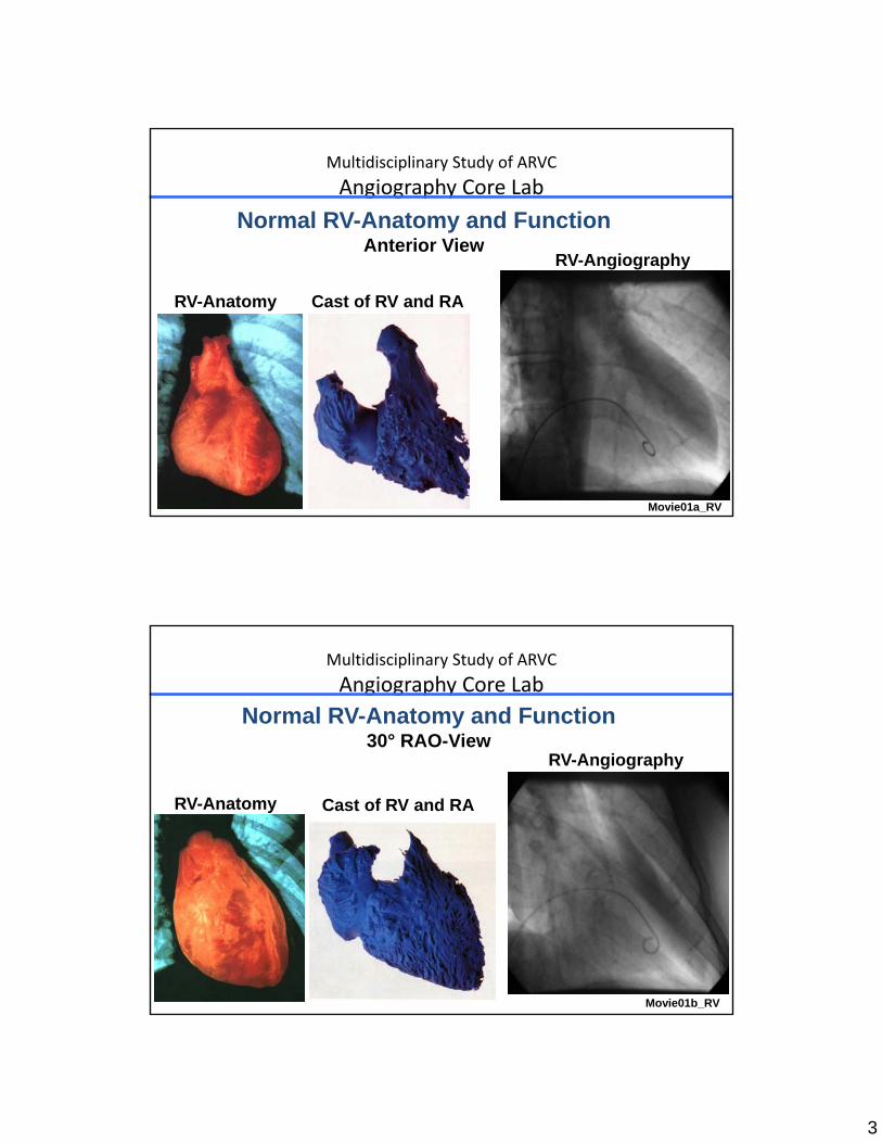
Normal RV-Anatomy and FunctionAnterior View
RV-Anatomy
RV-Angiography
Cast of RV and RA
Movie01a_RV
Multidisciplinary Study of ARVCAngiography Core Lab
Normal RV-Anatomy and Function30° RAO-View
RV-Anatomy
RV-Angiography
Cast of RV and RA
Movie01b_RV
4
Multidisciplinary Study of ARVCAngiography Core Lab
Normal RV-Anatomy and Function60° LAO-View
RV-Anatomy
RV-Angiography
Cast of RV and RA
Movie01c_RV
Multidisciplinary Study of ARVCAngiography Core Lab
Normal RV-Anatomy and FunctionLateral View
Left Lateral View
RV-Angiography
RV Lateral View
Movie01d_RV
5
Multidisciplinary Study of ARVCAngiography Core Lab
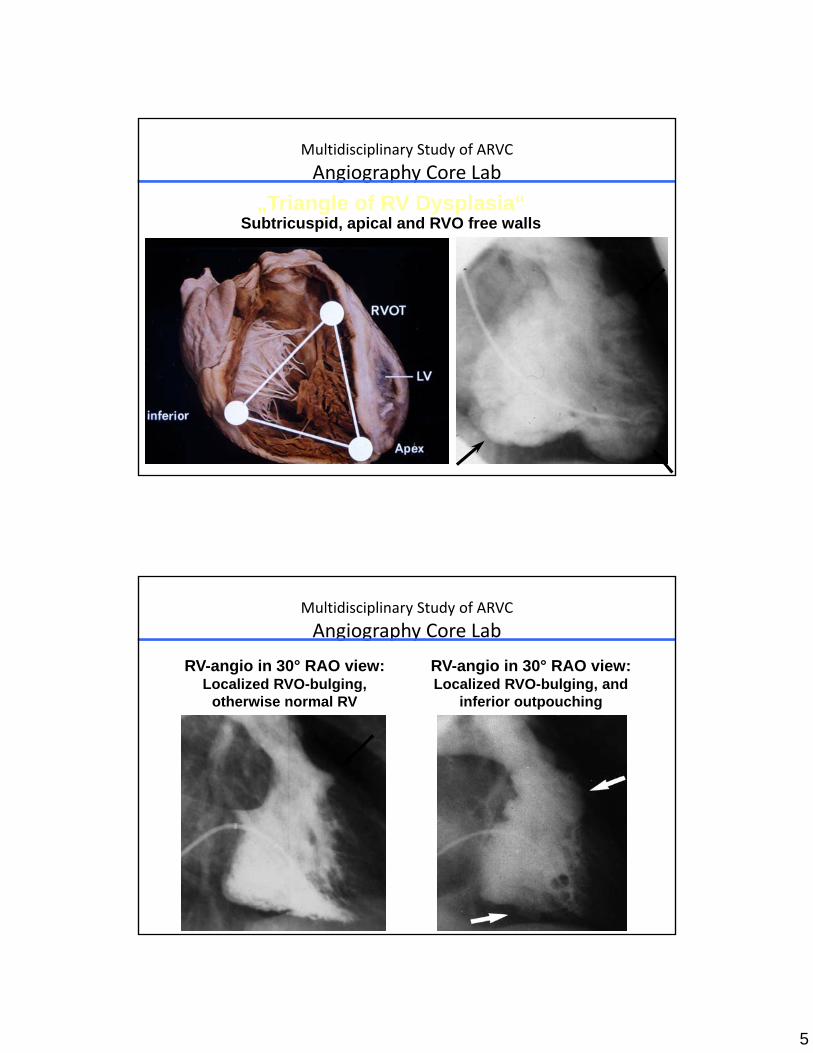
„Triangle of RV Dysplasia“Subtricuspid, apical and RVO free walls
Multidisciplinary Study of ARVCAngiography Core Lab
RV-angio in 30° RAO view:Localized RVO-bulging,
otherwise normal RV
RV-angio in 30° RAO view:Localized RVO-bulging, and
inferior outpouching
6
Multidisciplinary Study of ARVCAngiography Core Lab
RV-angio in 30° RAO view:Trabecular hypertrophy ant-sept, polycyclic contours infero-apical
Multidisciplinary Study of ARVCAngiography Core Lab
Multidisciplinary Study of ARVCAngiography Core Lab
40-50 ml low toxicity contrast dye (depending on global RV size)
Dye injection velocity of 12-15 ml/sec
Cine angio with 30 images/sec during deep inspiration(avoid overlap of inferior RV wall with diaphragm)
Protocol for RV Angiogram
8
Multidisciplinary Study of ARVCAngiography Core Lab
Make sure RV is fully within field of view in all projectionsAvoid ECG cables, connectors, etc. in the field of viewDO NOT MOVE THE TABLE during cine angio aquisitionAvoid PVCs by good catheter position and low dye velocityAvoid artificial TR by smooth passage of tricuspid valveFilm Long cine sequence to allow analysis of dye persistance
(and of LA/LV size and function after lung passage)Repeat angio if frequent PVCs or incomplete RV coverage
Protocol for RV Angiogram Improvement of Image Quality
Multidisciplinary Study of ARVCAngiography Core Lab
Angiography Worksheet
Please report all required information to the Core Lab on the Angiography Worksheet:
Patient data (age, height, weight, athleticactivity)Projections (views) performedCatheter used (type and size)Contrast used (amount and velocity of injection)Hemodynamics Please mail the filled worksheet together with the CD to the Angio Core Lab
9
Multidisciplinary Study of ARVCAngiography Core Lab
RV BIOPSY PROTOCOL FOR ARVC
Multidisciplinary Study of ARVCAngiography Core Lab
The histological diagnosis of arrhythmogenic right ventricular dysplasia (ARVC) is made from histological slides obtained
from right ventricular endomyocardial biopsy samples. Typical histological features of ARVC include myocardial atrophy with replacement by fibrous and/or fatty tissue interspersed with
surviving strands of hypertrophied myocytes. These changes affect primarily the RV free wall whereas the interventricular
septum is usually spared.
RV BIOPSY PROTOCOL FOR ARVC
10
Multidisciplinary Study of ARVCAngiography Core Lab
Because the septum is usually spared and because tissue abnormalities may be localized, there is significant sampling
error if standard septal sampling is performed. Predilection areas in the subtricuspid (basal inferior), apical and outflow tract areas of the right ventricular free wall. Therefore, biopsies should be sampled at the myocardial border of the RV
free wall and the septum at the areas of predilection or documented contraction abnormalities.
The following protocol for RV biopsy is designed to provide a description of RV biopsy technique in ARVC for the use within
the multidisciplinary study of ARVC.
RV BIOPSY PROTOCOL FOR ARVC
Multidisciplinary Study of ARVCAngiography Core Lab
Protocol for RV Biopsy (1)Myocardial biopsies obtained during right heart cath Femoral vein approach: use long sheath (8-9 F) in RVJugular vein approach: long sheath usually not requiredRecord RV and RA pressures before biopsy sampling
Long sheath and bioptome Bioptome in long sheath
11
Multidisciplinary Study of ARVCAngiography Core Lab
Protocol for RV Biopsy (2)Use bioptome with medium sized jawsCreate 70-90° smoothe curve at distal tip (5-7 cm) of bioptomeTake 5 samples of sufficient size (see pathology Core-Lab)Record RV and RA pressures after sampling is finished
70-90° smoothe distal curve Bioptome in long sheath Bioptome jaws (med. size)
Multidisciplinary Study of ARVCAngiography Core Lab
Technique of RV Biopsy in ARVCTarget sampling to abnormal (echo, angio) and predilection areas (RVO , apex, basal inferior free walls)Take samples from myocardial border of RV free wall and septum (post. orientation of bioptome), not strictly septal! Open bioptome just distal to long sheath (never within the trabecular system!), then approach RV wall with open jaws. Close jaws when good contact with RV sampling is assured at targeted area (induction of PVCs). Take sample by withdrawal of closed bioptome into sheath. Slight resistance is normal. In case of strong resistance at withdrawal: let loose by opening the jaws and make new attempt.
12
Multidisciplinary Study of ARVCAngiography Core Lab
Movie04b_RV30° RAO view
RV biopsy (RVO)
Open bioptome distal of long sheathApproach RV wall with open jawsClose jaws when good wall contactWithdraw bioptome and take sample