Cardiovascular disease (CVD) 12 Unit 1: Lifestyle, Transport, Genes and Health 1 Green Book 1.1,1.2 Orange Book 1.1 Blood clotting is a vital defence mechanism for the body. If you suffer a cut or graze, then clotting can: • minimise blood loss • help prevent the entry of pathogens • provide a framework for repair. But, if a clot occurs inside a blood vessel it can be very dangerous, blocking blood flow and sometimes leading to the death of tissues. Clot formation is stimulated when there is damage to a blood vessel. Damage exposes collagen fibres to which platelets (small cells with no nucleus formed when a precursor cell fragments) attach. The platelets release a clotting factor called thromboplastin. In the presence of calcium ions and vitamin K, thromboplastin converts inactive prothrombin into active thrombin. This in turn converts the soluble fibrinogen into insoluble fibrin, which forms a network of fibres, trapping cells and debris to make a clot. Atherosclerosis 13 Topic 1: Lifestyle, health and risk 1 Green Book 1.1, 1.2 Orange Book 1.1 Treatment of CVD Risk of CVD can be reduced by lifestyle changes: • stopping smoking • moderate exercise several times a week • stopping over-consumption of alcohol • dietary changes, especially lowering cholesterol and saturated fat intake. Medical treatments which can help are: • reducing high blood pressure (usually defined as over 160 (systolic)/100 (diastolic)) by antihypertensives • reduction of blood cholesterol, e.g. by diet or by drugs such as statins • anticoagulants • platelet inhibitors. The table summarises some drugs used for treating CVD and the risks associated with their use. Make sure you know that the two substances, prothrombin and fibrinogen, are the inactive forms. Pro means, roughly, first as in prototype. You may have met trypsinogen at GCSE and know that the –ogen suffix means the inactive form. The formation of an atherosclerotic plaque is a positive feedback phenomenon. Athero (artery) sclerosis (hardening) – the plaque makes the wall become less elastic and narrows the artery. CVD stroke – blood supply to part of the brain is cut off angina – narrowing of coronary blood vessels heart attack – blockage of coronary vessels peripheral vascular disease – thrombosis, narrowing of arteries to periphery, especially common in the legs The various types of cardiovascular disease (CVD), all caused as a result of atherosclerosis. Drug treatment Mode of action Risks/side effects diuretics (antihypertensive) increase volume of urine; lowers blood volume and pressure very occasional dizziness, nausea, muscle cramps beta blockers (antihypertensive) block response of heart to hormones and make contractions less frequent and less powerful possible link with diabetes ACE inhibitors (antihypertensive) block the production of angiotensin (ACE stands for angiotensin converting enzyme) which normally causes arterial constriction and a rise in blood pressure cough, dizziness, heart arrhythmia, impaired kidney function statins lower cholesterol level in the blood by blocking the liver enzyme that makes cholesterol muscle aches, nausea, constipation and diarrhoea; very rarely inflammation reactions can occur which, even more rarely, are fatal; also, again rarely, liver failure; also, people may stop trying to eat a healthy diet, leaving it all to the statins anticoagulants, e.g. warfarin reduce risk of clot formation risk of uncontrolled bleeding; dosage control is essential platelet inhibitory drugs, e.g. aspirin, clopidogrel make platelets less sticky aspirin irritates the stomach lining and can cause serious stomach bleeding; using clopidogrel with aspirin can make the risk even greater You probably will not know enough about what these treatments do to work out what the risks might be, so try to learn them thoroughly. Q1 List the factors which must be present in order for a blood clot to form. Q2 Even if all the blood clotting chemicals you have listed in Q1 were present, this would not allow a clot to form. What else is needed, apart from a range of chemicals? Q1 Use the outline flowchart below to summarise the clotting process by substituting the correct terms from the description on page 12. Thinking Task The risk of suffering from CVD is increased by: • genetic factors • age • gender • high blood pressure • lifestyle factors – diet, exercise and smoking. (See page 16 for more details.) increased risk of blood clotting in the artery inflammatory response white blood cells move into the artery wall cholesterol builds up, leading to formation of atheroma build-up of calcium salts and fibres leading to plaque formation narrowing of artery raised blood pressure damage to endothelial lining of artery (e.g. by smoking, high blood pressure) A B C D E F clot G H
Make sure you know that the two substances, prothrombin and fi brinogen, are the inactive forms. Pro means, roughly, fi rst as in prototype. You may have met trypsinogen at GCSE and know that the –ogen suffi x means the inactive form.
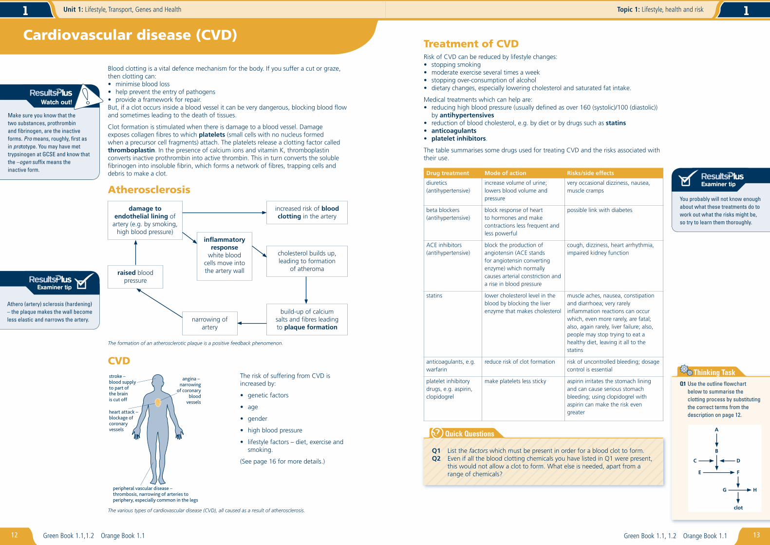
The formation of an atherosclerotic plaque is a positive feedback phenomenon.
Athero (artery) sclerosis (hardening) – the plaque makes the wall become less elastic and narrows the artery.
CVDstroke – blood supply to part of the brain is cut off
angina – narrowing
of coronaryblood
vessels
heart attack –blockage ofcoronary vessels
peripheral vascular disease –thrombosis, narrowing of arteries to periphery, especially common in the legs
The various types of cardiovascular disease (CVD), all caused as a result of atherosclerosis.
The genetic code must:• self replicate so that copies can pass to daughter cells during cell division• carry information that codes for proteins.
When DNA was confi rmed as the genetic code, there were competing theories about how it replicated. Meselson and Stahl’s classic experiment with Escherichia coli showed that the semi-conservativetheory was correct.
37
Topic 2: Genes and health 1
Green Book 2.1 Orange Book 2.4
Protein synthesisThe genetic code in DNA is in the nucleus, but the proteins formed using the code are made in the cytoplasm. So the DNA code is copied, making a molecule of messengerRNA in a process called transcription. The mRNA passes into the cytoplasm through nuclear pores and is used to make a polypeptide in a process called translation.
Meselson and Stahl’s experiment.
Students often confuse DNA replication with the process of mRNA synthesis (transcription). Try not to fall victim to this error.
How does DNA replication occur?During replication the two strands of DNA unwind and split apart. Free nucleotides line up along each strand, observing the complementary base pairing rules (page 34). The enzyme DNA polymerase bonds the nucleotides together as a phosphodiester bond forms between each deoxyribose and adjacent phosphate group. Hydrogen bonding links the two strands together.
DNA replication.
The words ‘transcription’ and ‘translation’ are very similar and very easily muddled. Try to remember this. When you go from English to French you are going from one language to another; you are translating. When you go from a base sequence (in mRNA) to an amino acid sequence in a polypeptide you are going from one language to another, so again translating. Once you have got this, you should also know what transcription is.
Q1 Given the following sequence of bases on a strand of DNA, what would be the sequence on the complementary strand and on the mRNA molecule formed from it?
TACGGTATGCCAACCTTC
Q2 Write a defi nition of each of the following terms, in the context of DNA replication and protein synthesis:
transcription translation template strand sense strand
If you answer a question, such as ‘How does DNA replicate?’ with a diagram, remember to explain it as well.
Summary of the steps in protein synthesis.
How does transcription work?During transcription the DNA unwinds and hydrogen bonds between base pairs split to separate the two strands. Only one strand is used in the formation of mRNA – the template (antisense) strand. The unused strand is called the sense strand.
Ribonucleotides are paired with their complement on the template strand: uracil pairs with adenine instead of thymine. The ribonucleotides are then joined up by RNApolymerase to form a strand of mRNA.
A
A
T
T
C
G
U
G
G
T
A
C
C
C
C
C
G
A
A
A
A
C
C
G
A
A
T
T
T
TC
G
G
A
T
C
G
G
G
GG
sensestrand
template(antisense)strand
ribonucleotides
mRNAforming
A
T
T
T
Transcription, mRNA synthesis, in progress.
Q1 If one strand of DNA, during replication, has had ACT added to it, what will be the next nucleotide added – A, C, T or G, or can you not say? Explain your answer.
Q2 Make a table to compare DNA replication with transcription.
Thinking Task
Make sure you have got the terms ‘sense strand’, ‘antisense strand’, ‘template’, ‘codon’ and ‘anticodon’ clear in your mind. Make a card with descriptions of each one on it.
What is translation?The mRNA carries the genetic message in the same base sequence language as the DNA. Transfer RNA (tRNA) translates the base sequence on the mRNA into the protein amino acid sequence. Each tRNA molecule carries an amino acid to the mRNA, where the amino acid joins others carried by other tRNAs to build a polypeptide.
DNA
mRNA
transcription
mRNA
protein
translation
cytoplasm
nucleus
tRNA–amino acid
after one generation after two generations
Experiment
Results
Conclusion
less dense
1 bacteria cultured in medium containing 15N
2 bacteria transferred to medium containing 14N
more dense
makes normal DNAwhich spins less farin centrifuge
expectedresults foreach theory
conservativemodel
semiconservativemodel
dispersivemodel
first replication second replication
makes heavy DNAwhich spins downfurther in centrifuge
cell does not have a nucleus but is still considered to be eukaryotic. Suggest two reasons why this cell is still considered to be eukaryotic.
Q2 Draw a diagram of a Golgi apparatus.
Q3 Make a fl owchart to describe protein traffi cking from when the protein is fi rst formed until it is released from the cell as an extracellular enzyme.
Magnifi cation and estimating sizeImagineacellthatmeasures10mmacrossonapagebecauseithasbeenmagnified1000times.Fromthiswecanworkoutthetruesizeofthecellbyusingtheformulabelowandtheexample.
Q1 Makeaflowcharttoexplainhowspeciesrichnesscanbemeasured.Q1 In a large population of
organisms, eight different alleles were found for one gene. However, when only half of the population was sampled, six different alleles were found for the same gene. Suggest two possible reasons for the absence of the two alleles.
Thinking Task
Make sure you know the difference between diversity and density of organisms.
1 Usingthedatainthetable:
aworkoutthenumberofspeciesfoundinthe3m2
sampled.
b constructabarcharttocomparethefrequencyofoccurrenceofeachspecies.
Answers
a Itdoesn’tmatterwhetherthespeciesoccursonlyonceormanytimesasthisquestionitemisaskingforpresenceorabsenceonly.Theansweris,therefore,5asAtoEoccuratleastonce.
b Frequencyisthetotalnumberoftimeseachspeciesisfound.ForspeciesAitis1andforspeciesDitis3.Thebarchartisshownbelow.
1m2quadratnumber
Species 1 2 3
A ✔
B ✔
C ✔ ✔
D ✔ ✔ ✔
E ✔ ✔
F
Freq
uenc
y of
occ
urre
nce
3
2
1
A B
Species
EC D F0
2 Measuringgeneticdiversity
Findthenumberofdifferentallelesinagenepool.
Eachgenemayhaveonetomanydifferentalleles.
Thetotalnumberofgenesaspecieshas
Make sure you know the difference between alleles and genes. A gene codes for a characteristic, e.g. eye colour. The alleles are the alternative forms, e.g. blue or brown, of a gene that codes for a variable characteristic, e.g. eye colour.
A new taxonomic groupingThreedomainshavebeenidentifiedbasedonmolecularphylogeny.Molecularphylogenycomparesthestructureofaparticularmoleculefromdifferentorganismstodiscovertheirdegreeofevolutionaryrelatedness.Themoresimilarthestructureofthemolecule,themorecloselyrelatedtheorganismsaretoeachother.Thisisbecausechangesinmoleculestructuregenerallyoccuronlyslowlyastheyarecausedbymutations.