INTRODUCTION Ascophyllum nodosum (L.) Le Jolis can be considered the most successful marine intertidal alga in the North Atlantic Ocean. It has a wide geographic distribution across several climatic zones on both sides of the Atlantic Ocean. It occurs in a wide range of habitats from estuaries and salt marshes to all but highly wave exposed habitats. In many sites it can be the dominant organism where it forms continuous stands in which overlapping fronds completely cover the substratum (Baardseth 1970; Chapman 1995). The basis for this success is difficult to explain; however, several studies suggest that the interaction of A. nodosum with its obligate endosymbiotic fungus, Mycophycias ascophylli (Cotton) Kohlmeyer and Volkmann-Kohlmeyer (= Mycosphaerella ascophylli Cotton) (Cotton 1909; Kohlmeyer and Kohlmeyer 1972, 1979; Garbary and Gautam 1989; Kohlmeyer and Volkmann-Kohlmeyer 1998) provide evidence for the success of the host fucoid (Garbary and MacDonald 1995; Garbary and London 1995; Garbary and Deckert 2001). Fungi are often found in intimate association with photosynthetic organisms, frequently as parasites. Remarkable interactions have been documented in which the association is so interdependent that it can be considered a composite organism. Grasses of the genera Festuca and Lolium associate with endophytic fungi of Neotyphodium spp. in combative-biochemical mutualisms that, to the host, confer resistance to herbivory, contribute to drought tolerance, improve competitive ability and enhance growth (Bacon and Hill 1996). The mycobiont in this case has forgone sexual reproduction and is solely transmitted vertically through host lines (Clay 1988). Neotyphodium is thus dependent on host fitness for the successful maintenance of its own germ line. Its influence, however, extends well beyond the host into the general community (Clay 1990; Clay and Holah 1999; Omacini et al. 2001; Rudgers et al. 2004) and it can therefore be considered a keystone species (Power et al. 1996). Algae Volume 20(3): 225-232, 2005 Ascophyllum and its symbionts. VI. Microscopic Characterization of the Ascophyllum nodosum (Phaeophyceae), Mycophycias ascophylli (Ascomycetes) Symbiotum R. J. Deckert and D. J. Garbary 1 * Department of Botany, Weber State University, Ogden, Utah, 84408 USA 1 Department of Biology, St. Francis Xavier University, Antigonish, Nova Scotia, B2G 2W5, Canada Optical microscopy of recently living and cleared material of the fucoid, Ascophyllum nodosum (L.) Le Jolis, revealed novel aspects of its interaction with the ascomycete Mycophycias ascophylli (Cotton) Kohlmeyer and Kohlmeyer (previously Mycosphaerella ascophylli Cotton). Most host cells are associated with hyphae by lateral attachment of cell walls. Hyphae form extensive networks throughout the host thallus and show considerable differentiation in the various host tissues. In the base of epidermal cells, hyphae form multicellular rings around each host cell to produce a continuous network. In medullary regions, long, relatively unbranched and longitudinally aligned hyphae occur, with radial branches extending into cortical regions. Scattered in the inner cortex of host tissue are numerous multicellular nodes of smaller, polygonal to irregular shaped cells with five or more radiating arms of hyphae. Individual hyphal cells show a variety of specializations including swellings and appressoria-like attachments to some host cells. These observations provide the morphological basis for the mutualistic symbiosis supported by recent experimental work. We conclude that this association is best described by the term “symbiotum.” Key Words: anatomy, Ascophyllum, Fucaceae, Mycophycias, mycophycobiosis, symbiosis, symbiotum *Corresponding author ([email protected])
Transcript
INTRODUCTION
Ascophyllum nodosum (L.) Le Jolis can be considered themost successful marine intertidal alga in the NorthAtlantic Ocean. It has a wide geographic distributionacross several climatic zones on both sides of the AtlanticOcean. It occurs in a wide range of habitats fromestuaries and salt marshes to all but highly waveexposed habitats. In many sites it can be the dominantorganism where it forms continuous stands in whichoverlapping fronds completely cover the substratum(Baardseth 1970; Chapman 1995). The basis for thissuccess is difficult to explain; however, several studiessuggest that the interaction of A. nodosum with itsobligate endosymbiotic fungus, Mycophycias ascophylli(Cotton) Kohlmeyer and Volkmann-Kohlmeyer(=Mycosphaerella ascophylli Cotton) (Cotton 1909;Kohlmeyer and Kohlmeyer 1972, 1979; Garbary andGautam 1989; Kohlmeyer and Volkmann-Kohlmeyer
1998) provide evidence for the success of the host fucoid(Garbary and MacDonald 1995; Garbary and London1995; Garbary and Deckert 2001).
Fungi are often found in intimate association withphotosynthetic organisms, frequently as parasites.Remarkable interactions have been documented inwhich the association is so interdependent that it can beconsidered a composite organism. Grasses of the generaFestuca and Lolium associate with endophytic fungi ofNeotyphodium spp. in combative-biochemical mutualismsthat, to the host, confer resistance to herbivory,contribute to drought tolerance, improve competitiveability and enhance growth (Bacon and Hill 1996). Themycobiont in this case has forgone sexual reproductionand is solely transmitted vertically through host lines(Clay 1988). Neotyphodium is thus dependent on hostfitness for the successful maintenance of its own germline. Its influence, however, extends well beyond the hostinto the general community (Clay 1990; Clay and Holah1999; Omacini et al. 2001; Rudgers et al. 2004) and it cantherefore be considered a keystone species (Power et al.1996).
AlgaeVolume 20(3): 225-232, 2005
Ascophyllum and its symbionts. VI. Microscopic Characterization of theAscophyllum nodosum (Phaeophyceae), Mycophycias ascophylli
(Ascomycetes) Symbiotum
R. J. Deckert and D. J. Garbary1*
Department of Botany, Weber State University, Ogden, Utah, 84408 USA1Department of Biology, St. Francis Xavier University, Antigonish, Nova Scotia, B2G 2W5, Canada
Optical microscopy of recently living and cleared material of the fucoid, Ascophyllum nodosum (L.) Le Jolis,revealed novel aspects of its interaction with the ascomycete Mycophycias ascophylli (Cotton) Kohlmeyer andKohlmeyer (previously Mycosphaerella ascophylli Cotton). Most host cells are associated with hyphae by lateralattachment of cell walls. Hyphae form extensive networks throughout the host thallus and show considerabledifferentiation in the various host tissues. In the base of epidermal cells, hyphae form multicellular rings aroundeach host cell to produce a continuous network. In medullary regions, long, relatively unbranched andlongitudinally aligned hyphae occur, with radial branches extending into cortical regions. Scattered in the innercortex of host tissue are numerous multicellular nodes of smaller, polygonal to irregular shaped cells with five ormore radiating arms of hyphae. Individual hyphal cells show a variety of specializations including swellings andappressoria-like attachments to some host cells. These observations provide the morphological basis for themutualistic symbiosis supported by recent experimental work. We conclude that this association is best describedby the term “symbiotum.”
The adaptive success of the association betweenspecies of eukaryotic algae and fungi, and occasionallycyanobacteria, to form lichens is exemplified by thecontinued success of these ancient composite organisms.Lichens tolerate rapid and extreme environmentalfluctuations in temperature and moisture. In tandem, themycobiont and photobiont often colonize habitats tooharsh for either symbiont on its own, and where fewother organisms succeed (Nash 1996). In lichens, thefungus provides a fairly stable environment for the algalcells, which, in turn, supply the fungus with fixedcarbon. In addition, if cyanobacteria are present, thefungus receives nitrogen. The thallus characteristic ofany particular lichen species is only produced when thealga and fungus live symbiotically (Büdel andScheidegger 1996), and axenic culture of the symbionts isonly possible in contrived laboratory conditions(Stocker-Wörgötter 1995). They are, in other words,ecologically obligate.
Kohlmeyer and Kohlmeyer (1972) coined the term“mycophycobiosis” to describe symbioses betweenspecies of macroalgae and fungi in which the photobiontis the exhabitant and the fungus is contained within thealgal thallus. The term refers to interactions potentiallyranging from parasitic to mutualistic. These biologicalsystems have not been studied extensively and are oftenignored in reviews of fungal-plant interactions (e.g., Issac1992; Schulz and Boyle 2005), or even in the context ofreviews of marine fungi (e.g., Fell and Newell 1998).Recently, the mycophycobiosis involving the green algaPrasiola crispa ssp. antarctica (Kützing) Knebel has beenreconfirmed as a lichen as Turgidosculum complicatumKohlmeyer and Kohlmeyer (Lud et al. 2001).
The adaptive significance of the Ascophyllum-Mycophycias symbiosis has been demonstrated. Thegrowth of Ascophyllum zygotes is enhanced by infectionof Mycophycias ascospores within the first week of life.Although other fungi have been described from A.nodosum (Kohlmeyer and Kohlmeyer 1979), labelling of
fungal cultures using fluorescent antibodies suggestedthat only one mycobiont is regularly present (Fries andThoren-Tolling 1978). In vitro experiments show that theendophyte increases desiccation tolerance of the alga(Garbary and London 1995), an important considerationin the intertidal habitat of Ascophyllum. The phenology ofgamete release for the alga and spore release for thefungus are closely tied. Uninfected zygotes exhibitaltered morphological development and decrease inseveral growth parameters in the laboratory compared tozygotes infected with the endophyte (Garbary andMacDonald 1995). Other potential benefits to the host,such as protection from herbivory (seen in terrestrialsystems) are not yet demonstrated for Ascophyllum.
The primary objective of this study was to investigatethe occupation of A. nodosum by M. ascophylli byapplying novel microscopic techniques to this system.These allow visualization of the hyphal organizationwithin the algal thallus. By clarifying morphologicalinteractions between the species we hoped to betterevaluate the causal basis for recently describedinteractions between the symbionts, and to better placethis symbiosis in the context of other fungal-plantinteractions, especially among terrestrial plants.
MATERIALS AND METHODS
Collection and sample preparationAscophyllum nodosum with its endophytic ascomycete,
Mycophycias ascophylli were collected in August 2000 atCaribou Harbour, Pictou Co., Captain’s Pond,Antigonish Co., and Tor Bay Provincial Park,Guysborough Co., all in Nova Scotia, Canada. Whole orportions of fronds were collected from the intertidalzone. Fresh material was hand-sectioned and mountedon microscope slides in lactoglycerol (1:1:1, lacticacid:glycerol:water) containing trypan blue (0.05%) andallowed to stain at least 1 h before viewing. Optimumstaining was achieved after several hours.
Deckert & Garbary: Ascophyllum and Mycophycias 227
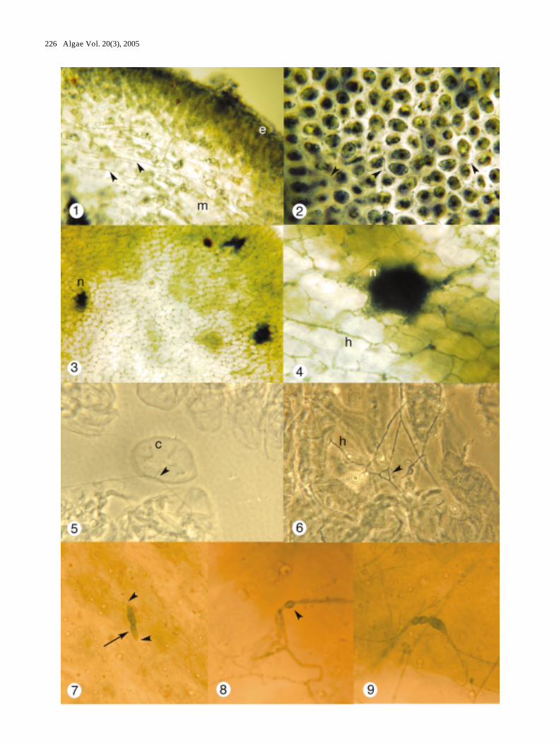
Figs 1-9. The Ascophyllum nodosum, Mycophycias ascophylli symbiotum. Figs 1-4, 7-9, brightfield microscopy; Figs 5-6, phase contrastmicroscopy. Fig. 1. Portion of longitudinal section of A. nodosum with outer epidermal (e) region and inner medullary (m) regionshowing longitudinally aligned (arrow heads) and radially aligned hyphae. X 200. Fig. 2. Paradermal section through cortex ofA. nodosum showing hyphal networks (arrow heads) surrounding each host cell. X 350. Fig. 3. Optical section through clearedtissue of A. nodosum showing abundant hyphal networks and three hyphal nodes (n). X 200. Fig. 4. Paradermal section of clearedmaterial of A. nodosum through hyphal node (n) showing numerous connections to filamentous hyphae (h). X 800. Fig. 5. Slightlysquashed preparation of cleared material of A. nodosum showing hyphal connection (arrow head) to cortical cell (c). X 1450. Fig.6. Hyphal detail in cleared material showing hyphal anastomosis (arrow head). X 950. Fig. 7. Swollen hypha with crosswall(arrow) connected at either end to normal hyphae (arrow heads). X 700. Fig. 8. Portion of hypha with conspicuous swelling(arrowhead). X 1000. Fig. 9. Differentiated swollen hypha connected to normal hyphae. X 1000.
228 Algae Vol. 20(3), 2005
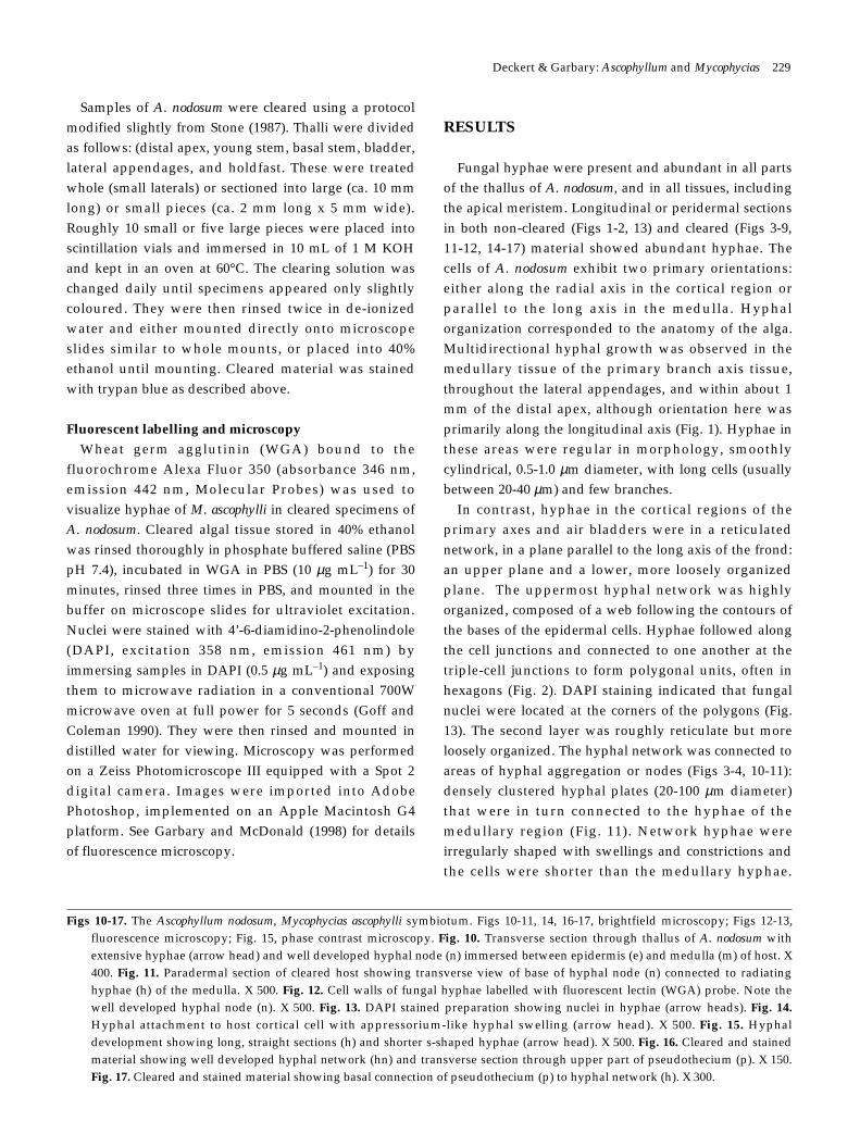
Samples of A. nodosum were cleared using a protocolmodified slightly from Stone (1987). Thalli were dividedas follows: (distal apex, young stem, basal stem, bladder,lateral appendages, and holdfast. These were treatedwhole (small laterals) or sectioned into large (ca. 10 mmlong) or small pieces (ca. 2 mm long x 5 mm wide).Roughly 10 small or five large pieces were placed intoscintillation vials and immersed in 10 mL of 1 M KOHand kept in an oven at 60°C. The clearing solution waschanged daily until specimens appeared only slightlycoloured. They were then rinsed twice in de-ionizedwater and either mounted directly onto microscopeslides similar to whole mounts, or placed into 40%ethanol until mounting. Cleared material was stainedwith trypan blue as described above.
Fluorescent labelling and microscopyWheat germ agglutinin (WGA) bound to the
fluorochrome Alexa Fluor 350 (absorbance 346 nm,emission 442 nm, Molecular Probes) was used tovisualize hyphae of M. ascophylli in cleared specimens ofA. nodosum. Cleared algal tissue stored in 40% ethanolwas rinsed thoroughly in phosphate buffered saline (PBSpH 7.4), incubated in WGA in PBS (10 µg mL–1) for 30minutes, rinsed three times in PBS, and mounted in thebuffer on microscope slides for ultraviolet excitation.Nuclei were stained with 4’-6-diamidino-2-phenolindole(DAPI, excitation 358 nm, emission 461 nm) byimmersing samples in DAPI (0.5 µg mL–1) and exposingthem to microwave radiation in a conventional 700Wmicrowave oven at full power for 5 seconds (Goff andColeman 1990). They were then rinsed and mounted indistilled water for viewing. Microscopy was performedon a Zeiss Photomicroscope III equipped with a Spot 2digital camera. Images were imported into AdobePhotoshop, implemented on an Apple Macintosh G4platform. See Garbary and McDonald (1998) for detailsof fluorescence microscopy.
RESULTS
Fungal hyphae were present and abundant in all partsof the thallus of A. nodosum, and in all tissues, includingthe apical meristem. Longitudinal or peridermal sectionsin both non-cleared (Figs 1-2, 13) and cleared (Figs 3-9,11-12, 14-17) material showed abundant hyphae. Thecells of A. nodosum exhibit two primary orientations:either along the radial axis in the cortical region orparallel to the long axis in the medulla. Hyphalorganization corresponded to the anatomy of the alga.Multidirectional hyphal growth was observed in themedullary tissue of the primary branch axis tissue,throughout the lateral appendages, and within about 1mm of the distal apex, although orientation here wasprimarily along the longitudinal axis (Fig. 1). Hyphae inthese areas were regular in morphology, smoothlycylindrical, 0.5-1.0 µm diameter, with long cells (usuallybetween 20-40 µm) and few branches.
In contrast, hyphae in the cortical regions of theprimary axes and air bladders were in a reticulatednetwork, in a plane parallel to the long axis of the frond:an upper plane and a lower, more loosely organizedplane. The uppermost hyphal network was highlyorganized, composed of a web following the contours ofthe bases of the epidermal cells. Hyphae followed alongthe cell junctions and connected to one another at thetriple-cell junctions to form polygonal units, often inhexagons (Fig. 2). DAPI staining indicated that fungalnuclei were located at the corners of the polygons (Fig.13). The second layer was roughly reticulate but moreloosely organized. The hyphal network was connected toareas of hyphal aggregation or nodes (Figs 3-4, 10-11):densely clustered hyphal plates (20-100 µm diameter)that were in turn connected to the hyphae of themedullary region (Fig. 11). Network hyphae wereirregularly shaped with swellings and constrictions andthe cells were shorter than the medullary hyphae.
Deckert & Garbary: Ascophyllum and Mycophycias 229
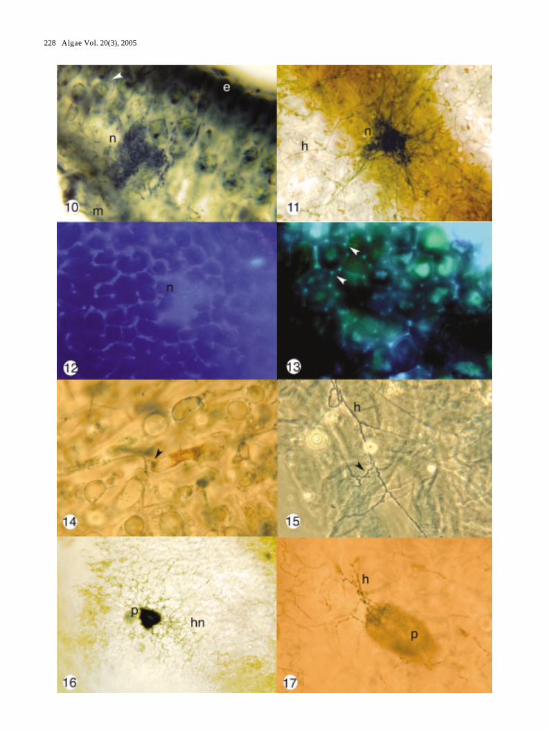
Figs 10-17. The Ascophyllum nodosum, Mycophycias ascophylli symbiotum. Figs 10-11, 14, 16-17, brightfield microscopy; Figs 12-13,fluorescence microscopy; Fig. 15, phase contrast microscopy. Fig. 10. Transverse section through thallus of A. nodosum withextensive hyphae (arrow head) and well developed hyphal node (n) immersed between epidermis (e) and medulla (m) of host. X400. Fig. 11. Paradermal section of cleared host showing transverse view of base of hyphal node (n) connected to radiatinghyphae (h) of the medulla. X 500. Fig. 12. Cell walls of fungal hyphae labelled with fluorescent lectin (WGA) probe. Note thewell developed hyphal node (n). X 500. Fig. 13. DAPI stained preparation showing nuclei in hyphae (arrow heads). Fig. 14.Hyphal attachment to host cortical cell with appressorium-like hyphal swelling (arrow head). X 500. Fig. 15. Hyphaldevelopment showing long, straight sections (h) and shorter s-shaped hyphae (arrow head). X 500. Fig. 16. Cleared and stainedmaterial showing well developed hyphal network (hn) and transverse section through upper part of pseudothecium (p). X 150.Fig. 17. Cleared and stained material showing basal connection of pseudothecium (p) to hyphal network (h). X 300.
Hyphal networks were present in all parts of the thallus(main axes, lateral branches, bladders, and receptacles),although they were somewhat disorganized at the apicalmeristems. Hyphae of the medulla were more regularlycylindrical with less branching and longer cells. Thesehyphae were oriented parallel to the longitudinal axis ofthe frond with occasional connections to the reticulatemycelium. Other hyphae were oriented along the radialaxis and extended to the surface of the algal frond.Hyphae were often seen strongly attached to host cells(Fig. 5) and hyphae in squash preparations werefrequently connected by anastomoses (Fig. 6) or shortsections of sinuous hyphae (Fig. 15). Infrequent hyphalswellings that were either septate or nonseptate wereobserved (Figs 7-9).
The hyphae displayed strong labelling by WGA-bound fluorochromes confirming presence of N-acetylglucosamine (chitin) or N-acetylneuraminic acid(sialic acid) residues in cell walls (Fig. 12). DAPI stainingconfirmed the presumed monokaryotic condition of cells(Fig. 13). Five to eight nuclei were in each net-like unitsurrounding the cortical cells. Occasionally butinfrequently, hyphae were attached to host cells byappressorium-like swellings. These host cells appeareddark in colour and necrotic (Fig. 14).
Pseudothecial ascocarps (Figs 16-17) were producedon receptacles; the base of these ascocarps were situateddeeper in the algal tissue than were the nodes and alsoappeared rounder in optical section. The upper necks ofthe pseudothecia were normally melanized, andappeared dark, even in cleared and stained specimens.The bases of the pseudothecia remained connected to thehyphal network when fully formed (Fig. 17).
DISCUSSION
Early accounts of Mycophycias ascophylli demonstrateda systemic infection of Ascophyllum nodosum, and similarobservations have been made on the related fucoid,Pelvetia canaliculata (L.) Decaisne and Thuret (Kohlmeyerand Kohlmeyer 1979; Kingham and Evans 1986). Thenature of the interaction between these species wasunclear; however, the obligate nature of the symbiosisand the absence of a conspicuous parasitism were usedto imply a mutualistic symbiosis (Kohlmeyer andKohlmeyer 1972, 1979). Smith and Ramsbottom (1915)initially queried whether or not P. canaliculata was alichen, and Kohlmeyer and Kohlmeyer (1972) asked thesame question with respect to A. nodosum. Although
Kohlmeyer and Kohlmeyer pointed out the lichen-likeattributes of this system, they used the more neutral term“mycophycobiosis” to describe this association andsimilar ones in other marine algae [i.e., Mycophycias (asMycosphaerella) in Apophlaea, Kohlmeyer and Hawkes1983]. Experiments demonstrated that infection by M.ascophylli modified the morphology of developingzygotes of A. nodosum and increased their protectionfrom desiccation (Garbary and London 1995; Garbaryand MacDonald 1995). Although the dominance of the A.nodosum thallus as the exhabitant preclude thedetermination of this system as a ‘lichen’ according toHawksworth (1988), thallus habit in lichens isoccasionally dictated by the photobiont (Budel andScheidegger 1996). In addition, the lichen Verrucariatavaresiae Moe was recently described in which thephotobiont is the crustose brown alga Petrodermamaculiforme (Wollny) Kuckuck (Moe 1997; Sanders et al.2004).
Nevertheless, we feel that the nature of themorphological and ecological integration in theAscophyllum-Mycophycias symbiosis makes thisequivalent to the “symbiotum” described for the grass/endophyte interaction (Schardl et al. 1991). The term“mycophycobiosis” of Kohlmeyer and Kohlmeyer (1972,1979) should be reserved for those marine algal-fungalassociations where the nature of the symbiosis is unclear.Based on the ability of the fungus to modifydevelopment of its algal partner, the associationpreviously considered to be a mycophycobiosis betweenPrasiola crispa ssp. antarctica and its fungal symbiont,Turgidosculum complicatum is now considered to be alichen (Lud et al. 2001). Based on the examples providedby V. tavaresiae and T. complicatum, the Ascophyllum-Mycophycias symbiosis also may be considered a lichen.Thus the terms, lichen, mycophycobiosis and symbiotumare not necessarily mutually exclusive.
The A. nodosum-M. ascophylli symbiotum hasadditional species that may be obligately or facultativelyassociated with it. The red alga, Vertebrata lanosa (L.)Christensen [=Polysiphonia lanosa (L.) Tandy], is anobligate epiphyte that is almost always associated withA. nodosum (see Garbary et al. 1991; Tian and Garbary1992; Garbary and Deckert 2001). The interaction of therhizoid of V. lanosa with the host symbiotum suggestscomplex interactions of both the fungal and algalcomponents with an invading species. Potentially moreimportant to the symbiotum is the association withepiphytic bacteria. Fries (1988) reported that when A.
230 Algae Vol. 20(3), 2005
nodosum thalli were treated to make them axenic, hyphaeof M. ascophylli grew out through the thallus wall andparasitized the host cells leading to host mortality. Thissuggests that a complex balance exists among thebacterial flora, A. nodosum and M. ascophylli.
We believe this is the first account of a highlyspecialized hyphal organization in an algal-fungalrelationship of the sort referred to as a“mycophycobiosis”. It is also the first report of this typeof organization and hyphal differentiation in theAscophyllum-Mycophycias system. The design of thehyphal network, which rings the perimeter of every cellin the frond at that plane, and represents a considerableenergy investment on the part of the fungus (and thehost), implies a specialized function. One of thedemonstrated benefits of this particular symbiosis is theability to tolerate periodic desiccation as the alga isexposed to air during tidal fluctuations. The mechanismis not known, but may involve the production ofosmoregulators by the fungus, as has been proposed as amechanism of drought tolerance in grass symbiota (Westet al. 1990). A hyphal network like the one seen inAscophyllum-Mycophycias would allow close monitoringof cell water relations and rapid response to detectedchanges.
ACKNOWLEDGEMENTS
We thank Larry Petersen, Alison Sherwood andJonathan Ferrier for critical comments on the ms, andTristan Moore and Mark Munro for technical assistancewith preparation of figures. This work was supported byresearch grants from the Natural Sciences andEngineering Research Council of Canada (NSERC) toDJG and NSERC postgraduate scholarships to RJD.
REFERENCES
Baardseth E. 1970. Synopsis of biological data on knobbedwrack Ascophyllum nodosum (Linnaeus) Le Jolis. FAO Fish.Bull. 38: 1-40.
Bacon C.W. and Hill N.S. 1996. Symptomless grass endophytes:products of coevolutionary symbioses and their role in theecological adaptations of grasses. In: Redlin S.C. andCarris L.M. (eds), Endophytic Fungi in Grasses and WoodyPlants: Systematics, Ecology and Evolution. APS Press, St.Paul, Minnesota. pp. 155-178.
Büdel B. and Scheidegger C. 1996. Thallus morphology andanatomy. In: Nash T.H. (ed.), Lichen Biology. CambridgeUniversity Press, Cambridge, U.K. pp. 37-64.
Chapman A.R.O. 1995. Functional ecology of fucoid algae:
twenty-three years of progress. Phycologia 34: 1-32.Clay K. 1988. Fungal endophytes of grasses: a defensive
mutualism between plants and fungi. Ecology 69: 10-16.Clay K. 1990. Fungal endophytes of grasses. Ann. Rev. Ecol. Syst.
21: 275-297.Clay K. and Holah J. 1999. Fungal endophyte symbiosis and
plant diversity in successional fields. Science 285: 1742-1743.Cotton A.D. 1909. Notes on marine pyrenomycetes. Trans. Br.
Mycol. Soc. 3: 92-99.Fell J.W. and Newell S.Y. 1998. Biochemical and molecular
methods for the study of marine fungi. In: Cooksey K.E.(ed.), Molecular Approaches to the Study of the Ocean.Chapman & Hall, London. pp. 259-283.
Fries L. 1988. Ascophyllum nodosum (Phaeophyta) in axenicculture and its response to the endophytic fungusMycosphaerella ascophylli and epiphytic bacteria. J. Phycol.24: 333-337.
Fries N. and Thorén-Tolling K. 1978. Identity of the fungalendophyte of Ascophyllum with Mycosphaerella ascophylliestablished by means of fluorescent antibody techniques.Bot. Mar. 21: 409-411.
Garbary D.J. and Deckert R.J. 2001. Three part harmony -Ascophyllum and its symbionts. In: Seckbach J. (ed.),Symbiosis: Mechanisms and Model Systems. Kluwer,Dortrecht, The Netherlands. pp. 309-321.
Garbary D.J. and Gautam A. 1989. The Ascophyllum/Polysiphonia/Mycosphaerella symbiosis. I. Populationecology of Mycosphaerella from Nova Scotia. Bot. Mar. 32:181-186.
Garbary D.J. and London J. 1995. The Ascophyllum/Polysiphonia/Mycosphaerella symbiosis. V. Mycosphaerellaprotects A. nodosum from desiccation. Bot. Mar. 38: 529-33.
Garbary D.J. and MacDonald K.A. 1995. The Ascophyllum/Polysiphonia/Mycosphaerella symbiosis. IV. Mutualism in theAscophyllum/Mycosphaerella symbiosis. Bot. Mar. 38: 221-225.
Garbary D.J. and McDonald A.R. 1998. Molecules, organellesand cells: fluorescence microscopy and red algaldevelopment. In: Cooksey K.E. (ed.), Molecular Approachesto the Study of the Ocean. Chapman & Hall, London. pp. 409-422.
Garbary D.J., Tian Lining and Burke J. 1991. The Ascophyllum/Polysiphonia/Mycosphaerella symbiosis. II. Aspects of theecology and distribution of Polysiphonia in Nova Scotia.Bot. Mar. 34: 391-401.
Goff L.J. and Coleman A.W. 1990. DNA: microfluorometricstudies. In: Cole K.M. and Sheath R.G. (eds), Biology of theRed Algae. Cambridge University Press, Cambridge, U.K.pp. 43-71.
Hawksworth D.L. 1988. The variety of fungal-algal symbioses,their evolutionary significance, and the nature of lichens.Bot. J. Lin. Soc. 96: 3-20.
Issac S. 1992. Fungal - Plant Interactions. Chapman & Hall,London.
Kingham D.L. and Evans L.V. 1986. The Pelvetia-Mycosphaerellainterrelationship. In: Moss S.T. (ed.), The Biology of Marine
Deckert & Garbary: Ascophyllum and Mycophycias 231
Fungi. Cambridge University Press, Cambridge, U.K. pp.177-187.
Kohlmeyer J. and Hawkes M.W. 1983. A suspected case ofmycophycobiosis between Mycosphaerella apophlaeae(Ascomycetes) and Apophlaea spp. (Rhodophyta). J. Phycol.19: 257-260.
Kohlmeyer J. and Kohlmeyer E. 1972. Is Ascophyllum nodosumlichenized? Bot. Mar. 15: 109-112.
Kohlmeyer J. and Kohlmeyer E. 1979. Marine Mycology, theHigher Fungi. Academic Press, New York.
Kohlmeyer J. and Volkmann-Kohlmeyer B. 1998. Mycophycias, anew genus for the mycobionts of Apophlaea, Ascophyllumand Pelvetia. Systema Ascomycetum 16: 1-7.
Lud D., Huiskes A.H.L. and Ott S. 2001. Morphologicalevidence for the symbiotic character of Turgidosculumcomplicatum Kohlm & Kohlm. (= Mastodia tesselata Hook. f.& Harvey). Symbiosis 31: 141-151.
Moe R. 1997. Verrucaria tavaresiae sp. nov., a marine lichen witha brown algal photobiont. Bull. Cal. Lichen Soc. 4: 7-11.
Nash T.H. 1996. Lichen Biology. Cambridge University Press,Cambridge, U.K.
Omacini M., Chaneton E.J., Ghersa C.M., and Mulller C.B. 2001.Symbiotic fungal endophytes control insect host-parasiteinteraction webs. Nature 409: 78-81.
Power M. E., Tilman D., Estes J.A., Menge B.A., Bond W.J. ,Mills L.S., Daily G., J. Castilla C., Lubchenco J. and PaineR.T. 1996. Challenges in the quest for keystones. Bioscience46: 609-620.
Rudgers J.A., Koslow J.M. and Clay K. 2004. Endophytic fungialter relationships between diversity and ecosystem
properties. Ecol. Let. 7: 42-51.Sanders W.B., Moe R.L. and Ascaso C. 2004. The intertidal
marine lichen formed by the pyrenomycete fungusVerrucaria tavaresiae (Ascomycotina) and the brown algaPetroderma maculiforme (Phaeophyceae): thallus organizationand symbiont interaction. Am. J. Bot. 91: 511-522.
Schardl C.L., Liu J.S., White J.F. Jr., Finkel R.A. and Burke J.1991. Molecular phylogenetic relationships of non-pathogenic grass mycosymbionts and clavicipitaceousplant pathogens. Pl. Syst. Evol. 178: 27-41.
Schulz B. and Boyle C. 2005. The endophytic continuum. Mycol.Res. 109: 661-686.
Smith A.L. and Ramsbottom J. 1915. Is Pelvetia canaliculata alichen? New Phytol. 14: 295-298.
Stocker-Wörgötter E. 1995. Experimental cultivation of lichensand lichen symbionts. Can. J. Bot. 73 (Suppl. 1): S579-S589.
Stone J.K. 1987. Initiation and development of latent infectionsby Rhabdocline parkeri on Douglas-fir. Can. J. Bot. 65: 2614-2621.
Tian Lining and Garbary D.J. 1992. The Ascophyllum/Polysiphonia/Mycosphaerella symbiosis. III. Culture studies onthe interaction between Polysiphonia lanosa and Ascophyllumnodosum. Bot. Mar. 35: 341-349.
West C.P., Oosterhuis D.M. and Wullschleger S.D. 1990.Osmotic adjustment in tissues of tall fescue in response towater deficit. Environ. Exp. Bot. 30: 1-8.