Page 1
ORIGINAL PAPER
Assessing Behavioural Effects of Chronic HPA Axis ActivationUsing Conditional CRH-Overexpressing Mice
Nina Dedic • Chadi Touma • Cristoph P. Romanowski • Marcel Schieven •
Claudia Kuhne • Martin Ableitner • Ailing Lu • Florian Holsboer •
Wolfgang Wurst • Mayumi Kimura • Jan M. Deussing
Received: 30 October 2011 / Accepted: 8 December 2011 / Published online: 25 December 2011
� Springer Science+Business Media, LLC 2011
Abstract The corticotropin-releasing hormone (CRH) and
its cognate receptors have been implicated in the patho-
physiology of stress-related disorders. Hypersecretion of
central CRH and elevated glucocorticoid levels, as a con-
sequence of impaired feedback control, have been shown to
accompany mood and anxiety disorders. However, a clear
discrimination of direct effects of centrally hypersecreted
CRH from those resulting from HPA axis activation has been
difficult. Applying a conditional strategy, we have generated
two conditional CRH-overexpressing mouse lines: CRH-
COEDel mice overexpress CRH throughout the body, while
CRH-COEAPit mice selectively overexpress CRH in the
anterior and intermediate lobe of the pituitary. Both mouse
lines show increased basal plasma corticosterone levels and
consequently develop signs of Cushing’s syndrome.
However, while mice ubiquitously overexpressing CRH
exhibited increased anxiety-related behaviour, overexpres-
sion of CRH in the pituitary did not produce alterations in
emotional behaviour. These results suggest that chronic
hypercorticosteroidism alone is not sufficient to alter anxi-
ety-related behaviour but rather that central CRH hyper-
drive on its own or in combination with elevated glucocor-
ticoids is responsible for the increase in anxiety-related
behaviour. In conclusion, the generated mouse lines repre-
sent valuable animal models to study the consequences of
chronic CRH overproduction and HPA axis activation.
Keywords Corticotropin-releasing hormone �Hypothalamic–pituitary–adrenal axis � Mouse model �Overexpression � Anxiety-related behaviour � Stress-coping
Introduction
The corticotropin-releasing hormone (CRH) plays a major
role in the adjustment of neuroendocrine, autonomic, and
behavioural adaptations to stressors. In this regard, CRH
functions as both, a neuroendocrine hormone within the line
of the hypothalamic–pituitary–adrenocortical (HPA) axis
(Vale et al. 1981) and a neuromodulator via hypothalamic
and extrahypothalamic neuronal pathways (Gallagher et al.
2008). Dysregulated and/or hyperactive CRH circuits have
been shown to be involved in neuroendocrine disturbances in
the context of stress-related disorders such as anxiety and
depression (Holsboer 1999; Deussing and Wurst 2005).
Elevated levels of CRH in the cerebrospinal fluid, hyperse-
cretion of CRH from the paraventricular nucleus of the
hypothalamus, elevated circulating cortisol as well as an
impaired glucocorticoid receptor (GR)-mediated negative
feedback are consistently replicated findings in patients with
Nina Dedic and Chadi Touma contributed equally to this work.
N. Dedic � C. Touma � C. P. Romanowski � M. Schieven �C. Kuhne � M. Ableitner � A. Lu � F. Holsboer �M. Kimura � J. M. Deussing (&)
Max Planck Institute of Psychiatry, Kraepelinstr.
2-10, 80804 Munich, Germany
e-mail: [email protected]
N. Dedic � W. Wurst
Institute of Developmental Genetics, German Research Center
for Environmental Health, Helmholtz Zentrum Munchen,
Ingolstadter Landstr. 1, 85764 Neuherberg, Germany
W. Wurst
Lehrstuhl fur Entwicklungsgenetik, c/o Helmholtz Zentrum
Munchen, Technical University Munchen-Weihenstephan,
Ingolstadter Landstr. 1, 85764 Neuherberg, Germany
W. Wurst
Deutsches Zentrum fur Neurodegenerative Erkrankungen e. V.
(DZNE), Standort Munchen, Schillerstrasse 44, 80336 Munich,
Germany
123
Cell Mol Neurobiol (2012) 32:815–828
DOI 10.1007/s10571-011-9784-0
Page 2
major depression (Nemeroff et al. 1984; Lowy et al. 1984;
Peeters et al. 2004). Therefore, animal models of CRH
excess have attracted major interest as tools to study the
consequences of a hyperactive CRH system.
The first CRH overexpressing mouse line was generated
via a classical transgenic approach applying the broadly
active metallothionine 1 promoter (Stenzel-Poore et al.
1992). These mice (CRF-OEMt1) showed a strong CRH
overexpression in the brain and peripheral organs including
lung, adrenal, heart, and testis. CRH overproduction resulted
in elevated plasma corticosterone levels and Cushing-like
symptoms. Moreover, CRF-OEMt1 showed increased anxi-
ety-related behaviour, which was reversible by the CRH
receptor antagonist a-helical CRH (Stenzel-Poore et al.
1994). Another CRH overexpressing mouse line was
developed using the Thy1.2 promoter driving CRH expres-
sion in postnatal and adult neurons of the brain (Dirks et al.
2001). However, CRH-OEThy1.2 did not show an altered
stress response or phenotype indicative of increased anxiety-
or depression-like behaviour (Dirks et al. 2001; Groenink
et al. 2002). Instead, CRH-OEThy-1.2 mice displayed reduced
startle reactivity as well as reduced freezing following fear
conditioning (Dirks et al. 2002b; Groenink et al. 2003). With
some delay CRH-OEThy1.2 also developed a mild Cushingoid
phenotype (Dirks et al. 2002a). In addition, CRH-over-
expressing mouse lines have been established in recent years
applying the ‘‘tet-ON/tet-OFF’’ system, which allows for
reversible and inducible overexpression of CRH (Vicentini
et al. 2009; Kolber et al. 2010). Although both studies
applied the Camk2a promoter combined with a tet-operator
driven CRH-construct, the behavioural and neuroendocrine
consequences of CRH excess were rather specific for each
mouse line (Vicentini et al. 2009; Kolber et al. 2010). Taken
together, these examples illustrate the difficulties to compare
results from different transgenic mouse lines even if they are
based on similar constructs.
To circumvent these problems, we have recently
developed a mouse model which permits conditional CRH
overexpression avoiding common uncertainties of classical
transgenesis such as unpredictable influences of the site of
transgene insertion and the number of inserted transgene
copies (Lu et al. 2008). This was achieved by introducing a
CRH expression unit into the ubiquitously expressed
ROSA26 (R26) locus. Undesired ubiquitous expression of
CRH driven by the R26 promoter is prevented unless a
loxP flanked transcriptional terminator is deleted via the
site-specific recombinase Cre. The ever growing ‘‘zoo’’ of
Cre recombinase transgenic mouse lines offers a plethora
of possibilities to induce specific spatial and temporal CRH
expression patterns by simple breeding. In this way CRH
expression is reproducibly driven by the endogenous R26
promoter, while the utilised Cre recombinase is only
determining its expression pattern. This approach allows
the meaningful comparison of different CRH-over-
expressing mouse lines.
Using this novel mouse model of CRH overexpression we
could demonstrate that CNS-restricted CRH overexpression
in CRH-COECNS mice, achieved by breeding with Nestin-
Cre mice (Tronche et al. 1999), leads to increased active
stress-coping behaviour and altered sleep regulation,
whereas forebrain-restricted CRH overexpression via
Camk2a-CreERT2 (Erdmann et al. 2007) results in increased
anxiety-related behaviour (Lu et al. 2008; Kimura et al.
2010; Kolber et al. 2010; Refojo et al. 2011). Both transgenic
lines show normal HPA axis activity demonstrating that the
dysregulation of the CRH system can lead to marked
behavioural alterations independent of basal HPA axis
alterations. However, several lines of evidence also suggest
that corticosteroids may cause mood and behaviour changes
in depression, but whether they directly contribute to altered
mood and anxiety symptoms remains unclear.
Here, we used our conditional mouse model of CRH
excess to discriminate between central effects of chronic
CRH hyperdrive and effects mediated via the HPA axis
and its final effector—corticosterone in mice and cortisol in
humans. To this end, we bred conditional CRH over-
expressing mice (CRH-COE) to deleter- and Pomc-Cre ani-
mals, respectively, thus enabling ubiquitous (CRH-COEDel)
and anterior pituitary-specific (CRH-COEAPit) CRH over-
expression at different dosages. The analyses of neuro-
endocrine parameters, physiological changes, and emotional
behaviour unravelled a predominant effect of central CRH
by itself or in combination with excessive corticosterone
on anxiety-related behaviour. Moreover, these conditional
models may represent very useful tools to study the behav-
ioural and neuroendocrine effects of hypercorticosteriodism
in the future.
Materials and Methods
Generation of Mice
Initially R26flopCrh/flopCrh mice (Lu et al. 2008) were bred to
Pomc-Cre mice (Akagi et al. 1997). Subsequently, mice
ubiquitously overexpressing CRH (CRH-COEDel) were
obtained by breeding female R26?/flopCrh Pomc-Cre mice
to male R26flopCrh/flopCrh mice. In this combination, Pomc-
Cre is transiently expressed during oogenesis and thus acts
as a deleter. Early deletion of the floxed stop (flop) cassette
results in ubiquitous expression of CRH. R26Crh/flopCrh
mice were intercrossed, however, no viable homozygous
R26Crh/Crh were obtained. Therefore, only R26flopCrh/flopCrh
(Ctrl) and heterozygous ubiquitously overexpressing
R26Crh/flopCrh (COE) mice were used. Anterior pituitary-
specific overexpressing mice (CRH–COEAPit) were
816 Cell Mol Neurobiol (2012) 32:815–828
123
Page 3
obtained by breeding male R26?/flopCrh Pomc-Cre mice to
female R26flopCrh/flopCrh mice. The following resulting
genotypes were used for further analyses: R26flopCrh/flopCrh
(Ctrl), R26?/flopCrh Pomc-Cre (COEhet), and R26flopCrh/flopCrh
Pomc-Cre (COEhom) mice. Genotyping was performed
by PCR using primers: ROSA-1, 50-AAA-GTC-GCT-CT
G-AGT-TGT-TAT-30; ROSA-5, 50-TAG-AGC-TGG-TTC-G
TG-GTG-TG-30; ROSA-6 50-GCT-GCA-TAA-AAC-CCC-
AGA-TG-30 and ROSA-7, 50-GGG-GAA-CTT-CCT-GAC-
TAG-GG-30. Standard PCR conditions resulted in a 398-bp
wild-type and a 646-bp mutant PCR product, respectively.
Animals with a premature deletion of the floxed transcrip-
tional terminator sequence were identified by the occurrence of
a 505-bp PCR product. The presence of Cre was evaluated
using primers CRE-F, 50-GAT-CGC-TGC-CAG-GAT-ATA-
CG-30 and CRE-R, 50-AAT-CGC-CAT-CTT-CCA-GCA-G-30
resulting in a PCR product of 574 bp. Mice used for this study
were kept on a mixed 129S2/Sv 9 C57BL/6 J background.
Pomc-Cre mice had been backcrossed to C57BL/6J for five
generations. The animal housing room as well as the experi-
mental room were maintained under standard laboratory con-
ditions (light–dark cycle: 12:12 h, lights on at 8 a.m.;
temperature: 22 ± 1�C; relative humidity: 55 ± 10%). Com-
mercial mouse diet (Altromin No. 1324, Altromin GmbH,
Lage, Germany) and bottled tap water were available
ad libitum.
Assessment of Physiological Parameters
At the age of 10–12 weeks, animals were weighed and then
sacrificed via decapitation. Thymus and adrenal glands
were extracted and stored in Ringer’s solution. In order to
determine the organ weight, additional surrounding tissue
was removed.
To assess the neuroendocrine profile of basal and stressed
animals, a second batch of 10–12-week-old animals was
separated 2 weeks prior to the experiment and singly housed
with a 12:12 h light:dark schedule (lights off at 07:00 p.m.).
All experiments and data analyses were performed sepa-
rately for male and female animals. To determine the basal
plasma hormone levels, mice were left undisturbed
throughout the night before the experiment. Blood sampling
was performed in the early morning (08:00–09:00 a.m.) and
afternoon (04:30–05:30 p.m.) by collecting trunk blood from
animals rapidly decapitated under isoflurane anaesthesia,
with the time from first handling of the animal to completion
of bleeding not exceeding 45 s. For evaluation of the endo-
crine response to stress, we collected blood samples imme-
diately after a 10-min restraint stress, for which animals were
placed in a 50-ml conical tube with the bottom removed.
Stress experiments were performed in the morning
(08:00–10:00 a.m.). Plasma corticosterone concentrations
were measured in duplicates by a commercially available
RIA kit (MP Biomedicals, Irvine, CA, USA) according to the
manufactures instructions. Plasma samples from CRH-
COEDel mice and from CRH-COEAPit mice were measured in
two independent RIAs.
Behavioural Phenotyping
At the time of testing, all animals were about 10–12-weeks of
age and were single housed in the experimental room for at
least 2 weeks. The battery of tests consisted of the open-field
test (OF), the elevated plus-maze test (EPM), the dark–light
box test (DaLi), and the forced swim test (FST; Touma et al.
2008). All tests were performed in the order listed between
9 a.m. and 12 a.m. The animals’ behaviour during the tests
was videotaped (for the FST) and scored by a trained
observer blind to the animals’ genotype using ‘Eventlog’
(version 1.0, Emco Software Ltd., Reykjavik, Iceland) or
was automatically analysed (for the OF, EPM, and DaLi) by
tracking the ‘centre of the animal’ using the ‘ANY-maze’
video-tracking software (Stoelting Co., Wood Dale, Illinois,
USA). All animal experiments were conducted in accor-
dance with the Guide for the Care and Use of Laboratory
Animals of the Government of Upper Bavaria, Germany.
Open-Field Test
The OF was used to measure locomotor activity and
explorative behaviour. Testing was performed in an evenly
and dimly lit (about 15 lux) OF consisting of a circular
arena (60 cm in diameter, surrounded by 40 cm high walls)
for 5 min. The total distance travelled, the number of
entries and the time the animal spent in the more aversive
inner zone (30 cm diameter) or the more protective outer
zone near the walls of the OF was measured.
Elevated Plus-Maze Test
Anxiety-related behaviour was measured by means of the
EPM. The plus-shaped apparatus was made of grey plastic
and consisted of two opposing closed/shielded arms (30 9
5 9 15 cm, dimly lit with about 10–20 lux) and two open/
unprotected arms (30 9 5 9 0.5 cm, brightly lit with about
300 lux) connected by a central platform (5 9 5 cm, illu-
minated with about 140 lux). The maze was elevated 40 cm
above the floor and each mouse was tested for 5 min on the
apparatus. At the beginning of each trial, the animals were
placed on the central platform facing one of the closed arms.
Parameters of interest included open arm time and entries.
Dark–Light Box Test
The DaLi, another commonly used paradigm to measure
anxiety-related behaviour in mice, was also employed. The
Cell Mol Neurobiol (2012) 32:815–828 817
123
Page 4
apparatus consisted of a rectangular box with two com-
partments, the relatively protected dark compartment
(15 9 20 9 25 cm, dimly lit with \10 lux) and the more
aversive light compartment (30 9 20 9 25 cm, brightly lit
with about 700 lux). At the beginning of the test, each
mouse was placed in the centre of the dark compartment
facing the back wall of the apparatus. The time as well as
the entries made into the lit compartment were measured
for 5 min.
Forced Swim Test
The FST was used to measure stress-coping behaviour.
Each animal was placed into a glass beaker (diameter
12 cm, height 24 cm) filled with water (temperature 23�C)
for a test period of 6 min. The parameters floating
(immobility except small movements to keep balance),
swimming and struggling (vigorous attempts to escape)
were recorded and scored throughout the 6-min test period
by a trained observer.
Sleep Recordings
Nine male CRH-COEAPit and nine of the respective male
control littermates were implanted with four EEG and two
EMG electrodes (for full surgical protocol, please refer to
(Romanowski et al. 2010). The animals were allowed to
recover from surgery for 2 weeks under standard labora-
tory conditions (23� ± 1�C, 12 h/12 h light–dark cycle,
food and water available ad libitum) before baseline
recordings were initiated. After recovery, all mice were
connected to an electrical swivel system by a recording
cable which allowed EEG and EMG recordings from freely
behaving animals. All signals were preamplified (1,0009,
custom made) and sent to a main amplifier (109, custom
made) before transformation by an analogue/digital card
(64 Hz sampling rate; National Instruments, Austin, TX).
The EEG signals were analogue band pass-filtered
(0.5–29 Hz, filter frequency roll off 48 dB/octave) and root
mean square was applied to the non-filtered EMG signals
before its digital conversion (sample rate: 64 Hz). Obtained
data were analysed by a LabView-based acquisition pro-
gram (EGEraVigilanz, SEA, Koln, Germany), and vigi-
lance states were manually defined as WAKE, non-rapid
eye movement sleep (NREMS), and rapid eye movement
sleep (REMS) in 4-s epochs.
Statistical Analysis
Data and statistical analysis were performed with the
computer programs GraphPad Prism 5.0 and SPSS 16.00.
All results are shown as means ± standard error of the
mean (SEM). Two-group comparisons of independent
samples were calculated using the Mann–Whitney–U test.
To examine differences between control, heterozygous and
homozygous CRH-COEAPit mice, the Kruskal–Wallis
(KW) H test followed by Dunn’s multiple comparison post
hoc test was applied. The effects of time and genotype on
corticosterone levels were examined by two-way-multi-
variate analysis of variance (ANOVA) with Bonferroni
post hoc tests. For the assessment of sleep recordings, a
two-factorial ANOVA with a repeated measures design
was applied and data were further statistically evaluated for
significances in each time period (light or dark), if appro-
priate, by a post hoc test for simple effects (Neumann–
Keuls test). Statistical significance was accepted at
P \ 0.05 and P B 0.1 was considered a trend.
Results
Ubiquitous and Pituitary-Specific Overexpression
of CRH
Breeding of CRH-COE mice (Fig. 1a; Lu et al. 2008) to
deleter- and Pomc-Cre mice (Fig. 1b; Akagi et al. 1997)
resulted in the excision of the transcriptional terminator
and expression of exogenous CRH throughout the body or
in the pituitary, respectively (Fig. 1c). Cre-mediated dele-
tion of the transcriptional terminator and concomitant
expression of CRH mRNA and b-galactosidase were
observed in all tissues of CRH-COEDel mice (data not
shown). In CRH-COEAPit mice CRH mRNA and b-galac-
tosidase expression was selectively observed in the anterior
and intermediate lobe of the pituitary as well as in a subset
of neurons in the arcuate nucleus (data not shown).
CRH-COEDel Mice Exhibit Endocrine Abnormalities
and Increased Anxiety-Related Behaviour
Already at the age of 3-week male and female CRH-
COEDel mice showed physical changes reminiscent of
Cushing’s syndrome such as hair loss and thin skin (data
not shown). Adult mice showed excess fat accumulation as
observed by visual inspection of subcutaneous and visceral
fat as well as an overall increased body weight (males:
Ctrl = 30.73 ± 0.53 g vs. COE = 35.73 ± 2.02 g, U =
37.0, P \ 0.05, n = 12; females: Ctrl = 25.00 ± 0.59 g
vs. COE = 31.50 ± 1.57 g, U = 15.0, P \ 0.01, n =
11–12) (Fig. 2a). Adrenal weights were also significantly
increased in male and female CRH-COEDel mice compared
to CRH-COECtrl littermates (males: Ctrl = 0.124 ±
0.0042 mg/g vs. 0.194 ± 0.024 mg/g, U = 13.0, P\0.01,
n = 8–12; females: Ctrl = 0.247 ± 0.0076 mg/g vs. COE =
0.312 ± 0.0140 mg/g, U = 9, P\0.05, n = 7–10) (Fig. 2b).
In addition, a reduction in thymus weight was observed in male
818 Cell Mol Neurobiol (2012) 32:815–828
123
Page 5
CRH-COEDel mice (Ctrl = 1.06 ± 0.095 mg/g vs. COE =
0.753 ± 0.105 mg/g, U = 8.0, P = 0.1, n = 4–10) (Fig. 2c).
In order to evaluate HPA axis rhythmicity, we measured cor-
ticosterone levels in the morning (a.m.) and the afternoon
(p.m.). Chronic CRH overproduction resulted in drastically
elevated levels of circulating plasma corticosterone, in male
and female mice compared to control littermates at both times
of the day (males: a.m.: Ctrl = 9.35 ± 2.45 ng/ml vs. COE =
162.5 ± 28.16, ng/ml; p.m.: Ctrl = 102.7 ± 14.07 ng/ml vs.
COE = 198.5 ± 23.54, two-way ANOVA, time effect
F(1,32) = 13.60, P\0.005, genotype effect F(1,32) = 49.47,
P\0.0001, time 9 genotype interaction F(1,32) = 0.10;
Bonferroni post-test, P\0.05 n = 8–12; females: a.m.:
Ctrl = 60.97 ± 14.20 ng/ml vs. COE = 131.4 ± 30.94 ng/ml,
p.m.: Ctrl = 179.6 ± 29.09 ng/ml vs. COE = 283.9 ±
34.01 ng/ml, two-way ANOVA, time effect F(1,31) = 21.33,
genotype effect F(1,31) = 8.89, Bonferroni post-test,
P\ 0.05, n = 8–10) (Fig. 2d). Thus, the regular circadian
rhythmicity of corticosterone secretion was virtually absent in
male CRH-COEDel mice, as illustrated by similarly elevated
corticosterone levels during the diurnal trough and diurnal
peak. Interestingly, this effect was much less pronounced in
female CRH-COEDel mice where elevated corticosterone
levels were still detectable in the afternoon. However, 10 min
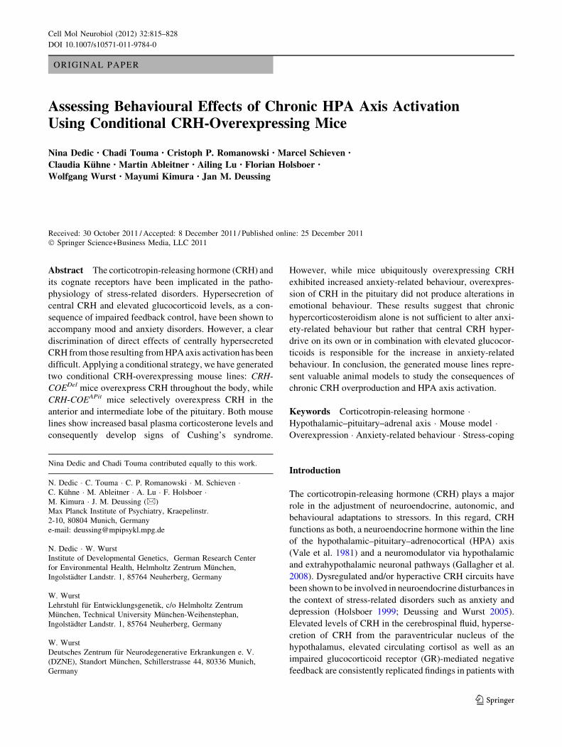
Fig. 1 Strategy for conditional
overexpression of CRH.
a Schematic representation of
the ROSA26 (R26) locus, which
was engineered to harbour a
Cre-inducible Crh-LacZexpression unit (R26flopCRH,
flop: floxed stop). b Breeding to
deleter-Cre or Pomc-Cre mice
to remove the transcriptional
terminator sequence (Cre
recombinase expression pattern
depicted in green). c Cre
recombinase induced expression
of CRH (depicted in orange)
and b-galactosidase throughout
the body (CRH-COEDel) or
within the anterior pituitary
(CRH-COEAPit). R26 exons are
indicated as black boxes, the
transcriptional terminator as a
STOP sign and loxP sites as
green arrowheads. SA splice
acceptor, IRES internal
ribosomal entry side, pA polyA
signal (Color figure online)
Cell Mol Neurobiol (2012) 32:815–828 819
123
Page 6
of acute restraint stress failed to induce a neuroendocrine
stress response in CRH-COEDel mice compared to littermate
controls, independent of gender (Fig. 2d). Generally, plasma
corticosterone concentrations were higher in control females
compared to control males (a.m.: m = 9.35 ± 2.45 ng/ml vs.
f = 60.97 ± 14.20 ng/ml, U = 3.0, P \0.001; p.m.: m =
102.7 ± 14.07 ng/ml vs. f = 179.6 ± 29.09 ng/ml, U = 23.0,
P = 0.059; stress: m = 174.3 ± 13.59 ng/ml vs. f = 301.0 ±
23.44 ng/ml, U = 4.0, P\0.001) (Fig. 2d). In the case of
CRH-overexpressing mice, gender-specific differences in cor-
ticosterone levels were only observed at the circadian peak, and
after acute restraint stress (p.m.: m = 198.5 ± 23.54 ng/ml vs.
f = 283.9 ± 34.01 ng/ml, U = 10.0, P = 0.075, stress:
m = 144.3 ± 24.22 ng/ml vs. f = 284.4 ± 20.94 ng/ml,
U = 5.0, P\0.001).
The OF test was employed to assess novelty-induced
locomotor and exploratory activity. CRH-COEDel mice
showed no significant differences in locomotion and inner
zone time compared to control littermates; however, they
made significantly fewer entries into the centre zone
(Ctrl = 12.33 ± 2.51 vs. COE = 4.58 ± 1.39, U = 33.0,
P \ 0.05, n = 12) (Fig. 3a). In the EPM CRH-COEDel
mice showed significantly increased anxiety-related
behaviour as evidenced by a decreased number of entries
(Ctrl = 38.24 ± 5.80 vs. COE = 21.35 ± 3.05%, U = 13.0,
P\0.05, n = 7–11) and time spent (Ctrl = 13.37 ± 3.88 vs.
COE = 4.01 ± 1.63%, U = 14, P\0.05, n = 7–11) in the
open arms compared to control littermates (Fig. 3b). Again,
general locomotor activity was not altered. An increase in
anxiety-related behaviour of CRH-COEDel mice was also
detected in the DaLi, indicated by decreased lit compartment
time (Ctrl = 10.96 ± 2.51 vs. COE = 3.37 ± 1.44%,
U = 24.0, P\0.05, n = 11–12) and number of entries
(Ctrl = 6.25 ± 0.83 vs. COE = 2.73 ± 0.63, U = 18.5,
P\0.05, n = 11–12) as well as an increased latency to enter
the lit compartment (Ctrl = 27.35 ± 14.84 vs. COE =
147.9 ± 36.12 s, U = 29.0, P\0.05, n = 11–12) (Fig. 3c).
To examine stress-coping behaviour, CRH-COEDel mice were
subjected to the FST. Ubiquitous CRH overexpression resulted
in a significantly increased struggling time and a trend towards
a decreased floating time (struggling: Ctrl = 11.26 ± 0.97%
vs. COE = 14.98 ± 1.49%, P = 0.05; Floating: Ctrl =
67.75 ± 1.8% vs. COE = 62.61 ± 2.6%, P = 0.1, n =
11–12; Fig. 3d).
CRH-COEAPit Mice Exhibit Endocrine Abnormalities
and Mild Behavioural Alterations
In contrast to CRH-COEDel mice, pituitary-specific CRH
overexpression led to a mild Cushing-like phenotype,
which was mainly associated with hair loss and thinning of
skin, starting at 5–6 months of age (data not shown).
Animals used for the assessment of the neuroendocrine
profile and behavioural analysis were between 10 and
12 weeks, and at that time were not distinguishable from
controls. We analysed heterozygous as well as homozy-
gous male CRH-COEAPit mice in order to assess the dos-
age-dependent effect of CRH overexpression. Interestingly,
heterozygous and homozygous CRH-COEAPit mice weight
significantly less than control littermates (males: Ctrl =
31.61 ± 0.43 g vs. COEhet = 27.94 ± 0.39 g vs. COEhom
28.99 ± 0.73 g, KW, H = 13.37, P \ 0.05, Dunn’s post-
test, P \ 0.05, n = 10–16; females: Ctrl = 23.0 ± 0.22 g
vs. COEhom = 21.58 ± 0.74 g, U = 31.5, P \ 0.05, n =
9–11) (Fig. 4a). Furthermore, CRH overexpression in the
Ctrl0
10
20
30
40
50*
Bo
dy
wei
gh
t
Ctrl0
10
20
30
40
50
*
Ad
ren
al g
lan
d w
eig
ht
/b
od
y w
eig
ht
Ctrl0.0
0.1
0.2
0.3
0.4*
Pla
sma
Co
rtic
ost
ero
ne
[g]
Ctrl0.0
0.1
0.2
0.3
0.4
*
[mg/
g]
[g]
[mg/
g]
Males Females
0.0
0.5
1.0
1.5
Ctrl
[mg/
g]
Thy
mu
s w
eig
ht
/b
od
y w
eig
ht
n.d.
[ng/
ml]
a.m. p.m. stress
*
*
0
100
200
300
400
500
[ng/
ml]
a.m. p.m. stress
* *
0
100
200
300
400
500
COE
COE
COE
COE
COE
A
B
C
D
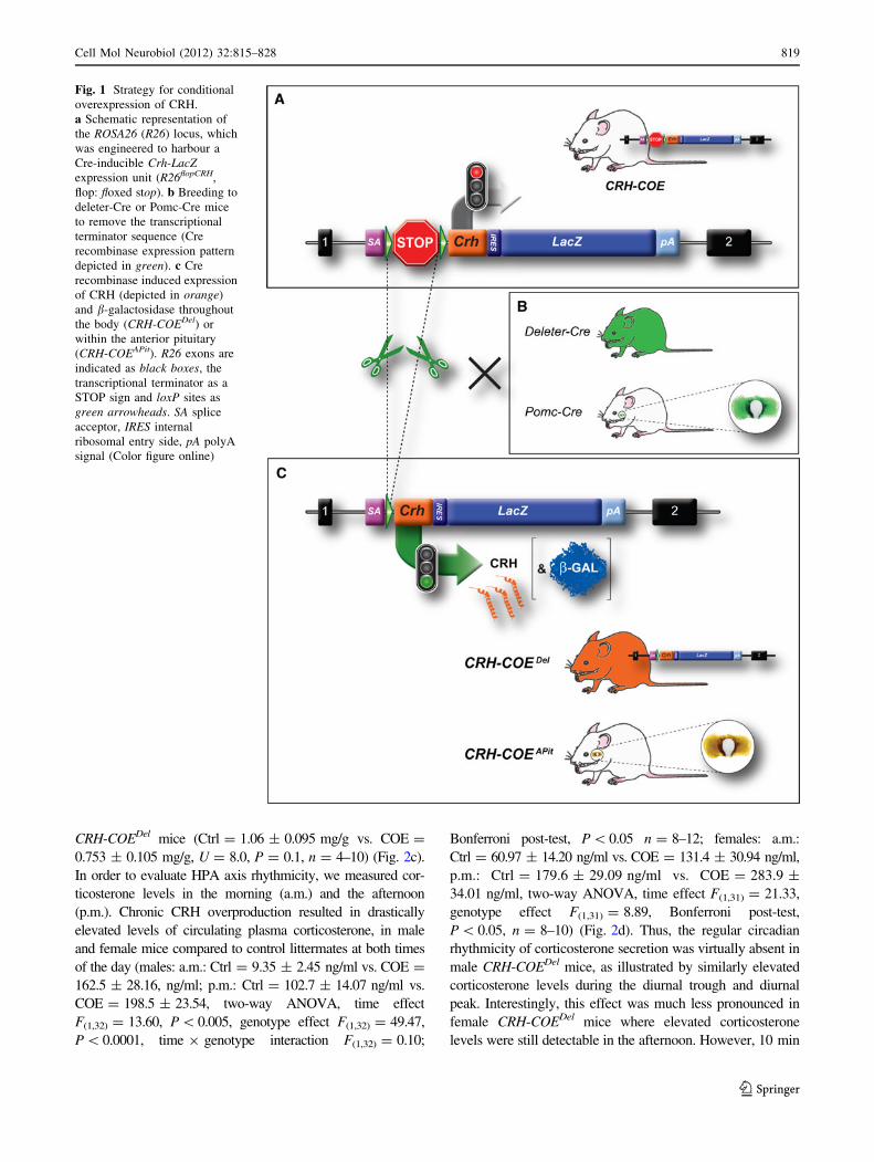
Fig. 2 Physiological and neuroendocrine alterations in male and
female CRH-COEDel mice. a Body weight and b relative adrenal
gland weights were significantly increased in male and female CRH–
COEDel mice (black bars) compared to control littermates (whitebars). c In addition, a decrease in relative thymus weight was
observed in male CRH-COEDel mice. d Corticosterone levels
measured in the morning (a.m.) and evening (p.m.) were significantly
elevated in male and female COE mice, which also showed an
attenuated stress response following 10-min of restraint stress
compared to control animals. *Significantly different from control
mice (P \ 0.05), t trend (P B 0.1), n.d. not determined, COEconditional overexpressing mice, Ctrl control littermates
820 Cell Mol Neurobiol (2012) 32:815–828
123
Page 7
pituitary resulted in a dose-dependent increase in relative
adrenal gland weight (Ctrl = 0.112 ± 0.005 mg/g vs.
COEhet = 0.174 ± 0.008 mg/g vs. COEhom = 0.196 ±
0.011 mg/g, KW, H = 20.65, P \ 0.0001, Dunn’s post-
test, P \ 0.05, n = 9–14) and a decrease in relative thymus
weight (Ctrl = 1.01 ± 0.046 mg/g vs. COEhet’ = 0.86 ±
0.01 mg/g vs. COEhom = 0.68 ± 0.05 mg/g, KW, H =
12.31, P \ 0.05, Dunn’s post-test, P \ 0.05) of male mice
(Fig. 4b, c). Similarly, female homozygous CRH-COEAPit
mice also showed enlarged adrenal glands (Ctrl = 0.28 ±
0.0076 mg/g vs. COEhom = 0.38 ± 0.012 mg/g, U = 7,
P \ 0.05, n = 10) (Fig. 4b). As was the case in CRH-
COEDel mice, circulating corticosterone levels were sig-
nificantly elevated in heterozygous and homozygous CRH-
COEAPit mice compared to littermate controls in the
morning (males: a.m.: Ctrl = 3.9 ± 0.9 ng/ml vs. COEhet =
72.52 ± 9.6 ng/ml vs. COEhom = 104.8 ± 8.4 ng/ml; two-
way ANOVA, time effect F(1,94) = 75.44, P\0.0001, geno-
type effect F(2,94) = 21.68, P\0.0001, time 9 genotype
interaction F(2,94) = 16.09, P\0.001, Bonferroni post-test,
P\0.05, n = 14–23; females: a.m.: Ctrl = 103.9 ±
20.14 ng/ml vs. COEhom = 227.2 ± 18.9 ng/ml; two-way
ANOVA, time effect F(1,39) = 6.39, P \ 0.05, genotype
effect F(1,39) = 6.16, P \ 0.05, time 9 genotype interaction
F(1,39) = 10.57, P \ 0.05, n = 11–12) (Fig. 3d). Again, a
dose-dependent increase in corticosterone could be observed
between heterozygous and homozygous CRH-COEAPit mice.
Female animals showed essentially the same phenotype.
However, differences in circulating corticosterone were not
observed in the afternoon, neither in male nor in female
CRH-COEAPit mice compared to control littermates. Thus,
similarly to male CRH-COEDel, animals, homozygous male
and female CRH-COEAPit mice exhibit marked alterations in
circadian corticosterone rhythmicity, showing only minimal
diurnal changes between morning and afternoon levels.
However, morning and afternoon plasma corticosterone
levels were generally much lower in male CRH-COEAPit
compared to male CRH-COEDel mice. Along these lines,
An
xiet
y-re
late
d b
ehav
iou
r(E
leva
ted
plu
s-m
aze)
0
5
10
15
20
25
Inne
r zo
ne ti
me
(%)
0
5
10
15
20
*
No.
inne
r zo
ne e
ntrie
s
An
xiet
y-re
late
d b
ehav
iou
r(D
ark-
ligh
t b
ox)
Str
ess-
cop
ing
beh
avio
ur
(Fo
rced
sw
im t
est)
(O
pen
fie
ld)
Lo
com
oto
r at
ivit
y
Tim
e st
rugg
ling
(%)
Tim
e sw
imm
ing
(%)
Tim
e flo
atin
g (%
)
0
10
20
30
40
50
Ope
n ar
m e
ntrie
s (%
)
*
0
50
100
150
200
Late
ncy
to fi
rst
lit c
omp.
ent
ry (
s)
*
0
2
4
6
8
Lit m
op. e
ntrie
s (%
)
*
Ope
n ar
m ti
me
(%)
0
5
10
15
20
*
Dis
tanc
e (m
)
Ctrl COE Ctrl COE0
5
10
15
20
0
2
4
6
8
Dis
tanc
e (m
)T
ime
in li
t com
p. (
%)
0
5
10
15
*
0
5
10
15
20
0
10
20
30
0
20
40
60
80
A
B
C
D
Ctrl COE
Ctrl COE Ctrl COE Ctrl COE
Ctrl COE Ctrl COE Ctrl COE
Ctrl COE Ctrl COE Ctrl COE
Fig. 3 Behavioural
characterization of male CRH-
COEDel mice. a Locomotor
activity in the open-field test
was not altered in CRH-COEDel
mice (black bars) compared to
control littermates (white bars).
Anxiety-related behaviour, as
assessed in the b elevated plus-
maze and c dark–light box test,
was significantly increased in
CRH-COEDel mice. d A mild
increase in active stress-coping
behaviour was observed in
CRH-COEDel mice compared to
control animals. *Significantly
different from control mice
(P \ 0.05), t trend (P B 0.1),
COE conditional overexpressing
mice, Ctrl control littermates
Cell Mol Neurobiol (2012) 32:815–828 821
123
Page 8
HPA axis reactivity was not inhibited in CRH-COEAPit mice.
Additionally, heterozygous and homozygous CRH-COEAPit
mice showed the same corticosterone response as control
littermates following 10 min of restraint stress (Fig. 4d).
In case of the CRH-COEAPit line, corticosterone plasma
concentrations were not only higher in control but also in
CRH-overexpressing females compared to males (Ctrl: a.m.:
m = 3.90 ± 0.88 ng/ml vs. f = 103.9 ± 20.14 ng/ml, U =
2.0, P \ 0.0001; p.m.: m = 120.3 ± 7.559 ng/ml vs. f =
228.2 ± 24.30 ng/ml, U = 23.0, P\0.001; stress; m =
261.5 ± 8.29 ng/ml vs. f = 413.3 ± 13.30 ng/ml, U = 0.0,
P\ 0.0001, n = 12–9/COEhom: a.m.: m = 104.8 ± 8.37 ng/
ml vs. f = 227.2 ± 18.94 ng/ml, U = 10.0, P \0.001; p.m.:
m = 121.8 ± 11.91 ng/ml vs. f = 211.6 ± 21.64 ng/ml, U =
33, P\0.05; stress: m = 260.4 ± 11.56 vs. f = 301.0 ±
16.47 ng/ml, U = 42.0, P\0.05, n = 11–23) (Fig. 4d).
Locomotor activity as well as the number of entries into
the inner zone of the OF were not altered in CRH-COEAPit
mice (Fig. 5a). Homozygous CRH-COEAPit mice, however,
tended to spent more time in the inner zone (Ctrl =
5.99 ± 0.83% vs. COEhom 9.65 ± 1.38%, U = 28.0, P =
0.062, n = 10–11). Anxiety-related behaviour, as assessed
in the EPM and DaLi test, was not changed in homozygous
CRH-COEAPit mice. In the FST, homozygous CRH-
COEAPit mice spent more time swimming than control
littermates (Ctrl = 19.18 ± 1.54% vs. COEhom = 25.30 ±
1.88%, U = 35.5, P \ 0.05, n = 12). However, struggling
and floating, which are considered the main readout
parameters of stress-coping behaviour in this test, were not
significantly altered.
CRH-COEAPit mice and control littermates displayed a
typical sleep/wake distribution of nocturnal rodents with
higher levels of NREMS and REMS during the light period
than during the dark period (Fig. 6). Under baseline con-
ditions CRH-COEAPit mice showed significantly higher
WAKE levels at ZT 4 (P = 0.002) and significantly lower
WAKE levels from ZT 16 to ZT 22 (P B 0.001; data not
shown) as compared to CRH-COECtrl animals. Inversely,
CRH-COEAPit mice showed significantly more NREMS
than CRH-COECtrl mice from ZT 16 to ZT 22 during
baseline recordings (P B 0.011; Fig. 6). As for REMS, no
differences could be detected between both genotypes on
the baseline day.
Discussion
Multiple lines of evidence suggest that a dysregulation of
the HPA axis plays an important role in the pathogenesis of
mood and anxiety disorders. However, to discriminate
between effects of centrally hypersecreted CRH from those
resulting from downstream peripheral effects due to HPA
axis activation is challenging. To unequivocally dissect
central from peripheral effects of CRH on physiology,
anxiety-related and stress-coping behaviour, we applied a
conditional mouse model that allows CRH overexpression
at different levels in a spatially restricted manner. As
previously described, this conditional approach provides
Bo
dy
wei
gh
t A
dre
nal
gla
nd
wei
gh
t /
Pla
sma
Co
rtic
ost
ero
ne
Males FemalesT
hym
us
wei
gh
t /
bo
dy
wei
gh
t
Ctrl
0.0
0.1
0.2
0.3
0.4
[mg/
g]
COEhomCtrl
n.d.
[g]
COEhet COEhom
**
[mg/
g]
0.0
0.1
0.2
0.3
0.4
COEhet COEhomCtrl
**
*
[g]
COEhomCtrl
*
0.0
0.5
1.0
1.5
[mg/
g]
COEhet COEhomCtrl
*
*
0
100
200
300
400
500
[ng/
ml]
0
100
200
300
400
500
[ng/
ml]
a.m. p.m. stress a.m. p.m. stress
*
bo
dy
wei
gh
t
0
10
20
30
40
50
0
10
20
30
40
50
***
A
B
C
D
Fig. 4 Physiological and neuroendocrine alterations in male and
female CRH-COEAPit mice. a Body weight in homozygous male and
female CRH-COEAPit mice (black bars) was significantly decreased
compared to control littermates (white bars). b A significant increase
in relative adrenal gland weight was observed in male heterozygous
(grey bars) and homozygous CRH-COEAPit mice compared to control
littermates. Relative adrenal gland weights were also significantly
increased in homozygous female CRH-COEAPit mice compared to
their respective controls. c Relative thymus weight, assessed in males
only, was significantly decreased in homozygous CRH-COEAPit mice
compared to control littermates. d A dosage-dependent increase in
morning corticosterone levels was observed in heterozygous and
homozygous male mice compared to control animals. Elevated
coticosterone levels in the morning (a.m.), but not in the evening
(p.m.) or in response to 10-min restraint stress, were also observed in
female homozygous CRH-COEAPit mice compared to control animals.
*Significantly different from control mice (P \ 0.05), not deter-
mined (n.d.). COEhet heterozygous conditional overexpressing mice,
COEhom homozygous conditional overexpressing mice, Ctrl control
littermates
822 Cell Mol Neurobiol (2012) 32:815–828
123
Page 9
the opportunity to create different CRH-overexpressing
mouse lines, avoiding well-known variables inherent to
classical transgenesis such as copy number or site of
transgene insertion (Lu et al. 2008). In our model, the
pattern of CRH overexpression depends solely on the
spatial and/or temporal properties of the introduced Cre
recombinase whereas the transcriptional control via the
endogenous R26 promoter guarantees stable and fully
reproducible expression levels. Moreover, it allows the
overexpression of CRH at two different dosages—from a
single or both R26 alleles, respectively. It is of note that the
homozygous disruption of the R26 locus has no phenotypic
consequences. Hence, the R26 locus is the most widely
used genomic location for reliable gene expression. Here
we generated two mouse lines of HPA axis hyperdrive with
and without direct alteration of central CRH expression to
distinguish more precisely between the effects of CRH and
corticosterone on physiology, anxiety-related and stress-
coping behaviour.
As expected, chronic and ubiquitous overexpression of
exogenous CRH led to prominent endocrine and physio-
logical changes in CRH-COEDel mice reminiscent of those
observed in patients with Cushing’s syndrome and largely
identical to those observed in CRF-OEMt1 mice (Stenzel-
Poore et al. 1992). These included excess fat accumulation,
thin skin, hair loss and severely elevated plasma cortico-
sterone levels. Chronic stress-like alterations, such as
enlarged adrenal glands and decreased thymus weight
caused by excessive glucocorticoid production and circu-
lation (van den Brandt et al. 2007; Hartmann et al. 2011;
Wagner et al. 2011), were also observed in CRH-COEDel
mice. Circadian rhythmicity of corticosterone secretion
was virtually absent in male but not female CRH-COEDel
mice. Circadian variation in HPA axis activity is known to
differ between genders, and could also explain the varia-
tions observed between male and female CRH-COEDel
animals (Seale et al. 2004; Atkinson et al. 2010). Gener-
ally, we found corticosterone levels to be higher in females
An
xiet
y-re
late
d b
ehav
iou
r(E
leva
ted
plu
s-m
aze)
A
nxi
ety-
rela
ted
beh
avio
ur
(Dar
k-lig
ht
box
) S
tres
s-co
pin
g b
ehav
iou
r(F
orc
ed s
wim
tes
t)
(Op
en f
ield
)L
oco
mo
tor
acti
vity
Ctrl COEhom
Tim
e st
rugg
ling
(%)
Ctrl COEhom
Tim
e sw
imm
ing
(%)
Ctrl COEhomT
ime
float
ing
(%)
Ctrl COEhom
Late
ncy
to fi
rst
lit c
omp.
ent
ry (
s)
Ctrl COEhom
Lit m
op. e
ntrie
s (%
)
Tim
e lit
com
p. (
%)
Ctrl COEhom
Dis
tanc
e (m
)
Ope
n ar
m ti
me
(%)
Ope
n ar
m e
ntrie
s (%
)
0
5
10
15
20
0
2
4
6
8
0
5
10
15
20
25
0
5
10
15
20
Dis
tanc
e (m
)
Inne
r zo
ne ti
me
(%)
No.
inne
r zo
ne e
ntrie
s
0
5
10
15
20
0
10
20
30
40
50
0
5
10
15
0
2
4
6
8
0
50
100
150
200
0
5
10
15
20
0
10
20
30
*
0
20
40
60
80
Ctrl COEhomCtrl COEhomCtrl COEhom
Ctrl COEhomCtrl COEhomCtrl COEhom
*
A
B
C
D
Fig. 5 Behavioural
characterization of male CRH-
COEAPit mice. a Locomotor
activity, measured in the open-
field test, and anxiety-related
behaviour, as assessed in the
b elevated plus-maze and
c dark–light box test, were not
altered in CRH-COEAPit (blackbars) mice compared to control
littermates (white bars). d An
increase in swimming time was
observed in homozygous CRH-
COEAPit mice in the FST.
*Significantly different from
control mice (P \ 0.05),
t = trend (P B 0.1). COEhom
homozygous conditional
overexpressing mice, Ctrlcontrol littermates
Cell Mol Neurobiol (2012) 32:815–828 823
123
Page 10
compared to males, which is most likely attributed to dif-
ferences in gonadal steroid levels (Rhees et al. 1999;
Drossopoulou et al. 2004; Andreano and Cahill 2009;
Garcia-Caceres et al. 2010). As displayed by CRH-COECtrl
mice of both lines, gender-specific HPA axis differences
are not only found at baseline but also in response to stress.
In contrast, restraint stress was not able to elicit a corti-
costerone response in neither male nor female CRH-
COEDel mice. It has been suggested that chronic HPA axis
activation desensitizes the HPA system to further stress-
dependent stimulation (Coste et al. 2001). However, the
fact that homozygous CRH-COEAPit mice, which also show
elevated glucocorticoid levels, are still able to respond to a
stressor, favours the conclusion that the absence of a stress
response in CRH-COEDel mice might rather reflect a ceil-
ing effect caused by sustained HPA axis hyperactivity.
Besides the mentioned endocrine abnormalities, CRH-
COEDel mice exhibited increased anxiety-related behaviour
in the EPM and DaLi test, which was also observed in
CRF-OEMt1 mice (Stenzel-Poore et al. 1994). We did not
see differences in general locomotor activity in the OF and
EPM, which might otherwise obscure the interpretation of
anxiety-related behaviour. The observation that CRH-
COEDel mice made less entries into the inner zone of the
OF additionally supports the phenotype of increased anx-
iety-related behaviour. In the FST, CRH-COEDel showed
increased active stress-coping behaviour compared to
CRH-COECtrl mice. However, these effects were not as
strong as previously observed in CRF-OEMt1 mice, which
showed a much more pronounced decrease in immobility
(van Gaalen et al. 2002). Similarly, intracerebroventricular
application of CRH or cortagine, a potent CRHR1 agonist,
decreased immobility in the FST (Garcia-Lecumberri and
Ambrosio 2000; Tezval et al. 2004). Along these lines,
CNS-restricted CRH overexpression (CRH-COECNS) also
induces a dosage-dependent reduction in immobility,
which is not an effect of excessive basal corticosterone
secretion since circulating corticosteroids are normal in
CRH-COECNS mice (Lu et al. 2008). In contrast, CRH-
OEThy1.2 mice did not show alterations in FST behaviour
(Dirks et al. 2001). These discrepancies might in the first
instance be related to the applied promoters, which differ
with respect to their spatial and temporal properties driving
CRH expression but also with respect to their strength and
subsequently triggered compensatory mechanisms. In
addition, the behavioural test conditions and genetic
background might explain some of the observed behav-
ioural differences. In contrast to CRH-COEDel mice,
chronic exposure to exogenous corticosterone has been
shown to reduce active stress-coping behaviour and to
increase immobility (Murray et al. 2008), suggesting once
more that enhanced active stress-coping behaviour in CRH-
COEDel and CRF-OEMt1 mice is a consequence of central
CRH hyperdrive. However, a mouse line-specific syner-
gistic effect of hypercorticosteroidism and CRH overpro-
duction on FST behaviour cannot be ruled out. Along these
lines, it is also not entirely clear whether the observed
anxiogenic phenotype in CRH-COEDel and CRF-OEMt1
mice is caused by a dysregulation and overproduction of
central CRH, secondary effects of glucocorticoids, or a
combination of both. Numerous lines of evidence suggest
that CRH and CRHR1 regulate behaviour in response to
stressors and under basal conditions independent of
downstream glucocorticoid action (Muller et al. 2003; Lu
et al. 2008; Kolber et al. 2010; Refojo et al. 2011; Fland-
reau et al. 2011). In addition, application of a CRHR1
0 2 4 6 8 10 12 14 16 18 20 22 0 2 4 6 8 10 12 14 16 18 20 22
0
20
40
60
80
100
*
COEhom
Ctrl
NR
EM
S (
%)
0
2
4
6
8
10
RE
MS
(%
)
ZT (h) ZT (h)
Sle
ep r
eco
rdin
gs
BA
Fig. 6 Baseline non-rapid eye movement sleep (NREMS) and rapid
eye movement sleep (REMS) of male CRH-COEAPit mice. NREMS
was significantly increased in CRH-COEAPit mice (filled circles)
during the second half of the dark period compared to control
littermates (open circles). No differences in REMS could be detected
between the two genotypes. White and black bars on the x-axis
indicate the light and dark period, respectively. Vigilance states are
indicated as percentages of 2 h means (±SEM). *Significantly
different from control mice (P \ 0.05). COEhom homozygous condi-
tional overexpressing mice, Ctrl control littermates
824 Cell Mol Neurobiol (2012) 32:815–828
123
Page 11
antagonist reverted the anxiogenic state observed in CRF-
OEMt1 mice (Stenzel-Poore et al. 1994) as well as the
active stress-coping phenotype in CRH-COECNS mice (Lu
et al. 2008). Furthermore, Heinrichs et al. (1997) showed
that adrenalectomy, leading to normalisation of plasma
corticosterone levels, did not attenuate the anxiogenic
effect of CRH overproduction. At the same time, long-term
exposure to exogenous corticosterone in rodents has been
shown to induce anxiety/depression-like changes in behav-
iour, neurochemistry, and brain morphology (Ardayfio and
Kim 2006; Murray et al. 2008; Gourley et al. 2008; David
et al. 2009).
However, chronic application of corticosterone ana-
logues hardly fulfils the criteria of construct validity and is
often applied at high and non-physiological concentrations.
In order to address the impact of excess glucocorticoids on
physiology and behaviour without directly altering central
CRH expression, we bred CRH-overexpressing mice to
Pomc-Cre mice (Akagi et al. 1997). In this mouse line,
CRH overexpression is mainly restricted to the anterior and
intermediate lobe of the pituitary as well as to a subset of
neurons of the arcuate nucleus. Similarly to CRH-COEDel
mice, heterozygous and homozygous CRH-COEAPit mice
displayed enlarged adrenal glands and an atrophy of the
thymus as a result of enhanced corticosterone secretion,
which is most likely a consequence of CRH acting in a
paracrine fashion directly within the pituitary. Despite
elevated plasma corticosterone levels, homozygous CRH-
COEAPit mice showed only a mild Cushing-like phenotype,
which became apparent only after 5-6 months of age. This
was associated with hair loss and thinning of skin, but not
with excessive fat accumulation. On the contrary, homo-
zygous CRH-COEAPit mice were significantly lighter than
control littermates. This is probably the result of Pomc-
directed CRH overexpression in the arcuate nucleus, which
is involved in the regulation of appetite (Schwartz et al.
2000) and where CRH might have elicited its well-known
anorectic effects (Heinrichs and Richard 1999). The fact
that high glucocorticoid levels have not been associated
with a reduction of food intake in experimental animals
(Warwick and Romsos 1988; Nieuwenhuizen and Rutters
2008) favours the assumption that CRH overexpression in
the arcuate nucleus is responsible for the observed body
weight alteration. It has been described that hypothalamic
CRH inhibits food intake and orexigenic effects of NPY in
the PVN independently of the HPA axis (Menzaghi et al.
1993; Heinrichs et al. 1993; Zorrilla et al. 2003). In addi-
tion, CRF-OEMt1 mice exhibit reduced food intake in
response to fasting due to neuronal activation in the arcuate
nucleus (Stengel et al. 2009). A possible explanation why
CRH-COEDel mice display substantial weight gain may be
linked to the heightened constitutive overexpression of
brain CRH-signalling pathways that override the NPY
signals in the arcuate nucleus, and the general anorexigenic
effects of CRH. In addition, CRH-COEDel mice showed
constantly elevated corticosterone levels. Thus, cortico-
sterone levels in CRH-COEDel mice are probably high
enough to induce hyperphagia, which is also observed after
central glucocorticoid administration. However, the exact
mechanism by which CRH overexpression in neurons of
the arcuate nucleus regulates weight loss/gain is subject of
further investigations.
As already mentioned, dosage-dependent differences in
corticosterone levels between heterozygous and homozy-
gous CRH-COEAPit mice and respective CRH-COECtrl
mice were only detectable at the circadian trough.
Although this led to disrupted circadian corticosterone
rhythmicity in male and female homozygous CRH-COEAPit
mice, these animals still displayed a comparatively normal
neuroendocrine stress response.
Similarly to CRH-COEDel mice, gender-specific differ-
ences in HPA axis activity were also observed in this
mouse line. Interestingly, we observed no alterations in
locomotor activity and anxiety-related behaviour in male
CRH-COEAPit mice, suggesting that chronic hypercortic-
osteroidism on its own is not sufficient to alter anxiety-
related behaviour. In support of this, conditional GR
knockout mice, which also display increased basal plasma
corticosterone levels and signs of a Cushing-like pheno-
type, show reduced anxiety (Tronche et al. 1999). Along
these lines, FKBP51 knockout mice, which show decreased
basal corticosterone levels as well as an enhanced recovery
following acute and chronic stress exposure, do not display
alterations in anxiety-related behaviour (Hartmann et al.
2011; Touma et al. 2011). This supports the notion that the
anxiogenic effects observed in CRH-COEDel and CRH-
OEMt1 mice are not solely caused by elevated glucocorti-
coids, but rather by central CRH hyperdrive or a synergistic
effect of both. However, the process by which central CRH
and glucocorticoids may synergistically modulate anxiety-
related behaviour is largely unknown. These observations
are not in line with studies of chronic corticosterone
application, where anxiety-related behaviour is induced
upon exogenous glucocorticoid application (Ardayfio and
Kim 2006; Murray et al. 2008; David et al. 2009). How-
ever, the assessment of hypercorticosteroidism-induced
behavioural effects via exogenous glucocorticoid admin-
istration faces major drawbacks: differential effects
strongly depend on the duration and dose of treatment
(Brotto et al. 2001; Gregus et al. 2005); HPA axis activity
is down-regulated which bears little resemblence to disease
etiology; observed outcomes have not been replicated by
many studies and are often contradictory especially con-
cerning effects of corticosterone application on the stress-
coping behaviour in the FST (Brotto et al. 2001; Murray
et al. 2008; Stone and Lin 2008; David et al. 2009).
Cell Mol Neurobiol (2012) 32:815–828 825
123
Page 12
Moreover, exogenous corticosteroids can have acute anti-
depressant and anti-stress effects (Reuter 2002; Het and
Wolf 2007; Stone and Lin 2008), but have also been shown
to induce depression-like behaviour in humans and animals
(Brown and Suppes 1998; Celano et al. 2011). These
controversies render the interpretation of the mild FST
phenotype in homozygous CRH–COEAPit mice difficult.
Additionally, it should be noted that CRH overexpression
in the anterior and intermediate lobes of the pituitary is
driven by the Pomc promoter, which is active from early
development onwards. Moreover, expression of Pomc in a
subset of trophoblast giant cells has been reported (Zhu and
Pintar 1998), which could result in a transient overex-
pression of CRH during gestation. Therefore, expression of
Pomc-Cre in the placenta needs to be analysed in the
future. In this regard, adaptive processes and compensatory
mechanisms in circuitries involved in anxiety-related
behaviour and feeding can not be ruled out. Furthermore,
expression levels and sensitivity of GRs and mineralocor-
ticoid receptors (MRs) might be altered in homozygous
CRH-COEAPit mice, partially blunting the effect of ele-
vated corticosterone levels, and thereby sustaining HPA
axis reactivity. Hence, the generation of inducible pitui-
tary-specific CRH-overexpressing mice would more pre-
cisely assess the role of elevated glucocorticoids during
adulthood.
CRH-COECNS and forebrain-specific CRH overexpress-
ing mice (CRH-COEFB) exhibit constantly elevated REM
sleep levels, suggesting that CRH originating from the
forebrain contributes to sleep disturbances in patients with
major depression (Kimura et al. 2010). Thus, altered REM
sleep architecture is likely to be a consequence of hyperse-
creted central CRH and may serve as a biomarker predicting
an upcoming clinical condition. Under baseline conditions,
CRH-COEApit mice showed a significant decrease in WAKE
compared to control littermates mice and significantly
increased NREMS, particularly during the latter half of the
dark period. The hyperdrive of CRH in the pituitary, through
an increase of corticosterone secretion from the adrenal
cortex, might entail an increase of negative feedback effects
in the CNS, including the PVN, where CRH expression and
secretion in turn would be suppressed. CRH is known to be
WAKE promoting and NREMS suppressing via its central
effects. Consequently, a stronger negative feedback in CRH-
COEAPit mice might have resulted in a decrease of WAKE
and an increase of NREMS during the latter half of the dark
period when endogenous CRH levels would be high. Human
studies as well have shown that cortisol, injected intrave-
nously, by presumably activating negative feedback path-
ways, leads to increased NREMS (Friess et al. 1994).
To our knowledge, CRH-COEAPit mice represent the
first animal model of hypercorticosteroidism independent
of direct genetic alterations in the brain. In this regard,
CRH-COEAPit mice offer valuable additional insights
regarding the physiological and behavioural effects of
excessive corticosterone production. Further studies will be
necessary to investigate, whether endogenous ACTH levels
are increased in response to chronic CRH overproduction
in CRH-COEDel and CRH-COEAPit mice, or whether the
effects are attributed to a hyper-responsiveness of the
adrenal cortex to ACTH. We only investigated CRH
overexpression and HPA axis hyperdrive in the context of
anxiety-related and stress-coping behaviour. However,
alterations in cognitive, social and reward-seeking behav-
iour also represent core endophenotypes of depression, and
remain to be assessed in CRH-COEDel and CRH-COEAPit
mice. Moreover, stress in combination with a genetic pre-
disposition can increase the risk to develop psychiatric
disorders (de Kloet et al. 2005) and should be examined in
both mouse models. In conclusion, the above described
mouse lines represent useful tools to address behavioural
and neuroendocrine effects of chronic CRH overproduction
and HPA axis activation. Nevertheless, the generation of
additional, site- and neurotransmitter-specific conditional
CRH-overexpressing mouse mutants is mandatory in order
to uncover the underlying neuronal circuits and brain
regions involved in mediating anxiety-related behaviour
via CRH.
Acknowledgments We would like to thank Sabrina Bauer, Ursula
Habersetzer and Cornelia Flachskamm for excellent technical assis-
tance. Moreover, we thank Carola Hetzel for careful reading of the
manuscript. This work was partially supported by the Bundesminis-
terium fur Bildung und Forschung within the framework of NGFN-
Plus (Forderkennzeichen: 01GS08151 and 01GS08155) and by the
Initiative and Networking Fund of the Helmholtz Association in the
framework of the Helmholtz Alliance for Mental Health in an Ageing
Society (HA-215).
References
Akagi K, Sandig V, Vooijs M, Van der Valk M, Giovannini M,
Strauss M, Berns A (1997) Cre-mediated somatic site-specific
recombination in mice. Nucl Acids Res 25:1766–1773
Andreano JM, Cahill L (2009) Sex influences on the neurobiology of
learning and memory. Learn Mem 16:248–266
Ardayfio P, Kim KS (2006) Anxiogenic-like effect of chronic
corticosterone in the light–dark emergence task in mice. Behav
Neurosci 120:249–256
Atkinson HC, Leggett JD, Wood SA, Castrique ES, Kershaw YM,
Lightman SL (2010) Regulation of the hypothalamic-pituitary-
adrenal axis circadian rhythm by endocannabinoids is sexually
diergic. Endocrinology 151:3720–3727
Brotto LA, Gorzalka BB, Barr AM (2001) Paradoxical effects of
chronic corticosterone on forced swim behaviours in aged male
and female rats. Eur J Pharmacol 424:203–209
Brown ES, Suppes T (1998) Mood symptoms during corticosteroid
therapy: a review. Harv Rev Psychiatry 5:239–246
Celano CM, Freudenreich O, Fernandez-Robles C, Stern TA, Caro
MA, Huffman JC (2011) Depressogenic effects of medications: a
review. Dialogues. Clin Neurosci 13:109–125
826 Cell Mol Neurobiol (2012) 32:815–828
123
Page 13
Coste SC, Murray SE, Stenzel-Poore MP (2001) Animal models of
CRH excess and CRH receptor deficiency display altered
adaptations to stress. Peptides 22:733–741
David DJ, Samuels BA, Rainer Q, Wang JW, Marsteller D, Mendez I,
Drew M, Craig DA, Guiard BP, Guilloux JP, Artymyshyn RP,
Gardier AM, Gerald C, Antonijevic IA, Leonardo ED, Hen R
(2009) Neurogenesis-dependent and -independent effects of
fluoxetine in an animal model of anxiety/depression. Neuron
62:479–493
de Kloet ER, Joels M, Holsboer F (2005) Stress and the brain: from
adaptation to disease. Nat Rev Neurosci 6:463–475
Deussing JM, Wurst W (2005) Dissecting the genetic effect of the
CRH system on anxiety and stress-related behaviour. Comptes
Rendus Biologies 328:199–212
Dirks A, Groenink L, Verdouw MP, Schipholt M, Jvd Gugten, Hijzen
T, Olivier B (2001) Behavioural analysis of transgenic mice
overexpressing corticotropin-releasing hormone in paradigms
emulating aspects of stress, anxiety, and depression. Int J Comp
Psychology 16:123–135
Dirks A, Groenink L, Bouwknecht JA, Hijzen TH, Van Der GJ,
Ronken E, Verbeek JS, Veening JG, Dederen PJ, Korosi A,
Schoolderman LF, Roubos EW, Olivier B (2002a) Overexpres-
sion of corticotropin-releasing hormone in transgenic mice and
chronic stress-like autonomic and physiological alterations. Eur J
Neurosci 16:1751–1760
Dirks A, Groenink L, Schipholt MI, Van Der GJ, Hijzen TH, Geyer
MA, Olivier B (2002b) Reduced startle reactivity and plasticity
in transgenic mice overexpressing corticotropin-releasing hor-
mone. Biol Psychiatry 51:583–590
Drossopoulou G, Antoniou K, Kitraki E, Papathanasiou G, Papalexi
E, Dalla C, Papadopoulou-Daifoti Z (2004) Sex differences in
behavioral, neurochemical and neuroendocrine effects induced
by the forced swim test in rats. Neuroscience 126:849–857
Erdmann G, Schutz G, Berger S (2007) Inducible gene inactivation in
neurons of the adult mouse forebrain. BMC Neurosci 8:63
Flandreau EI, Ressler KJ, Owens MJ, Nemeroff CB (2011) Chronic
overexpression of corticotropin-releasing factor from the central
amygdala produces HPA axis hyperactivity and behaviorial
anxiety associated with gene-expression changes in the hippo-
campus and paraventricular nucleus of the hypothalamus.
Psychoneuroendocrinology
Friess E, Bardeleben V, Wiedemann K, Lauer CJ, Holsboer F (1994)
Effects of pulsatile cortisol infusion on sleep-EEG and nocturnal
growth hormone release in healthy men. J Sleep Res 3:73–79
Gallagher JP, Orozco-Cabal LF, Liu J, Shinnick-Gallagher P (2008)
Synaptic physiology of central CRH system. Eur J Pharmacol
583:215–225
Garcia-Caceres C, Lagunas N, Calmarza-Font I, Azcoitia I, Diz-
Chaves Y, Garcia-Segura LM, Baquedano E, Frago LM, Argente
J, Chowen JA (2010) Gender differences in the long-term effects
of chronic prenatal stress on the HPA axis and hypothalamic
structure in rats. Psychoneuroendocrinology 35:1525–1535
Garcia-Lecumberri C, Ambrosio E (2000) Differential effect of low
doses of intracerebroventricular corticotropin-releasing factor in
forced swimming test. Pharmacol Biochem Behav 67:519–525
Gourley SL, Wu FJ, Kiraly DD, Ploski JE, Kedves AT, Duman RS,
Taylor JR (2008) Regionally specific regulation of ERK MAP
kinase in a model of antidepressant-sensitive chronic depression.
Biol Psychiatry 63:353–359
Gregus A, Wintink AJ, Davis AC, Kalynchuk LE (2005) Effect of
repeated corticosterone injections and restraint stress on anxiety
and depression-like behavior in male rats. Behav Brain Res
156:105–114
Groenink L, Dirks A, Verdouw PM, Schipholt M, Veening JG, Van Der
GJ, Olivier B (2002) HPA axis dysregulation in mice overexpress-
ing corticotropin releasing hormone. Biol Psychiatry 51:875–881
Groenink L, Pattij T, De JR, Van Der GJ, Oosting RS, Dirks A,
Olivier B (2003) 5-HT1A receptor knockout mice and mice
overexpressing corticotropin-releasing hormone in models of
anxiety. Eur J Pharmacol 463:185–197
Hartmann J, Wagner KV, Liebl C, Scharf SH, Wang XD, Wolf M,
Hausch F, Rein T, Schmidt U, Touma C, Cheung-Flynn J, Cox
MB, Smith DF, Holsboer F, Muller MB, Schmidt MV (2011)
The involvement of FK506-binding protein 51 (FKBP5) in the
behavioral and neuroendocrine effects of chronic social defeat
stress. Neuropharmacology 62:332–339
Heinrichs SC, Richard D (1999) The role of corticotropin-releasing
factor and urocortin in the modulation of ingestive behavior.
Neuropeptides 33:350–359
Heinrichs SC, Menzaghi F, Pich EM, Hauger RL, Koob GF (1993)
Corticotropin-releasing factor in the paraventricular nucleus
modulates feeding induced by neuropeptide Y. Brain Res
611:18–24
Heinrichs SC, Min H, Tamraz S, Carmouche M, Boehme SA, Vale
WW (1997) Anti-sexual and anxiogenic behavioral conse-
quences of corticotropin-releasing factor overexpression are
centrally mediated. Psychoneuroendocrinology 22:215–224
Het S, Wolf OT (2007) Mood changes in response to psychosocial
stress in healthy young women: effects of pretreatment with
cortisol. Behav Neurosci 121:11–20
Holsboer F (1999) The rationale for corticotropin-releasing hormone
receptor (CRH-R) antagonists to treat depression and anxiety.
J Psychiatr Res 33:181–214
Kimura M, Muller-Preuss P, Lu A, Wiesner E, Flachskamm C, Wurst
W, Holsboer F, Deussing JM (2010) Conditional corticotropin-
releasing hormone overexpression in the mouse forebrain
enhances rapid eye movement sleep. Mol Psychiatry 15:154–165
Kolber BJ, Boyle MP, Wieczorek L, Kelley CL, Onwuzurike CC,
Nettles SA, Vogt SK, Muglia LJ (2010) Transient early-life
forebrain corticotropin-releasing hormone elevation causes long-
lasting anxiogenic and despair-like changes in mice. J Neurosci
30:2571–2581
Lowy MT, Reder AT, Antel JP, Meltzer HY (1984) Glucocorticoid
resistance in depression: the dexamethasone suppression test and
lymphocyte sensitivity to dexamethasone. Am J Psychiatry
141:1365–1370
Lu A, Steiner MA, Whittle N, Vogl AM, Walser SM, Ableitner M,
Refojo D, Ekker M, Rubenstein JL, Stalla GK, Singewald N,
Holsboer F, Wotjak CT, Wurst W, Deussing JM (2008)
Conditional mouse mutants highlight mechanisms of corticotro-
pin-releasing hormone effects on stress-coping behavior. Mol
Psychiatry 13:1028–1042
Menzaghi F, Heinrichs SC, Pich EM, Tilders FJ, Koob GF (1993)
Functional impairment of hypothalamic corticotropin-releasing
factor neurons with immunotargeted toxins enhances food intake
induced by neuropeptide Y. Brain Res 618:76–82
Muller MB, Zimmermann S, Sillaber I, Hagemeyer TP, Deussing JM,
Timpl P, Kormann MS, Droste SK, Kuhn R, Reul JM, Holsboer
F, Wurst W (2003) Limbic corticotropin-releasing hormone
receptor 1 mediates anxiety-related behavior and hormonal
adaptation to stress. Nat Neurosci 6:1100–1107
Murray F, Smith DW, Hutson PH (2008) Chronic low dose
corticosterone exposure decreased hippocampal cell prolifera-
tion, volume and induced anxiety and depression like behaviours
in mice. Eur J Pharmacol 583:115–127
Nemeroff CB, Widerlov E, Bissette G, Walleus H, Karlsson I, Eklund
K, Kilts CD, Loosen PT, Vale W (1984) Elevated concentrations
of CSF corticotropin-releasing factor-like immunoreactivity in
depressed patients. Science 226:1342–1344
Nieuwenhuizen AG, Rutters F (2008) The hypothalamic–pituitary–
adrenal-axis in the regulation of energy balance. Physiol Behav
94:169–177
Cell Mol Neurobiol (2012) 32:815–828 827
123
Page 14
Peeters F, Nicolson NA, Berkhof J (2004) Levels and variability of
daily life cortisol secretion in major depression. Psychiatry Res
126:1–13
Refojo D, Schweizer M, Kuehne C, Ehrenberg S, Thoeringer C, Vogl
AM, Dedic N, Schumacher M, von Wolff G, Avrabos C, Touma
C, Engblom D, Schutz G, Nave KA, Eder M, Wotjak CT,
Sillaber I, Holsboer F, Wurst W, Deussing JM (2011) Glutama-
tergic and dopaminergic neurons mediate anxiogenic and
anxiolytic effects of CRHR1. Science 333:1903–1907
Reuter M (2002) Impact of cortisol on emotions under stress and
nonstress conditions: a pharmacopsychological approach. Neu-
ropsychobiology 46:41–48
Rhees RW, Al-Saleh HN, Kinghorn EW, Fleming DE, Lephart ED
(1999) Relationship between sexual behavior and sexually
dimorphic structures in the anterior hypothalamus in control
and prenatally stressed male rats. Brain Res Bull 50:193–199
Romanowski CP, Fenzl T, Flachskamm C, Wurst W, Holsboer F,
Deussing JM, Kimura M (2010) Central deficiency of cortico-
tropin-releasing hormone receptor type 1 (CRH-R1) abolishes
effects of CRH on NREM but not on REM sleep in mice. Sleep
33:427–436
Schwartz MW, Woods SC, Porte D Jr, Seeley RJ, Baskin DG (2000)
Central nervous system control of food intake. Nature 404:
661–671
Seale JV, Wood SA, Atkinson HC, Bate E, Lightman SL, Ingram CD,
Jessop DS, Harbuz MS (2004) Gonadectomy reverses the
sexually diergic patterns of circadian and stress-induced
hypothalamic-pituitary-adrenal axis activity in male and female
rats. J Neuroendocrinol 16:516–524
Stengel A, Goebel M, Million M, Stenzel-Poore MP, Kobelt P,
Monnikes H, Tache Y, Wang L (2009) Corticotropin-releasing
factor-overexpressing mice exhibit reduced neuronal activation
in the arcuate nucleus and food intake in response to fasting.
Endocrinology 150:153–160
Stenzel-Poore MP, Cameron VA, Vaughan J, Sawchenko PE, Vale W
(1992) Development of Cushing’s syndrome in corticotropin-
releasing factor transgenic mice. Endocrinology 130:3378–3386
Stenzel-Poore MP, Heinrichs SC, Rivest S, Koob GF, Vale WW (1994)
Overproduction of corticotropin-releasing factor in transgenic
mice: a genetic model of anxiogenic behavior. J Neurosci 14:
2579–2584
Stone EA, Lin Y (2008) An anti-immobility effect of exogenous
corticosterone in mice. Eur J Pharmacol 580:135–142
Tezval H, Jahn O, Todorovic C, Sasse A, Eckart K, Spiess J (2004)
Cortagine, a specific agonist of corticotropin-releasing factor
receptor subtype 1, is anxiogenic and antidepressive in the
mouse model. Proc Natl Acad Sci USA 101:9468–9473
Touma C, Bunck M, Glasl L, Nussbaumer M, Palme R, Stein H,
Wolferstatter M, Zeh R, Zimbelmann M, Holsboer F, Landgraf R
(2008) Mice selected for high versus low stress reactivity: a new
animal model for affective disorders. Psychoneuroendocrinology
33:839–862
Touma C, Gassen NC, Herrmann L, Cheung-Flynn J, Bull DR,
Ionescu IA, Heinzmann JM, Knapman A, Siebertz A, Depping
AM, Hartmann J, Hausch F, Schmidt MV, Holsboer F, Ising M,
Cox MB, Schmidt U, Rein T (2011) FK506 binding protein 5
shapes stress responsiveness: modulation of neuroendocrine
reactivity and coping behavior. Biol Psychiatry 70:928–936
Tronche F, Kellendonk C, Kretz O, Gass P, Anlag K, Orban PC, Bock
R, Klein R, Schutz G (1999) Disruption of the glucocorticoid
receptor gene in the nervous system results in reduced anxiety.
Nat Genet 23:99–103
Vale W, Spiess J, Rivier C, Rivier J (1981) Characterization of a
41-residue ovine hypothalamic peptide that stimulates secretion
of corticotropin and beta-endorphin. Science 213:1394–1397
van den Brandt J, Luhder F, McPherson KG, de Graaf KL, Tischner
D, Wiehr S, Herrmann T, Weissert R, Gold R, Reichardt HM
(2007) Enhanced glucocorticoid receptor signaling in T cells
impacts thymocyte apoptosis and adaptive immune responses.
Am J Pathol 170:1041–1053
van Gaalen MM, Stenzel-Poore MP, Holsboer F, Steckler T (2002)
Effects of transgenic overproduction of CRH on anxiety-like
behaviour. Eur J Neurosci 15:2007–2015
Vicentini E, Arban R, Angelici O, Maraia G, Perico M, Mugnaini M,
Ugolini A, Large C, Domenici E, Gerrard P, Bortner D, Mansuy
IM, Mangiarini L, Merlo-Pich E (2009) Transient forebrain over-
expression of CRF induces plasma corticosterone and mild
behavioural changes in adult conditional CRF transgenic mice.
Pharmacol Biochem Behav 93:17–24
Wagner KV, Wang XD, Liebl C, Scharf SH, Muller MB, Schmidt
MV (2011) Pituitary glucocorticoid receptor deletion reduces
vulnerability to chronic stress. Psychoneuroendocrinology
36:579–587
Warwick BP, Romsos DR (1988) Energy balance in adrenalecto-
mized ob/ob mice: effects of dietary starch and glucose. Am J
Physiol 255:R141–R148
Zhu Y, Pintar JE (1998) Expression of opioid receptors and ligands in
pregnant mouse uterus and placenta. Biol Reprod 59:925–932
Zorrilla EP, Tache Y, Koob GF (2003) Nibbling at CRF receptor
control of feeding and gastrocolonic motility. Trends Pharmacol
Sci 24:421–427
828 Cell Mol Neurobiol (2012) 32:815–828
123